nternational Rules for Seed Testing 2019 · 2018-04-12 · Zup primer sets by Rijlaarsdam . et al....
20
International Rules for Seed Testing 2019 Validated Seed Health Testing Methods Including changes and editorial corrections adopted at the Ordinary General Meeting 2018, Sapporo, Japan Effective from 1 January 2019 7-019a: Detection of Xanthomonas campestris pv. campestris in Brassica spp. seed
Transcript of nternational Rules for Seed Testing 2019 · 2018-04-12 · Zup primer sets by Rijlaarsdam . et al....
International Rules for Seed Testing 2019Validated Seed Health Testing Methods
Including changes and editorial corrections adopted at the Ordinary General Meeting 2018, Sapporo, Japan
Effective from 1 January 2019
7-019a: Detection of Xanthomonas campestris pv. campestris in Brassica spp. seed

Chapter 7: Validated Seed Health Testing Methods International Rules for Seed Testing
Effective 1 January 20197-019a-2
Validation reports
See References. Copies are available by e-mail from the ISTA Secretariat at [email protected].
Please send comments, suggestions or reports of problems relating to this method to the ISTA Seed Health Committee, c/o ISTA Secretariat.
Disclaimer
Whilst ISTA has taken care to ensure the accuracy of the methods and information described in this method descrip-tion, ISTA shall not be liable for any loss or damage, etc. resulting from the use of this method.
Safety precautions
Ensure you are familiar with hazard data and take appropriate safety precautions, especially during weighing out of ingredients. It is assumed that persons carrying out this test are in a laboratory suitable for carrying out microbiological procedures and familiar with the principles of Good Laboratory Practice, Good Microbiological Practice, and aseptic techniques. Dispose of all waste materials in an appropriate way (e.g. autoclaving, disinfection) and in accordance with local health, environmental and safety regulations.
Note on the use of the translations
The electronic version of the International Rules for Seed Testing includes the English, French and German versions. If there are any questions on interpretation of the ISTA Rules, the English version is the definitive version.
Published byThe International Seed Testing Association (ISTA)Zürichstr. 50, CH-8303 Bassersdorf, Switzerland
©2019 International Seed Testing Association (ISTA)
Online ISSN 2310-3655
All rights reserved. No part of this publication may be reproduced, stored in any retrieval system or transmitted in any form or by any means, electronic, mechanical, photocopying, recording or otherwise, without prior permission in writing from ISTA.

7-019a-3
Chapter 7: Validated Seed Health Testing MethodsInternational Rules for Seed Testing
Effective 1 January 2019
7-01
9a: X
anth
omon
as c
ampe
stris
pv.
cam
pest
ris o
n B
rass
ica
spp.
7-019a: Detection of Xanthomonas campestris pv. campestris in Brassica spp. seed
Host: Brassica spp.Pathogen(s): Xanthomonas campestris pv. campestris
(Pammel) Dowson
Prepared by: International Seed Health Initiative for Vegetable Crops, ISF (ISHI-Veg)
Authors: Roberts, S.J.1 & Koenraadt, H.2
1 Plant Health Solutions, 20 Beauchamp Road, Warwick, CV34 5NU, UK
E-mail: [email protected] Naktuinbouw, PO Box 40, 2370 AA Roelofarendsveen,
Netherlands E-mail: [email protected]
Revised by (Version 4.0): Grimault, V.3, Andro C.4, Oosterhof J.5 & Politikou, L.6
3 GEVES-SNES, rue Georges Morel, BP 90024, 49071 Beaucouzé CEDEX, France
E-mail: [email protected] BioGEVES, rue Georges Morel, BP 90024, 49071
Beaucouzé CEDEX, France E-mail: [email protected] Rijk Zwaan Breeding BV, PO Box 40, 2678 ZG, De
Lier, Netherlands E-mail: [email protected] ISF, 7 chemin du Reposoir, 1260 Nyon, Switzerland E-mail: [email protected]
Revised by (version 5.0): Sato, M.7, Asma, M.8 & Poli-tikou, L.6
7 Seed Health Laboratory, National Center for Seeds and Seedlings (NCSS), Fujimoto 2-2, Tsukuba, Ibaraki, 305-0852, Japan
E-mail:[email protected] Bejo Zaden BV, Seed Technology Laboratory, P.O. Box
50, 1749 ZH Warmenhuizen, Netherlands E-mail: [email protected]
Revised by (version 6.0): Bruinsma, M.2, Barnhoorn, R.9, Baldwin, T.4 & Ponzio, C.2
2 Naktuinbouw, PO Box 40, 2370 AA Roelofarendsveen, Netherlands. E-mail: [email protected]
4 BioGEVES, rue Georges Morel, BP 90024, 49071 Beaucouzé CEDEX, France
E-mail: [email protected] ISF, 7 chemin du Reposoir, 1260 Nyon, Switzerland E-mail: [email protected] Monsanto Holland B.V., PO Box 1050, 2660 BB Berg-
schenhoek, Netherlands E-mail: [email protected]
Revision history
Version 1.0, 2003-05-13Version 2.0, 2004-08-06 (Koenraadt et al., 2004)Version 3.0, 2006-07-05Version 3.1, 2010-01-01: Editorial change: correction of
autoclaving pressuresVersion4.0,2013-01-01:AdditionofPCRtest;definition
of sample sizeVersion 4.1, 2014-01-01: Renumbered 7-019a (Sato
et al., 2013)Version 5.0, 2015-01-01Version 5.1, 2017-01-01: Materials – numbers of Petri
dishes for media deletedVersion 6.0, 2018-01-01: Addition of pre-screening
methods and addition of TaqMan assay as a third op-tion for suspect screening
BackgroundThis method is based on methods originally published by Franken et al. (1991) and in the 2nd edition of Working Sheet No. 50 in the ISTA Handbook of Seed Health Test-ing (Schaad & Franken, 1996). Compared to the latter publication, this version incorporates a number of modi-fications resulting from comparative tests in 13 labora-tories (Koenraadt et al., 2004), a study done in a single laboratory (Roberts et al., 2004), a comparative test in ten laboratories (Bruinsma, 2017), a second comparative test in seven laboratories (Barnhoorn, 2017), and experience of routine testing in a number of laboratories. Summary ofmodifications:nofungicidesusedinextractionbuffer;NSCAA medium replaced by mCS20ABN; no centrifuga-tion step after 5 min; continuous shaking instead of static incubation; only one plate of each medium per dilution; removal of check for antagonistic bacteria; minor chang-es to media preparation; simplified pathogenicity testmethod;removalof immunofluorescence(IF)anddirectplating assays; addition of two PCR-based seed extract pre-screeningmethodsandathirdoptionforconfirmationof suspect isolates; changes to format and layout. This methoddiffersfromthatoriginallyproposedinthevalida-tion report (Roberts & Koenraadt, 2003) by the omission of a centrifugation step, which may theoretically give a reduced analytical sensitivity (Roberts et al., 2004). Users of this method should be aware that the values quoted for analytical sensitivity (detection limits) are theoretical; in practice the actual level of sensitivity achieved will vary with the background level of saprophytes.
Rules changes for 2019
Method 7-019a Revision history: new version number and date
Rules changes for 2019
Method 7-019a Background: ‘a comparative test in ten laboratories (Bruinsma, 2017), a second comparative test in seven laboratories (Barnhoorn, 2017)’ added
Rules changes for 2019
Method 7-019a Background: ‘addition of two PCR-based seed extract pre-screening methods and a third option for confirmation of suspect isolates’ added
Rules changes for 2019
Method 7-019a Background: ‘Details of how to use centrifugation with this method and other methods will be included in the ISTA Seed Health Handbook which is currently in preparation.’ deleted
Chapter 7: Validated Seed Health Testing Methods International Rules for Seed Testing
Effective 1 January 20197-019a-4
7-01
9a: X
anth
omon
as c
ampe
stris
pv.
cam
pest
ris o
n B
rass
ica
spp.
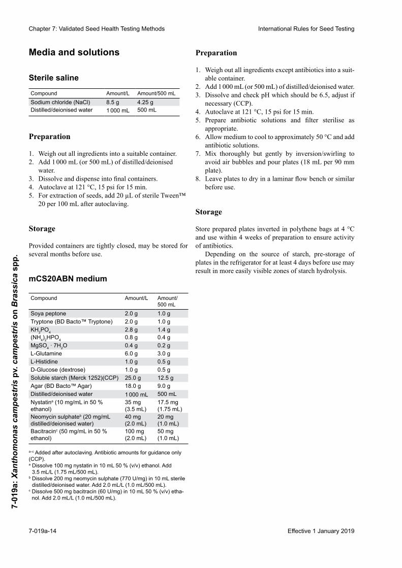
Version 4.0 includes the addition of a PCR test as an alternative to the pathogenicity test for confirmation ofsuspect isolates. The PCR test was derived from a multi-laboratory comparative test organised by the International Seed Health Initiative for Vegetable Crops, ISF (ISHI-Veg). The DLH primer sets by Berg et al. (2005) and the Zup primer sets by Rijlaarsdam et al. (2004) described for use were validated on X. campestris isolates by Far-gier and Manceau (2007). The primer sets were found to amplify X. campestris pv. incanae isolates as well as X. campestris pv. campestris (Xcc). However, the risk of Xcc false positives is considered negligible, as X. c. pv. incanae isolates were only pathogenic on Matthiola spp. and Erysimum cheiri (previously Cheiranthus cheiri) plants, and it is highly unlikely that X. c. pv. incanae is present on cultivated Brassica spp. Comparable results were shown between PCR and pathogenicity test for more than 97 percent of isolates of Xanthomonas campestris pathovars tested in the validation study by Grimault et al. (2011).TwoPCRoptionsareprovidedfortheconfirma-tionand/oridentificationofsuspectXcc colonies to enable selection and adaptation to a laboratory’s equipment and conditions.
Version 5.0 includes adapted recipes of the mCS-20ABN and mFS semi-selective media. The following changes have been made to the recipes described in the previous version of the method: in the mCS20ABN medi-um, the amount of KH2PO4 was increased from 1.59 g/L to 2.8 g/L and the (NH4)2HPO4 was increased from 0.33 g/L to 0.8 g/L.These increases resulted in a better bufferedmedium. The amount of agar was increased from 15.0 g/L to 18.0 g/L as it showed better absorption of the seed ex-tract. Finally, evidence was obtained that the sensitivity of Xanthomonas campestris pv. campestris with respect to neomycin activity is pH dependent (Olivier et al., 2006). In the mFS medium the starch concentration was increased from 10.0 g/L to 25.0 g/L, to improve the recognition of Xanthomonas campestris pv. campestris suspect colo-nies by typical halo formation. The KNO3 was added to a concentration of 0.5 g/L and gentamycin was removed. Finally, the expensive and very toxic cycloheximide was replaced by nystatin in both media for safety reasons and improvement of fungal control.
Version 6.0 includes the addition of seed extract PCR (SE-PCR) and bio-PCR as optional pre-screening meth-ods. One of these two options can be performed before the dilution plating. A negative result in the pre-screen PCRmayberegardedasthefinalresult;however,aposi-tive result in the pre-screen PCR should be confirmedby dilution plating to evaluate for the presence of viable Xcc.Anexampleof theworkflowusing thesemethodsis described in Fig. 1. The TaqMan primers and probes have been validated by Berg et al. (2006), and Köhl et al. (2011) and in an EU-based TESTA project. The PCR
primers for the SYBR Green assay have been published by Berg et al. (2006) and Rijlaarsdam et al. (2004).
Version 6.0 also includes the addition of a third option fortheconfirmationoftheidentityofisolatesfoundindi-lution plating by use of a TaqMan assay. Laboratories are free to choose the TaqMan assay or either of the two con-ventional PCR assays previously described in Version 4.0.
Treated seedSeed treatmentsmayaffect theperformanceof this test.The test must only be performed on untreated seed.
Note: Brassica seed subjected to a physical treatment, for example hot water, is regarded as treated seed.
Sample sizeThe sample (total number of seeds tested) and subsample size to be tested depends on the desired tolerance standard (maximum acceptable percentage of seeds infested) and detection limit (theoretical minimum number of pathogen propagules per seed which can be detected). The mini-mum recommended sample size is 30 000 seeds. In any case the maximum subsample size should be 10 000 seeds. A full discussion of these aspects can be found in Geng et al. (1987), Roberts et al. (1993) and Roberts (1999).
MaterialsReference material: known strain of Xanthomonas
campestris pv. campestris, X. campestris pv. raphani or standardised reference material. Known Acidovorax. cattleyae or A. citrulliisolateusedasInternalAmplifi-cation Control (IAC) for pre-screening PCR.
Plates of mFS medium: 90 mm Petri dishesPlates of mCS20ABN medium: 90 mm Petri dishesPlates of YDC: for subculture (at least 1 per subsample).Conical flasks or equivalent: containing sterile saline
(0.85 % (w/v) NaCl) plus Tween™ 20 (0.02 % (v/v); 20 µL per 100 mL) for soaking of seeds (10 mL per 1 000 seeds).
Dilution bottles: containing 4.5 mL of sterile saline (2 per subsample). Other volumes may be acceptable; see General methods.
70 % (v/v) ethanol: for disinfection of surfaces, equipment.
Incubator: operating at 28–30 °C.Balance: capable of weighing to the nearest 0.001 g.pH meter: capable of reading to the nearest 0.01 pH unit.Automatic pipettes: check accuracy and precision
regularly.
Rules changes for 2019
Method 7-019a Background: ‘increased from 0 g/L to 0.5 g/L’ changed to ‘added to a concentration of 0.5 g/L’
Rules changes for 2019
Method 7-019a Background: ‘Version 6.0 includes... described in Version 4.0.’ paragraphs added
Rules changes for 2019
Method 7-019a Background: ‘Version 6.0 includes... described in Version 4.0.’ paragraphs added
Rules changes for 2019
Method 7-019a Materials: under ‘Reference material’ – ‘X. campestris pv. raphani’ added and ‘Known A. cattleyae or A. citrulli isolate used as Internal Amplification Control (IAC) for pre-screening PCR.’ sentence added
Rules changes for 2019
Method 7-019a Materials: under ‘Conical flasks’ – ‘or equivalent’ added

7-019a-5
Chapter 7: Validated Seed Health Testing MethodsInternational Rules for Seed Testing
Effective 1 January 2019
7-01
9a: X
anth
omon
as c
ampe
stris
pv.
cam
pest
ris o
n B
rass
ica
spp.
Brassica seedlings: susceptible to all races of the patho-gen (e.g. B. oleracea ‘Wirosa’) for pathogenicity test.
Orbital shakerSterile pipette tipsSterile bent glass rodsDNA isolation: DNA isolation kit e.g. Qiagen DNeasy
Blood and Tissue kitReal-time PCR equipment and/or PCR thermal cyclerControls for PCR: The following positive and negative
controls should be included in the SE-PCR.– Positive Extraction Control (PEC) – Acidovorax.
cattleyae/A. citrulli spike (Step 2.1.3, also serves as InternalAmplificationControl(IAC));
– Negative Process Control (NPC) – seed extract from healthy seeds (Step 2.1.2);
– PositiveAmplificationControl(PAC)–X. campestris pv. campestris DNA;
– No Template Control (NTC) – PCR reaction mix with water (Step 2.4.5) and negative seed or buffer as aNPC. The controls should give the expected (absence of)amplificationinthePCRtest;
– Inclusion of a positive process control (positive seed sample) is recommended.
For SE-PCR
PCR primer set (Köhl et al., 2011): XCC-F: 5' gTg.CAT.Agg.CCA.CgA.TgT.Tg 3' XCC-R: 5' Cgg.ATg.CAg.AgC.gTC.TTA.CA 3' XCC-Pr: 5' FAM-CAA.gCg.ATg.TAC.TgC.ggC.
CgT.g-NFQ-MGB 3'PCR primer set (Berg et al., 2006): DLH153-F: 5' gTA.ATT.gAT.ACC.gCA.CTg.CAA 3' DLH154-R: 5' CAC.CgC.TCC.AgC.CAT.ATT 3' P7: 5' VICrepl-ATg.CCg.gCg.AgT.TTC.CAC.g-
BHQ1 3'PCR primer set Acidovorax cattleyae (Koenraadt et al., 2014): Acat 2-F: 5' TgT.AgC.gAT.CCT.TCA.CAA g 3' Acat 2-R: 5' TgT.CgA.TAg.ATg.CTC.ACA.AT 3' Acat1-Pr: 5' Texas Red-CTT.gCT.CTg.CTT.CTC.
TAT.CAC.g- BHQ2 3'
or (additional alternative option)
PCR primer set Acidovorax avenae subsp. citrulli (Sudar-shana, 2010): Contig21 F: 5' ACC GAA CAG AGA GTA ATT CTC
AAA GAC 3' Contig21 R: 5' GAG CGT GAT GGC CAA TGC 3' Contig21-Pr: 5' FAM/CAT +CG+C TT+G AGC
AG+C AA/3IABkFQ 3'
For bio-PCR
PCR primers (Berg et al., 2006): DLH153: 5' gTA.ATT.gAT.ACC.gCA.CTg.CAA 3' DLH154: 5' CAC.CgC.TCC.AgC.CAT.ATT 3'
PCR primers (Rijlaarsdam et al., 2004): Zup2309: 5' AAA.TCA.ggg.ggA.TgC.ggT.gg 3' Zup2310: 5' TCC.ggC.CAg.ggT.CgA.TAC.AgT.g 3'
PCR option 1 for suspect isolate confirmation
PCR primers (Berg et al., 2005): DLH120: 5' CCg.TAg.CAC.TTA.gTg.CAA.Tg 3' DLH125: 5' gCA.TTT.CCA.TCg.gTC.ACg.ATT.g 3'PCR primers (Rijlaarsdam et al., 2004): Zup2309: 5' AAA.TCA.ggg.ggA.TgC.ggT.gg 3' Zup2310: 5' TCC.ggC.CAg.ggT.CgA.TAC.AgT.g 3'Universal primers (adapted from Eden et al., 1991): 1052F: 5' gCA.Tgg.TTg.TCg.TCA.gCT.CgT. 3' BacR: 5' TAC.ggC.TAC.CTT.gTT.ACg.ACT.T 3'Agarose electrophoresis equipment
PCR option 2 for suspect isolate confirmation
PCR primers (Berg et al., 2005): DLH120: 5' CCg.TAg.CAC.TTA.gTg.CAA.Tg 3' DLH125: 5' gCA.TTT.CCA.TCg.gTC.ACg.ATT.g 3'PCR primers (Rijlaarsdam et al., 2004): Zup2311: 5' gCA.AAg.CCC.TCg.TTC.ACg.CAT 3' Zup2312: 5' ggT.ggT.gTg.gCC.gCT.CTT.CTC.AT 3'Universal primers (adapted from Eden et al., 1991): UpBacF: 5' TAC.ggC.TAC.CTT.gTT.ACg.ACT.T 3' UpBacR: 5' gAA.gAg.TTT.gAT.CCT.ggC.TCA.g 3'Agarose electrophoresis equipment
PCR option 3 for suspect isolate confirmation
PCR primer set (Köhl et al., 2011): XCC-F: 5' gTg.CAT.Agg.CCA.CgA.TgT.Tg 3' XCC-R: 5' Cgg.ATg.CAg.AgC.gTC.TTA.CA 3' XCC-Pr: 5' FAM-CAA.gCg.ATg.TAC.TgC.ggC.
CgT.g-NFQ-MGB 3'PCR primer set (Berg et al., 2006): DLH153-F: 5' gTA.ATT.gAT.ACC.gCA.CTg.CAA 3' DLH154-R: 5' CAC.CgC.TCC.AgC.CAT.ATT 3' P7: 5' VICrepl-ATg.CCg.gCg.AgT.TTC.CAC.g-
BHQ1 3'
Rules changes for 2019
Method 7-019a Materials: ‘DNA isolation’, ‘Real-time PCR equipment’ and ‘Controls for PCR’ added
Rules changes for 2019
Method 7-019a Materials: ‘For seed extract PCR’ new subsection added
Rules changes for 2019
Method 7-019a Materials: ‘For Bio-PCR’ new subsection added
Rules changes for 2019
Method 7-019a Materials: ‘PCR option 3 for suspect isolate confirmation’ new subsection added

Chapter 7: Validated Seed Health Testing Methods International Rules for Seed Testing
Effective 1 January 20197-019a-6
7-01
9a: X
anth
omon
as c
ampe
stris
pv.
cam
pest
ris o
n B
rass
ica
spp.
PCR primer set (Wu et al., 2008) Wu-F: 5' CAA.CgC.gAA.gAA.CCT.TAC.C 3' Wu-R: 5' ACg.TCA.TCC.CCA.CCT.TCC 3' Wu-P1: 5' Texas Red -ACg.ACA.ACC.ATg.CAC.
CAC.CTg-BHQ2 3' Wu-P2: 5' Texas Red- ACg.ACA.gCC.ATg.Cag.CAC.
CT-BHQ2 3'
Sample preparationThis can be done in advance of the assay.
It is vital to exclude any possibility of cross-contam-ination between seed samples; it is therefore essential to disinfect all surfaces, containers, hands, etc., both be-fore and after handling each sample. This can achieved by swabbing/spraying equipment and gloved hands with 70 % (v/v) ethanol.
If the submitted sample is received in several packets, these should be combined by emptying into a new, clean polythene bag and mixing by hand to give a composite sample.1. Count the number of seeds in a known weight. Esti-
mate the thousand-seed weight (TSW) as: TSW = (weight of seeds / number of seeds) × 1 0002. Based on the estimated TSW, weigh out subsamples
of the required size into new, clean polythene bags or containers.
MethodsCritical control points are indicated by CCP.
1. Extraction1.1 Suspend each subsample of seeds in pre-chilled
(2–4 °C) sterile saline plus Tween™ 20 (0.02 % v/v) inaconicalflaskorequivalent.Thevolumeofsalineshould be adjusted according to the number of seeds used (10 mL per 1 000 seeds).
1.2 Shake for 2.5 h at room temperature (20–25 °C) on an orbital shaker set at 100–125 rpm.
1.3Shaketheflaskstomixjustbeforefurtherprocessing.
For optional pre-screening SE-PCR, proceed with Step 2; for optional pre-screening with bio-PCR, proceed with Step 3; for dilution plating without pre-screening, proceed with Step 4.
2. Pre-screening SE-PCR2.1 Spiking of samples2.1.1 Transfer 10 mL of seed extract from each subsam-
ple into a 15 mL centrifuge tube.2.1.2 Includeaseedextractfromhealthyseedsorbuffer
without seeds as a Negative Process Control (NPC) (CCP).
2.1.3 Add a positive extraction control (PEC) spike to all subsamples for SE-PCR (e.g. 100 µL of A. cattleyae or A. citrulli stock OD600 = 0.6) (CCP).
Figure 1. Flowchart for the use of pre-screening and dilution plating methods for detection of Xanthomonas campestris pv. campestris in Brassica seeds.
Rules changes for 2019
Method 7-019a Materials: ‘PCR option 3 for suspect isolate confirmation’ new subsection added
Rules changes for 2019
Method 7-019a Methods: ‘or equivalent’ added to Step 1.1
Rules changes for 2019
Method 7-019a Methods: ‘1.3 Shake the flasks... processing.’ added ‘For optional... proceed with step 4.’ new sentence added
Rules changes for 2019
Method 7-019a Methods: new material for Step 2 (including tables) added
Rules changes for 2019
Method 7-019a Figures: new ‘Figure 1’ added; subsequent figures renumbered

7-019a-7
Chapter 7: Validated Seed Health Testing MethodsInternational Rules for Seed Testing
Effective 1 January 2019
7-01
9a: X
anth
omon
as c
ampe
stris
pv.
cam
pest
ris o
n B
rass
ica
spp.
2.2 Concentration of bacteria2.2.1 Centrifuge sample tubes for 5 min at 1 200 × g
(slow spin at room temperature). Carefully transfer supernatant to a clean 15 mL tube and discard the pellet.
Note: The pellet is not always stable. Take the tubes care-fully out of the centrifuge.
2.2.2 Centrifuge the supernatant for 20 min at a mini-mum of 3 400 × g (fast spin at room temperature).
Note: The pellet is not always stable. Take the tubes care-fully out of the centrifuge.
2.2.3 Carefully decant as much supernatant as possible without disturbing the pellet.
2.2.4 Use the pellet for the isolation of DNA.2.3 DNA isolation2.3.1 Resuspendbacterialpellet in lysisbuffer andex-
tract the bacterial DNA as described in the manual of the DNA extraction kit (e.g. DNeasy Blood and Tissue kit).
2.4 TaqMan PCRNote: Work on ice where possible and minimize amount
of time that probes are exposed to light.2.4.1 Use the X. campestris pv. campestris primer sets
from Köhl et al. (2011) and from Berg et al. (2006). Fluorophorelabelsareflexible.
2.4.2 Also include the appropriate primers and probes for the spike used. For detection of the spike A. cattleyae, use the primer set by Koenraadt et al. (2014). For detection of the spike A. citrulli use the primer set by Sudarshana (2010).
2.4.3 Prepare TaqMan PCR mix.2.4.4 Test each DNA extraction from each subsample in
duplicate.2.4.5 Use X. campestris pv. campestris DNA as Positive
AmplificationControl(PAC)andnucleicacidfreewater as No Template Control (NTC) (CCP).
2.4.6 CarryoutPCRreactioninafinalvolumeof25µL(20 µL reaction mixture and 5 µL sample).
2.4.7 Cover the plate after addition of DNA.2.4.8 Perform TaqMan PCR according to the PCR
conditions.2.4.9 Analysetheresults.Cut-offvaluesneedtobede-
termined in each laboratory. A. cattleyae is prefer-ably set between Ct 28 and 32. A. citrulli Ct value is set <35. Ct-value of A. cattleyae or A. citrulli, or another spike should be as expected.
Acut-offvalueof35wasused for thevalidationstudy.Ct-valuebelowcut-offforprimersetKöhlet al.(2011): X. campestris pv. campestris detected. Ct-value primer set Berg et al.(2006) below cut-off:X. campestris pv. campestris or X. campestris pv. raphani detected.
Interpretation & decisions – SE-PCR
Köhl TaqMan
Berg TaqMan
qPCR Result Follow-up
Positive Positive Expected result for Xcc
Dilution plating
Negative Positive Expected result for Xcr
Dilution plating
Positive Negative Inconclusive Dilution platingNegative Negative Expected result
for non-Xcc/XcrNo follow up: no Xcc/Xcr DNA detected
3. Optional pre-screening bio-PCR3.1 Enrichment3.1.1 Pipette 100 µL of the seed extract from each sam-
ple onto two plates of the selective medium (mFS) and spread over the surface with a sterile bent glass rod or sterile spreader.
3.1.2 Prepare control plates3.1.2.1 PCR control plates: spread one plate with 100 µL
of a dilution (approximatively 104 CFU/mL) of a control strain of X. campestris pv. campestris and one plate with a dilution (approximatively 104 CFU/mL) of a control strain of X. campestris pv. raphani.
3.1.2.2 Spiking control plates (PEC): Add 500 µL of the control strains, diluted to 105 CFU/mL, to a tube containing 4.5 mL seed extract for each sample. Spread 100 µL of each spiked seed extract on one plate of selective medium (mFS).
3.1.3 Incubate for 48 h at 28 °C.3.2 Washing3.2.1 Pipette 3 mL sterile saline solution (0.85 %) on each
plate, use a sterile spreader to suspend the bacterial colonies. Pipette 1 mL of the bacterial suspensions into sterile microtubes suitable for centrifugation.
3.2.2 Pipette 2 x 1 mL of the sterile saline solution into sterile microtubes to be used as a negative Bio-PCR control (NPC) (CCP).
3.3 DNA extraction3.3.1 Centrifuge the suspensions for 5 min at 5 000 × g
and remove the supernatant.3.3.2 Add 500 µL of 0.5 M NaOH and resuspend the pel-
let by pipetting.3.3.3 Incubate at 65 °C for 10 min.3.3.4 Add 5 µL of the lysed bacterial suspension to
495 µL of 20 mM Tris-HCl and mix gently with the pipette.
3.4 SYBR Green PCR3.4.1 Use the primers from Berg et al. (2006) and from
Rijlaarsdam et al. (2004). 3.4.2 Prepare SYBR Green PCR mix.3.4.3 Use X. campestris pv. campestris DNA as Positive
AmplificationControl(PAC)andnucleicacidfreewater as Non Template Control (NTC) (CCP).
Rules changes for 2019
Method 7-019a Methods: new material for Step 3 (including tables) added

Chapter 7: Validated Seed Health Testing Methods International Rules for Seed Testing
Effective 1 January 20197-019a-8
7-01
9a: X
anth
omon
as c
ampe
stris
pv.
cam
pest
ris o
n B
rass
ica
spp.
3.4.4 CarryoutPCRreactioninafinalvolumeof15µL(13 µL reaction mixture and 2 µL sample).
3.4.5 Cover the plate after addition of DNA.3.4.6 Perform SYBR Green PCR according to the PCR
conditions.3.4.7 Analyzetheresults.Cut-offvaluesneedtobede-
termined in each laboratory. A sample is declared positive if the Ct-value of one or both of the PCRs is less than 35 cycles (Ct<35) and the PCR product is detected at the same melting temperature as the positive controls (± 1.5 °C).
Interpretation & decisions – bio-PCR
Rijlaars-dam
Berg Sybr PCR Result Follow-up
Positive Positive Expected result for Xcc
Dilution plating
Negative Positive Expected result for Xcr
Dilution plating
Positive Negative Inconclusive Dilution platingNegative Negative Expected result
for non-Xcc/XcrNo follow up: no Xcc/Xcr DNA detected
4. Dilution and plating4.1 Prepare two serial tenfold dilutions from the seed
extract. Pipette 0.5 mL of the extract into 4.5 mL of sterile saline and vortex to mix (101 dilution). Pipette 0.5 mL of the 101 dilution into another 4.5 mL of sterile saline and vortex to mix (102 dilution) (see General methods).
4.2 Pipette 100 µL of each dilution and the undiluted seed extract onto plates of each of the selective media (mFS, mCS20ABN) and spread over the surface with a sterile bent glass rod (see General methods).
4.3 Incubate plates at 28–30 °C and examine after 3–4 d.4.4 Positive control (culture or reference material)4.4.1 Prepare a suspension of a known strain of X.
campestris pv. campestris in sterile saline or recon-stitute standardised reference material according to the supplier’s instructions.
4.4.2 Dilute sufficiently to obtain dilutions containingapprox. 102 to 104 cfu/mL. This may require up to seven ten-fold dilutions from a turbid suspension.
4.4.3 Pipette 100 µL of appropriate dilutions onto plates of each of the selective media (mFS, mCS20ABN) and spread over the surface with a sterile bent glass rod.
4.4.4 Incubate plates with the sample plates4.5 Sterility check4.5.1 Prepare a dilution series from a sample of the ex-
traction medium (i.e., saline plus Tween™ 20), containing no seeds, and plate on each of the media as for samples.
4.6 Examination of the plates4.6.1 Examine sterility check and positive control plates
(CCP).4.6.2 Examine the sample plates for the presence of typi-
cal X. campestris pv. campestris (Xcc) colonies by comparison with the positive control plates.
4.6.3 On mFS after 3–4 d, Xcc colonies are small, pale green, mucoid and surrounded by a zone of starch hydrolysis. This zone appears as a halo that may be easier to see with a black background (Fig. 2a). Colonies may show marked variation in size and may be visible on mFS after 3 d; if not, incubate for an additional day.
4.6.4 After 3–4 d on mCS20ABN, Xcc colonies are pale yellow, mucoid and surrounded by a zone of starch hydrolysis (Fig. 2b). Colonies may show marked variation in size. Depending on the number of col-onies present, it may be easier to evaluate plates after 3 d, before coalescence of starch hydrolysis zoneswhichcanmakeitmoredifficulttoidentifysuspect colonies.
4.6.5 Incubation of the plates at 4 °C for several hours before recording may result in sharper zones of starch hydrolysis with some starch sources.
4.6.6 Record the number of suspect and other colonies (see General methods).
5. Confirmation/identificationofsuspectcolonies5.1 Subculture suspect colonies to sectored plates of YDC.
To avoid the potential for cross-contamination of iso-lates, use a new sectored plate for each subsample. The precise numbers of colonies subcultured will depend on the number and variability of suspect colonies on the plate: if present, at least six colonies should be sub-cultured per subsample (CCP).
5.2 Subculture the positive control isolate to a sectored plate for comparison.
5.3 Incubate sectored plates for 24–48 h at 28–30 °C.5.4 Compare appearance of growth with positive control.
On YDC, X. campestris pv. campestris colonies are paleyellowandmucoid/fluidal(Fig.3).
5.5Confirm the identity of isolates by pathogenicity onBrassica seedlings (step 6) of known susceptibility or by using one of the three PCR options (steps 7, 8 or 9)(CCP).
5.6 Record results for each colony subcultured.6. Pathogenicity6.1 Grow seedlings of a Brassica cultivar known to be
susceptible to all races of X. campestris pv. campestris (e.g. cabbage ‘Wirosa’, see Vicente et al., 2001) in small pots or modules until at least 3–4 true leaf stage.
6.2 Scrape a small amount of bacterial growth directly from a 24–48 h YDC culture (e.g. sectored plate) with a sterile cocktail stick or insect pin.
Rules changes for 2019
Method 7-019a Methods: former Step 6 renumbered Step 5
Rules changes for 2019
Method 7-019a Methods: former Step 7 renumbered Step 6
Rules changes for 2019
Method 7-019a Methods: step 5.5 – ‘by PCR’ changed to ‘using one of the three PCR options’
Rules changes for 2019
Method 7-019a Methods: former Steps 2, 3 and 4 placed under ‘4. Dilution and plating’ ‘Shake the flasks to mix just before dilution.’ deleted

7-019a-9
Chapter 7: Validated Seed Health Testing MethodsInternational Rules for Seed Testing
Effective 1 January 2019
7-01
9a: X
anth
omon
as c
ampe
stris
pv.
cam
pest
ris o
n B
rass
ica
spp.
6.3 Inoculate six of the major veins at a point near the leaf edges on the two youngest leaves by stabbing with the cocktail stick or insect pin.
6.4 The number of plants which should be inoculated will depend on the variability of the cultivar and experi-ence of the operator, but 1–3 plants per isolate should usuallybesufficient.Itisbettertoinoculatemoreiso-lates with 1 plant per isolate than fewer isolates with 3 plants per isolate.
6.5 Inoculate with the positive control isolate and stab with a sterile cocktail stick or insect pin as a negative control (CCP).
6.6 Grow on plants at 20–25 °C.6.7 Examine plants for the appearance of typical progres-
sive V-shaped, yellow/necrotic lesions with blackened veins after 10–14 d (Fig. 4). Symptoms may be vis-ible earlier depending on temperature and the aggres-siveness of the isolate. Compare with positive control (CCP). It is important to discriminate between the progressive lesions caused by the vascular pathogen X. campestris pv. campestris and the limited dark ne-crotic lesions at the inoculation site caused by leaf spot Xanthomonas (often classified as eitherX. c. pv. armoraciae or X. c. pv. raphani (see Kamoun et al., 1992; Alvarez et al., 1994; Tamura et al., 1994; Vicente et al., 2001; Roberts et al., 2004).
7. Polymerase Chain Reaction (PCR) Option 17.1 Make a slightly turbid cell suspension (OD600 nm ap-
proximately 0.05) in 1.0 mL sterile saline from the suspected cultures on YDC medium and the positive control. In addition a non-suspect isolate should be used as a negative control (CCP). Centrifuge bacte-rial suspensions for 5 min at 8 000 rpm. Discard the supernatant and resuspend the pellet with 500 µL of 0.5 M NaOH. Incubate for 10 min at 65 °C by shaking at 1 000 rpm. Dilute 5 µL of solution into 495 µL of 20 mM Tris-HCl, pH 8, and vortex. Suspensions can bestoredat-20°Cuntilidentification.
7.2 Use the X. campestris pv. campestrisspecificpairofprimers from Rijlaarsdam et al. (2004) that will give a productof370bpandthespecificpairofprimersfromBerg et al. (2005) that will give a product of 619 bp
7.3 Universal bacterial primers should be used to validate the PCR reaction. Use the pair of primers adapted from Eden et al. (1991) that will give a product of 441 bp:
7.4 Prepare the reaction mixture (CCP). Carry out PCR reactions in0.2mLthin-walledPCRtubes inafinalvolume of 20 µL (17 µL reaction mixture + 3 µL bacte-rial suspension).
7.5PCRprofile:Aninitial3minincubationat95°Cfol-lowed by 6 cycles of 40 s at 95 °C, 40 s at 63 °C with a touchdown of 1 °C per cycle, 40 s at 72 °C followed by 29 cycles of 40 s at 95 °C, 40 s at 58 °C, 40 s at 72 °C.
Afinal5minincubationat72°Candinfinityat10°C(CCP).
7.6 Fractionate 10 µL of the PCR products and water (neg-ative PCR control) by gel electrophoresis in 1× Tris acetateEDTA(TAEbuffer)(CCP).Includea100bpladder. Stain with ethidium bromide in a bath and rinse in water.
7.7AnalysetheamplificationproductsforaX. campestris pv. campestris specific product of 370 bp/619 bpand a universal product of 441 bp (CCP; Fig. 5). Two bands (441 bp/619 bp) = positive identifica-tion of X. campestris, suspected presence of X. c. pv. armoraciae or X. c. pv. raphani; two bands (370 bp/441 bp) = indeterminate PCR result, patho-genicitytestmustbecarriedouttoconfirmthesuspectisolate;oneband(universal)=negativeidentification;no bands = bacterial template absent, repeat reaction.
8. Polymerase Chain Reaction (PCR) Option 28.1 Make a slightly turbid cell suspension (OD600 nm ap-
proximately 0.05) in 1.0 mL sterile saline from the suspended cultures on YDC medium and the positive control. In addition, a non-suspect isolate should be used as a negative control (CCP). Centrifuge bacte-rial suspensions for 5 min at 8 000 rpm. Discard the supernatant and resuspend the pellet with 500 µL of 0.5 M NaOH. Incubate for 10 min at 65 °C by shaking at 1 000 rpm. Dilute 5 µL of solution into 495 µL of 20 mM Tris-HCl, pH 8, and vortex. Suspensions can bestoredat-20°Cuntilidentification.
8.2 Use the X. campestris pv. campestrisspecificpairofprimers from Rijlaarsdam et al. (2004) that will give aproductof445bpandthefollowingspecificpairofprimers from Berg et al. (2005) that will give a product of 619 bp.
8.3 Universal bacterial primers should be used to validate the PCR reaction. Use the pair of primers adapted from Eden et al. (1991) that will give a product of 1511 bp:
8.4 Prepare the reaction mixture (CCP). Carry out PCR reactions in0.2mLthin-walledPCRtubes inafinalvolume of 25 µL (24 µL reaction mixture + 1 µL bacte-rial suspension).
8.5PCRprofile:Aninitial5minincubationat94°Cfol-lowed by 4 cycles of 1 min at 94 °C, 1 min at 65 °C with a touchdown of 1 °C per cycle, 1 min at 72 °C fol-lowed by 30 cycles of 1 min at 94 °C, 1 min at 60 °C, 1minat72°C.Afinal10minincubationat72°Candinfinityat10°C(CCP).
8.6 Fractionate 10 µL of the PCR products and water (neg-ative PCR control) by gel electrophoresis in 1× Tris acetateEDTA(TAEbuffer)(CCP).Includea100bpladder. Stain with ethidium bromide in a bath and rinse in water.
Rules changes for 2019
Method 7-019a Methods: former Step 8 renumbered Step 7
Rules changes for 2019
Method 7-019a Methods: step 7.4 – page number deleted
Rules changes for 2019
Method 7-019a Methods: step 8.4 – page number deleted
Rules changes for 2019
Method 7-019a Methods: former Step 9 renumbered Step 8

Chapter 7: Validated Seed Health Testing Methods International Rules for Seed Testing
Effective 1 January 20197-019a-10
7-01
9a: X
anth
omon
as c
ampe
stris
pv.
cam
pest
ris o
n B
rass
ica
spp.
8.7AnalysetheamplificationproductsforaX. campestris pv. campestrisspecificproductof445bp/619bpanda universal product of 1511 bp (CCP; Fig. 6). Two bands (619 bp/1511 bp) = positive identification ofXanthomonas campestris, suspected presence of X. c. pv. armoraciae or X. c. pv. raphani; two bands (445 bp/1511 bp) = indeterminate PCR result, patho-genicitytestmustbecarriedouttoconfirmthesuspectisolate;oneband(universal)=negativeidentification;no bands = bacterial template absent, repeat reaction.
9. Polymerase Chain Reaction (PCR) Option 39.1 Make a slightly turbid cell suspension (OD600 nm ap-
proximately 0.05) in 1.0 mL sterile saline from the suspected cultures on YDC medium and the positive control. In addition, a non-suspect isolate should be used as a negative control (CCP). Centrifuge bacte-rial suspensions for 5 min at 8 000 rpm. Discard the supernatant and resuspend the pellet with 500 µL of 0.5 M NaOH. Incubate for 10 min at 65 °C by shaking at 1 000 rpm. Dilute 5 µL of solution into 495 µL of 20 mM Tris-HCl, pH 8, and vortex. Suspensions can bestoredat-20°Cuntilidentification.
9.2 Use the following X. campestris pv. campestris spe-cificpairprimersandprobesfromKöhlet al. (2011) and from Berg et al. (2006).
9.3 Universal primers should be used to validate the PCR reaction. Use the primers and probes from Wu et al. (2008).
9.4 Prepare the reaction mixture (CCP). Carry out PCR reaction in 0.2mL thin-walled PCR tubes in a finalvolume 25 µl (20 µl reaction mixture + 5 µl bacterial suspension).
9.5 Carry out PCR reactions in a real-time PCR instrument accordingtofollowingPCRprofile:Aninitial10minincubationat95⁰Cfollowedby40cyclesof15sat95⁰Cand30sat60⁰C.
9.6 Determine Ct values; Ct values of positive control should consistently be lower than 30. The cut-offCt value of the internal amplification control (IAC)should be below 35, and the expected range is to be determined by the user based on experimental data.
In the case of universal bacterial primers, positive reactions may occur in non-template controls (NTC) due to the presence of residual DNA in Taq enzyme reagents. The IAC Ct values from reactions on sus-pect isolates should indicate at least a 10-fold higher concentration of bacterial DNA than the IAC Ct val-uesfromtheNTCreactions;thedifferencebetweenCtvalues should be more than 3.3.
Interpretation & decisions – suspect confirmn.
Köhl Taqman
Berg Taqman
PCR Result Follow-up
Positive Positive Expected result for Xcc
Pathogenicity test for confirmation
Negative Positive Expected result for Xcr
Pathogenicity test for confirmation
Positive Negative Inconclusive Pathogenicity test for confirmation
Negative Negative Expected result for non-Xcc/Xcr
Negative No follow-up, isolate is not Xcc/Xcr
General methodsPreparation of ten-fold dilution series: Each dilution
should be prepared by pipetting 0.5 mL (±5 %) from a well-mixed seed extract or previous dilution into a universal bottle (screw-capped) or similar containing 4.5 mL (±2 %) of sterile diluent and then vortexing to mix prior to the next dilution step. A new sterile pipette tip should be used for each dilution step. Pi-pettes should be checked regularly for accuracy and precision and re-calibrated as necessary. It is accept-able to prepare 10-fold dilutions using other volumes, provided that the laboratory can demonstrate that the required accuracy and precision can be achieved.
Plating of dilutions: This should be done as soon as pos-sible after dilutions have been prepared and certainly within 30 min. Working from the highest (most dilute) dilution to the undiluted extract, 0.1 mL is pipetted onto the centre of a surface-dry, labelled agar plate. The liquid should then be spread evenly over the entire surface of the medium with a bent glass rod. If care is taken to work from the highest to the lowest dilution (or undiluted extract), a single pipette tip and a single bent glass rod can be used for each sample. Ensure that all liquid has been absorbed by the agar before invert-ing and incubating plates. If necessary, allow plates to dryunderasterileair-flowinamicrobiologicalsafetycabinetorlaminarflowhood.
Recording of dilution plates: Record the results for all dilution plates. The most accurate estimate of bacterial numbers should be obtained from spread plates with total number between 30 and 300 colonies. However, this may be further complicated depending on the rela-tive numbers of suspect pathogen and other colonies. In order tominimise effort, start recordingwith thehighest dilution (most diluted) and count the number of suspect and the number of other colonies. If the to-tal number of colonies on a plate greatly exceeds 300,
Rules changes for 2019
Method 7-019a Methods: ‘9. Polymerase Chain Reaction (PCR) Option 3’ new step added (including table)

7-019a-11
Chapter 7: Validated Seed Health Testing MethodsInternational Rules for Seed Testing
Effective 1 January 2019
7-01
9a: X
anth
omon
as c
ampe
stris
pv.
cam
pest
ris o
n B
rass
ica
spp.
there is little value in trying to make a precise count if a more reliable count has already been obtained fromamorediluteplate,inwhichcaseitissufficientto record the number of colonies as ‘m’ (many) if they are still separate, or ‘c’ (confluent) if they have runtogether.
Sectored plates: Using a laboratory marker pen draw lines on the base of a standard 90 mm plate (Petri dish) to divide it into six equal sectors. Subculture single col-onies from dilution plates and make a single zigzagged streak within a single sector on the plate. Take care to leavesufficient spacebetweeneach isolate toensurethe growth does not coalesce. Thus, six suspect colo-nies can be subcultured to each sectored plate. Sepa-rate plates should be used for each sample/subsample. If the purity of subcultured isolates is doubtful, they should be further streaked out on whole plates.
Reporting results: The result of a seed health test should indicate the scientific name of the pathogendetected and the test method used. When reported on an ISTACertificate, results are enteredunder ‘OtherDeterminations’. The report must indicate the number of seeds tested. In the case of a negative result (pathogen not detected in any subsample), the results must be reported as ‘not detected’. In the case of a positive result, the report must indi-cate the mean number of pathogen propagules (cfu) per seed and the number of positive subsamples out of the total number tested.
Quality assuranceA record should be kept of the date and results of pipette calibration checks.It is essential that operators have re-ceived appropriate training and use automatic pipettes correctly.
Critical control points (CCP)
– Dilution plates prepared from positive control isolate(s) or reference material, should give single colonies with typical morphology (Step 4.6.1).
– The numbers of colonies on dilution plates prepared from the positive control isolate(s) or reference mate-rial should be similar on both media (Step 4.6.1).
– Numbers of bacteria on dilution plates should be con-sistent with the dilution (i.e. should decrease approx. 10-fold with each dilution) (Step 4.6.1).
– There should be no growth on dilution plates prepared as a sterility check (Step 4.6.1).
– Due to the potential for non-pathogenic isolates to be present in seed lots together with pathogenic iso-lates, it is essential to subculture, if present, at least the minimumnumberofsuspectcoloniesspecified(sixpersubsample) (Step 5.1), and to test all Xanthomonas-like subcultured isolates for pathogenicity or by PCR test (Step 5.5).
– The positive control isolate(s) or reference material should give colonies with typical morphology on YDC (Step 5.4).
– Positive control isolates should be included in every pathogenicity test (Step 6.5).
– The positive control isolate should give typical symp-toms in the pathogenicity test (Step 6.7).
– Positive and negative control isolates should be in-cluded in every PCR test (Steps 7.1, 8.1, 9.1).
– The preparation of the PCR mixture (Steps 7.4, 8.4, 9.4), theamplificationPCRprogram (Steps7.5,8.5,9.5) and the preparation of agarose gel for electropho-resis (Steps 7.6, 8.6, 9.6) should be adapted to avail-able material and equipment of individual laboratories testing for Xanthomonas campestris pv. campestris under the condition that results will be validated by PCR controls. Validation studies showed that PCR re-sults were more dependent on laboratory conditions than on PCR protocol when different PCR mixes,amplificationproductsandagarosegelswereusedinlaboratories.
– The positive control isolate should give the expected amplificationproduct in thePCRtest(steps7.7,8.7,9.7).
– The source of starch used in the selective media is critical for observation of starch hydrolysis. Verify that each new batch of starch gives clear zones of hy-drolysis with reference cultures of X. campestris pv. campestris (mFS and mCS20ABN media).
– The activity (units/mg) of some antibiotics may vary between batches. It may be necessary to adjust the weightorvolumeaddedtoensurethatthefinalnum-ber of units per litre of medium is consistent (mFS and mCS20ABN media).
– The activity of neomycin against some strains of Xcc isknowntobeaffectedbypH.ItisessentialthatthepH of the medium is less than 6.6 (see Media and solu-tions, mCS20ABN medium, Step 3)

Chapter 7: Validated Seed Health Testing Methods International Rules for Seed Testing
Effective 1 January 20197-019a-12
7-01
9a: X
anth
omon
as c
ampe
stris
pv.
cam
pest
ris o
n B
rass
ica
spp.
Figure 3. Typical yellow mucoid growth of isolates of Xanthomonas campestris pv. campestris on a sectored plate of YDC after 3 days at 28 °C. Only suspect cultures are indicated by arrows.
Figure 4. Cabbage leaves 7 days post-inoculation with Xanthomonas campestris pv. campestris. Typical symp-toms are black veins, wilting and chlorosis. The lower left leaf was used as a negative control.
Figure 2. Plates of mFS (a) and mCS20ABN (b) after 5 days of incubation at 28 °C showing typical colonies of Xanthomonas campestris pv. campestris surrounded by zones of starch hydrolysis.
a
b
– Prepare antibiotics stock solutions and other supple-ments in distilled/deionised water, or in 50 % (v/v) or 70 % (v/v) ethanol. Antibiotics stock solutions and other supplements prepared in distilled/deionised watermustbefiltersterilisedwitha0.2µmbacterial filter.Alternatively,itispossibletoaddtheamountofpowder to autoclaved distilled/deionised water. Solu-tions prepared in ethanol need no sterilisation (mFS and mCS20ABN media).
– The preparation of PCR mixture and the PCR condi-tions (Steps 2.4.3 and 2.4.8 for SE-PCR; Steps 3.4.2 and 3.4.6 for bio-PCR; and Steps 9.4 and 9.5 for suspect confirmation) shouldbeadapted toavailablematerial and equipment of individual laboratories un-der the condition that results will be validated by PCR and process controls.
Rules changes for 2019
Method 7-019a Quality assurance: new ‘Critical control point’ added

7-019a-13
Chapter 7: Validated Seed Health Testing MethodsInternational Rules for Seed Testing
Effective 1 January 2019
7-01
9a: X
anth
omon
as c
ampe
stris
pv.
cam
pest
ris o
n B
rass
ica
spp.
Figure 6. Agarose gel showing Xanthomonas campestris and X. c. pv. campestris amplification products using primer set combinations of PCR Option 2. 1: 100 bp ladder. 2, 3, 5: Two bands (619 bp/1511 bp) – positive sample with Xanthomonas campestris (Xca/Xcr suspected presence). 4: One band (1511 bp) – negative sample, no Xanthomonas campestris (Xc). 6, 7: Three bands (445 bp/619 bp/1511 bp) – positive sample with Xanthomonas campestris pv. campestris (Xcc) (with or without Xca/Xcr). 8: Water (negative PCR control) – no reaction.
1511 bp (universal primers)
619 bp (DLH120/DLH125)
445 bp (Zup2311/Zup2312)
1 2 3 4 5 6 7 8
Figure 5. Agarose gel showing Xanthomonas campestris and X. c. pv. campestris amplification products using primer set combinations of PCR Option 1. 1: 100 bp ladder. 2: Two bands (370 bp/441 bp) – indeterminate PCR result. 3: Three bands (441 bp/619 bp/370 bp) – positive sample with Xanthomonas campestris pv. campestris (Xcc) (with or without Xca/Xcr). 4: One band (441 bp) – negative sample, no Xanthomonas campestris (Xc). 5: Two bands (441 bp/619 bp) – posi-tive sample with Xanthomonas campestris (Xca/Xcr suspected presence). 6: Water (negative PCR control) – no reaction.
370 bp (Zup2309/Zup2310)
441 bp (universal primers)
619 bp (DLH120/DLH125)
Chapter 7: Validated Seed Health Testing Methods International Rules for Seed Testing
Effective 1 January 20197-019a-14
7-01
9a: X
anth
omon
as c
ampe
stris
pv.
cam
pest
ris o
n B
rass
ica
spp.
Media and solutions
Sterile saline
Compound Amount/L Amount/500 mLSodium chloride (NaCl) 8.5 g 4.25 gDistilled/deionised water 1 000 mL 500 mL
Preparation
1. Weigh out all ingredients into a suitable container.2. Add 1 000 mL (or 500 mL) of distilled/deionised
water.3. Dissolveanddispenseintofinalcontainers.4. Autoclave at 121 °C, 15 psi for 15 min.5. For extraction of seeds, add 20 µL of sterile Tween™
20 per 100 mL after autoclaving.
Storage
Provided containers are tightly closed, may be stored for several months before use.
mCS20ABN medium
Compound Amount/L Amount/ 500 mL
Soya peptone 2.0 g 1.0 gTryptone (BD Bacto™ Tryptone) 2.0 g 1.0 gKH2PO4 2.8 g 1.4 g(NH4)2HPO4 0.8 g 0.4 gMgSO4 ∙ 7H2O 0.4 g 0.2 gL-Glutamine 6.0 g 3.0 gL-Histidine 1.0 g 0.5 gD-Glucose (dextrose) 1.0 g 0.5 gSoluble starch (Merck 1252)(CCP) 25.0 g 12.5 gAgar (BD Bacto™ Agar) 18.0 g 9.0 gDistilled/deionised water 1 000 mL 500 mLNystatina (10 mg/mL in 50 % ethanol)
35 mg (3.5 mL)
17.5 mg (1.75 mL)
Neomycin sulphateb (20 mg/mL distilled/deionised water)
40 mg (2.0 mL)
20 mg (1.0 mL)
Bacitracinc (50 mg/mL in 50 % ethanol)
100 mg (2.0 mL)
50 mg (1.0 mL)
a–c Added after autoclaving. Antibiotic amounts for guidance only (CCP).a Dissolve 100 mg nystatin in 10 mL 50 % (v/v) ethanol. Add
3.5 mL/L (1.75 mL/500 mL).b Dissolve 200 mg neomycin sulphate (770 U/mg) in 10 mL sterile
distilled/deionised water. Add 2.0 mL/L (1.0 mL/500 mL).c Dissolve 500 mg bacitracin (60 U/mg) in 10 mL 50 % (v/v) etha-
nol. Add 2.0 mL/L (1.0 mL/500 mL).
Preparation
1. Weigh out all ingredients except antibiotics into a suit-able container.
2. Add 1 000 mL (or 500 mL) of distilled/deionised water.3. Dissolve and check pH which should be 6.5, adjust if
necessary (CCP).4. Autoclave at 121 °C, 15 psi for 15 min.5. Prepare antibiotic solutions and filter sterilise as
appropriate.6. Allow medium to cool to approximately 50 °C and add
antibiotic solutions.7. Mix thoroughly but gently by inversion/swirling to
avoid air bubbles and pour plates (18 mL per 90 mm plate).
8. Leaveplatestodryinalaminarflowbenchorsimilarbefore use.
Storage
Store prepared plates inverted in polythene bags at 4 °C and use within 4 weeks of preparation to ensure activity of antibiotics.
Depending on the source of starch, pre-storage of plates in the refrigerator for at least 4 days before use may result in more easily visible zones of starch hydrolysis.

7-019a-15
Chapter 7: Validated Seed Health Testing MethodsInternational Rules for Seed Testing
Effective 1 January 2019
7-01
9a: X
anth
omon
as c
ampe
stris
pv.
cam
pest
ris o
n B
rass
ica
spp.
mFS medium
(Schaad et al., 1989)
Compound Amount/L Amount/ 500 mL
K2HPO4 0.8 g 0.4 gKH2PO4 0.8 g 0.4 gKNO3 0.5 g 0.25 gMgSO4 ∙ 7H2O 0.1 g 0.05 gYeast extract 0.1 g 0.05 gMethyl Green (1 % aq.) 1.5 mL 0.75 mLSoluble starch (Merck 1252)(CCP)
25.0 g 12.5 g
Agar (BD Bacto™ Agar) 15.0 g 7.5 gDistilled/deionised water 1000 mL 500 mLNystatina (10 mg/mL 50 % ethanol)
35 mg (3.5 mL)
17.5 mg (1.75 mL)
D-Methionineb (1 mg/ mL 50 % ethanol)
3 mg (3.0 mL)
1.5 mg (1.5 mL)
Pyridoxine HClc (1 mg/ mL 50 % ethanol)
1 mg (1 mL)
0.5 mg (0.5 mL)
Cephalexind (20 mg/ mL 50 % ethanol)
50 mg (2.5 mL)
25 mg (1.25 mL)
Trimethoprime (10 mg/mL 70 % ethanol)
30 mg (3 mL)
15 mg (1.5 mL)
a–c, d,e Added after autoclaving. Antibiotic amounts for guidance only (CCP). a Dissolve 100 mg nystatine in 10 mL 50 % (v/v) ethanol. Add 3.5
mL/L (1.75 mL/500 mL).b Dissolve 10 mg D-methionine in 10 mL 50 % (v/v) ethanol. Add
3.0 mL/L (1.5 mL/500 mL).c Dissolve 10 mg pyridoxine HCl in 10 mL 50 % (v/v) ethanol. Add
1 mL/L (0.5 mL/500 mL).d Dissolve 200 mg cephalexin in 10 mL 50 % (v/v) ethanol. Add 2.5
mL/L (1.25 mL/500 mL).e Dissolve 100 mg trimethoprim in 10 mL 70 % (v/v) ethanol. Add
3 mL/L (1.5 mL/500 mL).
Preparation
1. Weigh out all ingredients except antibiotics, pyridox-ine HCl and D-methionine into a suitable container.
2. Add 1 000 mL (or 500 mL) of distilled/deionised water.3. Dissolve and check pH which should be 6.5, adjust if
necessary (important, CCP).4. Autoclave at 121 °C, 15 psi for 15 min.5. Prepare antibiotics, pyridoxine HCl and D-methionine
solutionsandfiltersteriliseasappropriate.6. Allow medium to cool to approximately 50 °C before
adding antibiotics, pyridoxine HCl and D-methionine solutions.
7. Mix the molten medium gently to avoid air bubbles and pour plates (18 mL per 90 mm plate).
8. Leaveplatestodryinalaminarflowbenchorsimilarbefore use.
Storage
Store prepared plates inverted in polythene bags at 4 °C and use within 4 weeks of preparation to ensure activity of antibiotics.
Depending on the source of starch, prestorage of plates in the refrigerator for at least 4 days before use may result in more easily visible zones of starch hydrolysis.
Yeast dextrose chalk (YDC) agar
Compound Amount/L Amount/500 mLAgar (BD Bacto™ Agar) 15.0 g 7.5 gYeast extract 10.0 g 5.0 gCaCO3 (light powder) 20.0 g 10.0 gD-Glucose (dextrose) 20.0 g 10.0 gDistilled/deionised water 1 000 mL 500 mL
Preparation
1. Weigh out all ingredients into a suitable oversize con-tainer(i.e.250mLofmediumina500mLbottle/flask)to allow swirling of medium just before pouring.
2. Add 1 000 mL (or 500 mL) of distilled/deionised water.3. Steam to dissolve.4. Autoclave at 121 °C, 15 psi for 15 min.5. Allow medium to cool to approximately 50 °C .6. Swirl to ensure even distribution of CaCO3 and avoid
air bubbles, and pour plates (22 mL per 90 mm plate).7. Leaveplatestodryinalaminarflowbenchorsimilar
before use.
Storage
Store prepared plates inverted in polythene bags at room temperature. Prepared plates can be stored for several months provided they do not dry out.
0.5 M NaOH
Compound Amount/LNaOH 20 gDistilled/deionised water 1 000 mL
Preparation
1. Weigh out ingredient into a suitable container.2. Add 1 000 mL distilled/deionised water.3. Dissolveanddispenseintofinalcontainers.4. Autoclave at 121 °C, 15 psi for 15 min.

Chapter 7: Validated Seed Health Testing Methods International Rules for Seed Testing
Effective 1 January 20197-019a-16
7-01
9a: X
anth
omon
as c
ampe
stris
pv.
cam
pest
ris o
n B
rass
ica
spp.
1M Tris-HCl, pH 8
Compound Amount/LTris base 121.1 gDistilled/deionised water 1 000 mL
Preparation
1. Weigh out ingredient into a suitable container.2. Add 800 mL of distilled/deionised water.3.Dissolveanddispenseintofinalcontainers.4. Check the pH with a pH meter and adjust if necessary.5. Autoclave at 121 °C, 15 psi for 15 min and dilute to
20 mM.
Examples of reaction mixture preparation for PCR
Primer set combinations and volumes for triplex TaqMan PCR (SE-PCR)
Preferably the triplex TaqMan PCR, as described in this method,isused.IfthePCRmachineusedisnotfitforatriplex including Texas Red, alternatively two duplex (tar-get and spike) reactions can be performed.
PCR mix triplex qPCR For 1 reaction (in µL)
Final concentration
PCR grade H2O 9.95 PerfeCta Multiplex qPCR ToughMix
5.0 1×
10 µM XCC F 0.75 0.3 µM10 µM XCC R 0.75 0.3 µM10 µM XCC Pr 0.5 0.2 µM10 µM DLH153-F 0.75 0.3 µM10 µM DLH154-R 0.75 0.3 µM10 µM P7 0.5 0.2 µM10 µM Acat 2-F or 10 µM contig21-F
0.4 0.16 µM
10 µM Acat 2-R or 10 µM contig21-R
0.4 0.16 µM
10 µM Acat 1-Pr or 10 µM contig21-Pr
0.25 0.1 µM
Subtotal 20.0Sample 5.0Total 25.0
PCR conditions for triplex Taqman PCR
PCR conditions
Step Temperature Duration Remarkshold 95 °C 2' 00"
95 °C 0' 15" 5 °C/sec.40 cycles 60 °C 0' 48" 5 °C/sec.
20 mM Tris-HClTrizma base 1.211 gH2O 500 mL
Preparation
1. Weigh out ingredient into a suitable container.2. Add 500 mL of distilled/deionized water.3. Dissolveanddispenseintofinalcontainers.4. Check the pH with a pH meter and adjust to pH 8.0
if necessary.5. Autoclave at 121 °C, 15 psi for 15 min.
DLH and Zup primer volumes for SYBR Green PCR (bio-PCR)
Mix DLH Initial conc. Volume (µL) Final conc.PCR grade H2O 4.5DLH153 20 µM 0.5 0.66 µMDLH154 20 µM 0.5 0.66 µMQuantitect SYBR Green PCR mix
2× 7.5 1×
DNA (extract) 2Total 15
Mix Zup Initial conc. Volume (µL) Final conc.PCR grade H2O 4.5Zup2309 20 µM 0.5 0.66 µMZup2310 20 µM 0.5 0.66 µMQuantitect SYBR Green PCR mix
2× 7.5 1×
DNA (extract) 2.0Total 15.0
Rules changes for 2019
Method 7-019a Media and solutions: new reaction mixture preparation for TaqMan PCR added
Rules changes for 2019
Method 7-019a Media and solutions: new reaction mixture preparation for TaqMan PCR added
Rules changes for 2019
Method 7-019a Media and solutions: new reaction mixture preparation for SYBR Green PCR added

7-019a-17
Chapter 7: Validated Seed Health Testing MethodsInternational Rules for Seed Testing
Effective 1 January 2019
7-01
9a: X
anth
omon
as c
ampe
stris
pv.
cam
pest
ris o
n B
rass
ica
spp.
PCR conditions for SYBR Green PCR
Step Temperature Time RemarksDenaturation 95 °C 15 minZup2309 20 µM 0.5 0.66 µMZup2310 20 µM 0.5 0.66 µMQuantitect SYBR Green PCR mix
2× 7.5 1×
DNA (extract) 2.06 cycles:DenaturationHybridisationElongation
94 °C63 °C72 °C
10 s15 s30 s
-1 °C/cycle
34 cycles:DenaturationHybridisationElongation
94 °C58 °C72 °C
10 s15 s30 s
Melt curve 72-95 °C
Primer set combinations and volumes for PCR Option 1 (suspect isolate confirmation)
Compound Final concentration
Volume in 20 µL (µL)
Sterile water 0.04Green Go Taq Buffer 5× 1 4MgCl2 (25 mM) 1.2 mM 0.96dNTP (2 mM each) 0.2 mM 2Zup2309 (5 µM) 0.2 µM 0.8Zup2310 (5 µM) 0.2 µM 0.8DLH120 (5 µM) 0.6 µM 2.4DLH125 (5 µM) 0.6 µM 2.41052F (1 µM) 0.085 µM 1.7BacR (1 µM) 0.085 µM 1.7Taq polymerase (5 U/µL) 0.05 U/µL 0.2DNA 3
Primer set combinations and volumes for PCR Option 2 (suspect isolate confirmation)
Compound Final concentration
Volume in 25 µL (µL)
Sterile water 15.9Taq buffer 10× (including 15 mM MgCl2)
1 (1.5 mM) 2.5
dNTP (5 mM each) 0.2 mM 1Zup2311 (20 µM) 0.8 µM 1Zup2312 (20 µM) 0.8 µM 1DLH120 (20 µM) 0.8 µM 1DLH125 (20 µM) 0.8 µM 1UpBacF (20 µM) 0.16 µM 0.2UpBacR (20 µM) 0.16 µM 0.2Taq polymerase (5 U/µL) 0.04 U/µL 0.2DNA 1
Primer set combinations and volumes for PCR Option 3 (suspect isolate confirmation)
Pipetting scheme duplex 1
Berg–Wu duplex Final concentration
Volume in 25 μL (µL)
DLH153 (10 µM) 0.5 µM 1.25DLH154 (10 µM) 0.5 µM 1.25P7 (10 µM, FAM) 0.2 µM 0.5Wu-F (10 µM) 0.2 µM 0.5Wu-R (10 µM) 0.2 µM 0.5Wu-P1 (TexRed, 10 µM) 0.2 µM 0.5Wu-P2 (TexRed, 10 µM) 0.2 µM 0.5PerfeCta Multiplex ToughMix (5×)*
1× 5.0
MQ 10.0Template DNA 5.0Total 25.0
*or equivalent
Pipetting scheme duplex 2
Köhl–Wu duplex Final concentration
Volumes in 25 μL (µL)
Xca-F (10 µM) 0.2 µM 0.5Xca-R (10 µM) 0.2 µM 0.5Xca-Pr (10 µM, Vic) 0.2 µM 0.5Wu-F (10 µM) 0.2 µM 0.5Wu-R (10 µM) 0.2 µM 0.5Wu-P1 (TexRed, 10 µM) 0.2 µM 0.5Wu-P2 (TexRed, 10 µM) 0.2 µM 0.5PerfeCta Multiplex ToughMix (5×)*
1× 5.0
MQ 11.5Template DNA 5.0Total 25.00
*or equivalent
Rules changes for 2019
Method 7-019a Media and solutions: new reaction mixture preparation for SYBR Green PCR added
Rules changes for 2019
Method 7-019a Media and solutions: new reaction mixture preparation for PCR Option 3 added

Chapter 7: Validated Seed Health Testing Methods International Rules for Seed Testing
Effective 1 January 20197-019a-18
7-01
9a: X
anth
omon
as c
ampe
stris
pv.
cam
pest
ris o
n B
rass
ica
spp.
Examples for visualisation of PCR products
Tris acetate EDTA (TBE) Buffer 1×
Compound Amount/LTris acetate EDTA (TAE 50×) 20 mLDistilled/deionised water 1 000 mL
1.5 % (w/v) agarose gel for electrophoresis
Compound 1 agarose gel (25 × 15 cm)
Amount/L
Tris acetate EDTA (TAE 1×) 300 mL 1 000 mLAgarose 4.5 g 15.0 g
Preparation
1. Make sure that the gel tray is clean and dry before use. Use the gel caster. Place the gel comb(s) in position in the gel tray.
2. Weigh out the desired amount of agarose and place in anErlenmeyerflaskwithameasuredamountofelec-trophoresisbuffer,e.g.fora300mLgeladd4.5gofagarose and300mLof 1×TAEbuffer to a 500mLflask.Thelargerflaskensuresthattheagarosewillnotboil over.
3. Dissolve the agarose in a microwave oven. All the grains of agarose should be dissolved and the solution clear.
4. Allow the medium to cool down to approx. 60 °C.5. After the gel is completely set, carefully remove the
gel comb(s).6. Remove the gels and place them in the electrophoresis
unit.7. Thesameelectrophoresisbufferusedinthegelmust
alsobeusedfortherunningbuffer.
Note: The amount of 1.5 % agarose gel for electrophore-sis to be prepared depends on the available electropho-resis apparatus of a laboratory.
ReferencesAlvarez, A. M., Benedict, A. A., Mizumoto, C. Y., Hunter,
J. E. & Gabriel, D. W. (1994). Serological, pathological, and genetic diversity among strains of Xanthomonas campestris infecting crucifers. Phytopathology, 84, 1449–1457.
Berg T., Tesoriero L. & Hailstones D. L. (2005). PCR-based detection of Xanthomonas campestris pathovars in Brassica seed. Plant Pathology, 54, 416–427.
Berg, T., Tesoriero, L. & Hailstones, D. L. (2006). A multiplex real-time PCR assay for detection of Xanthomonas campestris from brassicas. Letters in Applied Microbiology, 42, 624–630.
Chang, C. J., Donaldson, R., Crowley, M. & Pinnow, D. (1991). A new semi-selective medium for the isolation of Xanthomonas campestris pv. campestris from cru-cifer seed. Phytopathology, 81, 449–453.
Eden, P. A., Schmidt, T. M., Blackemore, R. P. & Pace, N. R. (1991). Phylogenetic analysis of Aquaspirillum magnetotacticum using polymerase chain reaction amplified 16S rRNA specific DNA. International Journal of Systematic Bacteriology, 41, 324–325.
Fargier, E. & Manceau, C. (2007). Pathogenicity assays restrict the species Xanthomonas campestris into three pathovars and reveal nine races within X. campestris pv. campestris. Plant Pathology, 56, 805–818.
Geng, S., Campbell, R. N., Carter, M. & Hills, M. (1987). Quality control programs for seedborne pathogens. Plant Disease, 67, 236–242.
Kamoun, S., Kamdar, H. V., Tola, E. & Kado, C. I. (1992). Incompatible interactions between crucifers and Xan-thomonas campestris involve a vascular hypersensitive response: role of the hrpX locus. Molecular Plant-Microbe Interactions, 5, 22–33.
Rules changes for 2019
Method 7-019a References: Berg et al. (2006) added

7-019a-19
Chapter 7: Validated Seed Health Testing MethodsInternational Rules for Seed Testing
Effective 1 January 2019
7-01
9a: X
anth
omon
as c
ampe
stris
pv.
cam
pest
ris o
n B
rass
ica
spp.
Koenraadt, H., van Bilsen, J. G. P. M. & Roberts, S. J. (2005). Comparative test for the detection of Xanthomonas campestris pv. campestris in Brassica seeds. Seed Science and Technology, 33, 115–125.
Koenraadt, H., van Vliet, A., Jodlowska, A., Woudt, B., Ebskamp, M. & Bruinsma, M. (2014). Detection of Acidovorax citrulli in seed extracts of melon and watermelon with TaqMan PCR. 7th ISTA Seed Health Symposium. Edinburgh, United Kingdom, 12–14 June 2014.
Köhl, J., Vlaswinkel, M., Groenenboom-de Haas, B. H., Kastelein, P., van Hoof, R. A., van der Wolf, J. M. & Krijger, M. (2011). Survival of pathogens of Brussels sprouts (Brassica oleracea Gemmifera Group) in crop residues. Plant Pathology, 60, 661–670.
Olivier, V., Guillaumes, J., Manceau, C. & Grimault, V. (2006). Risks of problems of Xanthomonas campestris pv. campestris growth on isolation medium mCS20ABN. GEVES report, 3 pp.
Rijlaarsdam, A., Woudt, B., Simons, G., Koenraadt, H., Oosterhof, J., Asma, M., Buddiger, P., Roorda, P., Grimault, V. & De Koning, J. (2004). Development of specific primer for the molecular detection ofXanthomonas campestris pv. campestris. EPPO Conference on Quality of Diagnosis and New Diagnostic Methods for Plant Pests. Noordwijkerhout, the Netherlands, 19–22 Apr 2004.
Roberts, S. J. (1999). Thresholds, standards, tests, trans-mission and risks. In: Proceedings of 3rd ISTA Seed Health Symposium, Ames, Iowa, USA, 16–19 August 1999, pp. 20–24. ISTA, Zurich, Switzerland.
Roberts, S. J., Brough, J., Everett, B. & Redstone, S. (2004). Extraction methods for Xanthomonas campestris pv. campestris from Brassica seed. Seed Science and Technology, 32, 439–453.
Roberts, S. J., Phelps, K., Taylor, J. D. & Ridout, M. S. (1993). Design and interpretation of seed health assays. In Proceedings of the First ISTA Plant Disease Committee Symposium on Seed Health Testing, Ottawa, Canada (ed. J. W. Shephard), pp. 115–125. Agriculture Canada, Ottawa, Canada.
Schaad, N. W. (1989). Detection of Xanthomonas campestris pv. campestris in Crucifers. In Detection of bacteria in seeds and other planting material (eds. A. W. Saettler, N. W. Schaad & D. A. Roth), pp. 68–75. American Phytopathological Society, St. Paul, Minnesota, USA.
Schaad, N. W. & Franken, A. A. J. M. (1996). ISTA Handbook on Seed Health Testing Working Sheet No. 50 (2nd ed.): Xanthomonas campestris pv. campestris. ISTA, Zurich, Switzerland.
Sudarshana, P. (2010). ISHI AAc primers evaluation. ISHI-Veg meeting, July 2010.
Tamura, K., Takikawa, Y., Tsuyumu, S. & Goto, M. (1994). Bacterial spot of crucifers caused by Xanthomonas campestris pv. raphani. Annals of the Phytopathological Society of Japan, 60, 281–287.
Vicente, J. G., Conway, J., Roberts, S. J. & Taylor, J. D. (2001). Identification and origin of Xanthomonas campestris pv. campestris races and related pathovars. Phytopathology 91, 492–499.
Yi-Dong Wu, Li-Hua Chen, Xiu-Jing Wu, Shi-Qiang Shang, Jin-Tu Lou, Li-Zhong Du, Zheng-Yan Zhao (2008).Gramstain-specificprobe-basedreal-timePCRfor diagnosis and discrimination of bacterial neonatal sepsis. Journal of Clinical Microbiology, Aug 2008, 2613–2619.
Validation references
Barnhoorn, R., Ponzio, C. (2017). ISHI-Veg comparative test plan for the replacement of conventional PCR by TaqMan PCR for the identification of Xanthomonas campestris pvs. campestris and raphani in Brassica seed. Submitted to ISTA for Method Validation Reports. International Seed Testing Association, Bassersdorf, Switzerland.
Bruinsma, M., Baldwin, T., Ponzio, C. (2017). ISHI-Veg comparative test for the detection of Xanthomonas campestris pv. campestris on cabbage seeds by bio-PCR and seed extract PCR. Submitted to ISTA for Method Validation Reports. International Seed Testing Association, Bassersdorf, Switzerland.
ISTA (2013). Proposal for replacement of mCS20ABN and mFS media recipes in ISTA Rule 7-019a (Xanthomonas campestris pv. campestris detection in Brassica spp. seed lots) by adapted versions. Method Validation Reports. International Seed Testing Association, Bassersdorf, Switzerland.
ISTA (2012). Report on validation of PCR as a new identificationmethod ofXanthomonas campestris pv. campestris on Brassica spp. seed. Method Validation Reports. International Seed Testing Association, Bassersdorf, Switzerland.
ISTA (2003). ISTA-PDC Technical report: Revised method for detection of Xanthomonas campestris pv. campestris in Brassica seed. Method Validation Reports. International Seed Testing Association, Bassersdorf, Switzerland.
Reproductability: dispersion : 1Repeatability: dispersion : 1Detection limits: 15 cfu/mL (theoretical, P = 0.95)
Rules changes for 2019
Method 7-019a References: new ‘Validation references’ added
Rules changes for 2019
Method 7-019a References: Yi-Dong et al. (2008) added
Rules changes for 2019
Method 7-019a References: Sudarshana (2010) added
Rules changes for 2019
Method 7-019a References: Koenraadt et al. (2014) and Köhl et al. (2011) added

Chapter 7: Validated Seed Health Testing Methods International Rules for Seed Testing
Effective 1 January 20197-019a-20
7-01
9a: X
anth
omon
as c
ampe
stris
pv.
cam
pest
ris o
n B
rass
ica
spp.