NOVYITATES - American Museum of Natural History
54
ASMEIR]ICAN MUSEUM N OVYITA TE S PUBLISHED BY THE AMERICAN MUSEUM OF NATURAL HISTORY CITY OF NEW YORK JANUARY 31, 1949 NUMBER 1400 THE HUMERUS FROM FISH TO MAN' BY WILLIAM K. GREGORY CONTENTS PREREQUISITE, PREADAPTIVE STAGES................................... 1 THE FIRST TRUE HUMERI ............................................ 8 THE HUMERUS OF PRIMITIVE TETRAPODS ............................... 10 SURVIVING AMPHIBIA ................................................. 16 ADAPTIVE DIVERSITY OF THE HUMERUS IN REPTILES AND BIRDS ........... 18 RISE AND DIVERSITY OF THE MAMMALIAN HUMERUS ..................... 24 THE HUMERUS FROM TREE SHREW TO MAN ......................... 36 RETROSPECT ........................................................ 41 REFERENCES TO LITERATURE ..................... 49 PREREQUISITE, PREADAPTIVE STAGES The humerus, the bone of the upper arm, is not found in any animal phylum except the Chordata, and among the latter it is, of course, absent in limbless or snake-like forms. The oldest known chordates, ranging from Ordovician to Upper Devonian times, belonged to the Class Ostracodermi, which even at that remote period were bilaterally symmetrical fishes (fig. 1) with segmentally arranged muscle flakes along the sides of the body. These myomeres were then the primitive locomotor organs, which have been transmitted with increasing modification to all later vertebrates. What is probably one of the oldest types of body form among the ostracoderms (fig. 1A) consisted of: (1) a rounded head shield, housing the brain and covering the mouth and branchial chamber, followed by (2) a muscular, thoracico- abdominal region, and (3) a muscular, tapefing tail. The ventral surface was already more or less flattened, and the paired eyes 1 Based partly on a paper read before the Viking Fund Conference on Locomotion, New York, February 7, 1948.
Transcript of NOVYITATES - American Museum of Natural History

ASMEIR]ICAN MUSEUM
NOVYITATESPUBLISHED BY THE AMERICAN MUSEUM OF NATURAL HISTORY
CITY OF NEW YORK JANUARY 31, 1949 NUMBER 1400
THE HUMERUS FROM FISH TO MAN'BY WILLIAM K. GREGORY
CONTENTS
PREREQUISITE, PREADAPTIVE STAGES................................... 1THE FIRST TRUE HUMERI............................................ 8THE HUMERUS OF PRIMITIVE TETRAPODS............................... 10SURVIVING AMPHIBIA................................................. 16ADAPTIVE DIVERSITY OF THE HUMERUS IN REPTILES AND BIRDS........... 18RISE AND DIVERSITY OF THE MAMMALIAN HUMERUS..................... 24THE HUMERUS FROM TREE SHREW TO MAN......................... 36RETROSPECT........................................................ 41REFERENCES TO LITERATURE..................... 49
PREREQUISITE, PREADAPTIVE STAGES
The humerus, the bone of the upper arm, is not found in anyanimal phylum except the Chordata, and among the latter it is, ofcourse, absent in limbless or snake-like forms.The oldest known chordates, ranging from Ordovician to Upper
Devonian times, belonged to the Class Ostracodermi, which evenat that remote period were bilaterally symmetrical fishes (fig. 1)with segmentally arranged muscle flakes along the sides of thebody. These myomeres were then the primitive locomotor organs,which have been transmitted with increasing modification to alllater vertebrates. What is probably one of the oldest types ofbody form among the ostracoderms (fig. 1A) consisted of: (1) arounded head shield, housing the brain and covering the mouthand branchial chamber, followed by (2) a muscular, thoracico-abdominal region, and (3) a muscular, tapefing tail. The ventralsurface was already more or less flattened, and the paired eyes
1 Based partly on a paper read before the Viking Fund Conference on Locomotion,New York, February 7, 1948.

AMERICAN MUSEUM NOVITATES
B Rhyncholepis
FIG. 1. Ostracoderms of Upper Silurian or Lower Devonian age. AfterKiaer.
were either on the side of the mouth or on top of the shield. Insome of the Pterapsida the body form, although somewhatflattened ventrally, was almost fish-like. In others it was dorso-ventrally flattened and more or less skate-like. In the Anaspida(fig. iB), which were probably specialized for rapid swimming, thehead shield was greatly reduced, exposing the large oralo-bran-chial region; the body was elongate, more or less laterally com-pressed. In all known ostracoderms the tail tip was turned moreor less downward.Both median and paired fins or lappets in the ostracoderms
(fig. 1A), as in fishes generally, served mostly as cutwaters, bilgekeels, and rudders, the main locomotor thrusts coming from thelateral muscles. In most of the cephalaspids there were more orless muscular extensions of the body wall at the outer rear marginsof the shield (fig. 1A). These "pectoral fins" were supported by an"endoskeletal pectoral girdle" (Stensio, 1927, 1932).The exoskeleton in most ostracoderms, from the Ordovician
Astrapis upward, was a highly organized complex of stratified,bony or near bony deposits, evidently a by-product of calciummetabolism. In Cephalaspida there was also extensive endo-skeletal tissue as in the interbranchial septa and in the endo-skeletal shoulder girdle (Stensio, op. cit.). This endoskeletal tissueof the earlier chordates was doubtless the material out of whichthe humerus, along with all other bones of the paired appendages,was later fashioned.
2 NO. 1400

THE HUMERUS FROM FISH TO MAN
B 4Lunaspis
A BothriolepisFIG. 2. Devonian antiarch (A) and macropetalichthyid (B). A after Patten;
B after Broili and Schr6der.
The Upper Silurian and Devonian Antiarchi, typified by thewell-known Pterichthys of Great Britain and Bothriolepis (fig. 2A)of North America, had a rounded head shield, a large domedthorax, and a tapering caudal sweep. Their long, pointed, swim-ming appendages were covered with an elaborate system of derm-bone armor plates and had neatly turned joints at the functionalshoulder and elbow. The upper-arm piece articulated by a convexhead, which was received into a goblet-like outgrowth from theanteroventral corner of the bony thorax. According to Stensio(1931, pp. 111, 112) there was also an endoskeletal core withconcentric layers of lime, at least in the upper arm, so that he re-
1949

AMERICAN MUSEUM NOVITATES
C Coccosteus
B RPA/%ycfcaena.spis
A Acant/haspis
FIG. 3. Structural evolution of the head shield and thoracic buckler in acan-thaspids and arthrodirans. After Heintz.
4 NO. 1400

THE HUMBRUS FROM FISH TO MAN
Euthacanthus mracnicoliFIG. 4. Restoration of a primitive acanthodian. After Watson.
gards the entire organ as the homologue of the pectoral fin offishes.Among the Placodermata, also of Upper Silurian to Devonian
age, we find a similarly wide range of body forms, including theshark-like Stegoselachia (fig. 2B), the skate-like Rhenanida, andthe joint-necked Arthrodira (fig. 3). In general the less specializedforms (e.g., fig. 3B) had a well-developed bony head shield andthorax. In the arthrodires, as Heintz (1931) has shown, the bonythoracic shell shortened up (fig. 3C, D) until it looked like theexoskeletal shoulder girdle of bony fishes; while the pectoralappendages, already spike-like in the Acanthaspida (fig. 3A),dwindled into vestiges. In the Stegoselachia, on the contrary,both the pectoral and pelvic appendages were supported by endo-skeletal pieces which collectively are remarkably like those ofsharks (Broili, 1933).The Devonian to Permian Acanthodii were formerly regarded
as a peculiar side branch of the sharks, but Watson (1937) es-pecially has shown that in some respects they are much moreprimitive than sharks in the structure of their skull, jaws, hyoidand gill arches. Their pectoral appendages (fig. 4) consistedchiefly of a pair of long spikes, apparently derived from minute,closely crowded and coalesced dermal denticles. At least in someacanthodians (fig. 5A) there was a web of skin near the base of thepectoral spine, and within this web were a few bony pieces which
1949 5
----------- ___AP_Poo--------
AMETICAN MUSEUM NOVITATES
A l .\" '' '' cvCrJwyrwrLU.>lcczntbzodes (ienacat/?a2s Cladose/ac J
FIG. 5. Morphology of the pectoral fins. A. Acanthodian. B-D. Fossiland Recent sharks. E. Surviving ganoid. F. Modern teleost. G, H. Lobefins (crossopterygians). I. Modern lung fish. From Gregory and Raven.
suggest the basals and radials of sharks. The pectoral girdle inacanthodians (fig. 4) may, as a whole, be homologous with thethoracic armor of placoderms (fig. 3). Ventrally it includes oneach side several pieces which supported the pectoral spines, butthe possible homologies of these pieces with the basal pieces ofsharks and with the humerus of Crossopterygii (fig. 6B) are un-certain.
Heintz (1931; 1938, pp. 19-25) has suggested that amongarthrodires the reduction and elimination of the large pectoralspines in coccosteid arthrodires might well have cleared the wayfor the expansion of the web and its supporting and activatingrods, as in the macropetalichthyids. In the earlier acanthodians(fig. 4) as well as in certain anaspid ostracoderms the pairedpectoral spikes form the first of a longitudinal series on either side.
6 NO. 1.400
RM I
D

THE HUMERtJB PROM FISH TO MAN
A Shark embrgoFIG. 6. Skeletal patterns of pectoral fins of lobe fin (B) and shark embryo
(A). A after Moy Thomas; B after Gregory and Raven. In A the area withinterrupted lines indicates the parts that are reduced or lost in B.
In the ventral view (fig. 4) these rows of spines converged towardsthe paired pelvic and median anal fins. Such rows of fins havebeen regarded as remnants of the spineless longitudinal finfoldsof larval sharks and of the Upper Devonian shark Cladoselache(fig. 5C), but there is evidence for the very early emphasis of theexoskeleton in the great number of Paleozoic spine-bearing fishes,including macropetalichthyids, acanthaspids, acanthodians, and
1949 7

AMERICAN MUSEUM NOVITATES
true sharks, and there is further evidence for the reduction ofspines in Microbrachius among the antiarchs and in the macro-petalichthyids (Lunaspis to Macropetalichthys). Thus the spine-lessness of Jaymoytius (E. I. White, 1946) and Cladoselache(Dean, 1909) may not safely be assumed as primitive. Moreoverthe absence of large paired fin spines in Cladoselache (fig. 5C) isassociated with a general reduction of the exoskeleton (even theshagreen denticles being extremely small) and with a great mul-tiplication of endoskeletal rods ("radials") which extend outwardalmost to the edge of the wide-based pectoral (fig. 5C) and pelvicfins. Doubtless these endoskeletal rods were secreted between themyocommata covering adjacent prolongations of the myomeres.
In typical sharks the radial rods converge medially towardsthree basal pieces, the propterygium, mesopterygium, and meta-pterygium (fig. 6A). Of these there is some evidence that themetapterygium is at least functionally analogous with a humerus.
THE FIRST TRUE HUMERI
A true humerus first appears in the Devonian Dipnoi andCrossopterygii. In the Dipnoi distal multiplication of the meta-pterygial units eventually produced a tapering, leaf-like paddle(fig. 7F), which in Protopterus (fig. 7G) and Lepidosiren becamefiliform. In Neoceratodus (fig. 7F) the diminishing cone-in-conearrangement of the muscles that move the pectoral fin indicatestheir derivation by the successive budding of myomeres. Anarrest of axial growth and a lateral spreading out of the series ofpre-axial radials would approach the fan-like pectoral paddle ofthe rhipidistian Crossopterygii, as long ago noted by Braus (1901),Goodrich (1909), and others. In Eusthenopteron (fig. 6B) distalmultiplication of the metapterygial segments had indeed ceasedwith the fifth mesomere (E), while the large proximal or humeralsegment (A) grew wider and developed a strong distal-medial(entocondylar) process, probably for chevron-like muscles thatfanned out towards the radials (a,b,c,d). Thus emerged (fig.8A2, C) a single shoulder joint and a two-part elbow joint. Thelarger elbow joint (AB) was for the short, succeeding mesomere(B), analogous in position with an ulna (U); the smaller one (Aa)for a slender, rod-like, pre-axial piece (a) that suggests a radius(R).These features were all prerequisites or preadaptations for the
tetrapod humerus, but as in every other major transformation
8 NO. 1400

T4THE IUMERUS FROM FISH TO MAN
G/ Pro opterus
E HoloptychiutS
FIG. 7. Divergent evolution of lobe fins (crossopterygians) and dipnoans.After Gregory.
1949 9
10AMERICAN MUSEUM NOVITATES
there were reductions and losses as well as new gains. That is, allthe dermal rays had to be eliminated and possibly one or more ofthe pre-axial radial branches of the paddle were lost, while fromthe postaxial, digitiferous border (figs. 8-10) sprouted the secondto fifth carpals, metacarpals, and digits (Gregory and Raven,1941).
THE HUMERUS OF PRIMITIVE TETRAPODS
When the choanate ancestors of the amphibians crawled outand wriggled about on muddy flats, a strong bending of the pec-toral paddles, especially between their first and second segments,corresponding to the elbow joint, would permit the more distalpart of the paddle to be applied to the substratum (figs. 8-10).Thus the thrusts of the forelimb would help to steer the wrigglingof the axial muscles and to raise the ventral surface a little from theground. That a pectoral fin with a strong muscular lobe can bebent and twisted -is seen in the African ganoid fish Polypteruswhich has stout and muscular pectoral paddles. The pectoralmusculature, as figured by Klaatsch (1896) was essentially of thecone-in-cone type, that is, an extension of the se-gmental musclesof the body wall. Although it is uncertain whether or not themetapterygium of Polypterus (fig. 5E) is homologous with thehumerus of Eusthenopteron (fig. 5H), the pectoral musculatureof the former suggests a division into a proximal group corre-sponding to the zono-humeral muscles and a distal group runningto the forearm and dermal rays. But this is a matter calling forfurther investigation.
In some of the early tetrapods, increasing multiplication ofaxial segments favored the wriggling movements of the body, andthe paired limbs remained small or became reduced and disap-peared, as in the serpent-like Aistopoda, the Caecilia, and manyothers. On the other hand, in the lines that led to the typicaltetrapods, natural selection evidently favored a genetically de-termined increase in size, on the ventral side, of the pectoral andcoracoid muscles and of the humerus- itself. Dorsally a corre-sponding increase occurred in the scapulo-humeral muscles and intheir insertion areas on the humerus.The pectoral girdle and limbs of fishes are usually situated at or
near the widest diameter of the body just behind the gill chamber(fig. 9A). The girdle itself is tied to the skull via the tabular, -post-temporal, and supracleithrum. The very large cleithrum bounds
10 NO. 1400

THE HUMERUS FROM FISH TO MAN
A?. Eusthenopteron
DCryptobranchus C Eryops E Telmatobius
FIG. 8. Skeletal patterns of uplifted pectoral appendages. Al. Devonianlobe fin, indicating humerus (A) in relation to succeeding mesomeres (B-E) andpre-axial radials (a-d). A2. The same, but with sharp flexure at the elbowjoint (aB). Dotted areas indicate inferred sites of budding metacarpals. C.Fully developed pectoral appendage of Carboniferous labyrinthodont. D.Pectoral appendage of embryo urodele. E. Pectoral appendage of larval anuran.After Gregory.
the gill chambers, affords an anchor for the lateral segmentalmuscles, and a base for the scapulacoracoid, mesocoracoid, and thepectoral limb. In the early tetrapod Eogyrinus, as Watson (1926)has shown, the pectoral girdle was still connected with the skulland the cleithrum was relatively very large. In typical tetrapods(fig. 9C), on the contrary, the connection with the skull is lost, thegill chamber is reduced and retreats to the ventral side, the cleith-rum later dwindles to a vestige. The very large scapular blade,
.1949 11
12 AME1CAN MUSEUM NOVITATES
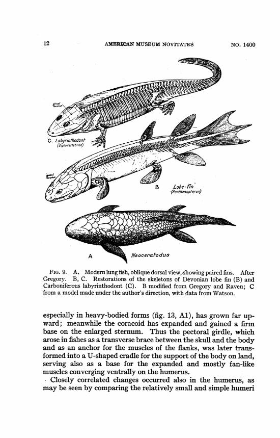
FIG. 9. A. Modern lung fish, oblique dorsal view,pshowing paired fins. AfterGregory. B, C. Restorations of the skeletons of Devonian lobe fin (B) andCarboniferous labyrinthodont (C). B modified from Gregory and Raven; Cfrom a model made under the author's direction, with data from Watson.
especially in heavy-bodied forms (fig. 13, Al), has grown far up-ward; meanwhile the coracoid has expanded and gained a firmbase on the enlarged sternum. Thus the pectoral girdle, whicharose in fishes as a transverse brace between the skull and the bodyand as an anchor for the muscles of the flanks, was later trans-formed into a U-shaped cradle for- the support of the body on land,serving also as a base for the expanded and mostly fan-likemuscles converging ventrally.on the humerus.
Closely correlated changes occurred also in the humerus, asmay be seen by comparing the relatively small and simple humeri
12 NO. 1400

THE HUMERUS410M FISH TOMAN1
EryopsFIG. 10. Skeletal patterns of the pectoral limbs. A. Devonian lobe fin. B.
Hypothetical intermediate. C. Permo-Carboniferous labyrinthodont. AfterGregory and Raven.
of Eusthenopteron (fig. 1OA) and Sauripterus (fig. 31) with thelarge and complexly built humeri of Eryops, Diadectes, Dimetrodon,and later forms.Romer (1922, p. 554) showed that in primitive tetrapods the
1949 13

AMEIkCAN MUSEtTM NOVITATES
4a te raI D orsalEryops_Right humerus
Medial Ventral
FIG. 11. Four views of the right humerus of a large Penno-Carboniferouslabyrinthodont, to illustrate the four "triangular areas" of Romer. Humerusafter Miner; triangular areas after Romer; muscle areas after Miner. PD,proximal dorsal; DD, distal dorsal; PV, proximal ventral; DV, distal ventralareas; ECTC, ectepicondylar process; ENTC, entepicondylar process; RC, capi-tellum for radius.
14 NO. 1400

THE HUMERUS FROM FISH TO MAN
humerus, with its wide proximal and distal ends nearly at rightangles to each other, "was built as a tetrahedron [fig. 11], com-posed of four roughly triangular surfaces, two having their basesproximally, and two distally, with the planes of these bases atright angles to one another." The tetrahedron is enclosed by fourroughly triangular surfaces, respectively, proximal-dorsal (PD),distal-dorsal (DD), proximal-ventral (PV), and distal-ventral(DV). Each of these serves as a triangular truss, and collectivelythey resist bending moments imposed by muscular force, byweight, and by the reactions of the medium, converging towardsthe humerus from various directions. In very short humeri ofprimitive tetrapods (figs. 11, 14, 15) the wide bases and lowheight of the four humeral triangles reflect the relative thicknessand shorter contractile ranges of their respective muscles ormuscle groups, while the relatively long shafts of many laterhumeri (figs. 14, 15, 29) indicate reduced thickness and strengthwith lengthened contractile ranges. Emphasis of any of the crosssections, crests, or processes implies greater strength of musclesor ligaments and involves changes in their angles of insertion andin the lengths of their lever arms. For example, the very largeentocondylar projection in primitive tetrapod humeri (fig. 11,ENTC), together with the shortness of the forearms (fig. 13A),implies (a) corresponding thickness and strength of the pronatorsand (b) relatively open insertion angles, with directions of pullmore nearly at right angles to the shaft of the radius and thus withfavorable leverage but short range.The spirally warped head or proximal articular facet (fig. 11)
of the primitive tetrapod humerus permitted partly rolling, partlytwisting movement of the humerus under the pull of successivesectors of the fan-like muscles covering the neck and pectoralgirdle (Miner, 1925). Thie capsular and other ligaments mustobviously have been so arranged as to permit this rocking andtwisting movement.The primitive tetrapod humerus was adapted for a crawling gait,
with sharply everted elbows, wide trackway, and short stride. It isbest exemplified in large-bodied labyrinthodonts such as thePermo-Carboniferous Eryops (fig. 13A, Al) and in the diadectiddivision of the Cotylosauria or primitive reptiles. In humeri withlengthy shafts (figs. 14, 15, 29) the proximal and distal humeraltriangles recede, and the humerus as a whole assumes somewhatthe functions of a loaded beam supported at either end, or more
1949 15

AMERICAN MUSEUM NOVITATES
FIG. 12. Humeri of primitive tetrapod (A, Al) and modern anuran (B, B1).After Gregory.
specifically of the boom or jib of a derrick crane (fig. 30). Whenthe head of the humerus (figs. 14, 15, 25) grows towards the dorsalor rear side the upper third of the bone may be bent backward, thelower third forward, producing a more or less sigmoid contour inside view. Strengthening of the bone may occur either by thick-ening the walls of the more or less hollow shaft or by the presenceof old or new crests between the muscle masses. Successive crosssections from the surgical neck downward suggest a system ofopposed (front and back) triangular struts or trusses (fig. 11).
SURVIVING AMPHIBIA
Among the smaller surviving Amphibia the ends of the humerusoften became cartilaginous.- In the urodeles (fig. 31) the usuallysmall humerus is frequently associated with aquatic habits andwith a wide, fan-like expansion of the coracoid cartilage and itsmuscles. The scapular blade sends forward a large "procoracoid"process which serves as a base for the fan-like supracoracoideusand procoraco-humeralis muscles. Reduction and loss of thecleithrum, clavicle, and interclavicle in these urodeles have con-tributed to considerable mobility of the right and left halves of the
16 NO. 1400

194 IHa ME1 ir O-
! 'IS TO 1THE HtJMBRTUS'M PH YOTMAN
Pelobates
FIG. 13. Skeletons of primitive tetrapod (A, Al) and modern anuran (B,B1). After Gregory.
coracoscapular arch, to a widely swinging movement of thehumerus and to a long reach in swimming.
In the frogs and allied forms (fig. 13B, B1) leaping and swim-
1949 17
AMnRICAN MUSEUM NOVITATES
AErythrosuchus
DCampy/ognathus
AMPonyx la/ticosaurus
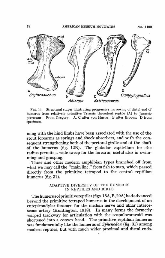
FIG. 14. Structural stages illustrating progressive narrowing of distal end ofhumerus from relatively primitive Triassic thecodont reptile (A) to Jurassicpterosaur. From Gregory. A, C after von Huene; B after Broom; D fromspecimen.
ming with the hind limbs have been associated with the use of thestout forearms as springs and shock absorbers, and with the con-sequent strengthening both of the pectoral girdle and of the shaftof the humerus (fig. 12B). The globular capitellum for theradius permits a wide sweep for the forearm, useful also in swim-ming and grasping.These and other modern amphibian types branched off from
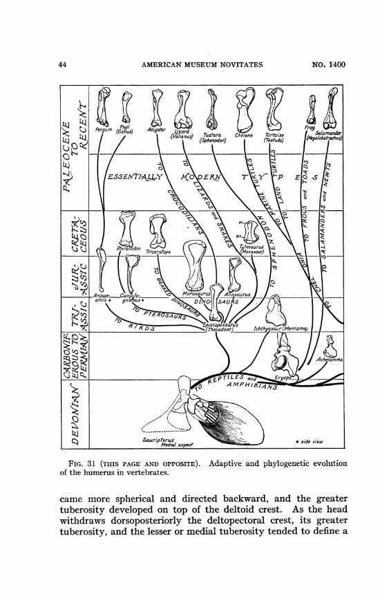
what we may call the "main line," from fish to man, which passeddirectly from the primitive tetrapod to the central reptilianhumerus (fig. 31).
ADAPTIVE DIVERSITY OF THE HUMERUSIN REPTILES AND BIRDS
The humerusofprimitivereptiles (figs. 18A, B, 29A) hadadvancedbeyond the primitive tetrapod humerus in the development of anentepicondylar foramen for the median nerve and ulnar interos-seous artery (Huntington, 1918). In many forms the formerlywarped trackway for articulation with the scapulocoracoid wasshortened into a convex head. The primitive reptilian humeruswas fundamentally like the humerus of Sphenodon (fig. 31) amongmodern reptiles, but with much wider proximal and distal ends.
18 NO. 14009
THE HUMERUS FROM FISH TO MAN
A/*.
Pgt
54tI
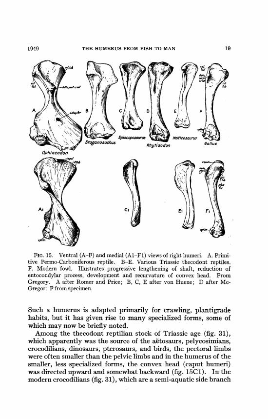
FIG. 15. Ventral (A-F) and medial (Al-Fl) views of right humeri. A. Primi-tive Permo-Carboniferous reptile. B-E. Various Triassic thecodont reptiles.F. Modern fowl. Illustrates progressive lengthening of shaft, reduction ofentocondylar process, development and recurvature of convex head. FromGregory. A after Romer and Price; B, C, E after von Huene; D after Mc-Gregor; F from specimen.
Such a humerus is adapted primarily for crawling, plantigradehabits, but it has given rise to many specialized forms, some ofwhich may now be briefly noted.Among the thecodont reptilian stock of Triassic age (fig. 31),
which apparently was the source of the aetosaurs, pelycosimians,crocodilians, dinosaurs, pterosaurs, and birds, the pectoral limbswere often smaller than the pelvic limbs and in the humerus of thesmaller, less specialized forms, the convex head (caput humeri)was directed upward and somewhat backward (fig. 15C1). In -themodern crocodilians (fig. 31), which are a semi-aquatic side branch
1949 19
AMERICAN MUSEUM NOVITATES
of the lizard-like thecodont stem, the humerus is fairly long andnarrow with somewhat sigmoid curve in side view. The ento-conidylar process, so large in primitive tetrapods, is here repre-sented only by a low convexity for the lateral ligaments of theelbow. The extensor-supinator crest is low and inconspicuous, andthe entire distal end forms a transversely placed rounded capitel-lum-trochlea. This type of humerus compares rather closely withthe humeri of relatively primitive saurischian dinosaurs (fig. 31),and Miner's study (1925) of the muscles and muscle areas oftetrapods gives reliable data for interpreting the homologousmuscular areas in dinosaur humeri.Among bipedal, bird-like dinosaurs the humerus and hand were
long and narrow; the humerus of Struthiomimus had a long,slender shaft, possibly supporting a patagium. In outer side viewthe humerus was slightly sigmoid, the distal concavity facingforward.
Increasing massiveness of body led on one side from the primi-tive Triassic thecodonts' to the huge, bipedal, flesh-eating dino-saurs, eventually with small hands and short humeri (fig. 31), andon the other to the ponderous, secondarily quadrupedal Sauropoda.The latter had very large humeri with a wide fossa for the thickcoraco-brachial muscles, and massive deltopectoral ridge (fig. 31).The distal epiphysis was long-growing and partly cartilaginous.The distal position of the large, radio-ulnar facet permitted amarked opening of the humero-radial angle, useful in walkingunder water.The beaked difiosaurs or Ornithischia, which had advanced
herbivorous jaws and teeth, were also secondarily quadrupedaland their ungual phalanges bore flat nails. Among them theduck-bill and cassowary-like dinosaurs were semi-aquatic, thefingers partly webbed; their humeri in side view were slightlysigmoid but not notably specialized. In the ponderous quadru-pedal ceratopsians, on the other hand, the humeri were verymassive (fig. 31) with large proximal crests and sharply bentelbows for the support of the enormous skull. The sharply pro-jecting entocondylar convexity of the early tetrapod humerus hadlong since been reduced in the small bipedal running ancestors ofthese huge animals, but with increasing weight the proximal anddistal ends of the humerus were secondarily widened and thick-ened, and the massive distal end was probably tied to the hugeulna and radius by very large and strong ligaments and by thick
20 NO. 1400

THE HUMERUS FROM FISH TO MAN
A Chenopsis atrata
FIG. 16. Humerus, pectoral girdle, and axial skeleton in a bird of strongflight. Showing stiffening of backbone, strong development of sternum, furcula,coracoid, spring-like ribs. The large deltopectoral crest flares out near theproximal end of the columnar humeral shaft. The forward and upward move-ment of the wing is effected in part by the deep pectoralis minor, the downwardmovement by the pectoralis major. From specimens.
muscles of great power. Still shorter and relatively wider werethe humeri of the armadillo-like ankylosaurs. In brief, humeri ofpnmitive dinosaurs, starting from the small, rather weak humeriof the Triassic thecodonts gave rise to: (1) the very long humerusof Struthiomimus with its almost cylindrical shaft- and smalltuberosities; (2) the long but enormous humeri of certain sauro-pods (e.g., Brachiosaurus); and (3) the extremely massive, widehumeri of the later Ceratopsia and Ankylosauria.
In quadrupeds the length of the humerus is obviously one of theseveral factors in the length of 'the stride. The long humerus of
1949 21

AM1tRIAN MUSEUMN-OVITATESN
FIG. 17. Skeleton and half section of box tortoise, to show partial enclosureof girdles and limbs by overgrowth of carapace and plastron. From specimens.
climbing animals contributes to the long reach and must withi-stand relatively great tensile stresses due to weight.Among the bat-like pterosaurs the shaft of the humerus (fig.
14D, 31) was narrow with thin walls and pneumatic interior. Inside view the bone was curved anteroventrally, and there were ahigh delto-pectoral crest and large coraco-brachialis fossa. Thejoint at the narrow distal end was more or less hinge-like.The long humerus (fig. 16) of birds of strong flight has a large
oval head facing dorsocaudally, a fairly large internal tuberosity,and a pneumatic interior. Thus the avian humerus, as comparedwith the central reptilian type, has undergone much less changethan has the avian pectoral girdle, in which the sternum hasgained a huge base and a high median keel (fig. 16). The humeriof ratite birds is longest in Rhea and becomes very small in emus,cassowaries, and Apteryx, i.e., as the wings degenerate. Thepectoral limb of penguins (fig. 31) has become paddle-like, but
NO. 1400

THE HUIMERtIS FROM FISH TO MAN
FIG. 18. Comparative series of humeri from primitive reptile (A) to modernsea turtle (D). After Gregory.
the elements of the avian wing are still present, though highlymodified (Simpson, 1946).The marked sigmoid curve of the tortoise humerus (figs. 17,
18), together with the globular, backwardly directed head, per-mits the humerus to be swung either far forward, so that the elbowlooks like a knee (fig. 17), or far backward to enable the pectorallimb to be tucked under the overhanging marginal ridge of thecarapace. In spite of its specialization the humerus of the tortoise(fig. 18C) is foreshadowed in basic features by the humerus ofpareiasaurs (fig. 18B), which in turn are readily derivable from theprimitive tetrapod type retained in diadectid cotylosaurs (fig. 18A).The very peculiar humerus of the sea turtle (fig. 18D) forms partof a pectoral paddle or wing; it is flattened, the distal end widened,the deltopectoral crest much modified, and the medial tuberositygreatly enlarged and flattened.
In ichthyosaurs (fig. 31) the limbs have become completelypaddle-like, the humerus was flattened, with wide distal ends, andthe rest of the limb has changed into more or less polygonalsecondary polyisomeres. The humerus of ichthyosaurs may havebeen derived from the slender, semi-aquatic humerus of Meso-saurus. The humerus of plesiosaurs (fig. 19E, F) is closely con-nected with that of nothosaurs (fig. 19C) which, in turn, leadbackward towards a Sphenodon-like type (fig. 19A, B). As inother advanced aquatic reptiles the facet for the ulna is shifted to
2X1949

AMERICAN MUSEUM NOVITATES
RI G H T HUMERIR mb6.erVENTRA L V/EW6icX
br
Plesiosa-urHydrotherosaurus
FPlacodont
DPlesiosaurus
antChampsosaurus NotAhosaurus SdSCapat.
t
ambulator laramienis C atdorA -
/ F:
,~~~~~~~~~~~~~~~~'~~~~
5, DORSAL VIW'S
FIG. 19. Comparative series of reptilian humeri; from relatively primitivesemi-aquatic form (A, B) to highly specialized marine plesiosaur (F). FromGregory. A, B after Barnum Brown; C, D after von Meyer; E after Watson;F after Wells.
the outer side of the distal end of the humerus so that the olecranalprocess is lateral to, not behind, the head of the radius, bothradius and ulna being flattened into the same plane.
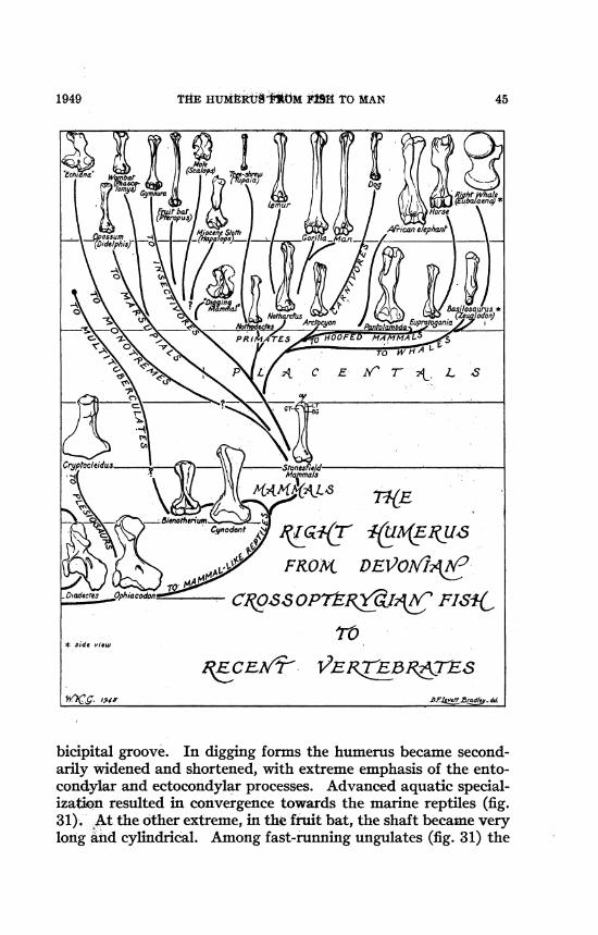
RISE AND DIVERSITY OF THE MAMMALIAN HUMERUS
Returning to the "main line," in the earlier mammal-likereptiles, the humerus of the less specialized pelycosaurs (fig. 29A)
24 NO. 1400

THE HUMERUS FROM FISH TO MAN
retained a large entocondylar process and was not very differentfrom the primitive tetrapod type in being adapted for a crawlinggait. In the giant South African dinocephalians the pectoralgirdles and humeri were relatively primitive, although the elbowin standing was less sharply bent and everted than in the primitivetetrapod. For the support of the great body weight the coraco-brachialis brevis muscle and its fossa were widely expanded, aswell as the areas for the extensors (Romer, 1922, pl. 36). In thegorgonopsians the fairly primitive humerus (fig. 29B), with itsmoderate sigmoid curve, permitted a somewhat cat-like stridewith the body raised well off the ground (Colbert, 1948), withelbows less everted than in primitive tetrapods. In the cynodontsthe pectoral girdle approached the mammalian stage especiallyin the scapula and coracoid, and so also did the humerus (fig.29C).Simpson (1928, p. 155) has shown that in the humerus of the
cynodont Diademodon (fig. 20A) the convex head was directeddorsoposteriorly but was not spherical, and the deltopectoralcrest at its proximal end was not yet expanded into a distinctgreater tuberosity, although the lesser tuberosity was well de-veloped. A large entepicondylar foramen was retained as well asan ectepicondylar foramen, both being characteristic of reptiles.The deltopectoral crest had begun to be turned medially towardsthe fossa for the coracobrachialis brevis and biceps muscles.Among the Tritylodontoidea the humerus (fig. 31) is well shown
in Bienotherium from the upper Triassic of China (Young, 1947,p. 582). This genus may represent an early side branch of themammalian Order Multituberculata. Its skull and jaws were ad-vanced beyond the cynodont stage and were almost mammalian.The descriptively multituberculate molars were adapted forcrushing hard objects. The humerus closely approaches theprimitive mammalian type, but the upper end of the deltoidcrest, although it is already raised into a swollen great tuberosity,is continuous with the head, which is not so fully spherical as intypical mammals. There are a large entepicondylar bridge andforamen, and there may be also an ectepicondylar groove.Among the Jurassic mammals the humerus is known only from
three specimens of uncertain ordinal position. As described bySimpson (1928, pp. 155-159) they retained many cynodontfeatures, but in others, such as the nearly spherical head, theywere more mammalian (fig. 20B). However, after careful com-
1949 25

AMERICAN MUVSEUM NOVITATES
FIG. 20. Median posterior view of right humeri. After Simpson. BG,bicipital groove; DPC, deltopectoral crest; ECEC, ectepicondyle; ECF, ectepi-condylar foramen; ENEC, entepicondyle; ENF, entepicondylar foramen;GT, greater tuberosity; H, head; LT, lesser tuberosity; OF, olecranon fossa;SC, supinator crest; TR, trochlea. Al-DI. Proximal views. Not to scale.
parative studies Simpson (op. cit.) concludes that these humeriwere normally held in a nearly horizontal plane as in cynodontsand monotremes. "The radial and ulnar articulations [of theStonesfield mammal]," writes Simpson (op. cit., p. 156), "are alsoextraordinarily reptilian. The true sharp-crested trochlea oflater mammals is not well developed. The radius articulates on aseparate rounded surface external and somewhat ventral to theulnar articulation."The humerus (fig. 20C) of the monotreme Ornithorhynchus at
first sight recalls that of primitive reptiles, but as Simpson (op.
26 NO. 1400

THE HUMERUS FROM FISH TO MAN1949 27
01-4
44
0)
0lS
S..
0
tn400C)C)co
c.;6.-

AMtRRICAN MUSEUM NOVITATES
cit.) and Howell (1937a) have shown, it is specialized for bothdigging and swimming. In the pectoral girdle the dorsoposteriorborder of the scapula is prolonged backward while the prespinousfossa is either vestigial or incipient. The thin, plate-like epicora-coid slides a little on the expanded sternum and strong inter-clavicle. Although these may be partly habitus features, thegirdle of Ornithorhynchus as a whole strongly recalls the conditionsin Cynognathus and indicates that also in the humerus the basicfeatures have been inherited from a remote cynodont ancestor,although the extreme projection of both the entepicondylar andextepicondylar processes (fig. 20C) together with the concavelycurved head (fig. 20C1) may be later specializations.The wide humerus (fig. 22B) of Tachyglossus ("Echidna")
clearly reflects great strength in digging, as do also the block-likecoracoid, the well-braced interclavicle, and stout sternum.
According to Simpson (1928, pp. 156-158), in the humerus(fig. 20D) of the opossum Didelphis the plane of the distal enddiverges only about 10 degrees from that of the proximal end(drawn through the greater and lesser tuberosities), whereas inthe Stonesfield humerus (fig. 20B1) of a Jurassic mammal thecorresponding angle is about 75 degrees and in Diademodon only40 degrees. This accords with much other evidence that, as wepass from early tetrapods to mammals, the angle between theproximal and distal ends of the humerus approaches zero. This isno doubt associated with the drawing inward of the elbows andopening of the angle at the elbow as in typical mammals.The humerus of Didelphis (figs. 21, 22) also is typically mam-
malian in possessing a distinct lateral or greater tuberosity on topof the deltoid crest. Upon it are inserted both the supraspinatusand infraspinatus muscles, which are pretty certainly derivedfrom the supracoracoscapula muscles of reptiles (Romer, 1922,p. 594). Likewise an internal or medial tuberosity was present, towhich was attached the subscapularis muscle. The "mammalian"posture of the humeri of Didelphis, with the elbow almost im-mediately beneath the caput humeri, is made possible not only bythe various mammalian features of the humerus itself, but (1) bythe freeing of the reduced coracoid from direct contact with thesternum, (2)-by the wide mobility of the whole complex of clavicle,coracoid, acromion, scapula, and humerus around the sternal artic-ulation of the clavicle, and (3) by the protection afforded to theshoulder joint by the overhanging acromion and its ligaments.
28 NO. 1400

THE HUMERUS FROM FISH TO MAN
Myrme-cophaga
G |
7Thyla-cynus
M Nj
CastIor Arctomys
Phalan- ArcAgisla cyorn
P
SAoen- Erin-odon aceus
Per"p-t chuIs
L
3@o- h'ver-
ratls
R,1
Euproto-5QSonia
FIG. 22. Left humeri of various primitive and adaptive types. From Greg-ory. A after Williston; E after Scott; others from the American Museum ofNatural History collections.
1949 29

AMERICAN NUSRVM NOVITATES
Doubtless the moving outward of the shoulder joint away fromthe midline exposed it to greater danger from breakage in falling,but the resulting mobility, the wider reach and swing of the fore-limb, contributed much to the increased speed, effectiveness, andendurance of mammalian over reptilian locomotion. The Didel-phis pattern of pectoral girdle and humerus appears to be arche-typical to that of all other marsupials.
In the "marsupial wolf" (Thylacynus) the marked elongation ofthe humerus (fig. 22) has helped to lengthen the stride of thiscursorial predator. The entepicondylar and ectepicondylar pro-jections were much reduced. The distal facet indicates a chieflyhinge-like movement at the elbow, with limited supination.At the other adaptive extreme, in the humerus of the "mar-
supial mole" (Notoryctes), digging powers are indicated by themarked widening of the distal end, by the strength of the crests,and by the winding surface for the brachioradialis.Among the diprotodont marsupials the phalanger humerus (fig.
22J, Phalangista) is close to the didelphid type, but with some in-crease of the supinator-extensor crest. The humerus (fig. 22) ofthe wombat (Phascolomys), a powerful digger, is evidently de-rived from the phalanger type, but it has become stouter, distallywider, with projecting tuberosities and crests. On the whole thereis a certain convergent resemblance to the cynodont humerus(fig. 29C), but the head is more spherical and the distal end com-pletely mammalian.The humerus (fig. 23) of the giant herbivorous Diprotodon and
related genera is associated with a very peculiar form of scapula inwhich there is a large, cap-like, dorsal protuberance directly abovethe vertical scapular spine. To this may have been attached athick dorsoscapular ligament, while below it was the serratus an-terior muscle, supporting the massive body. The vertebral borderof the scapula is extended far downward, and the axial margin isgreatly reduced. The inferior angle ends in a prominent, down-wardly projecting tuberosity, possibly for a robust teres minor.Perhaps in correlation with these and other features the ulna has anenormous, transversely widened olecranon for the triceps, and themiddle of the lateral aspect of the humerus bears a prominent,laterally projecting process and rugosity, probably for the deltoidmuscle. In short the humerus of Diprotodon as compared with theprimitive phalanger type, in becoming gigantic and graviportal,has on the whole been simplified.
30 NO. 1400

1949 THE HUMERUS FROM FISH TO MAN 31
FIG. 23. Skeleton of Diprotodon. From Gregory.
Among the placental mammals adaptive branching was alreadyprolific in the Paleocene and Lower Eocene epochs, and therewere many lines that subsequently became extinct as well as afew that branched in later times. Matthew (1904, 1937) showedthat the known hands and feet of Paleocene and early Eoceneplacental families were pentadactylate, with spreading digits andpartly divergent pollex and hallux, respectively, also that theradius and ulna could be freely supinated. He regarded theseand correlative characteristics of the girdles, limbs, and vertebraeas indicating for the placental mammals arboreal ancestry, asalready had been held to be so in marsupials by Dollo (1899) andby Bensley (1901).The humeri of the smaller Paleocene and Lower Eocene placen-
tal mammals, so far as known, were of the same basic pattern asthe opossum humerus (fig. 22F), but the extensor-supinator crest(fig. 22K, L, 0) was less flaring at the upper or proximal end. Itsloped evenly towards the shaft of the bone and did not end abovein a sharp notch. At the distal end the capitellum is less sphericalin front view than it is in Dideiphis (fig. 22F), and the trochleaoften has a sharp inner keel extending distally below the level ofthe capitellum. In the living Gymnura (fig. 31), a fairly primitive

32 AMERICAN MUSEUM NOVITATES NO. 1400
b0
0~~~~~~~~~~~~~~~~~~~~~Cd0
4-)
0-

THE HUMERUS FROM. FISH TO MAN
insectivore, as well as in Centetes (fig. 24), this kind of elbow jointis associated with a transverse widening of the head of the radius,which extends across to, and even a little beyond, the inner keelof the trochlea. With such a joint only a moderate supination ofthe radius is possible, and this condition predominates amongboth fast running and digging mammals. And among the primi-tive Paleocene and Lower Eocene placentals (except possiblyNothodectes) there is little or no suggestion of the "outer lip" ofthe trochlea (figs. 25, 28) which is developed in arboreal Primates(Gregory, 1920, p. 71; Simpson, 1935, p. 13).A review of the skeletons of Paleocene and Lower Eocene
mammals suggests that these early placentals were nearer toground-living, unguiculate, semi-fossorial ancestors than theywere to a more remote arboreal stock (fig. 31). Advanced fos-sorial habitus was already attained in the mole-like Paleocenehumerus (fig. 31) described by Simpson (1937, p. 140). Thisfurther indicates a fairly long antecedent history of partly ground-living habits for the placentals, extending well back into theUpper Cretaceous. As noted above the only known mammalianhumeri of the Triassic (fig. 31) and Jurassic (fig. 20B) ages tend toconnect the marsupials with the cynodont stock, and the detailedcharacters of the Stonesfield humerus indicate normally horizontalposture with elbows widely everted (Simpson, 1928). Such acombination may have been useful in a partly climbing, partlydigging, ambulatory stage, ancestral to the known diversifiedPaleocene humeri.With increasing speed and lengthening stride the humerus may
become either relatively long and narrow as in the dog (fig. 31), orshort and thick (when the power is concentrated in the proximalsegment) as in the horse (fig. 31) and other fast cursorial her-bivores. The elbow joint also becomes quite hinge-like, and themovement is limited to extension and flexion. Especially inmassive,.heavy-bodied mammals, the entocondylar eminence isoften pronounced, indicating very strong ligaments. The delto-pectoral crest is usually prominent, and so also are the windingsurface for the brachialis inferior muscle and the extensor-supina-tor crest.
In heavy-bodied ungulates which have large scapulae and heavyshoulder muscles the great tuberosity of the humerus sometimesbecomes very large and high, especially in elephants, titanotheres.and horses (fig. 31).
331949
AMkMUCAM MUSgtrM NOVITATES N.i0
ofnralaInma DfdUPh/i '"anyhMecles &M7zrtaP zeDZal ce&a n fYm.~~~~ ~ ~ ~ ~ ~~ -.
,*#toamhda 0/delphls Aroys i ds iZatardue5 Lsiar Cebu A/? /%/on
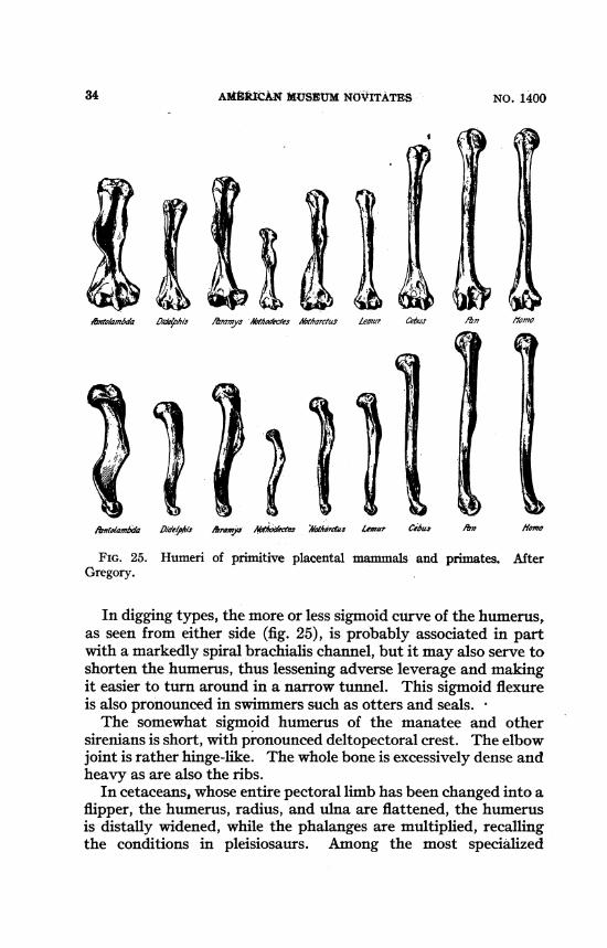
FIG. 25. Humeri of p tive placental mammals and primates. AfterGregory.
In digging types, the more or less sigmoid curve of the humerus,as seen from either side (fig. 25), is probably associated in partwith a markedly spiral brachialis channel, but it may also serve toshorten the humerus, thus lessening adverse leverage and makingit easier to turn around in a narrow tunnel. This sigmoid flexureis also pronounced in swimmers such as otters and seals.The somewhat sigmoid humerus of the manatee and other
sirenians is short, with pronounced deltopectoral crest. The elbowjoint is-rather hinge-like. The whole bone is excessively dense andheavy as are also the ribs.
In cetaceans, whose entire pectoral limb has been changed into aflipper, the humerus, radius, and ulna are flattened, the humerusis distally widened, while the phalanges are multiplied, recallingthe conditions in pleisiosaurs. Among the most specialized
34 NO. 1400
1949 THE HUM*RUS FROM FISH TO MAN 35
0
C4,~~~~~~~~~~~~~~~~~~~~-0
-4~
C6)cli

AMERICAN MUSEUM NOVITATES
cetacean humeri is the humerus of Eubalaena (fig. 31) with ex-tremely short shaft, huge spherical head, wide distal end, andsmall antero-internal tuberosity, possibly for the mastohumeralismuscle (cf. Schulte, 1916).The very long, somewhat cylindrical humeri of "flying lemurs"
(cobegos) are sufficiently strong to resist the bending momentsimposed by the weight of the body in skimming through the airand landing, and by the pulls of the long extensors and flexors.The cylindrical radius is much larger than the splint-like ulnawhich is coalesced distally with the radius. Thus the movementat the elbow is largely hinge-like, as indicated also by the widenedhead of the radius. In the fruit bats, the humerus (fig. 31) is alsovery long and slender. In the Microchiroptera two processes, oneon either side of the head of the humerus, sometimes articulatewith the scapulo-coracoid, thus limiting the movement, but in-creasing the strength, of the joint (Miller, 1907). Doubtless theassociated ligaments must be relatively strong.
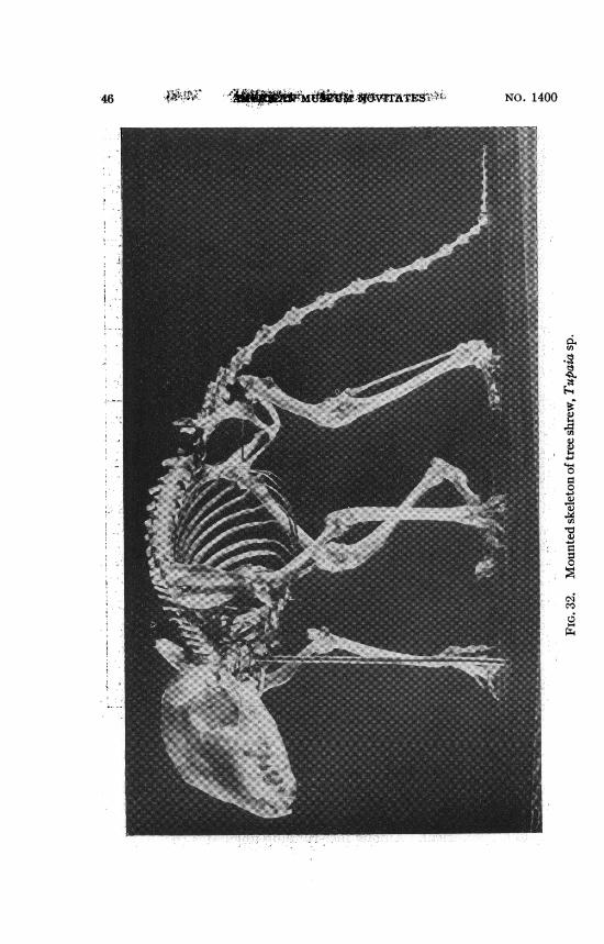
THE HUMERUS FROM TREE SHREW TO MAN
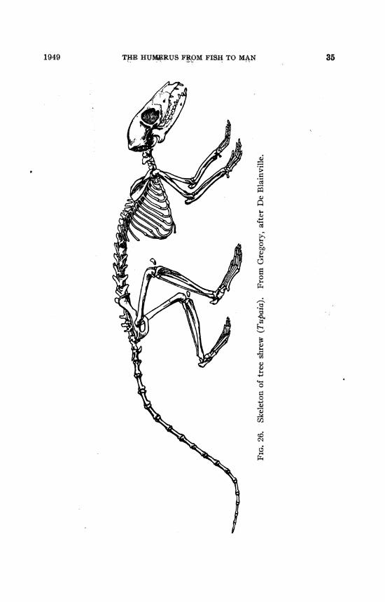
The Recent and extinct "tree shrews" (Tupaioidea) althoughlong classed with the Insectivora agree with or approach thelemurs in many significant structural features (cf. Gregory, 1910,pp. 269-285) and have been transferred to the lemuroid primatesby recent authors (cf. Simpson, 1945, pp. 61, 176). In the RecentTupaia the humerus (figs. 26, 32) and the forearm are both longand slender, and the radius is about as long as the humerus. Thehead of the humerus is large and spherical, the great tuberositylow, and so is the deltoid crest. The bicipital groove is barelyindicated, the extensor-supinator crest small, an entepicondylarforamen is present. The entocondylar eminence is conspicuous,indicating thick tendons of the pronator and flexor muscles.The capitellum is relatively large and globular, the head of theradius subcircular on the medial side. The trochlea is relativelysmall and, except perhaps in extreme flexure, the small coronoidprocess of the ulna seems to have little share in the front face ofthe trochlea, which in the front view is in contact solely with theradius. Nevertheless, moderate, though not extreme, supinationseems possible, as is indicated by the short, spreading hands withcompressed, curved, pointed claws and well-developed, partlydivergent pollex.
All these and other features may be preadaptations for arboreal
36 NO. 1400
THE HUMERUS FROM FISH TO MAN
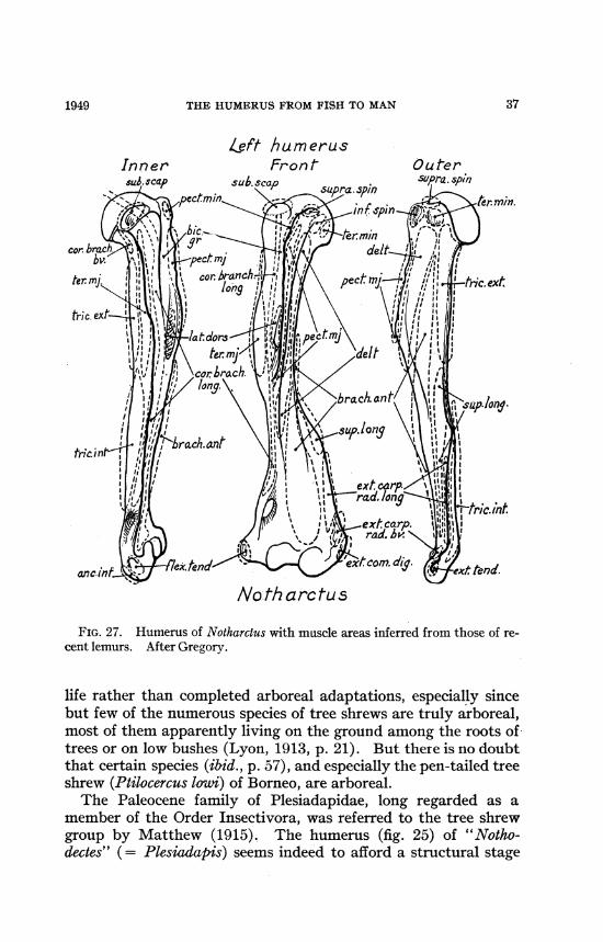
Left humerusInner Fron t Outer
No th arc tu s
FIG. 27. Humerus of Notharclus with muscle areas inferred from those of re-cent lemurs. After Gregory.
life rather than completed arboreal adaptations, especially sincebut few of the numerous species of tree shrews are truly arboreal,most of them apparently living on the ground among the roots oftrees or on low bushes (Lyon, 1913, p. 21). But there is no doubtthat certain species (ibid., p. 57), and especially the pen-tailed treeshrew (Ptilocercus low'i) of Borneo, are arboreal.The Paleocene family of Plesiadapidae, long regarded as a
member of the Order Insectivora, was referred to the tree shrewgroup by Matthew (1915). The humerus (fig. 25) of "Notho-dectes" (= Plesiadapis) seems indeed to afford a structural stage
371949

AMERICAN MUSEUM NOVITATES
leading from semi-terrestrial to the fully arboreal habitus of theEocene and Recent lemuroids. The humerus of Nothodectes wasrelatively shorter and wider distally than that of Ptilocercus(Gregory, 1920, p. 70, pl. 27), and its more strongly developedcrests and process suggest a stronger, probably ground-living type.Simpson (1935) during a careful analysis of the morphologicalcharacters of the Plesiadapis ("Nothodectes") skull, dentition,vertebrae, scapula, humerus, radius, etc., referred the Tu-paioidea, including Plesiadapis and the tree shrews, to theSuborder Lemuroidea.
In Notharctus, a quite primitive Eocene lemur, the humerus(fig. 25) somewhat resembles the climbing opossum type (figs. 21,22) especially in its large extensor-supinator crest (fig. 27). Butthe opossum has a relatively short, well-clawed hand and com-paratively strong flexor muscles, whereas in Notharctus the wholearm and hand was long and slender, with thin fingers and weakflexors of the digits; it also had nails, not claws. Here, as in manyother cases, two mammals with very different kinds of hands,conforming to different locomotor habits, may have less distinctlydissimilar humeri.The Notharctus type of humerus (figs. 25, 28) was evidently well
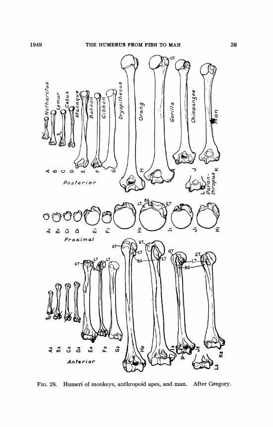
fitted to give rise by minor successive changes to the diversehumeri of later lemuroids and monkeys and eventually to those ofapes and man. The humerus of Tarsius appears to be a special-ized derivative of the Plesiadapis-Notharctus stem (Gregory, 1920,p. 75).The humeri of New World monkeys (fig. 28) often retain an
entepicondylar foramen, but their shafts are thinner than inNotharctus, and the supinator crest is less flaring distally. The"outer lip" (fig. 25) or ridge of the trochlea, which is beginningin Notharctus, is better developed in the New World monkeysand in all higher types, culminating in the anthropoids and man(fig. 28). Its presence marks the increase in width of the coronoidprocess of the ulna (fig. 33) which now supports the medial partof the circular radial head and takes up a part of the trochleawhich was formerly (in Paleocene mammals) occupied by themedial part of the transversely oval head of the radius. Thisarrangement permits extreme supination and greatly strengthensthe flexor side of the elbow joint.The humerus of the macaque (fig. 28), a fairly central Old
World monkey, is more or less intermediate between the lemuroid
38 NO. 1400
THE HUMERUS FROM FISH TO MAN 39
PostIer ior <,I, 0)/)c L
:3~~~~~~~~BG
oL
Proximal
w " w " s
< o a
An te ri or
FIG. 28. Humeri of monkeys, anthropoid apes, and man. After Gregory.
1949

AMREXCAIC MUSEUM NOVITATES
G Homo
FIG. 29. Anterior aspect of the right humerus, in standing pose; structuralseries, from primitive mammal-like reptile to man. From Gregory. A afterRomer and Price; B after Broili and Schroder; C-G from specimens.
and anthropoid types. The gibbons (fig. 28) with their extremelylong arms and fully brachiating habits are often regarded as ir-revocably specialized side lines (Howell and Straus, 1931). Butthis conclusion takes for granted the dogma of irreversibility anddisregards the principle of correlated changes in structure andfunction. The humerus of the orang (fig. 28) looks almost as ifit were a gigantic gibbon humerus. The fossil humerus referred tothe Miocene Dryopithecus (fig. 28) combines features of thegibbon and of the modem great apes (Gregory, 1928; Ehrenberg,1938).The apes all -have the ability to swing the arm around the
shoulder joint, and describe a very low cone or almost a circle.The human arm has lost little if any of this ability, and the wholeshoulder girdle and arm and hand of man (fig. 33) differ fromthose of the chimpanzee and gorilla only in the proportional em-phasis of certain parts, as of the thumb in man or the great widthof the hand in old male gorillas.The distal end of a humerus (fig. 28) found by Broom at Krom-
drai near the site of the Paranthropus type skull is confidently re-
40 NO. 1400

THE HUMERUS FROM FISH TO MAN
ferred by Broom to that genus. In other features (e.g., jaws andteeth) the Australopithecinae present a mixture of ape-like andhuman features, and it is not surprising to find more or less mixedor generalized features in the humerus. But even if we set thisspecimen aside for the present, the humeri (fig. 28) of chimpanzeesand gorillas differ from those of man chiefly in rather small pro-portional measurements.
RETROSPECT
Man has inherited from the earliest-vertebrates, exemplified bythe Silurian ostracoderms (fig. 1), the basic pattern of locomotionbased upon bilaterally arranged myomeres on either side of anelastic axis and directed by a nervous system of primitive fishtype. Man also owes to the ostracoderm grade the outgrowth ofpectoral buds from the lateral myomeres and the beginnings of apectoral girdle. To the same or some related group of pre-fishes,man owes the elaboration of bony tissue with its high adapta-bility in growth and plasticity in evolution.Man owes to the crossopterygians (fig. 9) of early Devonian
age, the modelling of the proximal metapterygial piece into apotential humerus, and of the second metapterygial piece into apotential ulna. The radius appears to be represented by thefirst pre-axial radial rod.The many-jointed pectoral paddle of Eusthenopteron (fig. 6)
and Sauripterus (fig. 31) was evidently quite flexible, the flexi-bility increasing towards the periphery. When such a fish paddlewas transformed into a primitive tetrapod limb the widening ofthe head of the humerus (fig. 11) limited its mobility to a rockingand twisting movement of the shoulder, within the limits imposedby a rather narrow and warped articular surface and doubtlessalso by the capsular and associated ligaments. Especially intetrapods further restraining and strengthening reactions wereexerted by the acromial and coracoid processes (figs. 21, 23, 24,26, and 32) and their ligaments surrounding the head of thehumerus. The clavicle, on the one hand, transmitted to thesternum medially directed thrusts of the humerus, and, on theother hand, by being tied to the sternum medially and to theacromion laterally, the clavicle helped to prevent the humerusfrom being pulled too far away from the sternum. At the otherextreme, in some specialized cursorial mammals (fig. 31, horse)the loss of the clavicles is compensated, and part of the weight of
1949 41

4 AMF*ICAN MUSEUM NOVITATES
FIG. 30. The human arm and hand, conceived as a modified derrick andmanual organ.
the thorax is supported, by the dorso-scapular ligament (Schmaltz,1905) which permits a wide fore-and-aft swing of the scapula andhumerus and lessens the danger of dislocation of the shoulder.
NO. 140042

1949 THE HUMERTJS 1:kOM iISII TO MAN 43
To the earliest tetrapod stage (figs. 9, 13), typified by theUpper Devonian, Carboniferous, and Permian labyrinthodonts,we owe the third major stage in the evolution of the pectoralgirdle and limbs. At that time the bend at the elbow was sharplyemphasized (fig. 13). Both ends of the humerus were wide (fig.11), the very short shaft was strongly twisted (fig. 12), the en-tocondyle and ectocondyle,-- the extensor-supinator and delto-pectoral crests were very prominent, while the articular surfaceat the head of the humeri formed a long, warped trackway (fig.11). These features were profoundly modified or sacrificed inlater stages, but they were nevertheless prerequisite for furtheradvance. For our ancestors had to swim before they could crawl,to crawl before they could run, to run on the ground before theycould climb and leap among the trees, to brachiate, at least tosome extent, before they could walk upright on the ground.To recapitulate, in general, -primitive tetrapod humeri in both
amphibians and reptiles were short and wide, with very wide distalends for powerful flexors and extensors of the elbow. The largecapitellum for the radius was chiefly ventral rather than distal inposition, the elbows being sharply bent. The "head" or proximalarticular facet was narrow, not spherical, and directed upward andinward rather than upward and backward. In modem urodeleamphibians the proximal and distal ends of the humerus oftenbecome cartilaginous. Among later quadrupedal reptiles, thebent-limbed type of humerus was retained in lizards and Spheno-don; but the head (caput humeri) has thickened, and in thetortoise it became spherical.When the length of the stride increases, as in crocodilians, the
humeral shaft becomes longer and narrower, the entocondylarand ectocondylar processes disappear, and the elbow joint be-comes more hinge-like. In side view such humeri are somewhatsigmoid. In the smaller pterosaurs, the shaft of the humerus wasslender, but the deltopectoral crest remained large. In the largestpterosaurs, the humerus (fig. 31) is relatively short in comparisonwith the very long wing, supported by the enlarged fourth digit.By somewhat parallel changes the primitive thecodont humerusgave rise to the slender and light but strongly built bird humerus(fig. 31). At the opposite extreme in the aquatic icthyosaurs thehumerus retained the wide distal end but was flattened to supportthe paddle-like forearm and hand.
In the line leading towards the mammals (fig. 31), the head be-
44 AMERICAN MUSEUM NOVITATES NO. 1400
~j Penguin (GIu~ Alligator 1 3ard Tutra Ceon oros 3lmnderarn (s5phenodon) Tetdo) (fglbfohs
0 \{Ed SE<I%$Y<t \P E|i 04
vr~ ~~~1
t 1-tER-C~~~1tJ C ( _don . 7jTosaurus \. -4
Ptrndn Triceratops5 (oasaur) -
-Archae-..Camp Mo rosaurus >A saurus / / , __ A294ornis D u;4S>1 1At~~i ij s CcoXauruI (don chthyoaur(Ma-)
0~~~~~~~~~~~~~~~~~~~~~~~~~~~~
Sauripterus side viewMedial aspecr
FIG. 31 (THIS PAGE AND OPPOSITE). Adaptive and phylogenetic evolutionof the humerus in vertebrates.
came more spherical and directed backward, and the greatertuberosity developed on top of the deltoid crest. As the headwithdraws dorsoposteriorly the deltopectoral crest, its greatertuberosity, and the lesser or medial tuberosity tended to define a
THE HUM tRtT4M FtSH TO MAN
bicipital groove. In digging forms the humerus became second-arily wldened and shortened, with extreme emphasis of the ento-condylar and ectocondylar processes. Advanced aquatic special-ization resulted in convergence towards the marine reptiles (fig.31). At the other extreme, in the fruit bat, the shaft became verylong and cylindrical. Among fast-running ungulates (fig. 31) the
1949 45
46 NO. 1400
4!4
'~~~~~~~~~~~~~~~~~~
I~~~~~~~~~~~~~~~~~~~~~~~~~~~~~~~~~~~~~~~~~~~~~~~~~~.....'..._dAs _ ~~~~~~~~~~~~~~~~~~~~~~~~~~~~~~~~~~~~~~~~~~~~~~~~~~~~............. .... .t
0J
..i_isi>2SsS*tsSS P~~~~~~~~~~~~~~~~a
aoF' . VX~~~~~~~~~~~~~~~~~~~~~~~~~~~~~~a!, _ ~~~~~~~~~~~~~~~~~~~~~~~~~~~~~~~~~~,,E
- | .. S S~~~~~~~~~~~~~~~~~~~~~~~~~~~~~a
!taXll S1 X)~~~~~~~~~~~

7Yf
FIG. 33. Right pectoral limb skeleton of- man and gorilla. Medial view ofhumerus and forearm, palmar view of wrist and hand.
humerus is shortened and thickened, the elbow sharply bent andrestricted to hinge-like movement.The forego-ing brief review has bearing on several classic
problems that are still of daily importance to the student of evolu-
19-49 47

4AMMkICAN MUSEUM NOVITATES
tion. The humerus in life is of course an indispensable part of acontinuous, organized whole. In the dried skeleton it appears tobe distinct from its neighbors because we do not usually see thejoints and ligaments which connect it with its fellows and setlimits to its movements, nor the opposing sets of the muscleswhich operate it, nor the endlessly complex nervous organizationwhich moves and checks it purposively and requires of it sufficientquickness, precision, and power as a condition of its owner'ssurvival.
In man as in other mammals the entire pectoral girdle andappendages serve as a system of compound extensible levers usedin locomotion (fig. 24)' and, by change of function, as an organ ofprehension (fig. 21, 26, 30). As an organ of prehension and manip-ulation man's pectoral limbs (fig. 30) comprise on either side amodified derrick-crane with a movable base (the shouldergirdle), and a boom, the humerus. The latter supports at theelbow joint a secondary boom, the ulna and radius. This segmentcan be extended, flexed, and rotated. The wrist and hand (fig.33) together form a third segment with a compound base (thewrist), and five jointed booms, the metacarpals and digits. In-dividually these act like little derricks, but collectively they serveas an organ for grasping, pulling, picking, hitting, batting, punch-ing, twisting, throwing, drumming, piano-playing, violin playing,drawing, writing, gesturing, or what you will. In every one ofthese the humerus plays its part like a well-trained member of anorchestra.The so-called mystery of adaptation, in the humerus as else-
where, appears to be due partly to the very human habit of lookingat only one finished product and imagining that it was "designed"first and made afterward. But the present sketch suggests (fig.28) that the human humerus has evolved out of a primitive apehumerus, and the ape humerus from a macaque-like humerus,that in turn from a lemuroid stage, and so on back to Eusthenop-teron and beyond that. Each earlier stage was attained by changeof emphasis or proportion of certain still earlier parts and bycorrelated loss or increase in dimension of other earlier parts;also each and every earlier stage was prerequisite to all its deriva-tives. Such a system is operated according to geneticists by thetesting of relative survival value in humeri of varying character-istics. The chance encounter of a given humerus type with for-tuitously developed new opportunities has made possible every
48 NO. 1400

THE HUMERUS FROM FISH TO MAN
"creative," "emergent" type that happened to do well both in itsolder set-up and in the newer niche. Thus according to the"palimpsest" theory some "habitus" features of older stages arealways modified or lost as new habitus features come in. In theevolution of the humerus from fish to man, anisomerism or chang-ing emphasis of parts has played the leading part and polyisom-erism is seen only in the tendency to develop separate centers ofossification for the epiphysis and tuberosities.
REFERENCES TO LITERATURE
ANDREWS, CHARLES W.1910. A descriptive catalogue of the marine reptiles of the Oxford clay.
Based on the Leeds Collection in the British Museum (Natural His-tory). London, pt. 1, xxiii + 205 pp., 94 figs., 10 pls.
ANDREWS, Roy CHAPMAN1916. The Sei whale (Balaenoptera borealis). Mem. Amer. Mus. Nat.
Hist., new ser., vol. 1, pp. 289-388, pls. 28-42.BENSLEY, B. A.
1901. A theory of the origin and evolution of the Australian Marsupialia.Amer. Nat., vol. 25, pp. 245-269.
DE BLAINVILLE, H. M. D.1839-1864. Ost6ographie ou description iconographique comparee du sque-
lette et du syst6me dentaire des mammif6res. Paris, vols. 1-4, nu-merous pls. [Numerous mounted skeletons.]
BRAus, H.1901. Die Muskeln u. Nerven der Ceratodusflosse. Denkschr. Med. Nat.
Gesellsch. Jena, vol. 4, pp. 137-300, 25 text figs., pls. 21-29.BROILI, F.
1929. Acanthaspiden aus dem rheinischen Unterdevon. Sitz. BayerischenAkad. Wiss., Math.-Nat. Abt., no. 2, pp. 143-163, 4 text figs., 2 pls.
1930. Neue Beobachten an Lunaspis. Ibid., no. 1, pp. 47-51, 2 figs.1933. Ein Macropetalichthyide aus den Hunsruckschiefern. Ibid., no. 3,
pp. 417-437, 7 text figs., 1 pl.BROOM, ROBERT
1899. On the development and morphology of the marsupial shoulder-girdle. Trans. Roy. Soc. Edinburgh, vol. 38, pt. 2, pp. 749-770.
1932. The mammal-like reptiles of South Africa. London, xvi + 376 pp.,figs. 1-111. [Morphology of pectoral girdle and limbs, pp. 322-330.]
1948. A contribution to our knowledge of the vertebrates of the Karroobeds of South Africa. Trans. Roy. Soc. Edinburgh, vol. 61, pt. 2,no. 21,--pp. 577-629, 44 figs. [Humerus of procynodont Leavachia,pp. 623-626.]
BRYANT, WILLIAM L.1919. On the structure of Eusthenopteron. Bull. Buffalo Soc. Nat. Sci.,
vol. 13, pp. 1-59, 8 figs., pls. 1-18.
491949

AMIIRCAN MUStUM NOVITATESN
COLBERT, EDWIN H.1948. The mammal-like reptile Lycaenops. Bull. Amer. Mus. Nat. Hist.,
vol. 89, pp. 353-404, text figs. 1-24, pls. 27-34. [Pectoral girdle andfore limb, pp. 379-383; humerus in mounted skeleton, with muscleinsertions, p. 399.]
CUNNINGHAM, D. J.1903. Text-book of anatomy. New York, xxix + 1309 pp.
CUVIER, GEORGES, AND C. AND L. LAURILLARD1849. Anatomie compar6e, recueil de planches de myologie. Paris, 340 pls.
DEAN, BASHFORD1909. Studies in fossil fishes (sharks, chimaeroids and arthrodires). Mem.
Amer. Mus. Nat. Hist. vol. 9, pp. 211-287, 65 figs., pls. 26-41. [Clad-oselache and origin of paired fins.]
DOLLO, L.1899. Les ancetres des marsupiaux etaient-ils arboricoles? Trav. Sta.
Zool. Wimereux, vol. 7, pp. 188-203, pl. 12.1900. Le pied du Diprotodon et l'origine arboricole des marsupiaux. Bull.
Sci. Ftance et Belgique, pp. 275-280.EATON, GEORGE F.
1910. Osteology of Pteranodon. Mem. Connecticut Acad. Arts and Sci.,vol. 2, 38 pp., 31 pls.
EHRENBERG, KURT1938. Austriacopithecus, ein neuer menschenaffenartiger Primate aus den
Miozan von Klein-Haderdorf bei Poysdorf in Nieder-osterreich (Nie-der-Donau). Sitzber. Akad. W'iss. Wien, div. 1, vol. 147, pt. 4,pp. 71-110, 9 tables. [Humerus referred by Schlosser to Dryopithe-cus.]
FRANCIS, ERIC T. B.1934. The anatomy of the salamander. Oxford, xxi + 381 pp., 25 pls.
GILMORE, CHARLES W.1920. Osteology of the carnivorous Dinosauria, in the U. S. National Mu-
seum, with special reference to the genera Antrodemus (Allosaurus)and Ceratosaurus. Bull. U. S. Natl. Mus., no. 110, xi + 159 pp.,79 text figs., 36 pls.
GOODRICH, E. S.1909. In Lankester, Ray, A treatise on zoology. Pt. IX. Vertebrata
craniata (first fascicle: cyclostomes and fishes). London, xvi +518 pp., 515 text figs.
GREGORY, WILLIAM K.1910. The orders of mammals. Bull. Amer. Mus. Nat. Hist., vol. 27, pp.
1-524. [Morphology of mammalian humerus, pp. 436-437.]1920. On the structure and relations of Notharctus, an American Eocene
primate. Mem. Amer. Mus. Nat. Hist., new ser., vol. 3, pt. 2, pp.51-243, pls. 23-59.
1926. The skeleton of Moschops capensis Broom. Bull. Amer. Mus. Nat.Hist., vol. 56, pp. 179-251, figs. 1-29, pls. 1-21. [Pectoral girdleand limb, pp. 196-211.]
1928. Were the ancestors of man primitive brachiators? Proc. Amer.Phil. Soc., vol. 67, no. 2, pp. 129-150, pls. 1-4.
50 NO. 1400

THE HUMERUS FROM FISH TO MAN
1929. The muscular anatomy and the restoration of the titanotheres. InOsborn, Henry Fairfield, The titanotheres of ancient Wyoming, Da-kota, and Nebraska. U. S. Geol. Surv. Monogr. 55, vol. 2, pp. 703-725, pls. 43, 44.
1935. Further observations on the pectoral girdle and fin of Sauripterustaylori Hall, a crossopterygian fish from the Upper Devonian ofPennsylvania, with special reference to the origin of the pentadac-tvlate extremities of Tetrapoda. Proc. Amer. Phil. Soc., vol. 75,no. 7, pp. 673-690, 7 figs.
1936. Habitus factors in the skeleton of fossil and recent mammals. Ibid.,vol. 76, no. 4, pp. 429-444, 14 figs. [Humerus in relation to otherparts, figs. 4-6, 8-12, 14.]
1946. Pareiasaurs versus placodonts as near ancestors to the turtles. Bull.Amer. Mus. Nat. Hist., vol. 86, pp. 275-326, text figs. 1-33, pls. 34, 35.[Evolution of chelonian humerus, pp. 305-307; evolution of plesio-saur humerus, p. 317.]
1947. The monotremes and the palimpsest theory. Ibid., vol. 88, pp.1-52. [Pectoral girdle and limbs, pp. 32-37, pl. 1. ]
In MS. Evolution emerging: A surVey of changing patterns from primevallife to man. In four parts.
GREGORY, WILLIAM K., AND CHARLES L. CAMP1918. Studies in comparative myology and osteology. III. Bull. Amer.
Mus. Nat. Hist., vol. 38, pp. 447-563, figs. 1-16, pls. 39-50. [Shouldergirdle and humerus of Sphenodon, Cynognathus, monotremes, andman, pp. 508-511, pls. 39-42.]
GREGORY, WILLIAM K., AND HENRY C. RAVEN1941. Studies on the origin and early evolution of paired fins and limbs.
Ann. New York Acad. Sci., vol. 42, pp. 273-360.HATCHER, JOHN B., OTHNIEL C. MARSH, AND RICHARD S. LULL
1907. The Ceratopsia. U. S. Geol. Surv. Monogr. 49, xxx + 299 pp., 125figs., 51 pls.
HEILMANN, GERHARD1927. The origin of birds. New York, vi + 210 pp., 140 figs., 2 pls.
HEINTZ, ANATOL1931. Untersuchungen uber den bau der Arthrodira. Acta Zool., vol. 12,
pp. 226-239, 2 pls.1938. Notes on Arthrodira. Norsk Geol. Tidsskr., vol. 18, pp. 1-27, 3 pls.1939. Cephalaspida from Downtonian of Norway. Skr. Norske Vidensk.
Akad. Oslo, Mat.-Nat. KI., vol. 1, no. 5, pp. 1-119, 35 text figs., 30pls.
HOWELL, A. BRAZIER1936. The musculature of antebrachium and manus in the platypus. Amer.
Jour. Anat., vol. 59, no. 3, pp. 425-432, figs. 1, 2.1937a. Morphogenesis of the shoulder architecture. Part V. Monotremata.
Quart. Rev. Biol., vol. 12, no. 2, pp. 191-205, figs. 1-4.1937b. The swimming mechanism of the platypus. Jour. Mammal., vol.
18, no. 2, pp. 217-222, figs. 1, 2.HOWELL, A. BRAZIER, AND W. L. STRAUS, JR.
1931. The brachial flexor muscles in primates. Proc. U. S. Natl. Mus.,vol. 8, pp. 1-31.
1949 51

AMPMICAN MUSEUM NOVITATES
HUENE, FRIEDRICH VON1915. On reptiles of the New Mexican Trias in the Cope collection. Bull.
Amer. Mus. Nat. Hist., vol. 34, pp. 485-507, 64 figs.HUNTINGTON, GEORGE S.
1918. Modern problems cf evolution, variation and inheritance in the ana-tomical part of the medical curriculum. Anat. Rec., vol. 14, no. 6,pp. 359-412, 18 text figs., pls. 1-17. [Variation in the entepicon-dylar foramen and associated parts from Sphenodon to man, pp.39-392; see fig. 15 and pls. 2-5. The median nerve and ulnar-inter-osseous artery pass beneath the supracondylar process or partialbridge above the former entepicondylar foramen, pl. 5.]
KELLOGG, REMINGTON1936. A review of the Archaeoceti. Carnegie Inst. Washington Publ., no.
482, pp. i-xv, 1-366, pls. 1-37.KIAER, JOHAN
1924. The Downtonian fauna of Norway, I. Anaspida. Skr. NorskeVidensk. Akad. Oslo, Mat.-Nat. Kl., vol. 1, no. 6, pp. 1-139, 50 textfigs., 14 pls.
KLAATSCH, HERMANN1896. Die Brustflosse der Crossopterygier. Ein Beitrag zur Anwendung
der Archipterygium-Theorie auf die Gleidmassen der Landwirbel-thiere. Festschrift zum 70 Geburtstage von Carl Gegenbaur, vol.1, pp. 259-391, 42 figs., 4 pls. [Polypterus, musculature of pectorallimb, pls. 2, 3.]
LYON, MARCUS WARD, JR.1913. Tree shrews: an account of the mammalian family Tupaiidae.
Proc. U. S. Natl. Mus., vol. 45, pp. 1-188, pls. 1-11.MATTHEW, WILLIAM D.
1904. The arboreal ancestry of the Mammalia. Amer. Nat., vol. 38, nos.455-456, pp. 811-818.
1909. The Carnivora and Insectivora of the Bridger Basin, Middle Eocene.Mem. Amer. Mus. Nat. Hist., vol. 9, pt. 6, pp. 291-565, pls. 43-52.
1937. Paleocene faunas of the San Juan Basin, New Mexico. Trans. Amer.Phil. Soc., new ser., vol. 30, pp. i-viii, 1-510, pls. 1-65. [Primitiveplacental humeri, pp. 48, 117, pls. 28, 45.]
MERRIAM, JOHN C.1908. Triassic Ichthyosauria, with special reference to the American forms.
Mem. Univ. of California, vol. 1, no. 1, 196 pp., 18 pls.MILLER, GERRIT S.
1907. The families and genera of bats. Bull. U. S. Natl. Mus., no. 57,pp. 1-282, 49 figs., pls. 1-14. [Development of trochin and trochiteron humerus, pp. 13-16.]
MINER, Roy WALDO1925. The pectoral limb of Eryops and other primitive tetrapods. Bull.
Amer. Mus. Nat. Hist., vol. 51, pp. 145-312, 104 text figs.OSBORN, HENRY F.
1899. A complete mosasaur skeleton, osseous and cartilaginous. Mem.Amer. Mus. Nat. Hist., vol. 1, pt. 4, pp. 165-188, 14 figs.
52 NO. 1400
1949 THE HTJMERUS FROM FISH TO MAN 53
OSBORN, HENRY F., AND WILLIAM K. GREGORY1929. Mechanics of locomotion and*habits of the titanotheres. In Osborn,
Henry Fairfield, The titanotheres of ancient Wyoming, Dakota, andNebraska. U. S. Geol. Surv. Monogr. 55, vol. 2, pp. 727-804.
OSBORN, HENRY F., AND CHARLES CRAIG MOOK1921. Camarasaurus, Amphicoelias, and other sauropods of Cope. Mem.
Amer. Mus. Nat. Hist., new ser., vol. 3, pt. 3, pp. 247-387, 127 figs.,pls. 60-85.
PARRINGTON, F. R.1933. On the cynodont reptile Thrinaxodon liorhinus Seeley. Ann. Mag.
Nat. Hist., ser. 10, vol. 11, pp. 16-24, 2 figs. [Including humerus.]ROMER, ALFRED S.
1922. The locomotor apparatus of certain primitive and mammal-like rep-tiles. Bull. Amer. Mus. Nat. Hist., vol. 46, pp. 517-606. [Theprimitive tetrahedral humerus, pp. 554-559.]
1942. Cartilage an embryonic adaptation. Amer. Nat., vol. 76, pp. 394-404.
1944. The development of tetrapod limb musculature-the shoulder regionof Lacerta. Jour. Morph., vol. 74, no. 1, pp. 1-41,- 18 figs.
ROMER, ALFRED S., AND L. W. PRINCE1940. Review of the Pelycosauria. Geol. Soc. Amer., Special Papers, no.
28, 538 pp., 71 figs., pls. 1-46. [Morphology of humerus, pp. 137-144.]
SCHLAIKJER, ERICH M.1933. Contributions to the stratigraphy and paleontology of the Goshen
Hole area, Wyoming. I. A detailed study of the structure and re-lationships of a new zalambdodont insectivore from the Middle Oli-gocene. Bull. Mus. Comp. Zool. Harvard Coll., vol. 76, no. 1, pp.1-23, 7 text figs., 1 pl. [Morphology of mole-like humeri, p. 22.]
SCHMALTZ, R.1905, 1909. Atlas der Anatomie des Pferdes, Teil 1, Das Skelett des Rumpfes
und der Gliedmassen, 1905; Teil 2, Topographische Myologie, 1909.2. Auflage. Berlin, Schoetz.
SCHULTE, H. VON W.1916. Anatomy of a foetus of Balaenoptera borealis. Mem. Amer. Mus.
Nat. Hist., new ser., vol. 1, pt. 6, pp. 389-502, 10 figs., pls. 43-57.SIMPSON, GEORGE GAYLORD
1928. A catalogue of the Mesozoic Mammalia in the geological departmentof the British Museum (Natural History). London, x + 215 pp.,56 figs., 12 pls. [Primitive humeri, pp. 155-160.]
1935. The Tiffany fauna, Upper Paleocene. 1. Multituberculata, Marsu-palia, Insectivora and ? Chiroptera. Amer. Mus. Novitates, no. 795,19 pp., 6 figs.
1937. The Fort Union of the Crazy Mountain field, Montana, and its mam-malian faunas. Bull. U. S. Natl. Mus., no. 169, pp. i-x, 1-287, pls.1-10. [Specialized humerus of insectivore, p. 140.]
1945. The principles of classification and a classification of mammals. Bull.Amer. Mus. Nat. Hist., vol. 85, pp. i-xvi, 1-350.
1946. Fossil penguins. Ibid., vol. 87, pp. 1-100, figs. 1-33.
AMERICAN MUSEUM NOVITATES
STENSI6, ERIK A:SON1927. The Downtonian and Devonian vertebrates of Spitsbergen. Part
I. Family Cephalaspidae. Skrifter om Svalbard og Nordishavet,no. 12, pt. A, text, pp. 1-391, figs. 1-103; pt. B, plates, pls. 1-112.
1931. Upper Devonian vertebrates from East Greenland. Meddel. Gr0n-land, vol. 86, no. 1, pp. 1-212, text figs. 1-95, pls. 1-36.
1932. The cephalaspids of Great Britain. London, British Museum (Nat-ural History), 220 pp., 70 figs., 66 pls.
STRAUS, WILLIAM L., JR.1940. The posture of the great ape hand in locomotion and its phylogenetic
implications. Amer. Jour. Phys. Anthrop., vol. 27, no. 2, pp. 199-207.
WATSON, D. M. S.1926. The evolution and origin of the Amphibia. Croonian Lecture. Phil.
Trans. Roy. Soc. London, ser. B, vol. 214, pp. 189-257, figs. 1-39.1937. The acanthodian fishes. Ibid., ser. B, vol. 228, pp. 49-146, figs. 1-25,
pls. 5-14.WHITE, ERROL I.
1946. Jaymoytius kervoodi, a new chordate from the Silurian of Lanarkshire.Geol. Mag., vol. 83, no. 2, pp. 89-97, 2 figs. [Type of new genus, fam-ily, order, Euphanerida, of ostracoderms, without armor and withwell-developed fin folds.]
YOUNG, CHUNG-CHIEN1947. Mammal-like reptiles from Lufeng, Yunnan, China. Proc. Zool.
Soc. London, vol. 117, pts. 2, 3, pp. 537-597, 23 text figs.. pls. 1-4.[Humeri, pp. 582-583.1
54 NO. 1400