
Novel Role for Vinculin in Ventricular Myocyte...
28
Novel Role for Vinculin in Ventricular Myocyte Mechanics and Dysfunction Jared R. Tangney, †6 Joyce S. Chuang, †6 Matthew S. Janssen, † Adarsh Krishnamurthy, † Peter Liao, ‡§ Masahiko Hoshijima, ‡‡‡ Xin Wu, { Gerald A. Meininger, jj Mariappan Muthuchamy, { Alice Zemljic-Harpf, ‡§ Robert S. Ross, ‡§‡‡ Lawrence R. Frank, †† Andrew D. McCulloch, †‡‡‡ and Jeffrey H. Omens †‡‡‡ * † Department of Bioengineering and ‡ Department of Medicine, University of California-San Diego, La Jolla, California; § Veterans Administration Healthcare San Diego, San Diego, California; and { Department of Systems Biology and Translational Medicine, Texas A&M Health Science Center, College of Medicine, College Station, Texas; jj Dalton Cardiovascular Research Center and Department of Medical Pharmacology and Physiology, University of Missouri, Columbia, Missouri; †† Department of Radiology and ‡‡ Cardiac Biomedical Science and Engineering Center, University of California-San Diego, La Jolla, California ABSTRACT Vinculin (Vcl) plays a key structural role in ventricular myocytes that, when disrupted, can lead to contractile dysfunction and dilated cardiomyopathy. To investigate the role of Vcl in myocyte and myocardial function, cardiomyocyte- specific Vcl knockout mice (cVclKO) and littermate control wild-type mice were studied with transmission electron microscopy (TEM) and in vivo magnetic resonance imaging (MRI) tagging before the onset of global ventricular dysfunction. MRI revealed significantly decreased systolic strains transverse to the myofiber axis in vivo, but no changes along the muscle fibers or in fiber tension in papillary muscles from heterozygous global Vcl null mice. Myofilament lattice spacing from TEM was significantly greater in cVclKO versus wild-type hearts fixed in the unloaded state. AFM in Vcl heterozygous null mouse myocytes showed a significant decrease in membrane cortical stiffness. A multiscale computational model of ventricular mechanics incorporating cross-bridge geometry and lattice mechanics showed that increased transverse systolic stiffness due to increased lattice spacing may explain the systolic wall strains associated with Vcl deficiency, before the onset of ventricular dysfunction. Loss of cardiac myocyte Vcl may decrease systolic transverse strains in vivo by decreasing membrane cortical tension, which decreases transverse compression of the lattice thereby increasing interfilament spacing and stress transverse to the myofibers. INTRODUCTION Vinculin (Vcl) is a 117-kDa membrane-associated protein expressed in all cell types. It localizes at both cell-matrix and cell-cell adhesion sites including costameres and inter- calated disks in cardiomyocytes (1–3). Vcl is a key struc- tural component in the formation of costamere protein complexes that link the actin cytoskeleton to integrins on the cell surface of muscle cells (4–7). Vcl mutations have been linked to both hypertrophic and dilated cardiomyopa- thies in humans (8). Previous studies have shown that suppression of Vcl expression by antisense oligonucleotide treatment of fetal cardiomyocytes causes a disturbance in normal myofibrillar arrangement (9). Additionally, investi- gation of heterozygous global Vcl knockout mice (VclKO) showed normal basal cardiac function, abnormal interca- lated disks, and a predisposition toward stress-induced cardiomyopathy provoked by aortic constriction (5). The mouse model utilized in this study has cardiomyocyte- specific reduction of Vcl (cVclKO) and displays abnormal intercalated disks (4). Half of the cVclKO mice die suddenly, before the age of three months, and the remainder develop dilated cardiomyopathy (DCM) by 16 weeks of age (4). The normal expression of Vcl may allow cells to resist stresses that result from mechanical forces on the cell exte- rior (4), suggesting a structural role in force transmission between the exterior and interior of the myocyte. It has also been suggested that Vcl plays a significant role in me- chanotransduction (5). Therefore Vcl deficiency may contribute to the development of ventricular remodeling leading to DCM, as was shown in the cVclKO mice. In addi- tion to transducing mechanical signals, Vcl could directly affect mechanical function of the myocardium in a direc- tion-dependent manner owing to its localization at the costamere, where it may contribute to the balance of myo- cyte forces between cortical membrane tension and trans- verse myocyte stresses. To better understand the role of Vcl in regional cardiac function, the three-dimensional geometric structure of the heart and fiber architecture must be taken into account. Muscle fibers have a distinct helical pattern in the wall of the left ventricle (10). In addition to fiber orientation, endo- mysial collagen struts link costameres of adjacent cells in the transverse direction (11) forming a laminar sheet struc- ture (10), and may be key to the transmission of forces transverse to the cells. Therefore, weakening of cell-extra- cellular matrix interactions or redistribution of intracellular forces, as could occur when cells have deficiencies in Vcl, may directly affect mechanics transverse to the myocyte fiber axis. The goal of this study was to investigate the structural role of Vcl in cardiac myocytes, and to determine the implica- tions for regional mechanics before the onset of global ventricular pump dysfunction in Vcl knockout mice. We hypothesized that altered cytoskeletal structure or force Submitted August 22, 2012, and accepted for publication February 7, 2013. 6 Jared R. Tangney and Joyce C. Chuang contributed equally to this article. *Correspondence: [email protected] Editor: Peter Hunter. Ó 2013 by the Biophysical Society 0006-3495/13/04/1623/11 $2.00 http://dx.doi.org/10.1016/j.bpj.2013.02.021 Biophysical Journal Volume 104 April 2013 1623–1633 1623
Transcript of Novel Role for Vinculin in Ventricular Myocyte...

Biophysical Journal Volume 104 April 2013 1623–1633 1623
Novel Role for Vinculin in Ventricular Myocyte Mechanics and Dysfunction
Jared R. Tangney,†6 Joyce S. Chuang,†6 Matthew S. Janssen,† Adarsh Krishnamurthy,† Peter Liao,‡§
Masahiko Hoshijima,‡‡‡ Xin Wu,{ Gerald A. Meininger,jj Mariappan Muthuchamy,{ Alice Zemljic-Harpf,‡§
Robert S. Ross,‡§‡‡ Lawrence R. Frank,†† Andrew D. McCulloch,†‡‡‡ and Jeffrey H. Omens†‡‡‡*†Department of Bioengineering and ‡Department of Medicine, University of California-San Diego, La Jolla, California; §Veterans AdministrationHealthcare San Diego, San Diego, California; and {Department of Systems Biology and Translational Medicine, Texas A&M Health ScienceCenter, College of Medicine, College Station, Texas; jjDalton Cardiovascular Research Center and Department of Medical Pharmacology andPhysiology, University of Missouri, Columbia, Missouri; ††Department of Radiology and ‡‡Cardiac Biomedical Science and Engineering Center,University of California-San Diego, La Jolla, California
ABSTRACT Vinculin (Vcl) plays a key structural role in ventricular myocytes that, when disrupted, can lead to contractiledysfunction and dilated cardiomyopathy. To investigate the role of Vcl in myocyte and myocardial function, cardiomyocyte-specific Vcl knockout mice (cVclKO) and littermate control wild-type mice were studied with transmission electron microscopy(TEM) and in vivo magnetic resonance imaging (MRI) tagging before the onset of global ventricular dysfunction. MRI revealedsignificantly decreased systolic strains transverse to the myofiber axis in vivo, but no changes along the muscle fibers or in fibertension in papillary muscles from heterozygous global Vcl null mice. Myofilament lattice spacing from TEM was significantlygreater in cVclKO versus wild-type hearts fixed in the unloaded state. AFM in Vcl heterozygous null mouse myocytes showeda significant decrease in membrane cortical stiffness. A multiscale computational model of ventricular mechanics incorporatingcross-bridge geometry and lattice mechanics showed that increased transverse systolic stiffness due to increased latticespacing may explain the systolic wall strains associated with Vcl deficiency, before the onset of ventricular dysfunction. Lossof cardiac myocyte Vcl may decrease systolic transverse strains in vivo by decreasing membrane cortical tension, whichdecreases transverse compression of the lattice thereby increasing interfilament spacing and stress transverse to the myofibers.
INTRODUCTION
Vinculin (Vcl) is a 117-kDa membrane-associated proteinexpressed in all cell types. It localizes at both cell-matrixand cell-cell adhesion sites including costameres and inter-calated disks in cardiomyocytes (1–3). Vcl is a key struc-tural component in the formation of costamere proteincomplexes that link the actin cytoskeleton to integrins onthe cell surface of muscle cells (4–7). Vcl mutations havebeen linked to both hypertrophic and dilated cardiomyopa-thies in humans (8). Previous studies have shown thatsuppression of Vcl expression by antisense oligonucleotidetreatment of fetal cardiomyocytes causes a disturbance innormal myofibrillar arrangement (9). Additionally, investi-gation of heterozygous global Vcl knockout mice (VclKO)showed normal basal cardiac function, abnormal interca-lated disks, and a predisposition toward stress-inducedcardiomyopathy provoked by aortic constriction (5). Themouse model utilized in this study has cardiomyocyte-specific reduction of Vcl (cVclKO) and displays abnormalintercalated disks (4). Half of the cVclKO mice diesuddenly, before the age of three months, and the remainderdevelop dilated cardiomyopathy (DCM) by 16 weeks of age(4). The normal expression of Vcl may allow cells to resiststresses that result from mechanical forces on the cell exte-rior (4), suggesting a structural role in force transmission
Submitted August 22, 2012, and accepted for publication February 7, 2013.6Jared R. Tangney and Joyce C. Chuang contributed equally to this article.
*Correspondence: [email protected]
Editor: Peter Hunter.
� 2013 by the Biophysical Society
0006-3495/13/04/1623/11 $2.00
between the exterior and interior of the myocyte. It hasalso been suggested that Vcl plays a significant role in me-chanotransduction (5). Therefore Vcl deficiency maycontribute to the development of ventricular remodelingleading to DCM, as was shown in the cVclKO mice. In addi-tion to transducing mechanical signals, Vcl could directlyaffect mechanical function of the myocardium in a direc-tion-dependent manner owing to its localization at thecostamere, where it may contribute to the balance of myo-cyte forces between cortical membrane tension and trans-verse myocyte stresses.
To better understand the role of Vcl in regional cardiacfunction, the three-dimensional geometric structure of theheart and fiber architecture must be taken into account.Muscle fibers have a distinct helical pattern in the wall ofthe left ventricle (10). In addition to fiber orientation, endo-mysial collagen struts link costameres of adjacent cells inthe transverse direction (11) forming a laminar sheet struc-ture (10), and may be key to the transmission of forcestransverse to the cells. Therefore, weakening of cell-extra-cellular matrix interactions or redistribution of intracellularforces, as could occur when cells have deficiencies in Vcl,may directly affect mechanics transverse to the myocytefiber axis.
The goal of this study was to investigate the structural roleof Vcl in cardiac myocytes, and to determine the implica-tions for regional mechanics before the onset of globalventricular pump dysfunction in Vcl knockout mice. Wehypothesized that altered cytoskeletal structure or force
http://dx.doi.org/10.1016/j.bpj.2013.02.021

FIGURE 1 (A) MRI tagging of the mouse heart at end-systole showing
deformed tag lines in a short-axis view predominantly of the LV wall
(circular cross-section) and the nontagged (shaded) area of the LV chamber.
(B) Three-dimensional geometric model of the mouse LV used for global
parameter estimation.
1624 Tangney et al.
transmission may affect tissue mechanics relative to themuscle fiber structure. Studying these animals before theydevelop heart failure allows more direct examination ofthe role of the protein defect, before secondary effects ofheart failure complicate the physiological response.Measurements of regional strains and isolated musclemechanical properties may also reveal early contractiledefects that are compensated at the scale of whole ventric-ular pump function. Three-dimensional cardiac strainswere measured in vivo with magnetic resonance imaging(MRI) tagging in control and cVclKO mice at an age of6–7 weeks before the onset of DCM in cVclKO. Therewere significant decreases in end-systolic transverse (radialand cross-fiber) strain components with no changes in fiberstrains, and no changes in global systolic function. Thestructural basis of this dysfunction was examined with ultra-structural quantification of sarcomere lattice spacing andAFM measurements of myocyte membrane mechanics. Amultiscale computational model showed that the measuredincreases in lattice spacing that occurred with Vcl deletioncould explain the observed alterations in three-dimensionalventricular wall mechanics by increasing the angle betweenstrongly bound crossbridges and the myofilaments therebystiffening the lattice transversely during systole. The resultssuggest that the primary effect of Vcl deficiency on myocytemechanics is not on axial shortening or force development.Rather, by decreasing membrane cortical tension, loss ofVcl increases lattice spacing and transverse force develop-ment by the sarcomeres, which in turn compromisesregional myocardial strains in the cross-fiber and radialdirections. These findings suggest a novel sarcomere-levelstructural mechanism for myocardial mechanical dysfunc-tion that may also be important in the early pathogenesisof heart failure associated with cytoskeletal defects.
MATERIALS AND METHODS
Mice
The cVclKO mice with cardiomyocyte-specific excision of the Vcl gene
have been described previously (4). Cre-negative littermates (WT) with
no Vcl excision were used as the controls. The mice used in this study
were 6–7 weeks of age from strains that had been maintained in a mixed
SV129/Black Swiss background and interbred for at least 10 generations.
All protocols were performed according to the National Institute of Health’s
Guide for the Care and Use of Laboratory Animals and was approved by the
University of California-San Diego’s Animal Subjects Committee.
MRI and global function
MRI protocols for cine and tagging sequences were based on the methods in
Chuang et al. (12) (see the Supporting Material for details). A sample
tagged image at end-systole is shown in Fig. 1 A. In the cine images, the
myocardium was segmented with a semiautomatic level set algorithm
(13) to extract the endocardial and epicardial boundaries. A three-
dimensional geometric model of the end-diastolic left ventricle (LV) was
created (Fig. 1 B) by fitting high-order prolate spheroidal finite element
meshes to the endocardial and epicardial points (12). Cavity and wall
Biophysical Journal 104(7) 1623–1633
volumes were measured from the three-dimensional mesh, and wall
mass was calculated by multiplying wall volume with myocardial density
(1.05 g/mL) (11).
Fiber strain analysis
Harmonic phase (HARP) analysis was used to automatically track myocar-
dial material points in the SPAMM images (14). To calculate three-dimen-
sional Lagrangian strains, a deformable model was created using material
point displacements from the HARP analysis. The end-diastolic LV model
was deformed to fit material point coordinates at end-systole. End-systolic
strains with respect to the cardiac coordinates (circumferential, longitu-
dinal, and radial) were calculated at the equator (midventricle) of the LV
free wall, at five transmural locations (epicardial, subepicardial, midwall,
subendocardial, and endocardial). Strains were then rotated about the radial
axis through the measured fiber angle (see measurements below) to give
regional strain in cardiac fiber coordinates (fiber, cross-fiber, and radial
axes; as seen in Fig. S5 in the Supporting Material). Details of the imple-
mentation of the deformable model can be found in Chuang et al. (12).
Histology
After MRI, hearts were arrested and fixed for measurement of fiber and
laminar sheet angles (see the Supporting Material for details).

Role of Vinculin in Cardiac Mechanics 1625
Isolated muscle mechanics
Right ventricular papillary muscles were isolated from adult WT and
heterozygous global Vcl null hearts, and mounted in a cardiac tissue culture
chamber as previously described in Raskin et al. (15). After precondition-
ing, the muscles were stimulated at 1 Hz, and stretched from slack length
to a passive muscle length with maximum developed systolic force. Peak
isometric twitch force was recorded at several passive lengths along with
muscle cross-sectional area to determine peak developed fiber stress as
a function of passive length.
AFM for cell membrane stiffness and adhesionforce
AFMwas performed in cardiomyocytes isolated from adult WTand hetero-
zygous global Vcl null hearts. In a third group vinculin gene transfer was
performed in heterozygous null myocytes. The force contact mode of oper-
ation was used to measure adhesion force and cortical membrane stiffness
(elastic modulus) from the retraction and approach curves, respectively
(16–18) (see the Supporting Material for details).
Myofilament image analysis
Samples were prepared for transmission electron microscopy (TEM) using
standard techniques (see the Supporting Material for details). Groups of
cVclKO and WT hearts were fixed either in diastole (arrested) or in systole
(barium contracture). Sarcomere length (SL) images and myofibril interfila-
ment lattice spacing images were recorded with TEM, and a fast Fourier
transform technique was used to measure lattice spacing from diffraction
patterns (see the Supporting Material for details).
Myofilament lattice mechanics model
A micromechanical model of myofilament mechanics was used to derive
transverse systolic sarcomere stiffness as a function of cross-bridge stiffness
based on cross-bridge geometry and actomyosin lattice spacing (Fig. 2).
Similar to the analysis by Schoenberg (19), transverse and axial cross-
bridge forces were resolved using a simplified two-dimensional model
where the S2 segment was modeled as a spring with a freely jointed hinge
at the origination site on the thick filament backbone with an axial tilt angle
at the attachment site (a, Fig. 2, A and B) of 45� (20,21). Because the S2
origin site was modeled as a freely rotating pivot point, the angle between
the thick filament and the S2 region of myosin (q in Fig. 2, E and F) is con-
strained by the other angles and varies as a function of actin-myosin lattice
spacing (D). These assumptions are sufficient to allow the cross-bridge
force FS2 to be resolved into longitudinal (fiber) and cross-fiber (transverse)
force components, Ff and Ft, respectively. An increase in lattice spacing
increases the cross-bridge tension by increasing cross-bridge strain and
increases the ratio of Ft to Ff by increasing q (Fig. 2, A and B). The changes
to Ft and Ff resulting from an incremental increase in D gives the transverse
and fiber cross-bridge stiffnesses whose ratio is
Kt
Kf
¼ lS2 �blS2 cos2qlS2 �blS2 sin2q ; (1)
where lS2 and blS2 are the instantaneous and resting lengths of the S2
segment of myosin, respectively.
To compute macroscopic three-dimensional transverse/fiber stiffness
ratios, we next consider the myofilament lattice geometry in planes trans-
verse and parallel to the myofilaments (Fig. 2, C and C) when all cross-
bridges are attached in the strongly bound state. By deriving the strain
energy associated with equibiaxial transverse strain of the lattice and
uniaxial fiber strain of the sarcomeres (see the Supporting Material for
details), we obtain the following expression for the ratio of the transverse
lattice stress (stt) to the fiber lattice stress (sff),
stt
sff
¼ 1
2
�D
d
�Kt
Kf
; (2)
where d is the distance along the thick filament between consecutive cross-
bridge pairs in the same plane (Fig. 2 E) and also represents the length of
a hexagonal unit element, with six crossbridges (three pairs) and a radius
equal to the actin-myosin lattice spacing (D) (Fig. 2, C and D).
To use these expressions in a continuum constitutive model of systolic
myocardial stress, the SL to be used is calculated from the macroscopic
fiber strain using the unloaded reference sarcomere length (S0) that was
measured for each genotype from the electron micrographs of unloaded car-
dioplegia-arrested hearts. The time-varying myofilament lattice spacing
was computed from the sarcomere length (S) using the assumption that
sarcomere volume (VS) remains constant during the cardiac cycle, i.e.,
S � D2 ¼ VS. The volume constant VS for each genotype was based directly
on TEM measurements in the diastolic and systolic arrested preparations,
which also supported the assumption that the lattice deformed isotropically
during contraction (i.e., even though the ventricle thickened radially and
FIGURE 2 Micromechanical model for sarco-
mere lattice spacing and cross-bridge architecture.
(A) Individual cross-bridge model consisting of
a myosin stalk based on the geometry by Schoen-
berg (19). (B) As the lattice spacing (D) increases
with Vcl deletion (transition from panels A to B),
binding angles (q) change and FS2 increases, and
attachment angle a remains constant. The result
is an increase in the transverse force, but little
change in the fiber force (Ft ¼ transverse force,
Ff ¼ fiber force, FS2 ¼ force along S2 segment).
(C and D) The unit cell of the lattice structure in
the axial (C) and transverse (D) directions is
marked (dotted outline, d ¼ 3 pairs of myosin
heads spacing). (E) A single myosin filament is
surrounded by six actin filaments. (F) Overall
lattice structure. Details of this myofilament lattice
model can be found in the Supporting Material.
Biophysical Journal 104(7) 1623–1633

1626 Tangney et al.
shortened transverse to the fibers in the plane of the wall, the interfilament
distances remained independent of orientation in the plane transverse to the
filaments). VS, derived from the electron micrographic measurements, was
scaled up by 20% in both genotypes to account for the effects of lattice
shrinkage due to dehydration and fixation previously reported by several
groups using similar techniques, which only affects lattice spacing and
not the SL (22–24).
In summary, by considering the mechanical equilibrium of the cross-
bridge and myofilament lattice in three dimensions, we derived a microme-
chanical model for myocyte systolic stiffnesses in the fiber and transverse
directions as a function of sarcomere length and lattice spacing. A kine-
matic model in turn enables the SL and lattice spacing to be determined
from macroscopic fiber strains. The only parameters in this analysis that
were varied between genotypes were the SL and Vs (calculated from SL
and D) as measured directly from the electron micrographs.
Ventricular mechanics model
This microstructural model was included in a finite element model of
ventricular mechanics to test whether measured alterations in sarcomere
geometry were sufficient to explain observed mechanics in vivo in young
WT and VclKO mice. The left ventricle was approximated by a truncated
prolate spheroidal geometry and fiber orientations were included based
on the histological measurements. Systolic active muscle stress was
computed as a function of time and regional fiber strain using a previously
published model (25) with parameters that were the same for both geno-
types as given in the Supporting Material. Using measured SL and lattice
spacing the dynamic transverse stiffness was computed from the model
fiber stress using the ratio obtained above from the micromechanical model.
The ventricular model was coupled to a Windkessel model of aortic imped-
ance and used to compute regional wall stresses and strains and ejection
fractions (see the Supporting Material for details).
Statistics
All measurements are presented as mean5 SD. End-systolic strains, sarco-
mere geometry measurements between cVclKO and WT mice, and fiber
angles were compared by two-way repeated measures analysis of variance
(ANOVA). P-values < 0.05 were considered significant. To identify the
effects of genotype on D independent of differences in SL, the relationship
was analyzed using analysis of covariance (ANCOVA) in which the
regressor was 1=ffiffiffiffiffiffiSL
pbased on the assumption of sarcomere volume conser-
vation. To test the statistical significance of the results predicted by the
model, z-scores were assigned to all the model strain results based on the
experimental means and standard deviations, and used to determine if
model results fall within the experimental variance.
RESULTS
Global geometry and function
From the geometric models based on the cine MRI imagesof the myocardium, end-diastolic volume (EDV), end-systolic volume (ESV), ejection fraction (EF), and left-ventricular mass index (LVMI) were measured (n ¼ 5 forboth WT and knockout (KO)). EDV (48 5 12 vs. 50 511 mL) and ESV (205 8 vs. 255 6 mL) were slightly largerin cVclKO, but not significantly different. There were alsono significant differences in EF (50 5 4 vs. 58 5 9%)and LVMI (4.25 0.9 vs. 4.15 0.7 mg/g) between cVclKOand WT mice, respectively. Mean lung/body weight ratios(0.60 5 0.11%, WT and 0.64 5 0.09%, KO) and liver/
Biophysical Journal 104(7) 1623–1633
body weight ratios (3.73 5 0.43%, WT and 4.05 50.67%, KO) were not different between the groups. Peaksystolic fiber stress was not different at any passive musclelength as determined from isolated papillary muscle studies(see the Supporting Material for data).
Fiber and sheet angles
Mean fiber angles at the equator of the LV free wallmeasured from histology displayed a linear transmuraldistribution between ~�65� at the epicardium and þ65� atthe endocardium with no differences between cVclKO andWT hearts (n ¼ 5 in each group). Sheet angles were onaverage negative throughout the wall, varying from �42�
to �65� at the midventricle, similar to previously reportedvalues (26). There were no significant differences in sheetangles between cVclKO and WT hearts. Both fiber andsheet angle measurement results can be seen in Fig. S1.
Regional strains
Regional midventricular strain tensors computed from theHARP analysis at three transmural locations and resolvedwith respect to local fiber coordinates (f ¼ fiber, c ¼cross-fiber, r ¼ radial), using the measured fiber angles,are shown in Fig. 3 at end-systole (referred to as an unde-formed reference state at end-diastole). In WT mice,systolic fiber strain (Eff) was negative (shortening) anduniform across the wall. Cross-fiber strain (Ecc) was alsonegative as expected and radial strain (Err) was positive,consistent with systolic wall thickening, and both of thesecomponents were largest at the endocardium and smallestat the epicardium, as seen previously in the mouse (12)and other species (27). In cVclKO hearts, two-wayANOVA analysis for the strain components (Table 1)showed that Eff was not different from WT; however, thetransverse components of strain (Ecc and Err) showed signif-icant differences of interaction (n ¼ 5, p ¼ 0.010, and p ¼0.001, respectively), implying a difference in the way thestrains vary transmurally. Err in the cVclKO heartsdecreased in magnitude with wall depth, opposite of thegradient observed in the WT hearts. There was also a signif-icant difference (p < 0.001, n ¼ 5) between genotypesregardless of transmural location for Err. All of the shearstrains were relatively small in magnitude, but a genotypedifference was observed for both Efc and Ecr (n ¼ 5,p ¼ 0.002 and p ¼ 0.015, respectively). Overall, Vcldeficiency significantly reduced myocardial systolic strainstransverse to the myocytes but had no effect on systolic fibershortening.
Membrane stiffness and adhesion force with AFM
Cortical membrane stiffnesses were computed from forcecurve data (see Fig. S3, with more details in the Supporting

FIGURE 3 Regional end-systolic fiber strains found from MRI and three-dimensional model analysis at five transmural locations (n ¼ 5). Six strain
components are referenced to the local fiber coordinate system (f ¼ fiber, c ¼ cross-fiber, and r ¼ radial axes). Values and gradients for WT are as expected,
and statistical comparison between WT (solid) and cVclKO (open) shows decreases in strain magnitude predominately for the radial strains (Err). (Endo,
endocardium; epi, epicardium; mid, midwall.)
Role of Vinculin in Cardiac Mechanics 1627
Material). Stiffness in cardiomyocytes from Vcl heterozy-gous null mice (14.8 5 0.6 kPa) decreased significantlyby 55% compared with WT myocytes (32.5 5 1.2 kPa,n ¼ 10). Rescue of vinculin restored cell stiffness to controllevels (34.5 5 0.9 kPa, n ¼ 10). There were no significantchanges in the initial peak adhesion force among Vclnull (39.1 5 0.2 pN, n ¼ 10), vinculin rescued (39.9 50.9 pN, n ¼ 10), and WT myocytes (38.6 5 1.6 pN,Fig. 4 B). The probability of adhesion to extracellular matrixprotein fibronectin decreased significantly by 40% in Vclheterozygous null myocytes compared with WT, and thisdifference was no longer significant in myocytes in whichvinculin expression was rescued.
Sarcomere geometry
Myofilament lattice dimensions measured from opticaldiffraction patterns computed from transmission electronmicrographs (Fig. 5 A) are shown in Table 2 (n ¼ 3 forboth WT and KO). Actin-myosin lattice spacing obtainedfrom the second-order diffraction distance and myosin-myosin lattice spacing (M-M D) from the first-order diffrac-tion pattern were both larger for cVclKO mice than WT
TABLE 1 Two-way ANOVA p-value results show the
difference between WT and cVclKO strains based on the five
locations through the wall and genotype
Strain component Genotype Wall location Interaction
Eff 0.799 0.210 0.638
Ecc 0.618 <0.001a 0.010a
Err <0.001a 0.976 0.001a
Efc 0.002a 0.053 0.548
Efr 0.879 0.965 0.990
Ecr 0.015a 0.629 0.871
aStatistically significant values (p < 0.05).
animals. The mean ratio of (M-M D)/D (1.70) was within1.9% of
ffiffiffi3
p, the ratio as derived for a hexagonal lattice.
Sarcomere lengths were slightly lower (p ¼ 0.051) in theKO mice, so to compare lattice dimensions independent ofSL we plotted D vs. 1=
ffiffiffiffiffiffiSL
p(Fig. 5 B). The slope of this rela-
tion is the sarcomere volume constant VS (p¼ 0.59 betweenWT and cVclKO). There was no significant effect of geno-type or contractile state on sarcomere volume. By ANCOVA
FIGURE 4 AFM measurements in cardiomyocytes from wild-type (WT),
heterozygous null (Vclþ/�), and vinculin rescued (Vclþ/� þ Vcl) groups.
(A) Cell cortical stiffness was significantly decreased in Vclþ/�mouse car-
diomyocytes. Rescue of vinculin expression in Vcl þ/� cardiomyocytes
restored stiffness to WT levels. (B) Adhesion force was not changed in
the three groups. *p < 0.05 vs. WT. n ¼ 10 for each group.
Biophysical Journal 104(7) 1623–1633

FIGURE 5 Lattice spacing image analysis. (A) Representative TEM
image montage at �47,000 magnification showing myofilament cross-
sections of a relaxed unloaded cVclKO heart. The pixel size is 0.19 nm,
and the image size is 2.3 mm � 2.3 mm. (B) Effect of Vcl deletion on
actin-myosin filament spacing in mouse hearts arrested at end-diastolic
(smaller 1=ffiffiffiffiffiffiSL
p) and barium-contracted states (larger 1=
ffiffiffiffiffiffiSL
p). Assuming
constant volume, D varies with 1=ffiffiffiffiffiffiSL
p. (Line) Connection of average
points.
1628 Tangney et al.
there was a significant effect of genotype and a significanteffect of contractile state on both myosin-myosin latticespacing and D. Refer to Table 2 for all p-values.
Ventricular strains from model analysis
Table S1 in the Supporting Material summarizes all of themyofilament lattice mechanics as well as the finite elementmodel parameters; the only parameters that were differentbetween the models of the WTand cVclKO mice were thosederived from the electron micrographs: unloaded referenceSL (1.88 mm WT and 1.74 mm cVclKO), unloaded systolicSL (1.50 mm WT and 1.43 mm cVclKO), and volume
TABLE 2 Sarcomere length (SL), myosin-myosin lattice spacing (M
for cVclKO (n ¼ 3) and WT (n ¼ 3) hearts arrested at end-diastole, a
each for WT and cVclKO)
Relaxed unloaded hearts VclKO (Avg 5 SD)
WT (Avg 5 SD)
Ba-contracted unloaded hearts VclKO (Avg 5 SD)
WT (Avg 5 SD)
Two-way ANCOVA (p-value)
The ANCOVA results show a significant genotype difference, for both M-M D
Biophysical Journal 104(7) 1623–1633
constant VS from which lattice spacing was computed(1620 nm2/mm WT and 2030 nm2/mm cVclKO). End-systolic strain components in fiber/cross-fiber coordinatesreferred to as end-diastole are shown in Fig. 6 for WT andcVclKO models and compared with experimental resultsfrom the MRI tagging experiments. Fiber strains computedwith the models showed a slightly larger difference betweenWT and cVclKO than what was observed experimentally.The radial strain decreased substantially with the increasedlattice spacing, but the reversal in the transmural gradientthat was observed experimentally was not quite capturedby the model. The cross-fiber strain decreased at the endo-cardium and midwall, but increased slightly at the epicar-dium with an increase in lattice spacing due to loss ofVcl, similar to the trend that was observed experimentally.In the simulation, end-systolic fiber stresses in the cVclKOmodel were 11% higher on average through the wallcompared to WT, where the largest increase was seen atthe epicardium (22%) and the smallest decrease was in themidwall (2%).
Z-scores showed that model results were within two stan-dard deviations of the mean experimental values for all sixstrain components at all transmural locations (z < 2) exceptfor 1 of the 60 component/location combinations. In addi-tion to the systolic strains, the model predicted an EF of44% for WT and 35% for cVclKO. Statistical analysis ofmodel-predicted values using the variance of the experi-mental EF showed no difference (p ¼ 0.06), similar to theexperimental finding (p ¼ 0.08).
DISCUSSION
In this study, we examined the effect of Vcl deletion and Vcldeficiency on myocyte and ventricular mechanics before theonset of global ventricular dysfunction in cVclKO mice. Atthis time we expected that Vcl deficiency is the main causeof the structural defects at the sarcomere level. The mecha-nistic multiscale model analysis and experimental resultssuggest that the observed early alterations in myocardialsystolic wall strains transverse but not parallel to the myo-fibers can be explained by the observed increase in systolicand diastolic myofilament lattice spacing. As the angle
-M D), and actin-myosin lattice spacing (D) raw measurements
nd hearts contracted with barium (Ba) against zero load (n ¼ 3
M-M D(nm) D (nm) SL (mm)
33.47 5 1.35 19.67 5 0.68 1.74 5 0.04
30.83 5 0.68 18.76 5 0.98 1.99 5 0.18
37.61 5 2.39 21.30 5 0.52 1.43 5 0.08
32.90 5 1.57 19.49 5 0.68 1.50 5 0.13
M-M D D
0.0333 0.0446
and D.

FIGURE 6 Model versus experiment comparison of fiber (Eff), cross-
fiber (Ecc), and radial (Err) systolic strains. Gradients and magnitudes
are mostly similar, indicating general agreement between model and
experiment.
Role of Vinculin in Cardiac Mechanics 1629
between the thick filament and the S2 region increases withincreased lateral myofilament spacing, the radial componentof the force in the crossbridge increases with little change inthe axial component. The lattice model suggests that thisresults in a comparable increase in transverse systolic stiff-ness relative to fiber stiffness at the level of the myocyte.The ventricular model suggests that these anisotropicchanges in systolic stiffness due to Vcl deletion result inlittle change in systolic fiber shortening but a substantialdecrease in radial wall thickening during systole—whichis now being opposed by the higher radial stiffness of thelattice in the cVclKO mice. Changes in shear strains weresmall in the model and hence, owing to incompressibility,similar fiber shortening and decreased radial thickening re-sulted in decreased cross-fiber shortening in the cVclKOmouse model.
Consistent with this hypothesis, papillary muscle testsshowed no changes in isometric tension in global Vclheterozygous null mice compared with controls. Atomicforce microscopy in global Vcl heterozygous null myocytesshowed a decrease in the probability of binding to fibro-
nectin-coated probe tips but no change in the force of adhe-sion, suggesting a decreased expression, availability, oraffinity of integrin receptors. However, membrane corticalstiffness calculated from the AFM force curves showeda significant decrease in Vcl heterozygous null myocytesthat was reversed when vinculin expression was rescuedin vitro. We suggest here that this finding may be due toa decrease in membrane cortical tension that is in equilib-rium with transverse compression of the myofilament latticethat is presumably transmitted via the Z-disk. The locationof Vcl in relation to the Z-disk, myofilaments, and the cellmembrane as well as its possible role in membrane tensioncan be seen in Fig. 7. When vinculin is deleted, there isa decrease in the stiffness of the protein complex at thecostamere which causes a decrease in the cortical tension,allowing for Z-disk expansion (increasing the latticespacing). By decreasing membrane cortical tension, thelattice compression is relieved, resulting in expansion thatincreases actin-myosin lattice spacing. Hence, we concludethat an early mechanism of ventricular mechanical dysfunc-tion before the onset of heart failure in vinculin-deficientmice is an increase in systolic transverse myofiber stressdevelopment due to increased myofilament lattice spacingmediated by a decrease in membrane cortical tension. Inthe cVclKO model, the change in material properties ledto a 13% increase in the subendocardial systolic fiber stress.Because increased wall stress is a known risk factor forhypertrophy and cardiac dilation, these structural changescould contribute to the subsequent development of heartfailure in cVclKO mice.
Global cardiac function in 6–7-week-old WT mice, asshown by EDV and EF derived from MRI, were near theranges reported in literature (28–30). Slight differencesmay be attributed to background strain, age, and anesthesialevels. On average, cVclKO hearts were slightly larger thanWT hearts and EF was minimally lower, but there were nosignificant differences detected in any of the global param-eters. Therefore, loss of Vcl did not significantly affectglobal systolic function or geometry in the animals studiedhere.
The measured three-dimensional end-systolic strains inthe WT free wall agree well with previously publishedmouse data in terms of magnitude and sign (31,32). OurWT strains showed that Err is positive, indicating systolicwall thickening, and in general larger at the endocardiumthan the epicardium. In-plane strains were negative as ex-pected, and torsional strain was also as seen before (30).Fiber architecture in the cVclKO hearts was not differentfrom that found in control hearts, and was consistent withpreviously published mouse studies (26). Transformingstrains into fiber coordinates provides insight into fiberand transverse-myocyte mechanics. The sign and magnitudeof systolic fiber strain components in WT mice were similarto values measured in large animals such as the dog (27,33).In cVclKO mouse hearts, end-systolic Eff strain was not
Biophysical Journal 104(7) 1623–1633

FIGURE 7 A hypothesized model of a possible role for vinculin in membrane cortical tension and compression of the Z-disk. In this model, vinculin main-
tains tension in the cortical cytoskeleton which compresses the Z-disk. When vinculin is deleted, the stiffness and force of the protein complex at the cos-
tamere (long dashed downwardarrows in panels A and B) is decreased (represented by shorter dashed arrows in panels C and D), which in turn leads to
a decrease in the cortical tension (represented by shorter horizontal solid arrows in the cortical cytoskeleton in panels C and D), corresponding to lower
AFM indentation force (open arrows). This decrease in cortical tension allows for the Z-disk to expand (owing to reduced compression of the Z-disk balanced
by the reduced cortical tension). The expansion of the Z-disk leads to a larger lattice spacing.
1630 Tangney et al.
significantly different from WT hearts, but Err and Ecc weredecreased at several locations. This change in transversefunction led to the hypothesis that sarcomere lattice spacingand myocyte force generation are altered by Vcl deficiency,and hence the histological and modeling studies.
Loss of normal Vcl expression led to increased myofila-ment lattice spacing that may be mediated by altered equi-librium of stresses in the cytoskeleton. On average, themyofilament lattice spacing in unloaded cVclKO heartswas larger than in the WT hearts at rest and in contracture,suggesting the presence of a cytoskeletal compressiveprestress acting transverse to the filaments (tangentially tothe Z-disk) that is partially released when Vcl is disrupted.Consistent with this hypothesis, membrane cortical stiffnessmeasured by AFM was ~55% lower in global heterozygousVcl null cardiomyocytes than controls, and this differencewas lost when vinculin expression was restored in thesecells. Wang et al. (34) suggested that membrane stiffnessis primarily a result of cortical pretension in adherentcontractile cells. This idea is consistent with the tensegrityhypothesis of cell mechanics (35) the details of which aredebated, but consistent with a growing number of cellmechanical studies. Thus we propose here that a decreasein membrane stiffness and expansion of the myofilamentlattice can be mediated by a decrease in membrane preten-sion transmitted via the Z-disk.
Previously, x-ray diffraction studies in rat heart muscle(36) reported a 34-nm distance between the planes of thethick filaments (d10 as described in Millman (24)) at a SLof 2.2 mm, which corresponds to a center-to-center distance
Biophysical Journal 104(7) 1623–1633
between myosin filaments of 39 nm. Lattice spacingmeasurements from WT mice in this study were consistent(within 6%) with these values after accounting for expectedlattice shrinkage due to TEM sample preparation methods(22–24). Although the WT TEM measurements wereconsistent with x-ray diffraction measurements, it shouldbe noted that it is uncertain how lattice shrinkage due toTEM sample preparation might change with genotype orsarcomere length, which is a drawback to using TEM forthis study. The lattice spacing in each mouse was alsonormalized by the inverse square-root of sarcomere lengthto exclude differences in lattice spacing due solely todifferences in SL between preparations. Viewed this way,sarcomere volume was higher in cVclKOmouse cardiomyo-cytes irrespective of loading.
The micromechanical model used here assumed that theincreased lattice spacing in the cVclKO hearts increasesthe cross-bridge angle, which would increase the transversesarcomere stiffness and generated transverse force assumingno change in the location of the actin binding site. Becausethe location of the actin binding site was assumed to be thesame between the two genotypes, the increased cross-bridgeangle caused an increase in strain in the S2 segment of themyosin head. Owing to the fact that the S2 segment wasmodeled as a spring, the increased strain resulted in anincreased force in this segment (as illustrated in Fig. 2).As a result, the cross-bridge active stress increased in thetransverse direction but remained approximately constantalong the fiber direction. This gave rise to a transverse stresson the lower end of the range recently determined by more

Role of Vinculin in Cardiac Mechanics 1631
complex models of the crossbridge (37). This analysisimplies that a more sophisticated model could show aneven more dramatic increase in the transverse componentof force with an increase in lattice spacing, possiblyincreasing the difference in Ecc between WT and cVclKOhearts.
The analysis of cross-bridge mechanics was performedusing angles and geometric lengths that were either previ-ously referenced in literature or measured in this study.The only values that were changed between the WT andcVclKO simulations were differences that were directlymeasured experimentally. No free or adjustable parameterswere used to model the effects of Vcl deficiency in the activecontraction simulations. The two-dimensional myosinsegment lengths were obtained via a projection of a three-dimensional model using parameters from literature, andare referenced in Table S1.
The altered sarcomere geometry of the cVclKO miceresults in decreased radial strain as predicted in the stressanalysis of the crossbridge. However, a limitation of themodel is that it does not predict the minor change in trans-mural gradient of Err between WT and cVclKO as seenexperimentally. Fiber strain in the model simulation changesmore between WT and cVclKO than the experiments, butthe magnitudes of strain match the experiments very closely.The larger difference in fiber strain in the model shows thelimitations of the simple model that could possibly beimproved in the future with a more complex model. Thecross-fiber strain is decreased in the cVclKO modelthroughout the wall, which agrees very closely to experi-ments at the endocardium, but the agreement differs slightlyat the midwall and epicardium. This disagreement is mostlikely due to the high levels of variability in the measuredcross-fiber strains. Additionally, it was determined that theincrease in transverse active stress decreases the amountof wall thickening at end-systole when Vcl function isaltered, which is consistent with experimental observations.The magnitudes of the genotypic strain changes in the simu-lation were less than the measured values, suggesting thatthe measured changes in sarcomeric structure may not bethe only mechanism responsible for the altered ventricularfunction associated with Vcl deletion.
There are several limitations in this study. In the activecontraction simulation the fiber angles were only measuredat the equator; these angles were then incorporated through-out the model. In addition, no transmural SL gradient wasincluded in the model because no data have been collectedto show that it exists in the mouse model being studied.However, if it was determined that this gradient did exist,it would affect the accuracy of the SL measurements aspreviously described, and its absence would alter the simu-lation results. Also, the simplifying assumptions of themodel (i.e., ignoring the higher-order sheet structure;modeling the myocardium as a transversely isotropic mate-rial; no fiber dispersion, etc.) together with the simple
axisymmetric geometry of the mesh result in a model thatmay not be capable of accurately depicting all of the mech-anistic subtleties associated with Vcl deletion. It should alsobe noted that the AFM stiffness protocol could be measuringa decrease in lattice stiffness as opposed to cortical tension;however, our analysis of lattice stiffness suggests that therewould be an increase in systolic lattice stiffness in the trans-verse direction.
The results of this study are an important step in under-standing the mechanistic link between cellular structuralalterations and contractile dysfunction associated with Vcldeletion and Vcl protein deficiency before the onset ofDCM remodeling. MRI tagging studies defined a specificmechanical defect in ventricular wall function that precedesany decrease in fiber shortening or global chamber function.This defect in mechanical function can be explained bya specific and measurable direction-dependent ultrastruc-tural change at the sarcomere level due to loss of costa-mere-associated Vcl. AFM measurements suggest a directmechanical link between this alteration in sarcomere geom-etry and the loss of costameric Vcl. We describe and quan-titatively tested what is to our knowledge, an entirely novelmechanism for contractile dysfunction that may also explainhow other proteins in the costamere and dystrophin-glyco-protein complex might lead to contractile dysfunction andsubsequent cardiomyopathy.
SUPPORTING MATERIAL
Materials, methods, twelve equations, five figures, one table, and references
(38-52).are available at http://www.biophysj.org/biophysj/supplemental/
S0006-3495(13)00238-5.
The authors thank Jennifer Stowe and Taylor Coe for their expert technical
assistance with these studies.
National Institutes of Health grants No. R01 HL103566, No. PO1
HL46345, grant No. HL088390, No. R01 HL105242, No. R01 HL96544,
No. R21 EB003888-01A1, No. KO2 HL-86650, No. R01 MH096100,
Veterans Administration grant No. BX001704, the National Biomedical
Computation Resource grant No. P41 GM103426-19,Texas A&M Health
Science Center Research Development Grant No. 244441-20702, and Elec-
tron microscopy was carried out at the National Center for Microscopy and
Imaging Research (RR004050) and assisted by Mason Mackey.
REFERENCES
1. Rudiger, M., N. Korneeva,., B. M. Jockusch. 1998. Differential actinorganization by vinculin isoforms: implications for cell type-specificmicrofilament anchorage. FEBS Lett. 431:49–54.
2. Schlaepfer, D. D., and T. Hunter. 1996. Signal transduction from theextracellular matrix—a role for the focal adhesion protein-tyrosinekinase FAK. Cell Struct. Funct. 21:445–450.
3. Hildebrand, J. D., M. D. Schaller, and J. T. Parsons. 1993. Identificationof sequences required for the efficient localization of the focal adhesionkinase, pp125FAK, to cellular focal adhesions. J. Cell Biol. 123:993–1005.
4. Zemljic-Harpf, A. E., J. C. Miller, ., R. S. Ross. 2007. Cardiac-myo-cyte-specific excision of the vinculin gene disrupts cellular junctions,
Biophysical Journal 104(7) 1623–1633

1632 Tangney et al.
causing sudden death or dilated cardiomyopathy. Mol. Cell. Biol.27:7522–7537.
5. Zemljic-Harpf, A. E., S. Ponrartana, ., R. S. Ross. 2004. Heterozy-gous inactivation of the vinculin gene predisposes to stress-inducedcardiomyopathy. Am. J. Pathol. 165:1033–1044.
6. Palmer, S. M., M. P. Playford,., S. L. Campbell. 2009. Lipid bindingto the tail domain of vinculin: specificity and the role of the N and Ctermini. J. Biol. Chem. 284:7223–7231.
7. Goldmann, W. H., R. Galneder, ., R. M. Ezzell. 1998. Differences inelasticity of vinculin-deficient F9 cells measured by magnetometry andatomic force microscopy. Exp. Cell Res. 239:235–242.
8. Zemljic-Harpf, A., A. M. Manso, and R. S. Ross. 2009. Vinculin andtalin: focus on the myocardium. J. Investig. Med. 57:849–855.
9. Shiraishi, I., D. G. Simpson,., T. K. Borg. 1997. Vinculin is an essen-tial component for normal myofibrillar arrangement in fetal mousecardiac myocytes. J. Mol. Cell. Cardiol. 29:2041–2052.
10. LeGrice, I. J., B. H. Smaill,., P. J. Hunter. 1995. Laminar structure ofthe heart: ventricular myocyte arrangement and connective tissue archi-tecture in the dog. Am. J. Physiol. 269:H571–H582.
11. Samarel, A. M. 2005. Costameres, focal adhesions, and cardiomyocytemechanotransduction. Am. J. Physiol. Heart Circ. Physiol. 289:H2291–H2301.
12. Chuang, J. S., A. Zemljic-Harpf,., J. H. Omens. 2010. Determinationof three-dimensional ventricular strain distributions in gene-targetedmice using tagged MRI. Magn. Reson. Med. 64:1281–1288.
13. Yushkevich, P. A., J. Piven,., G. Gerig. 2006. User-guided 3D activecontour segmentation of anatomical structures: significantly improvedefficiency and reliability. Neuroimage. 31:1116–1128.
14. Osman, N. F., W. S. Kerwin, ., J. L. Prince. 1999. Cardiac motiontracking using CINE harmonic phase (HARP) magnetic resonanceimaging. Magn. Reson. Med. 42:1048–1060.
15. Raskin, A. M., M. Hoshijima, ., J. H. Omens. 2009. Hypertrophicgene expression induced by chronic stretch of excised mouse heartmuscle. Mol. Cell. Biomech. 6:145–159.
16. Wolska, B. M., and R. J. Solaro. 1996. Method for isolation of adultmouse cardiac myocytes for studies of contraction and microfluorime-try. Am. J. Physiol. 271:H1250–H1255.
17. Wu, X., Z. Sun,., M. Muthuchamy. 2010. Cardiomyocyte contractilestatus is associated with differences in fibronectin and integrin interac-tions. Am. J. Physiol. Heart Circ. Physiol. 298:H2071–H2081.
18. Wu, X., S. Chakraborty, ., M. Muthuchamy. 2011. Fibronectinincreases the force production of mouse papillary muscles via a5b1 in-tegrin. J. Mol. Cell. Cardiol. 50:203–213.
19. Schoenberg, M. 1980. Geometrical factors influencing muscle forcedevelopment. I. The effect of filament spacing upon axial forces.Biophys. J. 30:51–67.
20. Julian, F. J., R. L. Moss, and M. R. Sollins. 1978. The mechanism forvertebrate striated muscle contraction. Circ. Res. 42:2–14.
21. Rayment, I., H. M. Holden, ., R. A. Milligan. 1993. Structure of theactin-myosin complex and its implications for muscle contraction.Science. 261:58–65.
22. Irving, T. C., and B. M. Millman. 1992. Z-line/I-band and A-bandlattices of intact frog Sartorius muscle at altered interfilament spacing.J. Muscle Res. Cell Motil. 13:100–105.
23. Palmer, B. M., B. K. McConnell, ., D. W. Maughan. 2004. Reducedcross-bridge dependent stiffness of skinned myocardium from micelacking cardiac myosin binding protein-C. Mol. Cell. Biochem.263:73–80.
24. Millman, B. M. 1998. The filament lattice of striated muscle. Physiol.Rev. 78:359–391.
25. Guccione, J. M., and A. D. McCulloch. 1993. Mechanics of activecontraction in cardiac muscle: part I—constitutive relations for fiberstress that describe deactivation. J. Biomech. Eng. 115:72–81.
Biophysical Journal 104(7) 1623–1633
26. Omens, J. H., T. P. Usyk, ., A. D. McCulloch. 2002. Muscle LIMprotein deficiency leads to alterations in passive ventricular mechanics.Am. J. Physiol. Heart Circ. Physiol. 282:H680–H687.
27. Ashikaga, H., J. H. Omens, ., J. W. Covell. 2004. Transmuralmechanics at left ventricular epicardial pacing site. Am. J. Physiol.Heart Circ. Physiol. 286:H2401–H2407.
28. Croisille, P., C. Rotaru, ., B. Hiba. 2007. Gender and strain varia-tions in left ventricular cardiac function and mass determined withmagnetic resonance imaging at 7 tesla in adult mice. Invest. Radiol.42:1–7.
29. Stegger, L., E. Heijman,., G. J. Strijkers. 2009. Quantification of leftventricular volumes and ejection fraction in mice using PET, comparedwith MRI. J. Nucl. Med. 50:132–138.
30. Zhong, J., W. Liu, and X. Yu. 2009. Transmural myocardial strain inmouse: quantification of high-resolution MR tagging using harmonicphase (HARP) analysis. Magn. Reson. Med. 61:1368–1373.
31. Zhong, J., W. Liu, and X. Yu. 2008. Characterization of three-dimen-sional myocardial deformation in the mouse heart: an MR taggingstudy. J. Magn. Reson. Imaging. 27:1263–1270.
32. Young, A. A., B. A. French, ., F. H. Epstein. 2006. Reperfusedmyocardial infarction in mice: 3D mapping of late gadoliniumenhancement and strain. J. Cardiovasc. Magn. Reson. 8:685–692.
33. Costa, K. D., Y. Takayama,., J. W. Covell. 1999. Laminar fiber archi-tecture and three-dimensional systolic mechanics in canine ventricularmyocardium. Am. J. Physiol. 276:H595–H607.
34. Wang, N., I. M. Toli�c-Nørrelykke, ., D. Stamenovi�c. 2002. Cellprestress. I. Stiffness and prestress are closely associated in adherentcontractile cells. Am. J. Physiol. Cell Physiol. 282:C606–C616.
35. Ingber, D. E., S. R. Heidemann, ., R. E. Buxbaum. 2000. Opposingviews on tensegrity as a structural framework for understanding cellmechanics. J. Appl. Physiol. 89:1663–1670.
36. Irving, T. C., J. Konhilas,., P. P. de Tombe. 2000. Myofilament latticespacing as a function of sarcomere length in isolated rat myocardium.Am. J. Physiol. Heart Circ. Physiol. 279:H2568–H2573.
37. Williams, C. D., M. Regnier, and T. L. Daniel. 2010. Axial and radialforces of cross-bridges depend on lattice spacing. PLOS Comput. Biol.6:e1001018.
38. Axel, L., and L. Dougherty. 1989. MR imaging of motion with spatialmodulation of magnetization. Radiology. 171:841–845.
39. Jynge, P., D. J. Hearse, ., M. V. Braimbridge. 1981. The St. Thomas’hospital cardioplegic solution: a characterization in two species. Scand.J. Thorac. Cardiovasc. Surg. Suppl. 30:1–28.
40. Chen, J., W. Liu,., X. Yu. 2005. Regional ventricular wall thickeningreflects changes in cardiac fiber and sheet structure during contraction:quantification with diffusion tensor MRI. Am. J. Physiol. Heart Circ.Physiol. 289:H1898–H1907.
41. Munch, D. F., H. T. Comer, and J. M. Downey. 1980. Barium contrac-ture: a model for systole. Am. J. Physiol. 239:H438–H442.
42. Hayashi, T., M. E. Martone, ., M. Hoshijima. 2009. Three-dimen-sional electron microscopy reveals new details of membrane systemsfor Ca2þ signaling in the heart. J. Cell Sci. 122:1005–1013.
43. Suzuki, S., T. Tsuchiya, ., H. Sugi. 1989. Electron microscopicstudies on the stretch-induced disordering of the myofilament latticein tetanized frog skeletal muscle fibers. J. Electron Microsc. (Tokyo).38:60–63.
44. Wu, X., Y. Yang, ., M. J. Davis. 2008. Potentiation of large conduc-tance, Ca2þ-activated Kþ (BK) channels by a5b1 integrin activation inarteriolar smooth muscle. J. Physiol. 586:1699–1713.
45. Sun, Z., L. A. Martinez-Lemus,., G. A. Meininger. 2005. Mechanicalproperties of the interaction between fibronectin and a5b1-integrin onvascular smooth muscle cells studied using atomic force microscopy.Am. J. Physiol. Heart Circ. Physiol. 289:H2526–H2535.
46. Lehenkari, P. P., and M. A. Horton. 1999. Single integrin moleculeadhesion forces in intact cells measured by atomic force microscopy.Biochem. Biophys. Res. Commun. 259:645–650.

Role of Vinculin in Cardiac Mechanics 1633
47. Costandi, P. N., L. R. Frank, ., J. H. Omens. 2006. Role ofdiastolic properties in the transition to failure in a mouse model ofthe cardiac dilatation. Am. J. Physiol. Heart Circ. Physiol. 291:H2971–H2979.
48. Doll, S., and K. Schweizerhof. 2000. On the development of volumetricstrain energy functions. J. Appl. Mech. 67:17–21.
49. Guccione, J. M., A. D. McCulloch, and L. K. Waldman. 1991. Passivematerial properties of intact ventricular myocardium determined froma cylindrical model. J. Biomech. Eng. 113:42–55.
50. Kerckhoffs, R. C., M. L. Neal,., A. D. McCulloch. 2007. Coupling ofa 3D finite element model of cardiac ventricular mechanics to lumpedsystems models of the systemic and pulmonic circulation. Ann.Biomed. Eng. 35:1–18.
51. Sonnenblick, E. H., and C. L. Skelton. 1974. Reconsideration of theultrastructural basis of cardiac length-tension relations. Circ. Res.35:517–526.
52. Craig, R., and J. L. Woodhead. 2006. Structure and function of myosinfilaments. Curr. Opin. Struct. Biol. 16:204–212.
Biophysical Journal 104(7) 1623–1633

Novel Role for Vinculin in Ventricular Myocyte Mechanics and Dysfunction
Jared R. Tangney,†8 Joyce S. Chuang,†8 Matthew S. Janssen,† Adarsh Krishnamurthy,† Peter Liao,‡§ Masahiko Hoshijima,‡‡‡ Xin Wu,¶ Gerald A. Meininger,|| Mariappan Muthuchamy,¶ Alice Zemljic-Harpf,‡§ Robert S. Ross,‡§‡‡ Lawrence R. Frank,†† Andrew D. McCulloch,†‡‡‡ and Jeffrey H. Omens†‡‡‡*
†Department of Bioengineering, ‡Department of Medicine, University of California-San Diego, La Jolla, California; §Veterans Administration Healthcare San Diego, San Diego, California; ¶Department of Systems Biology and Translational Medicine, Texas A&M Health Science Center, College of Medicine, College Station, Texas; ||Dalton Cardiovascular Research Center and Department of Medical Pharmacology and Physiology, University of Missouri, Columbia, Missouri; and ††Department of Radiology, ‡‡Cardiac Biomedical Science and Engineering Center, University of California-San Diego, La Jolla, California
Role of Vinculin in Cardiac Mechanics
Tangney et al.
Submitted August 22, 2012, and accepted for publication February 7, 2013. SUPPORTING MATERIAL MAGNET RESONANCE IMAGING (MRI)
MRI was performed on a 7T horizontal-bore magnet (Varian, Palo Alto, CA, USA) with an Avance II console (Bruker, Germany). Imaging protocols were based on methods in Chuang et al. (1). An ECG-triggered FLASH sequence was used for cine imaging with the following parameters: TE = 2.3 ms, TR = 6 ms, flip angle = 15°, slice thickness = 1 mm, NEX = 4, FOV = 2.0 cm, in-plane resolution = (156 µm)2. For MRI tagging, a spatial modulation of magnetization (SPAMM) sequence was used (2) to produce tag lines with a distance of 0.9 mm and width of 0.31 mm. Five short axis images and three longitudinal images were acquired. Cine and SPAMM images were collected at all slice locations. The entire imaging protocol took approximately 1 hour for each mouse (n=5 for both WT and KO hearts). PAPILLARY MUSCLE STUDIES
Passive and active stress vs. strain curves for the papillary muscles were similar in magnitude and shape as those seen in WT hearts in previous studies with the same experimental setup (Sheikh, 2008, JCI). Passive stress was 0.48±0.30 kPa (WT) and 0.54±0.25 kPa (KO) at a strain of 0.1, and 1.84±1.29 kPa (WT) and 2.30±0.91 kPa (KO) at a strain of 0.2. Active stress was a nearly linear function of strain for both groups, with values of 18.36±8.52 kPa (WT) and 16.14±9.70 kPa (KO) at a strain of 0.2. For both active and passive function, neither showed a statistical effect of animal type on stress or an interaction of animal type and strain by ANOVA.
HISTOLOGY
1

After MRI imaging the hearts were arrested with a hyperkalemic solution and fixed with 10% buffered formalin as described previously (3). Each heart was cut into three pieces for fiber and sheet angle measurement. Each tissue piece was embedded in optimal cutting temperature compound and cryo-sectioned to produce 10 µm slices. The first block was sectioned through the LV free wall parallel to the epicardial circumferential-longitudinal plane (1-2) for transmural fiber angles (α). A continuous fiber angle distribution through the wall from epicardium to endocardium was calculated by a linear least squares fit of the measured α. The remaining tissue pieces were sectioned parallel to the circumferential radial (1-3) plane or the longitudinal-radial (2-3) surface, for measurement sheet angles (β) as described previously (3).
SAMPLE PREPARATION FOR TRANSMISSION ELECTRON MICROSCOPY (TEM)
The first group of the hearts (n=3 each cVclKO and WT) was arrested at end-diastole by perfusion with a modified St. Thomas’ Hospital cardioplegic solution no. 2 (4); for the second group (n=3 each cVclKO and WT), barium contracture against zero-load was utilized to simulate end-systole (5, 6) by first perfusing the heart with a modified low-calcium Tyrode solution (0.078 mM CaCl2) for 5 min and then Tyrode solution with 0.078 mM CaCl2 and 2.5 mM BaCl2. Tissue samples were then prepared as described previously with minor modifications (7). Briefly, the hearts were perfusion fixed with 2% (wt/v) paraformaldehyde and 2.5 % glutaraldehyde. Two-hundred μm thick vibratome slices were incubated in 0.8% potassium ferrocyanide and 2% osmium tetroxide overnight before being stained with 1% uranyl acetate (UA), dehydrated in ethanol, and embedded in Durcupan ACM resin. MYOFILAMENT IMAGE ANALYSIS
Samples were prepared for TEM using standard techniques. Groups of cVclKO and WT hearts were fixed either in diastole (arrested) or in systole (barium contracture). 150-200 nm thickness sections for TEM were stained with 1% UA and Sato lead. Sets of images were obtained on a FEI Titan electron microscope operated at 300 kV. The tilt angle of the stage was adjusted manually such that the tissue was oriented for proper sectioning relative to the local fiber direction as quantified previously in the mouse (3). In order to quantify myofibril interfilament lattice spacing, images were recorded at 37,000x or 47,000x magnification by a Gatan 4K x 4K Ultrascan 4000 CCD camera with myocytes oriented approximately normal to the plane of the image. Sarcomere length (SL) images were recorded at 3,800x or 5,000x magnification by the same camera in the plane of the cell long axis. All images were processed within ImageJ. A square region of interest (ROI, 4096 by 4096 pixels) was selected at the A bands with both thick and thin filaments visible, and transformed into the spatial frequency domain by fast Fourier transformation (FFT). The distance to the first- and second-order diffraction patterns of the lattice images were converted into median inter-thick filament spacing and lattice spacing (thick to thin filament spacing) respectively across the region, while the first-order diffraction pattern from long axis sarcomere images was utilized to determine a median sarcomere length(8). A minimum of 12 ROIs was used in each animal for lattice spacing, and 18 ROIs from each for sarcomere length. FLUORESCENCE CONFOCAL MICROSCOPY
Overnight cultured cardiac myocytes were fixed with 2% paraformaldehyde for one hour followed by several glycine-PBS washes. Cells were permeabilized with ice-cold methanol for 3
2

3
min at 4C followed by several rinses with PBS. Cells were then incubated with blocking solution containing 1% BSA, 2.5% normal goat serum and 0.1% Triton X-100 for one hr. After the blocking step, cells were incubated together with primary mouse anti-vinculin (1:100, Chemicon International, Inc) or mouse IgG as control (1:100, Santa Cruz, CA) for one hour. Samples were rinsed and incubated with secondary antibodies of goat-anti-mouse Oregon green 488 IgG and goat-anti-rat red Cy5 IgG (1:200, Molecular Probes, Invitrogen) for 1 hour in the dark, washed extensively, and treated with ProLong AntiFade (Molecular Probes, Invitrogen). Serial image sections through focus with step size of 0.1-0.3 µm thickness were collected using the Leica AOBS SP2 Confocal microscope (Leica Microsystems GmbH Wetzlar, Germany). Normalized ratio of mean integrated fluorescent density of vinculin vs control IgG were compared. ADHESION FORCE AND CELL STIFFNESS WITH AFM
AFM force mode operation for force and stiffness measurements
AFM was performed in adult male mouse cardiomyocytes that were isolated from wild-type C57BL/6 and heterozygous global Vcl null mice (2-4 months) hearts using methods described previously (9). In a third group vinculin gene transfer was performed in heterozygous null myocytes (10). Transient transfection of myocytes was carried out in 35 mm tissue culture dishes using the lipofection technique. LipofectAMINE(20 μl, Invitrogen, Gran Island, NY) was mixed with 5 μg of total plasmid cDNA containing both vinculin and enhanced green fluorescent protein cDNA in 4 ml of 2% (v/v) Dulbecco’s modified Eagle’s medium (DMEM) without penicillin and streptomycin serum-free DMEM and placed on cells for overnight at 37°C in a humidified incubator containing 5% CO2. . The cDNA-containing medium was then aspirated and replaced with 2.5 % DMEM with 100 U/ml penicillin and 100 μg/ml streptomycin. Enhanced green fluorescent protein was used to identify successful transfected cells that were used in AFM protocols. Only transfected cells were used for AFM protocols, typically 2-3 days after transfection. The force contact mode of operation was used to measure of adhesion and cortical membrane stiffness (elastic modulus) from the retraction and approach curves, respectively (11, 12). The AFM experiments were performed using a Bioscope system (Model 3A, Digital Instruments, Santa Barbara, CA), which was mounted on an Axiovert 100 TV inverted microscope (Carl Zeiss, Germany). The AFM probes were silicon nitride microlevers with pyramidal tips with 20 nm radius and mean spring constant approximately 14.4 ± 0.6 pN/nm (Veeco). For each experiment, the position of the protein (e.g. fibronectin (FN)) labeled probe was controlled to repeatedly touch and retract (Z-axis) from the cell membrane surface. Force curves were recorded for these repeated cycles of probe approach and retraction at 0.5 Hz scan frequency and a Z-axis movement of 800 nm. With each group of experiments, 500 force curves were sampled from 10 randomly selected cells (obtained from 3 to 5 hearts; 50 curves/cell) for each treatment.
To measure cell cortical stiffness (i.e., elastic modulus or cell resistance to shape deformation, Fig. S3), approach force curves were used for analysis (gray solid trace in Fig. S3). When the FN-coated probe moves to approach the cell surface (point 1 to 2), force remains at zero level. The cantilever will bend, encounter a resistance and change the deflection signal after contact with the surface (gray solid ‘approach’ line, point 2 to 3). Point 2 represents a ‘reflection point or contact point’. The approach force curves were fitted with the Hertz Model assuming a cone shape indenting a flat surface between point 2 to 3 using MATLAB software (Mathwork, Inc.)

4
and NForceR software (copyright, 2004) to calculate the cortical stiffness based on tip displacement and membrane indentation. The stiffer the cell, the less the indentation and the steeper the upslope of the force curve. As the probe retraction starts (black dash ‘retraction’ line), the resistance force will decrease (point 3 to 4). The snap-off that represents bond rupture (i.e. termed adhesion force) between FN and the cardiomyocyte is shown in retraction line (black dash trace, point 5). To measure adhesion force, retraction force curves were used for analysis (solid dash trace in Fig. S3). Single-rupture forces were determined using Hooke’s Law: F=kd. Where d is the height of the step change in the retraction curve representing bond rupture (in Fig. S3 point ‘5’) and k is the spring constant (12, 13). The peak value (mode), which likely denotes a FN-integrin single bond unbinding force, represents the most frequent or maximum likelihood value obtained over 500 retraction curves in each experiment (11, 12, 14). A comparison of the FN adhesion force indicated that there was no difference among the three different regions, 25% from either end of the myocyte and in the lengthwise center of the cell (data not shown. Also see ref. (12)). As seen in Figure S3, the example trace shows two adhesion events (bond rupture) that occurred when the FN coated-probe retracted. When all adhesions between the FN-coated probe and cardiomyocyte have been broken, the retraction curve again overlies the initial approach curve level (point 6) because net forces acting on the cantilever are zero (i.e. equivalent to force acting on the probe during the approach).
Labeling of AFM probes
AFM probes were labeled with the FN, or control proteins using a method we have previously described (11, 12) that was adopted from Lehenkari and Horton (11, 13). Polyethylene glycol (PEG, Sigma) was used to cross-link proteins onto silicon nitride probes at room temperature. The probe was first incubated with 10 mg/ml PEG for 5 min, washed with phosphate buffered saline (PBS), and then incubated with FN (1 mg/ml, Invitrogen Corporation, Grand Island, NY) for 1 min. The tip was again washed with PBS. The spring constants were assumed to be unchanged after the protein labeling because only very end of the cantilever was coated. As a nonspecific protein, bovine serum albumin (BSA)-coated AFM probes were used. BSA coated probes (n=10) exhibited a significantly lower probability of binding and unbinding forces compared to FN coated probes (data not shown). General data analysis
For adhesion force and stiffness measurement, NForceR program, Matlab software (MathWorks, Natick, MA), Origin (OriginLab Corporation), and SAS were used. Adhesion force between FN and integrins on myocyte plotted as a function of the frequency (events) of occurrence. Single-rupture forces were determined using Hooke’s Law, with force being proportional to the height of the step change in the retraction curve representing bond rupture. Differences between means for the effect of a given treatment were determined using ANOVA, or with an independent two-tail t test, as appropriate. Averaged values were expressed as mean ± S.E.M. MYOFILAMENT LATTICE MECHANICS MODEL Cross-Bridge Stiffness Analysis
The single spring stiffness K of the S2 domain in the cross-bridge model can be resolved into fiber Kf and transverse Kt component stiffnesses by using the force balance equations(14) to derive the change in the axial (fiber) and radial (transverse) force components (Ff and Ft) due to

5
an infinitesimal change in the fiber and transverse displacement components in the strongly bound and strained state of the cross-bridge. The forces acting along the S2 segment can be resolved as Fcos and Fsin along the fiber and transverse directions.
sin
cos
sin cos
cos sin
t
f
t
f
F F
F F
dF dFF
d ddF dF
Fd d
(1)
Similarly, the displacements along the S2 segment can also be resolved into transverse and fiber components.
sin
cos
sin cos
cos sin
r l
x l
dr dll
d ddx dl
ld d
(2)
From the definition of spring stiffness, 2 2( )S SF K l l
, where 2Sl
is the rest length of the spring
and lS2 is the strained length. For the transverse stiffness, dx=0, giving tandl
ld
and
cos
dr l
d , and hence:
20
20
tan
( cos )
cos
1 cos
t
t t
dF dF dlKl
d dl d
dF K l l
ddF dF d
dr d drl
Kl
(3)
Similarly, for the fiber stiffness, dr=0, tan
dl l
d
and sin
dx l
d
, resulting in:
20
20
tan
( sin )
sin
1 sin
f
f f
dF dF dl Kl
d dl ddF K l l
ddF dF d
dx d dxl
Kl
(4)
From these two equations, we get the ratio of transverse to fiber stiffness to be given in terms of the S2 segment angle and lengths by:
20
20
cos
sint
f
l lK
K l l
(5)
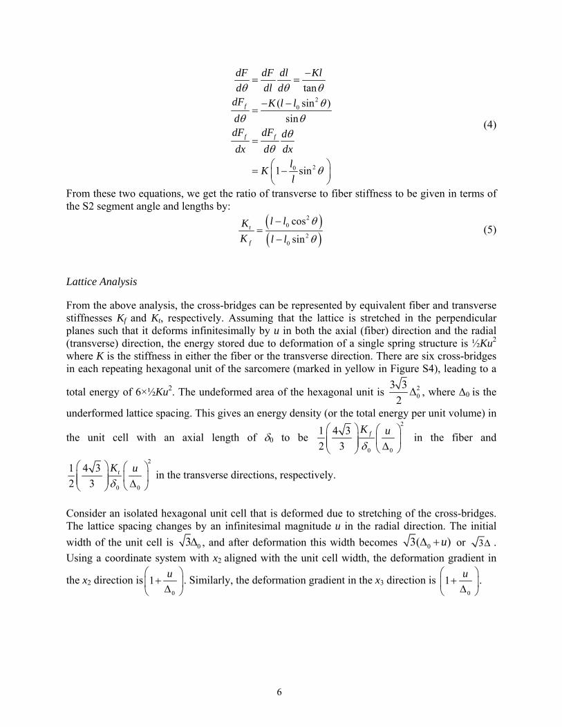
Lattice Analysis
From the above analysis, the cross-bridges can be represented by equivalent fiber and transverse stiffnesses Kf and Kt, respectively. Assuming that the lattice is stretched in the perpendicular planes such that it deforms infinitesimally by u in both the axial (fiber) direction and the radial (transverse) direction, the energy stored due to deformation of a single spring structure is ½Ku2 where K is the stiffness in either the fiber or the transverse direction. There are six cross-bridges in each repeating hexagonal unit of the sarcomere (marked in yellow in Figure S4), leading to a
total energy of 6×½Ku2. The undeformed area of the hexagonal unit is 20
3 3
2 , where Δ0 is the
underformed lattice spacing. This gives an energy density (or the total energy per unit volume) in
the unit cell with an axial length of 0 to be 2
0 0
1 4 3
2 3fK u
in the fiber and
2
0 0
1 4 3
2 3tK u
in the transverse directions, respectively.
Consider an isolated hexagonal unit cell that is deformed due to stretching of the cross-bridges. The lattice spacing changes by an infinitesimal magnitude u in the radial direction. The initial
width of the unit cell is 03 , and after deformation this width becomes 03( )u or 3 .
Using a coordinate system with x2 aligned with the unit cell width, the deformation gradient in
the x2 direction is0
1u
. Similarly, the deformation gradient in the x3 direction is
0
1u
.
6

The 2D deformation gradient tensor (F) and 2D finite strain tensor (E) can be expressed as
0
0
0 0
0 0
1 0
0 1
1 021
( 1)2
0 12
T
u
u
u u
u u
F
E F F
(6)
Similarly, in the transverse direction, the deformation gradient is given by 0
1u
and the strain
E11 by 0 0
12
u u
.
Assuming a linear strain energy relation, we can equate the strain energy in a unit cell to the energy from the deformation of the cross-bridges in the transverse direction as
, assuming that the transverse material stiffness Ct is constant (transversely
isotropic) in the plane perpendicular to the actin/myosin filaments. Substituting the strain values, we can equate the strain energy.
2 2 2
0 0 0 0 0
2
00 0
1 1 1 4 32 1
2 4 2 3
2 3
13 1
4
tt
t t
Ku u u uW C
C Ku u
(7)
This gives the relation to the transverse material stiffness as a function of the transverse cross-bridge stiffness Kt, the lattice spacing , the unit-cell axial spacing , and the cross-bridge lengthening the in the plane, u. Since u is small, we can neglect the square term leading to a
simpler relation, 0
0
2 1
3t tC K
where is the deformed lattice spacing.
In the axial fiber direction, 211
1
2 fW C E and again equating the energy in the fiber direction, we
obtain:
)(2
1 233
222 EECW t
7

2 2 2
0 0 0 0 0
2
02
0
00 0
1 1 1 4 31
2 4 2 3
4 3
13 1
4
ff
f f
Ku u u uW C
C Ku u
(8)
which, upon linearization, yields
2
0
0
4 1
3f fC K
and the macroscopic lattice stiffness ratio
can be computed to be
3
0
0
1
2t t
f f
C K
C K
.
The stress in the axial direction can be computed as 11ff fC E and in the transverse direction to
be 22tt rC E . This reduces to
2
02
0 0 0
00 0
2
0
00
4 31
213 1
4
4 3 1
32
ff f
f
u uK
u u
uK
u
(9)
Similarly in the transverse direction
0 0
2 3
32
tt t
uK
u
(10)
and the transverse to fiber macroscopic peak systolic stress ratio is:
8
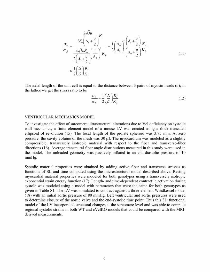
20 0 00
200 0
00
2 3
312 224 3 1
23
2
1
2
t
tt t
ff f
f
t
f
uK
u uK
u KuK
u
K
K
(11)
The axial length of the unit cell is equal to the distance between 3 pairs of myosin heads (; in the lattice we get the stress ratio to be
1
2tt t
ff f
K
K
(12)
VENTRICULAR MECHANICS MODEL
To investigate the effect of sarcomere ultrastructural alterations due to Vcl deficiency on systolic wall mechanics, a finite element model of a mouse LV was created using a thick truncated ellipsoid of revolution (15). The focal length of the prolate spheroid was 3.75 mm. At zero pressure, the cavity volume of the mesh was 30 l. The myocardium was modeled as a slightly compressible, transversely isotropic material with respect to the fiber and transverse-fiber directions (16). Average transmural fiber angle distributions measured in this study were used in the model. The unloaded geometry was passively inflated to an end-diastolic pressure of 10 mmHg. Systolic material properties were obtained by adding active fiber and transverse stresses as functions of SL and time computed using the microstructural model described above. Resting myocardial material properties were modeled for both genotypes using a transversely isotropic exponential strain energy function (17). Length- and time-dependent contractile activation during systole was modeled using a model with parameters that were the same for both genotypes as given in Table S1. The LV was simulated to contract against a three-element Windkessel model (18) with an initial aortic pressure of 80 mmHg. Left ventricular and aortic pressures were used to determine closure of the aortic valve and the end-systolic time point. Thus this 3D functional model of the LV incorporated structural changes at the sarcomere level and was able to compute regional systolic strains in both WT and cVclKO models that could be compared with the MRI-derived measurements.
9
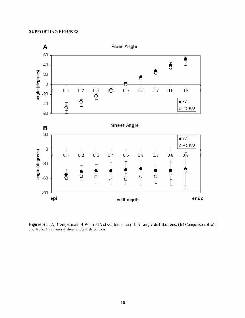
10
SUPPORTING FIGURES
Figure S1: (A) Comparison of WT and VclKO transmural fiber angle distributions. (B) Comparison of WT and VclKO transmural sheet angle distributions.

11
Figure S2: Immunofluorescence localization of cytoskeleton protein vinculin in cardiomyocytes from wild type adult mice (WT) and heterozygous null ((Vcl(+/-)), as well as GFP stained vinculin rescued [(Vcl(+/-) + Vcl] groups. (A) Isolated cardiac myocytes labeled green for vinculin with FITC-conjugated secondary antibody in WT and Vcl(+/-), or transfected cells expressing enhanced green fluorescent protein cDNA in [Vcl(+/-) + Vcl]. Vinculin staining is visualized at costameres (arrows, in cell edges) and intercalated disc (arrowheads). (B) Normalized mean integrated densities, i.e. normalized to control IgG staining or to non-transfected in each group before comparing among groups, were shown in cell body *p<0.05 vs. WT. The scale bar represents 10m.
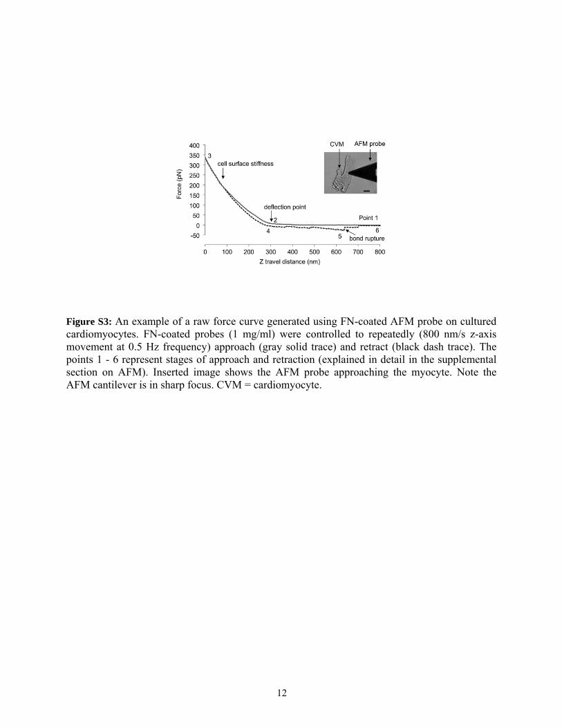
12
Figure S3: An example of a raw force curve generated using FN-coated AFM probe on cultured cardiomyocytes. FN-coated probes (1 mg/ml) were controlled to repeatedly (800 nm/s z-axis movement at 0.5 Hz frequency) approach (gray solid trace) and retract (black dash trace). The points 1 - 6 represent stages of approach and retraction (explained in detail in the supplemental section on AFM). Inserted image shows the AFM probe approaching the myocyte. Note the AFM cantilever is in sharp focus. CVM = cardiomyocyte.

13
Figure S4: Isolated unit cell marked in yellow showing the actin and myosin filament cross-sections. The lattice spacing at rest is . In the activated state, the lattice spacing is

14
Figure S5: Fiber, cross-fiber, and radial coordinates used for strain analysis. Rotated from cardiac coordinates about the radial axis through the fiber angles. Fiber axis runs parallel to fibers, cross-fiber axis is in the same plane but perpendicular to fiber axis, and radial axis is orthogonal to the fiber-cross-fiber plane.

15
SUPPORTING TABLE
** Parameter value derived from projections of a 3-dimensional model using values from Williams et. al.
(19), to the 2-dimensional model that is used here. * Parameter value derived from new 2-dimensional model lengths. Table S1. Model parameters used in the micromechanical and finite element models.
16
SUPPORTING REFERENCES (18-26) 1. Chuang, J. S., A. Zemljic-Harpf, R. S. Ross, L. R. Frank, A. D. McCulloch, and J. H.
Omens. 2010. Determination of three-dimensional ventricular strain distributions in gene-targeted mice using tagged MRI. Magn Reson Med 64:1281-1288.
2. Axel, L., and L. Dougherty. 1989. MR imaging of motion with spatial modulation of magnetization. Radiology 171:841-845.
3. Omens, J. H., T. P. Usyk, Z. Li, and A. D. McCulloch. 2002. Muscle LIM protein deficiency leads to alterations in passive ventricular mechanics. Am J Physiol Heart Circ Physiol 282:H680-H687.
4. Jynge, P., D. J. Hearse, D. Feuvray, W. Mahalu, S. Cankovic-Darracott, K. O'Brien, and M. V. Braimbridge. 1981. The St. Thomas' hospital cardioplegic solution: a characterization in two species. Scand J Thorac Cardiovasc Surg Suppl 30:1-28.
5. Chen, J., W. Liu, H. Zhang, L. Lacy, X. Yang, S. Song, S. A. Wickline, and X. Yu. 2005. Regional ventricular wall thickening reflects changes in cardiac fiber and sheet structure during contraction: quantification with diffusion tensor MRI. Am J Physiol Heart Circ Physiol 289:H1898-H1907.
6. Munch, D. F., H. T. Comer, and J. M. Downey. 1980. Barium contracture: a model for systole. Am J Physiol Heart Circ Physiol 239:H438-H442.
7. Hayashi, T., M. E. Martone, Z. Yu, A. Thor, M. Doi, M. J. Holst, M. H. Ellisman, and M. Hoshijima. 2009. Three-dimensional electron microscopy reveals new details of membrane systems for Ca2+ signaling in the heart. J Cell Sci 122:1005-1013.
8. Suzuki, S., T. Tsuchiya, Y. Oshimi, T. Takei, and H. Sugi. 1989. Electron microscopic studies on the stretch-induced disordering of the myofilament lattice in tetanized frog skeletal muscle fibers. J Electron Microsc (Tokyo) 38:60-63.
9. Wu, X., S. Chakraborty, C. L. Heaps, M. J. Davis, G. A. Meininger, and M. Muthuchamy. 2011. Fibronectin increases the force production of mouse papillary muscles via alpha5beta1 integrin. J Mol Cell Cardiol 50:203-213.
10. Wu, X., Y. Yang, P. Gui, Y. Sohma, G. A. Meininger, G. E. Davis, A. P. Braun, and M. J. Davis. 2008. Potentiation of large conductance, Ca2+-activated K+ (BK) channels by alpha5beta1 integrin activation in arteriolar smooth muscle. The Journal of physiology 586:1699-1713.
11. Sun, Z., L. A. Martinez-Lemus, A. Trache, J. P. Trzeciakowski, G. E. Davis, U. Pohl, and G. A. Meininger. 2005. Mechanical properties of the interaction between fibronectin and alpha5beta1-integrin on vascular smooth muscle cells studied using atomic force microscopy. Am J Physiol Heart Circ Physiol 289:H2526-2535.
12. Wu, X., Z. Sun, A. Foskett, J. P. Trzeciakowski, G. A. Meininger, and M. Muthuchamy. 2010. Cardiomyocyte contractile status is associated with differences in fibronectin and integrin interactions. Am J Physiol Heart Circ Physiol 298:H2071-2081.
13. Lehenkari, P. P., and M. A. Horton. 1999. Single integrin molecule adhesion forces in intact cells measured by atomic force microscopy. Biochem Biophys Res Commun 259:645-650.
14. Schoenberg, M. 1980. Geometrical factors influencing muscle force development: I. The effect of filament spacing upon axial forces. Biophys J 30:51-68.
15. Costandi, P. N., L. R. Frank, A. D. McCulloch, and J. H. Omens. 2006. Role of diastolic properties in the transition to failure in a mouse model of the cardiac dialation. Am J Physiol Heart Circ Physiol 291:H2971-H2979.

16. Doll, S., and K. Schweizerhof. 2000. On the development of volumetric strain energy functions. J Appl Mech 67:17-21.
17. Guccione, J. M., A. D. McCulloch, and L. K. Waldman. 1991. Passive material properties of intact ventricular myocardium determined from a cylindrical model. J Biomech Eng 113:42-55.
18. Kerckhoffs, R. C., M. L. Neal, Q. Gu, J. B. Bassingthwaighte, J. H. Omens, and A. D. McCulloch. 2007. Coupling of a 3D finite element model of cardiac ventricular mechanics to lumped systems models of the systemic and pulmonic circulation. Ann Biomed Eng 35:1-18.
19. Williams, C. D., M. Regnier, and T. L. Daniel. 2010. Axial and radial forces of cross-bridges depend on lattice spacing. PLoS Comput Biol 6:e1001018.
20. Matsubara, I., and B. M. Millman. 1974. X-ray diffraction patterns from mammalian heart muscle. J Mol Biol 82:527-536.
21. Rayment, I., H. M. Holden, M. Whittaker, C. B. Yohn, M. Lorenz, K. C. Holmes, and R. A. Milligan. 1993. Structure of the actin-myosin complex and its implications for muscle contraction. Science 261:58-65.
22. Julian, F. J., R. L. Moss, and M. R. Sollins. 1978. The mechanism for vertebrate striated muscle contraction. Circ Res 42:2-14.
23. Sonnenblick, E. H., and C. L. Skelton. 1974. Reconsideration of the ultrastructural basis of cardiac length-tension relations. Circ Res 35:517-526.
24. Craig, R., and J. L. Woodhead. 2006. Structure and function of myosin filaments. Curr Opin Struct Biol 16:204-212.
25. Millman, B. M. 1998. The filament lattice of striated muscle. Physiol Rev 78:359-391. 26. Guccione, J. M., L. K. Waldman, and A. D. McCulloch. 1993. Mechanics of active
contraction in cardiac muscle. J Biomech Eng 115:72-90.
17


