Novel Gold/Metal Oxide Nanocomposite Materials: Synthesis
17
Novel Gold/Metal Oxide Nanocomposite Materials: Synthesis, Structure, and Catalytic Activity Senior Honors Thesis Department of Chemical Engineering, Tufts University Forrest Gittleson May 2009
Transcript of Novel Gold/Metal Oxide Nanocomposite Materials: Synthesis

Novel Gold/Metal Oxide Nanocomposite Materials:
Synthesis, Structure, and Catalytic Activity Senior Honors Thesis
Department of Chemical Engineering, Tufts University
Forrest Gittleson
May 2009
Table of Contents
1. Introduction ........................................................................................................................................... 1
2. Experimental ......................................................................................................................................... 2
2.1 Gold Nanocrystal Synthesis ...................................................................................................... 2
2.2 Separation ................................................................................................................................. 2
2.2.1 Ionic Liquid Phase Transfer .................................................................................................. 2
2.3 Deposition ................................................................................................................................. 3
2.3.1 Coprecipitation ...................................................................................................................... 3
2.3.2 Deposition-Precipitation ....................................................................................................... 3
2.3.3 Gold Growth on Zinc Oxide ................................................................................................. 4
2.4 Catalytic Activity ...................................................................................................................... 4
3. Results and Discussion ......................................................................................................................... 5
3.1 Gold Nanoparticle Synthesis and Extraction ............................................................................ 5
3.2 Gold Deposition on Metal Oxides ............................................................................................ 6
3.3 Gold/Zinc Oxide ....................................................................................................................... 8
4. Conclusions and Future Work ............................................................................................................. 13
5. Acknowledgements ............................................................................................................................. 14
6. References ........................................................................................................................................... 15

1
1. Introduction Given the market and political imperatives
to develop and disseminate new clean
energy technologies, much research has
recently focused on the catalysis of chemical
reactions to improve the efficiency of energy
generation and decrease harmful emissions.
Some of this work has concentrated on
reactions that reduce emissions by
converting unwanted products (i.e. carbon
monoxide conversion to carbon dioxide) or
on fuel reforming reactions (i.e. generation
of hydrogen via the water-gas shift
reaction). Significant progress in catalysis
research has been made on multiple fronts
involving everything from processes to
improve selectivity and conversion to
synthesis of new materials that hold promise
for further advances.
There is great promise for catalysis research
in nano-scale materials synthesis and testing.
Devising materials at the nano-scale allows
researchers to broaden their focus to
materials that, though they may not be active
in bulk, hold special properties at the nano-
scale. Gold is one such material that exhibits
remarkably different characteristics in
atomic amounts. Published work to date has
shown that nano-scale gold is an active
catalyst for many reactions of interest
including the carbon monoxide (CO)
oxidation and water-gas shift (WGS)
reactions. Both of these reactions are
important for their applications to fuel cell
technology. The CO oxidation reaction
represents a method by which to remove
carbon monoxide from fuel reforming
product gases (syngas) that are used to
generate energy in a fuel cell. In this
circumstance, CO is known to poison
platinum electrocatalysts at the anode,
decreasing power ouput; therefore its
removal is essential. The WGS reaction,
which increases the yield of hydrogen from
steam reforming, also represents a target for
improved catalysis by gold.
Synthesis of gold-containing nanomaterials
for these purposes can be quite challenging
and thus poses an obstacle to their further
study. Nevertheless, this work seeks to
advance a unique line of research into gold-
containing nanomaterial synthesis,
characterization, and activity testing for the
catalysis of various energy-related reactions.
A more refined focus is specific to
nanocrystalline gold and its interactions with
metal oxide supports to provide potentially
better activity for the CO oxidation and
WGS reactions.
Preliminary studies involved gold
nanoparticle colloid synthesis and
separation, the goal of which was to develop
unsupported gold nanoparticles with islands
of metal oxide deposited on their surface.
However, separation of gold nanoparticles
from solution proved to be an
insurmountable obstacle. To synthesize gold
coordinated with metal oxides, several
deposition methods were then studied. The
structures formed were unique, yet again
difficult to extract, making activity testing a
challenge. These studies eventually led to a
narrowed focus on gold/zinc oxide
(Au/ZnO) nanocomposite materials.
Adapting several methods seen in the
literature concerning the Au/ZnO system,
procedures were tuned to obtain Au/ZnO
from zinc oxide nanocrystals, rods, and
wires. Subsequent activity testing for the CO
oxidation reaction showed that these
materials are active, though perhaps not
ideal at this early stage of study.
The work presented here should serve as a
foundation for further research into
gold/metal oxide nanocomposite materials
for the catalysis of energy-related reactions.
2
Particular emphasis is placed on the promise
of Au/ZnO nanocomposites.
2. Experimental The focus of this work has evolved
significantly since its inception and as such
covers several different topics, all of interest
to gold nanoparticle synthesis,
characterization, and activity evaluation.
What follows is a summary of the
significant experimental procedures
employed during this project.
2.1 Gold Nanocrystal Synthesis Many procedures for gold nanoparticle
colloid synthesis are available in the
literature. The procedure used for this
project was adapted from Sau and Murphy
(2004) and employs
cetyltrimethylammonium bromide (CTAB)
surfactant to stabilize sub-10nm gold
particles in aqueous solution. In a typical
procedure, 0.25mmol of chloroauric acid
(HAuCl4∙3H2O) was dissolved in 25mL of
water. Then 7.5mmol of CTAB was added
to 75mL of water and heated to
approximately 70°C until dissolution. The
CTAB solution was added dropwise into the
heated gold precursor solution. Next,
0.6mmol of sodium borohydride (NaBH4)
was dissolved in 60mL and chilled in an ice
bath. The chilled solution was then added
dropwise into the Au/CTAB solution to
reduce gold and form Au nanoparticles. The
colloid was allowed to mix for
approximately 2 hours.
2.2 Separation Because of the small particle size of the gold
suspended in aqueous solution and its
stability due to the CTAB surfactant, it was
judged that centrifugation would not be a
useful method of extracting gold particles
from solution1. As an alternative, phase
transfer using an ionic liquid was evaluated.
2.2.1 Ionic Liquid Phase Transfer
For the extraction of gold nanocrystals from
aqueous solution, 1-butyl-3-
methylimidazolium hexafluorophosphate
(ionic liquid) was employed in a phase
transfer procedure adapted from Wei, et al.
(2004). Typically, the ionic liquid was added
to aqueous gold stock solution in weight
ratios ranging from 1:5 to 1:20 ionic liquid
to aqueous stock solution. With vigorous
mixing of the two phases, the aqueous phase
(originally a deep purple hue) became
colorless and transparent while the
(originally yellow) organic phase became
purple, indicating the phase transfer of gold
particles. Following the extraction, the
aqueous portion was analyzed by UV
spectroscopy, showing that no gold was
present in the supernatant2.
For removal of the gold nanoparticles from
the ionic liquid phase, several methods were
tested. Because of the ionic liquid’s high
viscosity at room temperature, it was
necessary to heat that phase to reduce
viscosity and induce precipitation of the
gold particles coordinated in the imidizole
ring. Experiments involved heating the ionic
liquid with coordinated gold in the presence
of residual water. The result was a color
change in the ionic liquid from purple to
brown, possibly indicating decomposition of
ionic liquid or aggregation of gold 1 Testing of this hypothesis later showed that
centrifugation of the gold colloid produced an
aggregation of gold particles with particle sizes larger
than those in solution. This method was also limited
by the low gold concentration of the colloid and the
size of the centrifuge tubes available. 2 No peak was visible at the 520nm wavelength that
corresponds to the absorption by gold. UV
spectroscopy also showed that surfactant was present
in the supernatant following extraction, indicating
that a substantial portion of the surfactant did not
transfer into the ionic liquid phase.

3
nanoparticles into larger gold clusters3. To
avoid the presence of water, fresh ionic
liquid coordinated with gold was redispersed
(and dissolved) separately in methanol and
in ethanol by heating after the aqueous layer
was removed. Reprecipitation of the ionic
liquid from ethanol and methanol by cooling
in an ice bath produced a white organic
phase containing the gold particles4. After
removal of the alcohol, the ionic liquid was
heated again, boiling off any residual
alcohol, reducing the viscosity, and inducing
precipitation of the gold species. During
heating, gold particles precipitated out of
solution. Because of the small amount of
ionic liquid being used, the total amount of
gold precipitated was in the 1mg range,
close to the calculated weight of gold
present in the original aliquot of stock
solution used.
Removal of the precipitated gold from ionic
liquid was conducted by adding additional
ethanol and heating until ionic liquid
dissolution, then decanting off the
supernatant. Transmission electron
microscopy (TEM) was used to determine
the particle size of the extracted gold.
2.3 Deposition Because of the limitations of the ionic liquid
extraction procedure, a new method was
sought to enable unsupported nanoparticle
gold synthesis. Procedures for gold
deposition on metal oxides or vice versa
vary widely and include coprecipitation
(CP), deposition-precipitation (DP), as well
as other more unique methods of
nanocomposite synthesis.
3 Swatloski, et al. (2003) suggests that residual water
is the culprit, as decomposition of the ionic liquid
occurs readily in water. 4 For the ethanol sample after reprecipiation, a UV
spectrogram showed no gold particles in the ethanol
supernatant solution.
2.3.1 Coprecipitation
Preliminary attempts at coprecipitation of
gold and metal oxides varied considerably
from those found in Fu, et al. (2001).
Though ceria and zinc oxide were used, gold
stock solution with 5-10nm gold crystals
was substituted for gold precursor. In one
trial, an aliquot of gold stock solution was
mixed with cerium(III) nitrate solution in a
molar ratio of 1:9 gold to cerium (III)
nitrate. To maintain the pH around 10,
0.25M NaOH was added dropwise, leading
to the precipitation of the metal oxide
species. The same procedure was also
repeated with zinc nitrate in place of
cerium(III) nitrate. These trials did not
include the final drying and calcination steps
found in Fu, et al. (2001).
Another “coprecipitation” procedure was
devised involving the ionic liquid extraction
mechanism. It was thought that by
simultaneously extracting gold nanoparticles
and coordinated metal oxide, the size of
metal oxide structures would not be
significantly larger than those of the gold.
This, in fact, proved to be the case. Prior to
the ionic liquid extraction, cerium(III)
nitrate was mixed with aqueous stock
solution in a ratio of 10:1 gold to cerium(III)
nitrate by weight. That solution was then
contacted with ionic liquid and agitated.
Subsequent extraction steps were the same
as those above. TEM was used to determine
the presence of ceria as well as the
approximate sizes of gold and ceria
particles.
2.3.2 Deposition-Precipitation
A deposition-precipitation procedure similar
to that found in Fu, et al. (2001) was used to
produce gold/zinc oxide nanocomposite
materials from zinc oxide nanocrystals, rods,
and wires. Synthesis of ZnO rods and wires
was adapted from Cheng, et al. (2006),
which describes several factors that affect
zinc oxide crystal growth. For the synthesis

4
of ZnO wires, 1.15g of zinc acetate
dihydrate was mixed with 15mL of
methanol in a Teflon coated autoclave-safe
container. Next, 22.5mL of Me4NOH
(25w/w in water) and 7.5mL of Me4NOH
(25w/w in methanol) were mixed and added
to the zinc acetate solution. The solution was
stirred for 30 minutes then transferred to a
furnace for heating at 75°C for 24 hours.
Afterwards, the white precipitate was
centrifuged at 9,000 rpm for 20 minutes and
washed four times with water to obtain a
neutral pH. The ZnO precipitate was then
dried in an oven for 48 hours at 70°C and
crushed to a powder. ZnO nanorods were
synthesized in much the same way, with the
only difference being the water content.
Water content for ZnO nanorod synthesis
was decreased by using 7.5 of Me4NOH
(25w/w in water) and 22.5mL of Me4NOH
(25w/w in methanol). Zinc oxide
nanocrystals were synthesized using only
30mL of Me4NOH (25w/w in methanol).
After obtaining zinc oxide rods and wires,
deposition of gold was conducted by the DP
method. First, 0.2g of ZnO was dispersed in
10mL of water to which was added 0.24g of
(NH4)2CO3 (acting as a buffer). Next, 0.01g
of chloroauric acid was dissolved in 5mL of
water. The gold solution was added
dropwise to the zinc oxide suspension. After
stirring for 10 minutes the precipitate was
filtered under vacuum and washed twice
with water. The solid was dried on filter
paper at 60°C for 24 hours. After crushing
the dried powder, it was placed in an
autoclave-safe container and calcined in a
furnace at 250°C for four hours. Gold
crystal growth was obvious from the color
change following calcination.
2.3.3 Gold Growth on Zinc Oxide
As an alternative to deposition-precipitation,
another gold/zinc oxide structure was
synthesized by growing gold nanocrystals in
the presence of zinc oxide nanorods. This
procedure was adapted from that in Zhang,
et al. (2008), the only difference being that
the methanol solution containing zinc oxide
was not treated by nitrogen purging before
extended heating. ZnO nanorod synthesis
involved first preparing a zinc acetate in
methanol solution and a KOH in methanol
solution. The KOH solution was then added
dropwise to the zinc acetate solution and
allowed to stir at 60°C for two hours. The
mixture was cooled and purged with air to
reduce the volume and left to mix at 60°C
for 12 hours. Because most of the methanol
evaporated over the course of the 12 hours,
leaving only a white precipitate paste, the
zinc oxide was redispersed in ethanol,
washed several times with dilute trisodium
citrate solution, and centrifuged.
To promote gold crystal growth on zinc
oxide, 0.4mmol ZnO nanorods were
dispersed (via sonication) in 3.0mM
trisodium citrate solution. To that solution
was added 0.008mmol of chloroauric acid
dispersed in water. The solution was
allowed to stir for 12 hours. The precipitate
was collected by centrifugation at 50,000
rpm for 45min and dried at 60°C for 4
hours. In addition to this 50:1 ZnO to Au
sample, 100:1 and 25:1 samples were also
made. The 25:1 sample was produced in a
larger quantity to ensure sufficient yield
after collection. TEM images of these
samples show nanoparticulate gold and zinc
oxide coordinated together.
2.4 Catalytic Activity Testing catalytic activity for the CO
oxidation reaction involved using a packed
bed reactor with approximately 0.1g of
catalyst per trial. A mixture of 2% carbon
monoxide, 1% oxygen and the balance
helium was flowed over the bed at a flow
rate of 75mL/min. Programs were initiated
to evaluate CO conversion to CO2 at
temperatures ranging from 25°C to 250°C.
5
Light-off curves showing conversion as a
function of temperature were constructed
using peak area data from a gas
chromatograph connected to the output
stream of the packed bed. Only steady-state
data were collected. Accordingly, at each
temperature, data were collected for one
hour and averaged.
3. Results and Discussion The results of the experiments are presented
and discussed in the following sections.
3.1 Gold Nanoparticle Synthesis and
Extraction Though the original focus of this work was
to prepare nanostructures of gold with metal
oxide islands as a way of inverting the
structure of active water-gas shift catalysts
(including gold on ceria, lanthana, and zinc
oxide), this proved to be more difficult than
originally expected. In fact, the biggest
obstacle to the pursuit of these structures
was the extraction of intact gold
nanoparticles from aqueous solution.
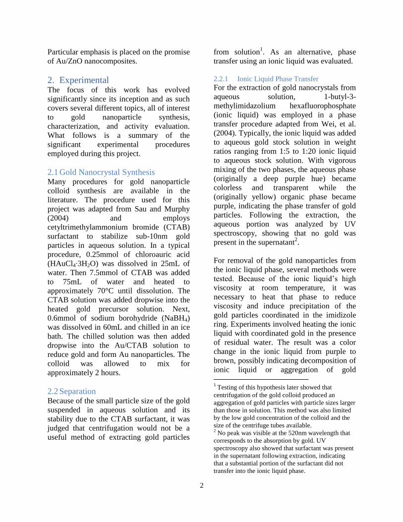
Nevertheless, gold colloidal suspensions
were synthesized and characterized by TEM.
Of the gold colloid syntheses conducted,
several trials did not succeed in producing
homogeneous suspensions, perhaps due to
insufficient dissolution of CTAB before
addition to the gold precursor solution.
Varying the procedure to allow for CTAB
dissolution prior to mixing with gold
precursor solved these problems. The
homogeneous gold colloid sample that
constituted the aqueous stock solution used
in subsequent procedures had a
concentration of 1.42mM Au. This sample
was subjected to a UV spectrograph, which
showed peaks at 520nm, consistent with
absorption by gold nanoparticles. TEM
images, such as that seen in Figure 1,
indicate the presence of single-crystal gold
of approximately 5-10nm. Though this size
is not ideal for water-gas shift catalysis, it
would likely be active for the CO oxidation
reaction once extracted from solution.
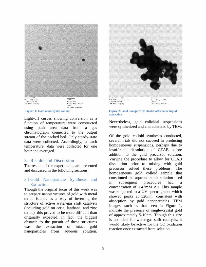
Figure 2: Gold nanoparticle cluster after ionic liquid
extraction Figure 1: Gold nanocrystal colloid
AU_2.002.'!!
Col, ~.5"P;I/".12:5J OJ/U/O,
TEM Mod .. : ]uging
HV~200.0~V
l>!r..ct Mag: 10DOOOxTil'·
AMTCn"ra Sys'''.
Au IIPo 20 '0 "001.'!!
Col, 5~'.1~'pix/.;"ron12:2010/31/0'TEM Mod .. : ...glng
"V~200.0~V
D;n.c' Hag 10000"
Til"AM'\" eo...,a Syot...
6
Because centrifugation of the aqueous gold
stock solution at 40,000 rpm was not
sufficient to collect gold, other extraction
methods were attempted. Simply boiling off
the solvent was, as expected, unsatisfactory
due to the precipitation of CTAB along with
gold. With the ionic liquid extraction
procedure adapted from Wei, et al. (2004),
gold nanocrystals were removed from the
aqueous solution. However, this procedure,
involving multiple dissolution and
precipitation steps, was labor intensive,
costly (primarily due to the cost of the ionic
liquid), and had low yield5. Furthermore,
TEM images of the gold collected via this
procedure (as seen in Figure 2), show that
aggregation occurs and particle size is not
preserved6. This suggests that even if a large
amount of gold could be extracted using this
method, it would not be ideal for either
WGS or CO oxidation reactions.
Nevertheless, these larger gold nanoparticles
may still be used to attempt a deposition of
metal oxide islands, per the initial goal of
this project. A material produced using
vapor deposition of ceria or zinc oxide on
gold may be quite active for CO oxidation or
WGS.
3.2 Gold Deposition on Metal Oxides With the obvious limitations of the gold
nanoparticle extraction procedures and the
desire to investigate the interaction between
gold nanoparticles and metal oxides, the
logical next step was to evaluate
coprecipitation methods. Because there is
5 Several weight ratios of gold stock solution to ionic
liquid were tested to determine the minimum amount
of ionic liquid that could be used to extract a given
amount of gold. That maximum ratio was found to be
approximately 20:1 gold solution to ionic liquid,
which still would only lead to the collection of gold
in milligram amounts per trial. 6 Gold particle size in these trials has noticeably
grown from 5-10nm to 50-200nm due to this
procedure, perhaps due to multiple heating steps and
the removal of surfactant during phase transfer.
much published literature on the topic of
gold on metal oxides (especially ceria,
lanthana, and zinc oxide), the target for
further research was defined as
nanocomposite materials in which metal
oxide particles would be roughly the same
size as the gold coordinated with them. This
concept suggested that an increase in surface
area interaction between gold and metal
oxide could be attainable, perhaps increasing
catalytic activity7.
For this study, gold/metal oxide composites
were synthesized with ceria and zinc oxide
starting from single-crystal gold of size 5-
10nm (from the aqueous gold stock
solution). It was thought that a
coprecipitation procedure might be able to
either encapsulate these gold particles in a
core-shell structure or alternatively form
nanoparticles that would coordinate with the
gold in a “dumbbell” type structure8. For the
gold/ceria nanocomposites, the results were
somewhat unanticipated. As seen in Figure
3, the structure of gold/ceria particles
produced from a ratio of 1:9 gold to
cerium(III) nitrate are non-spherical and of
roughly 100nm. Though it is difficult to
distinguish the gold from the ceria in these
images, we believe that this structure is
similar to the core-shell model in which the
gold particle is coated completely with ceria.
If this theory is true then the non-spherical
shape is likely caused by gradual formation
of ceria at different seed points on the
surface of the gold particle. Because CTAB
surfactant was present in solution during the
formation of these structures, it is
foreseeable that ceria selectively grew on the
gold particles in areas where the surfactant
7 It has been suggested in several papers including
Fu, et al. (2001) that coordination of gold with
oxygen containing materials improves catalytic
activity for the water-gas shift reaction. 8 This “dumbbell” structure is seen in the results of
Wang, et al. (2007) and Lee, et al. (2008) involving
gold/zinc oxide structures.

7
molecules were in low concentration – thus,
even growth over the entire surface did not
occur. This theory is contingent upon the
determination that both gold and ceria are
present in the structure viewed here. It is
generally recognized that in TEM images,
ceria is less dark than gold because it is less
dense. The fact that the outer lobes of the
particle are less dark than the interior is thus
the basis of this determination. Without x-
ray diffraction (XRD) analysis it is
impossible to know for sure the nature of
this particle. It may be the case that the
entire particle consists of smaller aggregated
gold particles stabilized by the surfactant in
this odd shape. If particles are determined to
contain gold and ceria, the use of scanning
transmission electron microscopy (STEM)
with mapping by energy dispersive x-ray
spectroscopy (EXD) would reveal whether
gold encapsulation has occurred. Further
study of this sample would be interesting but
may be stymied by difficulties in extraction
due to the presence of surfactant.
A second coprecipitation sample of gold and
ceria was made with higher gold content to
determine if the structure would change
based on concentration. A sample
synthesized from a 1:1 gold to cerium(III)
nitrate ratio indeed produced a noticeably
different structure. The particles seen in
Figure 4, from this sample, clearly adhere
more to the “dumbbell” model than to the
core-shell model suggested previously. It is
worth noting that several of the particles in
Figure 4 are individual – they are either gold
or ceria separately, as distinguished by their
shade – which is likely the result of
significant dilution and low ceria
concentration. The middle particle
representing the tangentially conjoined
“dumbbell” of gold and ceria is interesting
to consider because of its similarity to the
structures composed of gold and zinc oxide
from Wang, et al. (2007) and Lee, et al.
(2008). Of course the synthesis method used
was different from that in either paper
because of the use of ceria. Yet it was more
similar to that in Lee, et al. (2008), in which
gold nanoparticles were synthesized first,
then added to a solution from which zinc
oxide was precipitated. It was thought that
these “dumbbell” structures might have
Figure 4: Coprecipitation of Au/CeO2 in 1:1 ratio Figure 3: Coprecipitation of Au/CeO2 in 1:9 ratio
~o~CeOz 10 .01 N~otLOOl.hl
Col: l.lll~i,,/n.
lJ:J6 lVOVOITEMMooo: l ..g;ng
Il'h.oo,okvDirect M~g: .0000"Til':
AMT C~ ...<a "yah.
As_CeO. '0 .01 NaOH.OOJ,'itCol: l.lllpix/n_
13:" 1VOVO.T1!M Modo: "'glng
IlV_ZOO,OkV
Direct Mag: 20000"Tilt
AMT Cn...r~ "yah.

8
some unique catalytic activity because of the
intimacy of the two particles in the
conjoining region. Nevertheless, extraction
again proved to be an obstacle in the further
study of this structure.
An entirely separate coprecipitation
procedure, not similar to the others (and
likely unfit to be called coprecipitation),
involved the same ionic liquid extraction
method used earlier on gold colloidal
solutions. After using this method to extract
a solution of gold colloid containing
cerium(III) nitrate in 10:1 gold to
cerium(III) nitrate ratio, the product was
analyzed by TEM. The image seen in Figure
5 shows a unique nanocomposite structure
of gold and ceria. The needle-like shapes are
consistent with a known structure of ceria,
as are the amorphous lightly colored
regions. It is difficult to distinguish gold
particles in this picture because of the
darkness of the ceria needles; however, it is
known that they are present because of the
dark purple hue of the precipitate. In fact,
we would suspect gold to be present in this
structure simply by merit of the ionic liquid
extraction procedure9. The size of the gold
particles in this composite structure have
likely been preserved because there are no
signs of aggregation (no larger dark
particles). Thus, it is possible that with these
ceria needles and small-sized gold, this
structure could be active for either WGS or
CO oxidation reactions. Unfortunately, due
to the labor involved in this extraction
procedure and the cost of the ionic liquid,
this avenue was not pursued.
3.3 Gold/Zinc Oxide Further research concentrated on the
synthesis and activity testing of gold/zinc
oxide nanocomposites. Specific targets of
investigation included gold on zinc oxide
nanorods and wires. While there are some
unique findings from this work, further
research is necessary to confirm findings
and gain a better understanding of the
structure-activity relationship.
The first zinc oxide composite material was
synthesized with gold via the same
coprecipitation procedure used to generate
the Au/CeO2 composites from aqueous gold
stock solution. This produced large particles
of zinc oxide with 5-20nm gold on the
surface and in the interior (as seen in Figure
6). Though this sample is not unique in its
structure10
, it does show that coprecipitation
by this method can form Au/ZnO structures
with acceptable gold dispersion. It might
even exhibit catalytic activity for the CO
oxidation reaction, though it was not tested.
9 The imidazole ring in this particular ionic liquid
coordinates with metals like gold to enable
nanoparticle extraction. We have no mechanism to
suggest that ceria could be extracted via phase
transfer with the ionic liquid other than if it were
directly coordinated with the gold. 10
Coprecipitation of zinc oxide with gold
nanoparticles is common in papers such as Wang, et
al. (2002).
Figure 5: Au/CeO2 after ionic liquid extraction
Au_C<>0210_11L.00I.til
Cal'l.ll1pis/fi.
1."'0IUOV08
TE" "odo: lugi"g
HV.200.okv
Direct Hfig: 20000"Til<:
AHT Cn..ra Systa.
9
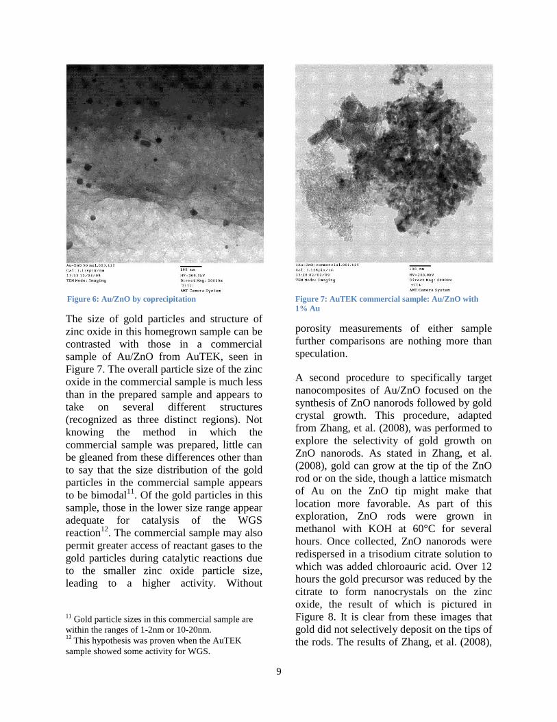
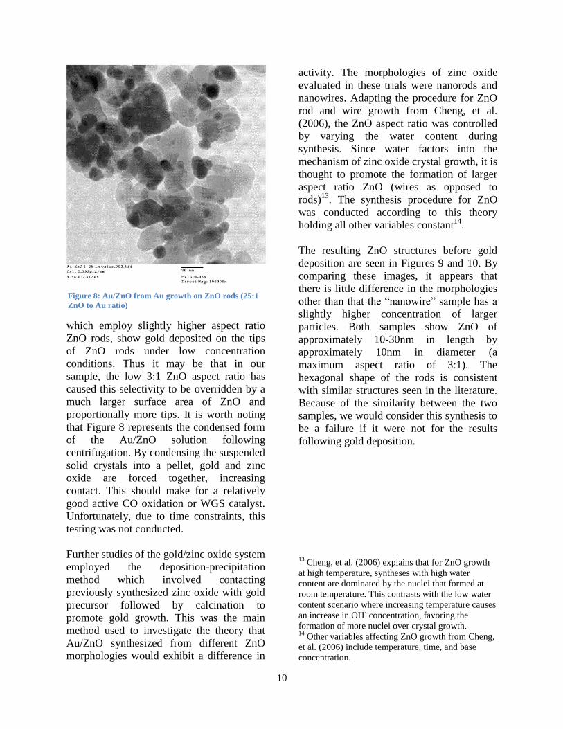
The size of gold particles and structure of
zinc oxide in this homegrown sample can be
contrasted with those in a commercial
sample of Au/ZnO from AuTEK, seen in
Figure 7. The overall particle size of the zinc
oxide in the commercial sample is much less
than in the prepared sample and appears to
take on several different structures
(recognized as three distinct regions). Not
knowing the method in which the
commercial sample was prepared, little can
be gleaned from these differences other than
to say that the size distribution of the gold
particles in the commercial sample appears
to be bimodal11
. Of the gold particles in this
sample, those in the lower size range appear
adequate for catalysis of the WGS
reaction12
. The commercial sample may also
permit greater access of reactant gases to the
gold particles during catalytic reactions due
to the smaller zinc oxide particle size,
leading to a higher activity. Without
11
Gold particle sizes in this commercial sample are
within the ranges of 1-2nm or 10-20nm. 12
This hypothesis was proven when the AuTEK
sample showed some activity for WGS.
porosity measurements of either sample
further comparisons are nothing more than
speculation.
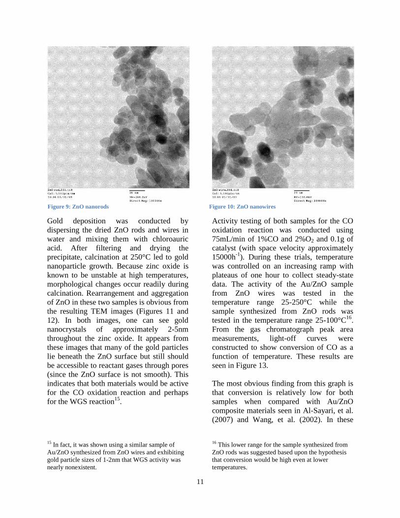
A second procedure to specifically target
nanocomposites of Au/ZnO focused on the
synthesis of ZnO nanorods followed by gold
crystal growth. This procedure, adapted
from Zhang, et al. (2008), was performed to
explore the selectivity of gold growth on
ZnO nanorods. As stated in Zhang, et al.
(2008), gold can grow at the tip of the ZnO
rod or on the side, though a lattice mismatch
of Au on the ZnO tip might make that
location more favorable. As part of this
exploration, ZnO rods were grown in
methanol with KOH at 60°C for several
hours. Once collected, ZnO nanorods were
redispersed in a trisodium citrate solution to
which was added chloroauric acid. Over 12
hours the gold precursor was reduced by the
citrate to form nanocrystals on the zinc
oxide, the result of which is pictured in
Figure 8. It is clear from these images that
gold did not selectively deposit on the tips of
the rods. The results of Zhang, et al. (2008),
Figure 7: AuTEK commercial sample: Au/ZnO with
1% Au
Figure 6: Au/ZnO by coprecipitation
Au_ZnO ~O .0J.00). II I
cal' l.lllpis/n_l.',~) lVOVOO
TE" "odo: luglng
HV.20o.okv
Direct Hog: 20000"Tilt:
AHT Cn..ra Systo_
1 A"_Z"o-co..er~1~ 1.00 I. t I Ic..l'l.llIp;',/n_13:lt OVOV09
T1!" "odo: "'glng~V·200.0kV
Direct "og: 20000"Tilt
AHT C~...,a Systo.
10
which employ slightly higher aspect ratio
ZnO rods, show gold deposited on the tips
of ZnO rods under low concentration
conditions. Thus it may be that in our
sample, the low 3:1 ZnO aspect ratio has
caused this selectivity to be overridden by a
much larger surface area of ZnO and
proportionally more tips. It is worth noting
that Figure 8 represents the condensed form
of the Au/ZnO solution following
centrifugation. By condensing the suspended
solid crystals into a pellet, gold and zinc
oxide are forced together, increasing
contact. This should make for a relatively
good active CO oxidation or WGS catalyst.
Unfortunately, due to time constraints, this
testing was not conducted.
Further studies of the gold/zinc oxide system
employed the deposition-precipitation
method which involved contacting
previously synthesized zinc oxide with gold
precursor followed by calcination to
promote gold growth. This was the main
method used to investigate the theory that
Au/ZnO synthesized from different ZnO
morphologies would exhibit a difference in
activity. The morphologies of zinc oxide
evaluated in these trials were nanorods and
nanowires. Adapting the procedure for ZnO
rod and wire growth from Cheng, et al.
(2006), the ZnO aspect ratio was controlled
by varying the water content during
synthesis. Since water factors into the
mechanism of zinc oxide crystal growth, it is
thought to promote the formation of larger
aspect ratio ZnO (wires as opposed to
rods)13
. The synthesis procedure for ZnO
was conducted according to this theory
holding all other variables constant14
.
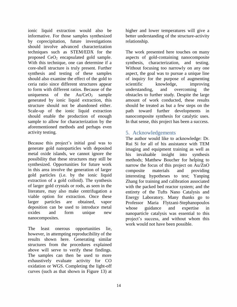
The resulting ZnO structures before gold
deposition are seen in Figures 9 and 10. By
comparing these images, it appears that
there is little difference in the morphologies
other than that the “nanowire” sample has a
slightly higher concentration of larger
particles. Both samples show ZnO of
approximately 10-30nm in length by
approximately 10nm in diameter (a
maximum aspect ratio of 3:1). The
hexagonal shape of the rods is consistent
with similar structures seen in the literature.
Because of the similarity between the two
samples, we would consider this synthesis to
be a failure if it were not for the results
following gold deposition.
13
Cheng, et al. (2006) explains that for ZnO growth
at high temperature, syntheses with high water
content are dominated by the nuclei that formed at
room temperature. This contrasts with the low water
content scenario where increasing temperature causes
an increase in OH- concentration, favoring the
formation of more nuclei over crystal growth. 14
Other variables affecting ZnO growth from Cheng,
et al. (2006) include temperature, time, and base
concentration.
Figure 8: Au/ZnO from Au growth on ZnO rods (25:1
ZnO to Au ratio)
Au_Z"O 1_25 in "a'H.001.\i1Cal, 5.591pix/n"U 0)/11/0.
~ttv.2oo.OkvDi.-..~, Hag
11
Gold deposition was conducted by
dispersing the dried ZnO rods and wires in
water and mixing them with chloroauric
acid. After filtering and drying the
precipitate, calcination at 250°C led to gold
nanoparticle growth. Because zinc oxide is
known to be unstable at high temperatures,
morphological changes occur readily during
calcination. Rearrangement and aggregation
of ZnO in these two samples is obvious from
the resulting TEM images (Figures 11 and
12). In both images, one can see gold
nanocrystals of approximately 2-5nm
throughout the zinc oxide. It appears from
these images that many of the gold particles
lie beneath the ZnO surface but still should
be accessible to reactant gases through pores
(since the ZnO surface is not smooth). This
indicates that both materials would be active
for the CO oxidation reaction and perhaps
for the WGS reaction15
.
15
In fact, it was shown using a similar sample of
Au/ZnO synthesized from ZnO wires and exhibiting
gold particle sizes of 1-2nm that WGS activity was
nearly nonexistent.
Activity testing of both samples for the CO
oxidation reaction was conducted using
75mL/min of 1%CO and 2%O2 and 0.1g of
catalyst (with space velocity approximately
15000h-1
). During these trials, temperature
was controlled on an increasing ramp with
plateaus of one hour to collect steady-state
data. The activity of the Au/ZnO sample
from ZnO wires was tested in the
temperature range 25-250°C while the
sample synthesized from ZnO rods was
tested in the temperature range 25-100°C16
.
From the gas chromatograph peak area
measurements, light-off curves were
constructed to show conversion of CO as a
function of temperature. These results are
seen in Figure 13.
The most obvious finding from this graph is
that conversion is relatively low for both
samples when compared with Au/ZnO
composite materials seen in Al-Sayari, et al.
(2007) and Wang, et al. (2002). In these
16
This lower range for the sample synthesized from
ZnO rods was suggested based upon the hypothesis
that conversion would be high even at lower
temperatures.
Figure 9: ZnO nanorods Figure 10: ZnO nanowires
Zl>Orod.OOl.tltc.l, ~.~~1p;~/".
\0:060)/31/09 ffV·'OO.O~V
Direct Mag: 100"OO~
ZnOw;cw.oOl.Ul
c.l ~.5~1p;~/".
10:050)/3\/00 ~V·'OO.O~V
Direct Mog: 10"OOO~

12
papers, 100 percent conversion was
achieved at temperatures below 100°C17
. In
contrast, the better of the two prepared
samples only achieved 70 percent
conversion at close to 200°C. Thus, the
activity of these materials seems less than
ideal for the CO oxidation reaction. On a
positive note, however, neither material is
prone to deactivation at raised temperatures
– a difficulty noted in the literature.
Conversion data collected on the cooling
ramp corresponds exactly to those on the
heating ramp for the Au/ZnO sample from
ZnO wires. For the Au/ZnO sample from
ZnO rods, there is a slight increase in
conversion apparent on the cooling ramp
(data not included in Figure 13). This might
indicate an increase in gold dispersion at
higher temperatures which is preserved after
cooling, or other morphological changes
which occur in zinc oxide that serve to
increase activity after a period of heating.
17
The flow rate in Al-Sayari, et al. (2007) was only
20mL/min with 0.5% CO (space velocity is not
given). Thus, comparison to these samples is not
direct or ideal.
The most interesting finding from this data
is the fact that, despite the noted similarities
in structure after calcination and initial ZnO
morphology, there is a distinct difference in
activity between the sample synthesized
from ZnO wires and that from rods. The
sample from ZnO wires is significantly more
active for CO oxidation than that from rods
at low temperatures. Because the trial for the
ZnO rods sample was limited to lower
temperature ranges, it is impossible to say if
activity remains lower than for the ZnO
wires sample at higher temperatures18
.
Explanation of these findings is difficult
having noted the structural similarities
between the two samples. Since gold content
was kept the same for both samples (2.7%
Au), it should not be a factor in the activity
difference. Access to subsurface gold, may
serve as an explanation since Figure 11
appears to show a more densely packed ZnO
structure than in Figure 12. Decreased
porosity in the ZnO support would certainly
affect the activity, although, with our current
18
This is likely the case from simple extrapolation or
intuition.
Figure 11: Au/ZnO from ZnO rods (2.7% Au) after
calcination
Figure 12: Au/ZnO from ZnO wires (2.7% Au) after
calcination
1f'/_2OU.OkV
Direct MUy'
Au_ZnO rod.oOl.<1l
Col, ~.~9'ph/".
10:01 OJ/31/09
~
~V~'OO.O~V
Diroct ..ny: 100000~
h_znOWir...~.~",~.~,,",-~~;:;';"'_~_"'''''''--'''''-':;'~-.;ICoI:5.5npi~/..9:5903/31/09

13
data, porosity cannot be determined19
.
Explanation beyond this would require
additional syntheses and testing of these
materials. Nevertheless, these results do
show that the difference in initial ZnO
morphology has an effect on activity after
gold deposition.
Figure 13: Au/ZnO light-off curve for CO oxidation,
1% CO, 2% O2 in He (SV=15000h-1)
4. Conclusions and Future Work Throughout the course of this research many
unique materials have been synthesized and
tested. New methods have been developed
for extraction and crystal growth of nano-
scale materials and gold-containing
nanocomposites have been explored in
greater depth as potential catalysts for CO
oxidation and WGS reactions. This work
should by no means be considered complete
at this early stage. There is significant
promise in developing catalysts from gold
and metal oxides. While some previous
literature has suggested that gold/zinc oxide
materials are not optimal catalysts for the
WGS and other reactions, the results
presented here indicate the potential for
improvements in structure that may make
Au/ZnO competitive with other widely used
materials.
19
The BET adsorption technique using the
Micromeretics apparatus would aid in surface area
determination for future work.
The finding that initial ZnO morphology has
a significant effect on catalytic activity for
the CO oxidation reaction suggests the
possibility of future improvements.
Continuing work should focus on the
relationship between ZnO aspect ratio
before calcination and gold particle size
afterwards. Surface area following
calcination should also be analyzed by BET
adsorption techniques. Correlating CO
oxidation activity to initial ZnO aspect ratio
in a more complete way should follow.
Because this DP method has been shown to
produce 2-5nm gold particles, it should also
be more thoroughly examined for WGS
activity.
Further understanding of activity can be
gained by leaching gold-containing samples
(usually with cyanide) to remove excess
gold. We might expect from this procedure
that atomic gold would remain on the ZnO
surface. In this way, the activity of leached
samples could be compared to those prior to
leaching to determine if activity stems from
atomic gold coordinated with ZnO or from
the larger gold particles. If leaching is a
success, we could reasonably expect some
catalytic activity for the WGS reaction.
Future work on the Au/ZnO system should
also involve the procedure of growing gold
crystals on ZnO nanorods and wires.
Though the sample synthesized from this
procedure was not tested for CO oxidation,
it is likely active – a claim that should be
verified. An expanded focus on this material
should also include studies that seek to more
conclusively determine if gold preferentially
grows on the tips of ZnO rods. Experiments
to evaluate the effect of ZnO polarity at the
rod tips as well as the lattice mismatch
theory should also be conducted.
Additional examination of the Au/CeO2
samples synthesized by coprecipitation and
0
10
20
30
40
50
60
70
80
0 50 100 150 200 250 300
Con
ver
sion
Temperature (C)
Au/ZnO Light-Off Curve for CO Oxidation
Au/ZnO from wires Au/ZnO from rods
14
ionic liquid extraction would also be
informative. For those samples synthesized
by coprecipitation, future investigations
should involve advanced characterization
techniques such as STEM/EDX for the
proposed CeO2 encapsulated gold sample.
With this technique, one can determine if a
core-shell structure is truly present. Further
synthesis and testing of these samples
should also examine the effect of the gold to
ceria ratio since different structures appear
to form with different ratios. Because of the
uniqueness of the Au/CeO2 sample
generated by ionic liquid extraction, this
structure should not be abandoned either.
Scale-up of the ionic liquid extraction
should enable the production of enough
sample to allow for characterization by the
aforementioned methods and perhaps even
activity testing.
Because this project’s initial goal was to
generate gold nanoparticles with deposited
metal oxide islands, we cannot ignore the
possibility that these structures may still be
synthesized. Opportunities for future work
in this area involve the generation of larger
gold particles (i.e. by the ionic liquid
extraction of a gold colloid). The synthesis
of larger gold crystals or rods, as seen in the
literature, may also make centrifugation a
viable option for extraction. Once these
larger particles are obtained, vapor
deposition can be used to introduce metal
oxides and form unique new
nanocomposites.
The least onerous opportunities lie,
however, in attempting reproducibility of the
results shown here. Generating similar
structures from the procedures explained
above will serve to verify these findings.
The samples can then be used to more
exhaustively evaluate activity for CO
oxidation or WGS. Completing the light-off
curves (such as that shown in Figure 13) at
higher and lower temperatures will give a
better understanding of the structure-activity
relationship.
The work presented here touches on many
aspects of gold-containing nanocomposite
synthesis, characterization, and testing.
Without focusing too narrowly on any one
aspect, the goal was to pursue a unique line
of inquiry for the purpose of augmenting
scientific knowledge, improving
understanding, and overcoming the
obstacles to further study. Despite the large
amount of work conducted, these results
should be treated as but a few steps on the
path toward further developments in
nanocomposite synthesis for catalytic uses.
In that sense, this project has been a success.
5. Acknowledgements The author would like to acknowledge: Dr.
Rui Si for all of his assistance with TEM
imaging and equipment training as well as
his invaluable insight into synthesis
methods; Matthew Boucher for helping to
narrow the focus of this project on Au/ZnO
composite materials and providing
interesting hypotheses to test; Yanping
Zhang for training and calibration associated
with the packed bed reactor system; and the
entirety of the Tufts Nano Catalysis and
Energy Laboratory. Many thanks go to
Professor Maria Flytzani-Stephanopoulos
whose guidance and expertise in
nanoparticle catalysis was essential to this
project’s success, and without whom this
work would not have been possible.
15
6. References
Al-Sayari, Saleh, Albert F Carley, Stuart H Taylor, and Graham J Hutchings. "Au/ZnO and
Au/Fe2O3 catalysts for CO oxidation at ambient temperature: comments on the effect of synthesis
conditions on the preparation of high activity catalysts prepared by coprecipitation." Topics in
Catalysis, 2007: 123-128.
Cheng, Bin, Wensheng Shi, Joette M Russel-Tanner, Lei Zhang, and Edward T Samulski. "Synthesis
of Variable-Aspect Ratio, Single Crystalline ZnO Nanostructures." Inorganic Chemistry, 2006:
1208-1214.
Fu, Qi, Adam Weber, and Maria Flytzani-Stephanopoulos. "Nanostructured Au-CeO2 catalysts for
low-temperature water-gas shift." Catalysis Letters, 2001: 87-95.
Imai, Hiroyuki, Masakazu Date, and Susumu Tsubota. "Preferential Oxidation of CO in H2-Rich Gas
at Low Temperatures over Au Nanoparticles Supported on Metal Oxides." Catalysis Letters, 2008:
68-73.
Lee, Myung-Ki, Tae Geun Kim, Woong Kim, and Yun-Mo Sung. "Surface Plasmon Resonance
(SPR) Electron and Energy Transfer in Noble Metal-Zinc Oxide Comopsite Nanocrystals." The
Journal of Physical Chemistry, 2008: 10079-10082.
Qian, Kun, et al. "Low-temperature CO oxidation over Au/ZnO/SiO2 catalysts: Some mechanism
insights." Journal of Catalysis, 2008: 269-278.
Sau, Tapan, and Catherine Murphy. "Seeded High Yield Synthesis of Short Au Nanorods in Aqueous
Solution." Langmuir, 2004: 6414-6420.
Swatloski, Richard, John Holbrey, and Robin Rogers. "Ionic liquids are not always green: hydrolysis
of 1-butyl-3-methylimidazolium hexafluorophosphate." Green Chemistry, 2003: 361-363.
Wang, Guiying, Wenxiang Zhang, Honglei Lian, Qingsheng Liu, Dazhen Jiang, and Tonghao Wu.
"Effect of Au Loading, H20 and CO Concentration on the Stability of Au/ZnO Catalysts for Room-
Temperature CO Oxidation." Reaction Kinetics Catalysis Letters, 2002: 343-351.
Wang, Xin, Xianggui Kong, Yi Yu, and Hong Zhang. "Synthesis and Characterization of Water-
Soluble and Bifunctional ZnO-Au Nanocomposites." The Journal of Physical Chemistry, 2007:
3836-3841.
Wei, Guor-Tzo, Zusing Yang, Chia-Ying Lee, Hsiao-Yen Yang, and C.R. Chris Wang. "Aqueous-
Organic Phase Transfer of Gold Nanoparticles and Gold Nanorods Using an Ionic Liquid." Journal
of the American Chemical Society, 2004: 5036-5037.
Zhang, Wei-Qing, Yang Lu, Tie-Kai Zhang, Weiping Xu, Meng Zhang, and Shu-Hong Yu.
"Controlled Synthesis and Biocompatibility of Water-Soluble ZnO Nanorods/Au Nanocomposites
with Tunable UV and Visible Emission Intensity." The Journal of Physical Chemistry, 2008: 19872-
19877.