Novel Fluorochromes for Detecting Toxicant-Induced ... its own advantages and disadvantages. ......
9
91 Fluoro-Jade: Novel Fluorochromes for Detecting Toxicant-Induced Neuronal Degeneration LARRY C. SCHMUED AND KERI J. HOPKINS Division of Neurotoxicology, National Center for Toxicological Research, Food and Drug Administration, Jefferson, Arkansas 72079 Address correspondence to: Dr Larry C. Schmued, Division of Neu- rotoxicology, National Center for Toxicological Research, Food and Drug Administration, Jefferson, Arkansas 72079-9502. ABSTRACT Two anionic fluorescein derivatives can be used for the simple and definitive localization of neuronal degeneration in brain tissue sections. Initial work on the first generation fluorochrome, Fluoro-Jade, demonstrated the utility of this compound for the detection of neuronal degeneration induced by a variety of well-characterized neurotoxicants, including kainic acid, 3-nitropropionic acid, isoniazid, ibogaine, domoic acid, and dizocilpine maleate (MK-801). After validation, the tracer was used to reveal previously unre- ported sites of neuronal degeneration associated with 1-methyl-4-phenyl-1,2,3,6-tetrahydropyridine (MPTP), methamphetamine, and d-fenfluramine. Preliminary findings with a second generation fluorescein derivative, Fluoro-Jade B, suggest that this tracer results in staining of optimal contrast and resolution in animals dosed with kainic acid. These 2 tracers can be combined with other histologic methods, including immunofluoresence and fluorescent Nissl stains. Recent preliminary findings on a number of specialized applica- tions of Fluoro-Jade include the detection of apoptosis, amyloid plaques, astrocytes, and dead cells in tissue culture. Keywords. Neuronal death; fluorescent probe; kainic acid; methamphetamine; d-fenfluramine; ibogaine; 1-methyl-4-phenyl-1,2,3,6- tetrahydropyridine (MPTP); dizocilpine maleate (MK-801) INTRODUCTION Until recently, pathologists interested in the detection and localization of toxicant-induced neuronal degenera- tion were limited to conventional histologic techniques such as hematoxylin and eosin staining (5, 14, 30) or suppressed silver methods (6, 9, 13, 19). Each method has its own advantages and disadvantages. Hematoxylin and eosin staining is technically simple and reproducible but does not result in the selective and unequivocal la- beling of degenerating neurons (4, 32). Suppressed silver methods can be used to selectively stain degenerating neurons; however, the technique is typically both labor intensive and notoriously capricious. Our goal was to de- velop tracers that are technically as simple and reliable as the hematoxylin and eosin methods and as sensitive and definitive for detecting neuronal degeneration as an ideal suppressed silver stain. Two novel anionic fluores- cein compounds, Fluoro-Jade and Fluoro-Jade B, fulfilled these requirements. Many neuropathologic conditions have been demon- strated with Fluoro-Jade since its introduction in 1997. These initial studies (24) involved validating this new tracer with compounds such as kainic acid, domoic acid, 3-nitropropionic acid, ibogaine, and dizocilpine maleate (MK-801) that are known to produce a characteristic pat- tern of neuronal degeneration. This tracer has also been used to localize previously unreported sites of neuronal degeneration, especially those associated with exposure to aromatic monoamine agonists such as methamphet- amine (3, 8, 25), d-Fenfluramine (28), and 1-methyl-4- phenyl-1,2,3,6-tetrahydropyridine (MPTP) (12). Recent developments in the field of fluorescent histochemical markers of neuronal degeneration include specialized ap- plications of Fluoro-Jade for staining amyloid plaques, astrocytes, apoptotic cells, and degenerating neurons in tissue culture. Another recent development is the synthe- sis of the next generation of anionic fluorescein deriva- tives, Fluoro-Jade B (29). This compound has an even greater affinity for degenerating neurons, resulting in even greater contrast and resolution. Strategies are de- scribed for combining Fluoro-Jade with other histochem- ical markers, including fluorescent Nissl stains, immu- nofluorescent methods, retrogradely transported axonal tracers, and a novel myelin stain. METHODS All animals used in these studies were adult (3-6 months old) male Sprague-Dawley rats except for studies on MPTP, in which adult C-57 mice were used, and stud- ies on methamphetamine, in which both mice and rats were used. All animals were given ad libitum food and water and were used in accordance with Institutional An- imal Care and Use Guidelines. Alzheimer’s dementia and age-matched control human temporal lobe autopsy tissue was obtained from Dr Sue Griffin (Veteran’s Administra- tion Hospital, Little Rock, AR). Drugs. The following neurotoxicants were obtained from Sigma Chemical Co (St. Louis, MO) unless other- wise indicated. Kainic acid (9 mg/kg) was administered IP, and rats were sacrificed 1-7 days later. Domoic acid (1-3 mg/kg), prepared by Dr Sherwood Hall (Food and Drug Administration), was administered IP, and animals were sacrificed 4 hours or 7 days later. Ibogaine (100 mg/ kg) was given IP, and the animals were sacrificed 1 day or 1 week later. Phencyclidine (PCP, 50 mg/kg) and MK- 801 (5 mg/kg) (Research Biochemicals International, Na- by guest on September 14, 2016 tpx.sagepub.com Downloaded from
Transcript of Novel Fluorochromes for Detecting Toxicant-Induced ... its own advantages and disadvantages. ......

91
Fluoro-Jade: Novel Fluorochromes for Detecting Toxicant-InducedNeuronal Degeneration
LARRY C. SCHMUED AND KERI J. HOPKINS
Division of Neurotoxicology, National Center for Toxicological Research, Food and Drug Administration,Jefferson, Arkansas 72079
Address correspondence to: Dr Larry C. Schmued, Division of Neu-rotoxicology, National Center for Toxicological Research, Food andDrug Administration, Jefferson, Arkansas 72079-9502.
ABSTRACT
Two anionic fluorescein derivatives can be used for the simple and definitive localization of neuronal degeneration in brain tissuesections. Initial work on the first generation fluorochrome, Fluoro-Jade, demonstrated the utility of this compound for the detectionof neuronal degeneration induced by a variety of well-characterized neurotoxicants, including kainic acid, 3-nitropropionic acid,isoniazid, ibogaine, domoic acid, and dizocilpine maleate (MK-801). After validation, the tracer was used to reveal previously unre-ported sites of neuronal degeneration associated with 1-methyl-4-phenyl-1,2,3,6-tetrahydropyridine (MPTP), methamphetamine, andd-fenfluramine. Preliminary findings with a second generation fluorescein derivative, Fluoro-Jade B, suggest that this tracer results instaining of optimal contrast and resolution in animals dosed with kainic acid. These 2 tracers can be combined with other histologicmethods, including immunofluoresence and fluorescent Nissl stains. Recent preliminary findings on a number of specialized applica-tions of Fluoro-Jade include the detection of apoptosis, amyloid plaques, astrocytes, and dead cells in tissue culture.
Keywords. Neuronal death; fluorescent probe; kainic acid; methamphetamine; d-fenfluramine; ibogaine; 1-methyl-4-phenyl-1,2,3,6-tetrahydropyridine (MPTP); dizocilpine maleate (MK-801)
INTRODUCTION
Until recently, pathologists interested in the detectionand localization of toxicant-induced neuronal degenera-tion were limited to conventional histologic techniquessuch as hematoxylin and eosin staining (5, 14, 30) orsuppressed silver methods (6, 9, 13, 19). Each methodhas its own advantages and disadvantages. Hematoxylinand eosin staining is technically simple and reproduciblebut does not result in the selective and unequivocal la-beling of degenerating neurons (4, 32). Suppressed silvermethods can be used to selectively stain degeneratingneurons; however, the technique is typically both laborintensive and notoriously capricious. Our goal was to de-velop tracers that are technically as simple and reliableas the hematoxylin and eosin methods and as sensitiveand definitive for detecting neuronal degeneration as anideal suppressed silver stain. Two novel anionic fluores-cein compounds, Fluoro-Jade and Fluoro-Jade B, fulfilledthese requirements.Many neuropathologic conditions have been demon-
strated with Fluoro-Jade since its introduction in 1997.These initial studies (24) involved validating this newtracer with compounds such as kainic acid, domoic acid,3-nitropropionic acid, ibogaine, and dizocilpine maleate(MK-801) that are known to produce a characteristic pat-tern of neuronal degeneration. This tracer has also beenused to localize previously unreported sites of neuronaldegeneration, especially those associated with exposureto aromatic monoamine agonists such as methamphet-amine (3, 8, 25), d-Fenfluramine (28), and 1-methyl-4-phenyl-1,2,3,6-tetrahydropyridine (MPTP) (12). Recent
developments in the field of fluorescent histochemicalmarkers of neuronal degeneration include specialized ap-plications of Fluoro-Jade for staining amyloid plaques,astrocytes, apoptotic cells, and degenerating neurons intissue culture. Another recent development is the synthe-sis of the next generation of anionic fluorescein deriva-tives, Fluoro-Jade B (29). This compound has an evengreater affinity for degenerating neurons, resulting ineven greater contrast and resolution. Strategies are de-scribed for combining Fluoro-Jade with other histochem-ical markers, including fluorescent Nissl stains, immu-nofluorescent methods, retrogradely transported axonaltracers, and a novel myelin stain.
METHODS
All animals used in these studies were adult (3-6months old) male Sprague-Dawley rats except for studieson MPTP, in which adult C-57 mice were used, and stud-ies on methamphetamine, in which both mice and ratswere used. All animals were given ad libitum food andwater and were used in accordance with Institutional An-imal Care and Use Guidelines. Alzheimer’s dementia and
age-matched control human temporal lobe autopsy tissuewas obtained from Dr Sue Griffin (Veteran’s Administra-tion Hospital, Little Rock, AR).
Drugs. The following neurotoxicants were obtainedfrom Sigma Chemical Co (St. Louis, MO) unless other-wise indicated. Kainic acid (9 mg/kg) was administeredIP, and rats were sacrificed 1-7 days later. Domoic acid(1-3 mg/kg), prepared by Dr Sherwood Hall (Food andDrug Administration), was administered IP, and animalswere sacrificed 4 hours or 7 days later. Ibogaine (100 mg/kg) was given IP, and the animals were sacrificed 1 dayor 1 week later. Phencyclidine (PCP, 50 mg/kg) and MK-801 (5 mg/kg) (Research Biochemicals International, Na-
by guest on September 14, 2016tpx.sagepub.comDownloaded from

92
tick, MA) were both given IP, and the animals were sac-rificed 1 day or 1 week after drug exposure. 3-Nitropro-pionic acid (3-NPA) was given as a single SC dose of 30mg/kg, and the animals were sacrificed 1-5 days later.MPTP (Research Biochemicals International) was admin-istered as a single 50 mg/kg IP dose, and the animalswere sacrificed 1, 2, or 5 days later. Methamphetaminewas administered as 4 15 mg/kg or 4 mg/kg IP injectionsgiven over an 8-hour period, and animals were sacrificed3 days later. d-Fenfluramine (Research Biochemicals In-ternational) was administered as a single 10 mg/kg IPinjection, and the rats were sacrificed 3-5 days later.
Histologic Processing. Animals were anesthetizedwith Ketamine (75 mg/kg) and Rompun (9 mg/kg) andthe majority were perfused with 500 ml of 0.1 M neutralphosphate-buffered formaldehyde (4%). A few of thekainic acid-dosed animals were anesthetized and hadtheir brains removed without fixation. Brains to be cuton a freezing sliding microtome were postfixed in thefixative solution plus 20% sucrose and then cut into 20-40-jjLm-thick sections that were collected in buffer. Cryo-stat sections were cut at 15 jjbm thickness and mounted
directly on gelatin-coated slides. Unfrozen vibratome-cutsections were cut at 50-100 Rm thickness and collectedin buffer. Tissue embedded in paraffin was dehydratedwith gradated alcohols, cleared with xylene, and infil-trated with paraffin overnight. This paraffin-embeddedtissue was then cut into 6-10-jjLm-thick sections, whichwere mounted on slides, deparaffinized with xylene, andrehydrated in gradated alcohols prior to staining.
Fluoro-Jade Dyes. Fluoro-Jade (Histo-Chem Inc, Jef-ferson, AR) is an anionic tribasic fluorescein derivativewith a molecular mass of 445 daltons. It has an emission
peak at 520 nm and an excitation peak at 480 nm. Thedry powder is stable when stored in an air-tight containeror desiccator. The 0.01% stock solution is stable for atleast 2 months when stored in the refrigerator; however,the working solution should be used the same day asprepared. Fluoro-Jade B, thought to be the bis homologueof Fluoro-Jade, was also provided by Histo-Chem Inc.
Fluoro-Jade Staining Procedure. Brain sections weremounted from distilled water onto gelatin-coated slidesand dried on a slide warmer at 45°C for at least 20 min-utes. When fully dry, the slides were immersed in 100%ethyl alcohol for 3 minutes followed by a 1-minute
change in 70% alcohol and a 1-minute change in distilledwater. The slides were then transferred to a solution of0.06% potassium permanganate and gently shaken on arotating platform for 15 minutes. This solution, whenkept in a sealed glass container, remains usable for a pe-riod of about 1 week. The slides were rinsed for 1 minutein distilled water and then transferred to the Fluoro-Jadestaining solution and stained for 30 minutes. A 0.01 %stock solution of the dye was prepared by dissolving 10mg of Fluoro-Jade or Fluoro-Jade B in 100 ml of distilledwater. The 0.001 % working solution of Fluoro-Jade wasprepared by adding 10 ml of the stock Fluoro-Jade so-lution to 90 ml of 0.1 % acetic acid in distilled water.After staining, the sections were rinsed 3 times for 1 min-ute each in distilled water. Excess water was drained off,and the slides were rapidly air dried on a slide warmer
or with a hot-air gun. When dry, the slides were im-mersed in xylene and then coverslipped with D.PX. (Al-drich Chemical Co, Milwaukee, WI), a nonaqueous andnonfluorescent plastic mounting medium. Sections wereexamined with an epifluorescence microscope using a fil-ter system suitable for visualizing fluorescein or fluores-cein isothiocyanate (FITC) (eg, the Nikon FITC filtercube B-2 has an excitation filter at 450-490 nm and abarrier filter at 520 nm). The resulting slides are verystable and require no special storage conditions or anti-fading agents.
Staining Variations for Multiple-Label Studies. Al-
though the aforementioned staining protocol has beendeemed to be of optimal sensitivity, resolution, and con-trast, some aspects of the pretreatment procedure may beincompatible with other desired histologic endpoints.Therefore, the procedure is basically quite flexible. Thepretreatment procedure with ethanol and potassium per-manganate reduces background staining but may also in-hibit costaining with immunofluorescent fluorescent Nissland fluorescent axonally transported tracers. The pretreat-ment steps may be omitted if alternative measures aretaken to reduce background staining. Alternative ways toreduce background staining include low dye concentra-tion (eg, 0.0001-0.0002%), low temperature (eg, 5°C) ofstaining solution, and high pH fixation (eg, pH 10.5 car-bonate or borate buffer). Fluorescent Nissl counterstain-ing of nondegenerating cells was accomplished with theuse of ethidium bromide or 4,6-diamidino-2-phenylindole(DAPI) as previously described (26). A 0.01 % stock so-lution of ethidium bromide or DAPI (Aldrich ChemicalCo) was prepared in distilled water, and the appropriateamount was added directly to the Fluoro-Jade stainingsolution. The final DAPI concentration should be
0.0002%, and the final ethidium bromide concentrationshould be 0.00005%. Nuclear counterstaining with DAPIcan be accomplished following pretreatment with potas-sium permanganate, but ethidium bromide Nissl counter--staining requires omission of this pretreatment step. An-other multiple-labeling technique involves combiningstandard immunofluorescent methods with Fluoro-Jade
histochemistry. For one such study, loose tissue sectionswere first rinsed in distilled water and then transferred tothe 0.06% potassium permanganate solution for 15 min-utes. The sections were then rinsed for 1 minute in dis-tilled water followed by 2 5-minute changes of vehicle(phosphate-buffered saline with 2% normal goat serumand 0.3% Triton-X) and then incubated in a primary an-tibody for 1-3 days at 5°C. For this study, an antibodyto glial fibrillary acidic protein (GFAP, DiaSorin, Still-
water, MN) was used at a dilution of 1:200 in vehicle.The sections were then rinsed with 2 5-minute changesof vehicle and subsequently immersed for 45 minutes atroom temperature in a secondary antibody solution con-sisting of a rhodamine-conjugated swine anti-rabbit an-tibody (Dako A/S, Denmark) diluted 1:30 in vehicle. Thetissue was then rinsed with 2 5-minute changes of 0.9%saline before being processed for Fluoro-Jade histochem-istry as previously described. In other studies, for someantigens (eg, tyrosine hydroxylase, p-amyloid, andGFAP) pretreatment with potassium permangenate tend-
by guest on September 14, 2016tpx.sagepub.comDownloaded from

93
ed to reduce the immunoreactivity. In these cases, thepotassium permanganate pretreatment was omitted, andthe background staining was minimized by using a stain-ing solution of low dye concentration (0.0002%) and ata low temperature (5°C).
Specialized Staining Applications. In preliminary stud-ies, we have investigated the possible use of Fluoro-Jadeat both extremes of the developmental scale, ie, in pre-natal animals and geriatric animals. An ongoing collab-oration with Dr Frank Scalzo (Arkansas Children’s Hos-pital Research Institute) involved examining 15-jjLm-thickcryostat-cut sections of the 6-day-old immersion forma-lin-fixed rat brains using the standard Fluoro-Jade meth-od. In a separate ongoing collaboration with Dr Sue Grif-fin (Little Rock Veteran’s Hospital) and Dr Guang JianWang, the standard Fluoro-Jade procedure was used tostain 10-jjLm-thick paraffin-embedded autopsy tissue sec-tions of temporal lobes from 5 humans diagnosed withAlzheimer’s disease and from 5 age-matched controls.Adjacent sections were stained immunohistochemicallyto allow the localization of (3-amyloid. Another recentapplication, a collaboration with Dr Xuemin Ye (NewYork State University) and Dr Andy Scallet (NationalCenter for Toxicological Research) involved looking atrat brains that had received scrapie prion protein stereo-taxically injected into the lateral ventricle. Two monthsafter injection, these animals were perfused with neutralbuffered formalin and their brains were embedded in par-affin and cut at 10 pLm. The sections were deparaffinizedwith xylene, rehydrated in graded alcohols, and stainedaccording to the standard Fluoro-Jade procedure. Afourth novel application of Fluoro-Jade involved a col-laboration with Dr Tim Freyaldenhoven (University ofArkansas Medical Center), who used Fluoro-Jade to stainPC 12 cells killed in tissue culture with MPTR One dayafter addition of the neurotoxicant to the tissue culture
medium, the cells were immersion fixed with standardfixative and then stained according to the standard Fluo-ro-Jade procedure. Reagent solutions were poured di-
rectly into the tissue culture dishes for the appropriateincubation times and subsequently decanted off. Cellswere examined using an inverted epifluorescent micro-scope.
RESULTS
Fluoro-Jade StainingIn general, the staining pattern seen with Fluoro-Jade
corresponds to the pattern of argyrophilia seen with anideal preparation using the suppressed silver method.Fluoro-Jade stains the cell bodies, dendrites, axons, andaxon terminals of degenerating neurons but does not stainhealthy neurons, myelin, vascular elements, or neuropil.If not completely removed during dissection, collagenicand elastic fibrils of the leptommeninges will exhibit anaffinity for Fluoro-Jade. When present, epithelial cells ofthe choroid plexus will also be Fluoro-Jade positive. Un-like the silver methods in which degenerating neuronsappear dark against a light background, degenerating neu-rons stained with Fluoro-Jade appear bright yellow-greenagainst a dark background. Thicker sections (40-100
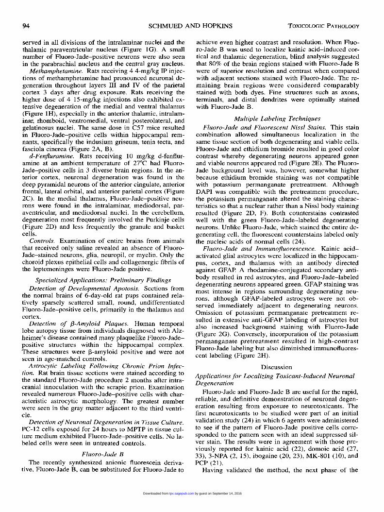
f.1m) allow for the visualization of dendritic arborizations(Figures 1E and 2D), and thinner sections (15-40 f.1m)provide the best resolution of axon terminals (Figures lA,B and 2B, F). The exact extent and pattern of Fluoro-Jade labeling depends on the nature of the neurotoxicinsult and the ensuing survival interval. The following isa more detailed description of the staining pattern ob-served following exposure to the following neurotoxicinsults.
Kainic Acid. This potent excitotoxin resulted in exten-sive Fluoro-Jade labeling within the forebrain. The dens-est labeling was found throughout the pyramidal cells ofthe hippocampus (Figures 1 A and 2E-H) and piriformcortex. Survival intervals of 1-4 days were optimal forrevealing cellular degeneration, whereas terminal degen-eration was optimally seen 4-14 days after drug expo-sure. Other structures with conspicuous labeling includedthe dorsal thalamus, septum, central nucleus of the amyg-dala, and substantia nigra pars reticulata. Also, portionsof the cortex exhibited a patchy distribution of Fluoro-Jade labeling with relatively extensive labeling of cin-gulate and temporal cortex regions and a more patchydistribution of layer II, III, and V pyramidal cells (Figure1B) throughout parietal and sensory-motor cortex.Domoic Acid. This seafood contaminant was relatively
similar to kainic acid in terms of the resulting distributionof Fluoro-Jade-stained cells (Figure 1C). Labeled cellswere detected at times as short as 4 hours or as long as7 days after drug exposure. Lower doses of domoic acid(eg, 1.5 mg/kg) occasionally produced a very localizedpatch of terminal degeneration within the stratum oriensnear the CA1-CA3 boundary. This degeneration may ormay not be accompanied by a few Fluoro-Jade positivecell bodies.
Ibogaine. This antidepressant alkaloid produced a veryrestricted pattern of Fluoro-Jade labeling. Only smallclusters of labeled Purkinje cells and their dendrites couldbe found in the paravermal region of the cerebellum (Fig-ure lE).MK-801/PCP. These N-methyl-D-aspartic acid
(NMDA) receptor-blocking anesthetics also resulted in arestricted Fluoro-Jade labeling pattern. Small stellate cellsof deep retrosplenial and cingulate cortex were positivelystained (data not shown).3-NPA. Administration of this inhibitor of metabolic
respiration resulted in extensive degeneration throughoutthe basal ganglia (Figure 1D), the thalamus, the hippo-campus (mostly CA 1 ), the deep nuclei of the cerebellum,and the cochlear nuclei. 3-NPA-induced degenerationwas typically characterized by a staining of virtually allneurons and neuropil within a specific structure or sub-structure. Degenerating cell body profiles were most ap-parent following a 1-day survival interval, whereas de-generating axonal profiles were most apparent followinga 5-day survival interval.MPTP. A single dose of MPTP resulted in labeled
fusiform cells throughout the substantia nigra pars com-pacta and the ventral tegmental area in mice kept at anambient temperature of 6°C (Figure 1F). These animalsalso had degenerating noncatecholaminergic neuronswithin the thalamus. Neuronal degeneration was ob-
by guest on September 14, 2016tpx.sagepub.comDownloaded from
94
served in all divisions of the intralaminar nuclei and thethalamic paraventricular nucleus (Figure 1 G). A smallnumber of Fluoro-Jade-positive neurons were also seenin the parabrachial nucleus and the central gray nucleus.
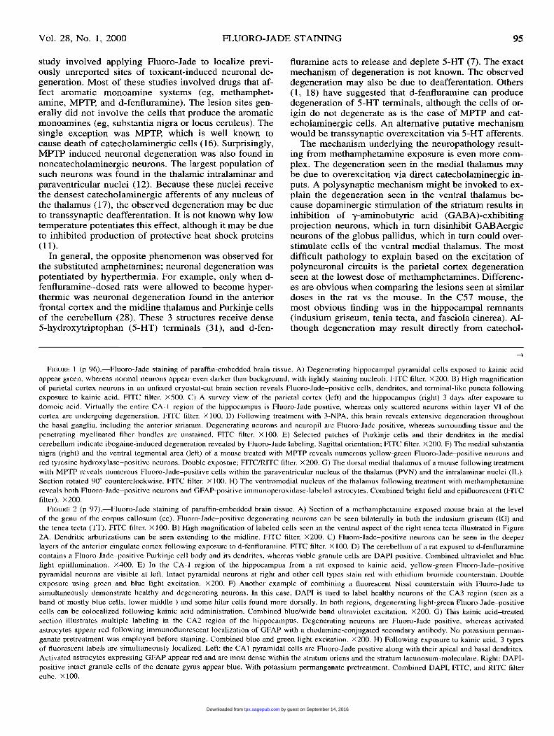
Methamphetamine. Rats receiving 4 4-mg/kg IP injec-tions of methamphetamine had pronounced neuronal de-generation throughout layers III and IV of the parietalcortex 3 days after drug exposure. Rats receiving thehigher dose of 4 15-mg/kg injections also exhibited ex-tensive degeneration of the medial and ventral thalamus(Figure 1 H), especially in the anterior thalamic, intralam-inar, rhomboid, ventromedial, ventral posterolateral, andgelatinosus nuclei. The same dose in C57 mice resultedin Fluoro-Jade-positive cells within hippocampal rem-nants, specifically the indusium griseum, tenia tecta, andfasciola cinerea (Figure 2A, B).
d-Fenfluramine. Rats receiving 10 mg/kg d-fenflur-amine at an ambient temperature of 27°C had Fluoro-Jade-positive cells in 3 diverse brain regions. In the an-terior cortex, neuronal degeneration was found in the
deep pyramidal neurons of the anterior cingulate, anteriorfrontal, lateral orbital, and anterior parietal cortex (Figure2C). In the medial thalamus, Fluoro-Jade-positive neu-rons were found in the intralaminar, mediodorsal, par-aventricular, and mediodorsal nuclei. In the cerebellum,degeneration most frequently involved the Purkinje cells(Figure 2D) and less frequently the granule and basketcells.
Controls. Examination of entire brains from animalsthat received only saline revealed an absence of Fluoro-Jade-stained neurons, glia, neuropil, or myelin. Only thechoroid plexus epithelial cells and collagenergic fibrils ofthe leptomeninges were Fluoro-Jade positive.
Specialized Applications: Preliminary FindingsDetection of Developmental Apotosis. Sections from
the normal brains of 6-day-old rat pups contained rela-tively sparsely scattered small, round, undifferentiatedFluoro-Jade-positive cells, primarily in the thalamus andcortex.
Detection of f3-Amyloid Plaques. Human temporallobe autopsy tissue from individuals diagnosed with Alz-heimer’s disease contained many plaquelike Fluoro-Jade-positive structures within the hippocampal complex.These structures were ¡3-amyloid positive and were notseen in age-matched controls.
Astrocytic Labeling Following Chronic Prion Infec-tion. Rat brain tissue sections were stained according tothe standard Fluoro-Jade procedure 2 months after intra-cranial innoculation with the scrapie prion. Examinationrevealed numerous Fluoro-Jade-positive cells with char-acteristic astrocytic morphology. The greatest numberwere seen in the gray matter adjacent to the third ventri-cle.
Detection of Neuronal Degeneration in Tissue Culture.PC-12 cells exposed for 24 hours to MPTP in tissue cul-ture medium exhibited Fluoro-Jade-positive cells. No la-beled cells were seen in untreated controls.
Fluoro-Jade B
The recently synthesized anionic fluorescein deriva-tive, Fluoro-Jade B, can be substituted for Fluoro-Jade to
achieve even higher contrast and resolution. When Fluo-ro-Jade B was used to localize kainic acid-induced cor-tical and thalamic degeneration, blind analysis suggestedthat 80% of the brain regions stained with Fluoro-Jade Bwere of superior resolution and contrast when comparedwith adjacent sections stained with Fluoro-Jade. The re-maining brain regions were considered comparablystained with both dyes. Fine structures such as axons,terminals, and distal dendrites were optimally stainedwith Fluoro-Jade B.
Multiple Labeling TechniquesFluoro-Jade and Fluorescent Nissl Stains. This stain
combination allowed simultaneous localization in thesame tissue section of both degenerating and viable cells.Fluoro-Jade and ethidium bromide resulted in good colorcontrast whereby degenerating neurons appeared greenand viable neurons appeared red (Figure 2E). The Fluoro-Jade background level was, however, somewhat higherbecause ethidium bromide staining was not compatiblewith potassium permanganate pretreatment. AlthoughDAPI was compatible with the pretreatment procedure,the potassium permanganate altered the staining charac-teristics so that a nuclear rather than a Nissl body stainingresulted (Figure 2D, F). Both counterstains contrastedwell with the green Fluoro-Jade-labeled degeneratingneurons. Unlike Fluoro-Jade, which stained the entire de-generating cell, the fluorescent counterstains labeled onlythe nucleic acids of normal cells (24).
Fluoro-Jade and Immunofluorescence. Kainic acid-activated glial astrocytes were localized in the hippocam-pus, cortex, and thalamus with an antibody directed
against GFAP A rhodamine-conjugated secondary anti-body resulted in red astrocytes, and Fluoro-Jade-labeleddegenerating neurons appeared green. GFAP staining wasmost intense in regions surrounding degenerating neu-rons, although GFAP-labeled astrocytes were not ob-served immediately adjacent to degenerating neurons.Omission of potassium permanganate pretreatment re-sulted in extensive anti-GFAP labeling of astrocytes butalso increased background staining with Fluoro-Jade
(Figure 2G). Conversely, incorporation of the potassiumpermanganate pretreatment resulted in high-contrastFluoro-Jade labeling but also diminished immunofluores-cent labeling (Figure 2H).
Discussion
Applications for Localizing Toxicant-Induced NeuronalDegeneration
Fluoro-Jade and Fluoro-Jade B are useful for the rapid,reliable, and definitive demonstration of neuronal degen-eration resulting from exposure to neurotoxicants. Thefirst neurotoxicants to be studied were part of an initialvalidation study (24) in which 6 agents were administeredto see if the pattern of Fluoro-Jade-positive cells corre-sponded to the pattern seen with an ideal suppressed sil-ver stain. The results were in agreement with those pre-viously reported for kainic acid (22), domoic acid (27,33), 3-NPA (2, 15), ibogaine (20, 23), MK-801 (10), andPCP (21).
Having validated the method, the next phase of the
by guest on September 14, 2016tpx.sagepub.comDownloaded from
95
study involved applying Fluoro-Jade to localize previ-ously unreported sites of toxicant-induced neuronal de-generation. Most of these studies involved drugs that af-fect aromatic monoamine systems (eg, methamphet-amine, MPTP, and d-fenfluramine). The lesion sites gen-erally did not involve the cells that produce the aromaticmonoamines (eg, substantia nigra or locus ceruleus). Thesingle exception was MPTP, which is well known tocause death of catecholaminergic cells (16). Surprisingly,MPTP induced neuronal degeneration was also found innoncatecholaminergic neurons. The largest population ofsuch neurons was found in the thalamic intralaminar and
paraventricular nuclei (12). Because these nuclei receivethe densest catecholaminergic afferents of any nucleus ofthe thalamus (17), the observed degeneration may be dueto transsynaptic deafferentation. It is not known why lowtemperature potentiates this effect, although it may be dueto inhibited production of protective heat shock proteins(11).
In general, the opposite phenomenon was observed forthe substituted amphetamines; neuronal degeneration waspotentiated by hyperthermia. For example, only when d-fenfluramine-dosed rats were allowed to become hyper-thermic was neuronal degeneration found in the anteriorfrontal cortex and the midline thalamus and Purkinje cellsof the cerebellum (28). These 3 structures receive dense5-hydroxytriptophan (5-HT) terminals (31), and d-fen-
fluramine acts to release and deplete 5-HT (7). The exactmechanism of degeneration is not known. The observeddegeneration may also be due to deafferentation. Others(1, 18) have suggested that d-fenfluramine can producedegeneration of 5-HT terminals, although the cells of or-igin do not degenerate as is the case of MPTP and cat-echolaminergic cells. An alternative putative mechanismwould be transsynaptic overexcitation via 5-HT afferents.The mechanism underlying the neuropathology result-
ing from methamphetamine exposure is even more com-plex. The degeneration seen in the medial thalamus maybe due to overexcitation via direct catecholaminergic in-puts. A polysynaptic mechanism might be invoked to ex-plain the degeneration seen in the ventral thalamus be-cause dopaminergic stimulation of the striatum results ininhibition of y-aminobutyric acid (GABA)-exhibitingprojection neurons, which in turn disinhibit GABAergicneurons of the globus pallidus, which in turn could over-stimulate cells of the ventral medial thalamus. The mostdifficult pathology to explain based on the excitation ofpolyneuronal circuits is the parietal cortex degenerationseen at the lowest dose of methamphetamines. Differenc-es are obvious when comparing the lesions seen at similardoses in the rat vs the mouse. In the C57 mouse, themost obvious finding was in the hippocampal remnants(indusium griseum, tenia tecta, and fasciola cinerea). Al-though degeneration may result directly from catechol-
-
FIGURE I (p 96).-Fluoro-Jade staining of paraffin-embedded brain tissue. A) Degenerating hippocampal pyramidal cells exposed to kainic acidappear green, whereas normal neurons appear even darker than background, with lightly staining nucleoli. FITC filter. X200. B) High magnificationof parietal cortex neurons in an unfixed cryostat-cut brain section reveals Fluoro-Jade-positive cells, dendrites, and terminal-like puncta followingexposure to kainic acid. FITC filter. X500. C) A survey view of the parietal cortex (left) and the hippocampus (right) 3 days after exposure todomoic acid. Virtually the entire CA-1 region of the hippocampus is Fluoro-Jade positive, whereas only scattered neurons within layer VI of thecortex are undergoing degeneration. FITC filter. X 100. D) Following treatment with 3-NPA, this brain reveals extensive degeneration throughoutthe basal ganglia, including the anterior striatum. Degenerating neurons and neuropil are Fluoro-Jade positive, whereas surrounding tissue and thepenetrating myelinated fiber bundles are unstained. FITC filter. X 100. E) Selected patches of Purkinje cells and their dendrites in the medialcerebellum indicate ibogaine-induced degeneration revealed by Fluoro-Jade labeling. Sagittal orientation; FITC filter. X 200. F) The medial substantianigra (right) and the ventral tegmental area (left) of a mouse treated with MPTP reveals numerous yellow-green Fluoro-Jade-positive neurons andred tyrosine hydroxylase-positive neurons. Double exposure; FITC/RITC filter. X200. G) The dorsal medial thalamus of a mouse following treatmentwith MPTP reveals numerous Fluoro-Jade-positive cells within the paraventricular nucleus of the thalamus (PVN) and the intralaminar nuclei (IL).Section rotated 90° counterclockwise. FITC filter. X 100. H) The ventromedial nucleus of the thalamus following treatment with methamphetaminereveals both Fluoro-Jade-positive neurons and GFAP-positive immunoperoxidase-labeled astrocytes. Combined bright field and epifluorescent (FITCfilter). X200.
FIGURE 2 (p 97).-Fluoro-Jade staining of paraffin-embedded brain tissue. A) Section of a methamphetamine-exposed mouse brain at the levelof the genu of the corpus callosum (cc). Fluoro-Jade-positive degenerating neurons can be seen bilaterally in both the indusium griseum (IG) andthe tenea tecta (TT). FITC filter. X 100. B) High magnification of labeled cells seen in the ventral aspect of the right tenea tecta illustrated in Figure2A. Dendritic arborizations can be seen extending to the midline. FITC filter. X200. C) Fluoro-Jade-positive neurons can be seen in the deeperlayers of the anterior cingulate cortex following exposure to d-fenfluramine. FITC filter. X 100. D) The cerebellum of a rat exposed to d-fenfluraminecontains a Fluoro-Jade-positive Purkinje cell body and its dendrites, whereas viable granule cells are DAPI positive. Combined ultraviolet and bluelight epiillumination. X400. E) In the CA-1 region of the hippocampus from a rat exposed to kainic acid, yellow-green Fluoro-Jade-positivepyramidal neurons are visible at left. Intact pyramidal neurons at right and other cell types stain red with ethidium bromide counterstain. Doubleexposure using green and blue light excitation. X200. F) Another example of combining a fluorescent Nissl counterstain with Fluoro-Jade tosimultaneously demonstrate healthy and degenerating neurons. In this case, DAPI is used to label healthy neurons of the CA3 region (seen as aband of mostly blue cells, lower middle ) and some hilar cells found more dorsally. In both regions, degenerating light-green Fluoro-Jade-positivecells can be colocalized following kainic acid administration. Combined blue/wide band ultraviolet excitation. X200. G) This kainic acid-treatedsection illustrates multiple labeling in the CA2 region of the hippocampus. Degenerating neurons are Fluoro-Jade positive, whereas activatedastrocytes appear red following immunofluorescent localization of GFAP with a rhodamine-conjugated secondary antibody. No potassium perman-ganate pretreatment was employed before staining. Combined blue and green light excitation. X200. H) Following exposure to kainic acid, 3 typesof fluorescent labels are simultaneously localized. Left: the CA 1 pyramidal cells are Fluoro-Jade positive along with their apical and basal dendrites.Activated astrocytes expressing GFAP appear red and are most dense within the stratum oriens and the stratum lacunosum-moleculare. Right: DAPI-positive intact granule cells of the dentate gyrus appear blue. With potassium permanganate pretreatment. Combined DAPI, FITC, and RITC filtercube. X 100.
by guest on September 14, 2016tpx.sagepub.comDownloaded from
98
aminergic inputs to this region (17), glutaminergic affer-ents from other areas receiving dopaminergic inputs (eg,olfactory bulb, entorhinal cortex, anterior thalamus, andlateral septum) probably are largely responsible for pro-ducing excitotoxic damage.
Other Applications and Recent DevelopmentsPreliminary data have shown that Fluoro-Jade will
stain (3-amyloid in temporal lobe brain sections from hu-mans with Alzheimer’s dementia. Although its potentialhas not been fully explored, Fluoro-Jade staining mayhave advantages over other methods for detecting (3-am-yloid plaques because it is technically simpler than (3-amyloid immunohistochemistry and more specific thanthioflavin S staining.
At the other end of the developmental spectrum, thestaining of small undifferentiated cells in the thalamusand cortex of 6-day-old rat pups suggests that Fluoro-Jade can detect cells that degenerate via an apoptoticmechanism. It is not, however, an exclusive marker forapoptosis because it will detect necrotic death as well.We have recently found that Fluoro-Jade can be used
to detect neuronal death in tissue culture. Thus, it pro-vides an alternative to the live-dead assays that typicallyinvolve identifying the incorporation and colorometricoxidation of a colorless fluorescein lactone and the activeexclusion of propidium iodide by living cells. It is pos-sible to freeze a degeneration pattern at a particular timeby fixing the cells and then staining them with Fluoro-Jade. This approach also avoids exposing living cells tovital dyes that might themselves compromise cellular vi-ability.
Another recent finding of possible significance is theobservation that at least certain types of chronic infla-mation, such as prion infection, can produce astrocyticlabeling with Fluoro-Jade (34). It is not known whetherthis staining indicates that the astrocytes are dead or
merely activated.The development of Fluoro-Jade B, the next generation
fluorochrome for the detection of degenerating neurons,has been in progress for the past year (29). Although theexact structure has not been definitively illucidated, thestochiometry and preliminary mass spectroscopy datasuggest that it is the bis-isomer of Fluoro-Jade. Althoughless fluorescent than Fluoro-Jade, Fluoro-Jade B has aneven greater affinity for degenerating neurons vs back-ground.
Comparing Fluoro-Jade with Other Markers:Respective Advantages and Disadvantages
Hematoxylin and Eosin. Advantages associated withhematoxylin and eosin staining include technical simplic-ity, reliability, and permanance. Disadvantages relate tothe fact that all brain structures, rather than only degen-erating neurons, are stained. Although inferences can bemade based on morphologic criteria such as neuronal
shrinkage, eosinophilia, vacuolation, and hyperchroma-tism (5, 14, 30), processing artifacts can result in bothshrunken and hyperchromatic cells (4, 32). Not only aresuch techniques prone to false-positive results, but it isalso possible to miss degenerating neurons because all
cells stain and only relatively subtle differences may existbetween normal and degenerating neurons. Moreover, he-matoxylin and eosin stain the neuropil a rather uniformpink color precluding visualization of dendrites, axons,and terminals.
Supressed Silver Stains. The primary advantage of anideal suppressed silver stain is that it selectively stainsonly degenerating neurons. The technique is also rela-
tively permanent and is of high contrast and resolution.The primary drawback to suppressed silver methods istheir notoriously capricious nature, which can result ineither false-positive or false-negative results. Other draw-backs are related to the fact that these techniques are verylabor intensive and time consuming and require the useof arsenic-based buffers for tissue perfusion and collec-tion.
Fluoro-Jade. Advantages associated with Fluoro-Jadeinclude technical simplicity, reliability, and unequivocaldetection of neuronal degeneration. Although archivalfading is not noticeable at the longest time examined (3years after preparation), prolonged examination at mag-nifications greater than X40 can result in detectable fad-ing. Generally, fading is not a problem because the back-ground fluoresence decreases at the same rate as the spe-cific signal, allowing high-quality photomicrographs tobe obtained even after 30 minutes of high-magnificationepifluoresent illumination. However, low-magnificationsurvey photomicrographs of a region should be obtainedprior to any prolonged illumination at high magnifica-tions to avoid the appearance of darker photobleachedareas in the survey micrographs.
Fluoro-Jade can be used as a marker for neurotoxicityin the brain. This relatively simple technique can be ap-plied to the testing of various new chemicals and drugs,both for special research questions and for more routinescreening. The real benefit will come as toxicologic pa-thologists utilize this technique and compare the resultswith those of more traditional procedures. We welcomesuch a comparison and encourage publication of the re-sults.
ACKNOWLEDGMENTS
We thank the following investigators who providedbrain tissue: Dr Andy Scallet who provided domoic acid-,ibogaine-, PCP-, and MK-801-exposed tissue, Dr SyedAli and Dr Tim Freyaldehoven who provided MPTP-ex-posed tissue, Dr Zbigniew Binienda who provided 3-NPA-exposed tissue, Dr John Bowyer and Dr PeterClausing who provided tissue exposed to methamphet-amine and d-fenfluramine, Dr Xuemen Ye who providedscrapie-exposed tissue, Dr Frank Scalzo who providedneonatal tissue, and Dr Sue Griffin and Dr Robert Mrakwho provided human autopsy tissue from individualswith Alzheimer’s disease. This work was supported inpart by FDA research protocol 7013.01.
REFERENCES
1. Appel NM, Contrera JF, De Sousa EB (1989). Fenfluramine selec-tivity differentially decreases the density of serotonergic nerve ter-minals in rat brain: Evidence from immunocytochemical studies. JPharmacol Exp Ther 249: 928-943.
by guest on September 14, 2016tpx.sagepub.comDownloaded from

99
2. Binienda Z, Frederick DL, Ferguson SA, Rountree RL, Paule MG,Schmued LC, Ali SF, Slikker W Jr, Scallet AC (1995). The effectof perinatal hypoxia on the behavioral, neurochemical, and neuro-histological toxicity of the metabolic inhibitor 3-nitropropionicacid. Metab Brain Dis 10: 269-281.
3. Bowyer JF, Peterson SL, Rountree RL, Tor-Agbidye J, Wang GJ(1998). Neuronal degeneration in rat forebrain resulting from d-amphetamine-induced convulsions is dependent on seizure severityand age. Brain Res 809: 77-90.
4. Cammermeyer J (1961). The importance of avoiding the ’dark’ neu-rons in experimental neuropathology. Acta Neuropathol 1: 245-
270.
5. Chassan JL (1997). Nervous system. In: Pathology, Anderson W,Kissane J (eds). Mosby, St. Louis, Missouri, pp 2074-2148.
6. de Olmos JS, Beltramino CA, de Olmos de Lorenzo S (1994). Useof an amino-cupric-silver technique for the detection of early andsemiacute neuronal degeneration caused by neurotoxicants, hy-poxia, and physical trauma. Neurotoxicol Terataol 16: 545-561.
7. Dunhault J, Verdavainne C (1967). Modification of the cerebralserotonin level in the rat by trifluoromethyl-2-phenyl ethyl ami-nopropane. Arch Int Pharmacodyn Ther 170: 276-286.
8. Eisch AJ, Schmued LC, Marshall JF (1998). Characterizing corticalneuron injury with Fluoro-Jade labeling after a neurotoxic regimenof methamphetamine. Synapse 30: 329-333.
9. Fink RP, Heimer L (1967). Two methods for the selective silverimpregnation of degenerating axons and their synaptic endings inthe central nervous system. Brain Res 4: 369-374.
10. Fix AS, Ross JF, Stitzel SR, Switzer RC (1996). Integrated evalu-ation of central nervous system lesions: Stains for neurons, astro-cytes, and microglia reveal the spatial and temporal features ofMK-801-induced neuronal necrosis in the rat cerebral cortex. Tox-icol Pathol 24: 291-304.
11. Freyaldenhoven TE, Ali SF (1995). MPTP- and MPP+-inducedeffects on body temperature exhibit age- and strain-dependence inmice. Brain Res 688: 161-170.
12. Freyaldenhoven TE, Ali SF, Schmued LC (1997). Systemic admin-istration of MPTP induces thalamic neuronal degeneration in mice.Brain Res 759: 9-17.
13. Gallyas F, Wolf JR, Bottcher H, Zaborsky L (1980). A reliable andsensitive method to localize terminal degeneration and lysosomesin the central nervous system. Stain Technol 55: 299-306.
14. Garcia JH, Kamijiyo Y (1974). Cerebral infraction: Evolution ofhistopathological changes after occlusion of a middle cerebral ar-tery in primates. J Neuropathol Exp Neurol 33: 409-421.
15. Hamilton BF, Gould DH (1987). Nature and distribution of brainlesions in rats intoxicated with 3-nitropropionic acid: A type ofhypoxic (energy deficient) brain damage. Acta Neuropathol 72:286-297.
16. Heikkila RE, Hess A, Duboisin RC (1983). Dopaminergic neuro-toxicity of 1-methyl-4-phenyl-1,2,5,6-tetrahydropyridine in mice.Science 224: 1451-1453.
17. Hokfelt T, Johansson O, Fuxe K, Goldstein M, Park D (1976).Immunohistochemical studies on the localization and distribution
of monoamine neuron systems in the rat brain. I. Tyrosine hydrox-ylase in the mes and diencephalon. Med Biol 54: 427-453.
18. Molliver DC, Molliver ME (1990). Anatomic evidence for a neu-rotoxic effect of (±)-fenfluramine upon serotonergic projections inthe rat. Brain Res 511: 165-168.
19. Nauta WJH, Gygax PA (1954). Silver impregnation of degeneratingaxons in the central nervous system: A modified technique. StainTechnol 29: 91-93.
20. O’Hearn E, Molliver ME (1993). Degeneration of Purkinje cells inparasagittal zones of the cerebellar vermis after treatment with ibo-gaine or harmaline. Neuroscience 55: 303-310.
21. Olney JW, Labruyere J, Price MT (1989). Pathological changesinduced in cerebrocortical neurons by phencyclidine and relateddrugs. Science 244: 1360-1362.
22. Olney JW, Rhee V, Ho OL (1974). Kainic acid: A powerful neu-rotoxic analogue of glutamate. Brain Res 77: 507-515.
23. Scallet AC, Ye X, Rountree R, Nony P, Ali SF (1996). Ibogaineproduces neurodegeneration in rat, but not mouse, cerebellum:
Neurohistological biomarkers of Purkinje cell loss. Ann NY AcadSci 801: 217-226.
24. Schmued LC, Albertson C, Slikker W Jr (1997). Fluoro-Jade: Anovel fluorochrome for the sensitive and reliable histochemical lo-calization of neuronal degeneration. Brain Res 751: 37-46.
25. Schmued LC, Bowyer JF (1997). Methamphetamine exposure canproduce neuronal degeneration in mouse hippocampal remnants.Brain Res 759: 135-140.
26. Schmued LC, Sawchenko PE, Swanson LW (1982). Some fluores-cent counterstains for neuroanatomical studies. J Histol Cytol 3012:123-128.
27. Schmued LC, Scallet AC, Slikker W Jr (1995). Domoic acid-in-duced neuronal degeneration in the primate forebrain revealed bydegeneration specific histochemistry. Brain Res 695: 64-70.
28. Schmued L, Slikker W, Clausing P, Bowyer J (1999). d-fenflur-amine produces neuronal degeneration in localized regions of thecortex, thalamus, and cerebellum of the rat. Toxicol Sci 48: 100-106.
29. Schmued LC, Slikker W Jr, Wang GJ (1998). Fluoro-Jade B: A bishomologue of Fluoro-Jade with improved degenerate neuron stain-ing properties. Soc Neurosci Abstr 24: 1064.
30. Siesjo BK (1981). Cell damage in the brain: A speculative synthe-sis. Cereb Blood Metab 1: 155-185.
31. Steinbusch HWM (1981). Distribution of serotonin-immunoreactiv-ity in the central nervous system of the rat—Cell bodies and ter-minals. Neuroscience 6: 557-618.
32. Stensaas SS, Edwards CQ, Stensaas LJ (1972). An experimentalstudy of hyperchromic nerve cells in the cerebral cortex. ExpNeurol 36: 472-487.
33. Tryphonas L, Truelove J, Nera E, Iverson F (1990). Acute neuro-toxicity of domoic acid in the rat. Toxicol Pathol 18: 1-9.
34. Ye X, Carp RI, Schmued LC, Meeker C, Rountree R, Scallet A(1997). Evaluation of neuronal and astrocytic pathology in scrapie-infected animals by specific degeneration-selective methods. SocNeurosci Abstr 23: 2442.
by guest on September 14, 2016tpx.sagepub.comDownloaded from