Notoginsenoside Ft1 Promotes Fibroblast Proliferation...
9
1521-0103/356/2/324–332$25.00 http://dx.doi.org/10.1124/jpet.115.229369 THE JOURNAL OF PHARMACOLOGY AND EXPERIMENTAL THERAPEUTICS J Pharmacol Exp Ther 356:324–332, February 2016 Copyright ª 2016 by The American Society for Pharmacology and Experimental Therapeutics Notoginsenoside Ft1 Promotes Fibroblast Proliferation via PI3K/Akt/mTOR Signaling Pathway and Benefits Wound Healing in Genetically Diabetic Mice s Eryun Zhang, Bo Gao, Li Yang, Xiaojun Wu, and Zhengtao Wang Department of Pharmacognosy, China Pharmaceutical University, Nanjing, China (E.Z, Z.W.); and Shanghai Key Laboratory of Complex Prescriptions, Institute of Chinese Materia Medica, Shanghai University of Traditional Chinese Medicine, Shanghai, China (B.G., L.Y., X.W., Z.W.) Received September 15, 2015; accepted November 12, 2015 ABSTRACT Wound healing requires the essential participation of fibroblasts, which is impaired in diabetic foot ulcers (DFU). Notoginsenoside Ft1 (Ft1), a saponin from Panax notoginseng, can enhance platelet aggregation by activating signaling network mediated through P 2 Y 12 and induce proliferation, migration, and tube formation in cultured human umbilical vein endothelial cells. However, whether it can accelerate fibroblast proliferation and benefit wound healing, especially DFU, has not been elucidated. In the present study on human dermal fibroblast HDF-a, Ft1 increased cell proliferation and collagen production via PI3K/Akt/ mTOR signaling pathway. On the excisional wound splinting model established on db/db diabetic mouse, topical application of Ft1 significantly shortened the wound closure time by 5.1 days in contrast with phosphate-buffered saline (PBS) treatment (15.8 versus 20.9 days). Meanwhile, Ft1 increased the rate of re- epithelialization and the amount of granulation tissue at day 7 and day 14. The molecule also enhanced mRNA expressions of COL1A1, COL3A1, transforming growth factor (TGF)-b1 and TGF-b3 and fibronectin, the genes that contributed to collagen expression, fibroblast proliferation, and consequent scar for- mation. Moreover, Ft1 facilitated the neovascularization ac- companied with elevated vascular endothelial growth factor, platelet-derived growth factor, and fibroblast growth factor at either mRNA or protein levels and alleviated the inflammation of infiltrated monocytes indicated by reduced tumor necrosis factor-a and interleukin-6 mRNA expressions in the diabetic wounds. Altogether, these results indicated that Ft1 might accelerate diabetic wound healing by orchestrating multiple processes, including promoting fibroblast proliferation, en- hancing angiogenesis, and attenuating inflammatory re- sponse, which provided a great potential application of it in clinics for patients with DFU. Introduction Wound healing is a well-orchestrated integration of complex biologic and molecular events, which requires the participa- tion of many types of cells, including macrophages, fibroblasts, keratinocytes, and endothelial cells. During the process, fibroblasts play an important role by depositing extracellular matrix (ECM) that guides angiogenesis and supports the migration and proliferation of other cells that eventually form the scar (Martin, 1997; Falanga, 2005; Brem and Tomic-Canic, 2007; Gurtner et al., 2008). Accordingly, drugs targeting fibroblast proliferation may contribute to the skin wound healing. Indeed, as known so far, two engineered living skin products approved by the Food and Drug Administration containing allogeneic fibroblasts have been successfully ap- plied in the therapy of patients with diabetic or venous skin ulcers (Griffith and Naughton, 2002). Diabetic foot ulcers (DFUs) are characterized by disrupted wound healing process, which is estimated to occur in 15% of all patients with diabetes (Boulton et al., 2005) and 84% of all diabetes-related lower-leg amputations (Reiber et al., 1999). Patients of DFUs often suffer from expensive hospital costs and poor quality of life (Ramsey et al., 1999). A number of factors accounting for the nonhealing wounds in diabetes have been implicated, which include abnormal fibroblast migration, proliferation, differentiation, and apoptosis (Al-Mulla et al., 2011). Unfortunately, in the past few decades, little improve- ment has been made in preventing the morbidity and disability of DFU. The best available treatment of chronic wounds only achieves a 50% healing rate, often with tempo- rary effect (Boulton et al., 2005). Therefore, more effective and specific drugs are urgently needed for the prevention or therapy of DFU. This work was supported financially by the National Natural Science Foundation of China [Grant 81530096], Key Research Innovation Project [Grant 13ZZ099], Key Project from Department of Education of China [Grant 20123107130002], Shanghai Three-Year Plan for Advancing Traditional Medicine [Grant ZY3-CCCX-3-3014] Shanghai Eastern Scholar Program [Grant 2013-59], and Shanghai E-research Institute of Bioactive Constituent in TCM plan. dx.doi.org/10.1124/jpet.115.229369 s This article has supplemental material available at jpet.aspetjournals.org. ABBREVIATIONS: Akt, protein kinase B; DFU, diabetic foot ulcer; ECM, extracellular matrix; ELISA, enzyme-linked immunosorbent assay; FGF, fibroblast growth factors; Ft1, notoginsenoside Ft1; GT, granulation tissue; HDF, human dermal fibroblast; HE, hematoxylin/eosin; IL-6, interleukin- 6; mTOR, mammalian target of rapamycin; PDGF, platelet-derived growth factor; PI3K, phosphatidylinositol-3-kinase; a-SMA, alpha-smooth muscle actin; TGF-b, transforming growth factor-b; TNF-a, tumor necrosis factor-a; VEGF, vascular endothelial growth factor. 324 http://jpet.aspetjournals.org/content/suppl/2015/11/13/jpet.115.229369.DC1 Supplemental material to this article can be found at: at ASPET Journals on July 14, 2018 jpet.aspetjournals.org Downloaded from
Transcript of Notoginsenoside Ft1 Promotes Fibroblast Proliferation...
1521-0103/356/2/324–332$25.00 http://dx.doi.org/10.1124/jpet.115.229369THE JOURNAL OF PHARMACOLOGY AND EXPERIMENTAL THERAPEUTICS J Pharmacol Exp Ther 356:324–332, February 2016Copyright ª 2016 by The American Society for Pharmacology and Experimental Therapeutics
Notoginsenoside Ft1 Promotes Fibroblast Proliferation viaPI3K/Akt/mTOR Signaling Pathway and Benefits Wound Healingin Genetically Diabetic Mice s
Eryun Zhang, Bo Gao, Li Yang, Xiaojun Wu, and Zhengtao WangDepartment of Pharmacognosy, China Pharmaceutical University, Nanjing, China (E.Z, Z.W.); and Shanghai Key Laboratory ofComplex Prescriptions, Institute of Chinese Materia Medica, Shanghai University of Traditional Chinese Medicine, Shanghai,China (B.G., L.Y., X.W., Z.W.)
Received September 15, 2015; accepted November 12, 2015
ABSTRACTWound healing requires the essential participation of fibroblasts,which is impaired in diabetic foot ulcers (DFU). NotoginsenosideFt1 (Ft1), a saponin from Panax notoginseng, can enhanceplatelet aggregation by activating signaling network mediatedthrough P2Y12 and induce proliferation, migration, and tubeformation in cultured human umbilical vein endothelial cells.However, whether it can accelerate fibroblast proliferation andbenefit wound healing, especially DFU, has not been elucidated.In the present study on human dermal fibroblast HDF-a, Ft1increased cell proliferation and collagen production via PI3K/Akt/mTOR signaling pathway. On the excisional wound splintingmodel established on db/db diabetic mouse, topical applicationof Ft1 significantly shortened the wound closure time by 5.1 daysin contrast with phosphate-buffered saline (PBS) treatment (15.8versus 20.9 days). Meanwhile, Ft1 increased the rate of re-epithelialization and the amount of granulation tissue at day 7
and day 14. The molecule also enhanced mRNA expressions ofCOL1A1, COL3A1, transforming growth factor (TGF)-b1 andTGF-b3 and fibronectin, the genes that contributed to collagenexpression, fibroblast proliferation, and consequent scar for-mation. Moreover, Ft1 facilitated the neovascularization ac-companied with elevated vascular endothelial growth factor,platelet-derived growth factor, and fibroblast growth factor ateither mRNA or protein levels and alleviated the inflammationof infiltrated monocytes indicated by reduced tumor necrosisfactor-a and interleukin-6 mRNA expressions in the diabeticwounds. Altogether, these results indicated that Ft1 mightaccelerate diabetic wound healing by orchestrating multipleprocesses, including promoting fibroblast proliferation, en-hancing angiogenesis, and attenuating inflammatory re-sponse, which provided a great potential application of it inclinics for patients with DFU.
IntroductionWound healing is a well-orchestrated integration of complex
biologic and molecular events, which requires the participa-tion ofmany types of cells, includingmacrophages, fibroblasts,keratinocytes, and endothelial cells. During the process,fibroblasts play an important role by depositing extracellularmatrix (ECM) that guides angiogenesis and supports themigration and proliferation of other cells that eventually formthe scar (Martin, 1997; Falanga, 2005; Brem andTomic-Canic,2007; Gurtner et al., 2008). Accordingly, drugs targetingfibroblast proliferation may contribute to the skin woundhealing. Indeed, as known so far, two engineered living skin
products approved by the Food and Drug Administrationcontaining allogeneic fibroblasts have been successfully ap-plied in the therapy of patients with diabetic or venous skinulcers (Griffith and Naughton, 2002).Diabetic foot ulcers (DFUs) are characterized by disrupted
wound healing process, which is estimated to occur in 15% ofall patients with diabetes (Boulton et al., 2005) and 84% of alldiabetes-related lower-leg amputations (Reiber et al., 1999).Patients of DFUs often suffer from expensive hospital costsand poor quality of life (Ramsey et al., 1999). A number offactors accounting for the nonhealing wounds in diabetes havebeen implicated, which include abnormal fibroblastmigration,proliferation, differentiation, and apoptosis (Al-Mulla et al.,2011). Unfortunately, in the past few decades, little improve-ment has been made in preventing the morbidity anddisability of DFU. The best available treatment of chronicwounds only achieves a 50% healing rate, often with tempo-rary effect (Boulton et al., 2005). Therefore, more effective andspecific drugs are urgently needed for the prevention ortherapy of DFU.
This work was supported financially by the National Natural ScienceFoundation of China [Grant 81530096], Key Research Innovation Project[Grant 13ZZ099], Key Project from Department of Education of China [Grant20123107130002], Shanghai Three-Year Plan for Advancing TraditionalMedicine [Grant ZY3-CCCX-3-3014] Shanghai Eastern Scholar Program[Grant 2013-59], and Shanghai E-research Institute of Bioactive Constituentin TCM plan.
dx.doi.org/10.1124/jpet.115.229369s This article has supplemental material available at jpet.aspetjournals.org.
ABBREVIATIONS: Akt, protein kinase B; DFU, diabetic foot ulcer; ECM, extracellular matrix; ELISA, enzyme-linked immunosorbent assay; FGF,fibroblast growth factors; Ft1, notoginsenoside Ft1; GT, granulation tissue; HDF, human dermal fibroblast; HE, hematoxylin/eosin; IL-6, interleukin-6; mTOR, mammalian target of rapamycin; PDGF, platelet-derived growth factor; PI3K, phosphatidylinositol-3-kinase; a-SMA, alpha-smoothmuscle actin; TGF-b, transforming growth factor-b; TNF-a, tumor necrosis factor-a; VEGF, vascular endothelial growth factor.
324
http://jpet.aspetjournals.org/content/suppl/2015/11/13/jpet.115.229369.DC1Supplemental material to this article can be found at:
at ASPE
T Journals on July 14, 2018
jpet.aspetjournals.orgD
ownloaded from
Panax notoginseng (Burk.) F. H. Chen, known as Sanqi inChina, is famous for its efficacy in treating trauma in EastAsia over 2000 years (Chen, 1987). Notoginsenoside Ft1 (Ft1;Fig. 1), a saponinmolecule, was first isolated from the leaves ofP. notoginseng in 2006 (Chen et al., 2006). Our group pre-viously reported that Ft1 was useful not only as a P2Y12
agonist in enhancing platelet aggregation (Gao et al., 2014)but also as a stimulator of proliferation, migration, and tubeformation in cultured human umbilical vein endothelial cells(Shen et al., 2012). These two properties indicated its possiblecurative effect on wound healing. However, whether thismolecule can promote the proliferation of fibroblast andbenefit wound healing has not been demonstrated elsewhere.The aim of this study was to investigate the effect of Ft1 on
fibroblast proliferation and disclose the underlying mecha-nism. Moreover, its in vivo function on wound healing wasassessed by an excisional wound splinting model establishedin genetically diabetic (db/db) mice. The research may con-tribute to the potential application of Ft1 in the therapy ofDFU.
Materials and MethodsChemicals and Regents. Ft1 was obtained from Shanghai R&D
Center for Standardization of Chinese Medicines (Shanghai, China).Its structure was confirmed using 1HNMR and 13C NMR spectralanalysis, and its purity was more than 98% as determined by highpressure liquid chromatography analysis.
Trizol reagent was purchased from Life Technology (Carlsbad, CA).PrimeScript RT Master Mix and SYBR Premix Ex Taq were fromTakara (Shiga, Japan). Phospho- PI3K p85 (Tyr458), PI3K, phospho-Akt (Thr308), Akt, phospho-mTOR (Ser2481) mTOR, and b-actinantibodies were obtained from Cell Signaling Technology (Danvers,
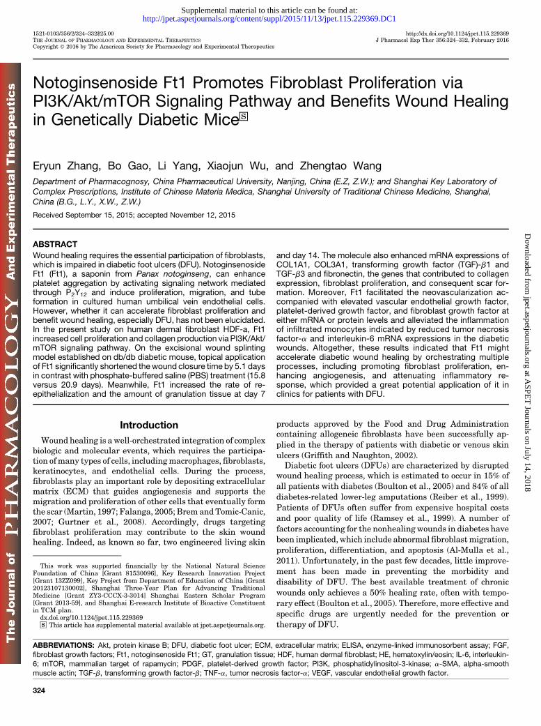
Fig. 1. Chemical structure of Ft1.
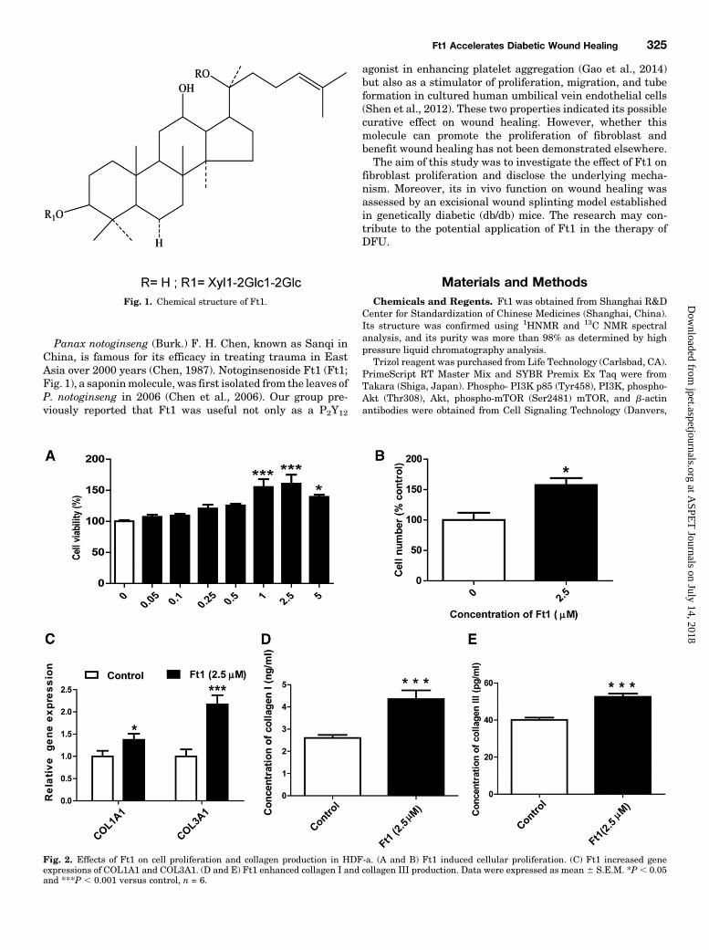
Fig. 2. Effects of Ft1 on cell proliferation and collagen production in HDF-a. (A and B) Ft1 induced cellular proliferation. (C) Ft1 increased geneexpressions of COL1A1 and COL3A1. (D and E) Ft1 enhanced collagen I and collagen III production. Data were expressed as mean 6 S.E.M. *P , 0.05and ***P , 0.001 versus control, n = 6.
Ft1 Accelerates Diabetic Wound Healing 325
at ASPE
T Journals on July 14, 2018
jpet.aspetjournals.orgD
ownloaded from
MA). Other antibodies against VEGF, CD31, and collagen III wereprovided byAbcam (Cambridge,UK). Pierce bicinchoninic acid proteinassay kit was purchased from Thermo Fisher Scientific (Waltham,MA). Human recombinant vascular endothelial growth factor (VEGF)was supplied by PeproTech (Rocky Hill, NJ). Enzyme-linkedimmunosorbent assay (ELISA) kits were purchased from R&D(Minneapolis, MN). All of the other reagents were from Sigma(St. Louis, MO) unless otherwise indicated.
Cell Culture and Cell Viability Measurement. Human dermalfibroblast-adult (HDF-a, ScienCell, SanDiego, CA) cells were culturedin fibroblast medium (ScienCell) supplemented with 2% fetal bovineserum, 100 IU/ml penicillin, and 100 IU/ml streptomycin in ahumidified atmosphere with 5% CO2 at 37°C. Cell viability aftertreatment with various concentrations of Ft1 for 48 hours wasdetermined by MTT assay. Optical density was measured at 570 nmwith 630 nm as the reference wavelength. Cell numbers were countedafter trypsinizing HDF-a treated with Ft1 for 48 hours. Final cellviability and number of the treated cells were presented as thepercentage to that of the control.
ELISA Analysis. After the cellswere treatedwithFt1 for 48 hours,the medium supernatant was collected and assayed for collagen I andIII using commercial ELISA kits according to the manufacturer’sinstructions. The concentrations of the target proteins in the mediumwere determined with respective standard curves prepared usingrecombinant proteins of known concentrations.
Animal Model and Treatment. Female leptin receptor-deficient (Lepr db/JNju, db/db) mice, 12 weeks old, with high bloodglucose (25.3 6 7.7 mM) were obtained from Nanjing BiomedicalResearch Institute of Nanjing University. The animals were single-house maintained under a 12 hour light/dark cycle at room temper-ature (23 6 2°C) with free access to food and water. All animalsreceived humane care according to the Institutional Animal Careguidelines approved by the Experimental Animal Ethical Committeeof Shanghai University of Traditional Chinese Medicine. The
excisional wound splinting model was generated according to themethod described previously (Wang et al., 2013). In brief, after hairremoval from the dorsal surface under anesthesia, two 6-mm full-thickness excisional skin wounds were created onmice at each side ofthe midline. A donut-shaped silicone splint was fixed around thewound with an immediate-bonding adhesive (Krazy Glue, Colum-bus, OH). Each wound was treated with 15 ml of Ft1 (6.7 mg/ml),vascular endothelial growth factor (VEGF) (1.0 mg/ml), or PBS everyother day. After drug administration, the wound was covered withTegaderm (Tegaderm, 3M, St. Paul, MN). The adhesive used wastested before the experiment, and no skin irritation or allergicreaction was observed.
Wound Analysis. Digital photographs of thewoundswere taken onthe day of surgery and every other day thereafter. Time to woundclosure was defined as the time needed for the wound bed to becompletely re-epithelialized and filled with new tissue. The pixel of thewound area was determined using Sigma Scan Pro Image AnalysisVersion 5.0 digital analysis software (Aspire Software International,Leesburg, VA) and was presented as the percentage of the initiativewound area. At day 7, 10, 14, and 28, the mice were killed, respectively,and thewound skin samples including the surrounding skin 6mmawaywere harvested using a 12-mm biopsy punch.
Histopathological Examination. For histologic preparations,the skin was fixed in 10% neutral buffered formalin and embeddedin paraffin. Skin tissues were sectioned in 4-mm-thick slices forhistopathological examination by hematoxylin/eosin (HE) stainingand for collagen formation by Masson’s trichrome staining.
For immunohistochemical staining, the sections were firstly in-cubated with 3% H2O2 for 10 minutes to deactivate the endogenousperoxidase. To recover antigen, these sections were soaked in 10 mMcitrate buffer solution (pH 6.0) and heated twice in the microwaveoven. The slides were then washed thoroughly with PBS (pH 7.4).After blocked with 5% bovine serum albumin in Tris-buffered salinefor 20 minutes, the sections were incubated with primary antibodies
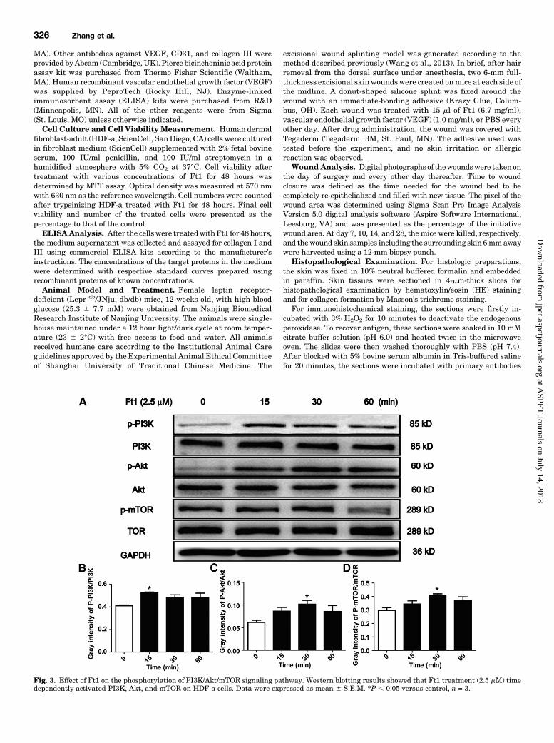
Fig. 3. Effect of Ft1 on the phosphorylation of PI3K/Akt/mTOR signaling pathway. Western blotting results showed that Ft1 treatment (2.5 mM) timedependently activated PI3K, Akt, and mTOR on HDF-a cells. Data were expressed as mean 6 S.E.M. *P , 0.05 versus control, n = 3.
326 Zhang et al.
at ASPE
T Journals on July 14, 2018
jpet.aspetjournals.orgD
ownloaded from
against CD31 at 4°C overnight followed by thorough wash with PBS.Afterward, the slides were sequentially incubated with biotinylatedsecondary antibody for 20 minutes and streptavidin-horseradishperoxidase for another 20 minutes. The staining was visualized afterincubation with a DAB-H2O2 solution. The slides were then counter-stained with hematoxylin for 1 minute, dehydrated with ethanol, andsealed in resinene for microscopic observation.
Real-Time Polymerase Chain Reaction. Total RNA from celland skin samples were isolated using Trizol reagent according to themanufacturer’s instructions. cDNAwas synthesized with PrimeScriptRT Master Mix kit. Real-time polymerase chain reaction wasperformed with SYBR green premix in accordance with the manufac-turer’s instructions. The following primers were used for cDNAamplification: for COL1A1 (human), 59-GCTACTACCGGGCTGAT-GAT-39 (forward) and 59-ACCAGTCTCCATGTTGCAGA-39 (reverse);for COL3A1 (human), 59-GAAGGGCAGGGAACAACTTG-39 (forward)and 59-TTTGGCATGGTTCTGGCTTC-39 (reverse); for VEGF (hu-man), 59-CGCAGCTACTGCCATCCAAT -39 (forward) and 59-CCCACAGGGATTTTCTTGTCTT-39 (reverse); for FGF (human), 59-GCCCAGTTCACTTCTTTGCA-39 (forward) and 59-AGATCCAAACCCAGACC-CAG-39 (reverse); for GAPDH (human), 59-TGTTGCCATCAATGACCCCTT-39 (forward) and 59-CTCCACGACFTACTCAGCG-39 (reverse);for VEGF (mouse), 59-GCACATAGAGAGAATGAGCTTCC-39 (for-ward) and 59-CTCCGCTCTGAACAAGGCT-39 (reverse); for PDGF
(mouse), 59-GAGGAAGCCGAGATACCCC-39 (forward) and 59-TGCTGTGGATCTGACTTCGAG-39 (reverse); for FGF (mouse), 59-ATGCTAGGGACCTGCCTTAGA-39 (forward) and 59-AGCCAAGCAATGGGGAAGTG-39 (reverse); for TGFb1 (mouse), 59-CTCCCGTGGCTTCTAGTGC-39 (forward) and 59-GCCTTAGTTTGGACAGGATCTG-39 (re-verse); for TGFb3 (mouse), 59-CCTGGCCCTGCTGAACTTG-39 (for-ward) and 59-TTGATGTGGCCGAAGTCCAAC-39 (reverse); for COL1A1 (mouse), 59-GCTCCTCTTAGGGGCCACT-39 (forward) and 59-CCACGTCTCACCATTGGGG-39 (reverse); for COL3A1 (mouse), 59-CTGTAACATGGAAACTGGGGAAA-39(forward) and 59-CCATAGCTGAACTGAAAACCACC-39 (reverse); for fibronectin (mouse), 59-ATGTGGACCCCTCCTGATAGT-39(forward) and 59-GCCCAGTGATTTCAGCAAAGG-39 (reverse); for TNFa (mouse), 59-CCCTCACACTCAGATCATCTTCT-39 (forward) and 59-GCTACGACGTGGGCTACAG-39 (reverse);for IL-6 (mouse), 59-TAGTCCTTCCTACCCCAATTTCC-39 (forward)and 59-TTGGTCCTTAGCCACTCCTTC-39 (reverse); for a-SMA(mouse), 59-AGGGAGTAATGGTTGGAATGG-39 (forward) and 59-GGTGATGATGCCGTGTTCTA-39 (reverse); for b-actin (mouse), 59-TGTCCACCTTCCAGCAGATGT-39 (forward) and 59-AGCTCAGTAACAGTCCGCCTAGA-39 (reverse). All of the gene expressions were normal-ized to that of the internal reference genes, namely GAPDH or b-actin,within the same samples using the delta delta CT method.
Western Blotting Analysis. Cell and skin tissues homogenateswere lysed in lysis buffer containing 50mMTris (pH 7.5), 1mMEDTA,
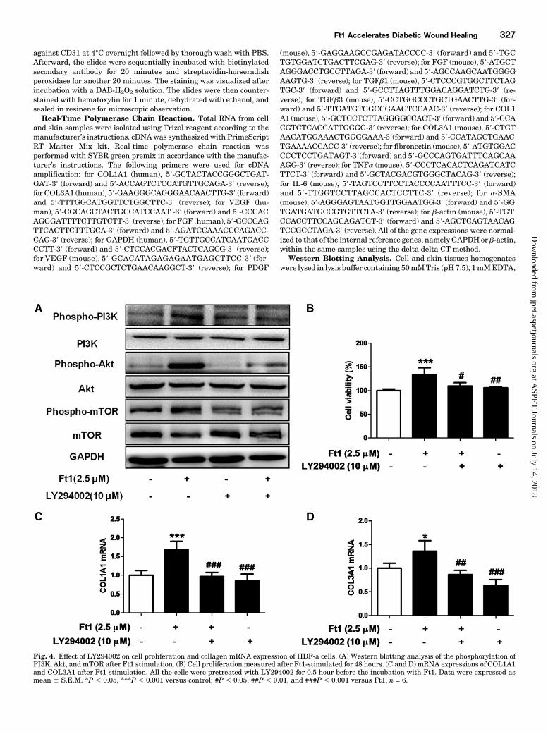
Fig. 4. Effect of LY294002 on cell proliferation and collagen mRNA expression of HDF-a cells. (A) Western blotting analysis of the phosphorylation ofPI3K, Akt, and mTOR after Ft1 stimulation. (B) Cell proliferation measured after Ft1-stimulated for 48 hours. (C and D) mRNA expressions of COL1A1and COL3A1 after Ft1 stimulation. All the cells were pretreated with LY294002 for 0.5 hour before the incubation with Ft1. Data were expressed asmean 6 S.E.M. *P , 0.05, ***P , 0.001 versus control; #P , 0.05, ##P , 0.01, and ###P , 0.001 versus Ft1, n = 6.
Ft1 Accelerates Diabetic Wound Healing 327
at ASPE
T Journals on July 14, 2018
jpet.aspetjournals.orgD
ownloaded from
150 mM NaCl, 20 mM NaF, 0.5%NP-40, 10% glycerol, 1 mMphenylmethylsulfonyl fluoride, 10 mg/ml aprotinin, 10 mg/mlleupeptin, and 10 mg/ml pepstatin A on ice. After centrifugationat 12,000 g for 15 minutes at 4°C, the supernatant was collected,and its protein concentration was determined using bicinchoninicacid method. Total proteins, 40 mg for each sample, were separatedon 12% SDS-PAGE and transferred onto PVDF membranes (Milli-pore, Bedford, MA). After blocked with 5% bovine serum albumin inPBST (0.1% Tween-20 in PBS) for 1 hour, the membranes wereincubated with respective primary antibodies at 4°C overnightfollowed by a thorough wash with PBST. Thereafter, the mem-branes were incubated with horseradish peroxidase-conjugatedsecondary antibody (1:5000) for 1 hour at room temperature. Theblots were developed by enhance chemiluminscence detectionregents (GE Healthcare, Waukesha, WI). Gray intensity of proteinbands was quantified with ImageJ and normalized to that ofb-actin in each sample.
Statistical Analysis. All data were presented as mean 6 S.E.M.Difference among multiple groups was analyzed using one-way anal-ysis of variance followed by Tukey’s multiple comparison test withGraphPad Prism 5.0 software. Unpaired t test was used to assess thedifference between two groups. A value of P , 0.05 was considered asstatistically significant.
ResultsFt1 Induced Cellular Proliferation of HDF-a. Ft1
prompted the proliferation of HDF-a cells when used at thedoses higher than 1 mM (Fig. 2, A and B). Compared with thecontrol, Ft1 used at 2.5 mM increased the cell viability (P ,0.001) and cell number (P , 0.05) of HDF-a by almost 50%.Therefore this concentration was chosen for the successiveexperiments. After treatment for 24 hours, significant mRNA
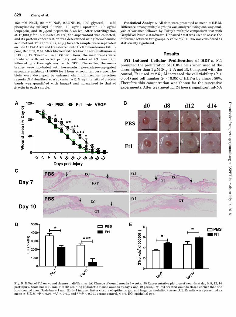
Fig. 5. Effect of Ft1 on wound closure in db/db mice. (A) Change of wound area in 3 weeks. (B) Representative pictures of wounds at day 0, 8, 12, 14postinjury. Scale bar = 10 mm. (C) HE staining of diabetic mouse wounds at day 7 and 10 postinjury. Ft1-treated wounds closed earlier than thePBS-treated ones. Scale bar = 1 mm. (D) Ft1 induced faster closure of epithelial gap and larger granulation tissue (GT). Results were presented asmean 6 S.E.M. *P , 0.05, **P , 0.01, and ***P , 0.001 versus control, n = 6. EG, epithelial gap.
328 Zhang et al.
at ASPE
T Journals on July 14, 2018
jpet.aspetjournals.orgD
ownloaded from
expressions of COL1A1 (P , 0.05) and COL3A1 (P , 0.001)was induced by Ft1 in HDF-a cells (Fig. 2C). In agreementwith the mRNA expression pattern, Ft1 also increased theprotein production of collagen I (P , 0.001) and collagen III
(P, 0.001) (Fig. 2, D and E). All of these results indicated theaccelerative effect of Ft1 on the proliferation of fibroblast.Ft1 Activated PI3K/Akt/mTOR Signaling Pathway in
HDF-a. Upon stimulation by Ft1, PI3K, Akt, andmTORwereall activated in HDF-a as the phosphorylation of thesesignaling proteins were enhanced within 1 hour (Fig. 3, Aand B). However, the phosphorylation peaks for these signal-ing molecules were reached at different time points. For PI3K,it seemed to be increasingly phosphorylated and reached itspeak at 15 minutes (P , 0.05). For Akt and mTOR, both ofthem reached their phosphorylation peak at 30 minutes (P ,0.05). To further confirm the involvement of PI3K/Akt/mTORsignaling pathway in Ft1-induced fibroblast proliferation, thePI3K inhibitor LY294002 was employed on HDF-a cells. Asshown in Fig. 4A, LY294002 could efficiently abrogate thephosphorylation of PI3K as well as Akt and mTOR induced byFt1. Accordingly, enhanced cell proliferation and COL1A1,COL3A1 mRNA expressions by Ft1 were all abolished by thechemical inhibitor (Fig. 4, B–D).Ft1 Enhanced Wound Healing Process in Genetically
Diabetic db/db Mice. Similar to VEGF, Ft1 acceleratedwound closure in genetically diabetic db/db mice. As shown inFig. 5A, compared with PBS-treated wounds, topical Ft1
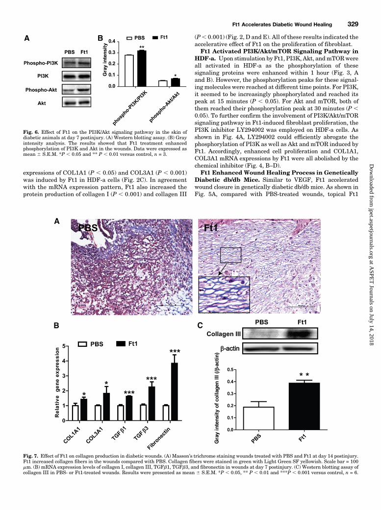
Fig. 6. Effect of Ft1 on the PI3K/Akt signaling pathway in the skin ofdiabetic animals at day 7 postinjury. (A) Western blotting assay. (B) Grayintensity analysis. The results showed that Ft1 treatment enhancedphosphorylation of PI3K and Akt in the wounds. Data were expressed asmean 6 S.E.M. *P , 0.05 and ** P , 0.01 versus control, n = 3.
Fig. 7. Effect of Ft1 on collagen production in diabetic wounds. (A) Masson’s trichrome staining wounds treated with PBS and Ft1 at day 14 postinjury.Ft1 increased collagen fibers in the wounds compared with PBS. Collagen fibers were stained in green with Light Green SF yellowish. Scale bar = 100mm. (B) mRNA expression levels of collagen I, collagen III, TGFb1, TGFb3, and fibronectin in wounds at day 7 postinjury. (C) Western blotting assay ofcollagen III in PBS- or Ft1-treated wounds. Results were presented as mean 6 S.E.M. *P , 0.05, ** P , 0.01 and ***P , 0.001 versus control, n = 6.
Ft1 Accelerates Diabetic Wound Healing 329
at ASPE
T Journals on July 14, 2018
jpet.aspetjournals.orgD
ownloaded from
treatment significantly accelerated wound healing process. Indiabetic mice, there was a significant decrease in terms ofaverage wound area in Ft1-treated wounds in comparisonwith the PBS-treated control at day 6 postinjury (P, 0.05). Onaverage, it took 15.8 days for Ft1-treated wounds to closecompletely in contrast with 20.9 days for PBS-treated controls.The increased healing rate of Ft1-treated wounds was clearlyshown by the representative photographs at 8, 12, and 14 dayspostinjury (Fig. 5B), in which the epithelium of Ft1-treatedwounds grew faster than that of PBS-treated controls at thesame day.Migration of the keratinocytes over the wounds for re-
epithelialization and ample granulation tissue formation arecritical early markers of successful wound healing assessment(Trautmann et al., 2000). As shown in Fig. 5C, histologicassessment of diabetic wounds confirmed the increased heal-ing rate of Ft1-treated wounds compared with PBS-treatedcontrols. The re-epithelialization process of Ft1-treatedwounds at day 7 and 10 postinjury was significantly fasterthan that of PBS-treated controls as demonstrated by reducedepithelial gaps in the Ft1-treated wounds (Fig. 5D; P , 0.05and P , 0.001). Granulation tissue (GT), largely composed of
fibroblasts synthesizing ECM proteins, in Ft1-treated woundsappeared to be thicker and larger. Quantitative measurementof the GT area of the wounds exposed that Ft1 treatmentsignificantly promoted GT growth at day 7 and day 14postinjury (Fig. 5E; P , 0.05).Consistent with its effect on PI3K/Akt signaling pathway in
HDF-a cells, Ft1 treatment enhanced phosphorylation of PI3K(P , 0.01) and Akt (P , 0.05) significantly in the skin ofdiabetic animals at day 7 postinjury (Fig. 6, A andB).Masson’strichrome staining exposed that Ft1 promoted collagen pro-duction in the wounds (Fig. 7A) asmore collagen fibers stainedin blue and regularly arranged could be found in Ft1-treatedwounds at day 14 postinjury compared with PBS-treated ones.Consistently, protein expression level of collagen III in Ft1-treated wound tissues was higher than that in the controls(Fig. 7C;P, 0.05).mRNA expression levels of proteins such asCOL1A1, COL3A1, TGFb1, TGFb3, and fibronectin that hadbeen reported to contribute to collagen formation were allelevated markedly (Fig. 7B; P , 0.05 or P , 0.001,respectively).Because neovascularization is also important for wound
repair, capillary formation was then examined in the wounds.
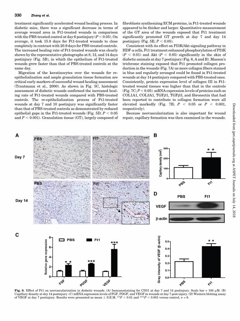
Fig. 8. Effect of Ft1 on neovascularization in diabetic wounds. (A) Immunostaining for CD31 at day 7 and 14 postinjury. Scale bar = 100 mM. (B)Capillary density at day 14 postinjury. (C) mRNA expression levels of FGF, PDGF, and VEGF in wounds at day 7 post-injury. (D) Western blotting assayof VEGF at day 7 postinjury. Results were presented as mean 6 S.E.M. **P , 0.01 and ***P , 0.001 versus control, n = 6.
330 Zhang et al.
at ASPE
T Journals on July 14, 2018
jpet.aspetjournals.orgD
ownloaded from
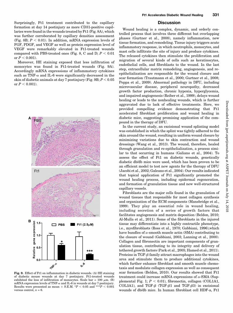
Surprisingly, Ft1 treatment contributed to the capillaryformation at day 14 postinjury as more CD31-positive capil-larieswere found in thewounds treated by Ft1 (Fig. 8A), whichwas further corroborated by capillary densities assessment(Fig. 8B; P , 0.01). In addition, mRNA expression levels ofFGF, PDGF, and VEGF as well as protein expression level ofVEGF were remarkably elevated in Ft1-treated woundscompared with PBS-treated ones (Fig. 8, C and D; P , 0.01or P , 0.001).Moreover, HE staining exposed that less infiltration of
monocytes was found in Ft1-treated wounds (Fig. 9A).Accordingly mRNA expressions of inflammatory cytokinessuch as TNF-a and IL-6 were significantly decreased in theskin of diabetic animals at day 7 postinjury (Fig. 9B; P, 0.05or P , 0.001).
DiscussionWound healing is a complex, dynamic, and orderly con-
trolled process that involves three different but overlappingphases (Gurtner et al., 2008), namely inflammation, newtissue formation, and remodeling. Tissue injury triggers acuteinflammatory response, in which neutrophils, monocytes, andmast cells infiltrate the site of injury and produce cytokines.The released cytokines then stimulate the proliferation andmigration of several kinds of cells such as keratinocytes,endothelial cells, and fibroblasts to the wound. In the laststep, extracellular matrix remodeling, angiogenesis, and re-epithelialization are responsible for the wound closure andscar formation (Trautmann et al., 2000; Gurtner et al., 2008;Peppa et al., 2009). Abnormal pathology in DFU, includingmicrovascular disease, peripheral neuropathy, decreasedgrowth factor production, chronic hypoxia, hyperglycemia,and impaired angiogenesis (Reiber et al., 1999), delays woundhealing or leads to the nonhealing wounds, which is furtheraggravated due to lack of effective treatments. Here, weprovided compelling evidence demonstrating that Ft1accelerated fibroblast proliferation and wound healing indiabetic mice, suggesting promising application of the com-pound in the therapy of DFU.In the current study, an excisional wound splinting model
was established in which the splint was tightly adhered to theskin around the wound, resulting in uniformwound closure byminimizing variations due to skin contraction and wounddressings (Wang et al., 2013). The wound, therefore, healedthrough granulation and re-epithelialization, a process simi-lar to that occurring in humans (Galiano et al., 2004). Toassess the effect of Ft1 on diabetic wounds, geneticallydiabetic db/db mice were used, which has been proven to bean efficient model to test new agents for the therapy of DFU(Jacobi et al., 2002; Galeano et al., 2004). Our results indicatedthat topical application of Ft1 significantly promoted thewound healing process, including epidermal regeneration,and formation of granulation tissue and new well-structuredcapillary vessels.Fibroblasts are the major cells found in the granulation of
wound tissues that responsible for most collagen synthesisand organization of the ECM components (Mansbridge et al.,1999). They play an essential role in wound healing,including secretion of a series of growth factors thatfacilitates angiogenesis and matrix deposition (Beldon, 2010;Al-Mulla et al., 2011). Some of the fibroblasts in the injuredtissue may differentiate into a highly contractile phenotype,i.e., myofibroblasts (Ross et al., 1970; Gabbiani, 1996),whichhave bundles of a-smooth muscle actin (SMA) contributing tothe closure of wound (Gabbiani, 2003; Lanning et al., 2000).Collagen and fibronectin are important components of gran-ulation tissue, contributing to its integrity and delivery oftethered growth factors (Park et al., 2005; Hamed et al., 2011).Proteins in TGF-b family attract macrophages into the woundarea and stimulate them to produce additional cytokines,which further enhance fibroblast and smooth muscle chemo-taxis and modulate collagen expression as well as consequentscar formation (Beldon, 2010). Our results showed that Ft1treatment could increase mRNA expressions of a-SMA (Sup-plemental Fig. 1; P , 0.01), fibronectin, collagen (COL1A1,COL3A1), and TGF-b (TGF-b1 and TGF-b3) in excisionalwounds of db/db mice. In human fibroblast cell HDF-a, Ft1
Fig. 9. Effect of Ft1 on inflammation in diabetic wounds. (A) HE stainingof diabetic mouse wounds at day 7 postinjury. Ft1-treated woundsexhibited the less of infiltration of monocytes. Scale bar = 100 mm. (B)mRNA expression levels of TNF-a and IL-6 in wounds at day 7 postinjury.Results were presented as mean 6 S.E.M. *P , 0.05 and ***P , 0.001versus control, n = 6.
Ft1 Accelerates Diabetic Wound Healing 331
at ASPE
T Journals on July 14, 2018
jpet.aspetjournals.orgD
ownloaded from
also could enhance mRNA expressions of COL1A1 andCOL3A1 as well as protein expressions of collagen I and III,which was mediated by PI3K/Akt/mTOR signaling pathway.Our findings suggested that Ft1 stimulated fibroblast pro-liferation and myofibroblast differentiation in the wound andthus accelerated wound closure.Neovascularization is considered to play a crucial patho-
physiological role in wound repair (Martin, 1997; Falanga,2005; Gurtner et al., 2008). The formation of new blood vesselsis essential to nourish the newly formed granulation tissueand the survival of keratinocytes. Our previous study showedthat Ft1 induces proliferation, migration, and tube formationof cultured human umbilical vein endothelial cells (Shen et al.,2012), which are used extensively as an in vitro model forangiogenesis research (Park et al., 2006). The proliferation,migration, and formation of tubular structure of endothelialcells are the indications for the development of new bloodvessels from pre-existing vascular bed in angiogenesis (Holashet al., 1999; Lamalice et al., 2007). In this study, Ft1-treatedwounds enhanced capillary density, suggesting that Ft1boosted local vessel growth in the wounds. Notably, Ft1induced significantly increased levels of proangiogenic mole-cules, such as VEGF, PDGF, and bFGF, in the wounds or inHDF-a cells (Supplemental Fig. 2; P , 0.05), which might bepartially responsible for its wound-healing function.Excessive inflammation, associated with a prolonged per-
sistence of neutrophil infiltration, is a consistent feature ofdiabetes-impaired wound healing (Nussler and Billiar, 1993).Some agents with an inhibitory ability in response to theinflammation, such as EPO, were shown to be effective intreating diabetic wounds (Eming et al., 2007). In the presentstudy, topical Ft1 treatment could reduce infiltration ofmonocytes and suppress the proinflammatory cytokines,namely IL-6 and TNF-a, in diabetic wound beds, implyingan inhibitory effect on the inflammation in diabetic wounds.In summary, we demonstrated that Ft1 enhanced fibroblast
proliferation via activating of PI3K/Akt/mTOR signalingpathway.Moreover, Ft1 accelerated wound healing in diabeticmice by orchestrating multifaceted factors in promoting re-epithelialization, granulation tissue formation, synthesis ofcollagen, angiogenesis, and preventing excessive inflamma-tory response. All of these results suggested potential appli-cation of Ft1 in the clinical therapy of DFU.
Acknowledgments
The authors thank all members of Dr. Wang’s, Dr. Wu’s, andDr. Yang’s laboratories at Shanghai University of Traditional ChineseMedicine for their helpful feedback and suggestions.
Authorship Contributions:
Participated in research design: Zhang, Gao, Yang, Wu, and Wang.Conducted experiments: Zhang and Gao.Performed data analysis: Zhang and Gao.Wrote or contributed to the writing of the manuscript: Zhang, Yang,
Wu, and Wang.
References
Al-Mulla F, Leibovich SJ, Francis IM, and Bitar MS (2011) Impaired TGF-b signalingand a defect in resolution of inflammation contribute to delayed wound healing in afemale rat model of type 2 diabetes. Mol Biosyst 7:3006–3020.
Beldon P (2010) Basic science of wound healing. Surgery 28:409–412.Boulton AJM, Vileikyte L, Ragnarson-Tennvall G, and Apelqvist J (2005) The globalburden of diabetic foot disease. Lancet 366:1719–1724.
Brem H and Tomic-Canic M (2007) Cellular and molecular basis of wound healing indiabetes. J Clin Invest 117:1219–1222.
Chen JT, Li HZ, Wang D, Zhang YJ, and Yang CR (2006) New dammarane mono-desmosides from the acidic deglycosylation of notoginseng-leaf saponins. HelvChim Acta 89:1442–1448.
Chen QS (1987) [Pharmacological studies on notoginseng saponins isolated from thefibrous root of Panax notoginseng]. Zhong Yao Tong Bao 12:45–47.
Eming SA, Krieg T, and Davidson JM (2007) Inflammation in wound repair: mo-lecular and cellular mechanisms. J Invest Dermatol 127:514–525.
Falanga V (2005) Wound healing and its impairment in the diabetic foot. Lancet 366:1736–1743.
Gabbiani G (1996) The cellular derivation and the life span of the myofibroblast.Pathol Res Pract 192:708–711.
Gabbiani G (2003) The myofibroblast in wound healing and fibrocontractive diseases.J Pathol 200:500–503.
Galeano M, Altavilla D, Cucinotta D, Russo GT, Calò M, Bitto A, Marini H, Marini R,Adamo EB, and Seminara P, et al. (2004) Recombinant human erythropoietinstimulates angiogenesis and wound healing in the genetically diabetic mouse.Diabetes 53:2509–2517.
Galiano RD, Michaels J, 5th, Dobryansky M, Levine JP, and Gurtner GC (2004)Quantitative and reproducible murine model of excisional wound healing. WoundRepair Regen 12:485–492.
Gao B, Huang L, Liu H, Wu H, Zhang E, Yang L, Wu X, and Wang Z (2014) PlateletP2Y₁₂ receptors are involved in the haemostatic effect of notoginsenoside Ft1, asaponin isolated from Panax notoginseng. Br J Pharmacol 171:214–223.
Griffith LG and Naughton G (2002) Tissue engineering–current challenges andexpanding opportunities. Science 295:1009–1014.
Gurtner GC, Werner S, Barrandon Y, and Longaker MT (2008) Wound repair andregeneration. Nature 453:314–321.
Hamed S, Ullmann Y, Egozi D, Daod E, Hellou E, Ashkar M, Gilhar A, and Teot L(2011) Fibronectin potentiates topical erythropoietin-induced wound repair in di-abetic mice. J Invest Dermatol 131:1365–1374.
Holash J, Wiegand SJ, and Yancopoulos GD (1999) New model of tumor angiogen-esis: dynamic balance between vessel regression and growth mediated by angio-poietins and VEGF. Oncogene 18:5356–5362.
Lamalice L, Le Boeuf F, and Huot J (2007) Endothelial cell migration during an-giogenesis. Circ Res 100:782–794.
Lanning DA, Diegelmann RF, Yager DR, Wallace ML, Bagwell CE, and Haynes JH(2000) Myofibroblast induction with transforming growth factor-beta1 and -beta3in cutaneous fetal excisional wounds. J Pediatr Surg 35:183–188.
Jacobi J, Jang JJ, Sundram U, Dayoub H, Fajardo LF, and Cooke JP (2002) Nicotineaccelerates angiogenesis and wound healing in genetically diabetic mice. Am JPathol 161:97–104.
Mansbridge JN, Liu K, Pinney RE, Patch R, Ratcliffe A, and Naughton GK (1999)Growth factors secreted by fibroblasts: role in healing diabetic foot ulcers. DiabetesObes Metab 1:265–279.
Martin P (1997) Wound healing–aiming for perfect skin regeneration. Science 276:75–81.
Nussler AK and Billiar TR (1993) Inflammation, immunoregulation, and induciblenitric oxide synthase. J Leukoc Biol 54:171–178.
Park HJ, Zhang Y, Georgescu SP, Johnson KL, Kong D, and Galper JB (2006) Humanumbilical vein endothelial cells and human dermal microvascular endothelial cellsoffer new insights into the relationship between lipid metabolism and angiogene-sis. Stem Cell Rev 2:93–102.
Park SG, Shin H, Shin YK, Lee Y, Choi EC, Park BJ, and Kim S (2005) The novelcytokine p43 stimulates dermal fibroblast proliferation and wound repair. Am JPathol 166:387–398.
Peppa M, Stavroulakis P, and Raptis SA (2009) Advanced glycoxidation products andimpaired diabetic wound healing. Wound Repair Regen 17:461–472.
Ramsey SD, Newton K, Blough D, McCulloch DK, Sandhu N, Reiber GE, and WagnerEH (1999) Incidence, outcomes, and cost of foot ulcers in patients with diabetes.Diabetes Care 22:382–387.
Reiber GE, Vileikyte L, Boyko EJ, del Aguila M, Smith DG, Lavery LA, and BoultonAJ (1999) Causal pathways for incident lower-extremity ulcers in patients withdiabetes from two settings. Diabetes Care 22:157–162.
Ross R, Everett NB, and Tyler R (1970) Wound healing and collagen formation.VI. The origin of the wound fibroblast studied in parabiosis. J Cell Biol 44:645–654.
Shen K, Ji L, Gong C, Ma Y, Yang L, Fan Y, Hou M, and Wang Z (2012) Noto-ginsenoside Ft1 promotes angiogenesis via HIF-1a mediated VEGF secretion andthe regulation of PI3K/AKT and Raf/MEK/ERK signaling pathways. BiochemPharmacol 84:784–792.
Trautmann A, Toksoy A, Engelhardt E, Bröcker EB, and Gillitzer R (2000) Mast cellinvolvement in normal human skin wound healing: expression of monocyte che-moattractant protein-1 is correlated with recruitment of mast cells which synthe-size interleukin-4 in vivo. J Pathol 190:100–106.
Wang X, Ge J, Tredget EE, and Wu Y (2013) The mouse excisional wound splintingmodel, including applications for stem cell transplantation. Nat Protoc 8:302–309.
Address correspondence to: Dr. Xiaojun Wu, Institute of Chinese MateriaMedica, Shanghai University of Traditional Chinese Medicine, 201203,Shanghai, P.R.China. E-mail: [email protected]; or Dr. Zhengtao Wang,Institute of Chinese Materia Medica, Shanghai University of TraditionalChinese Medicine, 201203, Shanghai, P.R.China. E-mail: [email protected]
332 Zhang et al.
at ASPE
T Journals on July 14, 2018
jpet.aspetjournals.orgD
ownloaded from