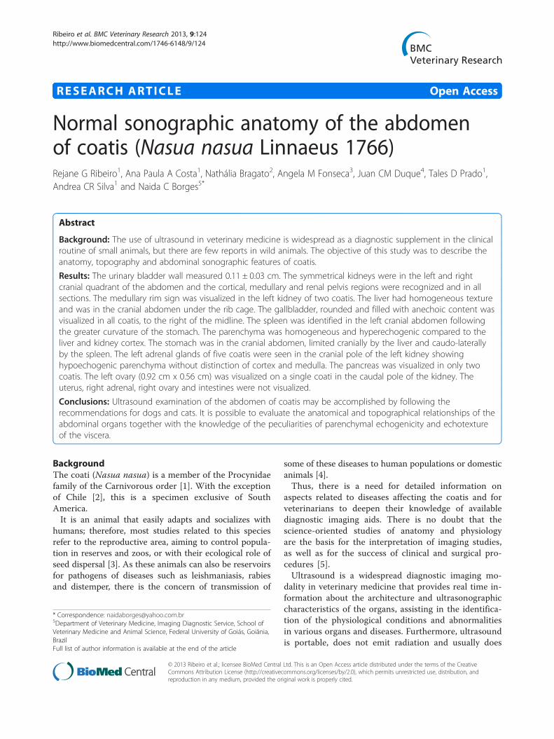
Normal sonographic anatomy of the abdomen of coatis (Nasua ...
10
RESEARCH ARTICLE Open Access Normal sonographic anatomy of the abdomen of coatis (Nasua nasua Linnaeus 1766) Rejane G Ribeiro 1 , Ana Paula A Costa 1 , Nathália Bragato 2 , Angela M Fonseca 3 , Juan CM Duque 4 , Tales D Prado 1 , Andrea CR Silva 1 and Naida C Borges 5* Abstract Background: The use of ultrasound in veterinary medicine is widespread as a diagnostic supplement in the clinical routine of small animals, but there are few reports in wild animals. The objective of this study was to describe the anatomy, topography and abdominal sonographic features of coatis. Results: The urinary bladder wall measured 0.11 ± 0.03 cm. The symmetrical kidneys were in the left and right cranial quadrant of the abdomen and the cortical, medullary and renal pelvis regions were recognized and in all sections. The medullary rim sign was visualized in the left kidney of two coatis. The liver had homogeneous texture and was in the cranial abdomen under the rib cage. The gallbladder, rounded and filled with anechoic content was visualized in all coatis, to the right of the midline. The spleen was identified in the left cranial abdomen following the greater curvature of the stomach. The parenchyma was homogeneous and hyperechogenic compared to the liver and kidney cortex. The stomach was in the cranial abdomen, limited cranially by the liver and caudo-laterally by the spleen. The left adrenal glands of five coatis were seen in the cranial pole of the left kidney showing hypoechogenic parenchyma without distinction of cortex and medulla. The pancreas was visualized in only two coatis. The left ovary (0.92 cm x 0.56 cm) was visualized on a single coati in the caudal pole of the kidney. The uterus, right adrenal, right ovary and intestines were not visualized. Conclusions: Ultrasound examination of the abdomen of coatis may be accomplished by following the recommendations for dogs and cats. It is possible to evaluate the anatomical and topographical relationships of the abdominal organs together with the knowledge of the peculiarities of parenchymal echogenicity and echotexture of the viscera. Background The coati (Nasua nasua) is a member of the Procynidae family of the Carnivorous order [1]. With the exception of Chile [2], this is a specimen exclusive of South America. It is an animal that easily adapts and socializes with humans; therefore, most studies related to this species refer to the reproductive area, aiming to control popula- tion in reserves and zoos, or with their ecological role of seed dispersal [3]. As these animals can also be reservoirs for pathogens of diseases such as leishmaniasis, rabies and distemper, there is the concern of transmission of some of these diseases to human populations or domestic animals [4]. Thus, there is a need for detailed information on aspects related to diseases affecting the coatis and for veterinarians to deepen their knowledge of available diagnostic imaging aids. There is no doubt that the science-oriented studies of anatomy and physiology are the basis for the interpretation of imaging studies, as well as for the success of clinical and surgical pro- cedures [5]. Ultrasound is a widespread diagnostic imaging mo- dality in veterinary medicine that provides real time in- formation about the architecture and ultrasonographic characteristics of the organs, assisting in the identifica- tion of the physiological conditions and abnormalities in various organs and diseases. Furthermore, ultrasound is portable, does not emit radiation and usually does * Correspondence: [email protected] 5 Department of Veterinary Medicine, Imaging Diagnostic Service, School of Veterinary Medicine and Animal Science, Federal University of Goiás, Goiânia, Brazil Full list of author information is available at the end of the article © 2013 Ribeiro et al.; licensee BioMed Central Ltd. This is an Open Access article distributed under the terms of the Creative Commons Attribution License (http://creativecommons.org/licenses/by/2.0), which permits unrestricted use, distribution, and reproduction in any medium, provided the original work is properly cited. Ribeiro et al. BMC Veterinary Research 2013, 9:124 http://www.biomedcentral.com/1746-6148/9/124
Transcript of Normal sonographic anatomy of the abdomen of coatis (Nasua ...
Ribeiro et al. BMC Veterinary Research 2013, 9:124http://www.biomedcentral.com/1746-6148/9/124
RESEARCH ARTICLE Open Access
Normal sonographic anatomy of the abdomenof coatis (Nasua nasua Linnaeus 1766)Rejane G Ribeiro1, Ana Paula A Costa1, Nathália Bragato2, Angela M Fonseca3, Juan CM Duque4, Tales D Prado1,Andrea CR Silva1 and Naida C Borges5*
Abstract
Background: The use of ultrasound in veterinary medicine is widespread as a diagnostic supplement in the clinicalroutine of small animals, but there are few reports in wild animals. The objective of this study was to describe theanatomy, topography and abdominal sonographic features of coatis.
Results: The urinary bladder wall measured 0.11 ± 0.03 cm. The symmetrical kidneys were in the left and rightcranial quadrant of the abdomen and the cortical, medullary and renal pelvis regions were recognized and in allsections. The medullary rim sign was visualized in the left kidney of two coatis. The liver had homogeneous textureand was in the cranial abdomen under the rib cage. The gallbladder, rounded and filled with anechoic content wasvisualized in all coatis, to the right of the midline. The spleen was identified in the left cranial abdomen followingthe greater curvature of the stomach. The parenchyma was homogeneous and hyperechogenic compared to theliver and kidney cortex. The stomach was in the cranial abdomen, limited cranially by the liver and caudo-laterallyby the spleen. The left adrenal glands of five coatis were seen in the cranial pole of the left kidney showinghypoechogenic parenchyma without distinction of cortex and medulla. The pancreas was visualized in only twocoatis. The left ovary (0.92 cm x 0.56 cm) was visualized on a single coati in the caudal pole of the kidney. Theuterus, right adrenal, right ovary and intestines were not visualized.
Conclusions: Ultrasound examination of the abdomen of coatis may be accomplished by following therecommendations for dogs and cats. It is possible to evaluate the anatomical and topographical relationships of theabdominal organs together with the knowledge of the peculiarities of parenchymal echogenicity and echotextureof the viscera.
BackgroundThe coati (Nasua nasua) is a member of the Procynidaefamily of the Carnivorous order [1]. With the exceptionof Chile [2], this is a specimen exclusive of SouthAmerica.It is an animal that easily adapts and socializes with
humans; therefore, most studies related to this speciesrefer to the reproductive area, aiming to control popula-tion in reserves and zoos, or with their ecological role ofseed dispersal [3]. As these animals can also be reservoirsfor pathogens of diseases such as leishmaniasis, rabiesand distemper, there is the concern of transmission of
* Correspondence: [email protected] of Veterinary Medicine, Imaging Diagnostic Service, School ofVeterinary Medicine and Animal Science, Federal University of Goiás, Goiânia,BrazilFull list of author information is available at the end of the article
© 2013 Ribeiro et al.; licensee BioMed CentralCommons Attribution License (http://creativecreproduction in any medium, provided the or
some of these diseases to human populations or domesticanimals [4].Thus, there is a need for detailed information on
aspects related to diseases affecting the coatis and forveterinarians to deepen their knowledge of availablediagnostic imaging aids. There is no doubt that thescience-oriented studies of anatomy and physiologyare the basis for the interpretation of imaging studies,as well as for the success of clinical and surgical pro-cedures [5].Ultrasound is a widespread diagnostic imaging mo-
dality in veterinary medicine that provides real time in-formation about the architecture and ultrasonographiccharacteristics of the organs, assisting in the identifica-tion of the physiological conditions and abnormalitiesin various organs and diseases. Furthermore, ultrasoundis portable, does not emit radiation and usually does
Ltd. This is an Open Access article distributed under the terms of the Creativeommons.org/licenses/by/2.0), which permits unrestricted use, distribution, andiginal work is properly cited.
Ribeiro et al. BMC Veterinary Research 2013, 9:124 Page 2 of 10http://www.biomedcentral.com/1746-6148/9/124
not require general anesthesia, except in wildlife animals.It can be used in clinics, laboratories and field, but its useis still limited in wild animals, due to the limited know-ledge of the topography and ultrasound anatomy of theirorgans [6].According Peixoto et al. [6] and Nyland et al. [7] it is
essential that the sonographer has an extensive know-ledge of anatomy, physiology and pathophysiology of thestudied species, because knowledge of the topographyand ultrasound anatomy of organs to be examined areprerequisites for an ultrasound, given that the accurateinterpretation depends directly on the differentiationbetween normal and abnormal structures.The aim of this study was to identify a technique for
abdominal ultrasonographic examination of the coatisand to describe the normal ultrasonographic anatomy ofits abdominal organs.
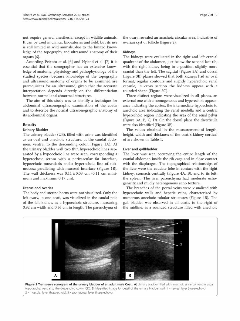
ResultsUrinary BladderThe urinary bladder (UB), filled with urine was identifiedas an oval and anechoic structure, at the caudal abdo-men, ventral to the descending colon (Figure 1A). Atthe urinary bladder wall two thin hyperechoic lines sep-arated by a hypoechoic line were seen, corresponding ahyperechoic serosa with a perivascular fat interface,hypoechoic muscularis and a hyperechoic line of sub-mucosa paralleling with mucosal interface (Figure 1B).The wall thickness was 0.11 ± 0.03 cm (0.11 cm mini-mum and maximum 0.17 cm).
Uterus and ovariesThe body and uterine horns were not visualized. Only theleft ovary, in one coati, was visualized in the caudal poleof the left kidney, as a hypoechoic structure, measuring0.92 cm width and 0.56 cm in length. The parenchyma of
Figure 1 Transverse sonogram of the urinary bladder of an adult maltopography, ventral to the descending colon (CD). B) Magnified image for2 - muscular layer (hypoechoic), 3 - submucosal layer (hyperechoic).
the ovary revealed an anachoic circular area, indicative ofovarian cyst or follicle (Figure 2).
KidneysThe kidneys were evaluated in the right and left cranialquadrant of the abdomen, just below the second last rib,with the right kidney being in a position slightly morecranial than the left. The sagittal (Figure 3A) and dorsal(Figure 3B) planes showed that both kidneys had an ovalformat, regular contours and slightly hyperechoic renalcapsule, in cross section the kidneys appear with arounded shape (Figure 3C).Three distinct regions were visualized in all planes, an
external one with a homogeneous and hyperechoic appear-ance indicating the cortex, the intermediate hypoechoic toanechoic area indicating the renal medulla and a centralhyperechoic region indicating the area of the renal pelvis(Figure 3A, B, C, D). On the dorsal plane the diverticulawere also identified (Figure 3B).The values obtained in the measurement of length,
height, width and thickness of the coati’s kidney corticalof are shown in Table 1.
Liver and gallbladderThe liver was seen occupying the entire length of thecranial abdomen inside the rib cage and in close contactwith the diaphragm. The topographical relationships ofthe liver were the caudate lobe in contact with the rightkidney, stomach centrally (Figure 4A, B), and to its left,the spleen. The liver parenchyma had moderate echo-genicity and mildly heterogenous echo texture.The branches of the portal veins were visualized with
hyperechoic walls and hepatic veins, characterized bynumerous anechoic tubular structures (Figure 4B). Thegall bladder was observed in all coatis to the right ofthe midline, as a rounded structure filled with anechoic
e Coati. A) Urinary bladder filled with anechoic urine content in usualdetail of the urinary bladder wall, 1 – serosal layer (hyperechoic),
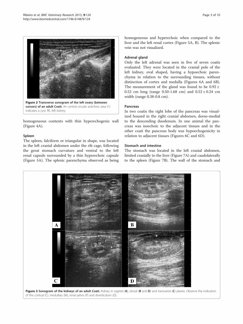
Figure 2 Transverse sonogram of the left ovary (betweencursors) of an adult Coati. An central circular anechoic area (1)indicates a cyst. RE, left kidney.
Ribeiro et al. BMC Veterinary Research 2013, 9:124 Page 3 of 10http://www.biomedcentral.com/1746-6148/9/124
homogeneous contents with thin hyperechogenic wall(Figure 4A).
SpleenThe spleen, falciform or triangular in shape, was locatedin the left cranial abdomen under the rib cage, followingthe great stomach curvature and ventral to the leftrenal capsule surrounded by a thin hyperechoic capsule(Figure 5A). The splenic parenchyma observed as being
Figure 3 Sonogram of the kidneys of an adult Coati. Kidney in sagittalof the cortical (C), medullary (M), renal pelvis (P) and diverticulum (D).
homogeneous and hyperechoic when compared to theliver and the left renal cortex (Figure 5A, B). The splenicvein was not visualized.
Adrenal glandOnly the left adrenal was seen in five of seven coatisevaluated. They were located in the cranial pole of theleft kidney, oval shaped, having a hypoechoic paren-chyma in relation to the surrounding tissues, withoutdistinction of cortex and medulla (Figures 6A and 6B).The measurement of the gland was found to be 0.92 ±0.52 cm long (range 0.50-1.68 cm) and 0.52 ± 0.24 cmwidth (range 0.38-0.8 cm).
PancreasIn two coatis the right lobe of the pancreas was visual-ized housed in the right cranial abdomen, dorso-medialto the descending duodenum. In one animal the pan-creas was isoechoic to the adjacent tissues and in theother coati the pancreas body was hypoechogenicity inrelation to adjacent tissues (Figures 6C and 6D).
Stomach and intestineThe stomach was located in the left cranial abdomen,limited cranially to the liver (Figure 7A) and caudolaterallyto the spleen (Figure 7B). The wall of the stomach and
(A), dorsal (B and D) and transverse (C) planes. Observe the indication
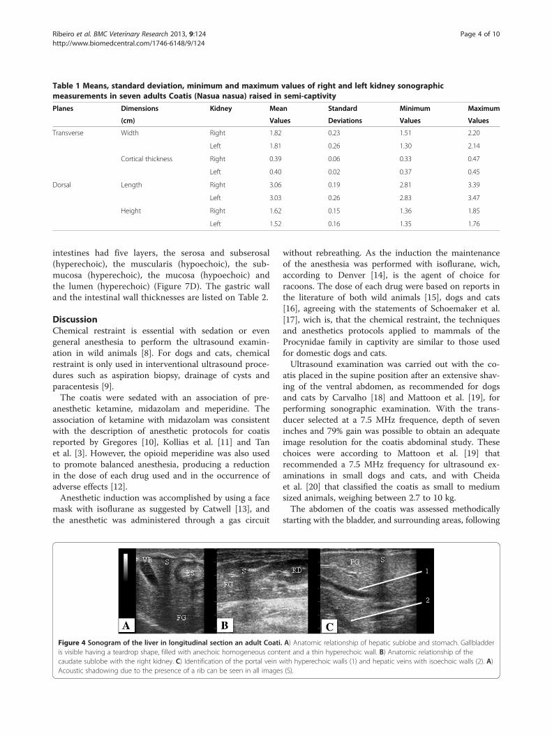
Table 1 Means, standard deviation, minimum and maximum values of right and left kidney sonographicmeasurements in seven adults Coatis (Nasua nasua) raised in semi-captivity
Planes Dimensions Kidney Mean Standard Minimum Maximum
(cm) Values Deviations Values Values
Transverse Width Right 1.82 0.23 1.51 2.20
Left 1.81 0.26 1.30 2.14
Cortical thickness Right 0.39 0.06 0.33 0.47
Left 0.40 0.02 0.37 0.45
Dorsal Length Right 3.06 0.19 2.81 3.39
Left 3.03 0.26 2.83 3.47
Height Right 1.62 0.15 1.36 1.85
Left 1.52 0.16 1.35 1.76
Ribeiro et al. BMC Veterinary Research 2013, 9:124 Page 4 of 10http://www.biomedcentral.com/1746-6148/9/124
intestines had five layers, the serosa and subserosal(hyperechoic), the muscularis (hypoechoic), the sub-mucosa (hyperechoic), the mucosa (hypoechoic) andthe lumen (hyperechoic) (Figure 7D). The gastric walland the intestinal wall thicknesses are listed on Table 2.
DiscussionChemical restraint is essential with sedation or evengeneral anesthesia to perform the ultrasound examin-ation in wild animals [8]. For dogs and cats, chemicalrestraint is only used in interventional ultrasound proce-dures such as aspiration biopsy, drainage of cysts andparacentesis [9].The coatis were sedated with an association of pre-
anesthetic ketamine, midazolam and meperidine. Theassociation of ketamine with midazolam was consistentwith the description of anesthetic protocols for coatisreported by Gregores [10], Kollias et al. [11] and Tanet al. [3]. However, the opioid meperidine was also usedto promote balanced anesthesia, producing a reductionin the dose of each drug used and in the occurrence ofadverse effects [12].Anesthetic induction was accomplished by using a face
mask with isoflurane as suggested by Catwell [13], andthe anesthetic was administered through a gas circuit
Figure 4 Sonogram of the liver in longitudinal section an adult Coatiis visible having a teardrop shape, filled with anechoic homogeneous contcaudate sublobe with the right kidney. C) Identification of the portal vein wAcoustic shadowing due to the presence of a rib can be seen in all images
without rebreathing. As the induction the maintenanceof the anesthesia was performed with isoflurane, wich,according to Denver [14], is the agent of choice forracoons. The dose of each drug were based on reports inthe literature of both wild animals [15], dogs and cats[16], agreeing with the statements of Schoemaker et al.[17], wich is, that the chemical restraint, the techniquesand anesthetics protocols applied to mammals of theProcynidae family in captivity are similar to those usedfor domestic dogs and cats.Ultrasound examination was carried out with the co-
atis placed in the supine position after an extensive shav-ing of the ventral abdomen, as recommended for dogsand cats by Carvalho [18] and Mattoon et al. [19], forperforming sonographic examination. With the trans-ducer selected at a 7.5 MHz frequence, depth of seveninches and 79% gain was possible to obtain an adequateimage resolution for the coatis abdominal study. Thesechoices were according to Mattoon et al. [19] thatrecommended a 7.5 MHz frequency for ultrasound ex-aminations in small dogs and cats, and with Cheidaet al. [20] that classified the coatis as small to mediumsized animals, weighing between 2.7 to 10 kg.The abdomen of the coatis was assessed methodically
starting with the bladder, and surrounding areas, following
. A) Anatomic relationship of hepatic sublobe and stomach. Gallbladderent and a thin hyperechoic wall. B) Anatomic relationship of theith hyperechoic walls (1) and hepatic veins with isoechoic walls (2). A)(S).
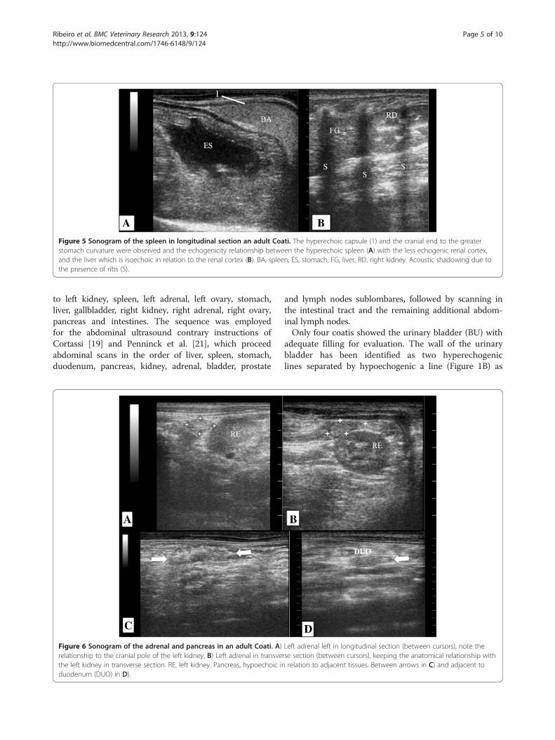
Figure 5 Sonogram of the spleen in longitudinal section an adult Coati. The hyperechoic capsule (1) and the cranial end to the greaterstomach curvature were observed and the echogenicity relationship between the hyperechoic spleen (A) with the less echogenic renal cortex,and the liver which is isoechoic in relation to the renal cortex (B). BA, spleen, ES, stomach, FG, liver, RD, right kidney. Acoustic shadowing due tothe presence of ribs (S).
Ribeiro et al. BMC Veterinary Research 2013, 9:124 Page 5 of 10http://www.biomedcentral.com/1746-6148/9/124
to left kidney, spleen, left adrenal, left ovary, stomach,liver, gallbladder, right kidney, right adrenal, right ovary,pancreas and intestines. The sequence was employedfor the abdominal ultrasound contrary instructions ofCortassi [19] and Penninck et al. [21], which proceedabdominal scans in the order of liver, spleen, stomach,duodenum, pancreas, kidney, adrenal, bladder, prostate
Figure 6 Sonogram of the adrenal and pancreas in an adult Coati. A)relationship to the cranial pole of the left kidney, B) Left adrenal in transvethe left kidney in transverse section. RE, left kidney. Pancreas, hypoechoic induodenum (DUO) in D).
and lymph nodes sublombares, followed by scanning inthe intestinal tract and the remaining additional abdom-inal lymph nodes.Only four coatis showed the urinary bladder (BU) with
adequate filling for evaluation. The wall of the urinarybladder has been identified as two hyperechogeniclines separated by hypoechogenic a line (Figure 1B) as
Left adrenal left in longitudinal section (between cursors), note therse section (between cursors), keeping the anatomical relationship withrelation to adjacent tissues. Between arrows in C) and adjacent to
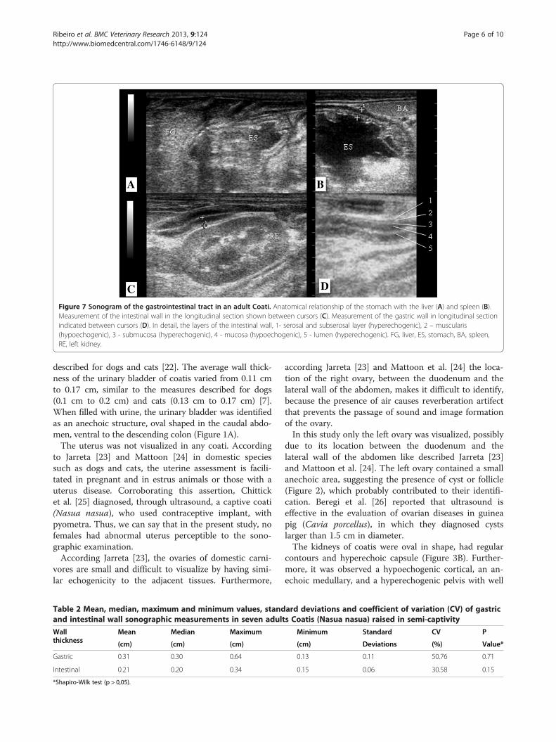
Figure 7 Sonogram of the gastrointestinal tract in an adult Coati. Anatomical relationship of the stomach with the liver (A) and spleen (B).Measurement of the intestinal wall in the longitudinal section shown between cursors (C). Measurement of the gastric wall in longitudinal sectionindicated between cursors (D). In detail, the layers of the intestinal wall, 1- serosal and subserosal layer (hyperechogenic), 2 – muscularis(hypoechogenic), 3 - submucosa (hyperechogenic), 4 - mucosa (hypoechogenic), 5 - lumen (hyperechogenic). FG, liver, ES, stomach, BA, spleen,RE, left kidney.
Ribeiro et al. BMC Veterinary Research 2013, 9:124 Page 6 of 10http://www.biomedcentral.com/1746-6148/9/124
described for dogs and cats [22]. The average wall thick-ness of the urinary bladder of coatis varied from 0.11 cmto 0.17 cm, similar to the measures described for dogs(0.1 cm to 0.2 cm) and cats (0.13 cm to 0.17 cm) [7].When filled with urine, the urinary bladder was identifiedas an anechoic structure, oval shaped in the caudal abdo-men, ventral to the descending colon (Figure 1A).The uterus was not visualized in any coati. According
to Jarreta [23] and Mattoon [24] in domestic speciessuch as dogs and cats, the uterine assessment is facili-tated in pregnant and in estrus animals or those with auterus disease. Corroborating this assertion, Chitticket al. [25] diagnosed, through ultrasound, a captive coati(Nasua nasua), who used contraceptive implant, withpyometra. Thus, we can say that in the present study, nofemales had abnormal uterus perceptible to the sono-graphic examination.According Jarreta [23], the ovaries of domestic carni-
vores are small and difficult to visualize by having simi-lar echogenicity to the adjacent tissues. Furthermore,
Table 2 Mean, median, maximum and minimum values, standand intestinal wall sonographic measurements in seven adult
Wallthickness
Mean Median Maximum
(cm) (cm) (cm)
Gastric 0.31 0.30 0.64
Intestinal 0.21 0.20 0.34
*Shapiro-Wilk test (p > 0,05).
according Jarreta [23] and Mattoon et al. [24] the loca-tion of the right ovary, between the duodenum and thelateral wall of the abdomen, makes it difficult to identify,because the presence of air causes reverberation artifectthat prevents the passage of sound and image formationof the ovary.In this study only the left ovary was visualized, possibly
due to its location between the duodenum and thelateral wall of the abdomen like described Jarreta [23]and Mattoon et al. [24]. The left ovary contained a smallanechoic area, suggesting the presence of cyst or follicle(Figure 2), which probably contributed to their identifi-cation. Beregi et al. [26] reported that ultrasound iseffective in the evaluation of ovarian diseases in guineapig (Cavia porcellus), in which they diagnosed cystslarger than 1.5 cm in diameter.The kidneys of coatis were oval in shape, had regular
contours and hyperechoic capsule (Figure 3B). Further-more, it was observed a hypoechogenic cortical, an an-echoic medullary, and a hyperechogenic pelvis with well
ard deviations and coefficient of variation (CV) of gastrics Coatis (Nasua nasua) raised in semi-captivity
Minimum Standard CV P
(cm) Deviations (%) Value*
0.13 0.11 50.76 0.71
0.15 0.06 30.58 0.15
Ribeiro et al. BMC Veterinary Research 2013, 9:124 Page 7 of 10http://www.biomedcentral.com/1746-6148/9/124
defined corticomedullary relationship (Figure 3). Thesefindings resemble those observed in dogs and cats[27-29], in cheetahs (Cinonyx jubatus) [30] and incapuchin monkeys (Cebus apella) [31].In contrast, Wagner et al. [32] evaluating the kidneys
of marmoset, found that in this species there is a poorcorticomedullary distinction and the cortex is hyper-echogenic. According to Vac [22] these descriptions ob-served in marmosets indicate glomerular or interstitialnephritis in dogs and cats. Extrapolating this oppositionresults to those obtained with the coatis, it can be seenthat the interpretation of the findings in a sonographicexamination is entirely related to the knowledge of ultra-sonographic anatomy and that the unawareness can leadto misdiagnosis.As there is little variation in weight between coatis, as
observed in cats, the linear measurements are more use-ful than in dogs in which the measurement is made sub-jectively because of the variety of size and body volumein animal of equal weight [27]. According to Vac [22]measures are more appropriate to compare the kidneysof animals that must be symmetrical, as seen in coatis(Table 1).The medullary rim sign was identified in two left kid-
neys of coatis (Figure 3d). According to Vac [22] is not apathognomonic ultrasound finding of renal disease.However, Mantins and Lamb [33] and Nyland et al. [27]reported that this finding was observed in dogs and catswith kidney disease and in clinically healthy animals,being investigations necessary to determine the causesand significance of this sign. Carsten et al. [30] observedmedullary rim sign in 21 of 26 kidneys of healthy chee-tahs. These results lead to the fact that is necessary toknow the specific ultrasound appearance of the studiedsystem and animal species.Cranio-medial to left kidney and cranial to the left
renal artery we encountered the left adrenal of five coatis(Figure 6). Similarly, Chapman et al. [34] and Nylandet al. [35] emphasized the ease of identifying the leftadrenal in dogs and cats compared to the right due tothe anatomic location, described for the study of coatis.Moreover, Wagner et al. [32] observed the adrenals ofmarmosets in an anatomical position similar to that ofcarnivores, however both adrenals were easily foundwhen employed silicone cushion for the sonographicevaluation.The adrenal glands of coatis had oval contour (Figure 6)
as observed in cats, different from dogs that usually have aresembling peanut-shape the adrenal gland [34]. As forthe echogenicity the adrenal glands were found to behypoechogenic relative to surrounding tissue and it wasnot possible to distinguish between cortical and medullaryregions (Figure 6). Santos et al. [29] reported this featurein the parenchyma of the adrenal glands of puppies and
kittens, unlike adult dogs and cats which is commonlyvisualized corticomedullary distinction according toCarvalho et al. [34] and Nyland et al. [35]. Despite thefind of this study, to support this definition, we suggestthat a greater number of coatis must be evaluated, con-sidering that sample of adult animals used in this studywas small.The liver of coatis is located entirely below the rib
cage, being the left lobe to the left of the midline, thequadrate in the middle plane, the right lobe to the rightof the midline and the caudate process of caudate lobein contact with the cranial pole the right kidney (Figure 4B).The cranial edge of the liver is bounded by the dia-phragm, a hyperechoic curvilinear structure, and thecaudal margin by the stomach, similar as seen in dogsand cats (Figure 4A).The portal veins have hyperechoic walls unlike the
hepatic veins, which were isoechoic to the surroundingtissue (Figure 4C), features also observed in domesticanimals by Mamprim [36] and Nyland et al. [37]. Thegallbladder, round or teardrop shaped and with an an-echoic content (Figure 4A), preserved the characteristicsdescribed in quotations from studies in dogs and cats[37]. Interestingly, Wagner et al. [32] found that in mar-mosets the gallbladder has a bilobed silhouette, which isconsidered naturally found in domestic cats as men-tioned Mamprim [36] and Nyland et al. [37]. Again, theanatomical knowledge proved to be crucial for the inter-pretation of sonographic findings, and should be followedin this coatis study.In this study, the spleen was easily visualized due to its
size and superficial location, without interference of gasfrom the intestinal contents. Our findings differ fromAlves et al. [31] who found that the spleen was the mostdifficult organ more to be evaluated in capuchin monkeydue to its small size in this species, allowing the evalu-ation of the spleen only in one animal among the tenstudied. In coatis, the spleen was falciform or triangularshaped, with thin echogenic capsule (Figure 5A), and ofa homogeneous echo texture similar to the spleen of thedog and cat, according to the descriptions of Santos [29]and Tannouz et al. [38].Analysing the relationship of echogenicity of the triad,
spleen, liver and kidney, it was found that in all coatispresented the spleen was more echogenic than the liver,and the liver was isoechoic to the renal cortex (Figure 5B).These characteristics are also reported in domestic carni-vores by Tannouz et al. [38] and Nyland et al. [39] and incheetahs by Carsten et al. [30]. Contrary to this descrip-tion, all healthy marmosets evaluated by Wagner et al.[32] presented the spleen more hypoechoic than the liverand renal cortex.When the transducer was craniomedial directed to the
spleen we identified the stomach, caudal located to the
Ribeiro et al. BMC Veterinary Research 2013, 9:124 Page 8 of 10http://www.biomedcentral.com/1746-6148/9/124
hepatic parenchyma (Figure 7a) and cranial to the leftkidney, similar to what is described for dogs and cats byFroes [40]. The gastric and intestinal wall was easilyevaluated and presented five layers with differentechogenicity (Figure 7C, D) as also verified by Mattoonet al. [19] in domestic carnivores and Wagner et al. [32]in marmosets. The thickness of the stomach (Figure 7D)and intestine (Figure 7C) was also similar to that de-scribed for dogs and cats by Mattoon et al. [19] andFroes [40].The pancreas was observed in only two coatis. The
parenchyma presented isoechoic to the surroundingtissue (Figure 6C) and slightly hypoechoic (Figure 6D).These differences of echogenicity also were observed inhealthy dogs and cats [19]. This limitation for identifica-tion can be justified if we consider the remarks of Zardoet al. [41] about the fact that even though the method ofchoice for assessing the pancreatic body in veterinarymedicine is the sonographic examination, it has limita-tions regarding the quality of the ultrasound equipment,experience of the sonographer, similarity with echo-genicity of the surrounding tissues, interfering of gasesand food contents of the gastrointestinal tract. Wagneret al. [32] justify that they could not visualized of thepancreas in a study with marmosets because the limitedtime of the examination (30–40 minutes).Thus, it is believed that the interpretation of ultra-
sound examinations is directly related to the knowledgeof topographic anatomy, as well as the sonographic dis-tinction of the various abdominal organs, of the variousanimals’ species. Considering all these aspects, the expe-rienced sonographer may assist clinicians and surgeonsin diagnostic and prognostic interpretations.
ConclusionsUltrasound examination of the abdomen of coatis maybe accomplished by following what is recommended fordogs and cats. It is effective to evaluate the anatomicaland topographical relationships of the abdominal organstogether with the knowledge of the peculiarities of par-enchymal echogenicity and echo texture of the viscera.
MethodsThis study was conducted under the authorization of theBrazilian Institute of Environment and Natural Renew-able Resources (Instituto Brasileiro do Meio Ambiente edos Recursos Naturais Renováveis - IBAMA), under li-cense number 014/2011 and was approved by the EthicsCommittee on Animal Experimentation of Federal Uni-versity of Goiás registration number 118/11.The study was conducted in seven adults coatis con-
sidered clinically healthy, four females and three males,with average weight of 2.67 ± 0.54 kg. The animals werefrom the Center for Screening of Wild Animals (Centro
de Triagem de Animais Silvestres - CETAS), an IBAMAagency, located in Goiânia, Goiás, Brazil. The examswere realized in the Veterinary Hospital of the Veterin-ary and Animal Science School, Federal University ofGoiás.As recommended, the animals underwent a 12 hour
fasting. At the Veterinary Hospital, coatis were sedatedwith 15 mg/kg of ketamine hydrochloride (KetamineAgener 10% União Química Farmacêutica Nacional S/A,Embu-Guaçu- SP, Brazil) associated with 0.2 mg/kg ofmidazolam (Dormine 0.5% Cristália Produtos QuímicosFarmacêuticos Ltda, Itapira- Lindóia- SP, Brazil) and5 mg/kg meperidine hydrochloride (5% Dolosal, CristáliaProdutos Químicos Farmacêuticos Ltda, Itapira- Lindóia-SP, Brazil), all in the same syringe, intramuscularly.The intravenous catheter of 22 gauges was placed in
the cephalic or femoral vein for drug administration andfluid-Care with sterile Ringer's lactate at a dose of10 ml/kg/hour (Ringer's Lactate. Equiplex IndústriaFarmacêutica Ltda., Aparecida de Goiânia-Go, Brazil).Afterwards, an anesthetic protocol was established, inwhich the coatis were induced with oxygen (O2) mask at100% and a four liters per minute (L/min) flow, withthe vaporizer calibrated to provide 3.5% isoflurane(Isoflurano, Instituto Biochimico Indústria FarmacêuticaLtda, Itatiaia-RJ, Brazil) in a rebreathing circuit of gases,of the Baraka type. and calibrated vaporizer melhor tirar.After induction, the animals were intubated with an
endotracheal tube, internal diameter of three and a halfor four millimeters, and maintained under spontaneousventilation with a 1.0 inspired oxygen fraction (FiO2)and a 300 mL/kg volume of fresh gas.
Sonographic evaluationFor the sonographic evaluation the hair of the abdominalventral region, from the costal arch to the inguinalregion, of the coatis was clipping. Then a heated acous-tic gel was applied to the animals’ skin to preventhypothermia and to assist in acoustic contact betweenthe transducer and the patient. The animals were posi-tioned a supine position to perform the sonographicevaluation with the ultrasound equipment My Lab™ 30Vet (The Esaote Group, Genova, Italy) coupled to themultifrequency linear transducer (7.5 MHz to 12.0 MHz),selected for 7.5 MHz frequency and a 79% gain.The positioning of the transducer to the achievement
of the sagittal and transverse of the urinary bladder,spleen, liver, gall bladder, adrenal, ovary, pancreas andgastrointestinal tract, as well as the evaluation of con-tour, margin, size, texture and echogenicity of these or-gans followed the instructions Carvalho [18].The kidneys were evaluated in the longitudinal, trans-
verse and dorsal planes to determine the contour,margin, echo texture and echogenicity following the
Ribeiro et al. BMC Veterinary Research 2013, 9:124 Page 9 of 10http://www.biomedcentral.com/1746-6148/9/124
recommendations of Vac [22]. The measurements wereperformed on images obtained with the dorsal andtransverse planes. According Nyland et al. [27], from thedorsal plane the measures of length (distance betweenthe cranial and caudal poles) and height (distance be-tween the ventral and dorsal surfaces) was determined.From the transversal plane the kidney width (distancebetween the dorsal and ventral surfaces and corticalthickness (distance between the outer edge of the kidneyand renal capsule) were measured.
Statistical analysisDescriptive statistical analysis was performed for presen-tations of the measurements of abdominal organs evalu-ated. The Shapiro-Wilk test was used to determine thenormal range of wall thickness of stomach and intestine.The same test was not applied in other organs studied,since these were not visualized at all coatis (n < 5).
Competing interestThe authors declare that they have no competing interest.
Author's contributionsJCDM and AMF anesthetized the animals. RGR, NCB, APC and NB did thesonographic examinations. RGR and NCB have written the manuscript. RGR,APC, NCB, TDP, ACRS helped in the review. All authors read and approvedthe final manuscript.
AcknowledgementsWe would like to thank the Screening Center of Wild Animals (CETAS) andthe Brazilian Institute of Environment and Natural Renewable Resources(IBAMA), who provided the animals for the study and the Federal Universityof Goias, where the study was conducted and also the Ethics Committee onAnimal Experimentation of Federal University of Goiás who approved it.
Author details1Pos graduation degree in Animal Science, School of Veterinary Medicine,Federal University of Goiás, Goiânia, Brazil. 2Medical resident in VeterinaryImaging Diagnostic, School of Veterinary Medicine and Animal Science,Federal University of Goiás, Goiânia, Brazil. 3Medical resident in VeterinaryAnesthesiology, School of Veterinary Medicine and Animal Science, FederalUniversity of Goiás, Goiânia, Brazil. 4Department of Veterinary Medicine,Anesthesiology Service, School of Veterinary Medicine and Animal Science,Federal University of Goiás, Goiânia, Brazil. 5Department of VeterinaryMedicine, Imaging Diagnostic Service, School of Veterinary Medicine andAnimal Science, Federal University of Goiás, Goiânia, Brazil.
Received: 22 August 2012 Accepted: 17 June 2013Published: 23 June 2013
References1. Whiteside DP: Nutrition and behavior of coatis and raccoons. Vet Clin
North Am Exot Anim Pract 2009, 12:187–195.2. Beisiegel BM: Notes on the coati, nasua nasua (carnivora: procyonidae)
in an Atlantic forest area. Braz J Biol 2001, 61:689–692.3. Teixeira RHF, Ambrósio SR: Carnívora – procyonidae (quati, Mão-pelada,
jupará). In Tratado de animais selvagens – medicina veterinária. Capitulo 33.Edited by Roca. São Paulo: Chapman and Hall; 2007:571–583.
4. Jorge RSP, Rocha FL, Junior JAM, Morato RG: Ocorrência de patogenoscarnívoros selvagens brasileiros e suas implicações para a conservação esaúde pública. Oecologia Australis 2010, 14:686–710.
5. King AM: Development, advances and applications of diagnosticultrasound in animals. Vet J 2006, 171:408–420.
6. Peixoto GCX, Lira RA, Alves ND, Silva AR: Bases físicas de formação deimagem ultrassonográfica. Acta Veterinária Brasilica 2010, 4:15–24.
7. Nyland TG, Mattoon JS, Herrsell EJ, Wisner ER: Princípios Físicos,instrumentação e segurança do diagnóstico por ultra-som. In Ultra-somdiagnóstico em pequenos animais. Capitulo 2. 2nd edition. Edited by Roca.São Paulo: Chapman and Hall; 2005:1–20.
8. Augusto A: Ultra-sonografia. In Tratado animais selvagens – medicinaveterinária. Capitulo 55. 1st edition. Edited by Roca. São Paulo: Chapmanand Hall; 2007:879–895.
9. Cortopassi SRG: Técnicas anestésicas utilizadas nos examesultrassonográficos. In Ultrassonografia em pequenos animais. Edited byRoca. São Paulo: Chapman and Hall; 2004:31–47.
10. Gregores GB: Topografia vertebra-medular e anesthesia espinhal em quati(Nasua nasua). 70f. Dissertação Mestrado, São Paulo: Medicina Veterinária,Universidade de São Paulo; 2006.
11. Kollias G, Abou-madi N: Procyonids and mustelids. In Wild mammals incaptivity: principles and techniques for zoo management. Capitulo 36. 2ndedition. Edited by London. Chicago: Chapman and Hall; 2010:417–428.
12. Corletto F: Multimodal and balanced analgesia. Vet Res Commun 2007,31:59–63.
13. Cantwell S: Ferrt, rabbit, and rodent anesthesia. Vet Anaesth Analg 2001,69:169–191.
14. Denver M: Procynidae and viveridae. In Zoo and wild animal medicine.1st edition. Edited by Saunders WB. Philadelphia: Chapman and Hall;2000:516–523.
15. Nunes ALV, Cruz ML, Cortopassi SRG: Anestesiologia. In Tratado animaisselvagens – medicina veterinária. Capitulo 63. 1st edition. Edited by Roca.São Paulo: Chapman and Hall; 2007:1040–1068.
16. Fantoni DT, Cortopassi SRG, Bernardi MM: Anestésicos intravenosos eoutros parenterais. In Farmacologia aplicada a medicina veterinária. Capitulo11. 3rd edition. Edited by Guanabara K. Rio de Janeiro: Chapman and Hall;2002:117–128.
17. Schoemaker NJ, Mol JA, Lumeij JT: Effects of anesthesia and manualrestraint on the plasma concentrations of pituitary and adrenocorticalhormones in ferrets. Vet Rec 2003, 152:591–595.
18. Carvalho CF: Ultrassonografia em pequenos animais. In Capitulo 3. Editedby Roca. São Paulo: Chapman and Hall; 2004:15–22.
19. Mattoon JS, Auld DM, Nyland TG: Técnica de varredura abdominal porultra-som. In Ultra-som diagnóstico em pequenos animais. Capitulo 4. 2ndedition. Edited by Roca. São Paulo: Chapman and Hall; 2005:53–84.
20. Cheida CC, Nakano OE, Fusco CR, Rocha MF, Quadros J: Carnívora. InMamíferos do Brasil. Edited by Paraná. Londrina: Chapman and Hall;2006:231–275.
21. Penninck D, D’anjou MA: Small animal ultrasonography. Iowa: BlackwellPublisning; 2008:520.
22. Vac MH: Sistema urinário: Rins, ureteres, bexiga urinária e uretra. InUltrassonografia em pequenos animais. Edited by Roca. São Paulo: Chapmanand Hall; 2004:111–144.
23. Jarreta GB: Ultrassonografia do aparelho reprodutor feminino. InUltrassonografia em pequenos animais. Capitulo 14. Edited by Roca. SãoPaulo: Chapman and Hall; 2004:181–206.
24. Mattoon JS, Nyland TG: Ovários e úteros. In Ultra-som diagnóstico empequenos animais. Capitulo 12. 2nd edition. Edited by Roca. São Paulo:Chapman and Hall; 2005:235–270.
25. Chittick E, Rotstein D, Brown T, Wolfe B: Pyometra and uterineadenocarcinoma in a melengestrol acetate-implanted captive coati(Nasua nasua). Journal of Zoo and Wilfelife Medicine 2001, 32:245–251.
26. Beregi A, Zorn S, Felka F: Ultrasonic diagnosis of ovarian guinea pigs. VetRadiol Ultrasound 1999, 40:74–76.
27. Nyland TG, MAttoon JS, Herrsell EJ, Wisner ER: Trato urinário. In Ultra-somdiagnóstico em pequenos animais. Capitulo 9. 2nd edition. Edited by Roca.São Paulo: Chapman and Hall; 2005:161–198.
28. Silva VC, Mamprim MJ, Vulcano LC: Ultrassonografia no diagnóstico dasdoenças renais em pequenos animais. Veterinária e Zootecnia 2008,15:435–444.
29. Santos IFC: Ultrassonografia abdominal de cães e gatos hígidos, adultos efilhotes. Dissertação Mestrado: Universidade Paulista, Medicina Veterinária; 2009.
30. Carstens A, Kirberger RM, Spotswood T, Wagner WM, Grimbeek RJ:Ultrasonography of the liver, spleen, and urinary tract of the cheetah(Acinonyx jubatus). Vet Radiol Ultrasound 2006, 47:376–383.
31. Alves FR, Costa FB, Arouche MS, Barros ACE, Miglino MA, Vulcano LC,Guerra PC: Avaliação ultrassonográfica do sistema urinário, fígado e úterodo macaco prego (Cebus apella). Pesqui Vet Bras 2007, 27:377–382.
Ribeiro et al. BMC Veterinary Research 2013, 9:124 Page 10 of 10http://www.biomedcentral.com/1746-6148/9/124
32. Wagner WM, Kirberger RM: Transcutaneous ultrasonography of thenormal commom marmoset (Callithrix jacchus). Vet Radiol Ultrasound2005, 46:251–258.
33. Mantins P, Lamb CR: Most dogs with medullary rim sign onultrasonography have no demonstrable renal dysfunction. Vet RadiolUltrasound 2000, 41:164–166.
34. Carvalho CF, Jericó MM: Adrenais. In Ultrassonografia em pequenos animais.Capitulo 9. Edited by Roca. São Paulo: Chapman and Hall; 2004:101–107.
35. Nyland TG, Mattoon JS, Herrsell EJ, Wisner ER: Glândulas adrenais. InUltra-som diagnóstico em pequenos animais. Capitulo 10. 2nd edition.Edited by Roca. São Paulo: Chapman and Hall; 2005:199–208.
36. Mamprim MJ: Fígado e vesícula biliar. In Ultrassonografia em pequenosanimais. Capitulo 6. Edited by Roca. São Paulo: Chapman and Hall;2004:51–70.
37. Nyland TG, Mattoon JS, Herrsell EJ, Wisner ER: Fígado. In Ultra-somdiagnóstico em pequenos animais. Capitulo 6. 2nd edition. Edited by Roca.São Paulo: Chapman and Hall; 2005:95–127.
38. Tannouz VGS: Baço. In Ultrassonografia em pequenos animais. Capitulo 8.Edited by Roca. São Paulo: Chapman and Hall; 2004:85–99.
39. Nyland TG, Mattoon JS, Herrsell EJ, Wisner ER: Baço. In Ultra-som diagnósticoem pequenos animais. Capitulo 7. Edited by Roca. São Paulo: Chapman andHall; 2005:131–145.
40. Froes TR: Ultrassonografia do trato gastrointestinal. In Ultrassonografia empequenos animais. Capitulo 11. Edited by Roca. São Paulo: Chapman andHall; 2004:147–162.
41. Zardo KM, Santos DR, Babicsak VR, Mamprim MJ: Avaliaçãoultrassonográfica abdominal modo-B em cães e gatos filhotes – revisãode literatura. Clínica Veterinária 2011, 95:80–86.
doi:10.1186/1746-6148-9-124Cite this article as: Ribeiro et al.: Normal sonographic anatomy of theabdomen of coatis (Nasua nasua Linnaeus 1766). BMC Veterinary Research2013 9:124.
Submit your next manuscript to BioMed Centraland take full advantage of:
• Convenient online submission
• Thorough peer review
• No space constraints or color figure charges
• Immediate publication on acceptance
• Inclusion in PubMed, CAS, Scopus and Google Scholar
• Research which is freely available for redistribution
Submit your manuscript at www.biomedcentral.com/submit