Normal Position of the Cerebellar Tonsils in Relation to ...
48
Normal Position of the Cerebellar Tonsils in Relation to the Anteroposterior Diameter of the Foramen Magnum in Individuals of Different Age Groups By Patience Namakau Buumba A dissertation Submitted in Partial Fulfilment of the Requirements for the Award of the Degree of Master of Science in Human Anatomy of the University of Zambia The University of Zambia Lusaka January, 2017
Transcript of Normal Position of the Cerebellar Tonsils in Relation to ...

Normal Position of the Cerebellar Tonsils in
Relation to the Anteroposterior Diameter of the
Foramen Magnum in Individuals of Different Age
Groups
By
Patience Namakau Buumba
A dissertation Submitted in Partial Fulfilment of the Requirements
for the Award of the Degree of Master of Science in Human
Anatomy of the University of Zambia
The University of Zambia
Lusaka
January, 2017

i
DECLARATION
I, Patience Namakau Buumba, declare that this Dissertation is my own work and that all
the sources I have cited have been indicated and acknowledged using complete
references. I further declare that this Dissertation has not been previously submitted for
a diploma, degree or for any other qualifications at this or any other university. It has
been written according to the guidelines for Master of Science in Human Anatomy
Degree dissertations of the University of Zambia.
Signature:…………………………………… Date:…………………………

ii
© 2017 by Patience Namakau Buumba. All rights reserved.

iii
CERTIFICATE OF COMPLETION OF DISSERTATION
I, Professor Erzingatsian Krikor, having supervised and read this dissertation is satisfied
that this is the original work of the author under whose name it is being presented. I
confirm that the work has been completed satisfactorily and is ready for final
submission.
Signature……………………………………………… Date………………………
Head of Department
Signature……………………………………………… Date………………………..
Department of Anatomy, School of Medicine, University of Zambia.

iv
CERTIFICATE OF APPROVAL
This dissertation of Patience Namakau Buumba has been approved in partial fulfillment
of the requirements for the award of the Degree of Master of Science in Human Anatomy
by the University of Zambia.
Examiner I
Signature:….….…………………… … Date:………………….….
Examiner II
Signature: ……………………………. Date…………...…………
Examiner III
Signature……………………………… Date………………………

v
ABSTRACT
BACKGROUND: The cerebellar tonsils are part of the nervous system which is the
chief controlling and coordinating system of the body. Their functional role is not yet
known but are believed to coordinate skilled and learned movements. The cerebellar
tonsils are prone to herniation through the foramen magnum in conditions causing
increased intracranial pressure. If increased pressure compresses important brain
structures, it can lead to serious, permanent problems or even death. Determining the
normal position of cerebellar tonsils and its relationship with the Anteroposterior
diameter of the foramen magnum in the Zambian population could provide guidance on
considerations which need to be taken into account when making a possible diagnosis
of individuals with cerebellar tonsillar herniation as well as in formulation of treatment
objectives.
OBJECTIVE: The objective of the study was to determine retrospectively the
relationship between the normal position of the cerebellar tonsils and the anteroposterior
diameter of the foramen magnum in individuals of different age groups.
METHODS: A cross sectional study design was used which analysed, retrospectively,
patient digital records of MRI scans of the brain taken at the Cancer Diseases Hospital
in Lusaka from the year 2012 to 2014. Computer software tools, which included rulers,
were used to measure lengths of the anteroposterior diameter of the foramen magnum
by drawing a line from the Basion to the Opisthion (Basion-Opisthion reference line)
and the position of the tonsils by drawing a perpendicular line from the inferior most
point of the tonsils to the Basion-Opisthion reference line. Data analysis was performed
using the Stata version 12 statistical software package.
RESULTS: From a sample of 127, 67 (52.8%) were female and 60 (47.2%) were male.
The median age was 28 years (Inter Quartile Range 1 - 75). The average anteroposterior
diameter of the foramen magnum and cerebellar tonsillar position were 36.5mm (range;
23.4mm - 47.6mm) and 0.5mm (range; -14.5mm – 13.1mm) respectively. Trends in the
means of the position of the cerebellar tonsils showed that the tonsils were below the
foramen magnum from ages 1-4 years ascending to a position above the foramen
magnum by age 5years and reaching their highest point between 12-17 years then
gradually descending again. Trends in the means of the AP diameter of the foramen
magnum showed an increase in the size of the foramen magnum up to the beginning of
adolescence at which point it remains almost constant. One unit (1mm) increase in the
diameter of the foramen magnum resulted into a significant lowering of 0.13mm in the
position of the cerebellar tonsils.
CONCLUSION: The study showed an association between cerebellar tonsillar position
and the anteroposterior diameter of the foramen magnum with 1mm increase in the AP
diameter of the foramen magnum causing a descent of 0.13mm of the cerebellar tonsils.
KEY WORDS: Cerebellar Tonsils, Anteroposterior Diameter, Foramen Magnum, age
groups.

vi
DEDICATION
To my baby Thabo

vii
ACKNOWLEDGMENTS
I would like to convey my gratitude to my research supervisors, Prof. Erzingatsian
Krikor and Dr. Boyd Mudenda who worked tirelessly with me and guided me through
the research process.
I would also like to sincerely thank and acknowledge contributions from the following:-
The Ministry of Health for granting me study leave and for sponsoring my studies.
The Dean, School of Medicine and Assistant Dean, Postgraduate
The Head of the Department of Human Anatomy Dr. E.B. Kafumukache
The Department of Physiotherapy for the support
Dr. V.K. Sunkutu-Sichizya for help rendered during the research.
The Executive Director, Cancer Diseases Hospital; the Head Radiographer, and
Support staff at the Cancer diseases Hospital who helped me during my data
collection.
My family, for their continued support, understanding, encouragement and
endurance throughout my period of study.
My colleagues, Hilda Zulu, Moono Silitongo, Dailesi Ndhlovu, Chileshe Mwaba,
Mutemwa Sikhanyiso and Martin Mulipilwa for the encouragement, moral and
academic support.
Other colleagues whose contributions I could not have done without, including; Mr.
Ephraim Zulu, Mr. Muchemwa Sinkala, and Mr. Benson Hamooya.
The patients whose Magnetic Resonance Imaging (MRI) records were used for the
study.

viii
CONTENTS
LIST OF FIGURES ................................................................................................................... x
DEFINITIONS OF TERMS USED ........................................................................................ xii
LIST OF ABBREVIATIONS ................................................................................................ xiii
CHAPTER ONE ....................................................................................................................... 1
INTRODUCTION ..................................................................................................................... 1
1.1 Statement of the Problem ............................................................................................... 6
1.2 Study Objectives .............................................................................................................. 6
1.2.1 Main Objective ......................................................................................................... 6
1.2.2 Specific Objectives ................................................................................................... 6
1.3 Research Questions ......................................................................................................... 6
1.4 Significance of the Study ................................................................................................ 7
1.5 Ethical Considerations .................................................................................................... 8
CHAPTER TWO ...................................................................................................................... 9
LITERATURE REVIEW......................................................................................................... 9
CHAPTER THRE ................................................................................................................... 12
METHODOLOGY ................................................................................................................. 12
3.1 Study Design .................................................................................................................. 12
3.2 Study Site ....................................................................................................................... 12
3.3 Study Population ........................................................................................................... 12
3.4 Criteria ........................................................................................................................... 12
3.4.1 Inclusion criterion .................................................................................................. 12
3.4.2 Exclusion Criteria .................................................................................................. 12
3.5 Sample Size .................................................................................................................... 13
3.6 Data Collection Instruments ........................................................................................ 13
3.7 Data Collection Procedure ........................................................................................... 13
3.8 Research Variables ....................................................................................................... 14
3.9 Data Management ......................................................................................................... 14
3.9.1 Data Analysis Instruments and Procedures ........................................................ 14
CHAPTER FOUR ................................................................................................................... 16
RESULTS AND INTERPRETATION ................................................................................. 16
4.1 0verview ......................................................................................................................... 16
4.2 position of the Cerebellar Tonsils and anteroposterior diameter of the foramen
magnum in different age groups ........................................................................................ 16
4.3 Comparing the different means of CT position and AP diameter with respect to age
groups ................................................................................................................................... 18
ix
4.4 Factors associated with cerebellar tonsillar (CT) position ........................................ 18
4.5 Factors associated with anteroposterior (AP) diameter ............................................ 19
CHAPTER FIVE .................................................................................................................... 20
DISCUSSION .......................................................................................................................... 20
5.1 Position of the Cerebellar Tonsils in different Age Groups ...................................... 20
5.2 Anteroposterior Diameter of the Foramen Magnum in Individuals of Different Age
Groups .................................................................................................................................. 21
5.3 Association of the Cerebellar Tonsillar Position and Anteroposterior Diameter of
the Foramen Magnum ........................................................................................................ 22
5.4 Factors Associated with Cerebellar Tonsillar Position .............................................. 22
5.5 Limitations ..................................................................................................................... 23
CHAPTER SIX ....................................................................................................................... 24
CONCLUSIONS AND RECOMMENDATIONS ................................................................ 24
6.1 Conclusions .................................................................................................................... 24
6.2 Recommendations ......................................................................................................... 24
REFERENCES ........................................................................................................................ 25
APPENDICES ......................................................................................................................... 29
10.1 Approval Letter from Assistant Dean (Postgraduate) ............................................. 29
10.2 Approval Letter from Eres Converge ....................................................................... 30
10.3 Letter of Permission to Conduct Research ............................................................... 32
10.4 Letter of Approval to Conduct Research .................................................................. 33
10.5 Data Collection Form .................................................................................................. 34

x
LIST OF FIGURES
Page
Figure 1.1: Inferior view of the cerebellum and the position of the tonsils on the
inferior aspect of each of the cerebellar hemispheres ………………...2
Figure 1.2: MRI sagittal section through the brain showing the cerebellar tonsil...3
Figure 1.3: Showing Cerebellar Tonsillar Ectopia (CTE) ………………………..3
Figure 1.4: Sagittal section through the brain showing a line drawn from the Basion
tothe Opisthion of the foramen magnum ……………………………..5
Figure 4.1: Mean position of the cerebellar tonsils in different age groups …… ...18
Figure 4.2: Mean AP diameter of the foramen magnum in different age groups ..18

xi
LIST OF TABLES
Page
Table 4.1: CT position and AP diameter of the foramen magnum for different age
groups ………………………………………………………………...17
Table 4.2: Difference in means of CT position and AP diameter for age groups 19
Table 4.3: Predictors of position of the cerebellar tonsils
(univariate and adjusted analysis) …………………………………….20
Table 4.4: Predictors of AP diameter of the foramen magnum
(univariate and adjusted analysis) …………………………………….20

xii
DEFINITIONS OF TERMS USED
Basion - The median point of the anterior border of
the foramen magnum of the occipital
bone.
Basion-Opisthion Reference Line - A straight line drawn from the Basion to
the Opisthion of the foramen magnum.
Chiari Malformationa - Congenital disorder that involves
malformation of the skull and downward
displacement of the cerebellar tonsils
through the foramen magnum.
Foramen Magnum - An opening at the base of the skull which
transmits the spinal cord.
Herniation - Abnormal protrusion of an organ or other
body part through a defect or natural
opening in a covering, membrane,
muscle, or bone into another space.
Intracranial Pressure - The pressure that is exerted on the brain
by both internal and external forces such
as cerebral spinal fluid and blood.
Opisthion - The median point of the posterior border of
the foramen magnum of the occipital bone.
xiii
LIST OF ABBREVIATIONS
ANOVA - Analysis of Variance
AP - Anteroposterior
CDH - Cancer Diseases Hospital
CNS - Central Nervous System
CSF - Cerebrospinal Fluid
CT - Cerebellar Tonsils
CTE - Cerebellar Tonsillar Ectopia
ICP - Intracranial Pressure
IQR - Interquartile Range
MRI - Magnetic Resonance Imaging
PNS - Peripheral Nervous System
RTA - Road Traffic Accident
UTH - University Teaching Hospital
1
CHAPTER ONE
INTRODUCTION
The cerebellar tonsils are part of the nervous system which is the chief controlling and
coordinating system of the body. The nervous system adjusts the body to the
surroundings and regulates all bodily activities both voluntary and involuntary
(Chaurasia, 2010). Chaurasia describes the nervous system as having been divided into
the Central Nervous System (CNS) and the Peripheral Nervous System (PNS). The CNS
comprises the brain and spinal cord whereas the PNS comprises 12 pairs of cranial
nerves and 31 pairs of spinal nerves.
The brain is the part of the central nervous system contained in the cranial vault (Fix,
2008). It is composed of the cerebrum, the cerebellum and the brain stem which is further
subdivided into mid brain, pons and medulla (Moore and Agur, 1995). Moore and Agur
have further described the brain as consisting of : two cerebral hemispheres which form
the largest part of the brain occupying the anterior and middle cranial fossae of the skull;
the diencephalon forming the central core of the brain; the midbrain (rostral part of the
brain stem) which lies at the junction of the middle and posterior cranial fossae; the pons
(middle part of the brain stem) which lies in the anterior part of the posterior cranial
fossa; the medulla oblongata (caudal part of the brain stem) which lies in the posterior
cranial fossa and is continuous with the spinal cord; and the cerebellum which is placed
posterosuperior to the pons and medulla and lies beneath the tentorium cerebelli in the
posterior cranial fossa.
The cerebellum as described by Standring (2008) is divided by numerous curved
transverse fissures, the deepest fissures divide it into lobes and lobules. The deepest
fissure in the vermis is the fissure prima, which curves ventrolaterally around the
superior surface of the cerebellum to meet the horizontal fissures; it appears early in
embryological development and marks the boundary between the anterior and posterior
lobes.
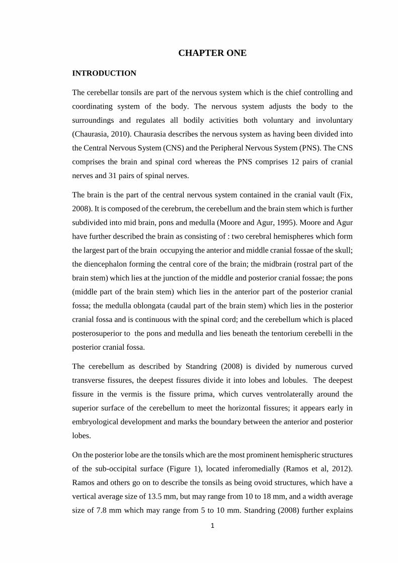
On the posterior lobe are the tonsils which are the most prominent hemispheric structures
of the sub-occipital surface (Figure 1), located inferomedially (Ramos et al, 2012).
Ramos and others go on to describe the tonsils as being ovoid structures, which have a
vertical average size of 13.5 mm, but may range from 10 to 18 mm, and a width average
size of 7.8 mm which may range from 5 to 10 mm. Standring (2008) further explains

2
that the nodule and attached flocculi constitute a separate flocculonodular lobe, which
is separated from the uvula and tonsils by the deep posterolateral fissure.
Furthermore, Ramos et al (2002) pointed out that the tonsils are anterosuperiorly related
to the inferior medullary velum and telachorioidea, laterally to the biventral lobule,
superolaterally to the tellovelotonsillar cleft, posteriorly and inferiorly to the cisterna
magna. These features can easily be seen on sagittal section of the brain (Figure 2).
The functional role of the cerebellar tonsils is not yet known but they are believed to
coordinate skilled and learned movements. According to Marvridis (2014), the
cerebellar tonsils belong to the neocerebellum (also called the pontocerebellum) which
comprises the posterior lobe (except the uvula and pyramids) and is the largest portion
of the cerebellum.
Figure 1.1: Inferior view of the cerebellum and the position of the tonsils on the inferior
aspect of each of the cerebellar hemispheres taken at the dissection laboratory,
University of Zambia, School of Medicine.

3
Figure 1.2: MRI sagittal section through the brain showing the cerebellar tonsil taken
at Cancer Diseases Hospital, Radiology Department.
The cerebellar tonsils are prone to herniating through the foramen magnum (an opening
at the base of the skull which transmits the spinal cord) (Figure 3), this can involve one
or both tonsils, particularly in the instance of a Chiari malformation, a disorder that may
be congenital or acquired.
Figure 1.3: Showing Cerebellar Tonsillar Ectopia (Mayfieldclinic.com)

4
Tonsillar herniation can also occur in conditions that cause a rise in Intra-Cranial
Pressure (ICP). Medical procedures such as lumbar puncture could cause a sudden drop
in icp in the presence of raised intracranial pressure causing sudden death (Hoffman,
2015). This is corroborated by Mokri (2000), who indicated that abnormalities
demonstrated on head MR imaging studies in patients with sudden CSF volume
depletion include sinking or sagging of the brain and descent of the cerebellar tonsils
sometimes mimicking Chiari I malformation.
Sudden increased ICP is a serious and often life threatening condition. If increased ICP
compresses brain structures and blood vessels, it can lead to serious, permanent
disability or mortality (Kantor, 2015). Causes of increased ICP as outlined by Dunn
(2002) include; localised mass lesions (traumatic haematomas; extradural, subdural,
intracerebral), neoplasms (glioma, meningioma, metastasis), abscesses, focal oedema
(secondary to trauma, infarction, tumour), disturbance of CSF circulation, obstruction
of major venous sinuses, defuse brain oedema (encephalitis, meningitis, diffuse head
injury) and idiopathic intracranial hypertension.
An increase in ICP is a serious medical problem. The Michigan State University module
for Increased ICP of 2007 explains that since the brain is enclosed in a rigid cranium,
the free space to expand is limited. When the mass of the intracranial contents increases
in the presence of disease, ICP increases. When ICP exceeds a critical point,
displacement of organs or organ parts near a potential space(s) occurs, and herniations
occur. The nature of herniation is determined by the location of the lesion. The pressure
can damage the brain or spinal cord by pressing on important brain structures and by
restricting blood flow into the brain (A.D.A.M. Medical Encyclopedia, 2003).
Cerebellar tonsillar herniation through the foramen magnum can occur with increased
ICP and could cause compression of the brainstem and upper cervical spinal cord as they
exit the skull. This could result in dysfunction of respiratory and cardiac centres in the
brain.
The position of the cerebellar tonsils can be measured with reference to a straight line
drawn from the Basion to the Opisthion of the foramen magnum (Figure 4). This line
also represents the Anteroposterior (AP) diameter of the foramen magnum (O'Connor et
al, 1973). The Basion and Opisthion of the foramen magnum can easily be identified on
sagittal section of the MRIs of the head.

5
Figure 1.4: sagittal section through the brain showing a line drawn from the Basion to
the Opisthion of the foramen magnum taken at Cancer Diseases Hospital, Radiology
Department
To measure properly the position of the cerebellar tonsils, Magnetic Resonance Imaging
(MRI) is used. MRI scan is a non-invasive test used to evaluate the brain, spinal cord,
and surrounding CSF (Mayfield clinic, 2013). Mayfield clinic further goes on to say that,
herniation may reach the level of the first two vertebrae (C1 or C2) of the cervical spine
and that herniation of the tonsils is often measured in millimetres (mm) below the
foramen magnum. It has been observed that it is not enough to measure herniation of the
tonsils below the foramen magnum. The normal position of the tonsils should be
considered in order to determine the true extent of their displacement. This is so because
many other studies, Mikulis et al (1992), Lakshmi (2015) and others, have confirmed
the existence of variations in the position of the cerebellar tonsils in individuals of
different age groups. These results have differed from study to study. Furthermore, it is
important that the size of the foramen magnum be considered when determining the
position of the cerebellar tonsils.
6
1.1 Statement of the Problem
Several studies carried out in various places around the world have indicated that there
is no standard normal position of the cerebellar tonsils. This has been attributed in part
to variations due to ethnicity of the study subjects. Since results of the studies done on
the subject have been different every time, there is need to carry out a study on the
Zambian population in order to have a proper reference range for diagnosis in our
patients. Furthermore, researchers from around the world have looked at the normal
position of the cerebellar tonsils in relation to age of the individual; gender; pathological
conditions such as Chiari 1 Malformation; and the size and shape of the skull. To date
there is no documented data about the normal position of the cerebellar tonsils in the
Zambian population, or literature detailing the relationship of the size of the foramen
magnum to the position of the cerebellar tonsils.
1.2 Study Objectives
1.2.1 Main Objective
The main objective of this study was to determine the relationship between the normal
position of the cerebellar tonsils and the size of the foramen magnum in individuals of
different age groups.
1.2.2 Specific Objectives
I. To determine the position of the cerebellar tonsils in individuals of different age
groups.
II. To determine the anteroposterior diameter of the foramen magnum in individuals
of different age groups.
III. To determine the correlation of the position of the tonsils to the AP diameter of
the foramen magnum.
IV. To correlate the position of the cerebellar tonsils in individuals on demographic
characteristics including gender.
1.3 Research Questions
1. What is the normal position of the cerebellar tonsils in individuals of different age
groups in the Zambian population?
2. Is there a correlation between the size of the foramen magnum and the position of the
cerebellar tonsils?

7
1.4 Significance of the Study
This study was aimed at finding out the normal position of the cerebellar tonsils in
different age groups of the Zambian population as well as how the position of the tonsils
is affected by the size (AP diameter) of the foramen magnum. Apparent herniation of
the cerebellar tonsils into the foramen magnum is a frequent normal variation and can
be misleading when interpreting the MRI scans conducted on patients. Therefore,
determining the normal position of cerebellar tonsils in the Zambian population could
provide guidance on considerations which need to be taken into account when making a
possible diagnosis of tonsillar herniation.
Tonsillar herniation is a problem that arises due to increase in Intra-Cranial Pressure
(ICP). This can be caused by many factors including space occupying lesions and
traumatic head injuries. Traumatic brain injury accounts for up to half of trauma related
fatalities worldwide (Winter et al, 2005). In fact, Faul et al (2010) have referred to
traumatic head injury as a silent epidemic. The Global Burden of Disease (2002) predicts
that by the year 2020, RTAs will become the third leading cause of death and disability
in the developing world.
Recently, in Zambia, there has been an increase in the number of Road Traffic Accidents
(RTAs). This therefore entails a high likelihood of people involved in these accidents
suffering head injuries which in turn may lead to development of raised intracranial
pressure, a risk factor for cerebellar tonsillar herniation.
Simoonga (2009) reported an increase in the number of RTAs in the country between
2004 and 2007. Statistics from the medical records at the University Teaching Hospital
showed an increase in the number of people admitted to the hospital following RTAs
from 203 in 2012 to 243 (20%) in 2013 and to 323 (33%) in 2014. Therefore, the number
of admissions following RTAs has risen from 2012 to 2014 by 59%. These statistics are
expected to translate into an increase in the number of patients suffering head injuries
with increased intracranial pressure and subsequent cerebellar tonsillar herniation.
Although similar research about CT position has been carried out in some other parts of
the world, there have been variations in the results obtained, therefore emphasizing the
fact that there is no standard position of the cerebellar tonsils that is universally
applicable. Various factors do affect the position of the cerebellar tonsils such as the
shape of the face and the size of the skull base in different ethnic groups (Flanagan,

8
2010). It is possible that such differences can be expected in the Zambian population
and the region. However, no information, during the study, was found within the region
on the subject, hence the importance of carrying out the study.
Furthermore, it is recognized that the age groups that were considered by many of the
other authors had a wide distribution implying that age had not been considered as a
possible factor contributing to the differences. For this reason the researcher used a
narrower age range in order to take into consideration rapid growth spurt especially
between the ages 1 to 17 years.
Relating the position of the cerebellar tonsils to the size of the foramen magnum is one
of the objectives of this study as there is paucity of information on the subject in the
literature. Information on this subject could provide an explanation as to why some
people can have their cerebellar tonsils below the foramen magnum and remain
asymptomatic whereas in others cerebellar tonsillar ectopia causes severe symptoms.
Such knowledge can help with early diagnosis of pathological conditions; improve
options for management and determine patient prognosis.
To the best knowledge of the researcher, such a study has not been performed in Zambia.
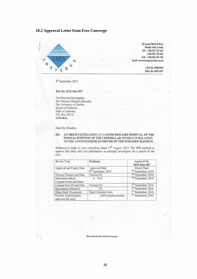
1.5 Ethical Considerations
Ethical approval was sought from Excellence in Research Ethics and Science (ERES)
CONVERGE IRB and authorization was obtained from the Cancer Diseases Hospital in
order to utilise the Magnetic Resonance Imaging facilities which the hospital offered.
The information obtained from the study was used strictly for this research project. To
guarantee confidentiality of the information obtained, the data collection forms which
were used were coded and did not reveal the names of the patients.
This study was retrospective and posed no risk to the patient as there was no direct
contact.

9
CHAPTER TWO
LITERATURE REVIEW
A number of similar studies have been carried out on the subject, most of them
comparing the position of the tonsils in normal subjects to those of individuals with a
firm diagnosis of Chiari malformation. Some of the studies have related the position of
the cerebellar tonsils to the age of the patient, gender as well as shape and size of the
skull.
A study was carried out by Lakshmi (2015) on 515 patients during MRI investigation
over a period of 8 months in India. It looked at the position of cerebellar tonsils with
reference to the level of the foramen magnum. Results of the study showed that the
highest number of cerebellar tonsillar ectopia was seen in the fifth decade of life while
a rise in the position of the tonsils was seen in the seventh decade in both sexes. In
children less than a year old and in old age, the position of tonsils was found to be above
the foramen magnum.
Another study was carried out in the United States of America by Barkovich et al (1986)
where the position of the cerebellar tonsils was measured with respect to the inferior
aspect of the foramen magnum in 200 normal patients and in 25 patients with an
established diagnosis of Chiari I malformation. Barkovich and his colleagues found the
mean position of the tonsils to be 1 mm above the foramen magnum with a range from
8 mm above the foramen magnum to 5 mm below the foramen magnum. They also found
that 14% of normal patients had tonsils extending slightly below the foramen magnum.
The researchers observed that a small degree of herniation of the cerebellar tonsils on
MRI was of debatable significance. Furthermore, Barkovich et al, in their study used
manual callipers to measure distances from the hard copies of the MRI, which was done
to the nearest millimetre. It has been noted that this method of measuring the position of
the cerebellar tonsils may be inaccurate and unreliable. This is because as compared to
digital measurements, those of manual vernier callipers are interpreted subjectively from
the scale by the user and this may not completely eliminate bias.
Mikulis et al (1992), in the United States of America, observed tonsillar elevation with
increasing age and therefore considered the following as criteria for determining ectopia.
In the first decade of life, 6 mm; in the second and third decades, 5 mm; in the fourth to

10
eighth decades, 4 mm; and in the ninth decade, 3 mm. Mikulis et al (1992) came to the
conclusion that a single reference standard that shows the normal distance of the
cerebellar tonsils from the foramen magnum is unsuitable unless age is considered.
Smith et al (2013), also based in the United States of America, found that the position
of the tonsils in normal participants was 7.5 mm below the foramen magnum for ages
0–10 years, 4 mm for 11–20 years, 5 mm for 21–30 years, 4 mm for 31–40 years, 4 mm
for 41–50 years, 3 mm for 51–60 years, 2.5 mm for 61–70 years, and 2 mm for 71 years
and older. These figures somewhat contradict the results by Mikulis and his colleagues
in that what was considered ectopia by Mikulis et al in 1992 was actually in the normal
range in a study conducted by Smith and his colleagues. Smith and colleagues further
observed a trend in their study where the mean position of the tonsils lowered slightly
with increasing age into young adulthood and ascended gradually with advancing age in
adulthood.
In a study carried out by O'Connor et al in 1973, they described the normal position of
the cerebellar tonsils as measured from the inferior most point to the Basion-Opisthion
reference line which always lay virtually at the same level bilaterally and was above the
reference line in all cases, the ages of the participants however were not specified in this
particular study. The average distance from the tonsils to the reference line in 100 cases
was 6 mm (+/-2 mm).
Cheng et al (2003) in China used MRI to compare quantitatively the position of the
cerebellar tonsils in age matched neurologically normal adolescents with that found in
idiopathic scoliosis patients. In the healthy subjects, results showed that the inferior most
point of the cerebellar tonsils was at an average of 2.8 mm above the basion-opisthion
reference line, with a range from 0 to10mm above the foramen magnum.
Freeman et al (2010) carried out a study in four groups of patients, two of the groups
were non traumatic (recumbent or upright) and the other two groups had history of
trauma (recumbent or upright). In the non-traumatic group, Cerebellar Tonsillar Ectopia
(CTE) was found in 5.7% (recumbent) and 5.3% (upright) groups. They reported rare
cases of CTE of 5mm and more.
In a study carried out by Acer et al (2006), the researchers measured the volumes of 28
human skulls and the cross-sectional area of the foramen magna, results showed that the
skulls with bigger volume had bigger foramen magna. They mentioned that because of

11
this, the consequences of nervous tissue displacement can be highly variable. However,
they did not mention whether this affects the position of the tonsils.
Huang et al (2013) carried out a study that compared cranial dimensions between
patients diagnosed with Chiari 1 Malformation and normal controls. They found that the
factors that were assessed in the study suggest underdevelopment of the occipital bone
in patients with Chiari 1 Malformation whereas in in the normal controls, the occipital
bone and hindbrain were normally developed. They also found that the shape of the
posterior cranial fossa in the group with Chiari 1 Malformation resembled a narrow
funnel.
From the studies that have been reviewed so far it is safe to conclude that there is no
particular standard that has been exhibited as to what the position of the cerebellar tonsils
should be in normal individuals. Many factors including age, gender and the shape and
size of the skull have been attributed to the differences in the position of the tonsils by
different authors. For the aforementioned reasons, the researcher undertook this study
with particular reference to the level of the tonsils and to the AP dimensions of the
foramen magnum.

12
CHAPTER THRE
METHODOLOGY
3.1 Study Design
The study design that was used in this research was a cross sectional study design which
analysed retrospectively patient records of MRI scans of the brain taken at the Cancer
Diseases Hospital in Lusaka from the year 2012 to 2014. The study endeavoured to
determine whether or not there was a relationship between the position of the cerebellar
tonsils and the size of the foramen magnum.
3.2 Study Site
The study was conducted in the Radiology Department at the Cancer Diseases Hospital
in Lusaka. This study site was chosen because the radiology department offers MRI
facilities and also receives referrals to the facility from all over the country including the
University Teaching Hospital which is the largest referral hospital in Zambia.
3.3 Study Population
The study population included all MRI records of patients, 1 year and older, who had
MRI brain scans taken at the CDH in Lusaka from the year 2012 to 2014.
3.4 Criteria
3.4.1 Inclusion criterion
All MRI brain scans of patients, 1 year and older, taken at the CDH between the years
2012 and 2014 that had been certified by a qualified radiologist as having no pathology.
3.4.2 Exclusion Criteria
All MRI brain scans of patients, 1 year and older, taken at the CDH between the years
2012 and 2014 that had been certified by a qualified radiologist as having pathology
were excluded.
All MRI brain scans which had no record of the patient’s age and gender were also
excluded from the study.

13
3.5 Sample Size
A census was used in this study. That is to say, all MRI brain scans that fit the inclusion
criterion within the period under review were included in the study. The CDH on average
takes 2000 scans each year, among these, half (1000) of them are brain scans. The reason
a census was chosen for this particular study was because while there are several (an
average of 1000) MRI brain scans taken at the CDH each year, patients who are referred
to the MRI facility are suspected of having pathology, therefore, there may not be many
MRI brain scans certified as being normal by the consultant radiologist. It was for this
reason that all the MRI brain scans that fit the inclusion criterion were considered in
order to have a sample size that would yield significant results.
3.6 Data Collection Instruments
The researcher used digital records of MRI scans of the brain that fit the inclusion
criterion.
Computer software tools including rulers were used to measure lengths for the position
of the cerebellar tonsils as well as the AP diameter of the foramen magnum.
A data collection form (appendix 10.5) was formulated and was used to record the
information collected from the digital MRIs. This information included; a code by which
to identify the patient, the age of the patient which was vital in this study since the
researcher was considering the age groups of individuals, the AP dimensions of the
foramen magnum (measured from the Basion to the Opisthion) as well as the lengths
from the inferior most point of the tonsils to the Basion-Opisthion reference line
(representing the position of the cerebellar tonsils).
3.7 Data Collection Procedure
Data collection began with retrieval of the hard copies of patients’ MRI reports and
selecting those that fit the inclusion criterion. These copies were then used to find digital
MRI brain scan records on the computer hard drive making sure all the scans chosen fit
the inclusion criterion. Individual patients’ digital MRI brain scans were then transferred
on to compact discs (CDs). The discs were then individually analysed and measurements
were taken using computer software tools. All the images were corrected for
magnification before any measurements were taken, this is to say that all images were
set to the same magnification before measurements were taken, to avoid bias. The AP

14
diameter of the foramen magnum was measured from the Basion to the Opisthion of the
foramen magnum on mid-sagittal section of the MRI brain scans to permit accuracy of
measurements. The measurements of the cerebellar tonsillar position were then carried
out from the inferior most point of the cerebellar tonsils to a line that was drawn from
the Basion to the Opisthion of the foramen magnum, this was also done on the mid-
sagittal section of the scans for the same reasons. The data collected was then recorded
on the data collection form.
3.8 Research Variables
Dependent Variable
Position of the cerebellar tonsils
Independent variable
Age
Gender
AP diameter of the foramen magnum
3.9 Data Management
3.9.1 Data Analysis Instruments and Procedures
Data analysis was performed using the Stata version 12 statistical software package, and
results were summarised on tables and graphs.
The Stata package was used to determine the proportions of demographic characteristics
of the sample. The data was then grouped according to different age groups and the mean
positions of the cerebellar tonsils and the AP diameter of the foramen magnum for each
age group were determined. The age groups that were used were according to Medlej
(2014) but slightly modified and were as follows:
Toddler (1 to 4 years)
Child (5 to 11 years)
Adolescent (12 to 17 years)
Young adult (18 to 39 years)
Middle age (40 to 59 years)
Old age (60 and older)

15
The one-way Analysis of Variance (ANOVA) was used to compare mean values in the
position of the cerebellar tonsils among the different age groups. Linear regression was
used to determine factors associated with the position of the cerebellar tonsils as well as
the anteroposterior diameter of the foramen magnum. All statistical tests were performed
at 5% significance level or 95% confidence interval with p-value of <0.05 to determine
statistical significance.

16
CHAPTER FOUR
RESULTS AND INTERPRETATION
4.1 0verview
The study investigated retrospectively the relationship between the normal position of
the cerebellar tonsils and the Anteroposterior diameter of the foramen magnum in
individuals of different age groups. The study also considered the patients’ gender as
one of the demographics that were likely to alter the results. A sample of 127 was
collected, out of which 67 (52.8%) were female and 60 (47.2%) were male. The median
age was 28 years (IQR 1 - 75). The average anteroposterior diameter of the foramen
magnum was 36.5mm (range; 23.4mm - 47.6mm) and the cerebellar tonsillar position
in the participants was 0.5mm (range; -14.5mm – 13.1mm).
4.2 position of the Cerebellar Tonsils and anteroposterior diameter of the foramen
magnum in different age groups
Table 1, illustrates the means and interquartile ranges of Cerebellar tonsillar position
and anteroposterior diameter of the foramen magnum in different age groups measured
in millimetres (mm).
Table 4.1: CT position and AP diameter of the foramen magnum for different age
groups
Age group (years) Mean CT position (IQR) Mean AP diameter (IQR)
1 - 4yrs -0.6 (-4.5 to 4) 31.6 (23.4 to 39.1)
5 - 11yrs 0.3 (-12.7 to 4.9) 36.6 (27.8 to 47.0)
12 - 17yrs 1.0 (-3.1 to 6.4) 36.0 (26.3 to 39.8)
18 - 39yrs 0.7 (-14.5 to 13.1) 36. 9 (26.3 to 47.6)
40 - 59yrs 0.4 (-2.7 to 5.7) 37.4 (32.0 to 44.2)
60 - 75yrs -0.5 (-3.6 to 1.3) 37.2 (34.2 to 43.2)
Abbreviations: CT; Cerebellar tonsils, IQR; interquartile range, AP; anteroposterior

17
Figure 4.1: Mean position of the cerebellar tonsils in different age groups
Figure 4.2: Mean Anteroposterior diameter of the foramen magnum in different
age groups
-0.8
-0.6
-0.4
-0.2
0
0.2
0.4
0.6
0.8
1
1.2
1 - 4yrs 5 - 11yrs 12 - 17yrs 18 - 39yrs 40 - 59yrs 60 - 75yrs
Cer
ebel
lar
ton
sils
po
siti
on
(m
m)
Age (in years)
28
29
30
31
32
33
34
35
36
37
38
1 - 4yrs 5 - 11yrs 12 - 17yrs 18 - 39yrs 40 - 59yrs 60 - 75yrs
Ante
ropost
eror
dia
met
er (
mm
)
Age (in years)
18
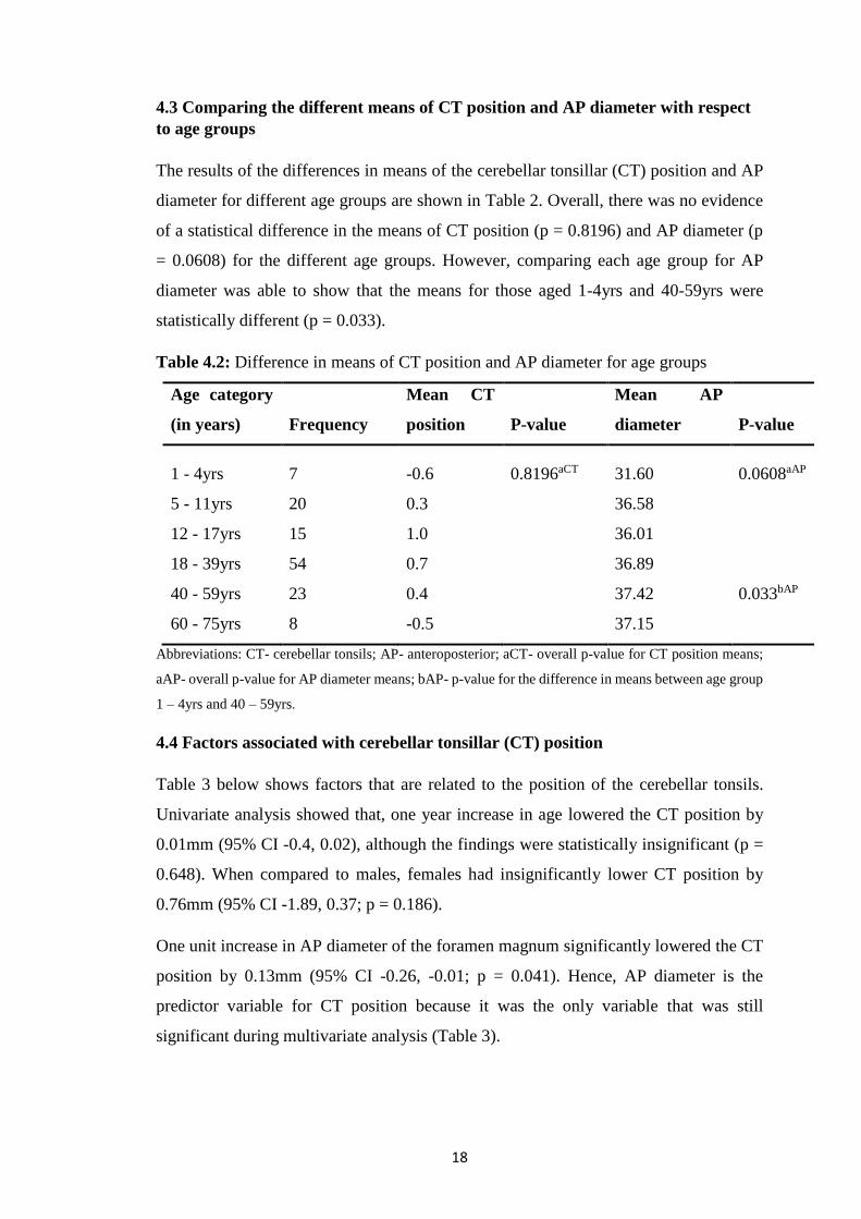
4.3 Comparing the different means of CT position and AP diameter with respect
to age groups
The results of the differences in means of the cerebellar tonsillar (CT) position and AP
diameter for different age groups are shown in Table 2. Overall, there was no evidence
of a statistical difference in the means of CT position (p = 0.8196) and AP diameter (p
= 0.0608) for the different age groups. However, comparing each age group for AP
diameter was able to show that the means for those aged 1-4yrs and 40-59yrs were
statistically different (p = 0.033).
Table 4.2: Difference in means of CT position and AP diameter for age groups
Age category
(in years) Frequency
Mean CT
position P-value
Mean AP
diameter P-value
1 - 4yrs 7 -0.6 0.8196aCT 31.60 0.0608aAP
5 - 11yrs 20 0.3 36.58
12 - 17yrs 15 1.0 36.01
18 - 39yrs 54 0.7 36.89
40 - 59yrs 23 0.4 37.42 0.033bAP
60 - 75yrs 8 -0.5 37.15
Abbreviations: CT- cerebellar tonsils; AP- anteroposterior; aCT- overall p-value for CT position means;
aAP- overall p-value for AP diameter means; bAP- p-value for the difference in means between age group
1 – 4yrs and 40 – 59yrs.
4.4 Factors associated with cerebellar tonsillar (CT) position
Table 3 below shows factors that are related to the position of the cerebellar tonsils.
Univariate analysis showed that, one year increase in age lowered the CT position by
0.01mm (95% CI -0.4, 0.02), although the findings were statistically insignificant (p =
0.648). When compared to males, females had insignificantly lower CT position by
0.76mm (95% CI -1.89, 0.37; p = 0.186).
One unit increase in AP diameter of the foramen magnum significantly lowered the CT
position by 0.13mm (95% CI -0.26, -0.01; p = 0.041). Hence, AP diameter is the
predictor variable for CT position because it was the only variable that was still
significant during multivariate analysis (Table 3).
19
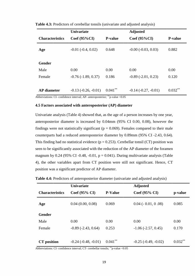
Table 4.3: Predictors of cerebellar tonsils (univariate and adjusted analysis)
Univariate Adjusted
Characteristics Coef (95%CI) P-value Coef (95%CI) P-value
Age -0.01 (-0.4, 0.02) 0.648 -0.00 (-0.03, 0.03) 0.882
Gender
Male 0.00 0.00 0.00 0.00
Female -0.76 (-1.89, 0.37) 0.186 -0.89 (-2.01, 0.23) 0.120
AP diameter -0.13 (-0.26, -0.01) 0.041** -0.14 (-0.27, -0.01) 0.032**
Abbreviations: CI- confidence interval; AP- anteroposterior; **p-value <0.05
4.5 Factors associated with anteroposterior (AP) diameter
Univariate analysis (Table 4) showed that, as the age of a person increases by one year,
anteroposterior diameter is increased by 0.04mm (95% CI 0.00, 0.08), however the
findings were not statistically significant (p = 0.069). Females compared to their male
counterparts had a reduced anteroposterior diameter by 0.89mm (95% CI -2.43, 0.64).
This finding had no statistical evidence (p = 0.253). Cerebellar tonsil (CT) position was
seen to be significantly associated with the reduction of the AP diameter of the foramen
magnum by 0.24 (95% CI -0.48, -0.01, p = 0.041). During multivariate analysis (Table
4), the other variables apart from CT position were still not significant. Hence, CT
position was a significant predictor of AP diameter.
Table 4.4: Predictors of anteroposterior diameter (univariate and adjusted analysis)
Univariate Adjusted
Characteristics Coef (95% CI) P-Value Coef (95% CI) p-value
Age 0.04 (0.00, 0.08) 0.069 0.04 (- 0.01, 0 .08) 0.085
Gender
Male 0.00 0.00 0.00 0.00
Female -0.89 (-2.43, 0.64) 0.253 -1.06 (-2.57, 0.45) 0.170
CT position -0.24 (-0.48, -0.01) 0.041** -0.25 (-0.49, -0.02) 0.032**
Abbreviations: CI- confidence interval; CT- cerebellar tonsils; **p-value <0.05
20
CHAPTER FIVE
DISCUSSION
5.1 Position of the Cerebellar Tonsils in different Age Groups
According to the results that were obtained from the sample collected in this study, the
trends in the means of the cerebellar tonsils in different age groups showed that the
tonsils were generally below the foramen magnum from ages 1-4 years ascending to a
position above the foramen magnum by age 5 and reaching their highest point between
12-17 years and then gradually descending again. Therefore, there was elevation of the
tonsils with increasing age until adolescence after which there was a gradual decent.
This finding could have been due to the fast rate at which the brain grows between ages
1-4years. According to the Centre on the Developing Child (2009,) about 700 new
nueronal connections are formed every second in the first few years of life. This and the
accumulation of neuroglia in the early years could cause an increase in the size of the
brain in a still expanding skull causing herniation of neuronal structures. Later in life,
brain matter begins to decrease while skull volume remains the same. This can be proven
thought a study by Ge et al in 2002 where results showed a significant linear decrease
in Both grey matter and white matter with age with a greater rate beginning only in adult
midlife. This phenomenon could explain why the tonsils in individuals over the age of
60 years have lower lying tonsils. The brain volume decreases while the skull remains
the same size causing the brain to sag and consequently herniate.
There were some similarities in trends between this study and studies from other regions.
Mikulis et al in 1992 for example found in a study they carried out that there was an
elevation in the position of the tonsils with age. Smith and colleagues in a study in 2013
observed lowering of cerebellar tonsillar position with increasing age into young
adulthood and from then on an elevation into adulthood. Lakshmi (2015) found the
largest number of cerebellar tonsillar ectopia was concentrated in the fifth decade of life
and thereafter a rise in the position in the seventh decade in both genders. The position
of the cerebellar tonsils in infancy and old age was found to be above the foramen
magnum. This was contrary to what was observed in this study where tonsillar ectopia
was seen in children from 1-4 years and adults over 60 years.
The mean positions of the cerebellar tonsils of different age groups were compared using
a one way analysis of variance (ANOVA). This was done in order to find out whether

21
there were any significant differences in the cerebellar tonsillar positions of the different
age groups. Results showed that even though there were differences from group to group
in the position of the tonsils, these differences were not statistically significant with p-
value of 0.8196. However, there were no studies found to compare these findings.
5.2 Anteroposterior Diameter of the Foramen Magnum in Individuals of
Different Age Groups
The average anteroposterior diameter of the foramen magnum was found to be 36.5mm
with a range from 23.4-47.6mm. These results were similar to those of other studies, for
instance a study by Lakshmi (2015) in India, found the mean value of the AP diameter
of the foramen magnum to be 35.57 with a range from 15-45mm. Osunwoke et al (2012)
in a study in Nigeria found the average length of the AP Diameter of the foramen
magnum to be 36.11mm.
Radhika et al (2014) in India also did a morphometric study of the foramen magnum and
results showed an average anteroposterior diameter of 35.3mm with a range from 27mm
to 43mm. The studies by Osunwoke et al and Radhika et al however only involved
adults.
The means of the anteroposterior diameter of the foramen magnum were also compared
using the one way ANOVA. When all measurements were compared against each other,
the differences were seen to be statistically insignificant with P-value of 0.0608.
However, when the means of age groups 1-4 years and 40-59 years were compared,
there was a significant difference seen with P-value of 0.033. This difference could have
been as a result of the fact that in the 1- 4 years age group, the skull is still growing and
is much smaller Compared to the 40-59 years age group where maximum growth has
been achieved and fusion of the skull bones has already taken place. The current study
as well as other studies like the one carried out by Lakshmi (2015) have shown an
increase in the diameter of the foramen magnum with increasing age. This could explain
the significant difference in the said age groups. However, there were no studies found
to compare these results with.
Trends in the means of the AP diameter of the foramen magnum in the different age
groups seen in this research showed an increase in the size of the foramen magnum up
to the beginning of adolescence (defined in this research as 12 years) at which point it

22
remains almost constant. In a study by Lakshmi (2015) results showed an increase in the
size of the foramen magnum up to the age of 14 after which the size remains constant.
The study also showed that the AP diameter of the foramen magnum was smaller in
females compared to males. The anteroposterior diameter of the foramen magnum in
females was less by 0.89mm though this finding was not statistically significant. This
was similar to results seen in other studies like one by Lakshmi (2015), in which the AP
diameter of the foramen magnum was found to be larger in males compared to females.
This has also been confirmed by Muralidhar et al (2014), Ukoha et al (2011) and many
others.
5.3 Association of the Cerebellar Tonsillar Position and Anteroposterior Diameter
of the Foramen Magnum
A univariate analysis of the position of the cerebellar tonsils against the anteroposterior
diameter of the foramen magnum showed that one unit (1mm) increase in the diameter
of the foramen magnum resulted into a significant lowering of 0.13mm in the position
of the cerebellar tonsils. Therefore, of all the variables that were analysed against the
position of the cerebellar tonsils, the anteroposterior diameter of the foramen magnum
is the one that was statistically seen to be a predictor of cerebellar tonsillar position
because it was the only one that was statistically significant with P-value of 0.041.
5.4 Factors Associated with Cerebellar Tonsillar Position
Linear regression was done to determine factors that affect the position of the cerebellar
tonsils. Position of the cerebellar tonsils was analysed against two (2) other factors that
could affect cerebellar tonsillar position including the age of the patient and their gender.
With respect to age, univariate analysis showed that an increase in the age of the patient
by one year caused lowering in the position of the cerebellar tonsils by 0.01mm. This
was done at 95% confidence interval. In a study by Bartholomeusz et al (2002), they
observed that in early childhood, there is an increase in both head circumference and
brain volume, and from adolescence onwards, brain volume decreases while head
circumference does not. However, there was no indication of the rate of increase of the
circumference of the skull in relation to that of the volume of the brain. Finding out the
rate of increase of both head circumference and brain volume could give reasons as to
why there is ectopia in the early years of life observed in this study.
23
When analysed with reference to the gender of the patient, females showed a lower
cerebellar tonsillar position by 0.76mm in comparison to their male counterparts,
although this difference was statistically insignificant. This difference could be due to
the difference in the size of the brain between males and females. Studies have shown
differences in brain size between males and females with males having larger brains.
Research carried out by Dekaban and Sadowsky (1978), shows that from birth the female
brain is slightly smaller compared to the male brain.
This finding is comparable to that seen in the study by Lakshmi (2015) where they
observed lower position of the cerebellar tonsils in females compared to males. The
study by Smith et al (2013) also established that females have, on average, a lower tonsil
position compared to males.
5.5 Limitations
This study was meant to be a population survey (census) where the researcher was to
include all the eligible samples to the study but the mode of data collection involved did
not allow for this. Therefore, this might have caused distortion of the results obtained
since the largest number of participants was between ages 18-39 years.

24
CHAPTER SIX
CONCLUSIONS AND RECOMMENDATIONS
6.1 Conclusions
A statistically significant relationship was observed between the size of the foramen
magnum and the position of the cerebellar tonsils. It was observed that the larger the
anteroposterior diameter of the foramen magnum, the lower the position of the cerebellar
tonsils.
i. The position of the cerebellar tonsils changes with increasing age, with the
tonsils situated below the foramen magnum between ages 1-4 years and
gradually ascending to a position above the foramen magnum reaching their
maximum position at about 17 years then gradually descending.
ii. The study showed an increase in the anteroposterior diameter of the foramen
magnum with age until about 12years after which it became somewhat constant
iii. The study showed an association between cerebellar tonsillar position and the
Anteroposterior diameter of the foramen magnum with 1mm increase in the AP
diameter of the foramen magnum causing a descent of 0.13mm of the cerebellar
tonsils.
iv. Based on the results of the study, females had lower cerebellar tonsillar position
compared to males, this trend was seen in all age groups.
6.2 Recommendations
i. It is recommended that other similar studies be carried out perhaps on larger
samples sizes and over a longer period of time to confirm the findings of this
study.
ii. It is recommended that in future studies on cerebellar tonsillar ectopia, age
should be taken into consideration.
iii. Studies should be carried out to correlate the size of the foramen magnum with
progression of symptoms in conditions involving cerebellar tonsillar ectopia.
25
REFERENCES
A.D.A.M. Medical Encyclopedia (2013). Increased Intracranial Pressure. Retrieved
31/12/14 from http://www.ncbi.nlm.nih.gov/pubmedhealth/PMH0001797/
Acer N. Sahin B. Ekinci N. Ergu H. and Basaloglu H. (2006). Relation between
Intracranial Volume and the Surface Area of the Foramen Magnum. The Journal of
Craniofacial Surgery. 17(2):326-330s
Barkovich AJ. Et al (1986). Significance of Cerebellar Tonsillar Position on MR.
American Journal of Neuroradiology. 7(5):795-799
Bartholomeusz HH. Courchesne E. and Karns CM. (2002). Relationship between
Head Circumference and Brain Volume in Healthy Normal Toddlers, Children and
Adults. Neuropediatrics. 33(5):239-241
Centre on the Developing Child (2009). Five Numbers to Remember About Early
Childhood Development (Brief). Retrieved 17th December, 2016 from
www.developingchild.harvard.edu.
Chaurasia BD. (2010). Human Anatomy: Regional and Applied Dissection and
Clinical. 5th ed, 3(307). New Delhi, CBS Publishers and Distributors PVT Ltd.
Cheng JC. Et al (2003). Redefining the Magnetic Resonance Imaging Reference Level
for the Cerebellar Tonsil: A Study of 170 Adolescents with Normal versus Idiopathic
Scoliosis. 28(8):815-818
Dekaban AS. and Sadowsky D. (1978). Changes in brain weights during the span of
human life: Relation of Brain Weights to Body Heights and Body Weights. Ann.
Neurology, 4:345-356
Dunn LT. (2002). Raised Intracranial Pressure. Journal of Neurology Neurosurgery
Psychiatry. 73:i23–i27. Retrieved 16/01/15 from http://jnnp.bmj.com/
Faul M. Xu L. Wald MM. and Coronado VG. (2010). Traumatic Brain Injury in the
United States: Emergency Department Visits, Hospitalizations and Deaths 2002–2006.
Retrieved 16/06/15 from www.cdc.gov/TraumaticBrainInjury
Fix JD. (2008). Neuroanatomy. 4th ed. 4(1). Philadelphia, Lippincott Williams &
Wilkins.

26
Flanagan M. (2010). The Upright Doctor’s Book: The Downside of Upright Posture.
Upright-health.com (2014). Retrieved 09/06/15 from http://www.upright-
health.com/cerebellar-tonsillar-ectopia.html.
Freeman MD. et al. (2010). A Case-Control Study of Cerebellar Tonsillar Ectopia
(Chiari) and Head/Neck Trauma (whiplash). Brain Injury, 24(7–8): 988–994
Ge Y. et al. (2002). Age-Related Total Gray Matter and White Matter Changes in
Normal Adult Brain. Part I: Volumetric MR Imaging Analysis. Americal journal of
neurology. 23: 1327-1333
Hoffman KR. et al. (2015). Management of Cerebellar Tonsillar Herniation following
Lumbar Puncture in Idiopathic Intracranial Hypertension. Case Reports in Critical Care.
Article ID 895035. Hindawi Publishing Corporation.
http://dx.doi.org/10.1155/2015/895035
http://www.mayfieldclinic.com/PE-Chiari.htm#.VKP5xHshZ89
Huang et al. (2013). The Comparative Morphometric Study of the Posterior Cranial
Fossa: What Is Effective Approaches to the Treatment of Chiari Malformation Type 1?
Journal of Korean Neurosurgery Society 54(5): 405-410.
Increased Intracranial Pressure Module (2007). College of Human Medicine,
Michigan, East Lansing, Michigan State University.
Kantor D. (2015). Increased Intrcranial Pressure. Retrieved 30/12/14 from
http://www.nlm.nih.gov/medlineplus/ency/article/000793.htm
Lakshmi AS. (2015). Position of Cerebellar Tonsils in Reference to Foramen Magnum:
An MRI Study. Journal of Evolution of Medical and Dental Sciences 4(94):15970-
15974, DOI: 10.14260/jemds/2015/2327.
Lakshmi AS. (2015). Sagittal Diameter of Foramen Magnum in Normal Population: An
MRI Study. Journal of Evolution of Medical and Dental Sciences 4(95): 16045-16047,
DOI: 10.14260/jemds/2015/2342.
Mavridis IN. (2014). Gross and Neurosurgical Anatomy of the Cerebellar Tonsil. OA
Anatomy 2(1):8
Mayfield Clinic (2013). Chiari Malformation. Retrieved 31/12/14 from
http://www.mayfieldclinic.com/PE-Chiari.htm#.VKP5xHshZ89

27
Medlej J. (2014). Human Anatomy Fundamentals: Drawing Different Ages. Retrieved
15/01/15 from http://design.tutsplus.com/tutorials/human-anatomy-fundamentals-
drawing -different-ages--cms-21905
Mikulis DJ. et al. (1992). Variance of the position of the cerebellar tonsils with age:
preliminary report. Radiology. 183(3):725-728.
Mokri B. (2000). Cerebrospinal Fluid Volume Depletion and its Emerging
Clinical/Imaging Syndromes. Neurosurgery Focus 9 (1).
Moore KL. and Agur (1995). Essential Clinical Anatomy. Pages 361-364. Baltimore,
Lippincott Williams & Wilkins.
Shepur MP. Magi M. Nanjundappa B. Havaldar PP. Gogi P. SH. (2014).
Morphometric Analysis of Foramen Magnum. International Journal of Anatomy and
Research. 2(1):249-55.
O'connor S. Du Boulay G. and Logue V. (1973). The Normal Position of the
Cerebellar Tonsils as Demonstrated by Myelography. Journal of Neurosurgery. 39:388
Osunwoke EA. Oladipo GS. Gwunireama IU. and Ngaokere JO. (2012).
Morphometric analysis of the foramen magnum and jugular foramen in adult skulls in
southern Nigerian population. American Journal of Science and Industrial Research.
3(6): 446-448
Radhika PM. Shailaja S. Prathap KJ. Sheshgiri C. and Jyothi KC. (2014).
Morphometric Study of the Foramen Magnum in Adult Human Skulls in Indian
Population. Asian Journal of Medical and Clinical Science. 3(2): 62-72
Ramos A. et al. (2012). Cerebellar Anatomy as Applied to Cerebellar Microsurgical
Resections. Arq Neuropsiquiatr. 70(6):441-446
Simoonga M. (2009). Road Safety in Lusaka City and Community Approaches to Road
Safety. Lusaka City Council.
Smith BW. et al. (2013). Distribution of cerebellar tonsil position: implications for
understanding Chiari malformation. Journal of Neurosurgery.
DOI:10.3171/2013.5.JNS121825
Standring S. (2008). Gray’s Anatomy: Cerebellum. 40th edition. London, Churchill
Livingstone, Elsevier limited.

28
The Global Burden of Disease. (2002). Prediction of Outcome in Trauma Patients.
JBJS 84-A (6):1090
Ukoha U. Egwu OA. Okafor IJ. Anyabolu AE. Ndukwe GU. Okpala I. (2011).
Sexual Dimorphism in the Foramen Magnum of Nigerian Adult. International Journal
of Biological and Medical Research. 2(4): 878 – 881
Winter CD. Adamides AA. Lewis PM. and Rosenfeld JV. (2005). A Review of the
Current Management of Severe Traumatic Brain Injury. Surgeon 3(5): 329-337

29
APPENDICES
10.1 Approval Letter from Assistant Dean (Postgraduate)
30
10.2 Approval Letter from Eres Converge
31

32
10.3 Letter of Permission to Conduct Research
THE UNIVERSITY OF ZAMBIA
SCHOOL OF MEDICINE
To : Dr Kennedy Lishimpi 11 February 2015 Senior Medical Superintendent
Cancer Diseases Hospital Lusaka. Sub. : Ms Patience Buumba. Research project & Radiology Dear Dr Lishimpi I wish to introduce to you Ms Buumba a second year MSc Anatomy student who is to undertake a research project on the radiological dimensions of cerebellar tonsils in relation to the foramen magnum. I would very much appreciate if you could give her permission to collect data from the MRI unit in your Department. With kind regards and best wishes Yours Sincerely
Professor Krikor Erzingatsian FRCSI: Hon FCS(ECSA)
cc : Dean SOM

33
10.4 Letter of Approval to Conduct Research

34
10.5 Data Collection Form