Normal Bone Anatomy - · PDF fileNormal Bone Anatomy ... hard compact cortical bone, ......
12
In: Textbook of Small Animal Orthopaedics, C. D. Newton and D. M. Nunamaker (Eds.) Publisher: International Veterinary Information Service (www.ivis.org), Ithaca, New York, USA. Normal Bone Anatomy ( 1-Jan-1985 ) A. W. Fetter and F. W. Rhinelander Bone tissue is the structural material that gives the bones the strength they require to act as levers for muscles, to give form to the soft tissues of the body, and to provide protective cavities for the vital organs. However, bone tissue also serves as a mineral bank that can be drawn upon in times of need. Morphologically bone tissue appears to be under the control of bone cells. Its surfaces are enveloped by active and resting osteoblasts and osteoclasts, and it is permeated by an interconnected canalicular system in which osteocytes are found. In the dog, no osteocyte is more than 0.3 mm from a blood vessel. This morphologic evidence combined with biochemical and endocrine evidence of the importance of bone mineral metabolism requires that bone tissue, along with its cells, be considered as an organ. These cells control the composition of the extracellular fluids of mineralized bone matrix within very narrow limits, and at the same time they can remove and replace the mineralized tissue to meet the anatomical needs of a mature skeleton. Young stated that "each problem in bone may be considered in the light of the relative distribution of bone cells among their various functional roles, for this system of cells acts as the ’final pathway’ through which the great variety of stimuli affecting bone must pass."(4) THE CELLS OF BONE The three major components of bone are osteogenic cells, organic matrix, and mineral. The osteogenic cells include osteoblasts, osteocytes, and osteoclasts, while the matrix consists predominantly of collagen and proteoglycans and constitutes approximately one third of the bone mass. The mineral that makes up approximately two thirds of bone is composed of calcium phosphate crystals deposited as hydroxyapatite. OSTEOBLASTS Osteoblasts form a cell layer over bone surfaces on which matrix is being formed. The cells are polarized, in that new osteoid, referred to as an osteoid seam, is deposited along the surface adjacent to bone (Fig. 1-1). The deeper portion of the osteoid seam undergoes mineralization along the so-called mineralization front. The bone is essentially enveloped by the osteoblasts, since the cells are in close contact with one another and tight junctions and gap junctions have been observed. Thus, the osteoblastic layer controls the transport of materials from the extracellular space to the osteoid seam and mineralization front. Ultrastructurally, osteoblasts feature a complement of organelles characteristic of cells actively involved in protein synthesis. They have abundant endoplasmic reticulum and numerous ribosomes, and the Golgi apparatus and mitochondria are quite prominent (Fig. 1-2). Procollagen molecules are produced by the ribosomes and extruded into the extracellular space, but only along the surface that faces bone. Proteolysis and polymerization within the extracellular space result in the formation of collagen fibrils. The combination of these intracellular and extracellular events leads to the production of the osteoid seam. Most of the proteoglycans are packaged in the Golgi apparatus, and vesicles containing these products then migrate to the surface of the cell and release their contents by exocytosis. The combination of proteoglycans and collagen fibers results in the production of a mineralizable matrix. FIG. 1-1 Osteoblasts (O) and osteoid seam (opposing arrows) on the surface of cancellous bone (B). Mesenchymal cells (M) and hematopoietic cells (H) are present within the intertrabecular space.
Transcript of Normal Bone Anatomy - · PDF fileNormal Bone Anatomy ... hard compact cortical bone, ......

In: Textbook of Small Animal Orthopaedics, C. D. Newton and D. M. Nunamaker (Eds.) Publisher: International Veterinary Information Service (www.ivis.org), Ithaca, New York, USA.
Normal Bone Anatomy ( 1-Jan-1985 ) A. W. Fetter and F. W. Rhinelander Bone tissue is the structural material that gives the bones the strength they require to act as levers for muscles, to give form to the soft tissues of the body, and to provide protective cavities for the vital organs. However, bone tissue also serves as a mineral bank that can be drawn upon in times of need. Morphologically bone tissue appears to be under the control of bone cells. Its surfaces are enveloped by active and resting osteoblasts and osteoclasts, and it is permeated by an interconnected canalicular system in which osteocytes are found. In the dog, no osteocyte is more than 0.3 mm from a blood vessel. This morphologic evidence combined with biochemical and endocrine evidence of the importance of bone mineral metabolism requires that bone tissue, along with its cells, be considered as an organ. These cells control the composition of the extracellular fluids of mineralized bone matrix within very narrow limits, and at the same time they can remove and replace the mineralized tissue to meet the anatomical needs of a mature skeleton. Young stated that "each problem in bone may be considered in the light of the relative distribution of bone cells among their various functional roles, for this system of cells acts as the 'final pathway' through which the great variety of stimuli affecting bone must pass."(4)
THE CELLS OF BONE The three major components of bone are osteogenic cells, organic matrix, and mineral. The osteogenic cells include osteoblasts, osteocytes, and osteoclasts, while the matrix consists predominantly of collagen and proteoglycans and constitutes approximately one third of the bone mass. The mineral that makes up approximately two thirds of bone is composed of calcium phosphate crystals deposited as hydroxyapatite.
OSTEOBLASTS Osteoblasts form a cell layer over bone surfaces on which matrix is being formed. The cells are polarized, in that new osteoid, referred to as an osteoid seam, is deposited along the surface adjacent to bone (Fig. 1-1). The deeper portion of the osteoid seam undergoes mineralization along the so-called mineralization front. The bone is essentially enveloped by the osteoblasts, since the cells are in close contact with one another and tight junctions and gap junctions have been observed. Thus, the osteoblastic layer controls the transport of materials from the extracellular space to the osteoid seam and mineralization front. Ultrastructurally, osteoblasts feature a complement of organelles characteristic of cells actively involved in protein synthesis. They have abundant endoplasmic reticulum and numerous ribosomes, and the Golgi apparatus and mitochondria are quite prominent (Fig. 1-2). Procollagen molecules are produced by the ribosomes and extruded into the extracellular space, but only along the surface that faces bone. Proteolysis and polymerization within the extracellular space result in the formation of collagen fibrils. The combination of these intracellular and extracellular events leads to the production of the osteoid seam. Most of the proteoglycans are packaged in the Golgi apparatus, and vesicles containing these products then migrate to the surface of the cell and release their contents by exocytosis. The combination of proteoglycans and collagen fibers results in the production of a mineralizable matrix.
FIG. 1-1 Osteoblasts (O) and osteoid seam (opposing arrows) on the surface of cancellous bone (B). Mesenchymal cells (M) and hematopoietic cells (H) are present within the intertrabecular space.
Membrane-bound vesicles containing amorphous calcium phosphate are extruded from the plasma membrane of the osteoblast into the extracellular space. It appears that these vesicles induce and actually activate crystal formation.(l,2) Alkaline phosphatase, which is produced by the osteoblast, is thought to act as a pyrophosphatase and may be involved in the initiation of the mineralization process. The transformation of amorphous calcium phosphate to crystalline hydroxyapatite appears to take place both inside and outside the matrix vesicles. When crystals formed within the vesicle contact the membrane of the vesicle, the membrane ruptures. The crystals, which are then exposed to a supersaturated solution, induce precipitation over the entire matrix surface.
OSTEOCYTES Approximately 10% of the osteoblastic population become enclosed in the developing matrix and are then referred to as osteocytes. They have structural features very similar to when they were on the surface of the matrix, but the endoplasmic reticulum may not be so profuse. As the cells become more deeply embedded in mineralized bone matrix, their cytoplasmic volume is reduced, as is their complement of cytoplasmic organelles (Fig. 1-3). Osteocytes have cytoplasmic processes that extend into the surrounding matrix for some distance and fill most of the canaliculi in which they are contained. The processes of osteocytes contact processes from other osteocytes and osteoblasts on the surface, forming tight junctions. This interconnection of osteoblastic lining cells with the osteocytes deep within bone regulates the flow of mineral ions from the extracellular fluid through the osteoblast to the osteocytes, from the osteocytes to the extracellular fluid surrounding them, and finally from this fluid to the mineralized bone matrix. Thus, the large surface area provided by the osteocytic population results in a regulatory mechanism for the exchange of mineral ions between the extracellular fluid and bone by means of the canalicular system. Osteocytes appear to be essential to the maintenance of bone, since when the cell dies, the matrix around it eventually is removed.
OSTEOCLASTS Osteoclasts are found in sites in which bone is being remodeled. They are large, multinucleated cells typically found on or near bone surfaces within concavities representing Howship's lacunae (Fig. 1-4). However, there is evidence that mononuclear cells with cytoplasmic features similar to osteoclasts may resorb bone. The size and number of nuclei in osteoclasts vary, but each nucleus usually is associated with a perinuclear Golgi apparatus in which Golgi vesicles are in all stages of development. The cytoplasm is filled with vacuoles and small vesicles. There is little endoplasmic reticulum and few ribosomes, and mitochondria are present in much greater number than in osteoblasts. The zone of contact of the plasma membrane with the bone surface consists of two areas. The ruffled border comprises fingerlike membranous folds that extend
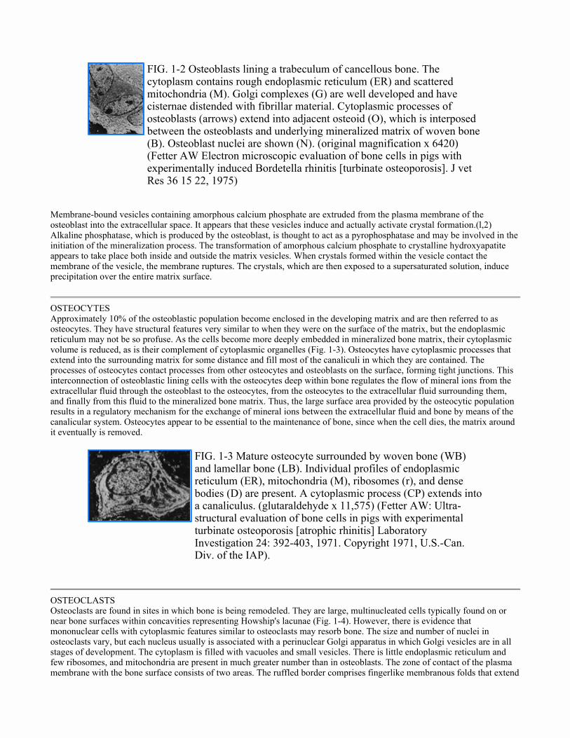
FIG. 1-2 Osteoblasts lining a trabeculum of cancellous bone. The cytoplasm contains rough endoplasmic reticulum (ER) and scattered mitochondria (M). Golgi complexes (G) are well developed and have cisternae distended with fibrillar material. Cytoplasmic processes of osteoblasts (arrows) extend into adjacent osteoid (O), which is interposed between the osteoblasts and underlying mineralized matrix of woven bone (B). Osteoblast nuclei are shown (N). (original magnification x 6420) (Fetter AW Electron microscopic evaluation of bone cells in pigs with experimentally induced Bordetella rhinitis [turbinate osteoporosis]. J vet Res 36 15 22, 1975)
FIG. 1-3 Mature osteocyte surrounded by woven bone (WB) and lamellar bone (LB). Individual profiles of endoplasmic reticulum (ER), mitochondria (M), ribosomes (r), and dense bodies (D) are present. A cytoplasmic process (CP) extends into a canaliculus. (glutaraldehyde x 11,575) (Fetter AW: Ultra- structural evaluation of bone cells in pigs with experimental turbinate osteoporosis [atrophic rhinitis] Laboratory Investigation 24: 392-403, 1971. Copyright 1971, U.S.-Can. Div. of the IAP).

varying distances into the cytoplasm, while the sealing area is characterized by a very dense homogeneous cytoplasm that surrounds the site of active bone resorption, namely, the ruffled border (Fig. 1-5). The cytoplasmic membrane in the sealing area is closely applied to the underlying surface of mineralized bone, functioning to isolate the region beneath the ruffled border and permitting Iysosomal enzymes and hydrogen ions produced by the osteoclasts to be concentrated in this area. Acid phosphatase is produced by osteoclasts, and the cells probably produce collagenase as well. Mineral is first dissolved, followed by removal of the organic matrix and disruption of mineralized matrix to a depth of 1 um to 2 um. Apatite crystals and collagen fibers can be observed in the extracellular space between the cytoplasmic folds. The degradation products of the matrix are thought to enter the cytoplasm of the osteoclasts through a process of endocytosis and are then transported across the cell and extruded into the extracellular space.
Osteogenic cells are derived from undifferentiated mesenchymal cells, which are also believed to give rise to the formed hematopoietic elements. There is evidence that bone marrow contains both predetermined osteoprogenitor cells and cells that will form bone in the presence of a suitable inducer. Recent studies in osteopetrosis have provided evidence that migrating mononuclear cells, possibly of splenic or thymic origin, may provide new osteoclasts directly or cells that influence osteoclastic function indirectly.(3) There is no clear evidence as to the fate of osteocytes when bone matrix is resorbed. It has been suggested that osteoclasts can undergo fission into mononuclear cells on the endosteal bone surface and that these mononuclear cells undergo a modulation of cell function to osteoblasts and then osteocytes.
THE MATRIX OF BONE Two types of bone matrix are observed in the mature skeleton: hard compact cortical bone, found largely in the shafts of the long bones that surround the marrow cavities; and spongy or cancellous bone, which comprises a network of fine, interlacing partitions, the trabeculae, enclosing cavities that contain either hematopoietic or fatty marrow. Cancellous bone is found in the vertebrae, in the majority of the flat bones, and in the ends of the long bones. The first bone to appear in embryonic development or in the repair of fractures is referred to as woven bone, coarse bundled bone, or fiber bone. It is characterized by coarse fiber bundles, approximately 30 um in diameter, running in a random, interlacing or "burlap" fashion. This woven bone is gradually replaced by lamellar bone. Thus, woven bone is a temporary structure and persists only in special locations such as the dental alveolus and osseous labyrinths. Lamellar bone, on the other hand, is not a primary structure but rather a replacement tissue formed during the remodeling of bone matrix. It consists of fine fiber bundles, 2 um to 4 um in diameter, that are arranged irregularly in parallel or concentric curving sheets.
FIG. 1-4 Osteoclast adjacent to lamellar bone (LB). Note the multiple nuclei (N) and vacuolated cytoplasm (V) in the vicinity of the ruffled border (RB) The mineralized section of bone is stained with Goldner's modified trichrome stain.
FIG. 1-5 Osteoclast in close proximity to a trabeculum of bone. Golgi complexes (G) are perinuclear in location. There are numerous mitochondria (M) and scattered profiles of endoplasmic reticulum (ER) in the cytoplasm. Many vacuoles (V) are present, especially in the vicinity of the ruffled border (RB). Numerous needle-shaped crystals and collagen fibers (arrows) are on the frayed bone surface beneath the ruffled border. (original magnification x 5775) (Fetter AW: Electron microscopic evaluation of bone cells in pigs with experimentally induced Bordetella rhinitis [turbinate osteoporosis. Am J Vet Res 36 15 22, 1975)

SECTION TWO NORMAL VASCULAR ANATOMY FREDERIC W. RHINELANDER
! Method of Vascular Evaluation
! General Anatomy of Long Bone
! Blood Supply of Immature Bone
! Blood Supply of Normal Mature Long Bones
! Afferent Vasculature System
! Direction of Blood Flow Through Cortex
! Efferent Vasculature System
! Intermediate Vascular System of Compact Bone
! Variability of Normal Circulation
METHOD OF VASCULAR EVALUATION The most representative unit of the appendicular skeleton is the so-called long-bone,(19) a well-recognized anatomical and vascular entity. It is not simply a bone that is long, like a rib; it is a straight bone with a tubular shaft and expanded ends (Fig. 1-6). It has been the principal site of problems in clinical orthopaedics and has therefore been the major object of vascular investigation over the years.
The osseous blood supplies of several species of animals have been studied by various investigators, and minor variations have been found. The bones of the dog, among the common laboratory animals, appear to be closest to those of humans from the vascular viewpoint. They have therefore been the subject of the vascular studies I have performed. Consequently, the long-bone of the dog will be the basis of this presentation of normal blood supply and its response to injury. The method of laboratory investigation has been that of microangiography and correlated histology, which Trueta(22) and Barclay(5) first developed for bone and I then expanded. (14,17) In each experiment, at the end of the observation period, the arterial tree of the limb under investigation is infused with an aqueous suspension of Micropaque (finely divided and uniform barium sulfate) at 120 mm Hg, the normal systolic blood pressure of the dog. The only blood vessels filled at this pressure are those that are functional at the time of the infusion and are on the afferent side of the circulation, up to and including the capillaries and their equivalents in cortical bone. Major veins are not demonstrated except as ghosts because the venous runoff is too rapid. The tissue specimens are decalcified (in order to render visible the minute infused blood vessels within cortex) and are then cut into slices 1-mm thick, from which the microangiograms are made with a special soft x-ray machine. After satisfactory microangiograms have been obtained, each tissue slice is sectioned by microtome at 6 um to 10 um to produce standard
FIG. 1-6 Diagrammatic representation of the parts of a long-bone. (Rhinelander FW: Circulation in bone. In Bourne G (ed): Vol 2, The Biochemistry and Physiology of Bone, 2nd ed, chap1. New York, Academic Press, 1972)

histologic sections, which are stained routinely. This technique is unique in portraying the histology of the same slice of tissue that is observed microangiographically.
GENERAL ANATOMY OF A LONG-BONE An idealized long-bone is shown in Figure 1-6. The epiphysis ceases to exist when maturity is reached. The entire expanded end of the bone is then the metaphysis, composed of trabecular (cancellous or spongy) bone. The diaphysis is a hollow tube of cortical (compact) bone. Its central cavity contains the medullary arterial supply and is occupied chiefly by fatty marrow. A portion of the medulla of some long-bones contains hematopoietic elements, but these are found chiefly in the cancellous bone of metaphyses. The entire surface of a long-bone, except at the ends at which articular cartilage is present, is covered by periosteum. The periosteum consists of an inner osteogenic layer (cambium), which provides appositional growth before maturity, and an outer fibrous layer, which is purely supportive. The presence of the active cambium, with longitudinal arterioles, makes the periosteum thick. However, for the mature long-bone the cambium is atrophic (thin and tenuous). The lack of longitudinal blood vessels in the periosteum has important surgical implications. Over most of the surface of mature long-bones, under normal conditions, the periosteum is loosely attached beneath the bellies of muscles. The only periosteal blood vessels in those areas are venules and capillaries. Where fasciae are firmly attached to the diaphysis, along ridges like the linea aspera of the femur, the afferent blood vessels are adequately protected; hence, there they can approach the cortical surface and enter. These afferent vessels are the principal nutrient artery (which may be dual, as in humans) and the periosteal arterioles.
BLOOD SUPPLY OF IMMATURE BONES The major difference between the blood supplies of a mature and an immature long-bone stems from the inability of blood vessels to cross the physis (cartilaginous growth plate), which separates the epiphysis from the expanded metaphysis at the end of the diaphysis (Fig. 1-6). The epiphysis and the metaphysis, therefore, have separate blood supplies.
To provide an adequate blood supply to this active area, the ends of growing long-bones are surrounded by a vascular circle the epiphyseometaphyseal arcade. Terminal capillary loops are present at the junction of articular cartilage and periosteum. The border of the epiphysis at the growth plate has an arborization of finely branching vessels. The major afferent vascular supply of the metaphysis comes from branches of the encircling arcade, which enter through numerous foramina. These periosteal arterioles anastomose with terminal branches of the medullary arterial supply of the diaphysis to form an arteriolar network that courses toward the active surface of the physis (Fig. 1-7, A). There they make a hairpin bend and retreat back upon themselves (Fig. 1-7, B). At those hairpin bends, the rate of blood flow is greatly reduced, and that is thought to account for the localization of blood-borne bacteria that causes the onset of hematogenous osteomyelitis.
FIG. 1-7 Normal immature canine radius. (A) Microangiogram of distal epiphysis, metaphysis, and adjacent diaphysis shows the overall afferent vascularization. Note that the only vascular connections between metaphysis and epiphysis are along the periosteum. (original magnification x 5). (B) Center of the growth plate (enlargement from A). Observe the massed end-arterioles beneath the plate, at the site of longitudinal growth. The arterioles on the epiphyseal side of the plate are inactive in comparison. (original magnification x 20) (Rhinelander FW, Wilson JW Blood supply to developing, mature and healing bone In Sumner-Smith G (ed): Bone in Clinical Orthopaedics: A Study of Clinical Osteology, chap 2. Philadelphia, WB Saunders 1982)
In addition to the growth in length at the juxtametaphyseal region of the physis, appositional growth occurs all around the periosteal surface of an immature long-bone. Longitudinal afferent blood vessels in the periosteum supply this appositional growth. With maturity the physis as such ceases to exist, and the entire expanded end of the long-bone becomes the metaphysis, with its periosteal arteriolar blood supply entering all surfaces except those covered by articular cartilage. The longitudinal afferent vessels in the periosteum of the metaphysis and diaphysis disappear, with atrophy of the osteogenic layer of the periosteum. This layer remains atrophic and dormant until activated by injury.
BLOOD SUPPLY OF NORMAL MATURE LONG BONES After multiple experimental observations by microangiography and correlated histology, I found it more clear and practical to break with established custom and to classify the normal blood vessels of a long-bone according to function, rather than according to anatomical location.(14,l6) In particular, I had observed that the normal components of the so-called periosteal circulation were predominantly veins and capillaries, engaged in the final states of the drainage of blood from cortex. Arterioles were a minor component, supplying blood to cortex only in the immediate vicinity of firm fascial attachments. I therefore recommended the elimination of the use of the term "periosteal circulation." The three basic vascular components according to function are the afferent vascular system (arteries and arterioles carrying nutrients), the efferent vascular system (veins and venules carrying waste products), and the intermediate vascular system (capillaries in cancellous bone and equivalent vessels of fixed size in the canals of cortical bone, linking the afferent and efferent systems).
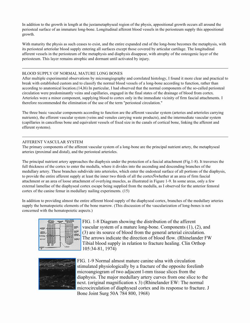
AFFERENT VASCULAR SYSTEM The primary components of the afferent vascular system of a long-bone are the principal nutrient artery, the metaphyseal arteries (proximal and distal), and the periosteal arterioles. The principal nutrient artery approaches the diaphysis under the protection of a fascial attachment (Fig.1-8). It traverses the full thickness of the cortex to enter the medulla, where it divides into the ascending and descending branches of the medullary artery. These branches subdivide into arterioles, which enter the endosteal surface of all portions of the diaphysis, to provide the entire afferent supply at least the inner two thirds of all the cortexÑwhether at an area of firm fascial attachment or an area of loose attachment of overlying muscles, as illustrated in Figure 1-9. In some areas, only a few external lamellae of the diaphyseal cortex escape being supplied from the medulla, as I observed for the anterior femoral cortex of the canine femur in medullary nailing experiments. (15) In addition to providing almost the entire afferent blood supply of the diaphyseal cortex, branches of the medullary arteries supply the hematopoietic elements of the bone marrow. (This discussion of the vascularization of long-bones is not concerned with the hematopoietic aspects.)
FIG. 1-8 Diagram showing the distribution of the afferent vascular system of a mature long-bone. Components (1), (2), and (3) are its source of blood from the general arterial circulation. The arrows indicate the direction of blood flow. (Rhinelander FW Tibial blood supply in relation to fracture healing. Clin Orthop 105:34-81, 1974)
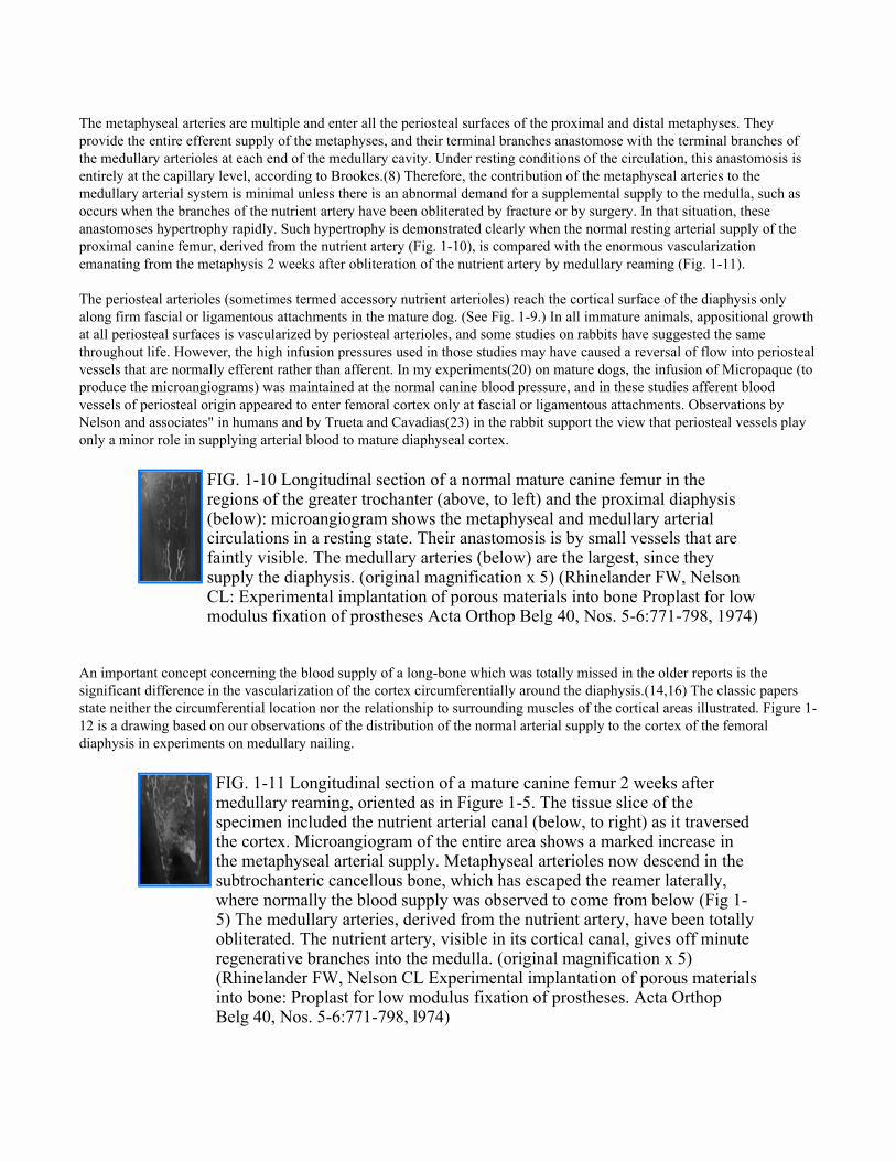
FIG. 1-9 Normal almost mature canine ulna with circulation stimulated physiologically by a fracture of the opposite forelimb microangiogram of two adjacent l-mm tissue slices from the diaphysis. The major medullary artery curves from one slice to the next. (original magnification x 3) (Rhinelander EW: The normal microcirculation of diaphyseal cortex and its response to fracture. J Bone Joint Surg 50A 784 800, 1968)
The metaphyseal arteries are multiple and enter all the periosteal surfaces of the proximal and distal metaphyses. They provide the entire efferent supply of the metaphyses, and their terminal branches anastomose with the terminal branches of the medullary arterioles at each end of the medullary cavity. Under resting conditions of the circulation, this anastomosis is entirely at the capillary level, according to Brookes.(8) Therefore, the contribution of the metaphyseal arteries to the medullary arterial system is minimal unless there is an abnormal demand for a supplemental supply to the medulla, such as occurs when the branches of the nutrient artery have been obliterated by fracture or by surgery. In that situation, these anastomoses hypertrophy rapidly. Such hypertrophy is demonstrated clearly when the normal resting arterial supply of the proximal canine femur, derived from the nutrient artery (Fig. 1-10), is compared with the enormous vascularization emanating from the metaphysis 2 weeks after obliteration of the nutrient artery by medullary reaming (Fig. 1-11). The periosteal arterioles (sometimes termed accessory nutrient arterioles) reach the cortical surface of the diaphysis only along firm fascial or ligamentous attachments in the mature dog. (See Fig. 1-9.) In all immature animals, appositional growth at all periosteal surfaces is vascularized by periosteal arterioles, and some studies on rabbits have suggested the same throughout life. However, the high infusion pressures used in those studies may have caused a reversal of flow into periosteal vessels that are normally efferent rather than afferent. In my experiments(20) on mature dogs, the infusion of Micropaque (to produce the microangiograms) was maintained at the normal canine blood pressure, and in these studies afferent blood vessels of periosteal origin appeared to enter femoral cortex only at fascial or ligamentous attachments. Observations by Nelson and associates" in humans and by Trueta and Cavadias(23) in the rabbit support the view that periosteal vessels play only a minor role in supplying arterial blood to mature diaphyseal cortex.
An important concept concerning the blood supply of a long-bone which was totally missed in the older reports is the significant difference in the vascularization of the cortex circumferentially around the diaphysis.(14,16) The classic papers state neither the circumferential location nor the relationship to surrounding muscles of the cortical areas illustrated. Figure 1-12 is a drawing based on our observations of the distribution of the normal arterial supply to the cortex of the femoral diaphysis in experiments on medullary nailing.
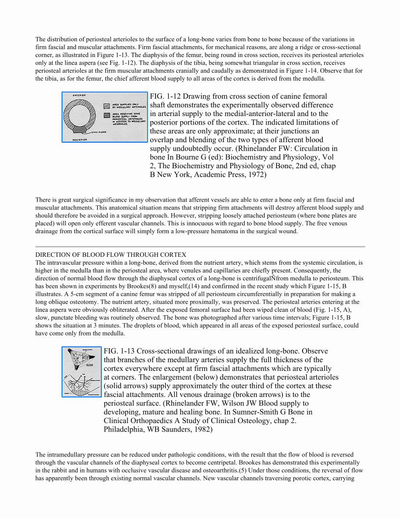
FIG. 1-10 Longitudinal section of a normal mature canine femur in the regions of the greater trochanter (above, to left) and the proximal diaphysis (below): microangiogram shows the metaphyseal and medullary arterial circulations in a resting state. Their anastomosis is by small vessels that are faintly visible. The medullary arteries (below) are the largest, since they supply the diaphysis. (original magnification x 5) (Rhinelander FW, Nelson CL: Experimental implantation of porous materials into bone Proplast for low modulus fixation of prostheses Acta Orthop Belg 40, Nos. 5-6:771-798, 1974)
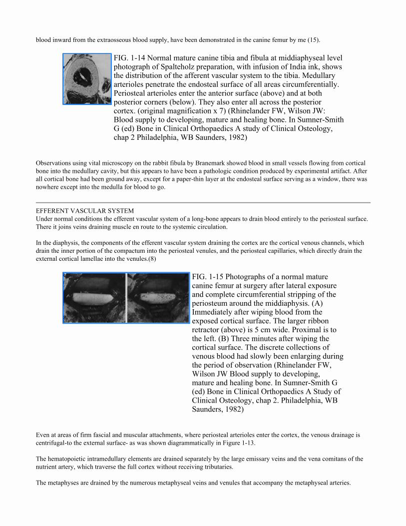
FIG. 1-11 Longitudinal section of a mature canine femur 2 weeks after medullary reaming, oriented as in Figure 1-5. The tissue slice of the specimen included the nutrient arterial canal (below, to right) as it traversed the cortex. Microangiogram of the entire area shows a marked increase in the metaphyseal arterial supply. Metaphyseal arterioles now descend in the subtrochanteric cancellous bone, which has escaped the reamer laterally, where normally the blood supply was observed to come from below (Fig 1-5) The medullary arteries, derived from the nutrient artery, have been totally obliterated. The nutrient artery, visible in its cortical canal, gives off minute regenerative branches into the medulla. (original magnification x 5) (Rhinelander FW, Nelson CL Experimental implantation of porous materials into bone: Proplast for low modulus fixation of prostheses. Acta Orthop Belg 40, Nos. 5-6:771-798, l974)
The distribution of periosteal arterioles to the surface of a long-bone varies from bone to bone because of the variations in firm fascial and muscular attachments. Firm fascial attachments, for mechanical reasons, are along a ridge or cross-sectional corner, as illustrated in Figure 1-13. The diaphysis of the femur, being round in cross section, receives its periosteal arterioles only at the linea aspera (see Fig. 1-12). The diaphysis of the tibia, being somewhat triangular in cross section, receives periosteal arterioles at the firm muscular attachments cranially and caudally as demonstrated in Figure 1-14. Observe that for the tibia, as for the femur, the chief afferent blood supply to all areas of the cortex is derived from the medulla.
There is great surgical significance in my observation that afferent vessels are able to enter a bone only at firm fascial and muscular attachments. This anatomical situation means that stripping firm attachments will destroy afferent blood supply and should therefore be avoided in a surgical approach. However, stripping loosely attached periosteum (where bone plates are placed) will open only efferent vascular channels. This is innocuous with regard to bone blood supply. The free venous drainage from the cortical surface will simply form a low-pressure hematoma in the surgical wound.
DIRECTION OF BLOOD FLOW THROUGH CORTEX The intravascular pressure within a long-bone, derived from the nutrient artery, which stems from the systemic circulation, is higher in the medulla than in the periosteal area, where venules and capillaries are chiefly present. Consequently, the direction of normal blood flow through the diaphyseal cortex of a long-bone is centrifugalÑfrom medulla to periosteum. This has been shown in experiments by Brookes(8) and myself,(14) and confirmed in the recent study which Figure 1-15, B illustrates. A 5-cm segment of a canine femur was stripped of all periosteum circumferentially in preparation for making a long oblique osteotomy. The nutrient artery, situated more proximally, was preserved. The periosteal arteries entering at the linea aspera were obviously obliterated. After the exposed femoral surface had been wiped clean of blood (Fig. 1-15, A), slow, punctate bleeding was routinely observed. The bone was photographed after various time intervals; Figure 1-15, B shows the situation at 3 minutes. The droplets of blood, which appeared in all areas of the exposed periosteal surface, could have come only from the medulla.
The intramedullary pressure can be reduced under pathologic conditions, with the result that the flow of blood is reversed through the vascular channels of the diaphyseal cortex to become centripetal. Brookes has demonstrated this experimentally in the rabbit and in humans with occlusive vascular disease and osteoarthritis.(5) Under those conditions, the reversal of flow has apparently been through existing normal vascular channels. New vascular channels traversing porotic cortex, carrying
FIG. 1-12 Drawing from cross section of canine femoral shaft demonstrates the experimentally observed difference in arterial supply to the medial-anterior-lateral and to the posterior portions of the cortex. The indicated limitations of these areas are only approximate; at their junctions an overlap and blending of the two types of afferent blood supply undoubtedly occur. (Rhinelander FW: Circulation in bone In Bourne G (ed): Biochemistry and Physiology, Vol 2, The Biochemistry and Physiology of Bone, 2nd ed, chap B New York, Academic Press, 1972)
FIG. 1-13 Cross-sectional drawings of an idealized long-bone. Observe that branches of the medullary arteries supply the full thickness of the cortex everywhere except at firm fascial attachments which are typically at corners. The enlargement (below) demonstrates that periosteal arterioles (solid arrows) supply approximately the outer third of the cortex at these fascial attachments. All venous drainage (broken arrows) is to the periosteal surface. (Rhinelander FW, Wilson JW Blood supply to developing, mature and healing bone. In Sumner-Smith G Bone in Clinical Orthopaedics A Study of Clinical Osteology, chap 2. Philadelphia, WB Saunders, 1982)
blood inward from the extraosseous blood supply, have been demonstrated in the canine femur by me (15).
Observations using vital microscopy on the rabbit fibula by Branemark showed blood in small vessels flowing from cortical bone into the medullary cavity, but this appears to have been a pathologic condition produced by experimental artifact. After all cortical bone had been ground away, except for a paper-thin layer at the endosteal surface serving as a window, there was nowhere except into the medulla for blood to go.
EFFERENT VASCULAR SYSTEM Under normal conditions the efferent vascular system of a long-bone appears to drain blood entirely to the periosteal surface. There it joins veins draining muscle en route to the systemic circulation. In the diaphysis, the components of the efferent vascular system draining the cortex are the cortical venous channels, which drain the inner portion of the compactum into the periosteal venules, and the periosteal capillaries, which directly drain the external cortical lamellae into the venules.(8)
Even at areas of firm fascial and muscular attachments, where periosteal arterioles enter the cortex, the venous drainage is centrifugal-to the external surface- as was shown diagrammatically in Figure 1-13. The hematopoietic intramedullary elements are drained separately by the large emissary veins and the vena comitans of the nutrient artery, which traverse the full cortex without receiving tributaries. The metaphyses are drained by the numerous metaphyseal veins and venules that accompany the metaphyseal arteries.
FIG. 1-14 Normal mature canine tibia and fibula at middiaphyseal level photograph of Spalteholz preparation, with infusion of India ink, shows the distribution of the afferent vascular system to the tibia. Medullary arterioles penetrate the endosteal surface of all areas circumferentially. Periosteal arterioles enter the anterior surface (above) and at both posterior corners (below). They also enter all across the posterior cortex. (original magnification x 7) (Rhinelander FW, Wilson JW: Blood supply to developing, mature and healing bone. In Sumner-Smith G (ed) Bone in Clinical Orthopaedics A study of Clinical Osteology, chap 2 Philadelphia, WB Saunders, 1982)
FIG. 1-15 Photographs of a normal mature canine femur at surgery after lateral exposure and complete circumferential stripping of the periosteum around the middiaphysis. (A) Immediately after wiping blood from the exposed cortical surface. The larger ribbon retractor (above) is 5 cm wide. Proximal is to the left. (B) Three minutes after wiping the cortical surface. The discrete collections of venous blood had slowly been enlarging during the period of observation (Rhinelander FW, Wilson JW Blood supply to developing, mature and healing bone. In Sumner-Smith G (ed) Bone in Clinical Orthopaedics A Study of Clinical Osteology, chap 2. Philadelphia, WB Saunders, 1982)
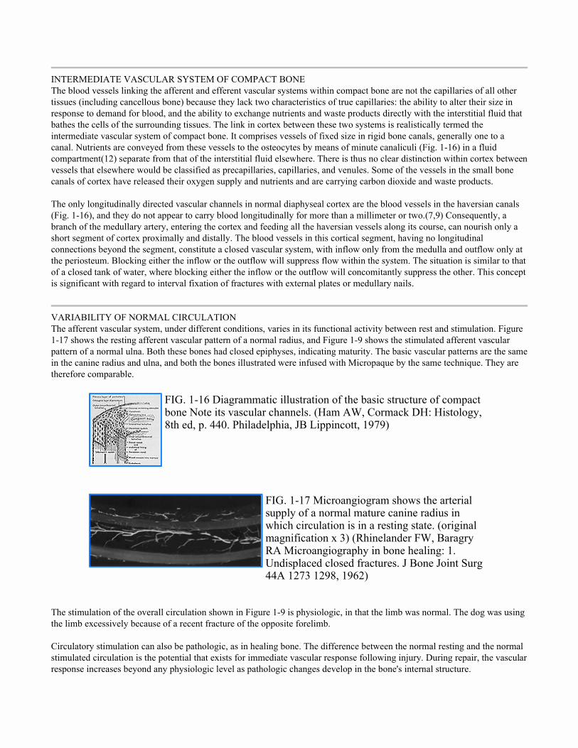
INTERMEDIATE VASCULAR SYSTEM OF COMPACT BONE The blood vessels linking the afferent and efferent vascular systems within compact bone are not the capillaries of all other tissues (including cancellous bone) because they lack two characteristics of true capillaries: the ability to alter their size in response to demand for blood, and the ability to exchange nutrients and waste products directly with the interstitial fluid that bathes the cells of the surrounding tissues. The link in cortex between these two systems is realistically termed the intermediate vascular system of compact bone. It comprises vessels of fixed size in rigid bone canals, generally one to a canal. Nutrients are conveyed from these vessels to the osteocytes by means of minute canaliculi (Fig. 1-16) in a fluid compartment(12) separate from that of the interstitial fluid elsewhere. There is thus no clear distinction within cortex between vessels that elsewhere would be classified as precapillaries, capillaries, and venules. Some of the vessels in the small bone canals of cortex have released their oxygen supply and nutrients and are carrying carbon dioxide and waste products. The only longitudinally directed vascular channels in normal diaphyseal cortex are the blood vessels in the haversian canals (Fig. 1-16), and they do not appear to carry blood longitudinally for more than a millimeter or two.(7,9) Consequently, a branch of the medullary artery, entering the cortex and feeding all the haversian vessels along its course, can nourish only a short segment of cortex proximally and distally. The blood vessels in this cortical segment, having no longitudinal connections beyond the segment, constitute a closed vascular system, with inflow only from the medulla and outflow only at the periosteum. Blocking either the inflow or the outflow will suppress flow within the system. The situation is similar to that of a closed tank of water, where blocking either the inflow or the outflow will concomitantly suppress the other. This concept is significant with regard to interval fixation of fractures with external plates or medullary nails.
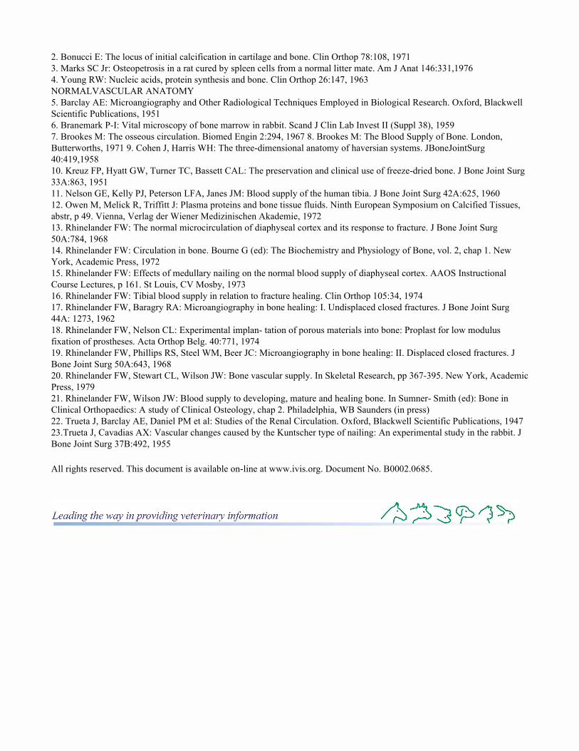
VARIABILITY OF NORMAL CIRCULATION The afferent vascular system, under different conditions, varies in its functional activity between rest and stimulation. Figure 1-17 shows the resting afferent vascular pattern of a normal radius, and Figure 1-9 shows the stimulated afferent vascular pattern of a normal ulna. Both these bones had closed epiphyses, indicating maturity. The basic vascular patterns are the same in the canine radius and ulna, and both the bones illustrated were infused with Micropaque by the same technique. They are therefore comparable.
The stimulation of the overall circulation shown in Figure 1-9 is physiologic, in that the limb was normal. The dog was using the limb excessively because of a recent fracture of the opposite forelimb. Circulatory stimulation can also be pathologic, as in healing bone. The difference between the normal resting and the normal stimulated circulation is the potential that exists for immediate vascular response following injury. During repair, the vascular response increases beyond any physiologic level as pathologic changes develop in the bone's internal structure.
FIG. 1-16 Diagrammatic illustration of the basic structure of compact bone Note its vascular channels. (Ham AW, Cormack DH: Histology, 8th ed, p. 440. Philadelphia, JB Lippincott, 1979)
FIG. 1-17 Microangiogram shows the arterial supply of a normal mature canine radius in which circulation is in a resting state. (original magnification x 3) (Rhinelander FW, Baragry RA Microangiography in bone healing: 1. Undisplaced closed fractures. J Bone Joint Surg 44A 1273 1298, 1962)

The direction of blood flow through diaphyseal cortex and the possibility of a change of direction through the normal vascular channels have been discussed at length above. Further insight into the subject may have been shed in preliminary/ experiments done in my laboratory with the use of a bone-grafting technique that was described by Bassett and his naval associates in a study of freeze-dried bone grafts.(10) Autogenous cortical inlay bone grafts were transplanted from one radius to the other in a series of dogs. Full-thickness segments of diaphyseal cortex were removed subperiosteally, with care to avoid thermal necrosis. The grafts were cut with size-60 drills turned at slow speed, through the same template for each radius, and were then removed by sharp osteotome. They were exchanged immediately between radii and were inserted with the same surface orientation that they had in the donor bone. They were pressed firmly into position and were secured by the soft-tissue closure of the wound. Examination of the bone specimens by standard microangiography and correlated histology revealed in-growth of blood vessels from the medulla by 2 weeks. The vessels appeared to enter old cortical channels, but that could not be determined definitely in these specimens. The striking vascular picture at 6 weeks is shown in Figure 1-18. Active vascularity, demonstrated by the number and size of injected blood vessels, is greater in the graft than in the neighboring cortex. This accounts for the greater porosity, observed histologically, of the graft than of this cortex. No osteoclasis was seen. By 6 weeks, there was advanced osseous union at each end of the graft, and by 12 weeks the graft appeared to be fully incorporated in the recipient bone. These experiments are chiefly of interest in the context of normal vascularization of diaphyseal cortex. Small longitudinal vessels entered the ends of the grafts from the adjacent host cortex, as occurs between cortical fragments in fracture repair. (See Chapter 38.) The most significant development was the invasion of the grafts by blood vessels along their endosteal surfaces but not along their periosteal surfaces. The chief source of regenerative vascularization was not the extraosseous blood supply, the usual source of vascularization of healing bone and the chief source of vascularization of cancellous chip grafts in incomplete bone defects. (See Chapter 39.) It was, rather, the medullary arterial system of the recipient radiusÑas if the graft were normal cortex of this radius. The recipient radius did recognize, however, that the cortical graft was necrotic and required a greatly supranormal supply of blood.
The fact that the primary invasion of the grafted segment of diaphyseal cortex is by blood vessels at only the endosteal surface has two possible explanations: that new afferent vessels, from whatever source, can primarily enter only the endosteal surface; or that only medulla-derived afferent vessels can primarily enter either surface, endosteal or periosteal. The latter concept would be consistent with the clinical observation in humans that massive grafts of pure cortex, used to bridge extensive defects in long-bones, unite promptly with the recipient bone at each end of the grafts but remain necrotic centrally for yearsÑeven though the central portions of the grafts have been residing in well vascularized soft-tissue beds.(21) These considerations are being examined as this chapter is being written. Using the same experimental model, I am reversing the surfaces of the inlay grafts in the recipient sites, placing the endosteal surface outward. In a parallel experimental series, grafts of pure cortex will be used to bridge complete bone defects. The timing, type, and direction of vascular invasion of the transplanted cortex will be observed
REFERENCES
STRUCTURE AND FUNCTION OF BONE 1. Anderson HC: Calcium-accumulating vesicles in the intercellular matrix of bone. In Hard Tissue Growth, Repair and Remineralization. Ciba Foundation's Symposium 11, North Holland, Excerpta Medica,1973
FIG. 1-18 Mature canine radial diaphysis 6 weeks after insertion of a cortical inlay autograft taken from the heterolateral radius: 8. microangiogram shows thorough vascularization of the graft from the medulla. The arrows indicate the two ends of the graft. g (original magnification x 5.5) (Rhinelander FW: Tibial blood supply in relation to fracture healing. Clin Orthop 105:34-81,10 1974)
2. Bonucci E: The locus of initial calcification in cartilage and bone. Clin Orthop 78:108, 1971 3. Marks SC Jr: Osteopetrosis in a rat cured by spleen cells from a normal litter mate. Am J Anat 146:331,1976 4. Young RW: Nucleic acids, protein synthesis and bone. Clin Orthop 26:147, 1963 NORMALVASCULAR ANATOMY 5. Barclay AE: Microangiography and Other Radiological Techniques Employed in Biological Research. Oxford, Blackwell Scientific Publications, 1951 6. Branemark P-I: Vital microscopy of bone marrow in rabbit. Scand J Clin Lab Invest II (Suppl 38), 1959 7. Brookes M: The osseous circulation. Biomed Engin 2:294, 1967 8. Brookes M: The Blood Supply of Bone. London, Butterworths, 1971 9. Cohen J, Harris WH: The three-dimensional anatomy of haversian systems. JBoneJointSurg 40:419,1958 10. Kreuz FP, Hyatt GW, Turner TC, Bassett CAL: The preservation and clinical use of freeze-dried bone. J Bone Joint Surg 33A:863, 1951 11. Nelson GE, Kelly PJ, Peterson LFA, Janes JM: Blood supply of the human tibia. J Bone Joint Surg 42A:625, 1960 12. Owen M, Melick R, Triffitt J: Plasma proteins and bone tissue fluids. Ninth European Symposium on Calcified Tissues, abstr, p 49. Vienna, Verlag der Wiener Medizinischen Akademie, 1972 13. Rhinelander FW: The normal microcirculation of diaphyseal cortex and its response to fracture. J Bone Joint Surg 50A:784, 1968 14. Rhinelander FW: Circulation in bone. Bourne G (ed): The Biochemistry and Physiology of Bone, vol. 2, chap 1. New York, Academic Press, 1972 15. Rhinelander FW: Effects of medullary nailing on the normal blood supply of diaphyseal cortex. AAOS Instructional Course Lectures, p 161. St Louis, CV Mosby, 1973 16. Rhinelander FW: Tibial blood supply in relation to fracture healing. Clin Orthop 105:34, 1974 17. Rhinelander FW, Baragry RA: Microangiography in bone healing: I. Undisplaced closed fractures. J Bone Joint Surg 44A: 1273, 1962 18. Rhinelander FW, Nelson CL: Experimental implan- tation of porous materials into bone: Proplast for low modulus fixation of prostheses. Acta Orthop Belg. 40:771, 1974 19. Rhinelander FW, Phillips RS, Steel WM, Beer JC: Microangiography in bone healing: II. Displaced closed fractures. J Bone Joint Surg 50A:643, 1968 20. Rhinelander FW, Stewart CL, Wilson JW: Bone vascular supply. In Skeletal Research, pp 367-395. New York, Academic Press, 1979 21. Rhinelander FW, Wilson JW: Blood supply to developing, mature and healing bone. In Sumner- Smith (ed): Bone in Clinical Orthopaedics: A study of Clinical Osteology, chap 2. Philadelphia, WB Saunders (in press) 22. Trueta J, Barclay AE, Daniel PM et al: Studies of the Renal Circulation. Oxford, Blackwell Scientific Publications, 1947 23.Trueta J, Cavadias AX: Vascular changes caused by the Kuntscher type of nailing: An experimental study in the rabbit. J Bone Joint Surg 37B:492, 1955
All rights reserved. This document is available on-line at www.ivis.org. Document No. B0002.0685.