Normal awake and sleep EEG
37
Normal awake and sleep EEG of paeds and adults Shehzad Hussain Technologist
-
Upload
sajjad-hussain-raja -
Category
Health & Medicine
-
view
262 -
download
1
Transcript of Normal awake and sleep EEG

Normal awake and sleep EEG of paeds and adults
Shehzad Hussain
Technologist
NEONATAL PERIOD
• The patterns observed in the neonatal EEG and the significance attributed to them depends on the conceptional maturity of the infant. Therefore, to evaluate the neonatal EEG, the reader must know the conceptional age of the infant (duration in weeks since last menstrual period/beginning of pregnancy), in addition to the postnatal age.

Premature Infants Less Than 29 Wk
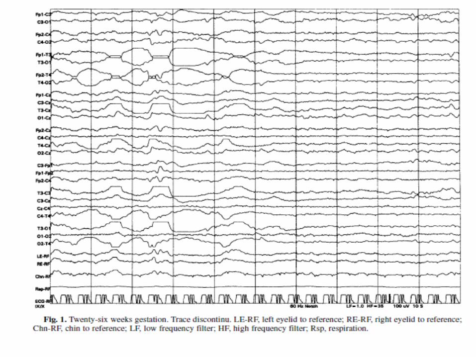
• The most striking feature of the early premature EEG is the discontinuity of activity.
• Bursts of high-voltage, predominantly delta activity, mixed with other frequencies and sharp waves, are interspersed with periods of low-voltage quiescent recordings. Interburst intervals may be prolonged, up to 90 s or more, whereas active bursts are generally shorter, but may last up to 1 min. This pattern is described as trace discontinue. (142-page primer soft)

Infancy (<1 Yr)
• With the end of the neonatal period (after 6-8 wk of age), trace alternant and frontal sharp waves are no longer observed in the healthy infant. In the awake state, an early, often poorly sustained, poorly reactive posterior dominant rhythm of three per second is first observed at 3 mo of age, and often increases to four per second at 4 mo. Reactivity to eye closure emerges quickly. The posterior dominant rhythm may be up to 6 to 7 Hz by 12 month of age.
• After the first to second month, infants move from wakefulness into QS.

Early Childhood (>1 to 3 Yr)
• The posterior dominant rhythm increases in frequency from 6 to 7 Hz in the second year to 7 to 8 Hz in the third year, and the blocking response to eye opening is now robust.
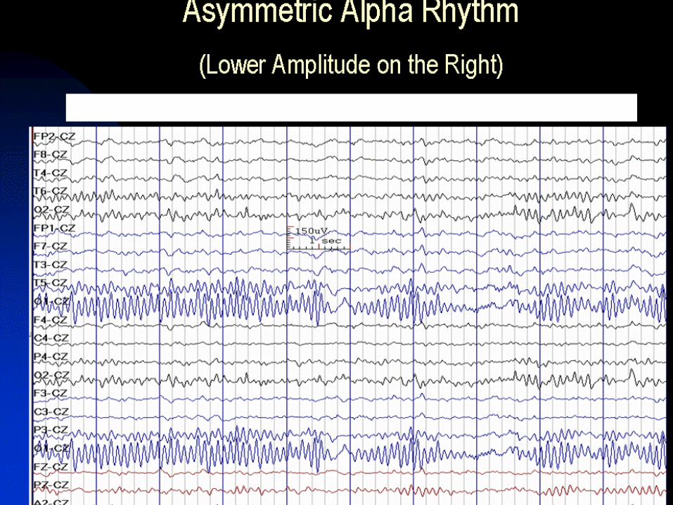
• As in adults, the dominant rhythm may be of greater amplitude in the non-dominant hemisphere.
• The difference should not be greater than 50%. In the waking background, delta activity remains prominent and may be observed diffusely or shifting in position throughout the record. There is a relative increase in the amount of theta activity, and this is visually the most striking frequency at this age. Throughout childhood, waking theta activity is prominent, often shifting in prominence from side to side.
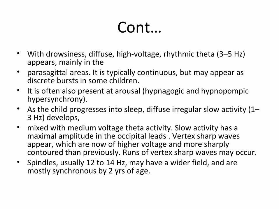
Cont…• With drowsiness, diffuse, high-voltage, rhythmic theta (3–5 Hz)
appears, mainly in the• parasagittal areas. It is typically continuous, but may appear as
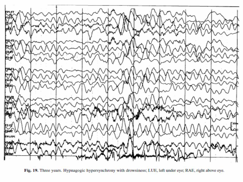
discrete bursts in some children.• It is often also present at arousal (hypnagogic and hypnopompic
hypersynchrony).• As the child progresses into sleep, diffuse irregular slow activity (1–
3 Hz) develops,• mixed with medium voltage theta activity. Slow activity has a
maximal amplitude in the occipital leads . Vertex sharp waves appear, which are now of higher voltage and more sharply contoured than previously. Runs of vertex sharp waves may occur.
• Spindles, usually 12 to 14 Hz, may have a wider field, and are mostly synchronous by 2 yrs of age.


Preschool Age (>3 to 6 Yr)
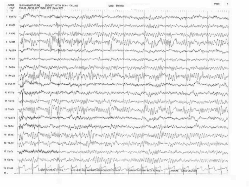
• At this age, the posterior basic rhythm consistently reaches alpha frequency. It is still of high amplitude, often greater than 100 μV. Throughout early childhood, low voltage background (<30 μV) is abnormal. Posterior slow waves of youth emerge at this age. These are 1.5- to 3-Hz waves, maximal in the occipital region. They are intermixed with posterior alpha, and, at times, fused slow waves can resemble occipital sharp waves, although lacking typical morphology and after-coming slow wave. Posterior slow waves, in common with the posterior dominant rhythm, block with eye opening.

Late Childhood (>6 to 12 Yr)
• Posterior dominant rhythm reaches 10 Hz by 10 yr of age, and reaches its maximum amplitude before that age. Posterior slow waves are prominent, and may be asymmetric, with higher amplitude on the right, as with the posterior dominant rhythm. Medium voltage semi-rhythmic frontal theta activity may be observed in healthy children at this age, and may persist into young adulthood. Hypnagogic hypersynchrony is disappearing, and is rare after the age of 6 yr.
• The drowsy pattern at this age is gradual alpha dropout, with increasing amounts of theta and delta activity.


Adolescence (>12 to 18 Yr)
• At this age, the EEG begins to resemble the adult EEG more closely, as the amount of underlying delta activity wanes completely (Fig. 27). The amplitude of the posterior dominant rhythm also declines gradually, although it remains higher than in adults throughout this period in many children. The mu rhythm reaches its maximum prominence at 15 to 16 yr, waning thereafter. Hyperventilation-related slowing is less pronounced, and the response to intermittent photic stimulation is mature, with a driving response occurring over the range 6- to 20-Hz stimulation.


NORMAL EEG PATTERN
• In Posterior Region
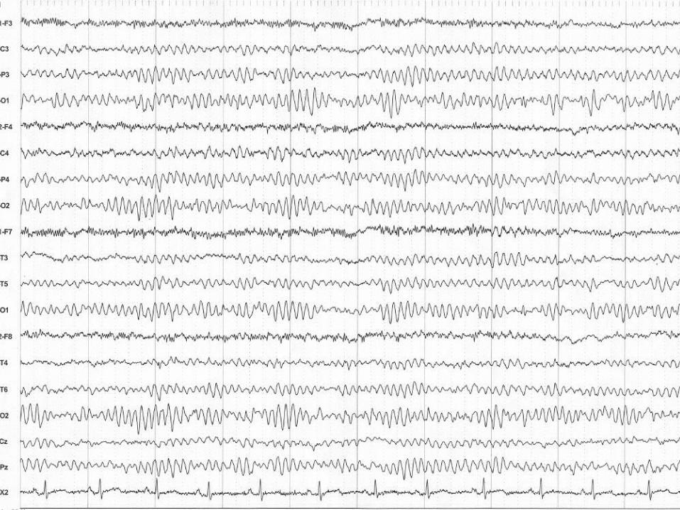
• A posterior alpha rhythm is recorded with highest amplitude from the O1 and O2 electrodes.
• A central alpha rhythm may also be recorded, but it is usually of lower amplitude than that recorded from occipital region.

Alpha Rhythm:-
• The trains of sinusoidal 8-13Hz activity recorded over the occipital region with eyes closure in awake alert adult.
• It should be bilaterally symmetrical (both in frequency & amplitudes).
• Reactivity of Alpha rhythm:-On eyes’ opening alpha rhythm attenuates which is called the reactivity of the alpha rhythm.


NORMAL EEG PATTERN
In Anterior Region:-Rhythmic beta activity is recorded in the frontal and central regions
with 14-35Hz frequency & its increased especially when sedatives are used. Drug enhanced beta is more commonly seen after sedation.
The beta rhythm is a low voltage fast frequency sinusoidal waves. Beta is presented during both awake and sleep states.Theta and delta are not prominent in the normal awake adult
EEG.


Normal Pattern Cont….
• Muscles or EMG activity:-
• An indication of patient tenseness is the recording of EMG activity in scalp muscles. EMG activity is faster in frequency and sharper in configuration than EEG activity.


Theta rhythm
• The Theta rhythm in normal adult appeared in a sleep stages while absent
In awake period.• Frequency;• It ‘s frequency range is >4 to<7HZ.• Distribution; Frontal/ frontocentral , occipital regions.


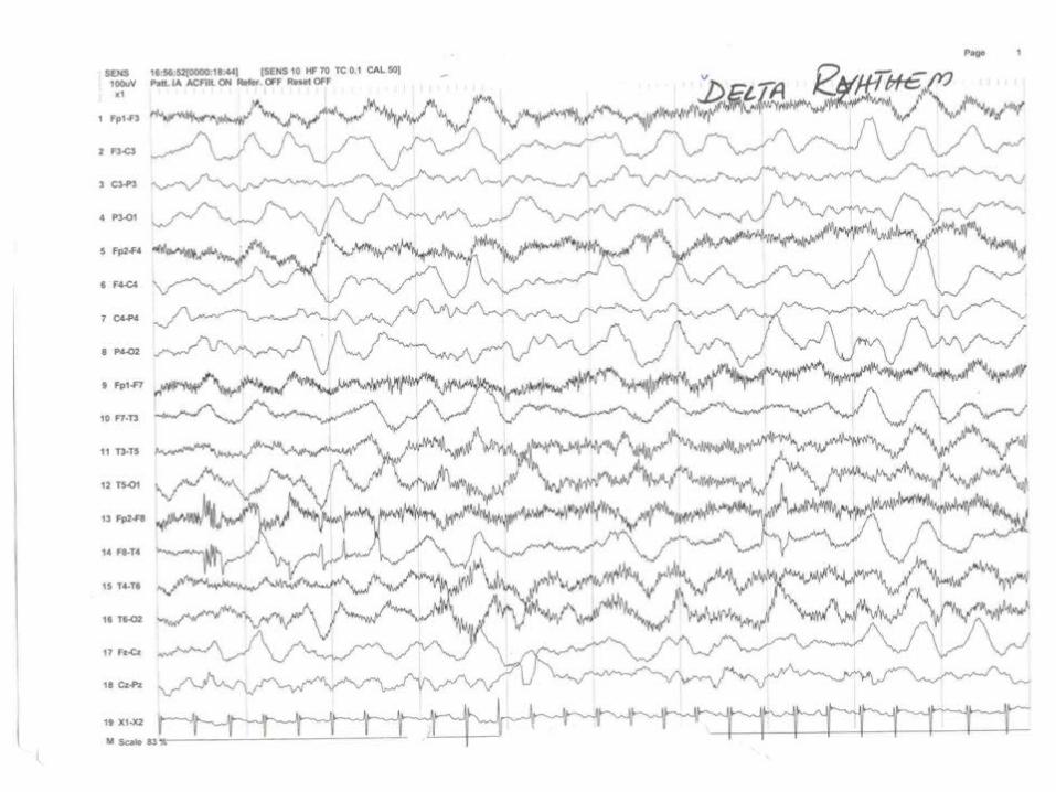
Delta activity
Describe by w.gray Walter English Describe by w.gray Walter English Physiologist in 1936.Physiologist in 1936.
The delta wave is termed as slow wave.The delta wave is termed as slow wave. Never seen in normal adult in awake state.Never seen in normal adult in awake state.
Delta wave occurring in a deep sleep in Delta wave occurring in a deep sleep in normal adult or showed Sever organic brain normal adult or showed Sever organic brain disease in adults. disease in adults.

Cont…..
• Frequency;
The frequency of delta wave is 0.5-3.5 HZ
Amplitude:
The amplitude of delta wave are variable.
Normally it may be around 50uv.• Distribution:-
Delta may occur diffused or it may be recorded as rhythmic wave in frontal.

Normal Pattern Cont….
• Symmetric and Asymmetric.
• The posterior dominant rhythm is usually symmetric, but asymmetries of up to 25% are often seen. Asymmetry should not be interpreted as abnormal unless it is at least 50%. The amplitude from the left hemisphere is often less than the right, so this should be considered when interpreting abnormalities.

The End