NON-NUCLEAR ESTROGEN RECEPTOR SIGNALING IN … · to invoke extranuclear signaling in endothelium...
16
NON-NUCLEAR ESTROGEN RECEPTOR SIGNALING IN ENDOTHELIUM* Qian Wu 1 , Ken Chambliss 1 , Michihisa Umetani 1 , Chieko Mineo 1 , and Philip W. Shaul 1 From Division of Pulmonary and Vascular Biology 1 , Department of Pediatrics University of Texas Southwestern Medical Center, Dallas, Texas 75390 Running head: Non-nuclear ER signaling in endothelium Address correspondence to: Philip W. Shaul, M.D., Division of Pulmonary and Vascular Biology, Department of Pediatrics, University of Texas Southwestern Medical Center, 5323 Harry Hines Blvd. Dallas, TX 75390-9063. Fax: 214-648-2096; Email: [email protected] In addition to the classical function of estrogen receptors (ER) as transcription factors, evidence continues to accumulate that they mediate non- nuclear processes in numerous cell types including endothelium, in which they activate endothelial NO synthase (eNOS). Non-nuclear ER signaling entails unique post-translational modifications and protein-protein interactions of the receptor with adaptor molecules, kinases, and G proteins. Recent in vitro and in vivo studies in mice using an estrogen dendrimer conjugate (EDC) that is excluded from the nucleus indicate that non-nuclear ER activation underlies the migration and growth responses of endothelial cells to estrogen, but not the growth responses of endometrial or breast cancer cells to the hormone. In this minireview, the features of ERα and protein-protein interactions that enable it to invoke extranuclear signaling in endothelium and the consequences of that signaling will be discussed. Although estrogen receptors (ER) function classically as transcription factors, more recently it has become apparent that ER also have the novel capacity to activate non-nuclear signaling in a variety of cell types(1-3). An understanding of the basis for non-nuclear ER signaling has been derived from the study of ER associated with plasmalemmal caveolae/lipid rafts in endothelial cells(4), which is the focus of this review. After a brief discussion of the actions of estrogen on endothelium that highlights non-nuclear processes, we will summarize the nature of non-nuclear ER function in endothelium. We will then discuss the signaling events that non-nuclear ER mediate in endothelium, the mechanisms by which they are coupled to kinases and other signaling or adapter molecules, and recent work interrogating these processes in vivo. Finally, we will highlight the current questions in the field. Endothelial Actions of Estrogen Estrogen has potentially potent cardiovascular protective actions, and these are primarly through direct effects on endothelial and vascular smooth muscle (VSM) cells(5-7), which express the two primary estrogen receptor (ER) isoforms, ERα and ERβ(4;5). In endothelial cells in culture, estrogen upregulates the expression of endothelial NO synthase (eNOS) (4), cyclooxygenase type 1(8), MMP-2 (9), and ERα (10), and it blunts the expression of endothelin(11) and the type 1 angiotensin II receptor(12). Along with these actions that involve the classical functions of ER as transcription factors, short-term effects of estrogen on the vasculature were demonstrated in humans that provided the initial suggestion that there are non-nuclear actions of the hormone and its receptors. In early studies, ethinyl estradiol acutely attenuated abnormal coronary vasoconstrictor reponses to acetylcholine (Ach) in postmenopausal women, and it also increased basal coronary blood flow and decreased coronary vascular resistance(13). Other studies of estradiol (E 2 ) administered to yield premenopausal serum levels showed no change in basal coronary vasomotor tone, but there were greater vasodilatory responses to Ach when Ach and E 2 were give simultaneously(14). In experiments in http://www.jbc.org/cgi/doi/10.1074/jbc.R110.191791 The latest version is at JBC Papers in Press. Published on February 22, 2011 as Manuscript R110.191791 Copyright 2011 by The American Society for Biochemistry and Molecular Biology, Inc. by guest on February 3, 2019 http://www.jbc.org/ Downloaded from
Transcript of NON-NUCLEAR ESTROGEN RECEPTOR SIGNALING IN … · to invoke extranuclear signaling in endothelium...

NON-NUCLEAR ESTROGEN RECEPTOR SIGNALING IN ENDOTHELIUM* Qian Wu1, Ken Chambliss1, Michihisa Umetani1, Chieko Mineo1, and Philip W. Shaul1
From Division of Pulmonary and Vascular Biology1, Department of Pediatrics University of Texas Southwestern Medical Center, Dallas, Texas 75390
Running head: Non-nuclear ER signaling in endothelium Address correspondence to: Philip W. Shaul, M.D., Division of Pulmonary and Vascular Biology, Department of Pediatrics, University of Texas Southwestern Medical Center, 5323 Harry Hines Blvd. Dallas, TX 75390-9063. Fax: 214-648-2096; Email: [email protected] In addition to the classical function of estrogen receptors (ER) as transcription factors, evidence continues to accumulate that they mediate non-nuclear processes in numerous cell types including endothelium, in which they activate endothelial NO synthase (eNOS). Non-nuclear ER signaling entails unique post-translational modifications and protein-protein interactions of the receptor with adaptor molecules, kinases, and G proteins. Recent in vitro and in vivo studies in mice using an estrogen dendrimer conjugate (EDC) that is excluded from the nucleus indicate that non-nuclear ER activation underlies the migration and growth responses of endothelial cells to estrogen, but not the growth responses of endometrial or breast cancer cells to the hormone. In this minireview, the features of ERα and protein-protein interactions that enable it to invoke extranuclear signaling in endothelium and the consequences of that signaling will be discussed. Although estrogen receptors (ER) function classically as transcription factors, more recently it has become apparent that ER also have the novel capacity to activate non-nuclear signaling in a variety of cell types(1-3). An understanding of the basis for non-nuclear ER signaling has been derived from the study of ER associated with plasmalemmal caveolae/lipid rafts in endothelial cells(4), which is the focus of this review. After a brief discussion of the actions of estrogen on endothelium that highlights non-nuclear processes, we will summarize the nature of non-nuclear ER function in endothelium. We will then
discuss the signaling events that non-nuclear ER mediate in endothelium, the mechanisms by which they are coupled to kinases and other signaling or adapter molecules, and recent work interrogating these processes in vivo. Finally, we will highlight the current questions in the field.
Endothelial Actions of Estrogen
Estrogen has potentially potent cardiovascular protective actions, and these are primarly through direct effects on endothelial and vascular smooth muscle (VSM) cells(5-7), which express the two primary estrogen receptor (ER) isoforms, ERα and ERβ(4;5). In endothelial cells in culture, estrogen upregulates the expression of endothelial NO synthase (eNOS) (4), cyclooxygenase type 1(8), MMP-2 (9), and ERα (10), and it blunts the expression of endothelin(11) and the type 1 angiotensin II receptor(12). Along with these actions that involve the classical functions of ER as transcription factors, short-term effects of estrogen on the vasculature were demonstrated in humans that provided the initial suggestion that there are non-nuclear actions of the hormone and its receptors. In early studies, ethinyl estradiol acutely attenuated abnormal coronary vasoconstrictor reponses to acetylcholine (Ach) in postmenopausal women, and it also increased basal coronary blood flow and decreased coronary vascular resistance(13). Other studies of estradiol (E2) administered to yield premenopausal serum levels showed no change in basal coronary vasomotor tone, but there were greater vasodilatory responses to Ach when Ach and E2 were give simultaneously(14). In experiments in
http://www.jbc.org/cgi/doi/10.1074/jbc.R110.191791The latest version is at JBC Papers in Press. Published on February 22, 2011 as Manuscript R110.191791
Copyright 2011 by The American Society for Biochemistry and Molecular Biology, Inc.
by guest on February 3, 2019http://w
ww
.jbc.org/D
ownloaded from

men using intravenous conjugated estrogens and phytoestrogens, there was rapid NO-dependent vasodilation 15 min after treatment, suggesting that estrogen promotes the bioavailability of NO and that these effects may be comparable in men and women(15). In 1997 it was reported by two laboratories that estrogen rapidly stimulates eNOS enzymatic activity in an ER-dependent manner(16;17). NO derived from eNOS has diverse potential beneficial vascular actions, including the promotion of endothelial cell growth and migration, the attenuation of VSM cell growth and migration, and the antagonism of platelet activation, thrombus formation and leukocyte-endothelial cell adhesion(18). Using genetically-modified mice, numerous pioneering studies begun in the late 1990’s then revealed that the basis for estrogen-related protection from atherosclerosis and vascular injury lies in direct actions of the hormone on ER expressed in vascular cells, and that along with other processes there is likely a primary role for E2/ER influencing eNOS activity (4).
Nature of Non-nuclear ER in Endothelium
Estrogen activates non-nuclear ER to result in rapid signaling in cell types in culture as diverse as oocytes, osteoblasts, osteoclasts, breast cancer cells, adipocytes and endothelial cells(1-3;19;20). The vast majority of studies in these various model systems have indicated that the rapid actions of estrogen originate at the cell surface and not in the nucleus. In the endothelium ERα was first implicated in non-nuclear signaling by the findings that overexpression of the receptor enhanced the acute activation of eNOS by E2, and that eNOS stimulation by endogenous receptor was inhibited by both ICI 182,780 and tamoxifen and by transfection with an ERα mutant lacking coding sequence distal to amino acid 271, which excludes the hormone binding domain(21). Initial evidence of potential involvement of endothelial cell ERβ in non-nuclear signaling arose from studies in
which the response to endogenous ER activation was inhibited by the ERβ-selective antagonist RR-tetrahydrochrysene(22). The localization of ER functional coupling to eNOS in isolated plasma membranes provided additional clarity of membrane receptor identity, with immunoidentification studies directed at multiple epitopes detecting the same 67kDa ERα protein and the same 54 kDa ERβ protein in purified plasma membrane fractions and in the nucleus(22;23). The requirement for ER and eNOS colocalization on the plasma membrane was further evident in reconstitution studies in COS-7 cells in which plasma membranes from cells expressing eNOS and ERα displayed rapid ER-mediated eNOS stimulation(24). The majority of studies in endothelium have identified full-length ERα (67 kDa) as the predominant ERα form associated with the plasma membrane involved in non-nuclear signaling(21;23;25). However, an N-terminus-deleted splice variant, ERα46, has been found in certain human endothelial cell lines, and it has been shown to colocalize with caveolin-1 in caveolae and effectively transduces membrane-initiated responses to E2(26;27). Interestingly, the transcriptional activity of ERα46 is greatly compromised compared to that of full-length ERα(28). The G protein-coupled receptor GPR30 has been proposed to be a third form of ER, and it has been localized to the plasma membrane in breast cancer cells and to endoplasmic reticulum when overexpressed in COS-7 cells(29;30). As a result of varying phenotypes in three mouse lines with diminished GPR30 expression and other conflicting findings, it is controversial whether GPR30 is a biologically-relevant ER or a collaborator in non-nuclear functions of the classical ER in certain contexts(31). The cell types expressing GPR30 have been evaluated in a mouse model with a lacZ reporter in the GPR30 locus. Regarding vascular expression, LacZ positive vascular cells were present in a number of arterial structures, but their distribution was
by guest on February 3, 2019http://w
ww
.jbc.org/D
ownloaded from

restricted to specific vascular beds, including arterioles in the kidney and vasa vasorum of the aorta, and the final branches of mesenteric arteries. The aorta, carotid, intercostal, renal and superior mesenteric arteries were negative for LacZ staining. In CNS vasculature the LacZ-positive cells were VSM cells and pericytes and staining was absent in endothelium, whereas in the kidney LacZ staining colocalized with the endothelial cell marker PECAM-1(32). Although GPR30 expression was not detected in the carotid arteries in the LacZ reporter mice, carotid arteries isolated from GPR30+/+ mice display relaxation in response to the GPR30 agonist G-1, whereas relaxation is absent in arteries from GPR30-/- mice(33). In recent studies of isolated precontracted rat carotid arteries, G-1 and another GPR30 agonist 5408-0877 both caused endothelium-dependent, NO-dependent relaxation(34). However, whereas primary endothelial cells from wild-type mice display E2 binding to whole cells or membranes, in endothelial cells from ERα-/-;ERβ-/- mice, in which GPR30 protein expression is demonstrable and unaltered, E2 binding is absent. In parallel, whereas wild-type endothelial cells display rapid signaling in response to E2, cells from ERα/ERβ double knockout mice do not, despite unchanged GPR30 expression(25). The additional key experiment is a test of responses to E2 by GPR30+/+ versus GPR30-
/- endothelium, but this has not yet been reported. Thus, although GPR30 is expressed in certain endothelial cells, there is currently a lack of clear evidence of a role for the receptor in estrogen action in endothelium. Instead, classical ERα and ERβ, and perhaps truncated forms of ERα (i.e. ERα46), mediate the non-nuclear actions of the hormone in endothelial cells.
ER Localization to Plasma Membrane Microdomains
Caveolae are specialized, cholesterol-rich plasma membrane organelles that are a subset of lipid rafts that compartmentalize signal transduction
molecules on the cell surface(35), and eNOS is targeted to caveolae via myristolyation and palmitoylation(24;36). Fractionation of endothelial cell plasma membranes has revealed both ERα and ERβ association with caveolae membranes. The proteins were also found associated with the noncaveolae plasma membrane fraction. Importantly, studies in isolated caveolae membranes demonstrated functional coupling of both ERα and ERβ to eNOS, and eNOS activation in caveolae in response to E2 was comparable to that observed with the classical eNOS agonist acetylcholine that signals through M2 muscarinic receptors(22;23). The basis for ER localization to the plasma membrane/caveolae has been evaluated by mutagenesis, primarily of ERα. Although not observed in all reports, there is evidence that ERα interacts with caveolin-1, that the interaction requires serine 522 of the receptor, which is located within the ligand binding domain (E domain) (Fig. 1), and that the interaction facilitates the trafficking of ERα to caveolae(23;37). The participation of caveolin-1 in ER function has been evaluated in caveolin-1 deficient mice, which are viable and fertile but display major changes in the structure and function of the heart, lung and blood vessels that may be due to the uncoupling of eNOS from caveolae(38;39). The mammary glands of caveolin-1-/- mice are hyper-responsive to estrogen and develop dysplastic lesions with adjacent stromal angiogenesis(40), suggesting crosstalk between caveolin-1 and ER in tumor microvasculature in vivo. In addition to potential participation of caveolin-1 in ERα targeting to the plasma membrane, cysteine 447 of ERα, which is also within the ligand binding domain, is a palmitoylation site important for membrane localization of the receptor (Fig. 1)(41), and additional residues flanking cysteine 447 are also required for plasma membrane targeting via palmitoylation(42). A role for ERα palmitoylation in endothelial cell receptor regulation is further supported by the observation in EA.hy927 immortalized
by guest on February 3, 2019http://w
ww
.jbc.org/D
ownloaded from
human endothelial cells that the N-terminally-truncated ERα46 is palmitoylated, and that palmitoylation is required for E2-enhanced plasma membrane recruitment of the receptor(27). The best available information regarding the topology of plasma membrane-associated ER is limited to studies of N-terminally myc-tagged ERα46 expressed in COS-7 cells. Without permeabilization before staining, fluorescence-activated cell sorting detected cells only with antibody to the C-terminus of ERα46, whereas signal with anti-c-myc antibody occurred only following permeabilization(43). These findings suggest that ERα46 possesses a C-terminal ectodomain, but the basis for such possible membrane orientation remains enigmatic and studies of the plasma membrane topography of endogenous receptor populations are needed. However, it is now well-established that caveolae are the key plasma membrane microdomain in which ERα and ERβ reside to regulate extranuclear events in endothelial cells, and that their localization to caveolae most likely depends on interaction with caveolin and/or receptor palmitoylation.
Signaling by Non-nuclear ER in Endothelium
Following the initial discovery of rapid eNOS activation by E2(16;17), the signaling underlying ER coupling to eNOS received considerable attention. In early studies roles for tyrosine kinases and MAP kinases were implicated by the findings that genistein and the MAP kinase kinase inhibitor PD98059 prevented eNOS activation by E2(21). Further work employing pharmacologic intervention or detection of changes in protein phosphorylation upon E2 treatment in arteries or cultured endothelial cells indicated requirements for PI3 kinase and Akt kinase and Erk 1/2, and enhanced phosphorylation of Ser1177 of eNOS (Fig. 2)(25;44-46). The enhancement of eNOS phosphorylation by Akt enacts activation of the enzyme by a variety of agonists besides
E2(18), and the signaling events immediately proximal to the Ser1177 phosphorylation invoked by E2 are similar to the upstream events by which a number of plasma membrane receptors regulate eNOS activity. However, in a unique manner ERα binds in a ligand-dependent fashion with the p85α regulatory subunit of PI3 kinase, the interaction is direct (Fig. 2), and it is not mediated by the Src-homology SH2/SH3 domains of p85α(46). The activation of PI3 kinase and its substrate Akt in response to E2 binding to non-nuclear ER is mediated by c-Src kinase, whose SH2 domain interacts with phosphorylated Tyr537 of ERα (Figs. 1,2), and this interaction may facilitate the plasma membrane recruitment of ERα(26;47;48). In studies primarily in MCF-7 breast cancer cells, it has been demonstrated that the methylation of Arg260 within the ERα DNA binding domain by the arginine methyltransferase PRMT1 (Fig. 1) triggers the interaction of the receptor with the p85 subunit of PI3 kinase and with c-Src, and that this process occurs in the cytoplasm and participates in non-nuclear downstream signaling(49). Whether Arg260 methylation is required for non-nuclear ERα signaling in endothelial cells is yet to be determined. In addition to activating kinases in endothelial cells, there is evidence that non-nuclear ER stimulation alters intracellular calcium homeostasis. In studies of human internal thoracic artery endothelium in situ and human arterial endothelial cells in culture, E2 caused a rapid increase in intracellular calcium at physiologically-relevant concentrations of the hormone (10-
9M), this was attenuated following intracellular calcium store depletion, and concurrent NO release was demonstrable. The ER antagonist tamoxifen blunted these responses. Similar findings were obtained using E2 conjugated to bovine serum albumin (E2-BSA), which has been employed to evaluate the role of cell surface receptors(50). However, observations with E2-BSA should be interpreted with caution because freshly-prepared solutions of E2-BSA contain free immunoassayable E2, E2-
by guest on February 3, 2019http://w
ww
.jbc.org/D
ownloaded from

BSA binds to ER only poorly because the E2 is linked to BSA through chemical groups in E2 that are important for ER binding, and certain E2-BSA preparations are of very high molecular weight, suggesting extreme protein crosslinking(51;52). E2 and E2-BSA also caused comparable calcium transients in cultured rat endothelial cells that were attenuated by the ER antagonist ICI 182,780(53). However, in other studies it has been found that E2 does not induce a discernable acute increase in intracellular calcium in endothelial cells(17;54) despite clear evidence that the non-nuclear activation of eNOS by E2 is calcium-dependent(16;55). These disparities are potentially related to the involvement of highly-localized, caveolae-associated calcium pools and caveolae-associated proteins that regulate calcium homeostasis(56), since E2 activation of the enzyme in isolated endothelial cell plasma membranes and caveolae occurs in the absence of added calcium but is completely prevented by calcium chelation(23). The localization of ERα to endothelial cell caveolae, and both physical and functional interaction of the receptor with other signaling molecules in that domain, suggests that adaptor proteins may also participate in non-nuclear actions of ERα in endothelial cells. One such candidate is the ERα binding protein striatin, which is a member of the WD-repeat protein family. In studies limited to the endothelial cell line Eahy926, striatin and ERα were coimmunoprecipitated, and overexpression of striatin increased the abundance of the receptor at the plasma membrane. In addition, E2 stimulated the formation of a complex containing ERα and striatin and Gαi, which is critically involved in non-nuclear ERα function in endothelial cells (see below). Pull-down experiments with purified proteins identified direct interaction between striatin and ERα involving amino acids 183-253 of the receptor (Fig. 1), and a peptide representing amino acids 176-253 of ERα prevented E2 activation of Erk1/2, Akt and eNOS(57). These findings support a role for striatin in non-nuclear ERα function
in endothelial cells, but further genetic evidence and studies in primary endothelial cells and in in vivo models are needed to strengthen such a conclusion. There are three additional molecules with direct interactions with ERα of consequence to non-nuclear signaling that have been studied in nonendothelial cells. Work in HeLa and MCF-7 cells has revealed that ERα interacts with hematopoietic PBX-interacting protein (HPIP), which mediates the binding of the receptor with tubulins and also recruits the p85 subunit of PI3 kinase and Src kinases to an ERα complex upon E2 binding, leading to the stimulation of Akt and Erk1/2. There is direct interaction of ERα with HPIP that requires amino acids 264-302 and 552-595 of the receptor (Fig. 1)(58). The protein Shc (Src-homology and collagen homology), which has direct interaction with ERα requiring residues between amino acids 1 and 367 of the receptor, is critically involved in linking E2 binding to non-nuclear ERα with morphologic changes and the growth of breast cancer cells(59). Furthermore, a scaffolding protein designated as MNAR (modulator of nongenomic activity of ER) has been described that promotes E2-induced interaction between ERα and Src family kinase(s) in non-endothelial cells(60). ERα-related functions of HPIP, Shc and MNAR in endothelium are yet to be queried. G protein Coupling of Non-nuclear ER in
Endothelium
As illustrated above, our deepest understanding of non-nuclear ER signaling in endothelium resides in the realm of the modulation of eNOS. The participation of c-Src, PI3 kinase, Akt and Erk1/2 mimicks the involvement of these kinases in eNOS modulation by numerous other agonist-receptor pairs(18). However E2-ER regulation of eNOS entails not only the unique direct interactions between ERα and c-Src and PI3 kinase, but also novel G protein coupling of the receptor. The potential involvement of G proteins was initially revealed in studies demonstrating that E2 activation of eNOS is pertussis toxin
by guest on February 3, 2019http://w
ww
.jbc.org/D
ownloaded from
(PTX) sensitive, and that ERα and Gαi can be coimmunoprecipitated from endothelial cell plasma membranes, with PTX preventing the coimmunoprecipitation(61). Pull-down experiments then revealed that ERα binds directly to Gαi and Gβγ, and mutagenesis as well as experiments with blocking peptide showed that this occurs via amino acids 251-260 and 271-595 of human ERα, respectively (Fig. 1). Studies of ERα complexed with heterotrimeric G proteins further showed that E2 causes the release of both Gαi and Gβγ without stimulating guanine nucletide exchange. Moreover, in COS-7 cells the disruption of ERα-Gαi interaction by deletion mutagenesis of the Gαi binding domain of ERα, or expression of blocking peptide, or Gβγ sequestration with β-adrenergic receptor kinase C terminus fully attenuates Src kinase and Erk1/2 activation by E2. In endothelial cells the disruption of ERα-Gαi interaction prevents eNOS activation and also the blunting of monocyte adhesion and the stimulation of cell migration caused by E2(62). Thus, direct ERα-G protein interaction underlies the functional coupling of the receptor to kinase cascades and the resulting downstream cellular responses in endothelium (Fig. 2).
Crosstalk Between Non-nuclear ER Signaling and Nuclear Processes
Along with the rapid cellular responses such as the activation of NO production that result from the immediate signaling caused by non-nuclear ER activation in endothelial cells, there are important potential consequences on endothelial cell gene expression. Studies in nonvascular cells indicate that ERα Ser104, Ser106 and Ser118 are potential targets for phosphorylation by the cyclin-dependent protein kinases Cdk2 (Ser104, Ser106) and Cdk7 (Ser118), with Cdk7 actions occurring in response to E2. In addition, ERα Ser118 can be phosphorylated by Erk1/2 in a ligand-independent manner. It is further known that these events modify the regulation of gene transcription by ERα(63-
65). The activation of NO production by non-nuclear E2 and ERα signaling also potentially influences nuclear actions of the hormone. S-nitrosylation of cysteine residues within ERα has been detected upon NO donor treatment of MCF-7 breast cancer cells, resulting in the inhibition of ERα DNA binding at specific estrogen response elements. Thus, although the studies to date have been in nonendothelial cells, the phosphorylation and also the S-nitrosylation of ERα invoked by its own non-nuclear signaling may have important influence on nuclear actions of E2 and ER in the endothelium. Direct evidence of crosstalk between non-nuclear ER signaling and nuclear processes has been obtained in studies in cultured endothelial cells. In human umbilical vein endothelial cells (HUVEC), E2 treatment for 40 min upregulates the expression of 250 genes, and their upregulation is PI3 kinase-dependent. Cyclooxygenase-2 is one such target gene, and its upregulation results in increased PGI2 and PGE2 secretion(66). The inhibitor of angiogenesis thrombospondin-1 is transiently downregulated by E2 in HUVEC via Erk1/2 and c-Jun NH2-terminal kinase/stress-activated protein kinase signaling(67). Furthermore, E2 activation of eNOS leads to the upregulation of telomerase activity via the stimulation of transcriptional transactivation of hTERT, the catalytic subunit of human telomerase(68). Additional studies have demonstrated that these processes participate in the pro-migratory response of endothelial cells to E2(66-68), to which the non-nuclear activation of the small GTPase RhoA, Rho kinase and moesin also contribute(69). It has also been demonstrated that both the pro-migratory response to E2 and the stimulation of endothelial cell growth by the hormone are Gαi-, Src-, and eNOS-dependent(70). Furthermore, the ability of E2 to preserve endothelial cell stress fiber formation and actin and membrane integrity under metabolic stress, and to attenuate hypoxia-induced apoptosis is dependent on p38 MAP kinase activation and resulting
by guest on February 3, 2019http://w
ww
.jbc.org/D
ownloaded from
activation of the mitogen-activated protein kinase-activated protein kinase-2 and heat shock protein 27 phosphorylation(71). These diverse processes take place in endothelial cells concurrently with the classical actions of ER serving as transcription factors binding directly to DNA via estrogen response elements or indirectly via tethered processes occuring via Sp1 or AP-1. Interestingly, the protein phosphatase PP2A interacts directly with ERα via amino acids 176-253 of the receptor (Fig. 1), and the phosphatase dephosphorylates ERαS118. Okadaic acid inhibition of PP2A activates ERα-mediated gene transcription, and in endothelial cells this leads to eNOS protein upregulation(72). Since PP2A also dephosphorylates eNOS Ser1177(73), which is phosphorylated by Akt to yield greater eNOS enzymatic activity upon E2 treatment, it is interesting to envision that a member of the complex of proteins interacting with non-nuclear ER may counterregulate the activating function of that complex. It is becoming more and more apparent that the ultimate physiologic responses of endothelium to E2 entail complex combinations of non-nuclear and nuclear processes and the influences of the diverse modes of crosstalk that occur between them. In Vivo Interrogation of Non-nuclear ER
Actions Up until recently, our ability to rigorously distinguish nuclear from non-nuclear ER actions in endothelium and other cell types was limited, and in vivo interrogation was not feasible. E2-BSA was initially used in studies of cultured endothelial cells and it caused the stimulation of eNOS phosphorylation(74). However, as noted above, the selectivity of E2-BSA for non-nuclear ER activation and its stability are uncertain. An estrogen-dendrimer conjugate (EDC) was then created in which estrogen is attached to a large, positively charged nondegradable poly(amido)amine (PAMAM) dendrimer via hydrolytically stable linkages, thereby
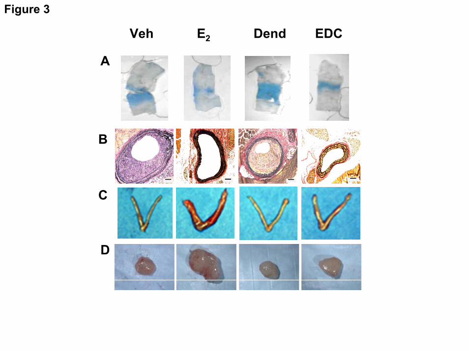
excluding EDC from the nucleus(75). EDC stimulates cultured endothelial cell proliferation and migration via ERα, Gαi, and the activation of Src kinase and eNOS that they invoke. Studies of ERE-luciferase reporter mice and ER target gene expression in the uterus further demonstrated that EDC causes the selective activation of non-nuclear ER signaling when administered in vivo. In mice, E2 and EDC equally stimulate carotid artery reendothelialization (Fig. 3A) and this is dependent on ER-Gαi coupling(70), and both agents attenuate the development of neointimal hyperplasia that occurs following endothelial injury in the setting of hypercholesterolemia (Fig. 3B). However, whereas uterine (Fig. 3C) and MCF-7 cell breast cancer xenograft growth in vivo (Fig. 3D) are stimulated by E2, they are not promoted by EDC(70). Further studies in cultured endometrial carcinoma cells and MCF-7 breast cancer cells demonstrated that although non-nuclear signaling and Erk1/2 activation indeed occur upon treatment with EDC, in contrast to the response of endothelial cells, the selective activation of non-nuclear ER signaling does not stimulate growth of the cancer cells(70;75). Thus, EDC is a non-nuclear SERM in vivo, and non-nuclear ER signaling provides potent cardiovascular protection without promoting uterine or breast cancer tumor growth. These processes potentially can be harnessed to provide vascular benefit without increasing cancer risk.
Conclusions and Current Questions
The study of non-nuclear ER signaling in endothelium has revealed novel mechanisms that provide greater diversity of function of the receptor. In its extranuclear locale, ERα has novel actions related to unique post-translational modifications and interactions with adaptor proteins, kinases and phosphatases, and also G proteins. The studies in mice using EDC revealed that non-nuclear ERα coupling to Gαi is operative in vivo, and a beneficial impact on neointima formation has been demonstrated.
by guest on February 3, 2019http://w
ww
.jbc.org/D
ownloaded from

We do not yet know whether selective non-nuclear ER activation affords atheroprotection, and although non-nuclear ERα activation in endothelium is likely of importance, the cell types in which non-nuclear ERα influence vascular disease pathogenesis are yet to be identified. It is also unclear how non-nuclear ER function contributes to estrogen modulation of metabolism. Considering the prevalence of crosstalk between non-nuclear and nuclear ER actions, it is important to recognize that although EDC provides a selective gain-of-function intervention targeting non-nuclear ER, loss-of-function strategies to specifically negate non-nuclear ER function are also needed. It is only through loss-of-function that new knowledge will be gained about the role of non-nuclear ER signaling in the actions of endogenous estrogens, whose levels are highly dynamic. The
structural features of ERα involved in non-nuclear signaling (Fig. 1) can potentially be modified to yield selective non-nuclear loss-of-function. Such approaches will be important in future studies attempting to understand the disparate roles of non-nuclear ER activation in the regulation cell growth, as observed in endothelial cells compared with endometrial or breast cancer cells(70). Numerous mysteries also remain about the topology of plasma membrane-associated ER and the basis by which trafficking to plasma membrane versus nucleus is determined. It is only through further investigation of this unique aspect of endocrinology that we will increase our understanding of how non-nuclear ER signaling influences the behavior of endothelium, as well as other cell types, under normal or pathologic conditions.
REFERENCES 1. Norman, A. W., Mizwicki, M. T., and Norman, D. P. (2004) Nat. Rev. Drug Discov. 3,
27-41 2. Osborne, C. K. and Schiff, R. (2005) J Clin. Oncol. 23, 1616-1622 3. Levin, E. R. (2005) Mol. Endocrinol. 19, 1951-1959 4. Chambliss, K. L. and Shaul, P. W. (2002) Endo. Rev. 23, 665-686 5. Mendelsohn, M. E. and Karas, R. H. (1999) N. Engl. J. Med. 340, 1801-1811 6. Hodgin, J. B. and Maeda, N. (2002) Endocrinology 143, 4495-4501 7. Mendelsohn, M. E. (2000) J. Steroid Biochem. Mol. Biol. 74, 337-343 8. Jun, S. S., Chen, Z., Pace, M. C., and Shaul, P. W. (1998) J. Clin. Invest 102, 176-183 9. Wingrove, C. S., Garr, E., Godsland, I. F., and Stevenson, J. C. (1998) Biochim. Biophys.
Acta 1406, 169-174 10. Ihionkhan, C. E., Chambliss, K. L., Gibson, L. L., Hahner, L. D., Mendelsohn, M. E., and
Shaul, P. W. (2002) Circ Res 91, 814-820 11. Akishita, M., Kozaki, K., Eto, M., Yoshizumi, M., Ishikawa, M., Toba, K., Orimo, H.,
and Ouchi, Y. (1998) Biochem Biophys. Res Commun. 251, 17-21 12. Nickenig, G., Strehlow, K., Wassmann, S., Baumer, A. T., Albory, K., Sauer, H., and
Bohm, M. (2000) Circulation 102, 1828-1833 13. Reis, S. E., Gloth, S. T., Blumenthal, R. S., Resar, J. R., Zacur, H. A., Gerstenblith, G.,
and Brinker, J. A. (1994) Circulation 89, 52-60 14. Gilligan, D. M., Quyyumi, A. A., and Cannon, R. O., III (1994) Circulation 89, 2545-
2551 15. Walker, H. A., Dean, T. S., Sanders, T. A., Jackson, G., Ritter, J. M., and Chowienczyk,
P. J. (2001) Circulation 103, 258-262 16. Lantin-Hermoso, R. L., Rosenfeld, C. R., Yuhanna, I. S., German, Z., Chen, Z., and
Shaul, P. W. (1997) Am. J. Physiol 273, L119-L126
by guest on February 3, 2019http://w
ww
.jbc.org/D
ownloaded from
17. Caulin-Glaser, T., Garcia-Cardena, G., Sarrel, P., Sessa, W. C., and Bender, J. R. (1997) Circ Res 81, 885-892
18. Shaul, P. W. (2002) Annu. Rev. Physiol 64, 749-774 19. Cheskis, B. J. (2004) J Cell Biochem 93, 20-27 20. Kelly, M. J. and Levin, E. R. (2001) Trends Endocrinol. Metab 12, 152-156 21. Chen, Z., Yuhanna, I. S., Galcheva-Gargova, Z., Karas, R. H., Mendelsohn, M. E., and
Shaul, P. W. (1999) J. Clin. Invest 103, 401-406 22. Chambliss, K. L., Yuhanna, I. S., Anderson, R. G., Mendelsohn, M. E., and Shaul, P. W.
(2002) Mol. Endocrinol. 16, 938-946 23. Chambliss, K. L., Yuhanna, I. S., Mineo, C., Liu, P., German, Z., Sherman, T. S.,
Mendelsohn, M. E., Anderson, R. G., and Shaul, P. W. (2000) Circ. Res. 87, E44-E52 24. Shaul, P. W., Smart, E. J., Robinson, L. J., German, Z., Yuhanna, I. S., Ying, Y.,
Anderson, R. G., and Michel, T. (1996) J. Biol. Chem. 271, 6518-6522 25. Pedram, A., Razandi, M., and Levin, E. R. (2006) Mol. Endocrinol. 20, 1996-2009 26. Haynes, M. P., Li, L., Sinha, D., Russell, K. S., Hisamoto, K., Baron, R., Collinge, M.,
Sessa, W. C., and Bender, J. R. (2003) J Biol. Chem. 278, 2118-2123 27. Li, L., Haynes, M. P., and Bender, J. R. (2003) Proc. Natl. Acad. Sci U. S. A 100, 4807-
4812 28. Figtree, G. A., McDonald, D., Watkins, H., and Channon, K. M. (2003) Circulation 107,
120-126 29. Revankar, C. M., Cimino, D. F., Sklar, L. A., Arterburn, J. B., and Prossnitz, E. R. (2005)
Science 307, 1625-1630 30. Filardo, E. J., Quinn, J. A., Bland, K. I., and Frackelton, A. R., Jr. (2000) Mol.
Endocrinol. 14, 1649-1660 31. Levin, E. R. (2009) Endocrinology 150, 1563-1565 32. Isensee, J., Meoli, L., Zazzu, V., Nabzdyk, C., Witt, H., Soewarto, D., Effertz, K., Fuchs,
H., Gailus-Durner, V., Busch, D., Adler, T., de Angelis, M. H., Irgang, M., Otto, C., and Noppinger, P. R. (2009) Endocrinology 150, 1722-1730
33. Haas, E., Bhattacharya, I., Brailoiu, E., Damjanovic, M., Brailoiu, G. C., Gao, X., Mueller-Guerre, L., Marjon, N. A., Gut, A., Minotti, R., Meyer, M. R., Amann, K., Ammann, E., Perez-Dominguez, A., Genoni, M., Clegg, D. J., Dun, N. J., Resta, T. C., Prossnitz, E. R., and Barton, M. (2009) Circ. Res. 104, 288-291
34. Broughton, B. R., Miller, A. A., and Sobey, C. G. (2010) Am. J. Physiol Heart Circ. Physiol 298, H1055-H1061
35. Shaul, P. W. and Anderson, R. G. W. (1998) Am. J. Physiol. 275, L843-L851 36. Garcia-Cardena, G., Oh, P., Liu, J., Schnitzer, J. E., and Sessa, W. C. (1996) Proc. Natl.
Acad. Sci. U. S. A 93, 6448-6453 37. Razandi, M., Alton, G., Pedram, A., Ghonshani, S., Webb, P., and Levin, E. R. (2003)
Mol. Cell Biol. 23, 1633-1646 38. Le, L. S. and Kurzchalia, T. V. (2005) Biochim. Biophys. Acta 1746, 322-333 39. Rahman, A. and Sward, K. (2009) Acta Physiol (Oxf) 195, 231-245 40. Mercier, I., Casimiro, M. C., Zhou, J., Wang, C., Plymire, C., Bryant, K. G., Daumer, K.
M., Sotgia, F., Bonuccelli, G., Witkiewicz, A. K., Lin, J., Tran, T. H., Milliman, J., Frank, P. G., Jasmin, J. F., Rui, H., Pestell, R. G., and Lisanti, M. P. (2009) Am. J. Pathol. 174, 1172-1190
41. Acconcia, F., Ascenzi, P., Fabozzi, G., Visca, P., and Marino, M. (2004) Biochem. Biophys. Res. Commun. 316, 878-883
42. Pedram, A., Razandi, M., Sainson, R. C., Kim, J. K., Hughes, C. C., and Levin, E. R. (2007) J. Biol. Chem. 282, 22278-22288
43. Li, L., Haynes, M. P., and Bender, J. R. (2003) Proc. Natl. Acad. Sci. U. S. A 100, 4807-4812
by guest on February 3, 2019http://w
ww
.jbc.org/D
ownloaded from
44. Florian, M., Lu, Y., Angle, M., and Magder, S. (2004) Steroids 69, 637-645 45. Guo, X., Razandi, M., Pedram, A., Kassab, G., and Levin, E. R. (2005) J. Biol. Chem.
280, 19704-19710 46. Simoncini, T., Hafezi-Moghadam, A., Brazil, D. P., Ley, K., Chin, W. W., and Liao, J. K.
(2000) Nature 407, 538-541 47. Migliaccio, A., Castoria, G., Di Domenico, M., de Falco, A., Bilancio, A., Lombardi, M.,
Barone, M. V., Ametrano, D., Zannini, M. S., Abbondanza, C., and Auricchio, F. (2000) EMBO J. 19, 5406-5417
48. Li, L., Hisamoto, K., Kim, K. H., Haynes, M. P., Bauer, P. M., Sanjay, A., Collinge, M., Baron, R., Sessa, W. C., and Bender, J. R. (2007) Proc. Natl. Acad. Sci. U. S. A 104, 16468-16473
49. Le Romancer, M., Treilleux, I., Leconte, N., Robin-Lespinasse, Y., Sentis, S., Bouchekioua-Bouzaghou, K., Goddard, S., Gobert-Gosse, S., and Corbo, L. (2008) Mol. Cell 31, 212-221
50. Stefano, G. B., Prevot, V., Beauvillain, J. C., Cadet, P., Fimiani, C., Welters, I., Fricchione, G. L., Breton, C., Lassalle, P., Salzet, M., and Bilfinger, T. V. (2000) Circulation 101, 1594-1597
51. Stevis, P. E., Deecher, D. C., Suhadolnik, L., Mallis, L. M., and Frail, D. E. (1999) Endocrinology 140, 5455-5458
52. Temple, J. L. and Wray, S. (2005) Endocrinology 146, 558-563 53. Rubio-Gayosso, I., Sierra-Ramirez, A., Garcia-Vazquez, A., Martinez-Martinez, A.,
Munoz-Garcia, O., Morato, T., and Ceballos-Reyes, G. (2000) J. Cardiovasc. Pharmacol. 36, 196-202
54. Miller, V. M., Li, L., and Sieck, G. C. (2002) Vascul. Pharmacol. 38, 109-113 55. Goetz, R. M., Thatte, H. S., Prabhakar, P., Cho, M. R., Michel, T., and Golan, D. E.
(1999) Proc. Natl. Acad. Sci. U. S. A 96, 2788-2793 56. Murata, T., Lin, M. I., Stan, R. V., Bauer, P. M., Yu, J., and Sessa, W. C. (2007) J. Biol.
Chem. 282, 16631-16643 57. Lu, Q., Pallas, D. C., Surks, H. K., Baur, W. E., Mendelsohn, M. E., and Karas, R. H.
(2004) Proc. Natl. Acad. Sci. U. S. A 101, 17126-17131 58. Manavathi, B., Acconcia, F., Rayala, S. K., and Kumar, R. (2006) Proc. Natl. Acad. Sci.
U. S. A 103, 15981-15986 59. Song, R. X., McPherson, R. A., Adam, L., Bao, Y., Shupnik, M., Kumar, R., and Santen,
R. J. (2002) Mol. Endocrinol. 16, 116-127 60. Barletta, F., Wong, C. W., McNally, C., Komm, B. S., Katzenellenbogen, B., and Cheskis,
B. J. (2004) Mol. Endocrinol. 18, 1096-1108 61. Wyckoff, M. H., Chambliss, K. L., Mineo, C., Yuhanna, I. S., Mendelsohn, M. E.,
Mumby, S. M., and Shaul, P. W. (2001) J. Biol. Chem. 276, 27071-27076 62. Kumar, P., Wu, Q., Chambliss, K. L., Yuhanna, I. S., Mumby, S. M., Mineo, C., Tall, G.
G., and Shaul, P. W. (2007) Mol. Endocrinol. 21, 1370-1380 63. Rogatsky, I., Trowbridge, J. M., and Garabedian, M. J. (1999) J. Biol. Chem. 274, 22296-
22302 64. Chen, D., Washbrook, E., Sarwar, N., Bates, G. J., Pace, P. E., Thirunuvakkarasu, V.,
Taylor, J., Epstein, R. J., Fuller-Pace, F. V., Egly, J. M., Coombes, R. C., and Ali, S. (2002) Oncogene 21, 4921-4931
65. Cheng, J., Zhang, C., and Shapiro, D. J. (2007) Endocrinology 148, 4634-4641 66. Pedram, A., Razandi, M., Aitkenhead, M., Hughes, C. C., and Levin, E. R. (2002) J Biol.
Chem. 277, 50768-50775 67. Sengupta, K., Banerjee, S., Saxena, N. K., and Banerjee, S. K. (2004) Mol. Cancer Res. 2,
150-158
by guest on February 3, 2019http://w
ww
.jbc.org/D
ownloaded from

68. Grasselli, A., Nanni, S., Colussi, C., Aiello, A., Benvenuti, V., Ragone, G., Moretti, F., Sacchi, A., Bacchetti, S., Gaetano, C., Capogrossi, M. C., Pontecorvi, A., and Farsetti, A. (2008) Circ. Res. 103, 34-42
69. Simoncini, T., Scorticati, C., Mannella, P., Fadiel, A., Giretti, M. S., Fu, X. D., Baldacci, C., Garibaldi, S., Caruso, A., Fornari, L., Naftolin, F., and Genazzani, A. R. (2006) Mol. Endocrinol. 20, 1756-1771
70. Chambliss, K. L., Wu, Q., Oltmann, S., Konaniah, E. S., Umetani, M., Korach, K. S., Thomas, G. D., Mineo, C., Yuhanna, I. S., Kim, S. H., Madak-Erdogan, Z., Maggi, A., Dineen, S. P., Roland, C. L., Hui, D. Y., Brekken, R. A., Katzenellenbogen, J. A., Katzenellenbogen, B. S., and Shaul, P. W. (2010) J. Clin. Invest 120, 2319-2330
71. Razandi, M., Pedram, A., and Levin, E. R. (2000) J. Biol. Chem. 275, 38540-38546 72. Lu, Q., Surks, H. K., Ebling, H., Baur, W. E., Brown, D., Pallas, D. C., and Karas, R. H.
(2003) J. Biol. Chem. 278, 4639-4645 73. Greif, D. M., Kou, R., and Michel, T. (2002) Biochemistry (Mosc). 41, 15845-15853 74. Haynes, M. P., Sinha, D., Russell, K. S., Collinge, M., Fulton, D., Morales-Ruiz, M.,
Sessa, W. C., and Bender, J. R. (2000) Circ. Res. 87, 677-682 75. Harrington, W. R., Kim, S. H., Funk, C. C., Madak-Erdogan, Z., Schiff, R.,
Katzenellenbogen, J. A., and Katzenellenbogen, B. S. (2006) Mol. Endocrinol 20, 495-502.
FOOTNOTES *The authors thank their many colleagues and collaborators who have contributed to the effort to better understand non-nuclear estrogen receptor signaling in endothelium. This work was supported by NIH grants HD030276 and HL087564, the Lowe Foundation, and the Crystal Charity Ball Center for Pediatric Critical Care Research. The abbreviations used are: eNOS, endothelial NO synthase; ER, estrogen receptor
FIGURE LEGENDS
Fig. 1. Post-translational modifications and protein-protein interactions involved in non-nuclear ERα signaling. The structure of ERα is shown in linear fashion, with N-terminal to C-terminal domains designated A-F. The amino acids spans of the domains are indicated by boxed numbers. The N-terminal A/B domain contains a hormone-independent transcription function (AF-1), the C domain contains the DNA binding function (DBD), the D domain contains nuclear localization signals, and the E domain contains the ligand binding domain (LBD) that also possesses hormone-dependent activation function (AF-2). Post-translational modifications and adaptor proteins that interact directly with ERα are shown above the receptor, and kinases, phosphatases and G proteins with direct interaction are shown below the receptor, with the relevant residues or regions of ERα that contain the interaction domains indicated numerically. Modifications or interacting proteins that regulate ERα non-nuclear functions in endothelium are shown in black, and those thus far identified in non-endothelial cells are shown in gray. Fig. 2. Non-nuclear ERα resides in a signaling complex associated with endothelial cell caveolae/lipid rafts. Upon estrogen binding, Gαi and Gβγ disassociate from ERα, and liberated Gβγ activates c-Src. This leads to the activation of PI3 kinase (PI3K), which activates Akt to phosphorylate eNOS Ser1177, and to the activation of Erk1/2 that is also required to yield an
by guest on February 3, 2019http://w
ww
.jbc.org/D
ownloaded from

increase in eNOS enzymatic activity. The resulting NO that is produced has both autocrine and paracrine actions. Fig. 3. Non-nuclear ER activation in mice promotes reendothelialization and provides protection from neointima formation, but it does not promote uterine or breast cancer growth. Experiments were performed in ovariectomized female mice administered vehicle (Veh), E2, control dendrimer (Dend) or EDC. (A) In studies of carotid artery reendothelialization following perivascular electric injury, the remaining area of endothelial denudation that has incorporated Evans blue dye 3 days post-injury is shown. (B) Neointima formation was evaluated in ApoE-/- mice 2 weeks following carotid artery endothelial denudation. (C) Uterotrophic responses to the treatments were also assessed. (D) Following the establishment of MCF-7 cell tumor xenografts in SCID mice, the growth responses of the tumors to 21 days of treatment were determined. Reprinted with permission(70).
by guest on February 3, 2019http://w
ww
.jbc.org/D
ownloaded from

A/B (AF-1) C (DBD) E (LBD, AF-2)D F 1 185 251 385 549 595
*Ser522
Caveo
lin
*Cys447
*Tyr537c-Src
Palmito
ylatio
n
*Arg260
Methyla
tion
183 253
Striati
n253176
PP2A251 260
Gαi
271 595
GβγShc1 367
264-302 552-595
HPIP
HPIP
Figure 1
by guest on February 3, 2019 http://www.jbc.org/ Downloaded from

ERα
Stria
tin
Gαi Gβγ
C-S
rcp8
5α
Erk1/2
Akt
eNOS
p110α
PI3KSer 1177P
NO NO
plasma membrane
caveola
Figure 2
by guest on February 3, 2019 http://www.jbc.org/ Downloaded from
A
C
D
Veh E2 Dend EDC
B
Figure 3
by guest on February 3, 2019 http://www.jbc.org/ Downloaded from
Qian Wu, Ken Chambliss, Michihisa Umetani, Chieko Mineo and Philip W. ShaulNon-nuclear Estrogen Receptor Signaling in Endothelium
published online February 22, 2011J. Biol. Chem.
10.1074/jbc.R110.191791Access the most updated version of this article at doi:
Alerts:
When a correction for this article is posted•
When this article is cited•
to choose from all of JBC's e-mail alertsClick here
by guest on February 3, 2019http://w
ww
.jbc.org/D
ownloaded from