NK-cell Editing Mediates Epithelial-to- Mesenchymal ... · MeDeBO, MeCoP, MeTU, MePA, and MeTA cell...
14
Tumor Biology and Immunology NK-cell Editing Mediates Epithelial-to- Mesenchymal Transition via Phenotypic and Proteomic Changes in Melanoma Cell Lines Leticía Huergo-Zapico 1 , Monica Parodi 1 , Claudia Cantoni 2,3,4 , Chiara Lavarello 5 , Juan L. Fern andez-Martínez 6 , Andrea Petretto 5 , Enrique J. DeAndr es-Galiana 6 , Mirna Balsamo 2 , Alejandro L opez-Soto 7 , Gabriella Pietra 1,2 , Mattia Bugatti 8 , Enrico Munari 9 , Marcella Marconi 9 , Maria Cristina Mingari 1,2,3 , William Vermi 8,10 , Lorenzo Moretta 11 , Segundo Gonz alez 7 , and Massimo Vitale 1 Abstract Tumor cell plasticity is a major obstacle for the cure of malig- nancies as it makes tumor cells highly adaptable to microenvi- ronmental changes, enables their phenotype switching among different forms, and favors the generation of prometastatic tumor cell subsets. Phenotype switching toward more aggressive forms involves different functional, phenotypic, and morphologic changes, which are often related to the process known as epithe- lial–mesenchymal transition (EMT). In this study, we report natural killer (NK) cells may increase the malignancy of melano- ma cells by inducing changes relevant to EMT and, more broadly, to phenotype switching from proliferative to invasive forms. In coculture, NK cells induced effects on tumor cells similar to those induced by EMT-promoting cytokines, including upregulation of stemness and EMT markers, morphologic transition, inhib- ition of proliferation, and increased capacity for Matrigel inva- sion. Most changes were dependent on the engagement of NKp30 or NKG2D and the release of cytokines including IFNg and TNFa. Moreover, EMT induction also favored escape from NK-cell attack. Melanoma cells undergoing EMT either increased NK-protective HLA-I expression on their surface or downregulat- ed several tumor-recognizing activating receptors on NK cells. Mass spectrometry–based proteomic analysis revealed in two different melanoma cell lines a partial overlap between proteo- mic profiles induced by NK cells or by EMT cytokines, indicating that various processes or pathways related to tumor progression are induced by exposure to NK cells. Significance: NK cells can induce prometastatic properties on melanoma cells that escape from killing, providing important clues to improve the efficacy of NK cells in innovative antitumor therapies. Cancer Res; 78(14); 3913–25. Ó2018 AACR. Introduction Attempts to exploit the natural killer (NK) cell antitumor potential for the cure of malignancies recently have proven highly promising for the therapy of certain hematologic malignancies, but have failed so far in solid tumors (1–5). The frustrating results on solid tumors can be explained, at least in part, by the presence at the tumor site of a specific microenvironment capable of interacting with the immune effectors in a complex and still partially unknown cross-talk, which may have unpredictable effects on immune-based therapeutic approaches. The tumor microenvironment can profoundly influence the availability and the function of different immune cell types, including NK cells (6, 7). Indeed, various elements present in the tumor site have been shown to induce downregulation of different activating NK receptors involved in tumor cell recognition (6, 7), including NKG2D, DNAM-1, and the natural cytotoxicity receptors (NCR) NKp30, NKp46, and NKp44 (8). Moreover, immune cells can participate in the tumor cell editing, favoring the selection/induction of tumor cells abnor- mally expressing immune-receptor ligands involved in either tumor cell recognition (HLA-I molecules and ligands for NKG2D, DNAM-1, and NCR) or immunoregulatory check- points (CTLA-4 and PDL-1; 5, 6, 9–13). Recently, it has also been proposed that certain immune cells present in the tumor microenvironment (namely MDSCs, macrophages, neutro- phils, and CD4 þ T cells) could even favor tumor progression by promoting epithelial–mesenchymal transition (EMT; refs. 14–18). According to recent viewpoints, in different tumors, including melanomas, transformed cells would be characterized by a 1 UOC Immunologia, Ospedale Policlinico San Martino Genova, Genoa, Italy. 2 Department of Experimental Medicine (DIMES), University of Genova, Genoa, Italy. 3 Center of Excellence for Biomedical Research (CEBR), University of Genova, Genoa, Italy. 4 Istituto Giannina Gaslini, Genoa, Italy. 5 Core Facilities - Proteomics Laboratory, Istituto Giannina Gaslini, Genoa, Italy. 6 Group of Inverse Problems, Optimization and Machine Learning, Departamento de Matem aticas, Universidad de Oviedo, Oviedo, Spain. 7 Department of Functional Biology, IUOPA, University of Oviedo, Facultad de Medicina, Oviedo, Spain. 8 Department of Pathology, University of Brescia, Brescia, Italy. 9 Department of Pathology, Sacro Cuore Don Calabria Hospital, Negrar (VR), Italy. 10 Department of Pathol- ogy and Immunology, Washington University School of Medicine, St. Louis, Missouri. 11 Immunology Area, Ospedale Pediatrico Bambin Ges u, Rome, Italy. Note: Supplementary data for this article are available at Cancer Research Online (http://cancerres.aacrjournals.org/). L. Huergo-Zapico and M. Parodi contributed equally to this article. Corresponding Author: Massimo Vitale, Ospedale Policlinico San Martino, Largo R. Benzi 10, Genoa 16132, Italy. Phone: 39-010-5558220; Fax: 39-010-354282; E-mail: [email protected] doi: 10.1158/0008-5472.CAN-17-1891 Ó2018 American Association for Cancer Research. Cancer Research www.aacrjournals.org 3913 on February 26, 2020. © 2018 American Association for Cancer Research. cancerres.aacrjournals.org Downloaded from Published OnlineFirst May 11, 2018; DOI: 10.1158/0008-5472.CAN-17-1891
Transcript of NK-cell Editing Mediates Epithelial-to- Mesenchymal ... · MeDeBO, MeCoP, MeTU, MePA, and MeTA cell...
Tumor Biology and Immunology
NK-cell Editing Mediates Epithelial-to-Mesenchymal Transition via Phenotypic andProteomic Changes in Melanoma Cell LinesLeticía Huergo-Zapico1, Monica Parodi1, Claudia Cantoni2,3,4, Chiara Lavarello5,Juan L. Fern�andez-Martínez6, Andrea Petretto5, Enrique J. DeAndr�es-Galiana6,Mirna Balsamo2, Alejandro L�opez-Soto7, Gabriella Pietra1,2, Mattia Bugatti8,Enrico Munari9, Marcella Marconi9, Maria Cristina Mingari1,2,3, William Vermi8,10,Lorenzo Moretta11, Segundo Gonz�alez7, and Massimo Vitale1
Abstract
Tumor cell plasticity is a major obstacle for the cure of malig-nancies as it makes tumor cells highly adaptable to microenvi-ronmental changes, enables their phenotype switching amongdifferent forms, and favors the generation of prometastatic tumorcell subsets. Phenotype switching toward more aggressive formsinvolves different functional, phenotypic, and morphologicchanges, which are often related to the process known as epithe-lial–mesenchymal transition (EMT). In this study, we reportnatural killer (NK) cells may increase the malignancy of melano-ma cells by inducing changes relevant to EMT and, more broadly,to phenotype switching from proliferative to invasive forms. Incoculture, NK cells induced effects on tumor cells similar to thoseinduced by EMT-promoting cytokines, including upregulationof stemness and EMT markers, morphologic transition, inhib-ition of proliferation, and increased capacity for Matrigel inva-
sion. Most changes were dependent on the engagement ofNKp30 or NKG2D and the release of cytokines including IFNgand TNFa. Moreover, EMT induction also favored escape fromNK-cell attack. Melanoma cells undergoing EMT either increasedNK-protective HLA-I expression on their surface or downregulat-ed several tumor-recognizing activating receptors on NK cells.Mass spectrometry–based proteomic analysis revealed in twodifferent melanoma cell lines a partial overlap between proteo-mic profiles induced by NK cells or by EMT cytokines, indicatingthat various processes or pathways related to tumor progressionare induced by exposure to NK cells.
Significance: NK cells can induce prometastatic properties onmelanoma cells that escape from killing, providing importantclues to improve the efficacy of NK cells in innovative antitumortherapies. Cancer Res; 78(14); 3913–25. �2018 AACR.
IntroductionAttempts to exploit the natural killer (NK) cell antitumor
potential for the cure ofmalignancies recently have proven highlypromising for the therapy of certain hematologic malignancies,
but have failed so far in solid tumors (1–5). The frustrating resultson solid tumors can be explained, at least in part, by the presenceat the tumor site of a specific microenvironment capable ofinteracting with the immune effectors in a complex and stillpartially unknown cross-talk, which may have unpredictableeffects on immune-based therapeutic approaches. The tumormicroenvironment can profoundly influence the availability andthe function of different immune cell types, including NK cells(6, 7). Indeed, various elements present in the tumor site havebeen shown to induce downregulation of different activating NKreceptors involved in tumor cell recognition (6, 7), includingNKG2D, DNAM-1, and the natural cytotoxicity receptors (NCR)NKp30, NKp46, and NKp44 (8).
Moreover, immune cells can participate in the tumor cellediting, favoring the selection/induction of tumor cells abnor-mally expressing immune-receptor ligands involved in eithertumor cell recognition (HLA-I molecules and ligands forNKG2D, DNAM-1, and NCR) or immunoregulatory check-points (CTLA-4 and PDL-1; 5, 6, 9–13). Recently, it has alsobeen proposed that certain immune cells present in the tumormicroenvironment (namely MDSCs, macrophages, neutro-phils, and CD4þ T cells) could even favor tumor progressionby promoting epithelial–mesenchymal transition (EMT;refs. 14–18).
According to recent viewpoints, in different tumors, includingmelanomas, transformed cells would be characterized by a
1UOC Immunologia, Ospedale Policlinico San Martino Genova, Genoa, Italy.2Department of Experimental Medicine (DIMES), University of Genova, Genoa,Italy. 3Center of Excellence for Biomedical Research (CEBR), University ofGenova, Genoa, Italy. 4Istituto Giannina Gaslini, Genoa, Italy. 5Core Facilities -Proteomics Laboratory, Istituto Giannina Gaslini, Genoa, Italy. 6Group of InverseProblems, Optimization and Machine Learning, Departamento de Matem�aticas,Universidad de Oviedo, Oviedo, Spain. 7Department of Functional Biology,IUOPA, University of Oviedo, Facultad de Medicina, Oviedo, Spain. 8Departmentof Pathology, University of Brescia, Brescia, Italy. 9Department of Pathology,Sacro Cuore Don Calabria Hospital, Negrar (VR), Italy. 10Department of Pathol-ogy and Immunology, Washington University School of Medicine, St. Louis,Missouri. 11Immunology Area, Ospedale Pediatrico Bambin Ges�u, Rome, Italy.
Note: Supplementary data for this article are available at Cancer ResearchOnline (http://cancerres.aacrjournals.org/).
L. Huergo-Zapico and M. Parodi contributed equally to this article.
CorrespondingAuthor:MassimoVitale, Ospedale Policlinico SanMartino, LargoR. Benzi 10, Genoa 16132, Italy. Phone: 39-010-5558220; Fax: 39-010-354282;E-mail: [email protected]
doi: 10.1158/0008-5472.CAN-17-1891
�2018 American Association for Cancer Research.
CancerResearch
www.aacrjournals.org 3913
on February 26, 2020. © 2018 American Association for Cancer Research. cancerres.aacrjournals.org Downloaded from
Published OnlineFirst May 11, 2018; DOI: 10.1158/0008-5472.CAN-17-1891
remarkable phenotypic and functional plasticity. This plasticitywould enable tumor cells to "switch" from a differentiated,proliferative, and poorly invasive state to an undifferentiated,poorly proliferative, and invasive (prometastatic) state (and viceversa; refs. 19, 20). These views unify in a comprehensive phe-nomenon, which is termed "phenotype switching," differentaspects and concepts of the tumor cell biology including theEMT, the generation of cancer stem cells, and the acquisition ofdrug or radiotherapy resistance (20, 21). Indeed, depending onthe tumor cell type, the "phenotype switching" often involves(i) transition toward an undifferentiated/mesenchymal morpho-logy, accompanied by E- and N-cadherin expression switch andcytoskeletal modifications; (ii) acquisition of stemness markers(such as CD166, CD133, and CD271); (iii) proliferationdecrease; and (iv) modulation of key transcription factors suchas SLUG, SNAIL, and SPARC (19, 22–24). In particular, inmelanoma cells, modulation of microphthalmia-associated tran-scription factor (MITF) appears to play a key role (20).
Various stimuli can favor tumor cell transition includingTGFb and WNT signaling, alteration of receptor Tyr-kinasepathways, hypoxia, and, as mentioned above, the interactionwith certain immune cell types (14–19). To our knowledge, noinformation is available on a possible NK:tumor cell cross-talkin the context of the tumor cell phenotype switching, althoughthe EMT could preferentially occur at the tumor borders, whereNK cells have been more frequently localized (6, 18). In thisstudy, we describe and characterize such NK:tumor cell cross-talk and provide evidence of the possible paradoxical activitythat NK cells could play in the tumor progression, at least incertain conditions.
Materials and MethodsGeneration of polyclonal NK-cell lines
NK cells from healthy donors were purified from peripheralblood and cultured on irradiated feeder cells and rhIL2 for 15to 20 days (see Supplementary Materials and Methods).
Melanoma cell lines, EMT induction, and coculturesMeDeBO, MeCoP, MeTU, MePA, and MeTA cell lines were
derived from metastatic melanoma resections provided by thelocal Cancer Surgery Unit and were described previously (25).Once established, the primary cell lines were initially expandedfor few passages, phenotypically characterized by FACS, andfrozen in multiple aliquots. For the experiments described in thisstudy, cells were thawed, tested for mycoplasma by specific PCR,and used within 30 days. The melanoma cell cultures werephenotypically characterized and assessed for purity by the anal-ysis of informative markers including Mel-CAM/CD146, GD2,and HLA-I. To evaluate EMT/phenotype switch, melanomacells were cultured in RPMI1640 10% FCS in 6-well plates to75% confluence, washed, and cultured in complete mediumplus 100 IU/mL IL2 either in the absence or in the presence ofpolyclonal NK cell lines, or in complete medium containing5 ng/mL TGFb1 þ 10 ng/mL TNFa (EMT cytokines; PeproTech).After 96 hours, melanoma cells were evaluated by FACS/microscopy, or in functional assays, or in proteomic analysis.Coculture melanoma:NK cell ratios were 5:1 (MeDeBO, MeTU,MePA, MeTA) or 2:1 (MeCoP). When indicated, cocultures wereperformed in transwell devices (Corning Incorporated)maintain-ing the same ratios and culture times.
Flow cytometry and cytolytic assaysCells were stained with appropriate mAbs, followed, when
needed, by PE-conjugated isotype-specific second reagent(Southern Biotechnology Associated). For intracellular stainingof fibronectin, MITF, and Ki67, melanoma cells were fixed andpermeabilized using Permeabilization/Fixation Kit (for cyto-plasmic proteins) or Foxp3/Transcription Factor Fixation/Permeabilization Kit (for nuclear proteins; eBioscience). Cellswere analyzed by FACSCalibur (Becton Dickinson). For anti-bodies and soluble NCRs, see Supplementary Materials andMethods.
Melanoma cell susceptibility and NK-cell killing capabilitywere evaluated in 4-hour 51Cr-release assays.
MicroscopyMicrophotographs were taken using Olympus IX70 micro-
scope equipped with the Hamamatsu ORCA-ER digital camera(images analyzed by CellR 1.2 Olympus).
Confocal microscopyMelanoma cells were seeded on glass coverslips and cul-
tured under the indicated conditions. After 4 days, melanomacells were washed, fixed, permeabilized, and stained withAlexa Fluor 488 phalloidin (Molecular Probes) and DAPI(see Supplementary Materials and Methods).
Matrigel invasion assaysHigh-density Matrigel (20 mL; 10 mg/mL) with reduced
growth factor content (BD Biosciences) were dropped in12-well plates. Melanoma cells alone, or pretreated for 72 hourswith EMT cytokines, or cocultured for 72 hours with NK cellswere added in the wells after drop solidification. Melanomacells were added to obtain 80% confluence (final volume:1 mL). NK cells and cytokine stimuli were maintained in(co-)cultures for 7 days before taking microphotographs.
Statistical analysesStatistical analyses were performed by Wilcoxon t test or
Mann–Whitney test as indicated in the figure legends (�, P �0.05; ��, P � 0.01; ���, P � 0.001).
Mass spectrometry–based proteomicsSamples were processed by in-Stage Tip protocol (26). Each
digested sample was analyzed by high-resolution LC/MS-MS)based on Orbitrap technology. The quantification strategy isbased on a label-free approach (LFQ) available in MaxQuantsuite. The proteomics data are subjected to a statistical valida-tion applying tools developed in Perseus Software (See Sup-plementary Materials and Methods; ref. 27).
Bioinformatics analysisThe methodology used in this study combines machine
learning approaches (28, 29). In each comparison, proteinsare ranked according to their discriminatory power of thecorresponding phenotype and measured by a combinationof fold change and Fisher ratio. Their predictive accuracy isestablished using a series of cross-validation experiments.Then, using GeneAnalytics software, we identified the path-ways or the biological processes associated with the proteinlist (see Supplementary Materials and Methods).
Huergo-Zapico et al.
Cancer Res; 78(14) July 15, 2018 Cancer Research3914
on February 26, 2020. © 2018 American Association for Cancer Research. cancerres.aacrjournals.org Downloaded from
Published OnlineFirst May 11, 2018; DOI: 10.1158/0008-5472.CAN-17-1891
IHC analysisSections were obtained from formalin-fixed paraffin-embed-
ded tissue blocks of human primary cutaneous melanomas.Briefly, for IHC, the reaction was revealed using NovolinkPolymer (Leica) or Labelled Polymer HRP (Dako) followedby DAB (single stainings of E-cadherin, N-cadherin, ZEB1,ZEB2). For double staining, sections were stained with twoconsecutive reactions with primary antibodies, using OptiViewDAB IHC Detection Kit and ultraView Universal Alkaline Phos-phatase RED Detection Kit (to assess E- or N-cadherin expres-sion in areas close to or far from CD56þ NK cells) or Mach 4MR-AP (Biocare Medical), followed by Ferangi Blue (to assessCD56 and SOX5). The quantification of marker expressionwas evaluated as percentage of positive cells or as "H-score."The H-score was assigned in each area as the sum of theproducts of the intensity (0 for negative, 1 for weakly positive,2 for moderately positive, and 3 for strongly positive) and theextent of immunoexpression (0%–100%).
Patient studiesFor the use of material from the patients, written informed
consent was obtained. The studies were conducted in accordancewith the Declaration of Helsinki and were approved by the localInstitutional Review Boards to WV (WW-IMMUNOCANCERhum,code NP-906) and to MCM (PR023REG2013).
ResultsNK cells promote EMT and phenotype switching in melanomacell lines
In order to study the possible effect of NK cells on the tumorcell phenotype switch, we set up mixed cell coculture experi-ments using primary polyclonal NK cell lines derived fromhealthy donors and 5 melanoma cell lines (MeDeBO, MeCoP,MeTU, MePA, and MeTA; ref. 25). These melanoma cell lineswere selected on the basis of their different EMT characteristicsat baseline and differential ability to undergo modulation oftheir phenotype and/or function in response to EMT-inducingcytokines (TNFa þ TGFb1; EMT cytokines) (23). Cocultureswere done at low NK:melanoma cell ratios, that is, cell-to-cellproportions coherent with the generally low NK-cell infiltrateobserved in tumors (6, 9). As controls, melanoma cells werecultured alone either in the absence of additional stimuli or inthe presence of EMT cytokines. After 72 hours of coculture,melanoma cells were analyzed by FACS or microscopy for theexpression and/or the distribution of informative markers andtypical EMT proteins.
Thus, we first analyzed the stemness markers CD271 andCD166, the transcription factor MITF, and the G protein–coupledreceptor GPR56. As also mentioned above, MITF expressiondecrease is a hallmark of the phenotype switch in melanoma,while GPR56 expression has been inversely correlated with theacquisition of prometastatic properties (30). As shown in Fig. 1A,upon exposure toNK cells,mostmelanoma cell lines significantlyincreased the expression of CD271 and CD166 and reduced theexpression of MITF and GPR56. Specifically, MeDeBO, MeCoP,and MeTUmodified, at various extents, all the analyzed markers,whereas MePA and MeTA showed changes only in CD271 andMITF, respectively. Noteworthy, the effect of coculture with NKcells was similar to (or even more pronounced than) that of EMTcytokines in all the analyzed melanoma cell lines.
We then evaluated typical features of the EMT such ascadherin switch (concomitant downregulation of E-cadherinand upregulation of N-cadherin expression), fibronectin expres-sion, F-actin filament reorganization, and morphologicchanges. Among the cell lines analyzed, MeDeBO and MeCoPcells showed clearest EMT-related changes in response to bothNK and cytokine stimulation, whereas MePA and MeTA cells,expressing EMT-related features at baseline, poorly modifiedtheir phenotype (Fig. 1B; Supplementary Fig. S1). MeDeBO cells(which highly expressed E-cadherin at baseline) showed cad-herin switch, fibronectin expression increases, and cytoskeletonreorganization, as indicated by the generation of F-actin stressfibers, whereas MeCoP cells, besides increasing F-actin reorga-nization, acquired a clear spindle-shaped morphology typicalof EMT.
We next analyzed Ki67 expression, a reliablemarker to evaluatecell growth. As shown in Fig. 1C, inhibition of proliferation inMeDeBO and MeCoP occurred after coculture with NK cells,further supporting thenotion thatNK cellsmay induce phenotypeswitching.
Finally, we analyzed whether NK cells had any effect on theinvasive capability of melanoma cells. To this end, melanomacells were assessed for the capacity to enter and move throughMatrigel spheres. Among the analyzed cell lines, MeCoP cellsshowed invasive capabilities after induction of EMT by NK cocul-ture or EMT cytokines. Thus, spindle-shaped MeCoP cellsacquired a bundle organization that favored the invasion of theMatrigel sphere (Fig. 1D; Supplementary Fig. S2).
Because the process of EMT, and in particular the down-regulation of E-cadherin, can be regulated by specific transcrip-tional factors, we assessed the effects of NK cells on SNAIL,SLUG, TWIST, and ZEB2 expression in MeDeBo cells. As shownin Supplementary Fig. S3A, NK cells induced increased expres-sion of SLUG and ZEB2 in MeDeBO cells after 24 and 48 hoursof coculture. This finding is in line with the progressivedecrease of E-cadherin expression in MeDeBO cells (Supple-mentary Fig. S3B).
Altogether, our experiments indicate that NK cells are capableof inducing different changes in melanoma cells compatible withEMT and phenotype switching.
NK-induced melanoma phenotype switching/EMT dependson cell-to-cell interaction and release of IFNg and TNFa
In order to define the mechanisms underlying the NK cell–mediated induction of the phenotype switching in melanomacells, we performed coculture experiments in transwells (out-lined in Supplementary Fig. S4). We chose the representativecell lines, MeDeBO and MeCoP, and analyzed parameterssuitable for evaluating the effect of NK cells. In particular,MeDeBO cells were analyzed for the expression of E-cadherin,CD271, MITF, GPR56, and Ki67, whereas MeCoP cells wereassessed essentially for their morphology. As shown in Fig. 2Aand B, MeDeBo and MeCoP cells underwent phenotypic ormorphologic changes when NK and melanoma cells were cul-tured in contact but not when cocultures were performed intranswell (i.e., NK and melanoma cells were cultured in theupper and lower chamber, respectively, TW). On the other hand,the phenotype switching (or the EMT) was induced in melano-ma cells alone in the lower chamber when NK:melanomacocultures were set up in the upper chamber of the transwell(TW/w). These experiments indicate that NK–tumor cell contact
NK Cells Edit Melanoma Cells: Phenotype Switching and EMT
www.aacrjournals.org Cancer Res; 78(14) July 15, 2018 3915
on February 26, 2020. © 2018 American Association for Cancer Research. cancerres.aacrjournals.org Downloaded from
Published OnlineFirst May 11, 2018; DOI: 10.1158/0008-5472.CAN-17-1891
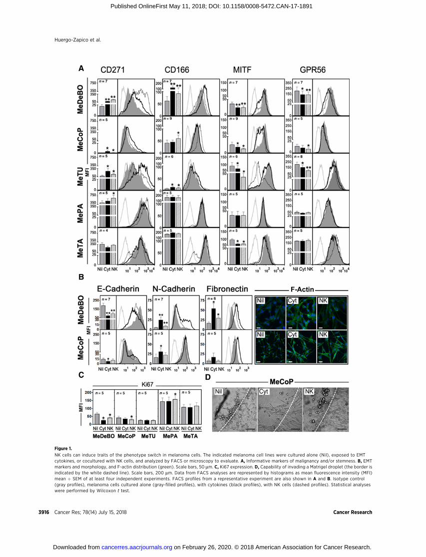
Figure 1.
NK cells can induce traits of the phenotype switch in melanoma cells. The indicated melanoma cell lines were cultured alone (Nil), exposed to EMTcytokines, or cocultured with NK cells, and analyzed by FACS or microscopy to evaluate. A, Informative markers of malignancy and/or stemness. B, EMTmarkers and morphology, and F-actin distribution (green). Scale bars, 50 mm. C, Ki67 expression. D, Capability of invading a Matrigel droplet (the border isindicated by the white dashed line). Scale bars, 200 mm. Data from FACS analyses are represented by histograms as mean fluorescence intensity (MFI)mean þ SEM of at least four independent experiments. FACS profiles from a representative experiment are also shown in A and B. Isotype control(gray profiles), melanoma cells cultured alone (gray-filled profiles), with cytokines (black profiles), with NK cells (dashed profiles). Statistical analyseswere performed by Wilcoxon t test.
Huergo-Zapico et al.
Cancer Res; 78(14) July 15, 2018 Cancer Research3916
on February 26, 2020. © 2018 American Association for Cancer Research. cancerres.aacrjournals.org Downloaded from
Published OnlineFirst May 11, 2018; DOI: 10.1158/0008-5472.CAN-17-1891
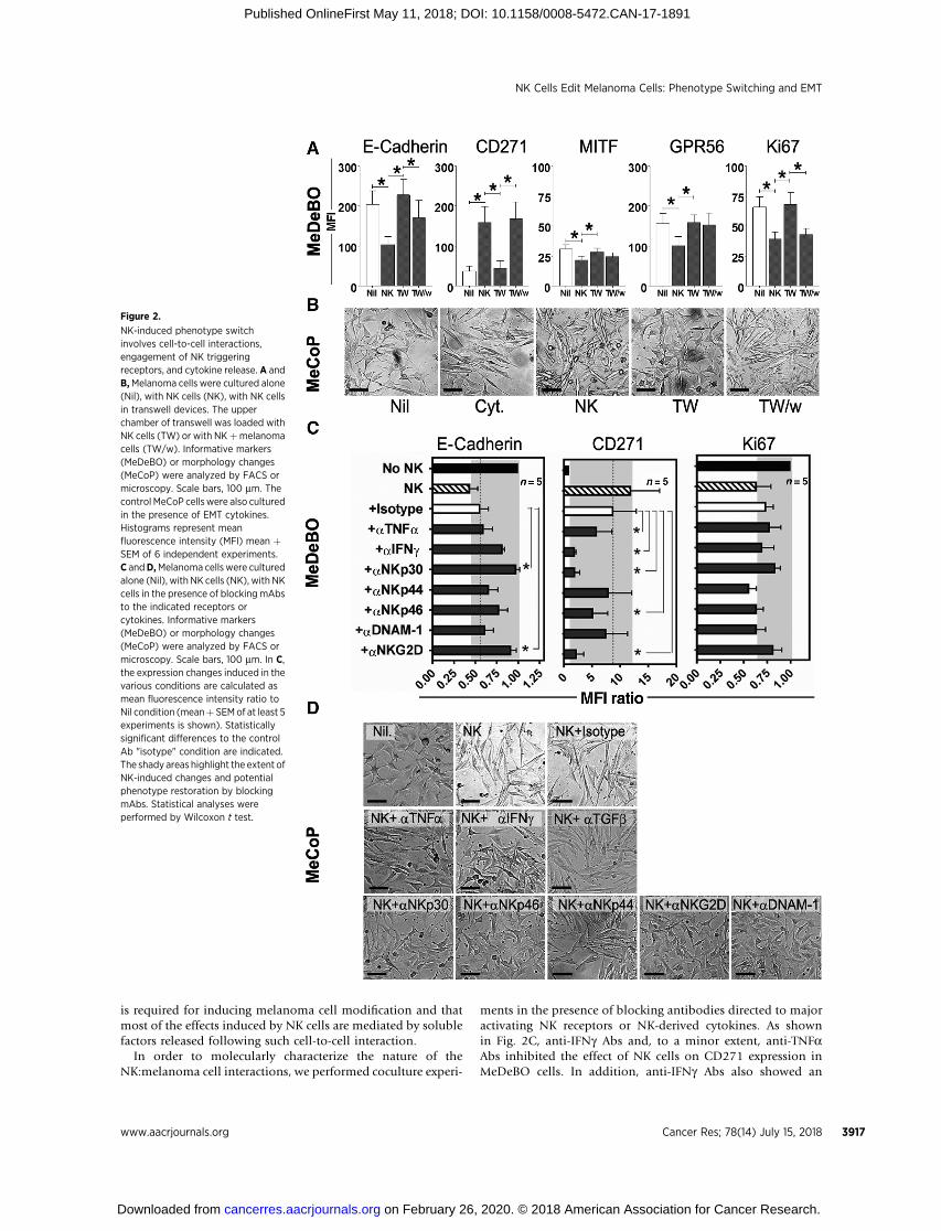
is required for inducing melanoma cell modification and thatmost of the effects induced by NK cells are mediated by solublefactors released following such cell-to-cell interaction.
In order to molecularly characterize the nature of theNK:melanoma cell interactions, we performed coculture experi-
ments in the presence of blocking antibodies directed to majoractivating NK receptors or NK-derived cytokines. As shownin Fig. 2C, anti-IFNg Abs and, to a minor extent, anti-TNFaAbs inhibited the effect of NK cells on CD271 expression inMeDeBO cells. In addition, anti-IFNg Abs also showed an
Figure 2.
NK-induced phenotype switchinvolves cell-to-cell interactions,engagement of NK triggeringreceptors, and cytokine release. A andB,Melanoma cells were cultured alone(Nil), with NK cells (NK), with NK cellsin transwell devices. The upperchamber of transwell was loaded withNK cells (TW) or with NKþmelanomacells (TW/w). Informative markers(MeDeBO) or morphology changes(MeCoP) were analyzed by FACS ormicroscopy. Scale bars, 100 mm. Thecontrol MeCoP cellswere also culturedin the presence of EMT cytokines.Histograms represent meanfluorescence intensity (MFI) mean þSEM of 6 independent experiments.C andD,Melanoma cells were culturedalone (Nil), with NK cells (NK), with NKcells in the presence of blocking mAbsto the indicated receptors orcytokines. Informative markers(MeDeBO) or morphology changes(MeCoP) were analyzed by FACS ormicroscopy. Scale bars, 100 mm. In C,the expression changes induced in thevarious conditions are calculated asmean fluorescence intensity ratio toNil condition (meanþ SEMof at least 5experiments is shown). Statisticallysignificant differences to the controlAb "isotype" condition are indicated.The shady areas highlight the extent ofNK-induced changes and potentialphenotype restoration by blockingmAbs. Statistical analyses wereperformed by Wilcoxon t test.
NK Cells Edit Melanoma Cells: Phenotype Switching and EMT
www.aacrjournals.org Cancer Res; 78(14) July 15, 2018 3917
on February 26, 2020. © 2018 American Association for Cancer Research. cancerres.aacrjournals.org Downloaded from
Published OnlineFirst May 11, 2018; DOI: 10.1158/0008-5472.CAN-17-1891
inhibitory trend toward the NK-induced effect on E-cadherin.Significant inhibitory effects were obtained also by blockingdifferent activating receptors, with maximal effects achievedupon NKp30 and NKG2D blockage. NKp30 and NKG2Dblockage also appeared to inhibit the NK-mediated effect onmelanoma proliferation (although without reaching statisticalsignificance; Fig. 2C, see Ki67 expression). Nevertheless, alsoNKp44, DNAM-1, and NKp46 receptors could contribute to theNK:melanoma interaction as their combined blockage resultedin an inhibitory effect comparable with that obtained byNKp30 masking (Supplementary Fig. S5A). Similarly, variousblocking Abs also showed an effect in the MeCoP:NK cellcoculture. Thus, anti-IFNg , anti-NKp30, anti-NKp46, anti-NKG2D, as well as anti–DNAM-1 Abs efficiently counteractedthe NK-induced morphologic transition of MeCoP cells, where-as anti-NKp44 and anti-TNFa Abs showed minor effects (Fig.2D). Finally, we also analyzed whether TGFb could be involvedin the NK-induced effect on melanoma cells, because suchEMT-inducing cytokine may be either produced by tumor cellsor induced in cocultures. As shown in Supplementary Fig. S5Band in Fig. 2D, anti-TGFb–blocking Abs did not interfere withthe phenotypic or morphologic changes induced by NK cellsin MeDeBO and MeCoP cells.
In conclusion, our results indicate that the induction oftumor phenotype switching/EMT by NK cells is primarilysustained by IFNg (and partly by TNFa), that is, cytokines thatare released upon NK:melanoma cell interaction and engage-ment of activating receptors.
Effect of phenotype switching/EMT on the NK:melanomacell cross-talk
We then analyzed whether undergoing a phenotype switchmodifies melanoma cell susceptibility to NK cell–mediatedattack, or their ability to suppress NK-cell activity.
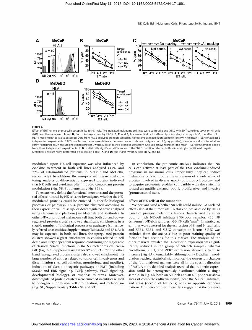
The phenotype switching/EMT on MeDeBO and MeCoPcells, induced either by exposure to EMT cytokines or by cellcoculture with NK cells, did not substantially modify thesurface expression of NKG2D or DNAM-1 ligands, nor did itmodify the binding of the soluble NCRs (to their ligands at thetumor cell surface; Supplementary Fig. S6). We also assessedthe expression of PD-L1, as this suppressive molecule can beinduced during EMT on breast cancer cells (13). As shown inSupplementary Fig. S6, exposure to both cytokine and NK cellsresulted in a slight induction of PD-L1 expression in MeCoPcells, whereas it was ineffective in MeDeBO cells. On the otherhand, MeCoP cells evidencing phenotype switch showed amarked HLA-I surface expression increase accompaniedby increased resistance to NK-cell killing activity (Fig. 3A andB). HLA-I masking by specific mAbs completely restored thesusceptibility of "cocultured" MeCoP cells to NK cell–mediatedkilling, an effect that was incomplete on cytokine-treated mel-anoma cells (Fig. 3C), suggesting that, besides HLA-I upregula-tion, additional mechanism of resistance may be induced byEMT cytokines. MeDeBO cells lacked surface HLA-I molecules.Accordingly, they did not show any modification of HLA-Iexpression and did not modify their susceptibility to NKcell–mediated killing (Fig. 3D and E). Exposure to EMT cyto-kines or coculture with NK cells led to HLA-I upregulationalso on MePA, MeTU, and MeTA cells, despite in these cases,the effects were less statistically significant (SupplementaryFig. S7). We also assessed whether exposure to NK cells could
increase resistance of MeCoP and MeDeBO cells to therapeutictreatments such as cytotoxic drugs or g-radiation. As shownin Supplementary Fig. S8, NK cells did not modify the sus-ceptibility of melanoma cells to taxol, while increased resist-ance to g-radiation in MeCoP (although without reachingstatistical significance).
In order to assess the effect of EMT on the immunosuppres-sive capability of melanoma cells, MeCoP and MeDeBO cellswere exposed to EMT cytokines to induce phenotype switching(EMT melanoma cells), and, afterwards, cocultured with NKcells for 3 days. As TGFb can affect both NK-cell function andphenotype, EMT melanoma cells were extensively washedbefore starting coculture. NK cells from cocultures were thenanalyzed for the expression of major activating receptors. Asshown in Fig. 4A, melanoma cells were able to induce down-regulation of NKp30, NKG2D, and DNAM-1 on NK cells. Thedifference between untreated and EMT melanoma cells indecreasing NCR expression was statistically significant in thecase of MeDeBo cells. Thus, NK cells exposed to MeDeBO cellswere also tested in a cytolytic assay. As shown in Fig. 4B, NK cellsconditioned by EMT-MeDeBO cells showed maximal inhibitionof their ability to kill melanoma cells as compared with NK cellsexposed to MeDeBO cells or cultured alone. Because inductionof immune checkpoint receptors on NK cells has been associatedwith tumor progression (12, 31), we also assessed whether EMTmelanoma cells could influence TIM-3 and PD-1 expression onNK cells. As shown in Fig. 4A, both melanoma cell lines inducedlittle fluctuations of TIM-3 expression on NK cells. In particular,exposure to EMT-MeDeBO cells resulted in slight, but notsignificant increase of TIM-3 expression. PD-1 expression wasnot induced in either the analyzed conditions.
Thus, the phenotype switch/EMT can favor the escape to NK-cell antitumor activity by enabling tumor cells to express higherlevels of HLA-I molecules and to increase their ability to targetactivating receptors on NK cells.
Analysis of proteomic profiles induced by NK cells onMeCoP and MeDeBO cells
The above functional and phenotypic data indicate that NKcells can induce, in melanoma cells, traits of increased malig-nancy that are hallmarks of phenotype switch or EMT. To gainfurther insight on this activity and acquire a more comprehen-sive characterization of the NK cell–mediated effect on mela-noma cells, we analyzed the proteomic changes induced by NKcells or by EMT cytokines on MeCoP and MeDeBO cell lines.
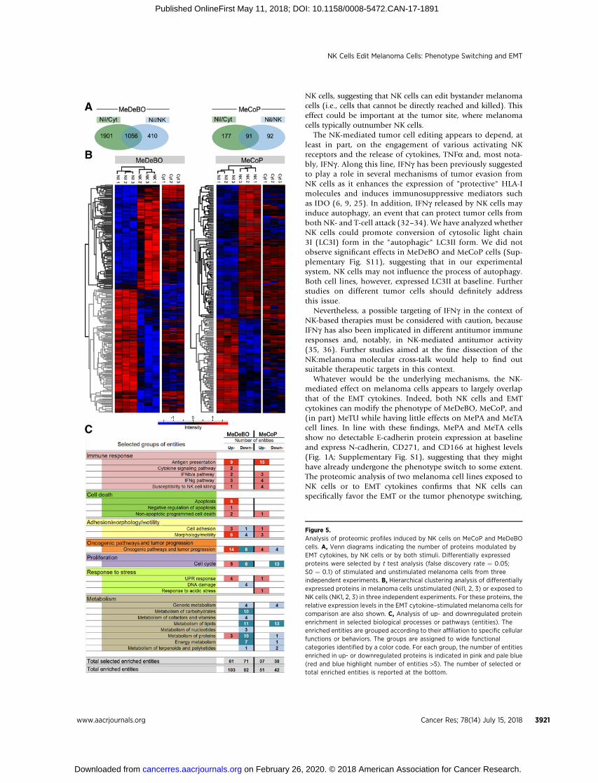
High-resolution mass spectrometry analysis of cell lysatesled to the definition of about 5,000 proteins for each cell line(in each individual experimental condition). The Spearmanrank correlation indicated an acceptable reproducibilityamong the analyzed biological replicates (average coefficients> 0.92 for each condition in both cell lines; SupplementaryFig. S9A). As shown by the Venn diagrams (Fig. 5A), exposureto NK cells or to EMT cytokines, respectively, induced themodulation of 183 or 268 proteins in MeCoP cells, and1,466 or 2,957 proteins in MeDeBo cells (T test false discoveryrate ¼ 0.05, S0 ¼ 0.1; also see the protein lists in Supplemen-tary Table S1). Thus, the number of differentially expressedproteins greatly differed between the two cell lines, suggestinga potential, marked variability among melanoma cells in theresponse to stimuli and, specifically, to NK cells. In spite ofthis variability, a consistent part of the proteins that were
Huergo-Zapico et al.
Cancer Res; 78(14) July 15, 2018 Cancer Research3918
on February 26, 2020. © 2018 American Association for Cancer Research. cancerres.aacrjournals.org Downloaded from
Published OnlineFirst May 11, 2018; DOI: 10.1158/0008-5472.CAN-17-1891
modulated upon NK-cell exposure was also influenced bycytokine treatment in both cell lines analyzed (49% and72% of NK-modulated proteins in MeCoP and MeDeBo,respectively). In addition, the unsupervised hierarchical clus-tering analysis of differentially expressed proteins indicatedthat NK cells and cytokines often induced concordant proteinmodulation (Fig. 5B; Supplementary Fig. S9B).
To extensively define the functional networks and the poten-tial effects induced by NK cells, we investigated whether the NK-modulated proteins could be enriched in specific biologicalprocesses or pathways. Thus, proteins clustered according totheir expression values as up- or downregulated were analyzedusing GeneAnalytic platform (see Materials and Methods). Ineither NK-conditioned melanoma cell line, both up- and down-regulated protein clusters showed significant enrichment in asizable number of biological processes or pathways (collective-ly referred to as entities; Supplementary Tables S2 and S3). As itmay be expected, in both cell lines, the upregulated proteinclusters showed a great enrichment in entities related to celldeath and IFNg-dependent response, confirming the major roleof classical NK-cell functions in the NK:melanoma cell cross-talk (Fig. 5C; Supplementary Tables S2 and S3). On the otherhand, upregulated protein clusters also showed enrichment in alarge number of entities related to tumor cell invasiveness anddissemination (i.e., cell adhesion, morphology, and motility),induction of classic oncogenic pathways or EMT (includingSMAD and ERK signaling, TGFb pathway, VEGF signaling,developmental biology), or response to stress. Moreover,downregulated protein clusters were enriched in entities relatedto oncogene suppression, cell proliferation, and metabolism(Fig. 5C; Supplementary Tables S2 and S3).
In conclusion, the proteomic analysis indicates that NKcells can activate at least part of the EMT cytokine–inducedprograms in melanoma cells. Importantly, they can inducemelanoma cells to modify the expression of a wide range ofproteins involved in diverse aspects of tumor cell biology, andto acquire proteomic profiles compatible with the switchingtoward an undifferentiated, poorly proliferative, and invasive(prometastatic) state.
Effects of NK cells at the tumor siteWe next analyzed whether NK cells could induce EMT-related
effects also at the tumor site. To this end, we assessed by IHC apanel of primary melanoma lesions characterized by eitherpoor or rich NK-cell infiltrate (NK-poor samples: <10 NKcells/mm2; NK-rich samples: >30 NK cells/mm2). In particular,samples were assessed for the expression of E- and N-cadherin,and ZEB1, ZEB2, and SLUG transcription factors. SLUG wasexcluded from the analysis due to poor staining quality offormalin-fixed sections for this marker. The analysis of theother markers revealed that E-cadherin expression was signif-icantly reduced in the group of NK-rich samples, whereasN-cadherin, ZEB1, and ZEB2 expression showed a trend toincrease (Fig. 6A). Remarkably, although only E-cadherin mod-ulation reached statistical significance, the expression changesof the four analyzed markers were all in the specific directionof EMT. A more detailed analysis revealed that cadherin expres-sion could be heterogeneously distributed within a singlesample. In Fig. 6B, both an NK-rich and an NK-poor case showareas of complete cadherin switch, near the NK-cell infiltrate,and areas (devoid of NK cells) with an opposite cadherinpattern. On their complex, these data suggest that the presence
Figure 3.
Effect of EMT on melanoma cell susceptibility to NK lysis. The indicated melanoma cell lines were cultured alone (Nil), with EMT cytokines (cyt), or NK cells(NK), and then analyzed. A and D, For HLA-I expression by FACS. B, C, and E, For susceptibility to NK-cell lysis in cytolytic assays. In C, the effect ofHLA-I masking mAbs is also assessed. Data from FACS analyses are represented by histograms as mean fluorescence intensity (MFI) mean þ SEM of at least 5independent experiments. FACS profiles from a representative experiment are also shown. Isotype control (gray profiles), melanoma cells cultured alone(gray-filled profiles), with cytokines (black profiles), with NK cells (dashed profiles). Data from cytolytic assays represent the meanþ SEM of 6 samples pooledfrom three independent experiments. In B, statistically significant differences to the "Nil" condition refer to both NK- and cyt-conditioned targets.Statistical analyses were performed by Wilcoxon t test (A and D) and Mann–Whitney test (B, C, and E).
NK Cells Edit Melanoma Cells: Phenotype Switching and EMT
www.aacrjournals.org Cancer Res; 78(14) July 15, 2018 3919
on February 26, 2020. © 2018 American Association for Cancer Research. cancerres.aacrjournals.org Downloaded from
Published OnlineFirst May 11, 2018; DOI: 10.1158/0008-5472.CAN-17-1891

and the proximity of the NK-cell infiltrate may favor theEMT. We also found samples showing rather homogeneousN- or E-cadherin expression, independent on the NK-cell local-ization. In these cases, the prevalence of various factors knownto affect EMT (i.e., cytokines, hypoxia, stromal cells, or otherimmune cells) may have masked the specific effect of NK cells.
To further dissect the effect of NK cells on EMT, we selecteda sample showing heterogeneous distribution of NK cellsand cadherins and quantified E- and N-cadherin expressionin the areas close to or distant from NK-cell infiltrate (see anexample in Supplementary Fig. S10). This quantification wasdone by calculating the H-score for each of the areas includedin the "close" or "far" group (see Supplementary Materialsand Methods). As shown in Fig. 6C, the areas close to NKcells showed a statistically significant lower expression ofE-cadherin and higher expression of N-cadherin as comparedwith areas distant from NK cells. These data definitely indicatethat, at least in certain cases, NK cells can influence the EMT atthe tumor site.
DiscussionThis study provides the first experimental evidence that NK
cells can induce the phenotype switching of melanoma cells toan undifferentiated, poorly proliferative, proinvasive state. Weshow that the presence of NK cells in coculture experiments caninduce on melanoma cells increased expression of stemnessmarkers, E- and N-cadherin switch, cytoskeletal and morpho-logic rearrangements, reduced tumor proliferation rates, andalso, in one melanoma cell line, increased invasiveness. More-over, the analysis of the NK-induced proteomic profiles indi-cates that NK cells may profoundly intervene in the overallbiology of melanoma cells, not only by affecting importantpathways related to tumorigenesis, cell survival/death, andresponse to stress, but also by influencing various metabolicprocesses.
Transwell experiments indicate that at least some of the NK-mediated effects are triggered by cell-to-cell interactions but canbe extended to melanoma cells that are not in close contact with
Figure 4.
EMT can increase the suppressive capability of melanoma cells. A, NK cells unconditioned (Nil), conditioned with melanoma cells (Mel), or conditionedwith melanoma cells exposed to EMT cytokines (Mel EMT) were analyzed by FACS for surface expression of the indicated NK receptors. Theconditioning melanoma cell lines are indicated to the left. Histograms represent mean fluorescence intensity (MFI) mean þ SEM of at least four independentexperiments. B, NK cells conditioned as indicated were analyzed for their ability to kill MeDeBO cells in a cytolytic assay. Data represent the mean þ SEM of6 samples pooled from three independent experiments. Statistical analyses were performed by Wilcoxon t test (A) and Mann–Whitney test (B). Beforephenotypic or functional analyses, NK cells were counted using trypan blue staining to evaluate dead cells. In all conditions analyzed, NK-cell viabilitywas approximately 90%.
Huergo-Zapico et al.
Cancer Res; 78(14) July 15, 2018 Cancer Research3920
on February 26, 2020. © 2018 American Association for Cancer Research. cancerres.aacrjournals.org Downloaded from
Published OnlineFirst May 11, 2018; DOI: 10.1158/0008-5472.CAN-17-1891
NK cells, suggesting that NK cells can edit bystander melanomacells (i.e., cells that cannot be directly reached and killed). Thiseffect could be important at the tumor site, where melanomacells typically outnumber NK cells.
The NK-mediated tumor cell editing appears to depend, atleast in part, on the engagement of various activating NKreceptors and the release of cytokines, TNFa and, most nota-bly, IFNg . Along this line, IFNg has been previously suggestedto play a role in several mechanisms of tumor evasion fromNK cells as it enhances the expression of "protective" HLA-Imolecules and induces immunosuppressive mediators suchas IDO (6, 9, 25). In addition, IFNg released by NK cells mayinduce autophagy, an event that can protect tumor cells fromboth NK- and T-cell attack (32–34). We have analyzed whetherNK cells could promote conversion of cytosolic light chain3I (LC3I) form in the "autophagic" LC3II form. We did notobserve significant effects in MeDeBO and MeCoP cells (Sup-plementary Fig. S11), suggesting that in our experimentalsystem, NK cells may not influence the process of autophagy.Both cell lines, however, expressed LC3II at baseline. Furtherstudies on different tumor cells should definitely addressthis issue.
Nevertheless, a possible targeting of IFNg in the context ofNK-based therapies must be considered with caution, becauseIFNg has also been implicated in different antitumor immuneresponses and, notably, in NK-mediated antitumor activity(35, 36). Further studies aimed at the fine dissection of theNK:melanoma molecular cross-talk would help to find outsuitable therapeutic targets in this context.
Whatever would be the underlying mechanisms, the NK-mediated effect on melanoma cells appears to largely overlapthat of the EMT cytokines. Indeed, both NK cells and EMTcytokines can modify the phenotype of MeDeBO, MeCoP, and(in part) MeTU while having little effects on MePA and MeTAcell lines. In line with these findings, MePA and MeTA cellsshow no detectable E-cadherin protein expression at baselineand express N-cadherin, CD271, and CD166 at highest levels(Fig. 1A; Supplementary Fig. S1), suggesting that they mighthave already undergone the phenotype switch to some extent.The proteomic analysis of two melanoma cell lines exposed toNK cells or to EMT cytokines confirms that NK cells canspecifically favor the EMT or the tumor phenotype switching,
Figure 5.Analysis of proteomic profiles induced by NK cells on MeCoP and MeDeBOcells. A, Venn diagrams indicating the number of proteins modulated byEMT cytokines, by NK cells or by both stimuli. Differentially expressedproteins were selected by t test analysis (false discovery rate ¼ 0.05;S0 ¼ 0.1) of stimulated and unstimulated melanoma cells from threeindependent experiments. B, Hierarchical clustering analysis of differentiallyexpressed proteins in melanoma cells unstimulated (Nil1, 2, 3) or exposed toNK cells (NK1, 2, 3) in three independent experiments. For these proteins, therelative expression levels in the EMT cytokine–stimulated melanoma cells forcomparison are also shown. C, Analysis of up- and downregulated proteinenrichment in selected biological processes or pathways (entities). Theenriched entities are grouped according to their affiliation to specific cellularfunctions or behaviors. The groups are assigned to wide functionalcategories identified by a color code. For each group, the number of entitiesenriched in up- or downregulated proteins is indicated in pink and pale blue(red and blue highlight number of entities >5). The number of selected ortotal enriched entities is reported at the bottom.
NK Cells Edit Melanoma Cells: Phenotype Switching and EMT
www.aacrjournals.org Cancer Res; 78(14) July 15, 2018 3921
on February 26, 2020. © 2018 American Association for Cancer Research. cancerres.aacrjournals.org Downloaded from
Published OnlineFirst May 11, 2018; DOI: 10.1158/0008-5472.CAN-17-1891
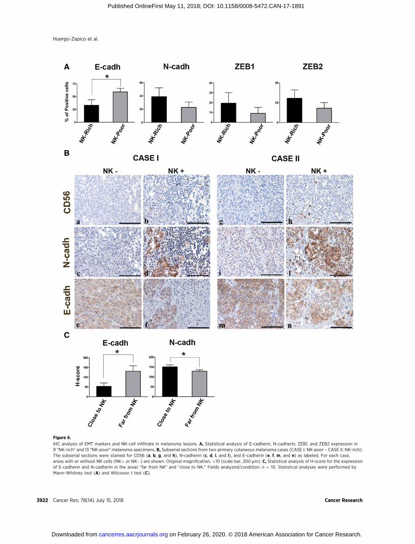
Figure 6.
IHC analysis of EMT markers and NK-cell infiltrate in melanoma lesions. A, Statistical analysis of E-cadherin, N-cadherin, ZEB1, and ZEB2 expression in9 "NK-rich" and 13 "NK-poor" melanoma specimens. B, Subserial sections from two primary cutaneous melanoma cases (CASE I: NK-poor – CASE II: NK-rich).The subserial sections were stained for CD56 (a, b, g, and h), N-cadherin (c, d, i, and l), and E-cadherin (e, f, m, and n) as labeled. For each case,areas with or without NK cells (NKþ or NK�) are shown. Original magnification, �10 (scale bar, 200 mm). C, Statistical analysis of H-score for the expressionof E-cadherin and N-cadherin in the areas "far from NK" and "close to NK." Fields analyzed/condition: n ¼ 10. Statistical analyses were performed byMann–Whitney test (A) and Wilcoxon t test (C).
Huergo-Zapico et al.
Cancer Res; 78(14) July 15, 2018 Cancer Research3922
on February 26, 2020. © 2018 American Association for Cancer Research. cancerres.aacrjournals.org Downloaded from
Published OnlineFirst May 11, 2018; DOI: 10.1158/0008-5472.CAN-17-1891
but also highlights the complexity of the NK-mediated effectson melanoma cells. As shown in Fig. 5A–C, the NK-inducedproteomic changes largely overlap those induced by EMTcytokines and involve biological processes or pathways relatedto tumor cell transition to aggressive forms. These changes alsoinclude the modulation of proteins involved in several meta-bolic processes or in cell-cycle regulation, suggesting that NKcells can contribute to the phenotype switch also through ageneral downregulation of tumor cell metabolism and prolif-eration. Remarkably, these effects may favor the generation ofthe so-called "dormant tumor cells" (37), which are thought togive rise to tumor relapse and metastasis. Finally, both theanalyzed melanoma cell lines show clear increments of proteinsinvolved in the interaction with immune cells and, in partic-ular, in the response to IFNg , confirming the pivotal role of thiscytokine in the NK:melanoma cell cross-talk.
The IHC analysis of NK-rich and NK-poor melanoma speci-mens shows that in NK-rich samples, four EMT markers, includ-ing E-cadherin, N-cadherin, ZEB1, and ZEB2, were all modu-lated in the direction of the EMT (although only for E-cadherinthis modulation reached statistical significance). In addition, ina sample showing heterogeneous cadherin and NK-cell distri-bution, the modulation of both E- and N-cadherin significantlycorrelated with the vicinity of melanoma cells to the localNK-cell infiltrate. On their complex, the IHC data indicate thatNK cells can effectively participate in the induction of the EMTat the tumor site. In this context, it should also be consideredthat several microenvironmental factors can influence EMTmarkers, and veil, in certain circumstances, the effect of NKcells. In search of EMT markers that could be more specificallymodulated by NK cells, we reevaluated the proteomic data andnoticed that MeDeBO cells exposed to either NK cells or cyto-kines showed upregulation of SOX5, a transcription factorinvolved in the negative regulation of MITF and in the inductionof EMT (see Supplementary Table S1; refs. 38, 39). We analyzedthis molecule in the melanoma lesions and found a clearstatistical correlation between the rich NK-cell infiltrate and thehigh expression of SOX5 (Supplementary Fig. S12A and S12B).In our opinion, this marker deserves further detailed studies todefine its possible role in the NK:tumor cell cross-talk.
MITF downregulation is distinctive of phenotype switch-ing in melanoma cells. However, the effects of NK cells maybe extended to tumors of epithelial origin, as suggested by theobservation that NK cells can promote the upregulation of twomaster regulators of EMT (Supplementary Fig. S3A) and caninduce morphologic changes (compatible with EMT) on thecervix adenocarcinoma HeLa cell line (Supplementary Fig. S13).
Our data indicate that NK cells promote EMT, but also showthat EMT profoundly influences NK cells. Indeed, EMT cansharply strengthen the suppressive capability of melanoma cellsby enhancing their ability to induce the downregulation ofimportant activating receptors on NK cells (Fig. 4A and B).Some recent studies suggested that the process of EMT plays arole in different mechanisms of tumor escape. Thus, for exam-ple, EMT has been shown to induce an immunosuppressivemicroenvironment in hepatocellular carcinoma (40), or toreduce the ability of breast cancer cells to form immunologicsynapses with cytotoxic T lymphocytes (34). On the otherhand, it has also been reported that EMT induction could pro-mote NKG2D-L upregulation on colorectal cells and on immor-talized keratinocytes, or induce increased NK cell–mediated
metastasis-specific immunosurveillance in lung cancer (41, 42).Nevertheless, besides HLA-I molecules, no major NK-receptorligands were significantly modified by EMT in our experimentson melanoma cells (Supplementary Fig. S6), suggesting that acertain variability among tumor cell types could exist in theresponse to EMT stimuli.
In summary, the NK:tumor cell cross-talk in the context ofEMT may occur in melanoma and affect the progression, andperhaps the fate, of the tumor. This issue opens importantquestions on how the tumor microenvironment could influ-ence the outcome of such "NK:melanoma:EMT" cross-talk.Indeed, tumor-associated immune and stromal cells, as wellas tumor-associated hypoxia, are known to favor EMT (19)and/or modulate NK-cell function (6, 7). Additional questionsregard the type and the functional status of the NK cells thatparticipate in the cross-talk at the tumor site. ConventionalCD56dimCD16bright NK cells have been reported to infiltratetumor tissues in several solid tumors (18, 43). TissueCD56dimCD16bright cells generally lack markers of tissue-resi-dent lymphocytes (i.e., CD69, CD103, or CD49a) and areconsidered as cells recirculating from peripheral blood(44, 45), where, indeed, CD56dimCD16bright cells are largelyrepresented. Along this line, peripheral blood–derived NK cellshave been recently shown to migrate in response to chemo-tactic stimuli released during NK:melanoma cell interaction(46). Intriguingly, tumor tissues are also frequently infiltratedby cells expressing the CD56brightCD16dim phenotype (47).These cells constitute a small fraction of circulating NK cellsbut are well represented in certain tissues or secondary lym-phoid organs (44, 47). CD56brightCD16dim cells are character-ized by low cytotoxicity and high IFNg production in responseto cytokines; hence, they may have a different, even morepronounced, effect on tumor phenotype switch and EMT.Thus, our data on PBNK cells offer reliable hints to characterizethe NK:tumor cell cross-talk, at least for tumors showing theCD56dimCD16bright NK-cell infiltrate. Further studies involvingspecific NK-cell subsets and dissecting the effect of specificcomponents of the tumor microenvironment would integrateand extend our data and provide important clues to designpersonalized and hopefully effective NK-based therapies.
Disclosure of Potential Conflicts of InterestNo potential conflicts of interest were disclosed.
Authors' ContributionsConception and design: L. Huergo-Zapico, E.J. DeAndr�es-Galiana,A. L�opez-Soto, L. Moretta, S. Gonz�alez, M. VitaleDevelopment of methodology: L. Huergo-Zapico, J.L. Fern�andez-Martínez,E.J. DeAndr�es-GalianaAcquisition of data (provided animals, acquired and managed patients,provided facilities, etc.): L.Huergo-Zapico,C.Cantoni, C. Lavarello, A. Petretto,G. Pietra, M. Bugatti, E. Munari, M. Marconi, W. Vermi, L. MorettaAnalysis and interpretation of data (e.g., statistical analysis, biostatistics,computational analysis): L. Huergo-Zapico, M. Parodi, C. Cantoni,C. Lavarello, J.L. Fern�andez-Martínez, A. Petretto, E.J. DeAndr�es-Galiana,M. Balsamo, A. L�opez-Soto, G. Pietra, M. Bugatti, E. Munari, W. Vermi,L. Moretta, S. Gonz�alez, M. VitaleWriting, review, and/or revision of the manuscript: L. Huergo-Zapico,M. Parodi, C. Cantoni, J.L. Fern�andez-Martínez, E.J. DeAndr�es-Galiana,A. L�opez-Soto, M.C. Mingari, L. Moretta, S. Gonz�alez, M. VitaleAdministrative, technical, or material support (i.e., reporting or organizingdata, constructing databases): L. Huergo-ZapicoStudy supervision: L. Huergo-Zapico, L. Moretta, M. Vitale
NK Cells Edit Melanoma Cells: Phenotype Switching and EMT
www.aacrjournals.org Cancer Res; 78(14) July 15, 2018 3923
on February 26, 2020. © 2018 American Association for Cancer Research. cancerres.aacrjournals.org Downloaded from
Published OnlineFirst May 11, 2018; DOI: 10.1158/0008-5472.CAN-17-1891
AcknowledgmentsThis work was supported by grants awarded by Associazione Italiana
Ricerca sul Cancro AIRC: IG 2014 project n. 15428 (to M. Vitale), IG 2014project n. 15283 (to L. Moretta), IG 2014 project n. 15378 (to W. Vermi),and "Special Program Molecular Clinical Oncology 5 � 100000 projectn. 9962 (to L. Moretta); PRA 2013 and FRA 2015, DIMES, University ofGenoa (to G. Pietra); 5 � 1000 Min. Sal. 2014 and 2015 (5M-2014-2353563 and 5M-2015-2360333 to M. Vitale and M.C. Mingari); the
Spanish grants of Instituto de Salud Carlos III (PI16/01485), and FEDEREuropean Union (to S. Gonzalez). M. Parodi was recipient of the AIRCfellowship n. 18274, year 2016. L. Huergo-Zapico was recipient of theEMBO short-term fellowship ASTF 520 - 2012. M. Bugatti is supported byFondazione Beretta (Brescia, Italy).
Received June 26, 2017; revised December 19, 2017; accepted May 8, 2018;published first May 11, 2018.
References1. Locatelli F, Pende D, Mingari MC, Bertaina A, Falco M, Moretta A, et al.
Cellular and molecular basis of haploidentical hematopoietic stem celltransplantation in the successful treatment of high-risk leukemias: role ofalloreactive NK cells. Front Immunol 2013;4:15.
2. Parkhurst MR, Riley JP, Dudley ME, Rosenberg SA. Adoptive transfer ofautologous natural killer cells leads to high levels of circulating naturalkiller cells but does not mediate tumor regression. Clin Cancer Res2011;17:6287–97.
3. Knorr DA, Bachanova V, Verneris MR, Miller JS. Clinical utility of naturalkiller cells in cancer therapy and transplantation. Semin Immunol 2014;26:161–72.
4. Cantoni C, Grauwet K, Pietra G, Parodi M, Mingari MC, Maria AD, et al.Role of NK cells in immunotherapy and virotherapy of solid tumors.Immunotherapy 2015;7:861–82.
5. Guillerey C, Huntington ND, Smyth MJ. Targeting natural killer cells incancer immunotherapy. Nat Immunol 2016;17:1025–36.
6. Vitale M, Cantoni C, Pietra G, Mingari MC, Moretta L. Effect of tumor cellsand tumor microenvironment on NK-cell function. Eur J Immunol2014;44:1582–92.
7. Pahl J, Cerwenka A. Tricking the balance: NK cells in anti-cancer immunity.Immunobiology 2017;222:11–20.
8. Vivier E, Raulet DH, Moretta A, Caligiuri MA, Zitvogel L, Lanier LL, et al.Innate or adaptive immunity? The example of natural killer cells. Science2011;331:44–9.
9. Balsamo M, Vermi W, Parodi M, Pietra G, Manzini C, Queirolo P, et al.Melanoma cells become resistant to NK-cell-mediated killing whenexposed to NK-cell numbers compatible with NK-cell infiltration in thetumor. Eur J Immunol 2012;42:1833–42.
10. Muntasell A, Ochoa MC, Cordeiro L, Berraondo P, Lopez-Diaz de Cerio A,Cabo M, et al. Targeting NK-cell checkpoints for cancer immunotherapy.Curr Opin Immunol 2017;45:73–81.
11. Abiko K, Matsumura N, Hamanishi J, Horikawa N, Murakami R, Yama-guchi K, et al. IFN-gamma from lymphocytes induces PD-L1 expressionandpromotes progressionof ovarian cancer. Br JCancer 2015;112:1501–9.
12. Pesce S, Greppi M, Tabellini G, Rampinelli F, Parolini S, Olive D, et al.Identificationof a subset of humannatural killer cells expressinghigh levelsof programmed death 1: A phenotypic and functional characterization.J Allergy Clin Immunol 2017;139:335–46.
13. Noman MZ, Janji B, Abdou A, Hasmim M, Terry S, Tan TZ, et al. Theimmune checkpoint ligand PD-L1 is upregulated in EMT-activated humanbreast cancer cells by amechanism involving ZEB-1 andmiR-200.Oncoim-munology 2017;6:e1263412.
14. Toh B, Wang X, Keeble J, Sim WJ, Khoo K, Wong WC, et al. Mesenchymaltransition and dissemination of cancer cells is driven by myeloid-derivedsuppressor cells infiltrating the primary tumor. PLoS Biol 2011;9:e1001162.
15. Fan QM, Jing YY, Yu GF, Kou XR, Ye F, Gao L, et al. Tumor-associatedmacrophages promote cancer stem cell-like properties via transforminggrowth factor-beta1-induced epithelial-mesenchymal transition in hepa-tocellular carcinoma. Cancer Lett 2014;352:160–8.
16. Mayer C, Darb-Esfahani S, Meyer AS, Hubner K, Rom J, Sohn C, et al.Neutrophil granulocytes in ovarian cancer - induction of epithelial-to-mesenchymal-transition and tumor cell migration. J Cancer 2016;7:546–54.
17. Goebel L, Grage-Griebenow E, Gorys A, Helm O, Genrich G, Lenk L,et al. CD4þ T cells potently induce epithelial-mesenchymal-transitionin premalignant and malignant pancreatic ductal epithelial cells-novelimplications of CD4þ T cells in pancreatic cancer development.Oncoimmunology 2015;4:e1000083.
18. Cantoni C, Huergo-Zapico L, Parodi M, Pedrazzi M, Mingari MC, MorettaA, et al. NK cells, tumor cell transition, and tumor progression in solidmalignancies: new hints for NK-based immunotherapy? J Immunol Res2016;2016:4684268.
19. Polyak K, Weinberg RA. Transitions between epithelial and mesenchymalstates: acquisition of malignant and stem cell traits. Nat Rev Cancer2009;9:265–73.
20. Kemper K, de Goeje PL, Peeper DS, van Amerongen R. Phenotype switch-ing: tumor cell plasticity as a resistance mechanism and target for therapy.Cancer Res 2014;74:5937–41.
21. Fukunaga-Kalabis M, Roesch A, HerlynM. From cancer stem cells to tumormaintenance in melanoma. J Invest Dermatol 2011;131:1600–4.
22. Lunter PC, van Kilsdonk JW, van Beek H, Cornelissen IM, Bergers M,Willems PH, et al. Activated leukocyte cell adhesion molecule (ALCAM/CD166/MEMD), a novel actor in invasive growth, controls matrix metal-loproteinase activity. Cancer Res 2005;65:8801–8.
23. Mikesh LM, Kumar M, Erdag G, Hogan KT, Molhoek KR, Mayo MW, et al.Evaluation of molecular markers of mesenchymal phenotype in melano-ma. Melanoma Res 2010;20:485–95.
24. Boiko AD, RazorenovaOV, van de RijnM, Swetter SM, JohnsonDL, Ly DP,et al. Human melanoma-initiating cells express neural crest nerve growthfactor receptor CD271. Nature 2010;466:133–7.
25. Pietra G, Manzini C, Rivara S, Vitale M, Cantoni C, Petretto A, et al.Melanoma cells inhibit natural killer cell function by modulating theexpression of activating receptors and cytolytic activity. Cancer Res 2012;72:1407–15.
26. Kulak NA, Pichler G, Paron I, Nagaraj N, Mann M. Minimal, encapsulatedproteomic-sample processing applied to copy-number estimation ineukaryotic cells. Nat Methods 2014;11:319–24.
27. Tyanova S, Temu T, Sinitcyn P, Carlson A, Hein MY, Geiger T, et al. ThePerseus computational platform for comprehensive analysis of (prote)omics data. Nat Methods 2016;13:731–40.
28. Saligan LN, Fernandez-Martinez JL, deAndres-Galiana EJ, Sonis S. Super-vised classification by filter methods and recursive feature eliminationpredicts risk of radiotherapy-related fatigue in patients with prostatecancer. Cancer Informat 2014;13:141–52.
29. deAndres-Galiana EJ, Fernandez-Martinez JL, Sonis ST. Design of Biomed-ical Robots for Phenotype Prediction Problems. J Comput Biol2016;23:678–92.
30. Xu L, BegumS,Hearn JD,Hynes RO.GPR56, an atypicalGprotein-coupledreceptor, binds tissue transglutaminase, TG2, and inhibits melanomatumor growth and metastasis. Proc Natl Acad Sci U S A 2006;103:9023–8.
31. SeoH, Jeon I, KimBS, ParkM, Bae EA, Song B, et al. IL-21-mediated reversalof NK cell exhaustion facilitates anti-tumour immunity in MHC class I-deficient tumours. Nat Commun 2017;8:15776.
32. Buchser WJ, Laskow TC, Pavlik PJ, Lin HM, Lotze MT. Cell-mediatedautophagy promotes cancer cell survival. Cancer Res 2012;72:2970–9.
33. Baginska J, Viry E, Berchem G, Poli A, Noman MZ, van Moer K, et al.Granzyme B degradation by autophagy decreases tumor cell susceptibilityto natural killer-mediated lysis under hypoxia. Proc Natl Acad Sci U S A2013;110:17450–5.
34. Akalay I, Janji B, Hasmim M, Noman MZ, Andre F, De Cremoux P, et al.Epithelial-to-mesenchymal transition and autophagy induction in breastcarcinoma promote escape from T-cell-mediated lysis. Cancer Res 2013;73:2418–27.
35. Menard C, Blay JY, Borg C, Michiels S, Ghiringhelli F, Robert C, et al.Natural killer cell IFN-gamma levels predict long-term survival with
Huergo-Zapico et al.
Cancer Res; 78(14) July 15, 2018 Cancer Research3924
on February 26, 2020. © 2018 American Association for Cancer Research. cancerres.aacrjournals.org Downloaded from
Published OnlineFirst May 11, 2018; DOI: 10.1158/0008-5472.CAN-17-1891
imatinib mesylate therapy in gastrointestinal stromal tumor-bearingpatients. Cancer Res 2009;69:3563–9.
36. Tu TC, Brown NK, Kim TJ, Wroblewska J, Yang X, Guo X, et al. CD160 isessential for NK-mediated IFN-gamma production. J Exp Med 2015;212:415–29.
37. Sosa MS, Bragado P, Aguirre-Ghiso JA. Mechanisms of disseminatedcancer cell dormancy: an awakening field. Nat Rev Cancer 2014;14:611–22.
38. Kordass T, Weber CE, Oswald M, Ast V, Bernhardt M, Novak D, et al. SOX5is involved in balanced MITF regulation in human melanoma cells.BMC Med Genet 2016;9:10.
39. Wang D, Han S, Wang X, Peng R, Li X. SOX5 promotes epithelial-mesenchymal transition and cell invasion via regulation of Twist1 inhepatocellular carcinoma. Med Oncol 2015;32:461.
40. Ye LY, Chen W, Bai XL, Xu XY, Zhang Q, Xia XF, et al. Hypoxia-inducedepithelial-to-mesenchymal transition in hepatocellular carcinoma inducesan immunosuppressive tumor microenvironment to promote metastasis.Cancer Res 2016;76:818–30.
41. Lopez-Soto A, Huergo-Zapico L, Galvan JA, Rodrigo L, de Herreros AG,Astudillo A, et al. Epithelial-mesenchymal transition induces an antitumor
immune response mediated by NKG2D receptor. J Immunol 2013;190:4408–19.
42. Chockley PJ, Chen J, Chen G, Beer DG, Standiford TJ, Keshamouni VG.Epithelial-mesenchymal transition leads to NK cell-mediated metasta-sis-specific immunosurveillance in lung cancer. J Clin Invest 2018;128:1384–96.
43. Platonova S, Cherfils-Vicini J, Damotte D, Crozet L, Vieillard V, Validire P,et al. Profound coordinated alterations of intratumoral NK cell phenotypeand function in lung carcinoma. Cancer Res 2011;71:5412–22.
44. Bjorkstrom NK, Ljunggren HG, Michaelsson J. Emerging insights intonatural killer cells in human peripheral tissues. Nat Rev Immunol 2016;16:310–20.
45. Freud AG, Mundy-Bosse BL, Yu J, Caligiuri MA. The broad spectrum ofhuman natural killer cell diversity. Immunity 2017;47:820–33.
46. Parodi M, Pedrazzi M, Cantoni C, AvernaM, PatroneM, Cavaletto M, et al.Natural Killer (NK)/melanoma cell interaction induces NK-mediatedrelease of chemotactic High Mobility Group Box-1 (HMGB1) capableof amplifying NK cell recruitment. Oncoimmunology 2015;4:e1052353.
47. Carrega P, Ferlazzo G. Natural killers are made not born: how to exploitNK cells in lung malignancies. Front Immunol 2017;8:277.
www.aacrjournals.org Cancer Res; 78(14) July 15, 2018 3925
NK Cells Edit Melanoma Cells: Phenotype Switching and EMT
on February 26, 2020. © 2018 American Association for Cancer Research. cancerres.aacrjournals.org Downloaded from
Published OnlineFirst May 11, 2018; DOI: 10.1158/0008-5472.CAN-17-1891
2018;78:3913-3925. Published OnlineFirst May 11, 2018.Cancer Res Leticía Huergo-Zapico, Monica Parodi, Claudia Cantoni, et al. Phenotypic and Proteomic Changes in Melanoma Cell LinesNK-cell Editing Mediates Epithelial-to-Mesenchymal Transition via
Updated version
10.1158/0008-5472.CAN-17-1891doi:
Access the most recent version of this article at:
Material
Supplementary
http://cancerres.aacrjournals.org/content/suppl/2018/05/11/0008-5472.CAN-17-1891.DC1
Access the most recent supplemental material at:
Cited articles
http://cancerres.aacrjournals.org/content/78/14/3913.full#ref-list-1
This article cites 47 articles, 13 of which you can access for free at:
Citing articles
http://cancerres.aacrjournals.org/content/78/14/3913.full#related-urls
This article has been cited by 1 HighWire-hosted articles. Access the articles at:
E-mail alerts related to this article or journal.Sign up to receive free email-alerts
Subscriptions
Reprints and
To order reprints of this article or to subscribe to the journal, contact the AACR Publications Department at
Permissions
Rightslink site. Click on "Request Permissions" which will take you to the Copyright Clearance Center's (CCC)
.http://cancerres.aacrjournals.org/content/78/14/3913To request permission to re-use all or part of this article, use this link
on February 26, 2020. © 2018 American Association for Cancer Research. cancerres.aacrjournals.org Downloaded from
Published OnlineFirst May 11, 2018; DOI: 10.1158/0008-5472.CAN-17-1891