Nitrogen Fixation Special Feature: On the feasibility of ... Spectroscopic studies of FeI(N2)FeI,...
7
cycle: Spectroscopic studies of FeI(N2)FeI, FeIVN, and related species Nitrogen Fixation Special Feature: On the feasibility of N2 fixation via a single-site FeI/FeIV Theodore A. Betley, Connie C. Lu, and Jonas C. Peters Michael P. Hendrich, William Gunderson, Rachel K. Behan, Michael T. Green, Mark P. Mehn, doi:10.1073/pnas.0604402103 2006;103;17107-17112; originally published online Nov 7, 2006; PNAS This information is current as of November 2006. & Services Online Information www.pnas.org/cgi/content/full/103/46/17107 etc., can be found at: High-resolution figures, a citation map, links to PubMed and Google Scholar, Supplementary Material www.pnas.org/cgi/content/full/0604402103/DC1 Supplementary material can be found at: References www.pnas.org/cgi/content/full/103/46/17107#BIBL This article cites 39 articles, 1 of which you can access for free at: www.pnas.org/cgi/content/full/103/46/17107#otherarticles This article has been cited by other articles: E-mail Alerts . click here at the top right corner of the article or Receive free email alerts when new articles cite this article - sign up in the box Subspecialty Collections www.pnas.org/cgi/collection/nitrogen_fixation Nitrogen Fixation Special Feature collection(s): This article, along with others on similar topics, appears in the following Rights & Permissions www.pnas.org/misc/rightperm.shtml To reproduce this article in part (figures, tables) or in entirety, see: Reprints www.pnas.org/misc/reprints.shtml To order reprints, see: Notes:
Transcript of Nitrogen Fixation Special Feature: On the feasibility of ... Spectroscopic studies of FeI(N2)FeI,...

cycle: Spectroscopic studies of FeI(N2)FeI, FeIVN, and related speciesNitrogen Fixation Special Feature: On the feasibility of N2 fixation via a single-site FeI/FeIV
Theodore A. Betley, Connie C. Lu, and Jonas C. Peters Michael P. Hendrich, William Gunderson, Rachel K. Behan, Michael T. Green, Mark P. Mehn,
doi:10.1073/pnas.0604402103 2006;103;17107-17112; originally published online Nov 7, 2006; PNAS
This information is current as of November 2006.
& ServicesOnline Information
www.pnas.org/cgi/content/full/103/46/17107etc., can be found at: High-resolution figures, a citation map, links to PubMed and Google Scholar,
Supplementary Material www.pnas.org/cgi/content/full/0604402103/DC1
Supplementary material can be found at:
References www.pnas.org/cgi/content/full/103/46/17107#BIBL
This article cites 39 articles, 1 of which you can access for free at:
www.pnas.org/cgi/content/full/103/46/17107#otherarticlesThis article has been cited by other articles:
E-mail Alerts. click hereat the top right corner of the article or
Receive free email alerts when new articles cite this article - sign up in the box
Subspecialty Collections
www.pnas.org/cgi/collection/nitrogen_fixation Nitrogen Fixation Special Feature
collection(s): This article, along with others on similar topics, appears in the following
Rights & Permissions www.pnas.org/misc/rightperm.shtml
To reproduce this article in part (figures, tables) or in entirety, see:
Reprints www.pnas.org/misc/reprints.shtml
To order reprints, see:
Notes:

On the feasibility of N2 fixation via a single-siteFeI�FeIV cycle: Spectroscopic studies of FeI(N2)FeI,FeIV'N, and related speciesMichael P. Hendrich†‡, William Gunderson†, Rachel K. Behan§, Michael T. Green‡§, Mark P. Mehn¶, Theodore A. Betley¶,Connie C. Lu¶, and Jonas C. Peters‡¶
†Department of Chemistry, Carnegie Mellon University, Pittsburgh, PA 15213; §Department of Chemistry and Biochemistry and Molecular Biology,Pennsylvania State University, University Park, PA 16802; and ¶Department of Chemistry and Chemical Engineering, Arnold and Mabel BeckmanLaboratories of Chemical Synthesis, California Institute of Technology, Pasadena, CA 55455
Edited by Richard R. Schrock, Massachusetts Institute of Technology, Cambridge, MA, and approved August 29, 2006 (received for review May 30, 2006)
The electronic properties of an unusually redox-rich iron system,[PhBPR
3]FeONx (where [PhBPR3] is [PhB(CH2PR2)3]�), are ex-
plored by Mossbauer, EPR, magnetization, and density-func-tional methods to gain a detailed picture regarding their oxi-dation states and electronic structures. The complexes ofprimary interest in this article are the two terminal iron(IV)nitride species, [PhBPiPr
3]Fe'N (3a) and [PhBPCH2Cy3]Fe'N (3b),
and the formally diiron(I) bridged-Fe(�-N2)Fe species,{[PhBPiPr
3]Fe}2(�-N2) (4). Complex 4 is chemically related to 3a viaa spontaneous nitride coupling reaction. The diamagneticiron(IV) nitrides 3a and 3b exhibit unique electronic environ-ments that are reflected in their unusual Mossbauer parameters,including quadrupole-splitting values of 6.01(1) mm�s and iso-mer shift values of �0.34(1) mm�s. The data for 4 suggest thatthis complex can be described by a weak ferromagnetic inter-action (J�D < 1) between two iron(I) centers. For comparison,four other relevant complexes also are characterized: a diamag-netic iron(IV) trihydride [PhBPiPr
3]Fe(H)3(PMe3) (5), an S � 3�2iron(I) phosphine adduct [PhBPiPr
3]FePMe3 (6), and the S � 2iron(II) precursors to 3a, [PhBPiPr
3]FeOCl and [PhBPiPr3]Fe-2,3:5,6-
dibenzo-7-aza bicyclo[2.2.1]hepta-2,5-diene (dbabh). The elec-tronic properties of these respective complexes also have beenexplored by density-functional methods to help corroborate ourspectral assignments and to probe their electronic structuresfurther.
dinitrogen activation � high-valent iron � nitrogenase �high-valent iron nitride � spectroscopy
B iological nitrogen reduction is an unusually difficult biocatalytictransformation to study because the nitrogenase apparatus
needs to be partially loaded with electron (and presumably proton)equivalents before substrate uptake can occur (1–6). Despite theabundant structural, spectroscopic, and biochemical data nowavailable, we still do not know definitively which metal(s) initiallybind(s) dinitrogen, what metal oxidation state(s) promote(s) bind-ing, or which reduced nitrogenous intermediates are generated(e.g., N3�, NH2�, N2H, N2H3, etc.) en route to ammonia formation.Among the various inorganic mechanisms for N2 fixation that havebeen broadly considered, one interesting scenario is a Chatt-type N2reduction cycle (7) mediated by a single iron center. Indeed, sucha scenario using a single metal center was proposed originally forthe Mo site in the cofactor (7) and recently has been demonstratedfor a Tris(amido)amine molybdenum system by Schrock and co-workers (8–12). For a related iron-mediated scheme (Fig. 1), a keyassumption is that a single iron site can accommodate ligands aselectronically distinct as �-acidic N2, and �-basic nitride or imide(N3� and NH2�, respectively).
Although a catalytic Fe cycle has yet to be established with anysmall-molecule model system, there are numerous theoreticaland biochemical articles implicating iron as the site of biologicalN2 fixation (13–22), and iron is the only metal known to be
common to all nitrogenases. Moreover, certain iron complexesdo mediate the conversion of N2 to NH3 and N2H4, albeit in lowyields (23, 24), and work by authors of this article has establishedthat four-coordinate iron complexes can accommodate both N2and nitride N3� at a single binding site. Indeed by using stericallyencumbered electron-releasing Tris(phosphino)borate ligandsof the type [PhBPR
3] (where [PhBPR3] is [PhB(CH2PR2)3]�),
examples of [PhBPR3]FeONx complexes have been character-
ized thoroughly in which a single iron center spans as many asfive formal iron oxidation states (25–31). As shown in Fig. 1, anintriguing scenario for the Fe-mediated Chatt-type cycle impli-cates Fe(I) and Fe(IV) as limiting oxidation states. To explorethe feasibility of these oxidation states in a pseudo-tetrahedralmodel system featuring N2 and nitride ligand types, we reporthere detailed studies on the electronic properties of severalrelated complexes with EPR and Mossbauer spectroscopy andmagnetization. In particular, we examine the complexes[PhBPR
3]FeIV'N, {[PhBPiPr3]FeI}2(�-N2) to explore whether
the formal oxidation states that have been used to describe theseredox-active iron complexes are valid representations.
Author contributions: M.P.H., M.T.G., and J.C.P. designed research; M.P.H., W.G., R.K.B.,M.T.G., M.P.M., T.A.B., C.C.L., and J.C.P. performed research; M.P.H., W.G., R.K.B., M.T.G.,M.P.M., and J.C.P. analyzed data; and M.P.H., M.P.M., and J.C.P. wrote the paper.
The authors declare no conflict of interest.
This article is a PNAS direct submission.
Abbreviation: dbabh, 2,3:5,6-dibenzo-7-aza bicyclo[2.2.1]hepta-2,5-diene.
‡To whom correspondence may be addressed. E-mail: [email protected],[email protected], or [email protected].
© 2006 by The National Academy of Sciences of the USA
Fig. 1. A hypothetical Chatt-type Fe-mediated N2 fixation cycle and com-plexes described herein.
www.pnas.org�cgi�doi�10.1073�pnas.0604402103 PNAS � November 14, 2006 � vol. 103 � no. 46 � 17107–17112
CHEM
ISTR
YSP
ECIA
LFE
ATU
RE
The nitride species [PhBPiPr3]FeIV'N (3a) is generated by the
addition of Li-2,3:5,6-dibenzo-7-aza bicyclo[2.2.1]hepta-2,5-diene (dbabh) to paramagnetic [PhBPiPr
3]FeCl (1) at low tem-perature in THF (Fig. 1). This process generates the paramag-netic amide species [PhBPiPr
3]Fe(dbabh) (2), which is thermallyunstable and releases anthracene to afford diamagnetic 3a.Nitride 3a is itself thermally unstable and, at ambient temper-ature, dimerizes at an appreciable rate to produce the diiron(I)bridged-N2 complex {[PhBPiPr
3]FeI}2(�-N2) (4). Characteriza-tion of the related nitride species, [PhBPCH2Cy
3]Fe IV'N (3b),also is described. For additional comparison, the tetravalenttrihydride complex [PhBPiPr
3]Fe(H)3(PMe3) (5) and the mono-valent complex [PhBPiPr
3]FePMe3 (6) similarly have been ex-amined. Crystal structures for 1, 4, 5, and 6 have been describedelsewhere (29–31).
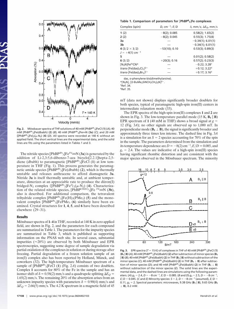
ResultsMossbauer spectra of 1–4 in THF, recorded at 140 K in zero appliedfield, are shown in Fig. 2, and the parameters for each compoundare summarized in Table 1. The parameters for the impurity speciesare summarized in Table 3, which is published as supportinginformation on the PNAS web site. In several cases, substantialimpurities (�20%) are observed by both Mossbauer and EPRspectroscopies, suggesting some degree of sample degradation viapartial oxidation of the complexes in solution or during storage afterfreezing. Partial degradation of a frozen solution sample of aniron(I) complex also has been reported by Holland, Munck, andcoworkers (32). The high-temperature Mossbauer spectrum of asample of [PhBPiPr
3]FeCl (1) (Fig. 2A) consists of two doublets.Complex 1 accounts for 80% of the Fe in the sample and has anisomer shift of � � 0.58(2) mm�s and a quadrupole splitting �EQ �1.65(2) mm�s. The remaining 20% of the absorption arises from anunknown impurity species with parameters � � 0.90(4) mm�s and�EQ � 2.66(5) mm�s. The 4.2 K spectrum in a magnetic field of 45
mT (data not shown) displays significantly broader doublets forboth species, typical of paramagnetic high-spin iron(II) centers inintermediate relaxation mode (33).
The EPR spectra of the high-spin iron(II) complexes 1 and 2 areshown in Fig. 3. The low-temperature parallel mode (15 K, B1 � B)EPR spectrum of 1 (40 mM in THF) shows a broad signal at g �12 (Fig. 3A); no other signals are observed up to 1,000 mT. Inperpendicular mode (B1 � B), the signal is significantly broader andapproximately three times less intense. The dashed line in Fig. 3Ais a simulation for an S � 2 species accounting for 70% of the spinin the sample. The parameters determined from the simulation andits temperature dependence are D � �8(2) cm�1, E�D � 0.085, andgz � 2.4. The values are indicative of a high-spin iron(II) specieshaving significant rhombic distortion and are consistent with themajor species observed in the Mossbauer spectrum. The minority
Fig. 2. Mossbauer spectra of THF solutions of 40 mM [PhBPiPr3]FeCl (1) (A); 40
mM [PhBPiPr3]Fe(dbabh) (2) (B); 40 mM [PhBPiPr
3]Fe'N (3a) (C); and 20 mM{[PhBPiPr
3]Fe}2(�-N2) (4) (D). All spectra were recorded at 140 K without anapplied field. The short vertical lines are the experimental data, and the solidlines are fits using the parameters listed in Tables 1 and 3.
Table 1. Comparison of parameters for �PhBPR3�Fe complexes
Complex (spin) D, cm�1; E�D �, mm�s; �EQ, mm�s
1 (2) �8(2); 0.085 0.58(2); 1.65(2)2 (2) �8(2); 0.045 0.55(3); 1.75(4)3a — �0.34(1); 6.01(1)3b — �0.34(1); 6.01(1)4 (3�2 � 3�2) �53(10); 0.10 0.53(3); 0.89(3)J � �4(1) cm�1
5 — 0.01(2); 0.58(2)6 (3�2) �20(3); 0.16 0.57(2); 0.23(3)�N3N�FeIVCN* — �0.22; 3.28†
trans-�Fe(das)2Cl2�2� — �0.12; 3.22‡
trans-�Fe(das)2Br2�2� — �0.17; 3.16‡
das, o-phenylene-bis(dimethylarsine).*�N3N�, �(t-BuMe2SiNCH2CH2)3N�3�.†Ref. 34.‡Ref. 35.
Fig. 3. EPR spectra (T � 15 K) of complexes in THF of 40 mM [PhBPiPr3]FeCl (1)
(B1 � B) (A); 40 mM [PhBPiPr3]Fe(dbabh) (2) after subtraction of minor species (B1
� B) (B); 40 mM [PhBPiPr3]Fe(dbabh) (2) in THF (B1 � B) without subtraction of the
minor species (C); 40 mM [PhBPiPr3]Fe(dbabh) (2) in THF (B1 � B) after subtrac-
tion of minor species (D); and 40 mM [PhBPiPr3]Fe(dbabh) (2) in THF (B1 � B)
without subtraction of the minor species (E). The solid lines are the experi-mental data, and the dashed lines are simulations using the following param-eters. (A) gz � 2.4, D � �8 cm�1, E�D � 0.085. (B and D) gz � 2.5, D � �8 cm�1,E�D � 0.045. (C and E) Minority species: S � 2, D � �8 cm�1 (assumed), E�D �0.11, gz � 2. Spectral parameters: microwaves, 9.38 GHz (B1 � B), 9.65 GHz (B1
� B), 0.2 mW.
17108 � www.pnas.org�cgi�doi�10.1073�pnas.0604402103 Hendrich et al.

species (30%) apparently has zero-field energies that render itunobservable with X-band EPR (36).
Metathesis of 1 with Li(dbabh) at low temperature in THFgenerates the iron amide [PhBPiPr
3]Fe(dbabh) (2). Complex 2 isthermally unstable but has been detected previously by 1H NMRand optical spectroscopies (30). A manganese analogue that wepresume to be isostructural to 2 has been characterized thoroughly,including an x-ray diffraction analysis. The iron(II) amide 2 exhibitsa doublet that accounts for �80% of the total Fe in the sample, withparameters � � 0.55(3) mm�s and �EQ � 1.75(4) mm�s (Fig. 2B).Approximately 20% of the absorption is from a presumed degra-dation product with parameters � � 0.72(5) mm�s and �EQ �3.10(5) mm�s. Both species have parameters that are typical ofhigh-spin FeII complexes. The Mossbauer spectrum at 4 K and 45mT shows broad features indicative of a paramagnetic species, inaddition to a small amount (5%) of the nitride complex[PhBPiPr
3]Fe'N (3a) (see below).The perpendicular mode EPR spectrum of 2 (40 mM in THF)
shows a feature near g � 9.8 on top of a broader feature (Fig. 3E).††
In parallel mode (Fig. 3C), both features near g � 9.8 sharpen andintensify. The signals near g � 9.8 can be quantitatively simulatedwith two Fe(II) species having concentrations of 28 mM (70%) and12 mM (30%). The dashed lines in Fig. 3 C and E are simulationsfor the minor S � 2 species with parameters as given in the figurelegend. The solid lines in Fig. 3 B and D are the experimentalspectra after subtraction of the simulation of this minor component.The major species is simulated (dashed lines, Fig. 3 B and D) as anS � 2 iron(II) species with D � �8(2) cm�1, E�D � 0.045, and gz �2.5. The spin states and relative ratios of these species are inagreement with those observed in the Mossbauer sample of 2.
Nitride 3a was prepared by allowing a sample of 2 to thermallydecay at 25°C over a period of minutes. Because 3a is itself thermallyunstable (decaying to {[PhBPiPr
3]Fe}2(�-N2) (4) over a period ofminutes at 40 mM at room temperature), preparing a high-puritysample of 3a at such high concentrations is difficult. The Mossbauerspectrum (T � 140 K) of 3a in THF, generated from a 40 mMoriginal stock solution of 1 and Li(dbabh), is shown in Fig. 2C. Thisspectrum contains a mixture of unresolved doublets from multiplespecies. We have attempted to fit this spectrum with combinationsof 1, 2, and 4; however, these combinations gave unsatisfactory fits.Instead, we must introduce two new impurities, which are presum-ably oxidation products, and a minor amount of 4. Most important,however, is the observation of a prominent new doublet withparameters � � �0.34(1) mm�s and �EQ � 6.01(1) mm�s, whichconstitutes �35% of the iron in the sample. The spectrum of 3a at4 K in a magnetic field of 45 mT (Fig. 4A) shows that the innerdoublets of the spectrum broaden, whereas the well resolved outerdoublet is unchanged, consistent with paramagnetic impurities anda diamagnetic [PhBPiPr
3]Fe'N.The chloride complex, [PhBPCH2Cy
3]FeCl also reacts with Li(d-babh) to produce a related nitride species, [PhBPCH2Cy
3]Fe'N(3b). Unlike [PhBPiPr
3]FeCl, the methylcyclohexyl-substituted de-rivative converts to the terminal nitride 3b over several hours attemperatures below �50°C without observation of an intermediateFeII(dbabh) species. Additionally, although 3b is unstable at tem-peratures above �50°C, it does not decay to a dinitrogen adductspecies akin to 4. It is therefore technically more straightforward togenerate a highly concentrated sample of 3b at low temperaturethan it is for 3a. The Mossbauer spectrum of 3b (Fig. 4B) exhibitsan outer doublet that constitutes 75% of the iron in the sample withthe same parameters as that observed for 3a, corroborating theirrespective assignments. The remaining 25% of the iron originatesfrom an unknown diamagnetic species, because the spectrum does
not depend on the magnetic field, with parameters � � 0.15(1)mm�s and �EQ � 1.65(1) mm�s.
For comparison, we have collected Mossbauer spectra of dia-magnetic [PhBPiPr
3]Fe(H)3(PMe3) (5) (Fig. 7A, which is publishedas supporting information on the PNAS web site). Structural andNMR data rigorously established the presence of three classicalhydride ligands for 5, rather than an alternative hydride�dihydrogenadduct iron(II) formulation (31). Therefore 3a, 3b, and 5 eachrepresent examples of formally iron(IV) species with diamagneticground states (30, 31). The T � 140 K Mossbauer spectrum of 5shows a single quadrupole doublet with parameters � � 0.01(2)mm�s and �EQ � 0.58(2) mm�s. The T � 4 K spectrum at 45 mTof 5 is unchanged, consistent with its diamagnetic character.
The low-temperature EPR spectrum of a sample of 3a (40 mMin THF) exhibits signals from the precursor 2 but with significantlylower intensities. The two species near g � 9.8 have decreased by60%, and the g � 1.95 minor species is approximately the same(�1% of sample). The EPR spectrum of 3b shows very weak signalsrepresenting 1% of the total sample. These changes are consistentwith the presence of the new diamagnetic Fe(IV) species observedin Mossbauer spectra.
Thermal decay of the nitride species 3a (but not 3b) affords adinitrogen complex, {[PhBPiPr
3]Fe}2(�-N2) 4. This complex hasbeen characterized structurally and also can be prepared by Na�Hgamalgam reduction of 1 in THF (28). The Mossbauer spectrum of4 (T � 140 K; Fig. 2D) can be fitted to two components: (i) a majorspecies with parameters � � 0.53(2) mm�s and �EQ � 0.89(3)mm�s (80%) and (ii) a minor species with parameters � � 1.10(4)mm�s and �EQ � 3.50(4)mm�s (20%) that appears to originatefrom an unknown iron(II) center. The spectrum at 4 K in a field of45 mT (data not shown) exhibits a significantly broadened doublet
††A sharp feature at g � 1.95 originates from a minor S � [1�2] species constituting 0.3 mMspins (�1% of sample). This feature vanishes in parallel mode. No other signals areobserved up to 1,000 mT.
Fig. 4. Mossbauer spectra of 40 mM [PhBPiPr3]Fe'N in THF (3a) (A); 40 mM
[PhBPMeCy3]Fe'N (3b) in THF (B); and 46 mM [PhBPiPr
3]FePMe3 (6) in toluene(C). All spectra are recorded at 4.2 K with a parallel applied field of 45 mT. Theshort vertical lines are the experimental data, and the solid lines are fits usingthe following parameters. (A) � � �0.34(1) mm�s, �EQ � 6.01(1) mm�s (35%).(B) Two species with � � �0.34(1) mm�s, �EQ � 6.01(1) mm�s (75%) and � �0.15(1) mm�s, �EQ � 1.65(1) mm�s (25%). (C) S � 3�2, D � 20 cm�1, E�D � 0.16,g � 2.2, � � 0.57(2) mm�s, �EQ � 0.23(3) mm�s, Ax � Ay � 0, Az � �8.8 mT.
Hendrich et al. PNAS � November 14, 2006 � vol. 103 � no. 46 � 17109
CHEM
ISTR
YSP
ECIA
LFE
ATU
RE

with the same parameters, indicative of a paramagnetic species inintermediate relaxation mode.
The low-temperature (2 K) parallel mode EPR spectrum of 4 (13mM in THF) exhibits a prominent signal at g � 13.5 with a 1:3:3:1hyperfine splitting pattern and a � 4.0 mT (Fig. 5A). The perpen-dicular mode spectrum shows a much broader feature near thissame g value with 10-fold lower intensity.‡‡ The simulation shownin Fig. 5B is for the ground doublet of a spin-coupled system SA �SB � 3�2 using the spin Hamiltonian of Eq. 1 (see SupportingMaterials and Methods, which is published as supporting informa-tion on the PNAS web site). The parameters of the simulation aregiven in the figure legend. The exchange value (J) and axialzero-field splitting parameter (DA � DB) are determined from themagnetization data (see below). EPR simulations that match thespectra require a ferromagnetic interaction (J 0) between thespins and site values of DA, DB 0. In addition, the simulation isquantitative; the intensity of the simulation is in agreement with thesample concentration. To match the hyperfine splitting, the simu-lation includes three equivalent I � 1�2 nuclei with Az � 77 � 10�4
cm�1. The reason for the occurrence of the four-line pattern,instead of the expected seven-line pattern from the six equivalentphosphines, is as yet unclear. This value is unusually large for aligand hyperfine coupling constant. The EPR signals of solutions of4 change drastically at higher temperatures, and these changes willneed to be explored further.
Isofield magnetization data were collected on a powder sampleof 4 at magnetic fields of 0.5, 2.5, and 5.0 T. A plot of �T versus Tof the data are shown in Fig. 8, which is published as supportinginformation on the PNAS web site. The data have been correctedfor the following: diamagnetic susceptibility of 4 (�dia � �8 � 10�4
cm3�mole), temperature-independent paramagnetism (�TIP ��10 � 10�4 cm3�mole), and field-independent magnetization(MS � �8 � 10�3 J�T per mole) (37, 38). The least-squaressimulation of the data (solid line) is shown in Fig. 8 for two identicalexchange-coupled S � 3�2 Fe(I) sites with J � �4 cm�1 and DA �DB � �53 cm�1. For reference, the Brillouin curve (D � 0, g � 2.0)for an S � 3 state at 0.5 T also is shown. Simulations of the data witheither an antiferromagnetic interaction (J 0) or DA � DB 0 didnot fit the data.
The complex [PhBPiPr3]FePMe3 (6) (31) was studied, in part, as
an aid to establish expected parameters for iron(I) complexesrelevant for the present work. The Mossbauer spectrum of 6 intoluene, recorded at 140 K in zero applied field, is shown in Fig. 7B.The spectrum of 6 shows a single species with parameters � �0.57(2) mm�s and �EQ � 0.23(3) mm�s. At 4 K and a field of 45mT, the spectrum (Fig. 4C) shows a paramagnetic six-line patternthat can be fit with an S � 3�2 species having the highly anisotropicFe hyperfine constants given in the figure legend. Interestingly,although the [PhBPiPr
3]FePMe3 complex has an isomer shift that issimilar to that of the Fe(II) complexes 1 and 2, the spin state S �3�2 of [PhBPiPr
3]FePMe3 is indicative of an Fe(I) valence. To ourknowledge, the Mossbauer parameters of only one other iron(I)coordination complex have been reported. The parameters ofLFeI(HC'CPh) (where L is HC(C[tBu]N-[2,6-diisopropylphe-nyl])2
�) (� � 0.44 mm�s and �EQ � 2.02 mm�s) differ significantly,presumably because of the different geometric and electronicstructure imposed by the �-diketiminate ligand and a symmetry thatgives rise to an orbital degeneracy (32).
The low-temperature (15 K) perpendicular mode EPR spec-trum of 6 exhibits a single signal with g values of 5.44 and 2.17(see Fig. 9, which is published as supporting information on thePNAS web site). The simulation (dashed line) overlaid on thespectrum is for a paramagnetic center with S � 3�2, D � �20(3)cm�1, E�D � 0.16, and g � 2.2. The experimental spectrum isextremely broad, apparently because of molecular interactions.Consequently, the simulation does not match particularly well.The spin concentration based on the simulation is in approxi-mate agreement with the sample concentration, and the spinstate and amount are in agreement with the species observed inthe Mossbauer sample. The spectrum does not show broadeningfor T 100 K. Thus, the value of D was determined from a fitto the temperature dependence of the S � 3�2 signal. Impor-tantly, with respect to the large hyperfine value observed forcomplex 4, simulations of the EPR spectrum of 6 indicate thatthe signal is sufficiently broad to accommodate an unresolvedhyperfine splitting of a magnitude similar to that observed for 4.By contrast, there is no evidence for 31P hyperfine in the EPRspectrum of S � 2 Fe(II)(dbabh) 2. Simulations of the signal of2, which include three equivalent 31P nuclei and a comparablelarge hyperfine A value, show an unmistakably large hyperfinesplitting that is not observed in the experimental spectrum.
To aid in the analysis of these complexes, density-functionalcalculations were performed on 1–6. Spin densities and Moss-bauer parameters were determined by using both optimized andcrystal structure geometries. Geometry optimizations were per-formed at the B3LYP�6–311G level. The same level of theorywas used to determine electric field gradients for �EQ and �. TheMulliken spin densities and FeOX bond distances are listed inTables 4 and 5, which are published as supporting informationon the PNAS web site. The spin densities are in good agreementwith the formal iron oxidation states of these species. Complexes1 and 2, which are each formally high-spin Fe(II), have 3.6unpaired electrons, and the S � 3�2 Fe(I) centers in[PhBPiPr
3]FePMe3 and 4 each possess three unpaired spins. Theclosed shell singlets 3a and [PhBPiPr
3]Fe(H)3(PMe3) have nospin density. Differences between the spin densities obtained atthe optimized and crystal structure geometries are negligible.Calculated bond distances are in reasonable agreement withexperiment. The FeOP bonds, however, appear to follow thegeneral trend observed for second-row ligand donor atoms inthat they are consistently (�0.1-Å) too long.
There is generally good agreement between theory and experi-ment for 3a, 4, 5, and 6 (Table 2). Differences in most cases arewithin the range of previously reported errors (39, 40). Interestingly,the Mossbauer parameters obtained from directly calculated crystalstructure geometries are generally in better agreement with exper-iment than the parameters obtained from the density-functional
‡‡The only other significant signal at 2 K is from a 1% minor species at g � 2.05 inperpendicular mode.
Fig. 5. EPR data for 4. (A) EPR spectrum (T � 2.3 K, B1 � B) of 20 mM{[PhBPiPr
3]Fe}2(�-N2) (4) in THF. (B) Simulation with parameters: SA � SB � 3�2, J ��4 cm�1, DA � DB � �53 cm�1, E�DA � E�DB � 0.10, gAz � gBz � 2.25, ALz � 77 �10�4 cm�1 (3 equivalent IL � 1�2). Spectral parameters: microwaves, 9.38 GHz, 0.2mW; modulation amplitude, 0.1 mT.
17110 � www.pnas.org�cgi�doi�10.1073�pnas.0604402103 Hendrich et al.

method (DFT) optimized geometries. This finding may be a con-sequence of the inaccurately long FeOP bond lengths obtainedfrom density-functional methods. Although the isomer shifts cal-culated for 1 and 2 are in good agreement with experiment, theircalculated �EQ values have unusually large errors of �1.5 mm�s.These errors most likely result from our attempts to model theground states of 1 and 2 as single determinant states. In bothcomplexes, three electrons occupy the lowest two orbitals (e") of theiron manifold. The correct description of these quintet groundstates requires a linear combination of determinants in which bothmembers of the e" set are alternately doubly occupied. In contrast,the ground states of 3, 4, 5, and 6 are well described by singledeterminants.
DiscussionIn this study, we have used both physical and density-functionalmethods to explore whether the formal oxidation states Fe(I) andFe(IV) are apt assignments for complexes such as the terminallybonded iron nitrides 3a and 3b and the diiron bridged-N2 complex4. Such information is germane to our ongoing consideration of ahypothetical Fe(I)�Fe(IV) Chatt-type N2 fixation cycle mediated byiron. The model complexes described in this article are unique inthat they provide a synthetic platform in which an iron center canaccommodate both N2 and N3� ligands at a single binding site. Sucha feature would presumably be critical to a catalytic cycle thatsampled intermediates bearing both types of ligand functionalities.
The crystallographic structures of [BP3]Fe'Nx complexes showa pseudo-threefold symmetric environment with the borate, the Fecenter, and the nitride or imide group lying along a common z axis(25, 27, 28, 41). Under this approximate C3v symmetry, the dorbitals split into a low-lying nonbonding e set of dxy, dx2–y2
parentage (Fig. 6), an intermediate-energy a1 orbital (dz2-type), anda highest-energy e set of dxz, dyz parentage. Complexes 1 and 2 bothare quintets (S � 2), and both exhibit Mossbauer parameters withinthe range expected for pseudo-tetrahedral high-spin Fe(II) com-plexes (33). From the EPR data, the zero-field terms for 2 are D ��8 cm�1 and E�D � 0.085. Under the symmetry C3v, within the 5Dterm, we derive from ligand-field theory the following approximaterelations between the zero-field and orbital energies: D � ��2(0.5��a1 � 1��e), E � � 0.5�2��a1. Here, �e and �a1 are the energiesfrom the ground orbital to the first and second excited orbitals,respectively. Using a spin-orbit constant of � � �100 cm�1 forFe(II), and the D and E values for 2, gives �e � 1,400 cm�1 and�a1 � 7,400 cm�1. The near-IR spectrum of 1 has a band at 7,360cm�1 with an extinction coefficient of 100 M�1 cm�1. The lowextinction coefficient and the agreement in energy with the calcu-lated value indicates that this is a d–d transition to the a1 orbital (dz2
parentage) lying above the nonbonding e set (dxy and dx2–y2
parentage).The Mossbauer data obtained for the terminally bonded nitride
complexes 3a and 3b are generally in agreement with the electronicstructure picture that has been previously advanced (27, 30). For the
nitride complex in the Fe(IV) oxidation state, density-functionalmethods predict four spin-paired electrons in the lower e set and anempty a1 orbital with a highest occupied molecular orbital�lowestunoccupied molecular orbital (HOMO�LUMO) gap of almost 4eV. This electronic structure is distinct from that of the trihydridecomplex [PhBPiPr
3]Fe(H)3(PMe3) 5, in which an orbital of a1symmetry lies much closer to the lower e set. For comparison, theMossbauer parameters for a few other low-spin Fe(IV) complexesare given in Table 1. The Tris(amido)amine FeIVOCN complex ofSchrock has a significantly negative isomer shift and large quad-rupole splitting. Wieghardt and coworkers (42, 43) have character-ized two S � 3�2 Fe(V)Onitrido complexes at very low temper-atures with � � �0.04 mm�s, �EQ values �1.90 to �1.0 mm�s, and�-N bridged complexes having an S � 1 Fe(IV) center � � �0.04to �0.14 mm�s and �EQ values between 0.79 and 1.13 mm�s.Interestingly, the large quadrupole-splitting parameter observed for3a and 3b is larger than those of any other FeIV species and, to ourknowledge, the largest of any known diamagnetic Fe complex. Thegeometry of the complexes places the hard nitride and boratefunctionalities along a pseudo-threefold z axis and polarizablephosphines around the periphery of the complex. This situationgenerates an unusually large electric field gradient along the z axisand, consequently, a large quadrupole splitting. In complex 5, theanionic charges are more symmetrically disposed, implying anisotropic electric field gradient relative to that for 3a or 3b and,consequently, a much smaller quadrupole-splitting parameter.
The electronic description of the dinitrogen adduct 4 remainssomewhat more enigmatic. The crystal structure of 4 shows arelatively short FeON distance of �1.82 Å and an NON distanceof 1.138(6) Å (30). These parameters reflect a degree of �-back-bonding from iron into the N'N �* orbitals, but it is not so muchas to suggest true electron transfer. For instance, the NON distancein complexes featuring a bridged NAN2� ligand is expected to becloser to 1.24 Å (44). The NON distance in 4, however, is muchshorter and is rather close to that of free N2. The x-ray data are thusmost indicative of an Fe(I) valence for each iron center of 4, ratherthan an FeIIONANOFeII formulation. Within the resolution ofthe Mossbauer spectrum, the iron sites of the dimeric complex 4appear equivalent. Although the isomer shift often can be identifiedwith a particular Fe valence state, similar isomer shifts are observedfor the monomeric Fe(I) and Fe(II) complexes and for the dini-trogen adduct 4. Thus, the isomer shift does not give a definitiveindication of the iron valence for the highly covalent complexesdescribed herein. The complementary EPR spectra that are pro-vided are additionally informative and thus have helped to provide
Table 2. Computationally predicted and experimentally observedMössbauer parameters
Complex S
Experiment Crystal structureOptimizedstructure
�EQ � � �EQ � � �EQ �
1 2 1.65 0.58 0.55 3.18 0.48 0.15 3.18 0.592 2 1.75 0.55 0.71 �3.45 0.58 0.88 2.79 0.593 0 6.01 �0.34 0.01 6.22 �0.154 3�2 0.89 0.53 0.78 �1.32 0.49 0.78 �1.64 0.69
3�2 0.90 1.37 0.49 0.58 �1.58 0.675 0 0.58 0.01 0.38 1.08 �0.05 0.66 1.05 0.136 3�2 0.23 0.57 0.68 �0.23 0.55 0.55 �0.20 0.80
Fig. 6. Qualitative MO splitting diagrams to aid the discussion of thed-electron configurations for Fe(I), Fe(II), and Fe(IV) centers with the geome-tries described in this article.
Hendrich et al. PNAS � November 14, 2006 � vol. 103 � no. 46 � 17111
CHEM
ISTR
YSP
ECIA
LFE
ATU
RE

a more complete picture of their appropriate spin and oxidationstate assignments.
Although it is as yet unclear whether either of the idealizedconfigurations, FeION'NOFeI or FeIIONANOFeII, accuratelydescribes the electronic structure of 4, they do provide an initialscheme for consideration of the magnetic information from thecomplex. As demonstrated above, the EPR and magnetization dataof 4 support an FeION'NOFeI electronic configuration. Inparticular, the least-squares simulation of the isofield magnetizationdata are appropriate for two identical exchange-coupled S � 3�2Fe(I) sites with J � �4 cm�1 and DA � DB � �53 cm�1. We alsohave considered an alternative formulism representing theFeIIONANOFeII configuration, wherein the identical Fe(II) siteshave local spins of SA � SB � 2, and two electrons are assumed tobe localized on the bridging dinitrogen to give a local dinitrogenspin of SC � 1. For this three-spin model, an antiferromagneticinteraction between both iron centers to the central NN spinproduces a ground system spin SS � 3. Simulations of the magne-tization data were extensively examined for the symmetric linear,three-spin model, with strong antiferromagnetic interactions, JAC �JBC �50 cm�1, and weak exchange, �JAB� 5 cm�1, with valuesof �DA � DB� ranging up to 100 cm�1 and gA � gB values between2 and 2.5. The C site is representative of an NAN2� triplet withDC � 0 and gC � 2. The simulation routine, which we have written,calculates the magnetic moment from full diagonalization of thethree-spin Hamiltonian, including zero-field terms. A suitable fit tothe magnetization data for the three-spin model over this entirerange of parameters could not be found.
In addition, the unusually large magnitude of the 31P hyperfinecoupling observed for 4 is indicative of strongly covalent FeOPinteractions and consistent with an Fe(I) valence state. This hyper-fine splitting is not observed from the Fe(II) complex 2 but may bepresent in the Fe(I) complex 6. A significantly larger hyperfineconstant for an Fe(I) valence may be attributed to a higher degreeof �-backbonding from the iron center into the phosphines for theFe(I) valence relative to Fe(II). We cannot yet explain the appear-ance of a 1:3:3:1 31P hyperfine coupling pattern for 4, rather thana seven-line pattern because of six equivalent FeOP interactions.Currently, we can only speculate that this may be a dynamic effectcorrelated to the lifetime of the 31P nuclear states. We haveconsidered the possibility that the hyperfine pattern is caused bytwo equivalent 14N nuclei in the three-spin formulism of theFe(II)ONANOFe(II) configuration. Two observations argue
strongly against this. First, even if the fifth line is missing (althoughwe believe not), two equivalent 14N nuclei would require a five-line1:2:3:2:1 pattern, which gives poor simulations of the EPR spec-trum. Second, the hyperfine constant of the 14N nucleus derivedfrom vector coupling of the three-spin model is AN � �4AS � 3,giving a site value of AN � 160 � 10�4 cm�1. This value is eight timesgreater than that observed for NO• or N2
�•. Future work will benefitfrom 15N isotopes to verify the conclusion.
Given all of the data presently available, we prefer an Fe(I)oxidation state assignment for 4 as most appropriate. Interestingly,Holland has prepared a diiron bridged-N2 complex supported by�-diketiminate ligands, also with an S � 3 ground state (45). For thissystem, an iron(I) formulation also can be posited. However, theNON bond distance that has been determined is more significantlyelongated (1.18 � 0.01 Å) relative to free N2 than in 4. Given thisobservation, and the harder coordination environment present forthis �-diketiminate diiron system, it has been advanced that thethree-spin exchange model is most appropriate for anFe(II)ONANOFe(II) complex (45). We suspect that the softphosphine ligands used in our own studies help to provide access tolower valent iron(I) because of their �-acidity.
To conclude, a single iron site is able to support terminallybonded N2 and nitride (N3�) ligands and also can accommodate arange of multielectron redox reactions. These features are requisitefor the development of a single-site Fe-mediated N2 fixation schemebased on the classic Chatt-type cycle. We also are consideringmechanisms that would be initiated by a single iron site but thatmight thereafter sample bimetallic intermediates, such as diiron�-N3� and �-NH2� species. Ongoing work therefore includes thepreparation and physical characterization of examples of suchspecies (26, 27).
Materials and MethodsComplete materials and methods are provided in SupportingMaterials and Methods.
Squid data were collected at the Molecular Materials Research Centerof the Beckman Institute of the California Institute of Technology. Thiswork was supported by National Institutes of Health Grant GM-070757(to J.C.P.), Postdoctoral Fellowship GM-072291 (to M.P.M.), and GrantGM-077387 (to M.P.H.). R.K.B. is grateful for a Herman FraschFoundation Fellowship, and M.T.G. acknowledges the Arnold andMabel Beckman Foundation and the Alfred P. Sloan Foundation.
1. Burgess BK, Lowe DJ (1996) Chem Rev 96:2983–3011.2. Eady RR (1996) Chem Rev 96:3013–3030.3. Howard JB, Rees DC (1996) Chem Rev 96:2965–2982.4. Thorneley RNF, Lowe DJ (1985) in Molybdenum Enzymes, ed Spiro TG (Wiley, New York),
Vol 7, pp 221–284.5. Dance I (2005) J Am Chem Soc 127:10925–10942.6. Seefeldt LC, Dean DR (1997) Acc Chem Res 30:260–266.7. Chatt J, Dilworth JR, Richards RL (1978) Chem Rev 78:589–625.8. Yandulov DV, Schrock RR (2002) J Am Chem Soc 124:6252–6253.9. Schrock RR (2003) Chem Commun 2389–2391.
10. Yandulov DV, Schrock RR (2003) Science 301:76–78.11. Schrock RR (2005) Philos Trans R Soc London A 363:959–969.12. Yandulov DV, Schrock RR (2005) Inorg Chem 44:1103–1117.13. Barney BM, Laryukhin M, Igarashi RY, Lee H-I, Dos Santos PC, Yang T-C, Hoffman BM,
Dean DR, Seefeldt LC (2005) Biochemistry 44:8030–8037.14. Igarashi RY, Laryukhin M, Dos Santos PC, Lee H-I, Dean DR, Seefeldt LC, Hoffman BM
(2005) J Am Chem Soc 127:6231–6241.15. Barney BM, Yang T-C, Igarashi RY, Dos Santos PC, Laryukhin M, Lee H-I, Hoffman BM,
Dean DR, Seefeldt LC (2005) J Am Chem Soc 127:14960–14961.16. Lee H-I, Benton PMC, Laryukhin M, Igarashi RY, Dean DR, Seefeldt LC, Hoffman BM
(2003) J Am Chem Soc 125:5604–5605.17. Lee H-I, Hales BJ, Hoffman BM (1997) J Am Chem Soc 119:11395–11400.18. Yang T-C, Maeser NK, Laryukhin M, Lee H-I, Dean DR, Seefeldt LC, Hoffman BM (2005)
J Am Chem Soc 127:12804–12805.19. Vrajmasu V, Munck E, Bominaar EL (2003) Inorg Chem 42:5974–5988.20. Huniar U, Ahlrichs R, Coucouvanis D (2004) J Am Chem Soc 126:2588–2601.21. Kastner J, Blochl PE (2005) ChemPhysChem 6:1724–1726.22. Hinnemann B, Nørskov JK (2003) J Am Chem Soc 125:1466–1467.23. Leigh GJ (1992) Acc Chem Res 25:177–181.
24. George TA, Rose DJ, Chang YD, Chen Q, Zubieta J (1995) Inorg Chem 34:1295–1298.25. Brown SD, Betley TA, Peters JC (2003) J Am Chem Soc 125:322–323.26. Brown SD, Mehn MP, Peters JC (2005) J Am Chem Soc 127:13146–13147.27. Brown SD, Peters JC (2005) J Am Chem Soc 127:1913–1923.28. Betley TA, Peters JC (2003) J Am Chem Soc 125:10782–10783.29. Betley TA, Peters JC (2003) Inorg Chem 42:5074–5084.30. Betley TA, Peters JC (2004) J Am Chem Soc 126:6252–6254.31. Daida EJ, Peters JC (2004) Inorg Chem 43:7474–7485.32. Stoian SA, Yu Y, Smith JM, Holland PL, Bominaar EL, Munck E (2005) Inorg Chem
44:4915–4922.33. Munck E (2000) in Physical Methods in Bioinorganic Chemistry, ed Que L, Jr (University
Science, Sausalito, CA), pp 287–319.34. Cummins CC, Schrock RR (1994) Inorg Chem 33:395–396.35. Paez EA, Oosterhuis WT, Weaver DL (1970) J Chem Soc Chem Commun 506–507.36. Palmer G (2000) in Physical Methods in Bioinorganic Chemistry, ed Que L, Jr (University
Science, Sausalito, CA), pp 121–185.37. Carlin RL (1986) Magnetochemistry (Springer, Berlin).38. Girerd J-J, Journaux Y (2000) in Physical Methods in Bioinorganic Chemistry, ed Que L, Jr
(University Science, Sausalito, CA), pp 321–374.39. Zhang Y, Mao JH, Godbout N, Oldfield E (2002) J Am Chem Soc 124:13921–13930.40. Neese F (2003) Curr Opin Chem Biol 7:125–135.41. Jenkins DM, Betley TA, Peters JC (2002) J Am Chem Soc 124:11238–11239.42. Meyer K, Bill E, Mienert B, Weyhermuller T, Wieghardt K (1999) J Am Chem Soc
121:4859–4876.43. Grapperhaus CA, Mienert B, Bill E, Weyhermuller T, Wieghardt K (2000) Inorg Chem
39:5306–5317.44. MacKay BA, Fryzuk MD (2004) Chem Rev 104:385–401.45. Stoian SA, Vela J, Smith JM, Sadique AR, Holland PL, Munck E, Bominaar EL (2006) J Am
Chem Soc 128:10181–10192.
17112 � www.pnas.org�cgi�doi�10.1073�pnas.0604402103 Hendrich et al.