NIRTriggered Anticancer Drug Delivery by...
5
Drug Delivery DOI: 10.1002/anie.201300183 NIR-Triggered Anticancer Drug Delivery by Upconverting Nanoparticles with Integrated Azobenzene-Modified Mesoporous Silica** Jianan Liu, Wenbo Bu,* Limin Pan, and Jianlin Shi* Cancer is one of the most common causes of death in the world, and chemotherapy remains to be one of the most frequent treatments for many cancers. However, its success has been greatly limited because of systemic toxicity due to the toxic effects by the nonspecific distribution of anti-cancer drugs. The drug delivery systems with the features of sustained drug release have attracted much attention owing to their enhanced therapeutic efficacy and reduced side effect. [1] For example, mesoporous silica nanoparticles (MSNs) with interesting properties, such as thermal and photostability, tunable sizes, high loading capacity, and the ease of functionalization according to currently available results, have made the MSNs one of the most promising carriers for drug delivery. [2] Although MSNs-based drug delivery systems have been proven effective, however, the use of cytotoxic drugs that are only released in the target area is much more preferred in clinical application. One way to prevent any premature release of anticancer drugs before they reach the target cells is to develop stimuli-responsive systems with controlled release features. Over the past decade, researches on MSNs-based light-controlled drug delivery systems have been carried out extensively because light as an external stimulus offers controllable drug release both spatially and temporally and thus exhibits great poten- tials for further biomedical applications. [3] However, most of them have achieved limited success in applications in vitro and in vivo mainly because of the easy damages to both biological samples and living tissues by UV light used to excite the photosensitizer and extremely quick attenuation of UV light in tissues. To tackle this issue, it is highly desirable to develop NIR remote-controllable MSNs-based system which can be used both in vitro and in vivo, which, however, has not been well addressed and remains a great challenge. Compared to UV light, near-infrared (NIR) light is much less damaging to biological specimens and living tissues involved and has remarkably deeper tissue penetration. [4] For example, Parak and co-workers reported that polyelectrolyte multilayer capsules loaded with cargo and plasmonic (Au or Ag) NPs can be used to release the cargo by NIR-photo- thermal heating. [5] The cargo can be easily released with high efficiency under NIR exposure in one second. However, the size of these capsules can be as large as several microns, which will inevitably suffer from phagocytosis by reticuloendothelial systems (RES) when these systems are intravenously injected. Recently, upconverting nanoparticles (UCNPs) have emerged as an appealing candidate for the application of NIR light. [6] Because of the unique ladder-like energy level structures of lanthanide ions (such as Tm 3+ , Er 3+ , and Ho 3+ ), UCNPs are able to absorb NIR light and convert it into high- energy photons in a very broad range from the UV to the NIR region. Such a unique and fascinating photoluminescence property enables UCNPs to function as a NIR-induced mediator by coating caged compounds on the nanoparticle surface, [7] and as a NIR-controlled photoswitch for reversible ring-closing and ring-opening transformation of a dithienyle- thene compound. [8] Very recently, Branda and co-workers successfully used the NIR laser to dissociate block copolymer micelles by encapsulating UCNPs inside micelles. [9] Since the copolymer micelles will inevitably suffer from self-degrada- tion in biological environment, a combination between UCNPs and micelles may find few opportunities in practical bio-applications for remote-controlled drug delivery using a NIR laser in vitro and in vivo. In more recent studies, our group successfully demonstrated UCNP/methylene blue- based photodynamic therapy (PDT) through controlled singlet oxygen release triggered by NIR light. [10] However, in spite of all the above efforts, reports on the direct NIR- light-controlled anticancer drug release for cancer therapy from a UCNPs-containing structure has not been found in the literature to date, which remains a great challenge in light- controlled drug delivery studies. Herein, we report a novel and general strategy for NIR light-triggered anticancer drug release based on a mesoporous silica-coated UCNPs structure, designated as UCNP@mSiO 2 . Figure 1 a shows the synthetic procedure for UCNP@mSiO 2 . The strategy consists of preparing NaYF 4 : TmYb@NaYF 4 core–shell nanoparticles and subsequently coating the Tm- doped core–shell UCNPs with mesoporous silica. After [*] J. N. Liu, Prof. W. B. Bu, L. M. Pan, Prof. J. L. Shi State Key Laboratory of High Performance Ceramics and Superfine Microstructure Shanghai Institute of Ceramics, Chinese Academy of Sciences 1295 Ding-Xi Road, Shanghai 200050 (China) E-mail: [email protected] [email protected] [**] This work was financially supported by the National Natural Science Foundation of China Research (grant numbers 50823007, 50972154, 51132009, 51072212, and 21172043), the Shanghai Rising-Star Program (grant number 12QH1402500), the Nano special program of the Science and Technology Commission of Shanghai (grant number 11nm0505000), the Development Foun- dation for Talents of Shanghai (grant number2012035), the National Basic Research Program of China (973 Program, grant number 2011CB707905). Supporting information for this article is available on the WWW under http://dx.doi.org/10.1002/anie.201300183. A ngewandte Chemi e 4375 Angew. Chem. Int. Ed. 2013, 52, 4375 –4379 # 2013 Wiley-VCH Verlag GmbH & Co. KGaA, Weinheim
Transcript of NIRTriggered Anticancer Drug Delivery by...
Drug DeliveryDOI: 10.1002/anie.201300183
NIR-Triggered Anticancer Drug Delivery by UpconvertingNanoparticles with Integrated Azobenzene-Modified MesoporousSilica**Jianan Liu, Wenbo Bu,* Limin Pan, and Jianlin Shi*
Cancer is one of the most common causes of death in theworld, and chemotherapy remains to be one of the mostfrequent treatments for many cancers. However, its successhas been greatly limited because of systemic toxicity due tothe toxic effects by the nonspecific distribution of anti-cancerdrugs. The drug delivery systems with the features ofsustained drug release have attracted much attention owingto their enhanced therapeutic efficacy and reduced sideeffect.[1] For example, mesoporous silica nanoparticles(MSNs) with interesting properties, such as thermal andphotostability, tunable sizes, high loading capacity, and theease of functionalization according to currently availableresults, have made the MSNs one of the most promisingcarriers for drug delivery.[2] Although MSNs-based drugdelivery systems have been proven effective, however, theuse of cytotoxic drugs that are only released in the target areais much more preferred in clinical application. One way toprevent any premature release of anticancer drugs beforethey reach the target cells is to develop stimuli-responsivesystems with controlled release features. Over the pastdecade, researches on MSNs-based light-controlled drugdelivery systems have been carried out extensively becauselight as an external stimulus offers controllable drug releaseboth spatially and temporally and thus exhibits great poten-tials for further biomedical applications.[3] However, most ofthem have achieved limited success in applications in vitroand in vivo mainly because of the easy damages to bothbiological samples and living tissues by UV light used toexcite the photosensitizer and extremely quick attenuation ofUV light in tissues. To tackle this issue, it is highly desirable to
develop NIR remote-controllable MSNs-based system whichcan be used both in vitro and in vivo, which, however, has notbeen well addressed and remains a great challenge.
Compared to UV light, near-infrared (NIR) light is muchless damaging to biological specimens and living tissuesinvolved and has remarkably deeper tissue penetration.[4] Forexample, Parak and co-workers reported that polyelectrolytemultilayer capsules loaded with cargo and plasmonic (Au orAg) NPs can be used to release the cargo by NIR-photo-thermal heating.[5] The cargo can be easily released with highefficiency under NIR exposure in one second. However, thesize of these capsules can be as large as several microns, whichwill inevitably suffer from phagocytosis by reticuloendothelialsystems (RES) when these systems are intravenously injected.Recently, upconverting nanoparticles (UCNPs) haveemerged as an appealing candidate for the application ofNIR light.[6] Because of the unique ladder-like energy levelstructures of lanthanide ions (such as Tm3+, Er3+, and Ho3+),UCNPs are able to absorb NIR light and convert it into high-energy photons in a very broad range from the UV to the NIRregion. Such a unique and fascinating photoluminescenceproperty enables UCNPs to function as a NIR-inducedmediator by coating caged compounds on the nanoparticlesurface,[7] and as a NIR-controlled photoswitch for reversiblering-closing and ring-opening transformation of a dithienyle-thene compound.[8] Very recently, Branda and co-workerssuccessfully used the NIR laser to dissociate block copolymermicelles by encapsulating UCNPs inside micelles.[9] Since thecopolymer micelles will inevitably suffer from self-degrada-tion in biological environment, a combination betweenUCNPs and micelles may find few opportunities in practicalbio-applications for remote-controlled drug delivery usinga NIR laser in vitro and in vivo. In more recent studies, ourgroup successfully demonstrated UCNP/methylene blue-based photodynamic therapy (PDT) through controlledsinglet oxygen release triggered by NIR light.[10] However,in spite of all the above efforts, reports on the direct NIR-light-controlled anticancer drug release for cancer therapyfrom a UCNPs-containing structure has not been found in theliterature to date, which remains a great challenge in light-controlled drug delivery studies.
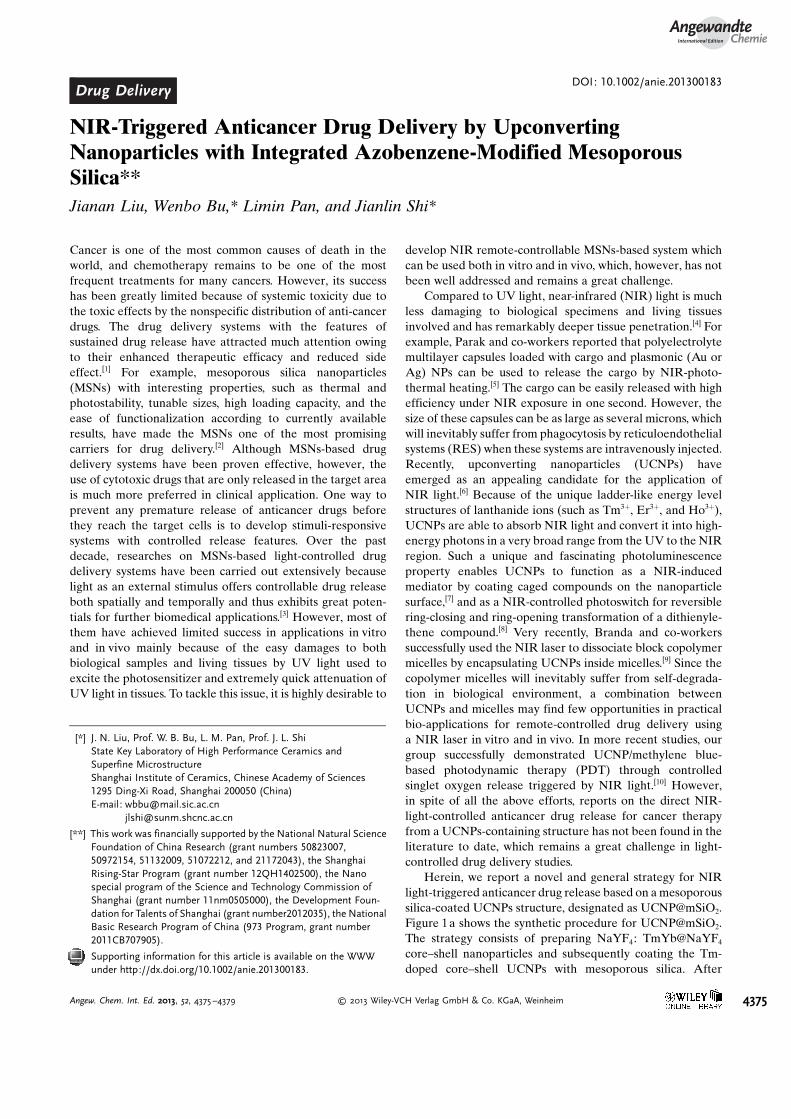
Herein, we report a novel and general strategy for NIRlight-triggered anticancer drug release based on a mesoporoussilica-coated UCNPs structure, designated as [email protected] 1a shows the synthetic procedure for [email protected] strategy consists of preparing NaYF4: TmYb@NaYF4
core–shell nanoparticles and subsequently coating the Tm-doped core–shell UCNPs with mesoporous silica. After
[*] J. N. Liu, Prof. W. B. Bu, L. M. Pan, Prof. J. L. ShiState Key Laboratory of High Performance Ceramics andSuperfine MicrostructureShanghai Institute of Ceramics, Chinese Academy of Sciences1295 Ding-Xi Road, Shanghai 200050 (China)E-mail: [email protected]
[**] This work was financially supported by the National Natural ScienceFoundation of China Research (grant numbers 50823007,50972154, 51132009, 51072212, and 21172043), the ShanghaiRising-Star Program (grant number 12QH1402500), the Nanospecial program of the Science and Technology Commission ofShanghai (grant number 11nm0505000), the Development Foun-dation for Talents of Shanghai (grant number2012035), the NationalBasic Research Program of China (973 Program, grant number2011CB707905).
Supporting information for this article is available on the WWWunder http://dx.doi.org/10.1002/anie.201300183.
AngewandteChemie
4375Angew. Chem. Int. Ed. 2013, 52, 4375 –4379 � 2013 Wiley-VCH Verlag GmbH & Co. KGaA, Weinheim
installing “photomechanical” azobenzene groups (azo) intothe mesopores of silica that act as “stirrer” in the mesoporoussilica, the TAT peptide was conjugated on the surface of theNPs to enhance the cellular uptake.[11] The above materialswere called UCNP@mSiO2-azo. Finally, the anticancer drugdoxorubicin (Dox) was loaded into the mesopores (Dox-UCNP@mSiO2-azo). Dox (zeta potential: + 2.2 mV) canstrongly attach to silica (zeta potential: �40.6 mV) throughformation of strong hydrogen bonds and charge interactionswith the surface silanol groups,[12] which resulted in a minimalDox release in aqueous environments. While the trans isomerof the azo molecules will transform into the cis isomer under
UV light irradiation, and in contrast the cis isomer will formthe trans isomer under irradiation of visible light (Figure S1 inthe Supporting Information). Upon absorption of NIR light(980 nm), the nanoparticles emit photons in the UV/Visregion, which can be absorbed immediately by the photo-responsive azo molecules in the pore network of themesoporous silica layer (Figure 2a). The reversible photo-isomerization by simultaneous UVand visible light emitted bythe UCNPs creates a continuous rotation–inversion move-ment. The back and forth wagging motion of the azomolecules will act as a molecular impeller that propels therelease of Dox. As a result, the Dox loaded in the mesopores
Figure 1. a) Synthetic procedure for upconverting nanoparticles coatedwith a mesoporous silica outer layer. b) NIR light-triggered Dox releaseby making use of the upconversion property of UCNPs and trans–cisphotoisomerization of azo molecules grafted in the mesopore networkof a mesoporous silica layer. TEM images of c) NaYF4:Tm, Yb,d) NaYF4:Tm, Yb@NaYF4, e) NaYF4:Tm, Yb@NaYF4@mSiO2, andf) NaYF4:Tm, Yb@NaYF4@mSiO2-azo. g) Bright-field and h) dark-fieldSTEM image of TAT-modified NaYF4:Tm, Yb@NaYF4@mSiO2-azo.
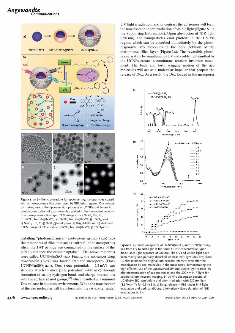
Figure 2. a) Emission spectra of UCNP@mSiO2 and UCNP@mSiO2-azo from UV to NIR light at the same UCNP concentration upondiode laser light exposure at 980 nm. The UV and visible light havebeen mostly and partially absorbed whereas NIR light (800 nm) fromUCNPs retained the original luminescent intensity even after themodification by azo molecules in the mesopores, demonstrating thehigh efficient use of the upconverted UV and visible light in trans--cisphotoisomerization of azo molecules and the 800 nm NIR light foradditional luminescent imaging. b) UV/Vis absorption spectra ofUCNP@mSiO2-azo before and after irradiation with 980 nm light(8.9 Wcm�2) for 0.5 to 8 h. c) Drug release in PBS under NIR lightirradiation and dark conditions, alternatively. Every duration of NIRirradiations is 1 h.
.AngewandteCommunications
4376 www.angewandte.org � 2013 Wiley-VCH Verlag GmbH & Co. KGaA, Weinheim Angew. Chem. Int. Ed. 2013, 52, 4375 –4379
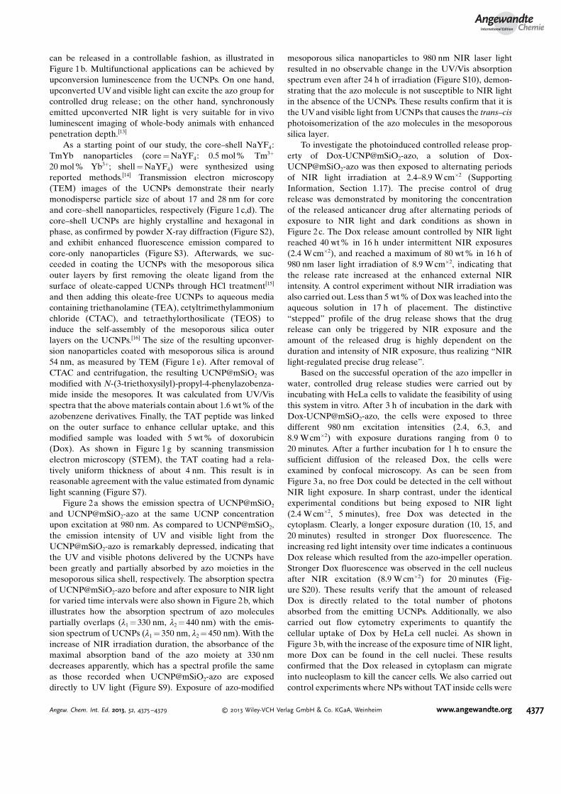
can be released in a controllable fashion, as illustrated inFigure 1b. Multifunctional applications can be achieved byupconversion luminescence from the UCNPs. On one hand,upconverted UV and visible light can excite the azo group forcontrolled drug release; on the other hand, synchronouslyemitted upconverted NIR light is very suitable for in vivoluminescent imaging of whole-body animals with enhancedpenetration depth.[13]
As a starting point of our study, the core–shell NaYF4:TmYb nanoparticles (core = NaYF4: 0.5 mol% Tm3+
20 mol% Yb3+; shell = NaYF4) were synthesized usingreported methods.[14] Transmission electron microscopy(TEM) images of the UCNPs demonstrate their nearlymonodisperse particle size of about 17 and 28 nm for coreand core–shell nanoparticles, respectively (Figure 1c,d). Thecore–shell UCNPs are highly crystalline and hexagonal inphase, as confirmed by powder X-ray diffraction (Figure S2),and exhibit enhanced fluorescence emission compared tocore-only nanoparticles (Figure S3). Afterwards, we suc-ceeded in coating the UCNPs with the mesoporous silicaouter layers by first removing the oleate ligand from thesurface of oleate-capped UCNPs through HCl treatment[15]
and then adding this oleate-free UCNPs to aqueous mediacontaining triethanolamine (TEA), cetyltrimethylammoniumchloride (CTAC), and tetraethylorthosilicate (TEOS) toinduce the self-assembly of the mesoporous silica outerlayers on the UCNPs.[16] The size of the resulting upconver-sion nanoparticles coated with mesoporous silica is around54 nm, as measured by TEM (Figure 1e). After removal ofCTAC and centrifugation, the resulting UCNP@mSiO2 wasmodified with N-(3-triethoxysilyl)-propyl-4-phenylazobenza-mide inside the mesopores. It was calculated from UV/Visspectra that the above materials contain about 1.6 wt% of theazobenzene derivatives. Finally, the TAT peptide was linkedon the outer surface to enhance cellular uptake, and thismodified sample was loaded with 5 wt% of doxorubicin(Dox). As shown in Figure 1g by scanning transmissionelectron microscopy (STEM), the TAT coating had a rela-tively uniform thickness of about 4 nm. This result is inreasonable agreement with the value estimated from dynamiclight scanning (Figure S7).
Figure 2a shows the emission spectra of UCNP@mSiO2
and UCNP@mSiO2-azo at the same UCNP concentrationupon excitation at 980 nm. As compared to UCNP@mSiO2,the emission intensity of UV and visible light from theUCNP@mSiO2-azo is remarkably depressed, indicating thatthe UV and visible photons delivered by the UCNPs havebeen greatly and partially absorbed by azo moieties in themesoporous silica shell, respectively. The absorption spectraof UCNP@mSiO2-azo before and after exposure to NIR lightfor varied time intervals were also shown in Figure 2 b, whichillustrates how the absorption spectrum of azo moleculespartially overlaps (l1 = 330 nm, l2 = 440 nm) with the emis-sion spectrum of UCNPs (l1 = 350 nm, l2 = 450 nm). With theincrease of NIR irradiation duration, the absorbance of themaximal absorption band of the azo moiety at 330 nmdecreases apparently, which has a spectral profile the sameas those recorded when UCNP@mSiO2-azo are exposeddirectly to UV light (Figure S9). Exposure of azo-modified
mesoporous silica nanoparticles to 980 nm NIR laser lightresulted in no observable change in the UV/Vis absorptionspectrum even after 24 h of irradiation (Figure S10), demon-strating that the azo molecule is not susceptible to NIR lightin the absence of the UCNPs. These results confirm that it isthe UVand visible light from UCNPs that causes the trans–cisphotoisomerization of the azo molecules in the mesoporoussilica layer.
To investigate the photoinduced controlled release prop-erty of Dox-UCNP@mSiO2-azo, a solution of Dox-UCNP@mSiO2-azo was then exposed to alternating periodsof NIR light irradiation at 2.4–8.9 W cm�2 (SupportingInformation, Section 1.17). The precise control of drugrelease was demonstrated by monitoring the concentrationof the released anticancer drug after alternating periods ofexposure to NIR light and dark conditions as shown inFigure 2c. The Dox release amount controlled by NIR lightreached 40 wt% in 16 h under intermittent NIR exposures(2.4 W cm�2), and reached a maximum of 80 wt % in 16 h of980 nm laser light irradiation of 8.9 W cm�2, indicating thatthe release rate increased at the enhanced external NIRintensity. A control experiment without NIR irradiation wasalso carried out. Less than 5 wt% of Dox was leached into theaqueous solution in 17 h of placement. The distinctive“stepped” profile of the drug release shows that the drugrelease can only be triggered by NIR exposure and theamount of the released drug is highly dependent on theduration and intensity of NIR exposure, thus realizing “NIRlight-regulated precise drug release”.
Based on the successful operation of the azo impeller inwater, controlled drug release studies were carried out byincubating with HeLa cells to validate the feasibility of usingthis system in vitro. After 3 h of incubation in the dark withDox-UCNP@mSiO2-azo, the cells were exposed to threedifferent 980 nm excitation intensities (2.4, 6.3, and8.9 Wcm�2) with exposure durations ranging from 0 to20 minutes. After a further incubation for 1 h to ensure thesufficient diffusion of the released Dox, the cells wereexamined by confocal microscopy. As can be seen fromFigure 3a, no free Dox could be detected in the cell withoutNIR light exposure. In sharp contrast, under the identicalexperimental conditions but being exposed to NIR light(2.4 W cm�2, 5 minutes), free Dox was detected in thecytoplasm. Clearly, a longer exposure duration (10, 15, and20 minutes) resulted in stronger Dox fluorescence. Theincreasing red light intensity over time indicates a continuousDox release which resulted from the azo-impeller operation.Stronger Dox fluorescence was observed in the cell nucleusafter NIR excitation (8.9 Wcm�2) for 20 minutes (Fig-ure S20). These results verify that the amount of releasedDox is directly related to the total number of photonsabsorbed from the emitting UCNPs. Additionally, we alsocarried out flow cytometry experiments to quantify thecellular uptake of Dox by HeLa cell nuclei. As shown inFigure 3b, with the increase of the exposure time of NIR light,more Dox can be found in the cell nuclei. These resultsconfirmed that the Dox released in cytoplasm can migrateinto nucleoplasm to kill the cancer cells. We also carried outcontrol experiments where NPs without TAT inside cells were
AngewandteChemie
4377Angew. Chem. Int. Ed. 2013, 52, 4375 –4379 � 2013 Wiley-VCH Verlag GmbH & Co. KGaA, Weinheim www.angewandte.org
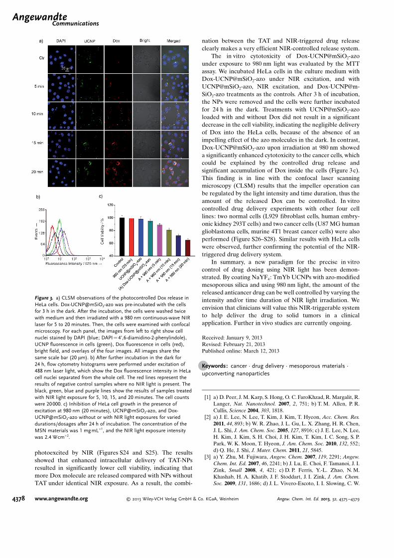
photoexcited by NIR (Figures S24 and S25). The resultsshowed that enhanced intracellular delivery of TAT-NPsresulted in significantly lower cell viability, indicating thatmore Dox molecule are released compared with NPs withoutTAT under identical NIR exposure. As a result, the combi-
nation between the TAT and NIR-triggered drug releaseclearly makes a very efficient NIR-controlled release system.
The in vitro cytotoxicity of Dox-UCNP@mSiO2-azounder exposure to 980 nm light was evaluated by the MTTassay. We incubated HeLa cells in the culture medium withDox-UCNP@mSiO2-azo under NIR excitation, and withUCNP@mSiO2-azo, NIR excitation, and Dox-UCNP@m-SiO2-azo treatments as the controls. After 3 h of incubation,the NPs were removed and the cells were further incubatedfor 24 h in the dark. Treatments with UCNP@mSiO2-azoloaded with and without Dox did not result in a significantdecrease in the cell viability, indicating the negligible deliveryof Dox into the HeLa cells, because of the absence of animpelling effect of the azo molecules in the dark. In contrast,Dox-UCNP@mSiO2-azo upon irradiation at 980 nm showeda significantly enhanced cytotoxicity to the cancer cells, whichcould be explained by the controlled drug release andsignificant accumulation of Dox inside the cells (Figure 3c).This finding is in line with the confocal laser scanningmicroscopy (CLSM) results that the impeller operation canbe regulated by the light intensity and time duration, thus theamount of the released Dox can be controlled. In vitrocontrolled drug delivery experiments with other four celllines: two normal cells (L929 fibroblast cells, human embry-onic kidney 293T cells) and two cancer cells (U87 MG humanglioblastoma cells, murine 4T1 breast cancer cells) were alsoperformed (Figure S26–S28). Similar results with HeLa cellswere observed, further confirming the potential of the NIR-triggered drug delivery system.
In summary, a new paradigm for the precise in vitrocontrol of drug dosing using NIR light has been demon-strated. By coating NaYF4: TmYb UCNPs with azo-modifiedmesoporous silica and using 980 nm light, the amount of thereleased anticancer drug can be well controlled by varying theintensity and/or time duration of NIR light irradiation. Weenvision that clinicians will value this NIR-triggerable systemto help deliver the drug to solid tumors in a clinicalapplication. Further in vivo studies are currently ongoing.
Received: January 9, 2013Revised: February 21, 2013Published online: March 12, 2013
.Keywords: cancer · drug delivery · mesoporous materials ·upconverting nanoparticles
[1] a) D. Peer, J. M. Karp, S. Hong, O. C. FaroKhzad, R. Margalit, R.Langer, Nat. Nanotechnol. 2007, 2, 751; b) T. M. Allen, P. R.Cullis, Science 2004, 303, 1818.
[2] a) J. E. Lee, N. Lee, T. Kim, J. Kim, T. Hyeon, Acc. Chem. Res.2011, 44, 893; b) W. R. Zhao, J. L. Gu, L. X. Zhang, H. R. Chen,J. L. Shi, J. Am. Chem. Soc. 2005, 127, 8916; c) J. E. Lee, N. Lee,H. Kim, J. Kim, S. H. Choi, J. H. Kim, T. Kim, I. C. Song, S. P.Park, W. K. Moon, T. Hyeon, J. Am. Chem. Soc. 2010, 132, 552;d) Q. He, J. Shi, J. Mater. Chem. 2011, 21, 5845.
[3] a) Y. Zhu, M. Fujiwara, Angew. Chem. 2007, 119, 2291; Angew.Chem. Int. Ed. 2007, 46, 2241; b) J. Lu, E. Choi, F. Tamanoi, J. I.Zink, Small 2008, 4, 421; c) D. P. Ferris, Y.-L. Zhao, N. M.Khashab, H. A. Khatib, J. F. Stoddart, J. I. Zink, J. Am. Chem.Soc. 2009, 131, 1686; d) J. L. Vivero-Escoto, I. I. Slowing, C. W.
Figure 3. a) CLSM observations of the photocontrolled Dox release inHeLa cells. Dox-UCNP@mSiO2-azo was pre-incubated with the cellsfor 3 h in the dark. After the incubation, the cells were washed twicewith medium and then irradiated with a 980 nm continuous-wave NIRlaser for 5 to 20 minutes. Then, the cells were examined with confocalmicroscopy. For each panel, the images from left to right show cellnuclei stained by DAPI (blue; DAPI=4’,6-diamidino-2-phenylindole),UCNP fluorescence in cells (green), Dox fluorescence in cells (red),bright field, and overlays of the four images. All images share thesame scale bar (20 mm). b) After further incubation in the dark for24 h, flow cytometry histograms were performed under excitation of488 nm laser light, which show the Dox fluorescence intensity in HeLacell nuclei separated from the whole cell. The red lines represent theresults of negative control samples where no NIR light is present. Theblack, green, blue and purple lines show the results of samples treatedwith NIR light exposure for 5, 10, 15, and 20 minutes. The cell countswere 20000. c) Inhibition of HeLa cell growth in the presence ofexcitation at 980 nm (20 minutes), UCNP@mSiO2-azo, and Dox-UCNP@mSiO2-azo without or with NIR light exposures for varieddurations/dosages after 24 h of incubation. The concentration of theMSN materials was 1 mgmL�1, and the NIR light exposure intensitywas 2.4 Wcm�2.
.AngewandteCommunications
4378 www.angewandte.org � 2013 Wiley-VCH Verlag GmbH & Co. KGaA, Weinheim Angew. Chem. Int. Ed. 2013, 52, 4375 –4379
Wu, V. S. Y. Lin, J. Am. Chem. Soc. 2009, 131, 3462; e) S.Angelos, Y. W. Yang, N. M. Khashab, J. F. Stoddart, J. I. Zink, J.Am. Chem. Soc. 2009, 131, 11344; f) P. Yang, S. Gai, J. Lin, Chem.Soc. Rev. 2012, 41, 3679.
[4] K. Szaciłowski, W. Macyk, A. Drzewiecka-Matuszek, M. Brin-dell, G. Stochel, Chem. Rev. 2005, 105, 2647.
[5] a) M. Ochs, S. Carregal-Romero, J. Rejman, K. Braeckmans,S. C. De Smedt, W. J. Parak, Angew. Chem. 2013, 125, 723;Angew. Chem. Int. Ed. 2013, 52, 695; b) A. G. Skirtach, A.MuÇoz Javier, O. Kreft, K. Kçhler, A. Piera Alberola, H.Mçhwald, W. J. Parak, G. B. Sukhorukov, Angew. Chem. 2006,118, 4728; Angew. Chem. Int. Ed. 2006, 45, 4612.
[6] a) M. Haase, H. Sch�fer, Angew. Chem. 2011, 123, 5928; Angew.Chem. Int. Ed. 2011, 50, 5808; b) Y. Yang, Q. Shao, R. Deng, C.Wang, X. Teng, K. Cheng, Z. Cheng, L. Huang, Z. Liu, X. Liu, B.Xing, Angew. Chem. 2012, 124, 3179; Angew. Chem. Int. Ed.2012, 51, 3125; c) R. Deng, X. Xie, M. Vendrell, Y.-T. Chang, X.Liu, J. Am. Chem. Soc. 2011, 133, 20168; d) W. Wu, L. Yao, T.Yang, R. Yin, F. Li, Y. Yu, J. Am. Chem. Soc. 2011, 133, 15810;e) Y. Dai, P. a. Ma, Z. Cheng, X. Kang, X. Zhang, Z. Hou, C. Li,D. Yang, X. Zhai, J. Lin, ACS Nano 2012, 6, 3327; f) Y. Dai, D.Yang, P. a. Ma, X. Kang, X. Zhang, C. Li, Z. Hou, Z. Cheng, J.Lin, Biomaterials 2012, 33, 8704; g) S. Gai, P. Yang, C. Li, W.Wang, Y. Dai, N. Niu, J. Lin, Adv. Funct. Mater. 2010, 20, 1166;h) C. Li, J. Lin, J. Mater. Chem. 2010, 20, 6831; i) R. Kumar, M.Nyk, T. Y. Ohulchanskyy, C. A. Flask, P. N. Prasad, Adv. Funct.Mater. 2009, 19, 853; j) F. Vetrone, R. Naccache, V. Mahalingam,C. G. Morgan, J. A. Capobianco, Adv. Funct. Mater. 2009, 19,2924; k) K. G. Jayakumar, N. M. Idris, Y. Zhang, Proc. Natl.Acad. Sci. USA 2012, 109, 8483; l) Q. Zhan, J. Qian, H. Liang, G.Somesfalean, D. Wang, S. He, Z. Zhang, S. Andersson-Engels,ACS Nano 2011, 5, 3744; m) Y. I. Park, H. M. Kim, J. H. Kim,K. C. Moon, B. Yoo, K. T. Lee, N. Lee, Y. Choi, W. Park, D. Ling,
K. Na, W. K. Moon, S. H. Choi, H. S. Park, S.-Y. Yoon, Y. D. Suh,S. H. Lee, T. Hyeon, Adv. Mater. 2012, 24, 5755; n) C. Wang, H.Tao, L. Cheng, Z. Liu, Biomaterials 2011, 32, 6145.
[7] C. J. Carling, F. Nourmohammadian, J.-C. Boyer, N. R. Branda,Angew. Chem. 2010, 122, 3870; Angew. Chem. Int. Ed. 2010, 49,3782.
[8] a) J.-C. Boyer, C.-J. Carling, B. D. Gates, N. R. Branda, J. Am.Chem. Soc. 2010, 132, 15766; b) C.-J. Carling, J.-C. Boyer, N. R.Branda, J. Am. Chem. Soc. 2009, 131, 10838.
[9] B. Yan, J.-C. Boyer, N. R. Branda, Y. Zhao, J. Am. Chem. Soc.2011, 133, 19714.
[10] F. Chen, S. Zhang, W. Bu, Y. Chen, Q. Xiao, J. Liu, H. Xing, L.Zhou, W. Peng, J. Shi, Chem. Eur. J. 2012, 18, 7082.
[11] a) M. Lewin, N. Carlesso, C.-H. Tung, X.-W. Tang, D. Cory, D. T.Scadden, R. Weissleder, Nat. Biotechnol. 2000, 18, 410; b) H.Yuan, A. M. Fales, T. Vo-Dinh, J. Am. Chem. Soc. 2012, 134,11358.
[12] N. Z. Knezevic, B. G. Trewyn, V. S. Y. Lin, Chem. Eur. J. 2011, 17,3338.
[13] a) J. Zhou, Y. Sun, X. X. Du, L. Q. Xiong, H. Hu, F. Y. Li,Biomaterials 2010, 31, 3287; b) H. Xing, W. Bu, Q. Ren, X.Zheng, M. Li, S. Zhang, H. Qu, Z. Wang, Y. Hua, K. Zhao, L.Zhou, W. Peng, J. Shi, Biomaterials 2012, 33, 5384.
[14] a) F. Wang, R. Deng, J. Wang, Q. Wang, Y. Han, H. Zhu, X.Chen, X. Liu, Nat. Mater. 2011, 10, 968; b) F. Chen, W. Bu, S.Zhang, X. Liu, J. Liu, H. Xing, Q. Xiao, L. Zhou, W. Peng, L.Wang, J. Shi, Adv. Funct. Mater. 2011, 21, 4285.
[15] N. Bogdan, F. Vetrone, G. A. Ozin, J. A. Capobianco, Nano Lett.2011, 11, 835.
[16] a) L. Pan, Q. He, J. Liu, Y. Chen, M. Ma, L. Zhang, J. Shi, J. Am.Chem. Soc. 2012, 134, 5722; b) J. Kobler, K. Moller, T. Bein, ACSNano 2008, 2, 791.
AngewandteChemie
4379Angew. Chem. Int. Ed. 2013, 52, 4375 –4379 � 2013 Wiley-VCH Verlag GmbH & Co. KGaA, Weinheim www.angewandte.org