Niosomes: An Updateimpactfactor.org/PDF/IJCPR/8/IJCPR,Vol8,Issue3,Article14.pdf · 2018-04-25 ·...
12
Available online on www.ijcpr.com International Journal of Current Pharmaceutical Review and Research; 8(3); 272-283 ISSN: 0976 822X Research Article *Author for Correspondence: [email protected] Niosomes: An Update Shilpi Arora * , Yashwant Himachal Institute of Pharmacy, Paonta Sahib, Himachal Pradesh, India Available Online: 17 th June, 2017 ABSTRACT Over decades researchers are striving to use the drugs in an efficient manner to treat various diseases. The efficient use can be explained as reduced dose, reduced side effects, reduced dosage frequency, greater patient compliance and maximum concentration of the drug at the site of action so as to reduce the undue exposure to the entire body. The article focuses on various advantages of vesicular systems (niosomes) to develop the effective delivery system to achieve maximum effective concentration. Niosomes, nonionic surfactant vesicles with lamellar structure which may be unilamellar and multilamellar serve to be efficient in providing these required advantages. The bilayer structure of niosomes being amphiphillic in nature can be used to deliver hydrophilic drugs in its aqueous core and lipophilic drugs in the bilayer made up of surfactants. Various additives in niosomes include nonionic surfactant as film forming agent, cholesterol as stabilizing and rigidizing agent for the bilayer and various charge inducers which develop a charge on the surface of niosomes and stabilize the prepared formulation by the resulting repulsive forces. This article also comprises of various breakthroughs in niosomal delivery of drugs representing various classes. On the basis of above information, the niosomes have been thoroughly exploited for the drug delivery system and still offer scope for research on various drugs for their maximum therapeutic utilization in management and treatment of various dreadful diseases. Keywords: Vesicular system, niosomes, nonionic surfactant, cholesterol. INTRODUCTION Niosomes are non-ionic surfactant vesicles having a bilayer structure formed by selfassembly of hydrated surfactant monomers. The bilayer is multilamellar or unilamellar which enclose aqueous solution of solutes and lipophilic components are in the bilayer itself. Niosomes are formed by hydration of non-ionic surfactant dried film resulting in imbibing or encapsulating the hydrating solution. Major component of niosomes is non-ionic surfactant which give it an advantage of being more stable when compared to liposomes thus overcoming the problems associated with liposomes i.e. susceptibility to oxidation, high price and the difficulty in procuring high purity levels which influence size, shape and stability (Vyas and Khar, 2011). Niosomes can entrap both hydrophilic and lipophilic drugs in aqueous layer and vesicular membrane respectively. The bilayers of niosomes have both inner and outer surfaces to be hydrophilic with sandwiched lipophilic area in between. Thus, a large number of drugs and other materials can be delivered using niosomes (Udupa, 2004). Salient features Niosomes serve as drug depots in the body which release the drug in a controlled manner through its bilayer providing sustained release of the enclosed drug. Targeted drug delivery can also be achieved using niosomes the drug is delivered directly to the body part where the therapeutic effect is required. Thereby reducing the dose required to be administered to achieve the desired effect (Mujoriya and Bodla, 2011; Karim et al., 2010). The therapeutic efficacy of the drugs is improved by reducing the clearance rate, targeting to the specific site and by protecting the encapsulated drug. Drug targeting reduces the dose which leads to subsequent decrease in the side effects. Niosomes are amphiphillic i.e. both hydrophilic and lipophillic in nature and can accommodate a large number of drugs with a wide range of solubilities. The formulation is in the form of aqueous vehicle based suspension having greater patient compliance when compared to oily dosage forms. Niosomal dispersion being aqueous can be emulsified in a non aqueous phase to regulate the drug release rate and to administer the vesicles in non-aqueous phase. They improve the oral bioavailability of poorly soluble drugs and also enhance the skin permeability of drugs when applied topically. Niosomes provide advantage of usage through various routes viz. oral, parentral, topical, ocular etc. The bilayers of the niosomes protect the enclosed Active pharmaceutical ingredient from the deterogenous factors present both inside and outside the body. So niosomes can be used for the delivery of labile and sensitive drugs. Niosomes are osmotically active and stable and also increase the stability of the entrapped drug. The surfactants used and also the prepared niosomes are biodegradable, biocompatible and non-immunogenic. Handling and storage of surfactants does not require any special conditions. The characteristics and the performance of the prepared niosomes can be controlled by altering the composition, concentration of various additives, size, lamellarity and surface charge of vesicles (Khan et al., 2011) doi: 10.25258/ijcprr.v8i03.9217
Transcript of Niosomes: An Updateimpactfactor.org/PDF/IJCPR/8/IJCPR,Vol8,Issue3,Article14.pdf · 2018-04-25 ·...
Available online on www.ijcpr.com
International Journal of Current Pharmaceutical Review and Research; 8(3); 272-283
ISSN: 0976 822X
Research Article
*Author for Correspondence: [email protected]
Niosomes: An Update
Shilpi Arora*, Yashwant
Himachal Institute of Pharmacy, Paonta Sahib, Himachal Pradesh, India
Available Online: 17th June, 2017
ABSTRACT
Over decades researchers are striving to use the drugs in an efficient manner to treat various diseases. The efficient use can
be explained as reduced dose, reduced side effects, reduced dosage frequency, greater patient compliance and maximum
concentration of the drug at the site of action so as to reduce the undue exposure to the entire body. The article focuses on
various advantages of vesicular systems (niosomes) to develop the effective delivery system to achieve maximum effective
concentration. Niosomes, nonionic surfactant vesicles with lamellar structure which may be unilamellar and multilamellar
serve to be efficient in providing these required advantages. The bilayer structure of niosomes being amphiphillic in nature
can be used to deliver hydrophilic drugs in its aqueous core and lipophilic drugs in the bilayer made up of surfactants.
Various additives in niosomes include nonionic surfactant as film forming agent, cholesterol as stabilizing and rigidizing
agent for the bilayer and various charge inducers which develop a charge on the surface of niosomes and stabilize the
prepared formulation by the resulting repulsive forces. This article also comprises of various breakthroughs in niosomal
delivery of drugs representing various classes. On the basis of above information, the niosomes have been thoroughly
exploited for the drug delivery system and still offer scope for research on various drugs for their maximum therapeutic
utilization in management and treatment of various dreadful diseases.
Keywords: Vesicular system, niosomes, nonionic surfactant, cholesterol.
INTRODUCTION
Niosomes are non-ionic surfactant vesicles having a
bilayer structure formed by selfassembly of hydrated
surfactant monomers. The bilayer is multilamellar or
unilamellar which enclose aqueous solution of solutes and
lipophilic components are in the bilayer itself. Niosomes
are formed by hydration of non-ionic surfactant dried film
resulting in imbibing or encapsulating the hydrating
solution. Major component of niosomes is non-ionic
surfactant which give it an advantage of being more stable
when compared to liposomes thus overcoming the
problems associated with liposomes i.e. susceptibility to
oxidation, high price and the difficulty in procuring high
purity levels which influence size, shape and stability
(Vyas and Khar, 2011). Niosomes can entrap both
hydrophilic and lipophilic drugs in aqueous layer and
vesicular membrane respectively. The bilayers of
niosomes have both inner and outer surfaces to be
hydrophilic with sandwiched lipophilic area in between.
Thus, a large number of drugs and other materials can be
delivered using niosomes (Udupa, 2004). Salient features
Niosomes serve as drug depots in the body which release
the drug in a controlled manner through its bilayer
providing sustained release of the enclosed drug. Targeted
drug delivery can also be achieved using niosomes the
drug is delivered directly to the body part where the
therapeutic effect is required. Thereby reducing the dose
required to be administered to achieve the desired effect
(Mujoriya and Bodla, 2011; Karim et al., 2010). The
therapeutic efficacy of the drugs is improved by reducing
the clearance rate, targeting to the specific site and by
protecting the encapsulated drug. Drug targeting reduces
the dose which leads to subsequent decrease in the side
effects. Niosomes are amphiphillic i.e. both hydrophilic
and lipophillic in nature and can accommodate a large
number of drugs with a wide range of solubilities. The
formulation is in the form of aqueous vehicle based
suspension having greater patient compliance when
compared to oily dosage forms. Niosomal dispersion being
aqueous can be emulsified in a non aqueous phase to
regulate the drug release rate and to administer the vesicles
in non-aqueous phase. They improve the oral
bioavailability of poorly soluble drugs and also enhance
the skin permeability of drugs when applied topically.
Niosomes provide advantage of usage through various
routes viz. oral, parentral, topical, ocular etc. The bilayers
of the niosomes protect the enclosed Active
pharmaceutical ingredient from the deterogenous factors
present both inside and outside the body. So niosomes can
be used for the delivery of labile and sensitive drugs.
Niosomes are osmotically active and stable and also
increase the stability of the entrapped drug. The surfactants
used and also the prepared niosomes are biodegradable,
biocompatible and non-immunogenic. Handling and
storage of surfactants does not require any special
conditions. The characteristics and the performance of the
prepared niosomes can be controlled by altering the
composition, concentration of various additives, size,
lamellarity and surface charge of vesicles (Khan et al.,
2011)
doi: 10.25258/ijcprr.v8i03.9217

Shilpi et al. / Niosomes: An Update
IJCPR, Volume 8, Issue 3, May- June 2017 Page 273
Structural Components of Niosomes
Surfactants A wide range of surfactants and their
combinations in different molar ratios have been used to
entrap many drugs in niosomes of varying features such as
size (Giddi et al., 2007). Ether linked surfactants These are
polyoxyethylene alkyl ethers which have hydrophilic and
hydrophobic moieties are linked with ether. The general
formula of this group is (CnEOm), where n can be 12-18
and m can be 3-7. Surfactants with polyhydroxyl head and
ethylene oxide units are also reported to be used in
niosomes formation (Vyas and Khar, 2011). Single alkyl
chain surfactant C16 mono alkyl glycerol ether with an
average of three glycerol units is one of the examples of
this class of surfactants used for the preparation of
niosomes (Baillie et al., 1985). Polyoxyethelene 4 lauryl
ether (Brij30) has an HLB value of 9.7, phase transition
temperature mercury salts, phenolic substances,
salicylates, sulfonamides and tannins as it cause oxidation
leading to discoloration of product. Polyoxyethylene cetyl
ethers (Brij58) and Polyoxyethylene stearyl ethers
(Brij72and76) are also used in preparation of niosomes
(Gannu and Pogaku, 2011). Ester linked surfactants These
surfactants have ester linkage between hydrophilic and
hydrophobic groups and have been studied for its use in
the preparation and delivery of sodium stibogluconate to
the experimental marine visceral leishmaniasis (Hunter et
al., 1988). Sorbitan Esters These are most widely used
ester linked surfactants especially in food industry. The
commercial sorbitan esters are mixtures of the partial
esters of sorbital and its mono and di-anhydrides with oleic
acid. These have been used to entrap wide range of drugs
viz doxorubicin (Uchegbu et al., 1996) Alkyl Amides
These are alkyl galactosides and glucosides which have
incorporated amino acid spacers. The alkyl groups are
fully or partially saturated C12 to C22 hydrocarbons and
some novel amide compounds have fluorocarbon chains.
Fatty Acids and Amino Acid Compounds These are amino
acids which are made amphiphilic by addition of
hydrophobic alkyl side chains and long chain fatty acids
which form “Ufasomes” vesicles formed from fatty acid
bilayers. Cholesterol Steroids bring about changes in
fluidity and permeability of the bilayer and are thus
important components. Cholesterol a waxy steroid
metabolite is usually added to the non-ionic surfactants to
provide rigidity and orientational order. It does not form
the bilayer itself and can be incorporated in large molar
ratios. Cholesterol is an amphiphilic molecule; it orients its
OH group towards aqueous phase and aliphatic chain
towards surfactant’s hydrocarbon chain. Rigidization is
provided by alternative positioning of rigid steroidal
skeleton with surfactant molecules in the bilayer by
restricting the movement of carbons of hydrocarbon.
Cholesterol is also known to prevent leakage by abolishing
gel to liquid phase transition (Dahiya et al., 2011) Charge
Inducers Charge inducers increase the stability of the
vesicles by induction of charge on the surface of the
prepared vesicles. It act by preventing the fusion of
vesicles due to repulsive forces of the same charge and
provide higher values of zeta potential. The commonly
used negative charge inducers are dicetyl phosphate,
dihexadecyl phosphate and lipoamine acid and positive
charge inducers are sterylamine and cetyl pyridinium
chloride (Shan et al., 2008; Bandyopadhyay and Johnson,
2007).
METHODS OF PREPARATION
Passive Trapping Techniques This category include most
of the techniques used in preparation of niosomes in which
drug is incorporated during the preparation of niosomes i.e.
during their formation (Udupa, 2004). Thin Film
Hydration All vesicles forming Components i.e.
surfactant, cholesterol and charge inducers are dissolved in
a volatile organic solvent in a round bottom flask. Using
rotary evaporator the organic solvent is evaporated at room
temperature forming a thin dry film of Dissolved
components. The dried thin film is hydrated with aqueous
phase with gentle agitation which leads to formation of
niosomes. The drug can be added to the aqueous phase if
hydrophilic and can be dissolved in organic solvent with
other components if hydrophobic (Baillie et al.,1986;
Palozza et al., 2006) Ether Injection Surfactant and other
components are dissolved in ether (diethyl ether) and is
then slowly injected into aqueous solution maintained at
60°C through a needle. This addition leads to evaporation
of ether and formation of single layered vesicles. This
method offers advantage of control of size, which can be
obtained by controlling size of needle and other conditions.
It suffers from disadvantage of limited solubility of
materials in ether and difficulty of removal of ether from
final formulation (Yasin et al., 2012; Guinedi et al., 2005)
Reverse Phase Evaporation The ingredients are dissolved
in a mixture of volatile organic solvents (ether and
chloroform) and drug is dissolved in aqueous phase. Water
in oil emulsion is formed of the two phases in a bath
sonicator. The basic principle involves evaporation of
organic solvent to form niosomes. This emulsion is dried
in a rotary evaporator at 40°C to form a semi solid gel of
large vesicles. Small quantity of buffer is added and the
semi solid form is sonicated at 4-5°C to form small
unilamellar vesicles (Guinedi et al., 2005) Multiple
Membrane Extrusion The basic principle involves
extrusion that is forced passage of
mixture/suspension/emulsion of the components through
polycarbonate membranes repeatedly to obtain niosomes
of desired size. The organic phase is dried in a rotary
evaporator and is hydrated by aqueous phase, the resultant
is extruded through the membrane (Khandare et al., 1994)
Microfluidization The two phases are allowed to interact
at ultra high speed in micro channels in an interaction
chamber. The high speed impingement and the energy
involved leads to formation of uniform and small
niosomes. This method has a high degree of
reproducibility (Khandare et al., 1994). Sonication The
mixture of drug solution in buffer, surfactant and
cholesterol is sonicated with titanium probe sonicator at
60°C for 3 minutes to yield niosomes. This method is also
used to produce small unilemellar vesicles from large
multilamellar vesicles prepared by other techniques
(Carter et al., 1988; Yoshida et al., 1992 and Hofland,
1992) The Bubble Method This method prepares niosomes

Shilpi et al. / Niosomes: An Update
IJCPR, Volume 8, Issue 3, May- June 2017 Page 274
in one step without the use of organic solvent. All the
components are dispersed in buffer and the dispersion is
placed in a round bottom flask immersed in a water bath
with controlled temperature. The flask has three necks
attached to water cooled reflux, thermometer and nitrogen
supply. The dispersion is mixed with a shear homogenizer
for 15 seconds and then bubbled with nitrogen in this
assembly to form niosomes (Chauhan and Luorence, 1989)
Active Trapping Techniques This includes the loading of
drug after the formation of niosomes. The niosomes are
prepared and then drug is loaded by maintaining pH
gradient or ion gradient to facilitate uptake of drug into
niosomes. It offers various advantages of 100%
entrapment, high drug lipid ratios, absence of leakage, cost
effective and suitability for labile drugs (Udupa, 2004).
Trans Membrane pH Gradient The organic phase with
dissolved components is evaporated to form a thin layer
and hydrated with citric acid, multilamellar vesicles are
formed which are freeze thawed 3 times and sonicated. To
this niosomal suspension aqueous solution with drug is
added, vortexed and pH is raised upto 7.0-7.2 with 1M
disodium phosphate. The mixture is later heated at 60 °C
for 10 minutes to get drug loaded niosomes (Biju et al.,
2006; Mayer et al., 1985) APPLICATIONS OF
NIOSOMES Antineoplastic Agents A biggest drawback of
cancer chemotherapy is side effects and lesser therapeutic
efficiency. Various attempts have been made to overcome
these drawbacks including niosomes as a novel drug
delivery system. Negatively charged niosomes of
Paclitaxel showed slow release being beneficial in storage,
administration, reduced toxic side effects and efficient oral
delivery (Bayindir and Yuksel, 2010). 5-fluorouracil (5-
FU) used for the treatment of actinic keratosis and non
melanoma skin cancer shows a poor percutaneous
permeation which was enhanced by delivery through
niosomes and also showed (Alvi et al., 2011). Further the
suitability of niosomes was also studied for 5-FU using
different preparation techniques and surfactants which
provided promising results (Namdeo and Jain, 1999).
Innovative niosomes of alpha, omega-hexadecyl-bis-(1-
aza-18-crown-6) (bola) were proved to be delivery systems
for the administration of 5- fluorouracil (Cosco et al.,
2009). Use of immunomodulatorsmuramyl dipeptide and
tuftsin on niosomes showed enhanced delivery of
bleomycin by macrophage activation. Thus present greater
efficacy of bleomycin in cancer chemotherapy (Raja et al.,
1996). Doxorubicin niosomes of span 60 provided
prolonged release with double tumoricidal activity, 10
times decreased clearance and increased levels of
metabolites in liver (Uchegbu et al., 1995). Preparation of
niosomes of doxorubicin has also lead to slow release with
peak plasma concentration same as the free drug with no
pulmonary side effects, reduced IC50 in doxorubicin
resistant cell lines and increased activity in ovarian cancer
cell line (Uchegbu et al.,1994; Uchegbu et al., 1996).
Innovative doxorubicin niosomes of dimethylsuberimidate
with transferrin covalently bound to the surface showed
higher uptake and cytotoxicity as compared to glucose
targeted niosomes of Npalmitoyl glucosamine also the
effect of cholesterol was studied which resulted in More
sustained release and reduced tumor growth in presence of
cholesterol (Dufes et al., 2004; Rogerson et al., 1988).
High degree of chemical purity, high entrapment values
and also reduced light induced degradation of adriamycin
was observed when encapsulated in niosomes and on
further evaluation it showed delayed growth of tumor
volume in human lung tumor cells, monolayered and
spheroid cultures and in xenografted nude mice (Rogerson
et al., 1987; Kerr et al., 1988). Metabolic profile and
urinary and faecal excretion of methotrexate was altered
by niosomes, there is rapid formation of 7-hydroxy
methotrexate, prolong levels in blood, large amounts in
liver and brain, reduced excretion of drug and its 7-
hydroxy metabolite into urine and bile and in vivo
protection of drug (Azmin et al., 1985; Azmin et al., 1986).
Plumbagin niosomes showed better anticancer activity and
less toxicity when compared to free drug (Naresh et al.,
1996). Combination of the stealth action and active
targeting function of polyethylene glycol cyanoacrylate-
co-hexadecyl cyanoacrylate (PEG-PHDCA) and
transferrin (Tf) was used to promote drug delivery to solid
tumor of hydroxycamptothecin (Hong et al., 2009).
Niosomes are also proved to be efficacious in prolonging
the action by sustaining the release of cytarabine
hydrochloride leading to decreased side effects and
toxicities (Ruckmani et al., 2000). NSAIDS With a view
to prepare stable niosomes with reduced leakage
cholesterol and surfactant ratios were optimized using
Aceclofenac as model drug. The prepared niosomes
showed good entrapment efficiency, which may improve
bioavailability of Aceclofenac (Srinivas et al., 2010).
Aceclofenac niosomes have also been prepared for topical
use after incorporation into carbopol gel. The gel showed
improved penetration and therapeutic efficacy of the drug
(Solankia et al., 2010). Niosomal gel was compared with
plain nimesulide gel in terms of drug delivery. It was
concluded that niosomal gel showed prolonged release of
nimesulide, thereby enhancing the anti-inflammatory
activity (Shahiwala and Misra, 2002). Despite of having
high oral bioavailability rofecoxib was withdrawn due to
its gastrointestinaladverse effects and cardiac toxicities. So
an attempt was made to reduce its side effects and
toxicities by encapsulation in niosomes. The niosomal gel
showed prolong drug release that is sustaining the action
of rofecoxib, thereby reducing its severe adverse effects
(Das and Palei, 2011). Niosomes with sustained release of
Diclofenac Sodium were prepared to achieve longer
duration of action with release upto 72 hours thus
providing an effective and improved therapeutic effect to
treat rheumatoid arthritis (Raja et al., 1994). Ketoprofen
was encapsulated in niosomes of span 60 for topical
application which released the drug in slow and sustained
manner (Arora and Sharma, 2010). Pharmacokinetic
parameters of flurbiprofen (FBP) were compared of
niosomal and non-niosomal formulations in dairy cattle
which showed long circulation of FBP niosomes offering
a potential application in improving short half-life
(Confalonieri et al., 2010). Formulation variables have
been optimized for flurbiprofen proniosomes for
preparation niosomes using a twofactor, three-level

Shilpi et al. / Niosomes: An Update
IJCPR, Volume 8, Issue 3, May- June 2017 Page 275
randomized full factorial strategy and were proved to be
critical in performance of formulation (Zidan and Mokhtar,
2011). Stable precursors of niosomes have been developed
using different non-ionic surfactants and also cholesterol
was proven to be an important ingredient of stable
niosomes of flurbiprofen (Mahmoud et al., 2008).
Meloxicam was entrapped in niosomal gel which showed
decreased side effects and increased pharmacological
activity thus proving to be an promising vehicles for
transdermal delivery and an alternative to the conventional
dosage form as shown by increased permeation through
skin (ElMenshawe and Hussein, 2011). Antiplatelet effect
of indomethacin have been sustained by incorporation into
niosomes, the effect may be shown due to greater quantity
of the drug reaching the specific site of inhibition in the
interior of the platelets and acting directly on the cyclo-
oxygenase system to prevent thromboxane formation
(Pillai and Salim, 1999). Antileishmanial Agents
Niosomal sodium stibogluconate was shown to be more
active than free drug against experimental murine visceral
leishmaniasis. High levels in liver and low serum levels
were obtained with high drug levels in infected
reticuloendothelial systelm (Baillie et al., 1986).
Paromomycin niosomes showed increased activity when
tested in-vitro and in-vivo for activity against Leishmania
donovani. They showed increased activity against liver
parasites without any significant suppression of
bonemarrow parasites (Williams et al., 1998).
Amarogentin, a secoiridoid glycoside has been evaluated
for its antileishmanial property using niosomes, liposomes
and free drug. The niosomal form was found to be more
efficacious than the liposomal form at the same membrane
microviscosity level and may have clinical application in
the treatment of leishmaniasis (Medda et al., 1999). The
appreciable efficacy in destroying intracellular parasites as
well as non-hepatotoxic and nonnephrotoxic nature,
harmine in niosomes can be considered for clinical
application in humans (Lalaa et al., 2004) Bacopasaponin
C, an indigenous glycoside, was tested for antileishmanial
properties both in free and in various delivery modes, e.g.,
niosomes, microspheres, and nanoparticles.
Bacopasaponin C was found to be very active in all the
vesicular forms analyzed from tissue histology, blood
pathology, and specific tests related to normal liver and
kidney functions without any side effects (Sinha et al.,
2002). Toxicity remains the major obstacle for the most
potent drugs known in the therapy of leishmaniasis so an
attempt was made to deliver nanocapsulated quercetin to
treat experimental leishmaniasis in the hamster model so
as to increase its efficacy as well as to reduce the toxicity.
It showed reduced parasite burden in the spleen as well as
reduced hepatotoxcity and renal toxicity as compared to
free drug so may be considered for clinical trials (Sarkar et
al., 2002). 14-deoxy-11-oxo-andrographolide, an
antileishmanial compound reduced the spleen parasite load
by 39% with subcutaneous injection of free drug and 78%,
91% and 59% in liposomes, niosomes and microspheres
respectively. The niosomal formulation showed greater
efficacy and lesser toxicity so might have clinical
application to combat visceral Leishmaniasis (Lala et al.,
2003). Gene Delivery Antisense oligonucleotides (OND)
were delivered effectively using cationic niosomes of
sorbitan monoesters which showed positive cellular uptake
of the antisense oligonucleotides from the prepared
niosomes (Huang et al., 2005). Surface of cationic
liposomes was modified by auto-coacervation through
electrostatic effect to develop a new gene carrier for
antisenseoligonucleotides. The efficacy was shown by
facilitated cellular uptake by COS-7 cells and HeLa cells
and positive effect on gene expression (Huang et al., 2006).
Polysorbate cationic niosomes exhibited the binding
capacity and the gene transfer study showed high
efficiency in mediating cellular uptake and transferred
gene expression (Huang et al., 2011). Elastic cationic
niosomes were used to enhance transdermal absorption of
luciferase plasmid (pLuc) by application of iontophoresis
or stratum corneum stripping method. The prepared
nanovesicles showed high degree of flux thus, presenting
niosomes as suitable carriers for luciferase plasmid
transdermal delivery (Manosroi et al., 2009). Niosomes of
Npalmitoylglucosamine bearing glucose or transferrin
ligands were prepared for drug targeting. It was shown that
glucose units were accessible on vesicle surface which
may be available for glucose receptors in vivo. Over
expression of GLUT receptors in cancer cells may lead to
targetting of these niosomes. Thus presenting them as gene
targetting agents to the cancer cells (Dufes et al., 2000).
PEGylated niosomes when compared to cationic niosomes
showed enhanced cellular uptake of oligonucleotide in
serum by providing protection against serum nucleases and
preventing serum proteins from approaching the
encapsulated oligonucleotide (Huang et al., 2008).
Niosomes have also served to be an alternative carrier of
Herring sperm DNA with added a dvantages over the
conventional liposomes (Yang et al., 2008). Cosmetic
Niosomes of N-acetyl glucosamine are prepared due to its
potential in the delivery of hydrophilic and hydrophobic
drugs in topical form and improved penetration into the
skin. N-acetyl glucosamine (NAG) has been considered in
the treatment of hyperpigmentation disorders due to its
inhibitory effect on thyrosinase enzymes in melanocytes.
Prepared formulations improved the extent of drug
localized in the skin, as needed in hyperpigmentation
disorders (Shatalebi et al., 2010). Elastic and non elastic
niosomes of gallic acid were prepared and non elastic
niosomes showed a slight increase in entrapment
efficiency. But the elastic niosomes showed increased
permeation through the skin which will be beneficial for
topical antiaging application (Manosroi et al.,2011). The
advantages of niosomes in aqueous dispersions were
shown in comparison with classical formulations such as
emulsions. These systems exhibit lower toxicity and
permit closer control of the availability of active
substances at the stratum corneum suitable for skin
moisturising and tanning products (Handjani et al., 1979).
Niosomes were prepared as possible approach to improve
the low skin penetration and bioavailability shown by
conventional topical vehicle for minoxidil. The results
suggest that niosomal formulations could constitute a
promising approach for the topical delivery of minoxidil in
Shilpi et al. / Niosomes: An Update
IJCPR, Volume 8, Issue 3, May- June 2017 Page 276
hair loss treatment (Mura et al., 2007). Finasteride
niosomes have been formulated for effective treatment of
androgenetic alopecia and the in vitro permeation and in
vivo deposition studies, demonstrated the potentials of
niosomes for successful delivery of finasteride to the
pilosebaceous unit (PSU) (Tabbakhian et al., 2006).
Ellagic acid (EA) is a potent antioxidant phytochemical
substance which has limited use due to poor
biopharmaceutical properties, low solubility and low
permeability. Niosomes with added solubilizers enhanced
the permeation of ellagic acid into the skin with increased
efficacy of ellagic acid (Junyaprasert et al., 2012). Proteins
Elastic anionic niosomes of n-terminal tat-GFP fusion
protein were prepared with various concentrations of
ethanol and edge activators sodium cholate and sodium
deoxycholate compared with nonelastic anionic niosomes.
Elastic anionic niosomes showed larger vesicular size,
higher negative zeta potential, elasticity (deformability
index), entrapment efficiency and flux through rat skin in
a transdermal study (Manosroi et al., 2010). Further the
Tat-GFP fusion protein has also been loaded in elastic
niosomes which enhanced the cellular uptake and chemical
stability of the peptide. Thus presenting to be an useful tool
for efficient delivery of many therapeutic proteins
(Manosoroi et al., 2011). Insulin was entrapped in the
niosomes prepared with different lipid composition and
doses. Significant decrease in blood glucose levels were
observed for longer duration thereby increasing the
therapeutic value of entrapped insulin (Khaksa et al., 2000)
Niosomes have also shown high protection of insulin
against proteolytic enzymes and good stability in the
presence of sodium desoxycholate and storage
temperatures (Varshosaz et al., 2003). Enhancement of the
entrapment of various charged peptide drugsviz. bacitracin
(BCT), insulin and bovine serum albumin (BSA) in
niosomes have been achieved by modifying the vesicular
charge compositions. The resulted formulation was found
to be appropriate to entrap the peptides with different
charges and polarity for pharmaceutical application as
shown by entrapment efficiency of the peptides in
niosomes determined by gel electrophoresis and gel
documentation (Mansoroi et al., 2010). Peroral vaccine
delivery system was developed by encapsulating
ovalbumin in non-ionic surfactant vesicles which were
evaluated using BALB/c mice. Encapsulation of
ovalbumin into Wasag7 (70% stearate sucrose ester, 30%
palmitate sucrose ester (40% mono-, 60% di/tri-ester))
niosomes resulted in a significant increase in antibody
titres (Rentel et al., 1999). Anti-Fungal Agents
Griseofulvin has a poor and variable oral bioavailability,
so niosomes were prepared using different methods of
preparation by varying nonionic surfactants, their
concentration and cholesterol concentration. It was
concluded that span 60 provided highest entrapment
efficiency and sustained release so could be one of the
promising delivery system for griseofulvin (Jadon et al.,
2009). Niosomal encapsulation provided means for
parental administration of nystatin, reducing its toxicity
and making it a more active antifungal agent. Nystatin
niosomes exerted less nephrotoxicity and hepatotoxicity in
vivo, showed higher level of drug in vital organs and
revealed pronounced efficacy in elimination of the fungal
burden in experimental animals compared with those
treated with free nystatin (Abdelbary et al., 2011).
Clotrimazole is widely used for the treatment of mycotic
infections of the genitourinary tract. An alternative
formulation was developed for the vaginal administration
of clotrimazole to provide sustained and controlled release
for local vaginal therapy by formulation in niosomes. The
prepared vesicle gel system was evaluated by antifungal
activity and tolerability on tissue level in rat which showed
sustained and controlled release of the drug, Clotrimazole
(Ning et al., 2005). Fluconazole-loaded niosomes of Span
40, Span 60, and Brij 72 surfactant were prepared and
evaluated. The prepared formulation accumulated and
formed localized drug depots in the skin, thereby releasing
the contents in a sustained manner and is able to greatly
enhance cutaneous retention of the drug (Gupta et al.,
2011). Presence of 50% alcohol in marketed gel of
naftifine hydrochloride an antifungal drug has been
detrimental to skin after repeated exposure. Non alcoholic
niosomal formulation of the drug was prepared and
incorporated in the gel to overcome the problem (Barakat
et al., 2009). Anti-Tubercular Drugs Niosomal formulation
of isoniazid was prepared to achieve effective treatment of
tuberculosis which showed 61.80% of cellular uptake
obtained from the drug-loaded niosomes by macrophage
cells. The cellular uptake obtained is sufficient to achieve
effective treatment of tuberculosis. The prepared
formulations also showed reduced dose, reduced toxicity
andfrequency and increased patient compliance. The
added advantage was macrophage targeting at the sites
where tuberculosis bacteria are harbored (Singh et al.,
2011). Isoniazid was encapsulated as niosomal
formulation which showed in vitro release pattern
indicating sustained release for 48 hours and lesser toxicity
in vivo than free drug (Karki et al., 2008). Encapsulation
of rifampicin in niosomes delivered the drug at the target
site with reduced toxicity and effective uptake. The
prepared formulations showed site specific tagetting by
controlling the niosome size and prolonged release, so can
be a promising approach in delivery of antitubercular drug
Rifampicin (Jain and Vyas, 1995). Pyrazinamide (PZA)
plays a unique role in shortening therapy because it kills a
population of semilatent tubercle bacilli residing in an
acidic environment. Encapsulation in niosomes targeted
the maximum concentration of PZA to the affected site
(lungs), excluded undesirable side effects, decreased
toxicity and may overcome the problem of drug resistance
(El-Ridy et al., 2011). Triton X 100 niosomes were used to
deliver anti-tubercular drugs whose release patterns were
studied. Rifampicin and isoniazid had fickian or
diffusional release and pyrazinamide had non-Fickian
release mechanism (Mehta et al., 2011). Antibiotics The
feasibility of using non-ionic surfactant vesicles
(niosomes) as carriers for the ophthalmic controlled
delivery of a water soluble local antibiotic, gentamicin
sulphate was investigated and the results demonstrated
niosomes to be promising ophthalmic carriers for the
topical application of gentamicin sulphate (Abdelbary and
Shilpi et al. / Niosomes: An Update
IJCPR, Volume 8, Issue 3, May- June 2017 Page 277
El-Gendy, 2008). Preparation and evaluation of
cefpodoxime proxetil niosomes showed controlled release
of 65.25% for 24 hours with zero order kinetics, thus
reducing the chances of dose dumping during usage
(Sambathkumar et al., 2011). The bioavailability of
Cefuroxime axetil which is just 25% was improved by
preparing niosomes. The prepared niosomes showed good
entrapment efficiency and in vitro release and also were
stable in bile salts (Sambhakar et al., 2011). Antibacterial
Drugs The influence of nonionic surfactant vesicles was
studied on physicochemical property, stability and in vitro
percutaneous absorption of enoxacin and was compared
with liposomes. The results showed the ability of niosomes
to modulate drug delivery without significant toxicity
making them useful to formulate topical enoxacin (Jia-You
et al., 2001). A higher and longer-lasting inhibition effect
was observed of nisin encapsulated in niosome and EDTA
against S. aureus as compared to their free forms. Thus
niosomes are offered as an alternative approach to
encapsulate nisin in a delivery system better than
liposomes (Kopermsuba et al., 2011). Gallidermin was
entrapped in niosomes in order to increase its stability and
efficiency for pharmaceutical and cosmeceutical uses. The
drug was shielded when entrapped in niosomes thereby
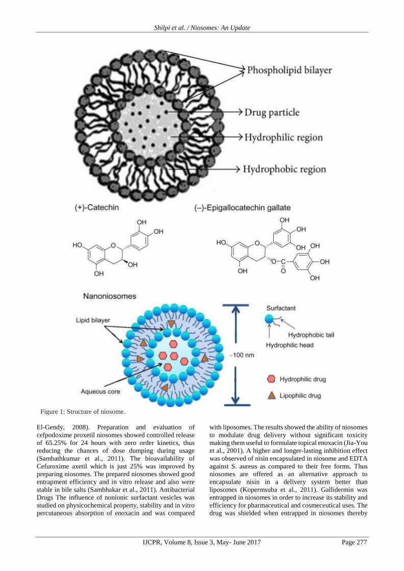
Figure 1: Structure of niosome.
Shilpi et al. / Niosomes: An Update
IJCPR, Volume 8, Issue 3, May- June 2017 Page 278
protecting the drug from the oxidation environments.
Gallidermin loaded in anionic niosomes and incorporated
in gel is the superior topical antibacterial
formulationbecause of the high accumulation in the skin
with no risk of systemic effect (Manosroia et al., 2010).
Antiviral Zidovudine (ZDV), an anti-HIV drug was
formulated in proniosomes and niosomes and their
distributions in lungs, kidney, heart, liver and spleen of
mice were studied after intravenous bolus injection.
Formulation prepared using Tween 80 was found to be
optimized with increased half-life, mean residence time
and reduced leakage of drug at 4⁰C (Ruckmani et al.,
2010). Liver targeting of ribavirin was enhanced upto 6
folds by using niosomes as drug delivery system when
compared to free drug solution. Ribavirin niosomes have
significant liver targeting property, which is expected to
improve the efficacy of low doses of ribavirin and
minimize its toxic side-effects at higher doses (Hashim et
al., 2010). Drug release was significantly affected by the
compositional factors in tenofovir niosomes. The
niosomes were prepared using different compositions and
were evaluated for vesicular sizing parameters, electrical
properties, drug entrapment data and drug release
characteristics. The results demonstrated the usefulness of
the microfluidization for the production and further scale-
up of anti-HIV niosomes with very small mean vesicular
sizes (Zidana et al., 2011). Immunization The topical
immunization with cholera toxin B is potential adjuvant for
cutaneous immune responses when coadministered with
the HBsAg encapsulated niosomes. Thus the niosomes for
topical delivery of vaccines using hepatitis B surface
protein as an antigen and cholera toxin B as an adjuvant
can be effective as topical delivery of vaccines
(Maheshwari et al., 2011). Mannosylated niosomes were
formulated as a topical vaccine delivery carrier and
adjuvant for the induction of both humoral and cellular
immunity. The proposed system would be simple, stable,
and cost effective and might be clinically acceptable (Jain
and Vyas, 2005). Mannosylated niosomes as oral DNA
vaccine carriers for the induction of humoral, cellular and
mucosal immunity were prepared. It was concluded that
niosomes produced both humoral (both systemic and
mucosal) and cellular immune response upon oral
administration and serve as DNA, vaccine carrier and
adjuvant for effective oral immunization (Jain et al., 2005).
The ability of non-ionic surfactant vesicles (NISV) to
stimulate humoral responses to bovine serum albumin
were studied and results suggest that adjuvants cannot only
circumvent antigen-specific non-responsiveness or low
responsiveness, but also can induce antibody isotype
switching independent of major histocompatibility
complex controls (Brewer and Alexander, 1994). Vitamins
Tretinoin cutaneous delivery is strongly affected by vesicle
composition and thermodynamic activity of the drug. In
particular, small, negatively charged niosomal
formulations, whichare saturated with tretinoin, have
shown to give higher cutaneous drug retention (Manconi
et al., 2006). A-tocopherol showed improve efficacy,
reduce toxicity and enhance therapeutic index when
enclosed in nonionic surfactant vesicles. The prepared
vesicles had 1-5μm diameter, entrapment efficiency
between 61.17%-79.63% and cumulative release 75.92%-
96.01% (Desai et al., 2010). Tretinoin-loaded niosomes as
multilamellar vesicles (MLV), large unilamellar vesicles
(LUV) and sonicated unilamellar vesicles (SUV) were
prepared and evaluated. The in vitro release of tretinoin
niosomes was found to be greater than tretinoin solution
by using Franz diffusion cells. Release data showed that
tretinoin delivery is mainly affected by the vesicular
structure and that tretinoin delivery increased from MLVs
to LUVs to SUVs (Manconi et al., 2002). Anti-
Inflammatory Niosomes have been immune-targeted to the
inflammation areas by conjugation with a purified
monoclonal antibody to CD44 (IM7) through a cyanuric
chloride (CC) linkage on the polyoxyethylene group of the
Tween 61 molecule (Hood et al., 2007). Non-ionic
surfactant vesicles (NSVs) were proposed for the
pulmonary delivery of glucocorticoids such as
beclomethasone dipropionate (BDP) for the treatment of
inflammatory lung diseases, e.g. asthma, chronic
obstructive pulmonary disease and various type of
pulmonary fibrosis. The obtained data indicated that the
investigated non-ionic surfactant vesicles (NSVs)
represent a promising tool as a pulmonary drug delivery
system (Marianeccia et al., 2010). A natural compound
with an efficacious anti-inflammatory activity, ammonium
glycyrrhizinate was loaded into Bola-niosomes. These
loaded niosomes depicted noticeable improvement of the
in vivo anti-inflammatory activity of the drug (Paolino et
al., 2007). Anti-Glaucoma Agents Chitosan or Carbopol
coated niosomal formulations of timolol maleate were
prepared which showed a sustained effect upto 8 hours.
The study concluded that the prepared formulations were
significantly better considering that half the concentration
is required indicating lesser systemic side effects, which
include cardiovascular side effects associated with ocular
timolol maleate therapy (Aggarwal and Kaur, 2005).
Timolol maleate is conventionally applied in form of eye
solutions which results in almost 80% of the instilled dose
being lost and results in systemic side-effects especially in
patients suffering from heart diseases or bronchial asthma
thus limiting its usefulness for the control of glaucoma.
Bioadhesive niosomal formulation showed a sustained and
controlled effect to eliminate the side effects (Kaur et al.,
2010). Diagnosis Non-ionic surfactant vesicles (niosomes)
are considered as carriers of iobitridol, a diagnostic agent
used for X-ray imaging. Increase of the rate of
encapsulation and the stability of the vesicles were found
to be satisfactory and in addition the physico-chemical and
morphological properties of the vesicles have been studied
(muller et al., 2000) Hormones Luteinizing hormone
releasing hormone (LHRH) was formulated in niosomes of
Hexadecyl diglycerol ether (C16G2), cholesterol, and
poly-24-oxyethylene cholesteryl ether (Solulan C24) in the
ratio 91:0:9 which resulted in polyhedral niosomes. The
prepared niosomes were stable in both muscle homogenate
and plasma and had clearance of about 49 hours with
sustained release (Arunothayanun et al., 1999). Muscle
Relaxants Niosomes of baclofen a centrally acting muscle
relaxant have been prepared to improve the low skin

Shilpi et al. / Niosomes: An Update
IJCPR, Volume 8, Issue 3, May- June 2017 Page 279
penetration and bioavailability characteristics shown by
conventional topical vehicle. The prepared niosomes
revealed advantages in vesicle surface morphology,
entrapment efficiency, in vitro drug release, Osmotic
fragility, stability studies and showed improved muscle
relaxation activity (Keservani et al., 2010). Anaesthetics
Interest in new delivery systems for local anaesthetics led
to non-ionic surfactant vesicles of lidocaine. The
performance of niosomes containing lidocaine
hydrochloride is remarkably better than that observed with
classical liposomes and Tween 20 micelles. The neutral
vesicles, prepared with Tween 20 and cholesterol, entrap a
higher lidocaine amount presenting it as novel delivery
system for lidocaine hydrochrolide (Carafa et al., 2002).
Anti-Diabetic Oral bioavailability of Gliclazide an oral
antidiabetic drug was improved by entrapment in nonionic
surfactant vesicles, also the release was sustained over a
period of 24 hours for better therapeutic efficacy. The high
values of zeta potential indicate stabilization of niosomes
by electrostatic repulsive forces (Tamizharasi et al., 2009).
Contraceptive The anti-fertility effect of cantchroman was
enhanced by incorporation into niosomes. The prepared
formulation showed 48.73% release in 8 hours and in vivo
anti-fertility studies showed 83.3% protection against
pregnancy. Histopathological studies showed no side
effects and no other toxic effects. So the study presents the
niosomes as suitable delivery system for contraceptives
(Shenoy et al., 1997). Miscellaneous Niosomes with
enclosed hemoglobin showed a visible spectrum
superimposable to that of free hemoglobin. Vesicles are
permeable to oxygen, hemoglobin dissociation curve can
be modified similar to free hemoglobin, have some
deformability and are more viscous than red blood cells but
have similar rheological behavior (Moser et al., 1989).
Topical and systemic application of naltrexone markedly
improves the characteristic signs of diabetic keratopathy
like impaired corneal sensation and delayed wound repair.
Niosomes of naltrexone for ocular delivery had high
entrapment efficiency and thermoresponsive properties
desirable for ocular delivery (Abdelkader et al., 2010).
Non-ionic surfactant vesicles (niosomes) appended with a
polysaccharide cap were prepared using hydrophobized
polysaccharides, O-palmitoyl pullulan (OPPu) and
cholesteroyl pullulan (CHPu) anchored onto propranolol
hydrochloride containing preformed niosomes. No
significant difference was observed in percent
encapsulation of polysaccharide coated and uncoated
vesicles. The influence of the hydrophobized
polysaccharide cap on niosomal membrane integrity and
stabilization against harsh bio-environment conditions was
also investigated using detergent and bile, freeze-thaw
cycling, osmotic stress, and long term and shelf stability
studies (Sihorkar and Vyas, 2000). Niosomes of capsaicin
were prepared to improve performance of its variety of
pharmacological actions on the cardiovascular, respiratory
and nervous systems. The prepared formulations were
compared to microemulsions prepared from the same
surfactants in the same ratio and better promote the
transdermal delivery of Capsaicin, with respect to
microemulsions for topical delivery of this drug (Tavano
et al., 2011)
CONCLUSION
Niosomes are novel drug delivery system which offers a
large number of advantages over other conventional and
vesicular delivery systems. Namely targeted delivery,
reduction of dose, stability and compatibility of non-ionic
surfactants, easy modification, delayed clearance,
suitability for a wide range of Active Pharmaceutical
Agents etc. From the above compilation of work it can be
concluded that niosomes have suitability for encapsulating
a varied variety of drugs and also the benefits offered by
niosomes are also widely exploited. Niosomes have
evolved for treatment of many dreadful diseases efficiently
with reduced side effects and better patient compliance.
Thus niosomes present itself as a versatile tool in
therapeutics.
REFERENCES
1. Abdelbary A., Essam T., Abd El-Salam R. M.,
AlyKassem A. A. Niosomes as a potential drug
delivery system for increasing the efficacy and safety
of nystatin (antifungal). Drug Dev Ind Pharm. 2011;
37: 149- 508.
2. Abdelbary G., El-Gendy N. Niosome-Encapsulated
Gentamicin for Ophthalmic Controlled Delivery.
AAPS PharmSciTech. 2008; 9: 740- 747.
3. Abdelkader H., Ismail S., Kamal A., Alany R. G.
Preparation of niosomes as an ocular delivery system
for naltrexone hydrochloride: physicochemical
characterization. Pharmazie. 2010; 65: 811-817.
4. Aggarwal D., Kaur I.P. Improved pharmacodynamics
of timolol maleate from a mucoadhesive niosomal
ophthalmic drug delivery system. Int J Pharm. 2005;
290: 155-159.
5. Alvi I. A., Madan J., Kaushik D., Sardana S., Pandey
R.S., Ali A. Comparative study of transfersomes,
liposomes, and niosomes for topical delivery of 5-
fluorouracil to skin cancer cells: preparation,
characterization, in-vitro release, and cytotoxicity
analysis. Anticancer Drugs. 2011; 22: 774-782.
6. Arora R., Sharma A. Release Studies of Ketoprofen
Niosome Formulation. J Chem Pharm Res. 2010; 2:
79-82.
7. Arunothayanun P., Turton J. A., Uchegbu I. F.,
Florence A. T. Preparation and in vitro/in vivo
evaluation of luteinizing hormone releasing hormone
(LHRH)-loaded polyhedral and spherical/tubular
niosomes. J Pharm Sci. 1999; 88 :34-38.
8. Azmin M. N., Florence A. T., Handjani-Vila R.M.,
Stuart J.F., Vanlerberghe G., Whittaker J. S. The
effect of non-ionic surfactant vesicle (niosome)
entrapment on the absorption and distribution of
methotrexate in mice. J Pharm Pharmacol. 1985; 37:
237-242.
9. Azmin M. N., Florence A. T., Handjani-Vila R. M.,
Stuart J. F., Vanlerberghe G., Whittaker J. S. The
effect of niosomes and polysorbate 80 on the
Shilpi et al. / Niosomes: An Update
IJCPR, Volume 8, Issue 3, May- June 2017 Page 280
metabolism and excretion of methotrexate in the
mouse. J Microencapsul. 1986; 3: 95-100.
10. Baillie A. J., Coombs G. H., Dolan T.F., Laurie J.
Non-ionic surfactant vesicles, niosomes, as a delivery
system for the anti-leishmanial drug, sodium
stibogluconate. J Pharm Pharmacol. 1986; 38: 502-
505.
11. Baillie A. J., Florence A. T., Hume L. R., Muirhead
G. T., Rogerson A. The preparation and properties of
niosomes--non-ionic surfactant vesicles. J Pharm
Pharmacol. 1985; 37: 863-868.
12. Bandyopadhyay P., Johnson M. Fatty alcohols or fatty
acids as niosomal hybrid carrier: effect on vesicle size,
encapsulation efficiency and in vitro dye release.
Colloids Surf B Biointerfaces. 2007; 58: 68-71.
13. Barakat H. S., Darwish I. A., El-Khordagui L. K.,
Khalafallah N. M. Development of naftifine
hydrochloride alcohol-free niosome gel. Drug Dev Ind
Pharm. 2009; 35: 631-637.
14. Bayindir Z. S., Yuksel N. Characterization of
niosomes prepared with various nonionic surfactants
for paclitaxel oral delivery. J Pharm Sci. 2010; 99:
2049-2060.
15. Biju S. S., Talegaonkar S., Mishra P. R., Khar R. K.
Vesicular systems: An overview. Indian J Pharm Sci.
2006; 68: 141-153.
16. Brewer M., Alexander J. Studies on the adjuvant
activity of nonionic surfactant vesicles: adjuvant-
driven IgG2a production independent of MHC
control. Vaccine. 1994; 12: 613-619.
17. Carafa M., Santucci E., Lucania G. Lidocaine-loaded
non-ionic surfactant vesicles characterization and in
vitro permeation studies. Int J Pharm. 2002; 231: 21–
32.
18. Carter K.C. et al. The therapeutic effect of sodium
stibogluconate in BALB:c mice infected with
Leishmania donobani is organ dependent. J Pharm
Pharmacol. 1988; 40: 370–373.
19. Chauhan S., Luorence M. J. The preparation of
polyoxyethylene containing non-ionic surfactant
Vesicles. J Pharm Pharmacol. 1989; 41: 6.
20. Confalonieri E. O., Soraci A. L., Becaluba M.,
Denzoin L., Rodriguez E., Riccio B., Tapia O. The
disposition of free and niosomally encapsulated Rac-
flurbiprofen in dairy bovines. J Vet Pharmacol Ther.
2010; 33: 9-14.
21. Cosco D., Paolino D., Muzzalupo R., Celia C., Citraro
R., Caponio D., Picci N., Fresta M. Novel PEG-coated
niosomes based on bola-surfactant as drug carriers for
5-fluorouracil. Biomed Microdevices. 2009; 11:
1115-1125.
22. Dahiya N. K., Rao R., Nanda S. Preparation and
characterization techniques in niosomal vesicular
systems- A review. J. Pharm. Biomed. Sci. 2011; 5:1-
8.
23. Desai A. R., Raghuveer I., Chitme H. R., Chandra R.
Development and characterization of niosomal drug
delivery of atocopherol. Int. J. Chem. Anal. Sci. 2010;
1: 146-158.
24. Dufes C., Muller J. M., Couet W., Olivier J. C.,
Uchegbu I. F., Schatzlein A. G. Anticancer drug
delivery with transferrin targeted polymeric chitosan
vesicles. Pharm Res. 2004; 21: 101-107.
25. Dufes C., Schatzlein A. G., Tetley L., Gray A. I.,
Watson D. G., Olivier J. C., Couet W., Uchegbu I. F.
Niosomes and Polymeric Chitosan Based Vesicles
Bearing Transferrin and Glucose Ligands for Drug
Targeting. Pharm Res. 2000; 17: 1250-1258.
26. El-Menshawe S. F., Hussein A. K. Formulation and
evaluation of meloxicam niosomes as vesicular
carriers for enhanced skin delivery. Pharm Dev
Technol. 2011. (Epub ahead of print). El-Ridy M. S.,
Abdelbary A., Nasr E. A., Khalil R. M., Mostafa D.
M., El-Batal A. I., Abd El-Alim S. H. Niosomal
encapsulation of the antitubercular drug,
pyrazinamide. Drug Dev Ind Pharm. 2011; 37: 1110-
1118.
27. Gannu P. K., Pogaku R. Nonionic surfactant vesicular
systems for effective drug delivery-an overview. Acta
Pharmaceutica Sinica B. 2011; 1: 208–219.
28. Giddi H. S., Arunagirinathan M. A., Bellare J. R. Self-
assembled surfactant nano-structures important in
drug delivery: A review. Indian J Exp Biol. 2007; 45:
133-159.
29. Guinedi A. S., Nahed D. M., Samar M., Rania M. H.
Preparation and evaluation of reverse-phase
evaporation and multilamellar niosomes as
ophthalmic carriers of acetazolamide. Int J Pharm.
2005; 306: 71–82.
30. Gupta M., Vaidya B., Mishra N., Vyas S. P. Effect of
surfactants on the characteristics of fluconazole
niosomes for enhanced cutaneous delivery. Artif Cells
Blood Substit Immobil Biotechnol. 2011; 39: 376-
384.
31. Handjani-Vila R. M., Ribier A., Rondot B.,
Vanlerberghie G. Dispersions of lamellar phases of
non-ionic lipids in cosmetic products. Int J Cosmet
Sci. 1979; 1: 303-314.
32. Hashim F., El-Ridy M., Nasr M., Abdallah Y.
Preparation and characterization of niosomes
containing ribavirin for liver targeting. Drug Deliv.
2010; 17: 282-287.
33. Hofland H. E. J. Safety aspects of non-ionic surfactant
vesicles-a toxicity study related to the
physicochemical characteristics of non-ionic
surfactants. J Pharm Pharmacol. 1992; 44 :287-294.
34. Hong M., Zhu S., Jiang Y., Tang G., Pei Y. Efficient
tumor targeting of hydroxycamptothecin loaded
PEGylated niosomes modified with transferrin. J
Control Release. 2009; 133: 96-102.
35. Hood E., Gonzalez M., Plaas A., Strom J., VanAuker
M. Immuno-targeting of nonionic surfactant vesicles
to inflammation. Int J Pharm. 2007; 339: 222-230.
36. Huang Y., Chen J., Chen X., Gao J., Liang W.
PEGylated synthetic surfactant vesicles (Niosomes):
novel carriers for oligonucleotides. J Mater Sci Mater
Med. 2008; 19: 607-614.
37. Huang Y. Z., Gao J. Q., Chen J. L., Liang W. Q.
Cationic liposomes modified with non-ionic
Shilpi et al. / Niosomes: An Update
IJCPR, Volume 8, Issue 3, May- June 2017 Page 281
surfactants as effective non-viral carrier for gene
transfer. Colloids Surf B Biointerfaces. 2006; 49: 158-
164
38. Huang Y. Z., Han G., Wang H., Liang W. Q. Cationic
niosomes as gene carriers: preparation and cellular
uptake in vitro. Pharmazie. 2005; 60: 473-474.
39. Huang Y. Z., Rao Y., Chen JL., Yang V. C., Liang W.
Polysorbate cationic synthetic vesicle for gene
delivery. J Biomed Mater Res. 2011; 96: 513-519.
40. Hunter C. A., Dolan T. F., Coombs G. H., Baillie A.
J. Vesicular systems(niosomes and liposomes) for
delivery of sodium stibogluconate in experimental
murine visceral leishmaniasis. J Pharm Pharmacol.
1988; 40: 161-165.
41. Jadon P. S., Gajbhiye V., Jadon R. S., Gajbhiye K. R.,
Ganesh N. Enhanced Oral Bioavailability of
Griseofulvin via Niosomes. AAPS PharmSciTech.
2009; 10: 1186-1192.
42. Jain C. P., Vyas S. P. Preparation and characterization
of niosomes containing rifampicin for lung targeting.
J Microencapsul. 1995; 12: 401-407.
43. Jain S., Singh P., Mishra V., Vyas S. P. Mannosylated
niosomes as adjuvant–carrier system for oral genetic
immunization against Hepatitis B. Immunol Lett.
2005; 101: 41–49.
44. Jain S., Vyas S. P. Mannosylated niosomes as carrier
adjuvant system for topical immunization. J Pharm
Pharmacol. 2005; 57: 1177– 1184.
45. Jia-You F., Chi-Tzong H., Wen-Ta C., Ying-Yue W.
Effect of liposomes and niosomes on skin permeation
of enoxacin. Int J Pharm. 2001; 219: 61-72.
46. Junyaprasert V. B., Singhsa P., Suksiriworapong J.,
Chantasart D. Physicochemical properties and skin
permeation of Span 60/Tween 60 niosomes of ellagic
acid. Int J Pharm. 2012; 423: 303-311.
47. Karim K. M., Mandal A. S., Biswas N., Guha A.,
Chatterjee S., Behera M., Kuotsu K. Niosome: A
future of targeted drug delivery systems. J Adv Pharm
Tech Res. 2010; 1: 374-380.
48. Karki R., Mamatha G. C., Subramanya G., Udupa N.
Preparation, characterization and tissue disposition of
niosomes containing isoniazid. Rasayan J Chem.
2008; 1: 224-227.
49. Kaur I. P., Aggarwal D., Singh H., Kakkar S.
Improved ocular absorption kinetics of timolol
maleate loaded into a bioadhesive niosomal delivery
system. Graefes Arch Clin Exp Ophthalmol. 2010;
248: 1467- 1472.
50. Kerr D. J., Rogerson A., Morrison G. J., Florence A.
T., Kaye S. B. Antitumour activity and
pharmacokinetics of niosome encapsulated
adriamycin in monolayer, spheroid and xenograft. Br
J Cancer. 1988; 58: 432-436.
51. Keservani R. K., Sharma A. K., Ramteke S. Novel
Vesicular Approach for Topical Delivery of Baclofen
via Niosomes. Lat Am J Pharm. 2010; 29: 1364-1370.
52. Khaksa G., D'Souza R., Lewis S., Udupa N.
Pharmacokinetic study of niosome encapsulated
insulin. Indian J Exp Biol. 2000; 38: 901- 905. Khan
A., Sharma P. K., Visht S., Malviya R. Niosomes as
colloidal drug delivery system: A review. Journal of
Chronotherapy and Drug Delivery. 2011; 2: 15-21.
53. Khandare J. N., Madhavi G., Tamhankar B. M.
Niosomes novel drug delivery system. The Eastern
Pharmacist. 1994; 37: 61-64.
54. Kopermsuba P., Mayena V., Warin C. Potential use of
niosomes for encapsulation of nisin and EDTA and
their antibacterial activity enhancement. Food
Research International. 2011; 44: 605–612.
55. Lala S., Nandy A. K., Mahato S. B., Basu M. K.
Delivery in vivo of 14-deoxy-11-oxoandrographolide,
an antileishmanial agent, by different drug carriers.
IJBB. 2003; 40: 169-174.
56. Lalaa S., Pramanickb S., Mukhopadhyayb S.,
Bandyopadhyayc S., Basua M. K. Harmine:
Evaluation of its Antileishmanial Properties in
Various Vesicular Delivery Systems. J Drug Target.
2004; 12: 165-175.
57. Maheshwari C., Pandey R. S., Chaurasiya A., Kumar
A., Selvam D. T., Prasad G. B., Dixit V. K. Non-ionic
surfactant vesicles mediated transcutaneous
immunization against hepatitis B. Int
Immunopharmacol. 2011; 11: 1516-1522.
58. Mahmoud M., Omaima A. S., Mohammed A. H.,
Nagia A. M. Effect of some formulation parameters
on flurbiprofen encapsulation and release rates of
niosomes prepared from proniosomes. Int J Pharm.
2008; 361: 104-111.
59. Das M. K., Palei N. N. Sorbitan ester niosomes for
topical delivery of rofecoxib. Indian J Exp Biol. 2011;
49: 438-445.
60. Manconi M., Sinico C., Valenti D., Lai F., Fadda A.
M. Niosomes as carriers for tretinoin:III. A study into
the in vitro cutaneous delivery of vesicle incorporated
tretinoin. Int J Pharm. 2006; 311: 11-9.
61. Manconi M., Sinico C., Valenti D., Loy G., Fadda A.
M. Niosomes as carriers for tretinoin. I. Preparation
and properties. Int J Pharm. 2002; 234: 237–248.
62. Manosroi A., Jantrawut P., Akazawa H., Akihisa T.,
Manosroi W., Manosroi J. Transdermal absorption
enhancement of gel containing elastic niosomes
loaded with gallic acid from Terminalia chebula galls.
Pharm Biol. 2011; 49: 553-562.
63. Manosroi A., Khanrin P., Werner R. G., Götz F.,
Manosroi W., Manosroi J. Entrapment enhancement
of peptide drugs in niosomes. J Microencapsul. 2010;
27: 272-280.
64. Manosroi A., Khositsuntiwong N., Götz F., Werner R.
G., Manosroi J. Transdermal enhancement through rat
skin of luciferase plasmid DNA loaded in elastic
nanovesicles. J Liposome Res. 2009; 19: 91-98.
65. Manosroi A., Lohcharoenkal W., Götz F., Werner R.
G., Manosroi W., Manosroi J. Cellular uptake
enhancement of Tat-GFP fusion protein loaded in
elastic niosomes. J Biomed Nanotechnol. 2011;7:366-
376.
66. Manosroi J., Lohcharoenkal W., Götz F., Werner R.
G., Manosroi W., Manosroi A. Transdermal
absorption enhancement of nterminal tat-GFP fusion

Shilpi et al. / Niosomes: An Update
IJCPR, Volume 8, Issue 3, May- June 2017 Page 282
protein (TG) loaded in novel low-toxic elastic anionic
niosomes. J Pharm Sci. 2010; 100: 1525-1534.
67. Manosroia A., Khanrina P., Lohcharoenkala W.,
Werner R. G., Götzd F., Manosroie W., Manosroia J.
Transdermal absorption enhancement through rat skin
of gallidermin loaded in niosomes. Int J Pharm. 2010;
392: 304–310.
68. Marianeccia C., Paolino D., Celia C., Fresta M.,
Carafa M., Alhaique F. Non-ionic surfactant vesicles
in pulmonary glucocorticoid delivery:
Characterization and interaction with human lung
fibroblasts. J Control Release. 2010; 147: 127–135.
69. Mayer L. D., Bally M. B., Hope M. J., Cullis P. R.
Uptake of antineoplastic agents into large unilamellar
vesicles in response to a membrane potential.
Biochem Biophys Acta. 1985; 816: 294-302.
70. Medda S., Mukhopadhyay S., Basu M. K. Evaluation
of the invivo activity and toxicity of amarogentin, an
antileishmanial agent, in both liposomal and niosomal
forms. J Antimicrob Chemother. 1999; 44: 791- 794.
71. Mehta S. K., Jindal N., Kaur G. Quantitative
investigation, stability and in vitro release studies of
anti-TB drugs in Triton niosomes. Colloids Surf B
Biointerfaces. 2011; 87: 173-179.
72. Moser P., Marchand-Arvier M., Labrude P.,
Handjani-Vila R. M., Vigneron C. Hemoglobin
niosomes: Preparation, functional and physico-
chemical properties, and stability. Pharm Acta Helv.
1989; 64: 192-202.
73. Mujoriya R. Z., Bodla R. B. Niosomes – challenge in
preparation for pharmaceutical scientist. Int J App
Pharm. 2011; 3: 11-15. Muller D., Foulon M.,
Bonnemain B., Vandamme T. F. Niosomes as carriers
of radiopaque contrast agents for X-ray imaging. J
Microencapsul. 2000; 17: 227-243.
74. Mura S., Pirot F., Manconi M., Falson F., Fadda A. M.
Liposomes and niosomes as potential carriers for
dermal delivery of minoxidil. J Drug Target. 2007; 15:
101-108.
75. Namdeo A., Jain N. K. Niosomal delivery of 5-
fluorouracil. J Microencapsul. 1999; 16: 731-740.
76. Naresh R. A., Udupa N., Devi P. U. Niosomal
plumbagin with reduced toxicity and improved
anticancer activity in BALB/C mice. J Pharm
Pharmacol. 1996; 48: 1128-1132.
77. Ning M., Guo Y., Pan H., Chen X., Gu Z. Preparation,
in Vitro and in Vivo Evaluation of
Liposomal/Niosomal Gel Delivery Systems for
Clotrimazole. Drug Dev Ind Pharm. 2005; 31: 375-
383.
78. Palozza P., Muzzalupo R., Trombino S., Valdannini
A., Picci N. Solubilization and stabilization of beta-
carotene in niosomes: delivery to cultured cells. Chem
Phys Lipids. 2006; 139: 32–42.
79. Paolino D., Muzzalupo R., Ricciardi A., Celia C.,
Picci N., Fresta M. In vitro and in vivo evaluation of
Bola-surfactant containingniosomes for transdermal
delivery. Biomed Microdevices. 2007; 9: 421- 433.
80. Patel R., Patel K. P. Advances in novel parentral drug
delivery systems. Asian J Pharm. 2010; 4: 193-199.
81. Pillai G. K., Salim M. L. Enhanced inhibition of
platelet aggregation in-vitro by niosome-encapsulated
indomethacin. Int J Pharm. 1999; 193: 123-127.
82. Raja R. A. N., Pillai G. K., Udupa N., Chandrashekar
G. Antiinflammatory activity of niosome
encapsulated diclofenac sodium in arthritic rats.
Indian J Pharmacol. 1994; 26: 46-48.
83. Raja R. A. N., Udupa N., Devi P. U. Effect of
macrophage activation on niosome encapsulated
bleomycin in tumor bearing mice. Indian J Pharmacol.
1996; 28: 175-180.
84. Rentel C. O., Bouwstra J. A., Naisbett B., Junginger
H. E. Niosomes as a novel peroral vaccine delivery
system. Int J Pharm. 1999; 186: 161-167.
85. Rogerson A., Cummings J., Florence A. T.
Adriamycin-loaded niosomes: drug entrapment,
stability and release. J Microencapsul. 1987; 4: 321-
328.
86. Rogerson A., Cummings J., Willmott N., Florence A.
T. The Distribution of Doxorubicin in Mice Following
Administration in Niosomes. J Pharm Pharmacol.
1988; 40: 337-342.
87. Ruckmani K., Jayakar B., Ghosal S. K. Nonionic
surfactant vesicles (niosomes) of cytarabine
hydrochloride for effective treatment of leukemias:
encapsulation, storage, and in vitro release. Drug Dev
Ind Pharm. 2000; 26: 217-222.
88. Ruckmani K., Sankar V., Sivakumar M. Tissue
distribution, pharmacokinetics and stability studies of
zidovudine delivered by niosomes and proniosomes. J
Biomed Nanotechnol. 2010; 6: 43-51.
89. Sambathkumar R., Sekharbabu V., Perumal P.,
Murthy N. V., Kanagasabi R., Vijaya R., Manikander
M. Development and evaluation of cefpodoxime
proxetil niosomes using various sorbitan esters. Res J
Pharm Biol and Chem Sci. 2011; 2: 213-219.
90. Sambhakar S., Singh B., Paliwal S. K., Mishra P. R.
Niosomes as a Potential Carrier for Controlled
Release of Cefuroxime Axetil. Asian J Biochem and
Pharm Res. 2011; 1: 126-136.
91. Sarkar S., Mandal S., Sinha J., Mukhopadhyay S., Das
N., Basu M. K. Quercetin: Critical Evaluation as an
Antileishmanial Agent In Vivo in Hamsters Using
Different Vesicular Delivery Modes. J Drug Target.
2002; 10: 573-578.
92. Shahiwala A., Misra A. Studies in topical application
of niosomally entrapped Nimesulide. J Pharm
Pharmaceut Sci. 2002; 5: 220- 225.
93. Shan W., Liu H., Shi J., Yang L., Hu N. Self-assembly
of electroactive layer-by-layer films of heme proteins
with anionic surfactant dihexadecyl phosphate.
Biophys Chem. 2008; 134: 101-109.
94. Shatalebi M. A., Mostafavi S. A., Moghaddas A.
Niosome as a drug carrier for topical delivery of N-
acetyl glucosamine. Res Pharm Sci. 2010; 5: 107-117.
95. Shenoy B. D., Udupa N., Singh U. V., Kumari N. In-
vitro and in-vivo evaluation of novel injectable
contraceptive preparations of centchroman. Indian J
Pharmacol. 1997; 29: 233-237.
Shilpi et al. / Niosomes: An Update
IJCPR, Volume 8, Issue 3, May- June 2017 Page 283
96. Sihorkar V., Vyas S.P. Polysaccharide coated
niosomes for oral drug delivery: formulation and in
vitro stability studies. Pharmazie. 2000; 55: 107-113.
Singh G., Dwivedi H., Saraf S. K., Saraf S. A.
Niosomal delivery of isoniazid - development and
characterization. Trop J Pharm Res. 2011; 10: 203-
210.
97. Sinha J., Raay B., Das N., Medda S., Garai S., Mahato
S. B., Basu M. K. Bacopasaponin C: Critical
Evaluation of Anti-Leishmanial Properties in Various
Delivery Modes. Drug Deliv. 2002; 9: 55-62.
98. Solankia A. B., Parikh J. R., Parikh R. H., Patel M. R.
Evaluation of different compositions of niosomes to
optimize Aceclofenac transdermal delivery. AJPS.
2010; 5: 87-95
99. Srinivas S., Kumar Y. A., Hemanth A., Anitha M.
Preparation and Evaluation of Niosomes Containing
Aceclofenac. Dig J Nanomater Bios. 2010; 5: 249–
254.
100. Tabbakhian M., Tavakoli N., Jaafari M. R.,
Daneshamouz S. Enhancement of follicular delivery
of finasteride by liposomes andniosomes 1. In vitro
permeation and in vivo deposition studies using
hamster flank and ear models. Int J Pharm. 2006; 323:
1-10.
101. Tamizharasi S., Dubey A., Rathi V., Rathi J. C.
Development and characterization of niosomal drug
delivery of gliclazide. J Young Pharm. 2009; 1: 205-
209.
102. Tavano L., Alfano P., Muzzalupo R., Cindio B.
Niosomes vs microemulsions: New carriers for topical
delivery of Capsaicin. Colloids Surf B Biointerfaces.
2011; 87: 333-339.
103. Uchegbu I. F., Double J. A., Kelland L. R., Turton J.
A., Florence A. T. The activity of doxorubicin
niosomes against an ovarian cancer cell line and three
in vivo mouse tumour models. J Drug Target. 1996; 3:
399-409.
104. Uchegbu I. F., Double J. A., Turton J. A., Florence A.
T. Distribution, Metabolism and Tumoricidal activity
of doxorubicin administered in sorbitan monostearate
(Span60) Niosomes in the mouse. Pharm Res. 1995;
7: 1019-1024.
105. Uchegbu I. F., Turton J. A., Double J. A., Florence A.
T. Drug distribution and pulmonary adverse effect of
intraperitoneally administered doxorubicin niosomes
in the mouse. Biopharm Drug Dispos. 1994; 15: 691-
707.
106. Udupa, N. (2004). Niosomes as drug carriers. In: N.
K. Jain. Controlled and Novel Drug Delivery (pp. 292-
303). New Delhi: CBS Publishers & Distributors.
Varshosaz J., Pardakhty A., Hajhashemi V. I.,
Najafabadi A. R. Development and physical
characterization of sorbitan monoester niosomes for
insulin oral delivery. Drug Deliv. 2003; 10: 251-262.
107. Vyas SP and Khar RK. Targeted and Controlled Drug
Delivery Novel Carrier Systems. CBS Publishers and
Distributors, New Delhi (2011) 249 -279.
108. Williams D., Mullen A. B., Baillie A. J., Carter K. C.
Comparison of the efficacy of free and non-ionic-
surfactant vesicular formulations of paromomycin in
a murine model of visceral leishmaniasis. J Pharm
Pharmacol. 1998; 50: 1351-1356.
109. Yang C. Y., Yin C., Zhou Y. Y., Dai Z. X.
Electrochemical properties of niosomes modified Au
electrode and DNA recognition. Colloids Surf B
Biointerfaces. 2008; 67: 179–182.
110. Yasin M. N., Hussain S., Malik F., Hameed A., Sultan
T., Qureshi F., Riaz H., Perveen G., Wajid A.
Preparation and characterization of chloramphenicol
niosomes and comparison with chloramphenicol eye
drops (0.5%w/v) in experimental conjunctivitis in
albino rabbits. Pak J Pharm Sci. 2012; 25: 117-121.
Yoshida H. et al. Niosomes for oral delivery of peptide
drugs. J Control Release. 1992; 21: 145–153.
111. Zhang M., Kataoka K. Nano-structured composites
based on calcium phosphate for cellular delivery of
therapeutic and diagnostic agents. Nanotoday. 2009;
4: 508-517.
112. Zidan A. S., Mokhtar M. Multivariate optimization of
formulation variables influencing flurbiprofen
proniosomes characteristics. J Pharm Sci. 2011; 100:
2212-2221.
113. Zidana A. S., Rahmana Z., Khan M. A. Product and
process understanding of a novel pediatric anti-HIV
tenofovir niosomes with a high-pressure
homogenizer. Eur J Pharm Sci. 2011; 44: 93–102.