NIH Public Access Mutagenesis as Tools To Identify Agonist ... · Stefano Moro†, Danping Guo†,...
22
Human P2Y 1 Receptor: Molecular Modeling and Site-Directed Mutagenesis as Tools To Identify Agonist and Antagonist Recognition Sites Stefano Moro † , Danping Guo † , Emidio Camaioni † , José L. Boyer ‡ , T. Kendall Harden ‡ , and Kenneth A. Jacobson *,† † Molecular Recognition Section, Laboratory of Bioorganic Chemistry, National Institute of Diabetes, Digestive and Kidney Diseases, National Institutes of Health, Bethesda, Maryland 20892-0810 ‡ Department of Pharmacology, University of North Carolina, School of Medicine, Chapel Hill, North Carolina 27599-7365 Abstract The molecular basis for recognition by human P2Y 1 receptors of the novel, competitive antagonist 2′-deoxy-N 6 -methyladenosine 3′,5′-bisphosphate (MRS 2179) was probed using site-directed mutagenesis and molecular modeling. The potency of this antagonist was measured in mutant receptors in which key residues in the transmembrane helical domains (TMs) 3, 5, 6, and 7 were replaced by Ala or other amino acids. The capacity of MRS 2179 to block stimulation of phospholipase C promoted by 2-methylthioadenosine 5′-diphosphate (2-MeSADP) was lost in P2Y 1 receptors having F226A, K280A, or Q307A mutations, indicating that these residues are critical for the binding of the antagonist molecule. Mutation of the residues His132, Thr222, and Tyr136 had an intermediate effect on the capacity of MRS 2179 to block the P2Y 1 receptor. These positions therefore appear to have a modulatory role in recognition of this antagonist. F131A, H277A, T221A, R310K, or S317A mutant receptors exhibited an apparent affinity for MRS 2179 that was similar to that observed with the wild-type receptor. Thus, Phe131, Thr221, His277, and Ser317 are not essential for antagonist recognition. A computer-generated model of the human P2Y 1 receptor was built and analyzed to help interpret these results. The model was derived through primary sequence comparison, secondary structure prediction, and three-dimensional homology building, using rhodopsin as a template, and was consistent with data obtained from mutagenesis studies. We have introduced a “cross-docking” procedure to obtain energetically refined 3D structures of the ligand–receptor complexes. Cross-docking simulates the reorganization of the native receptor structure induced by a ligand. A putative nucleotide binding site was localized and used to predict which residues are likely to be in proximity to agonists and antagonists. According to our model TM6 and TM7 are close to the adenine ring, TM3 and TM6 are close to the ribose moiety, and TM3, TM6, and TM7 are near the triphosphate chain. © 1998 American Chemical Society * Address correspondence to: Dr. Kenneth A. Jacobson, Chief, Molecular Recognition Section, NIH/NIDDK/LBC, Bldg. 8A, Rm. B1A-19, Bethesda, MD 20892-0810. Tel.: (301) 496-9024. Fax: (301) 480-8422. [email protected]. Supporting Information Available: Coordinate files for models of complexes of MRS 2179, ATP, and 2-MeSATP with the human P2Y 1 receptor (63 pages). Ordering information is given on any current masthead page. NIH Public Access Author Manuscript J Med Chem. Author manuscript; available in PMC 2012 October 11. Published in final edited form as: J Med Chem. 1998 April 23; 41(9): 1456–1466. doi:10.1021/jm970684u. NIH-PA Author Manuscript NIH-PA Author Manuscript NIH-PA Author Manuscript
Transcript of NIH Public Access Mutagenesis as Tools To Identify Agonist ... · Stefano Moro†, Danping Guo†,...

Human P2Y1 Receptor: Molecular Modeling and Site-DirectedMutagenesis as Tools To Identify Agonist and AntagonistRecognition Sites
Stefano Moro†, Danping Guo†, Emidio Camaioni†, José L. Boyer‡, T. Kendall Harden‡, andKenneth A. Jacobson*,†
†Molecular Recognition Section, Laboratory of Bioorganic Chemistry, National Institute ofDiabetes, Digestive and Kidney Diseases, National Institutes of Health, Bethesda, Maryland20892-0810‡Department of Pharmacology, University of North Carolina, School of Medicine, Chapel Hill,North Carolina 27599-7365
AbstractThe molecular basis for recognition by human P2Y1 receptors of the novel, competitive antagonist2′-deoxy-N6-methyladenosine 3′,5′-bisphosphate (MRS 2179) was probed using site-directedmutagenesis and molecular modeling. The potency of this antagonist was measured in mutantreceptors in which key residues in the transmembrane helical domains (TMs) 3, 5, 6, and 7 werereplaced by Ala or other amino acids. The capacity of MRS 2179 to block stimulation ofphospholipase C promoted by 2-methylthioadenosine 5′-diphosphate (2-MeSADP) was lost inP2Y1 receptors having F226A, K280A, or Q307A mutations, indicating that these residues arecritical for the binding of the antagonist molecule. Mutation of the residues His132, Thr222, andTyr136 had an intermediate effect on the capacity of MRS 2179 to block the P2Y1 receptor. Thesepositions therefore appear to have a modulatory role in recognition of this antagonist. F131A,H277A, T221A, R310K, or S317A mutant receptors exhibited an apparent affinity for MRS 2179that was similar to that observed with the wild-type receptor. Thus, Phe131, Thr221, His277, andSer317 are not essential for antagonist recognition. A computer-generated model of the humanP2Y1 receptor was built and analyzed to help interpret these results. The model was derivedthrough primary sequence comparison, secondary structure prediction, and three-dimensionalhomology building, using rhodopsin as a template, and was consistent with data obtained frommutagenesis studies. We have introduced a “cross-docking” procedure to obtain energeticallyrefined 3D structures of the ligand–receptor complexes. Cross-docking simulates thereorganization of the native receptor structure induced by a ligand. A putative nucleotide bindingsite was localized and used to predict which residues are likely to be in proximity to agonists andantagonists. According to our model TM6 and TM7 are close to the adenine ring, TM3 and TM6are close to the ribose moiety, and TM3, TM6, and TM7 are near the triphosphate chain.
© 1998 American Chemical Society*Address correspondence to: Dr. Kenneth A. Jacobson, Chief, Molecular Recognition Section, NIH/NIDDK/LBC, Bldg. 8A, Rm.B1A-19, Bethesda, MD 20892-0810. Tel.: (301) 496-9024. Fax: (301) 480-8422. [email protected].
Supporting Information Available: Coordinate files for models of complexes of MRS 2179, ATP, and 2-MeSATP with the humanP2Y1 receptor (63 pages). Ordering information is given on any current masthead page.
NIH Public AccessAuthor ManuscriptJ Med Chem. Author manuscript; available in PMC 2012 October 11.
Published in final edited form as:J Med Chem. 1998 April 23; 41(9): 1456–1466. doi:10.1021/jm970684u.
NIH
-PA Author Manuscript
NIH
-PA Author Manuscript
NIH
-PA Author Manuscript

IntroductionThe physiological actions of extracellular ATP1,2 have been comprehensively described andinclude contractile regulation of the heart, as well as vascular and visceral smooth muscle,excitatory and inhibitory effects on neurons of the central and peripheral nervous system,and activation of neuroendocrine secretion. These effects are apparently mediated by afamily of G-protein-coupled receptors (P2Y) and ligand-gated ion channels (P2X).3 Sevensubtypes of P2X receptors and at least six distinct subtypes of P2Y receptors have beencloned. P2Y1 receptors were the first in the series to be cloned4 and were shown to have theseven transmembrane helical domain (TM) structure common for the superfamily ofrhodopsin-like G-protein-coupled receptors.5 Like other P2Y receptors, P2Y1 receptorsactivate phospholipase C (PLC), which generates inositol phosphates and diacylglycerolfrom phosphatidyl-inositol(4,5)bis-phosphate.6
Detailed structural studies of P2Y1 receptors have been limited by the lack of selective P2Y1antagonists.7 Recently, we have synthesized a new, selective antagonist for P2Y1 receptors:2′-deoxy-N6-methyladenosine 3′,5′-bisphosphate (MRS 2179),8 the chemical structure ofwhich is shown in Figure 1. A Ki of approximately 100 nM was determined for MRS 2179acting as an antagonist at the PLC-coupled P2Y1 receptor in turkey erythrocytemembranes.8 Due to its potency and structural similarity to agonists, we chose MRS 2179 asa ligand to probe the P2Y1 receptor binding pocket.
Site-directed mutagenesis of human P2Y receptors was utilized for localizing agonistrecognition elements in two previous studies.9,10 Amino acid residues in TM3, TM5, TM6,and TM7 were found to be involved in nucleotide binding to P2Y1 receptors.10 The potentP2Y1 receptor agonist 2-methylthioadenosine 5′-triphosphate (2-MeSATP) had no activityin cells expressing the R128A, R310A, and S314A mutant receptors (see Figure 2), and amarkedly reduced potency of 2-MeSATP was observed with the K280A and Q307A mutantreceptors. These and additional residues in the same transmembrane regions were selected toprobe the influence of individual side chains on the molecular recognition of the antagonistMRS 2179 in the present study. Site-directed mutagenesis studies have been used incombination with molecular modeling to predict the environment of agonist and antagonistrecognition pockets of the P2Y1 receptor.
ResultsFunctional Characterization of Mutant P2Y1 Receptors
Site-directed mutagenesis was carried out by standard methodology10–16 usingoligonucleotide primers. Wild-type and mutant HA-tagged human P2Y1 receptors wereexpressed in Cos-7 cells and shown to be present in the plasma membrane by ELISAmeasurements, as shown previously.10 The agonist 2-MeSADP, previously shown to be 5-fold more potent than 2-MeSATP at this receptor,10 displayed an EC50 value for stimulationof PLC17 in wild-type receptors of 1.94 nM (Table 1). Recently, we have synthesized 2′-deoxy-N6-methyladenosine 3′,5′-bisphosphate, MRS 2179, as a selective and competitiveP2Y1 antagonist.8 The potency of this nucleotide antagonist was measured in the wild-type(Figure 3A) and mutant (Figure 3A,B) human P2Y1 receptors. In wild-type receptors theantagonist MRS 2179 at a concentration of 1 µM resulted in a 10-fold right shift of theconcentration–response curve of 2-MeSADP. This shift corresponded to a KB value forMRS 2179 of 0.177 µM. Thus, the potency of MRS 2179 as a competitive antagonist at thehuman P2Y1 receptor was similar to its reported potency at the turkey P2Y1 receptor.8
The effects of point mutations in the P2Y1 receptor on antagonist recognition wereexamined and correlated with effects on agonist potency (Figure 3C). These mutant
Moro et al. Page 2
J Med Chem. Author manuscript; available in PMC 2012 October 11.
NIH
-PA Author Manuscript
NIH
-PA Author Manuscript
NIH
-PA Author Manuscript

receptors can be divided into three groups based on the effects observed on antagonistpotency. For most mutations there is a linear relationship between EC50 for agonist vs KBfor antagonist. Thus, the effect of most mutations on agonist potency is proportional to theeffect on antagonist affinity. However, F131A, H277A, and R310K mutant receptors showessentially the same antagonist affinity as wild-type receptors, while agonist affinity ismarkedly reduced.
Residues Essential for Antagonist RecognitionAlthough 2-MeSADP is a potent activator of the F226A mutant receptor,10 1 µM MRS 2179had no significant effect (p = 0.06) on the potency of 2-MeSADP in the stimulation of PLC(<2-fold) in cells expressing this mutant receptor. Thus, amino acid residue Phe226 isapparently more critical for recognition of the antagonist than for agonist. At K280A andQ307A mutant receptors, 1 µM MRS 2179 was also ineffective as an antagonist of PLCactivation. For the K280A, Q307A, and Y136A mutant receptors, additional concentration–response curves for 2-MeSADP were measured at a much higher concentration of MRS2179 (50 µM). This concentration was selected based on the concentration effectrelationships for the antagonist at fixed concentrations of 2-MeSADP (Figure 3B) at thesethree and at the S314T mutant receptors. Approximate IC50 values (µM) for antagonism ofPLC activation by MRS 2179 were 5 (S314T), 42 (Y136A), 30 (K280A), and 42 (Q307A).In these experiments, the concentration of agonist was 3.3- (K280A), 7.7- (Q307A), 9.6-(S314T), and 81- (Y136A) fold greater than the EC50 value at each mutant receptor in theabsence of MRS 2179. It was not feasible to measure the effect of MRS 2179 at the R128A,R310A, and S314A mutant receptors, because the agonist did not fully activate thesereceptors.10
Mutations Modulatory to Antagonist RecognitionAt the H132A, Y136A, T222A, and S314T mutant receptors, MRS 2179 (1 µM) producedan intermediate shift, i.e. a 2- to 3-fold reduction in agonist potency, of the concentrationresponse curve of 2-MeSADP. Thus, the His132, Tyr136, and Thr222 residues located inTM3 and TM5 appear to have a modulatory role in recognition of this antagonist. Stericrequirements are present at Ser314, since the Thr substitution reduced the affinity of theantagonist.
Mutations That Do Not Affect Antagonist RecognitionThe shift in EC50 of 2-MeSADP promoted by 1 µM MRS 2179 at the F131A, T221A,H277A, R310K, and S317A mutant receptors was nearly identical to that observed in wild-type receptors (roughly an order of magnitude reduction in agonist potency). Thus, theresidues Phe131, Thr221, His277, and Ser317 are not essential for recognition of MRS2179, and at amino acid residue Arg310, a side chain of similar positive charge (Lys) couldbe substituted with no effect on antagonist potency.
Model of the Agonist Recognition SiteOn the basis of the structure of rhodopsin which has sequence homology to GPCRs, we havederived a human P2Y1 receptor model using the Sybyl program18 and other computationalmethods,19–28 and docked ATP, the natural agonist, in the hypothetical binding site, in afashion that is consistent with all available pharmacological data.
Although a three-dimensional rhodopsin-like model of the chick P2Y1 receptor waspublished previously,5 our description of ligand/P2Y1 receptor interactions has beenimproved by including additional computational steps to explore the reorganization of thenative receptor structure induced by the ATP coordination (cross-docking). Figure 4 shows
Moro et al. Page 3
J Med Chem. Author manuscript; available in PMC 2012 October 11.
NIH
-PA Author Manuscript
NIH
-PA Author Manuscript
NIH
-PA Author Manuscript

the 3D structural models of the human P2Y1 receptor before and after the application ofcross-docking with ATP. Several geometric parameters were unaffected by cross-docking:the total length of the membrane-spanning region is about 40 Å; the interhelical distancebetween the pairs of adjacent helical axes is about 10 Å, consistent with a commoninterhelical contact distance;25 the interhelical angles, measured between the principal axesof adjacent helices, are between −150° and 170° for antiparallel and between 10° and 25° forthe parallel helices (typical of a 3–4 type helix–helix contact associated with optimalinteractions between nearly parallel aligned helices).25 Each helix maintained almost thesame position and tilting found in the published rhodopsin 2D electron density map.26,27
TM5 in the ATP-bound cross-docked model has been rotated clockwise 60° about itstransmembrane axis with respect to the ligand-free receptor model. Consequently, theposition of Thr222 is shifted inside the helical bundle. This residue seems to be moderatelyimportant in the coordination of the γ-phosphate of ATP, as demonstrated by site-directedmutagenesis.10 Moreover, in the cross-docked model TM3, TM4, TM6, and TM7 wererotated clockwise 5°, 15°, 10°, and 5°, respectively, about its transmembrane axis withrespect to the ligand-free receptor model. The energy of cross-docked ATP–receptorcomplex structure is about 65 kcal/mol lower with respect to the original one.
As in the earlier modeling study of van Rhee et al.,5 ATP was present in the anticonformation (χ, the torsion angle of the glycosidic bond C9–N9–C1′-O4′ was −3.8°),consistent with the typical conformation based on crystallographic data for protein-boundnucleotides. The ring puckering, defined by the dihedral angle C1′–C2′–C3′–C4′ was18.8°, resulting in a 2′-exo, 3′-endo (3T2, North) conformation of the tetrahydrofuran ring.
Figure 5A represents the final helical bundle with ATP docked into the putative ligandbinding cavity. The putative orientation of bound nucleotide is slightly different from thatpredicted in the previous modeling study,5 based on new specific interactions proposed as aresult of our site-directed mutagenesis studies.10 As shown in Figure 5A, the recognition ofATP seems to occur in the upper region of the helical bundle. The adenine moiety of theATP molecule is most favorably oriented perpendicular to the plane of the lipid bilayer, withthe γ-phosphate of the triphosphate chain in proximity to the exofacial portion of TM5 andthe adenosine moiety near the midpoints of TM6 and TM7. From Figure 5A it is possible todistinguish three different parts of the transmembrane domain responsible for ATPrecognition: TM6 and TM7 are close to the adenine ring; TM3 and TM6 are close to theribose moiety; and TM3, TM6, and TM7 are near the triphosphate chain.
There appear to be three favorable interactions between the adenine moiety of ATP and thereceptor. The side chain of Gln307(TM7) is within hydrogen-bonding distance of the N6
atom at 2.7 Å, and the side chain of Ser314(TM7) is positioned 2.0 Å from the N1 atom ofthe purine ring. Arg310(TM7) is within ionic coupling range to both N7 and N9 atoms of theadenine moiety. Three residues are tentatively implicated in the coordination of the ribosestructure. The side chain of His132(TM3) and O4′ are separated by 3.2 Å, His277-(TM6)and O3′ are separated by 3.6 Å, and the Ser317-(TM7) is within hydrogen-bonding distanceof O2′ at 3.9 Å. The triphosphate side chain appears to be coordinated by Arg128 (TM3; 1.8Å, O3γ and 1.7 Å, O2β), Tyr136 (TM3; 3.6 Å, O2α), Thr222 (TM5; 4.1 Å, O2γ), Lys280(TM6; 1.7 Å, O3α), Arg310 (TM7; 2.6 Å, O3β).
The presence of three important basic residues (Arg128, Lys280, and Arg310) near theextracellular environment seems to indicate that these amino acids are essential to thecoordination of the triphosphate moiety. Figure 6 shows the molecular surface of the P2Y1receptor model color coded by electrostatic potential as calculated using GRASP25 withAmber all-atoms atomic charges. The view is from the extracellular side directly into thebinding site, and the structure of ATP is highlighted. It is interesting to note the depth of the
Moro et al. Page 4
J Med Chem. Author manuscript; available in PMC 2012 October 11.
NIH
-PA Author Manuscript
NIH
-PA Author Manuscript
NIH
-PA Author Manuscript

pocket and its positive electrostatic nature (blue color). The positive electrostatic potential ofthe binding cavity overlaps very well with the negative potential generated by the negativecharges of the triphosphate moiety of ATP.
We also docked 2-MeSATP into the helical bundle. 2-MeSATP is 350-fold more potent thanATP in activating P2Y1 receptors.2,30 We have tried to rationalize the higher potency of 2-MeSATP vs ATP by comparing the geometry of the two structures docked within thebinding cavity. The methylthio derivative has almost the same position as the natural ligand,although the coordination environment is slightly different. The presence of the bulkymethylthio substituent at the 2-position shifts the molecule further away from TM7compared to ATP, as demonstrated by the increased distance of 3.4 Å between the sidechain of the Ser314-(TM7) and the N1 atom of the adenine ring. At the same time, morefavorable hydrogen-bonding interactions are possible between the side chains ofGln307(TM7) and Ser314(TM7) with the N6 atom (distance 2.0 and 2.6 Å respectively) ofthe 2-MeSATP. Also the interactions between Arg128 (TM3; 1.7 Å, O3γ and 1.6 Å, O2β)and Thr222 (TM5; 3.1 Å, O2γ) with the triphosphate chain seem to be more geometricallyfavorable with respect to those observed for ATP.
Model of the Antagonist Recognition SiteThe possible modes of recognition of the 3′,5′-bisphosphate MRS 2179 in the P2Y1 bindingpocket was examined. There are two possible ways of overlapping ATP and MRS 2179: onein which all the adenosine ring atoms are perfectly overlayed and another in which ATP α-and γ-phosphorus atoms are overlayed to 3′- and 5′-phosphorus atoms of MRS 2179 (seeFigure 7). Ligand/receptor complexes with the two different arrangements of the MRS 2179structure were constructed and optimized as previously described. The complexcorresponding to the adenosine overlay appeared more stable by approximately 40 kcal/mol.Consistent with their structural similarity, the cross-docking procedure demonstrated that thereceptor architecture found for ATP recognition was energetically appropriate also for MRS2179. Figure 5B represents the lowest energy docked complex of MRS 2179 in the proposedligand binding cavity. In the MRS 2179 cross-docked model, TM3, TM4, TM5, TM6, andTM7 were rotated clockwise 10°, 15°, 55°, 5°, and 5°, respectively, about theirtransmembrane axes with respect to the ligand-free receptor model. The energy of cross-docked MRS 2179–receptor complex structure is about 50 kcal/mol lower with respect tothe original one. MRS 2179 was present in the anti conformation (χ, the torsion angle of theglycosidic bond C9–N9–C1′-O4′ was −6.5°), consistent with the typical conformationbased on crystallographic data for protein-bound nucleotides. The ring puckering, defined bythe dihedral angle C1′–C2′–C3′–C4′ was 22.3°, resulting in a 2′-exo, 3′-endo (3T2, North)conformation of the tetrahydrofuran ring.
In this model, the side chain of Gln307(TM7) is within hydrogen-bonding distance of the N6
atom at 1.7 Å, and the side chain of Ser314(TM7) is positioned at 1.9 Å from the N1 atomand at 2.7 Å from the N6 of the purine ring. Another three amino acids are important for thecoordination of the phosphate groups in this antagonist: Arg128(TM3), Tyr136(TM3), andLys280(TM6). As shown in Figure 5B, Lys280 may interact directly with both 3′- and 5′-phosphates (1.6 Å, O3′ and 1.6 Å, O5′), whereas Arg128(TM3) is within ionic couplingrange to both the O2 and O3 atoms of the 5′-phosphate. Tyr136 is also within hydrogen-bonding distance of the 5′-phosphate at 3.8 Å. Thr222 is positioned at 4.8 Å from the 5′-phosphate group of MRS 2179.
DiscussionFundamental understanding of the molecular details of ligand/GPCR interactions remainsvery rudimentary. How an agonist binds and transforms a resting GPCR into its active form
Moro et al. Page 5
J Med Chem. Author manuscript; available in PMC 2012 October 11.
NIH
-PA Author Manuscript
NIH
-PA Author Manuscript
NIH
-PA Author Manuscript
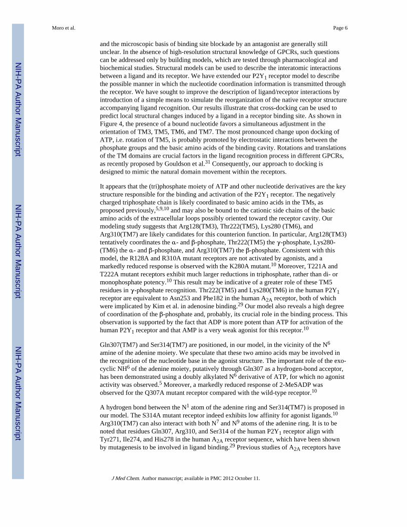
and the microscopic basis of binding site blockade by an antagonist are generally stillunclear. In the absence of high-resolution structural knowledge of GPCRs, such questionscan be addressed only by building models, which are tested through pharmacological andbiochemical studies. Structural models can be used to describe the interatomic interactionsbetween a ligand and its receptor. We have extended our P2Y1 receptor model to describethe possible manner in which the nucleotide coordination information is transmitted throughthe receptor. We have sought to improve the description of ligand/receptor interactions byintroduction of a simple means to simulate the reorganization of the native receptor structureaccompanying ligand recognition. Our results illustrate that cross-docking can be used topredict local structural changes induced by a ligand in a receptor binding site. As shown inFigure 4, the presence of a bound nucleotide favors a simultaneous adjustment in theorientation of TM3, TM5, TM6, and TM7. The most pronounced change upon docking ofATP, i.e. rotation of TM5, is probably promoted by electrostatic interactions between thephosphate groups and the basic amino acids of the binding cavity. Rotations and translationsof the TM domains are crucial factors in the ligand recognition process in different GPCRs,as recently proposed by Gouldson et al.31 Consequently, our approach to docking isdesigned to mimic the natural domain movement within the receptors.
It appears that the (tri)phosphate moiety of ATP and other nucleotide derivatives are the keystructure responsible for the binding and activation of the P2Y1 receptor. The negativelycharged triphosphate chain is likely coordinated to basic amino acids in the TMs, asproposed previously,5,9,10 and may also be bound to the cationic side chains of the basicamino acids of the extracellular loops possibly oriented toward the receptor cavity. Ourmodeling study suggests that Arg128(TM3), Thr222(TM5), Lys280 (TM6), andArg310(TM7) are likely candidates for this counterion function. In particular, Arg128(TM3)tentatively coordinates the α- and β-phosphate, Thr222(TM5) the γ-phosphate, Lys280-(TM6) the α- and β-phosphate, and Arg310(TM7) the β-phosphate. Consistent with thismodel, the R128A and R310A mutant receptors are not activated by agonists, and amarkedly reduced response is observed with the K280A mutant.10 Moreover, T221A andT222A mutant receptors exhibit much larger reductions in triphosphate, rather than di- ormonophosphate potency.10 This result may be indicative of a greater role of these TM5residues in γ-phosphate recognition. Thr222(TM5) and Lys280(TM6) in the human P2Y1receptor are equivalent to Asn253 and Phe182 in the human A2A receptor, both of whichwere implicated by Kim et al. in adenosine binding.29 Our model also reveals a high degreeof coordination of the β-phosphate and, probably, its crucial role in the binding process. Thisobservation is supported by the fact that ADP is more potent than ATP for activation of thehuman P2Y1 receptor and that AMP is a very weak agonist for this receptor.10
Gln307(TM7) and Ser314(TM7) are positioned, in our model, in the vicinity of the N6
amine of the adenine moiety. We speculate that these two amino acids may be involved inthe recognition of the nucleotide base in the agonist structure. The important role of the exo-cyclic NH6 of the adenine moiety, putatively through Gln307 as a hydrogen-bond acceptor,has been demonstrated using a doubly alkylated N6 derivative of ATP, for which no agonistactivity was observed.5 Moreover, a markedly reduced response of 2-MeSADP wasobserved for the Q307A mutant receptor compared with the wild-type receptor.10
A hydrogen bond between the N1 atom of the adenine ring and Ser314(TM7) is proposed inour model. The S314A mutant receptor indeed exhibits low affinity for agonist ligands.10
Arg310(TM7) can also interact with both N7 and N9 atoms of the adenine ring. It is to benoted that residues Gln307, Arg310, and Ser314 of the human P2Y1 receptor align withTyr271, Ile274, and His278 in the human A2A receptor sequence, which have been shownby mutagenesis to be involved in ligand binding.29 Previous studies of A2A receptors have
Moro et al. Page 6
J Med Chem. Author manuscript; available in PMC 2012 October 11.
NIH
-PA Author Manuscript
NIH
-PA Author Manuscript
NIH
-PA Author Manuscript

shown that TM7 is the critical region for the interaction of the adenine moiety in agonistrecognition.29
As previously reported, substitution at the 2-position of the adenine ring of the agonistderivatives is tolerated and in some cases favored for P2Y receptor agonists.30 For example,2-MeSADP is more potent than ADP, and 2-MeSATP is more potent than ATP. We suggestthat the presence of the bulky methylthio substituent at the 2-position shifts the position ofthe agonist inside the binding cavity farther away from TM7, and this change enhances theelectrostatic interaction between the basic amino acids, Arg128 and Lys280, and thenegative charges of the phosphate side chain.
Tyr136(TM3) can also interact with the α-phosphate group of the ATP molecule. Wesuggest that this interaction is a “secondary” interaction. In fact, alanine replacement ofPhe131(TM3), His132(TM3), Tyr136-(TM3), Phe226(TM5) or His277(TM6) resulted inmutant receptors that exhibited 7–18-fold reductions in the potency of 2-MeSATP comparedwith the wild-type receptor.10 These five residues in TM3, TM5, and TM6 are all close tothe ATP binding site and seem to play a modulatory role in ligand binding to the P2Y1receptor.
From the present modeling study, the antagonist MRS 2179 can overlap the ATP structureby superimposition of all adenine ring atoms (Figure 7). If this hypothesis is correct, allamino acids important in the recognition of the adenine moiety of the ATP molecule shouldalso be important for MRS 2179 binding. Accordingly, the antagonist at 1 µM did not affectthe potency of 2-MeSADP in the Q307A and S314T mutant receptors, indicating that theseresidues are important for the binding of the antagonists. This is additional evidence that theTM7 region may be very important for the accommodation of the adenine moiety for bothagonists and antagonists.
We speculate that hydrogen bonding occurs between the N6 position of the adenine moietyand the NH2 of Gln307. The apparent affinity of antagonists related to MRS 2179 isdramatically dependent on the size of the N6 substituent.8 For example, the N6 propyl groupcompletely abolished antagonist properties, and double alkylation of the N6 amino groupresulted in a 300-fold decrease in apparent affinity of MRS 2179. It is possible to rationalizethese results from our model by proposing an unfavorable steric interaction between thelarge N6 substituent and the side chain of Gln307.
Ser314(TM7) in the P2Y1 receptor–MRS 2179 complex can interact with both the N6 andN1 atoms of the adenine moiety, but the N1 position seems to be preferred. This is inaccordance with the experimental evidence that the N1-methyl analogue of 2′-deoxyadenosine 3′,5′-bisphosphate is a weak antagonist (13-fold loss of potency vs thecorresponding adenosine bisphosphate).8 The proximity between Ser314 and the adeninemoiety is also supported by the fact that the S314T mutation reduces the antagonist affinity.
Previously reported structure–activity relationships for adenosine bisphosphates at P2Y1receptors8 suggest that either 2′- or 3′-deoxy modifications are completely tolerated in thecase of antagonists, while 2′- and 3′-deoxyATP are less potent (6.9- and 27-fold,respectively) than ATP. For nucleotide antagonists, phosphate groups at either 2′- or 3′-positions are nearly equivalent and also equally essential as a 5′-phosphate group forantagonist properties. Our model suggests that two amino acids are important for thecoordination of the phosphate groups: Arg128(TM3) and Lys280(TM6). In particular,Lys280 can interact directly with both 3′- and 5′-phosphates, and Arg128 is directlyinvolved in the coordination of the 5′-phosphates. The high flexibility of the side chain ofLys280, and consequently the possibility that the amino group can also interact with the 2′-
Moro et al. Page 7
J Med Chem. Author manuscript; available in PMC 2012 October 11.
NIH
-PA Author Manuscript
NIH
-PA Author Manuscript
NIH
-PA Author Manuscript

phosphate, may be the reason for the antagonist activity of both 2′,5′- and 3′,5′-bisphosphates.
Thr222 seems to be too far from the 5′-phosphate group (4.8 Å) to participate in a direct,strong hydrogen-bonding interaction with the ligand, but only one water molecule would besufficient to bridge the two groups. Phe226(TM5) is also located in proximity to the 5′-phosphate group. Since a hydrophobic interaction occurring between the receptor and thisportion of the nucleotide is untenable, we speculate that this amino acid has an importantfunction in delineating the binding site cavity. It is likely that Arg128(TM3) andArg310(TM7) are very important for the binding of MRS 2179, just as they are for agonist.However, the lack of responsiveness of these Ala mutant receptors to 2-Me-SADP made itimpossible to measure antagonist-binding properties.
Tyr136(TM3) seems to be modulatory for antagonist potency, just as for agonist potency. Inour model, Tyr136 can interact with the 5′-phosphate group of MRS 2179.
Alanine replacement of Phe131(TM3), His132(TM3), His277(TM6), or Ser317(TM7)resulted in mutant receptors that exhibited apparent antagonist affinities similar to thoseobserved wisth the wild-type receptor. Thus, these residues are not essential for antagonistrecognition. Mutation of Ser317 is inconsequential to activation;10 however, the aromaticresidues Phe131, His132, and His277 are modulatory for agonist action.10 We speculate thatcoordination of residues in TM3 and TM6 by the nucleotide may be related toconformational changes leading to receptor activation. The differential effects on antagonistaffinity and agonist potency in F131A, H277A, and R310K mutant receptors (Figure 3C) donot appear to be due to reduced intrinsic activity in these mutant receptors, since maximalefficacy noted for three different agonists in these mutant receptors reached levels similar tothat type of wild-type receptors.10
Taken together, these results suggest that the adenosine and α-phosphate moieties arecritical for the binding of both agonists and antagonists to the P2Y1 receptor and that thepresence of β- and γ-phosphates are probably essential for the agonist activity of ATP. MRS2179 and its related molecules are the only clearly competitive antagonists at the P2Y1receptor. Other known antagonists such as PPADS, suramin, etc. likely have differentbinding requirements at the receptor.8 Further studies may differentiate these requirementsand prove helpful for antagonist drug development.
ConclusionsBinding of the adenine nucleotide antagonist MRS 2179 to the human P2Y1 receptorinvolves many of the same amino acid residues that are important for recognition of agonistATP derivatives: Lys280 and Gln307 are very important, and His132, Tyr136, and Thr222are modulatory. Furthermore, residue S317 is not involved in binding of either agonists orthe antagonist MRS 2179. However, differences between agonist and antagonist recognitionwere observed. In particular, Phe226 in TM5 is more important for the antagonist MRS2179 than for agonists, and residues Phe131, Thr221, and His277 are less important for theantagonist than for agonists. This has allowed us to construct rhodopsin-based molecularmodels of the P2Y1–MRS 2179 and P2Y1–ATP complexes, allowing for minor adjustmentsof the helical positions upon nucleotide binding. The binding modes for these two ligandsare very similar, especially in the adenine region; however a rotation of the antagonistmolecule allows direct interaction of both 5′- and 3′-monophosphate groups with the samepositively charged residues of the receptor that are putatively involved in recognition of the5′-triphosphate moiety.
Moro et al. Page 8
J Med Chem. Author manuscript; available in PMC 2012 October 11.
NIH
-PA Author Manuscript
NIH
-PA Author Manuscript
NIH
-PA Author Manuscript

Experimental ProceduresMolecular Biological Methods
Single amino acids of the human P2Y1 receptor cDNA (pcDNA3P2Y1) were mutated aspreviously described.10 Briefly, the coding region of pCDNA3P2Y1 was subcloned into thepCD-PS expression vector,12 yielding pCDP2Y1. All mutations were introduced intopCDP2Y1 using standard polymerase chain reaction (PCR) techniques.10,13
Oligonucleotides (Bioserve Biotechnologies, Laurel, MD) were designed and used togenerate a PCR fragment, which was then used to replace the corresponding wild-type P2Y1sequence. The accuracy of all PCR-derived sequences was confirmed by dideoxysequencing of the mutant plasmids.14 In addition to single amino acid replacement withinthe TMs, the N-terminus of the receptor was appended with an epitope tag, consisting of a 9-amino acid sequence derived from the influenza virus hemagglutinin (HA) protein (TACCCA TAC GAC GTG CCA GAC TAC GCG; peptide sequence: YPYDVPDYA) which wasinserted after the first Met residue.10,11 A HexaHis tag15 was also included at the C-terminusimmediately after the Leu residue resulting in a construct potentially suitable for affinitychromatography using a chelated nickel column.
Mutant receptors were transiently expressed in COS-7 cells transfected approximately 24 hafter seeding with plasmid DNA (4 µg of DNA/dish) using the DEAE-dextran method.16
Cells were grown for an additional 48 h at 37 °C in Dulbecco’s modified Eagle’s mediumsupplemented with 10% fetal bovine serum.
Pharmacological and Immunological MethodsInositol phosphate determination was used as the measure of activity of the ligands at themutant receptors. Stimulation by the agonist 2-methylthioadenosine 5′-diphosphate (2-Me-SADP) (RBI, Natick, MA) and its antagonism by MRS 2179 (synthesized as the ammoniumsalt, as described8) were measured in the wild-type and mutant receptors. The assay wascarried out according to the general approach of Harden et al.6 using [3H]-myo-inositol(American Radiolabeled Chemicals, St. Louis, MO) in the presence of 10 mM LiCl for 30min at 37 °C, 5% CO2. The [3H]inositol monophosphate fraction was isolated by anionexchange chromatography17 and eluted with 4.5 mL of 0.1 M formic acid/0.2 M ammoniumformate. Pharmacological parameters were analyzed using the Kaleida-Graph program(Abelbeck Software, version 3.01). Statistical analysis was performed using the unpaired t-test (Prism, GraphPad, San Diego, CA).
Indirect cellular ELISA measurements were employed to demonstrate that the wild-type andmutant receptors were expressed in the plasma membrane with the proper orientation of N-terminus. Approximately 72 h after transfection, cells were fixed in 4% formaldehyde,incubated with HA-specific monoclonal antibody (12CA5, Boehringer-Mannheim,Indianapolis, IN), and then washed and incubated with a 1:2000 dilution of a peroxidase-conjugated goat anti-mouse IgG antibody (Sigma, St. Louis, MO) for 1 h at 37 °C.10,29
Color reaction took place in the presence of hydrogen peroxide and o-phenylenediamine(each 2.5 mM in 0.1 M phosphate–citrate buffer, pH 5.0) and was measured bichromaticallyin the BioKinetics reader (EL 312, Bio Tek Instruments, Inc., Winooski, VT) at 490 and 630nm (baseline).
Computational MethodsThe human P2Y1 receptor model was built and optimized using Sybyl 6.318 and Macro-model 5.019 modeling packages, respectively, based on the approach described by van Rheeet al.5 All calculations were performed on a Silicon Graphics Indigo2 R8000 workstation.Briefly, transmembrane domains were identified with the aid of Kyte–Doolittle
Moro et al. Page 9
J Med Chem. Author manuscript; available in PMC 2012 October 11.
NIH
-PA Author Manuscript
NIH
-PA Author Manuscript
NIH
-PA Author Manuscript

hydrophobicity20 and Emini20 surface probability parameters. Transmembrane helices were
built from the sequences and minimized individually. The minimized helices were thengrouped together to form a helical bundle that matching the overall characteristics of theelectron density map of rhodopsin. The helical bundle was minimized using the Amber21
all-atoms force field until the rms value of the conjugate gradient (CG) was <0.1 kcal/molper Å. A fixed dielectric constant = 4.0 was used throughout these calculations.
A model of adenosine 5′-triphosphate (ATP) was constructed from the crystallographiccoordinates of ATP-phosphoglycerate kinase.22 2-MeSATP and MRS 2179 models wereconstructed from ATP using the “Sketch Molecule” module of Sybyl. The ligands were thenfully minimized using AM123 of MOPAC24 and were rigidly docked into the helical bundleusing graphical manipulation coupled to continuous energy monitoring (Dock module ofSybyl). When a final position was reached, consistent with a local energy minimum, thecomplexes of receptor and ligand were subjected to an additional CG minimization run of300 steps. Partial atomic charges for the ligands were imported from the MOPAC outputfiles.
We wanted to explore possible ligand-induced rearrangements of 7TM bundle by sampling7TM conformations in the presence of the docked ligands. The docking process wasrepeated for each model agonist– and antagonist–receptor complex after manually adjustingthe relative positions of TM3, TM4, TM5, TM6, and TM7 around the initial docked ligands(our cross-docking procedure). Cross-docking was carried out using the Dock module ofSybyl. Each helix was separated from the ligand–receptor complex structure, and its relativeposition was changed until a new lower energy geometry was obtained. These adjustmentsconsisted of small translations and rotations of the principal axis of the helix with respect toits original position. When a new final position was reached, consistent with the lowest localenergy minimum, the separated helix was merge again into the ligand–receptor complex.The hydropathy profile for the new oriented helix was checked using Kyte–Doolittlemethod.20 The new complex was subjected to an additional CG minimization run of 300steps. This procedure was repeated for TM3, TM4, TM5, TM6, and TM7. The manualadjustments were followed by 25 ps of molecular dynamics (MD module of Macromodel)performed at a constant temperature of 300 K using time step of 0.001 ps and a dielectricconstant = 4.0. This procedure was followed by another sequence of CG energyminimization to a gradient threshold of <0.1 kcal/mol per Å. Energy minimization of thecomplexes was performed using the Tripos (SYBYL) and AMBER all-atom (MacroModel)force field. The three-dimensional energy-minimized structures appear to be force-field-independent, particularly in the binding region, as judged by backbone superposition withSYBYL’s root-mean-square superposition procedure (rms = 0.014–0.22 Å).
Supplementary MaterialRefer to Web version on PubMed Central for supplementary material.
AcknowledgmentsS. Moro is grateful to Gilead Sciences (Foster City, CA) for a postdoctoral fellowship grant. The authors aregrateful for helpful comments and encouragement provided by Dr. Robert Pearlstein of the Center for MolecularModeling (DCRT), NIH. We thank Dr. Carsten Hoffmann and Dr. Ivar von Kügelgen of Molecular RecognitionSection, NIDDK/NIH, for helpful discussion.
Abbreviations
ATP adenosine 5′-triphosphate
Moro et al. Page 10
J Med Chem. Author manuscript; available in PMC 2012 October 11.
NIH
-PA Author Manuscript
NIH
-PA Author Manuscript
NIH
-PA Author Manuscript

DMEM Dulbecco’s modified Eagle’s medium
ELISA enzyme-linked immunosorbent assay
FBS fetal bovine serum
GPCR G-protein-coupled receptor
HA hemagglutinin
HBSS Hank’s balanced salt solution
2-Me-SADP 2-methylthioadenosine 5′-diphosphate
2-Me-SATP 2-methylthioadenosine 5′-triphosphate
MRS 2179 2′-deoxy-N6-methyladenosine 3, 5′-bisphosphate
PBS phosphate-buffered saline
PCR polymerase chain reaction
PLC phospholipase C
PPADS pyridoxal-5′-phosphate-6-azophenyl-2, 4-disulfonate
rms root-mean-square
TM transmembrane helical domain
Tris tris-(hydroxymethyl)aminomethane
References1. Burnstock, G. Purinergic Approaches in Experimental Therapeutics. Jacobson, KA.; Jarvis, M.,
editors. New York: Wiley; 1997.
2. Fredholm BB, Abbracchio MP, Burnstock G, Daly JW, Harden TK, Jacobson KA, Leff P, WilliamsM. Nomenclature and classification of purinoceptors. Pharmacol. Rev. 1994; 46:143–156.[PubMed: 7938164]
3. Burnstock G, King BF. Numbering of cloned P2 purinoceptors. Drug Dev. Res. 1996; 38:67–71.
4. Webb TE, Simon J, Krishek BJ, Bateson AN, Smart TG, King BF, Burnstock G, Barnard EA.Cloning and functional expression of a brain G protein-coupled ATP receptor. FEBS Lett. 1993;324:219–225. [PubMed: 8508924]
5. van Rhee AM, Fischer B, van Galen PJM, Jacobson KA. Modelling the P2Y purinoceptor usingrhodopsin as template. Drug Design Discovery. 1995; 13:133–154.
6. Harden TK, Hawkins PT, Stephens L, Boyer JL, Downes P. Phosphoinositide hydrolysis byguanosine 5′-(gamma-thio) triphosphate-activated phospholipase C of turkey erythrocytemembranes. Biochem. J. 1988; 252:583–593. [PubMed: 2843174]
7. Jacobson, KA.; Kim, Y-C.; Camaioni, E.; van Rhee, AM. Structure activity relationships of P2receptor agonists and antagonists. Chapter 4 in The P2 Nucleotide Receptors. In: Turner, JT.;Weisman, G.; Fedan, J., editors. The Receptors. Clifton, NJ: Humana Press; 1997. p. 81-107.
8. Camaioni E, Boyer JL, Mohanram A, Harden TK, Jacobson KA. Deoxyadenosine-bisphosphatederivatives as potent antagonists at P2Y1 receptors. J. Med. Chem. 1998; 41:183–190. [PubMed:9457242]
9. Erb L, Garrad R, Wang YJ, Quinn T, Turner JT, Weisman GA. Site-directed mutagenesis of P2Upurinoceptors—positively charged amino acids in transmembrane helix-6 and helix-7 affect agonistpotency and specificity. J. Biol. Chem. 1995; 270:4185–4188. [PubMed: 7876172]
10. Jiang Q, Guo D, Lee BX, van Rhee AM, Kim YC, Nicholas R, Schachter J, Harden TK, JacobsonKA. Mutational analysis of residues essential for ligand recognition at the human P2Y1 receptor.Mol. Pharmacol. 1997; 52:499–507. [PubMed: 9281613]
Moro et al. Page 11
J Med Chem. Author manuscript; available in PMC 2012 October 11.
NIH
-PA Author Manuscript
NIH
-PA Author Manuscript
NIH
-PA Author Manuscript

11. Schachter JB, Li Q, Boyer JL, Nicholas RA, Harden TK. Second messenger cascade specificityand pharmacological selectivity of the human P2Y1-purinoceptor. Br. J. Pharmacol. 1996;118:167–173. [PubMed: 8733591]
12. Okayama H, Berg PA. A cDNA cloning vector that permits expression of cDNA inserts inmammalian cells. Mol. Cell. Biol. 1983; 3:280–289. [PubMed: 6300662]
13. Higuchi, R. Using PCR to Engineer DNA. In: Ehrlich, HA., editor. PCR Technology. New York:Stockton Press; 1989. p. 61-70.
14. Sanger R, Nicklen S, Coulson AR. DNA sequencing with chain-terminating inhibitors. Proc. Natl.Acad. Sci. U.S.A. 1977; 74:5463–5467. [PubMed: 271968]
15. Robeva AS, Woodard R, Luthin DR, Taylor HE, Linden J. Double tagging recombinant A1- andA2A-adenosine receptors with hexahistidine and the FLAG epitope. Development of an efficientgeneric protein purification procedure. Biochem. Pharmacol. 1996; 51:545–555. [PubMed:8619901]
16. Cullen BR. Use of eukaryotic expression technology in the functional analysis of cloned genes.Methods Enzymol. 1987; 152:684–704. [PubMed: 3657593]
17. Berridge MJ, Dawson RM, Downes CP, Heslop JP, Irvine RF. Changes in the levels of inositolphosphates after agonist-dependent hydrolysis of membrane phosphoinositides. Biochem J. 1983;212:473–482. [PubMed: 6309146]
18. The program SYBYL 6.3 is available from TRIPOS Associates. St. Louis, MO: 1993.
19. Mohamadi F, Richards NGJ, Guida WC, Liskamp R, Lipton M, Caufield C, Chang G,Hendrickson T, Still WC. MacroModel-An Integrated Software System for Modeling Organic andBioorganic Molecules using Molecular Mechanics. J. Comput. Chem. 1990; 11:440–450.
20. Kyte J, Doolittle RF. A simple method for displaying the hydrophobic character of a protein. J.Mol. Biol. 1982; 157:105–132. [PubMed: 7108955]
21. Weiner SJ, Kollman PA, Nguyen DT, Case DA. An all-atom force field for simulation of proteinand nucleic acids. J. Comput. Chem. 1986; 7:230–252.
22. Watson HC, Walker NP, Shaw PJ, Bryant TN, Wendell PL, Fothergill LA, Perkins RE, ConroySC, Dobson MJ, Tuite MF. Sequence and structure of yeast phosphoglycerate kinase. EMBO J.1982; 1:1635–1640. [PubMed: 6765200]
23. Dewar MJSE, Zoebisch G, Healy EF. AM1: A new general purpose quantum mechanicalmolecular model. J. Am. Chem. Soc. 1985; 107:3902–3909.
24. MOPAC 6.0 available from Quantum Chemistry Program Exchange.
25. Chiota C, Levitt M, Richardson D. Helix to helix packing in proteins. J. Mol. Biol. 1981; 145:215–250. [PubMed: 7265198]
26. Schertler GF, Villa C, Henderson R. Projection structure of rhodopsin. Nature. 1993; 362:770–772.[PubMed: 8469290]
27. Urger VM, Hargrave PA, Baldwin JM, Schertler GFX. Arrangement of rhodopsin transmenbraneα-helices. Nature. 1997; 389:203–206. [PubMed: 9296501]
28. Nicholls, A. Grasp: Graphical Representation and Analysis of surface Properties. New York:Columbia University; 1992.
29. Kim J, Wess J, van Rhee MA, Schoneberg T, Jacobson KA. Site-directed mutagenesis identifiesresidues involved in ligand recognition in the human A2a adenosine receptor. J. Biol. Chem. 1995;270:13987–13997. [PubMed: 7775460]
30. Fischer B, Boyer JL, Hoyle CHV, Ziganshin AU, Brizzolara AL, Knight GE, Zimmet J, BurnstockG, Harden TK, Jacobson KA. Identification of potent, selective P2Y-purinoceptor agonists–structure–activity-relationships for 2-thioether derivatives of adenosine 5′-triphosphate. J. Med.Chem. 1993; 36:3937–3946. [PubMed: 8254622]
31. Gouldson PR, Snell CR, Reynolds CA. A new approach to docking in the β2-adrenergic receptorthat exploits the domain structure of G-protein-coupled receptors. J. Med. Chem. 1997; 40:3871–3886. [PubMed: 9397168]
Moro et al. Page 12
J Med Chem. Author manuscript; available in PMC 2012 October 11.
NIH
-PA Author Manuscript
NIH
-PA Author Manuscript
NIH
-PA Author Manuscript

Figure 1.Chemical structures of ATP and MRS 2179 (charges not shown).
Moro et al. Page 13
J Med Chem. Author manuscript; available in PMC 2012 October 11.
NIH
-PA Author Manuscript
NIH
-PA Author Manuscript
NIH
-PA Author Manuscript

Figure 2.Partial topology of the human P2Y1 receptor showing residues proposed to be involved inrecognition of the antagonist MRS 2179. Mutation sites in which replacement by alaninesignificantly impedes the ability of MRS 2179 to block activation of the receptor arehighlighted. Gray shading indicates residues that are conserved between P2Y1 and P2Y2receptors. Solid highlighted circles indicate a 1–3-fold change in the EC50 for 2-MeSADP inthe presence of 1 µM MRS2179; circles with thick outline, a 3–6-fold change; solidnonhighlighted circles, a 6–12-fold change. Residues R128, R310, and S314 are highlighted;however the effects of antagonist at these Ala mutant receptors could not be evaluatedexperimentally due to lack of activation by agonist. The effects of non-Ala substitutions onthe potency of MRS 2179 are S314T, moderate reduction; R310K, identical to wild-typereceptors.
Moro et al. Page 14
J Med Chem. Author manuscript; available in PMC 2012 October 11.
NIH
-PA Author Manuscript
NIH
-PA Author Manuscript
NIH
-PA Author Manuscript

Figure 3.Stimulation of phospholipase C by 2-MeSADP in human P2Y1 receptors. TransfectedCOS-7 cells were incubated for 30 min at 37 °C (see the Experimental Procedures fordetails). Data are presented as absolute accumulations of tritiated inositol phosphates abovebasal levels in the absence of 2-MeSADP for representative experiments. EC50 values(average of two to four independent experiments, each carried out in duplicate) are given inTable 1. (A) Concentration–response curves for stimulation of PLC in the absence (opensymbols) or presence (closed symbols) of MRS 2179 in COS-7 cells transiently expressingwild-type HA-tagged-hP2Y1 (○) or mutant hP2Y1 receptors (K280A, △; F131A, □)Concentrations of MRS 2179 used were K280A, 50 µM; wild-type and F131A, 1 µM. (B)
Moro et al. Page 15
J Med Chem. Author manuscript; available in PMC 2012 October 11.
NIH
-PA Author Manuscript
NIH
-PA Author Manuscript
NIH
-PA Author Manuscript

Concentration–response curves for antagonism by MRS 2179 of agonist-induced stimulationof PLC in four mutant receptors. Concentrations of 2-MeSADP used were: K280A, 5 µM(○); Q307A, 2.5 µM (●); S314T, 100 nM (◆); Y136A, 1 µM (◇). (C) Correlation ofagonist (EC50, nM) vs antagonist (KB, µM) potencies at mutant hP2Y1 receptors (mutationshaving little or no effect on antagonist affinity, △; mutations having an intemediate effect onantagonist affinity; □, mutations having the largest effect on antagonist affinity, ●). Anestimated KB value for F226A of 2.6 µM has been used.
Moro et al. Page 16
J Med Chem. Author manuscript; available in PMC 2012 October 11.
NIH
-PA Author Manuscript
NIH
-PA Author Manuscript
NIH
-PA Author Manuscript

Figure 4.Stereoview of human P2Y1 receptor transmembrane helical bundle model viewed along thehelical axes from the extracellular end (A, top) and perpendicular to the helical axes (B,bottom), before (left) and after (right) the “cross-docking” procedure for the P2Y1–ATPcomplex (see the Experimental Procedures for details). The docked ATP molecule is notshown. Side chains of some amino acids important for ligand recognition are highlighted.
Moro et al. Page 17
J Med Chem. Author manuscript; available in PMC 2012 October 11.
NIH
-PA Author Manuscript
NIH
-PA Author Manuscript
NIH
-PA Author Manuscript

Figure 5.(A, Top) Side view of the P2Y1–ATP complex model. The side chains of the importantresidues in proximity to the docked ATP molecule are highlighted and labeled. Residues inproximity (≤5 Å) to the docked ATP molecule: adenine Q307, R310, S314; ribose H132,H277, S317; triphosphate R128, Y136, T222, K280, R310. (B, Bottom) Side view of P2Y1–MRS 2179 complex model. The side chains of the important residues in proximity to thedocked MRS 2179 molecule are highlighted and labeled. Residues in proximity (≤5 Å) tothe docked MRS2179 molecule: adenine Q307, R310, S314; ribose H132, H277;bisphosphate R128, Y136, K280, R310 (R128 and K280 present different conformationswith respect to the ATP binding model).
Moro et al. Page 18
J Med Chem. Author manuscript; available in PMC 2012 October 11.
NIH
-PA Author Manuscript
NIH
-PA Author Manuscript
NIH
-PA Author Manuscript

Figure 6.Surface potential of P2Y1–ATP complex model, displayed with GRASP.25 The molecularsurface is color coded by electrostatic potential. Potentials less than −20 kT are red, thosegreater than 20 kT are blue, and neutral potentials (0 kT) are white. The ATP binding site isclearly distinguishable as a region of intense positive potential.
Moro et al. Page 19
J Med Chem. Author manuscript; available in PMC 2012 October 11.
NIH
-PA Author Manuscript
NIH
-PA Author Manuscript
NIH
-PA Author Manuscript

Figure 7.Possible superpositions of ATP and MRS 2179. (A) Superposition of all adenosine ringatoms or (B) superposition of ATP α- and γ-phosphate atoms with 3′- and 5′-phosphate ofMRS 2179.
Moro et al. Page 20
J Med Chem. Author manuscript; available in PMC 2012 October 11.
NIH
-PA Author Manuscript
NIH
-PA Author Manuscript
NIH
-PA Author Manuscript

NIH
-PA Author Manuscript
NIH
-PA Author Manuscript
NIH
-PA Author Manuscript
Moro et al. Page 21
Tabl
e 1
Ant
agon
ism
of
the
Act
ivat
ion
of P
hosp
holip
ase
C in
Mut
ant H
uman
P2Y
1 R
ecep
tors
a
EC
50(2
-MeS
AD
P, n
M)
KBe,
f
(MR
S 21
79, µ
M)
cons
truc
tbre
sidu
ecn
alon
ed+M
RS
2179
(1
±M)
WT
41.
94 ±
0.8
012
.9 ±
2.6
** 0
.177
F131
A3.
323
19.2
± 0
.710
3 ±
20*
0.2
29
H13
2A3.
334
17.7
± 4
.753
.0 ±
9.1
** 0
.501
Y13
6A3.
374
12.3
± 3
.736
.0 ±
8.3
** 0
.519
T22
1A5.
423
2.45
± 0
.60
12.4
± 1
.1**
0.2
46
T22
2A5.
432
10.9
± 1
.125
.0 ±
0.2
** 0
.773
F226
A5.
473
19.5
± 1
.927
.0 ±
4.7
>1
H27
7A6.
526
115
± 2
973
8 ±
4**
0.1
85
K28
0A6.
556
1520
± 3
1015
80 ±
110
**10
.4g
Q30
7A7.
367
323
± 5
337
8 ±
46*
* 4
.25g
R31
0K7.
394
324
± 4
122
70 ±
840
** 0
.166
S314
T7.
435
10.4
± 1
.330
.9 ±
5.1
** 0
.507
S317
A7.
464
1.88
± 0
.35
12.1
± 0
.5**
0.1
84
a Dat
a ar
e pr
esen
ted
as m
eans
± S
D o
f tw
o to
sev
en in
depe
nden
t exp
erim
ents
, eac
h pe
rfor
med
in d
uplic
ate.
b All
cons
truc
ts c
onta
in th
e H
A-e
pito
pe ta
g se
quen
ce a
t the
N-t
erm
inus
and
a h
exa-
His
tag
at th
e C
-ter
min
us.1
0
c Usi
ng th
e se
quen
ce id
entif
ier
as d
efin
ed in
ref
10.
d Ago
nist
at t
he h
ighe
st c
once
ntra
tions
cau
sed
only
a s
mal
l inc
reas
e or
no
chan
ge in
inos
itol p
hosp
hate
s in
the
follo
win
g m
utan
ts: R
128A
(2.
29);
R31
0A (
7.39
); R
310S
/S31
4R (
7.39
/7.4
3); S
314A
(7.
43).
e Cal
cula
ted
acco
rdin
g to
the
Schi
ld e
quat
ion:
KB
= [
anta
goni
st]/
((E
C50
with
ant
agon
ist/E
C50
ago
nist
alo
ne)
− 1
).
f EC
50 v
alue
s, a
s fo
llow
, for
the
stim
ulat
ion
of P
LC
by
2-M
eSA
DP
at e
ach
of th
e fo
llow
ing
mut
ant r
ecep
tors
, in
the
pres
ence
of
50 µ
M M
RS
2179
: 188
± 1
8 nM
(Y
136A
); 8
.82
± 3
.51
µM (
K28
0A);
4.1
2 ±
0.35
µM
(Q
307A
); o
r in
the
pres
ence
of
20 µ
M M
RS
2179
: 106
nm
(S3
14T
).
g KB
val
ues
for
MR
S 21
79 w
ere
dete
rmin
ed u
sing
incr
ease
s in
EC
50 f
or 2
-MeS
AD
P in
the
pres
ence
of
50 µ
M M
RS
2179
(se
e fo
otno
te f)
.
J Med Chem. Author manuscript; available in PMC 2012 October 11.

NIH
-PA Author Manuscript
NIH
-PA Author Manuscript
NIH
-PA Author Manuscript
Moro et al. Page 22St
atis
tical
sig
nifi
canc
e: *
p <
0.0
5;
**p
< 0
.005
.
J Med Chem. Author manuscript; available in PMC 2012 October 11.