NIH Public Access and Through Altering Cell Metabolism 1,2 ...
25
Graviola: A Novel Promising Natural-Derived Drug That Inhibits Tumorigenicity and Metastasis of Pancreatic Cancer Cells In Vitro and In Vivo Through Altering Cell Metabolism María P. Torres 1,2 , Satyanarayana Rachagani 1 , Vinee Purohit 2 , Poomy Pandey 3 , Suhasini Joshi 1 , Erik D. Moore 1 , Sonny L. Johansson 2,4 , Pankaj K. Singh 1,2 , Apar K. Ganti 5 , and Surinder K. Batra 1,2,4 1 Department of Biochemistry and Molecular Biology, University of Nebraska Medical Center, Omaha, NE 68198-5870, USA 2 Eppley Institute for Research in Cancer and Allied Diseases, University of Nebraska Medical Center, Omaha, NE 68198-5870, USA 3 Department of Environmental, Agricultural & Occupational Health, University of Nebraska Medical Center, Omaha, NE 68198-5870, USA 4 Department of Pathology and Microbiology, University of Nebraska Medical Center, Omaha, NE 68198-5870, USA 5 Department of Internal Medicine Division of Hematology and Oncology, University of Nebraska Medical Center, Omaha, NE 68198-5870, USA Abstract Pancreatic tumors are resistant to conventional chemotherapies. The present study was aimed at evaluating the potential of a novel plant-derived product as a therapeutic agent for pancreatic cancer (PC). The effects of an extract from the tropical tree Annona Muricata, commonly known as Graviola, was evaluated for cytotoxicity, cell metabolism, cancer-associated protein/gene expression, tumorigenicity, and metastatic properties of PC cells. Our experiments revealed that Graviola induced necrosis of PC cells by inhibiting cellular metabolism. The expression of molecules related to hypoxia and glycolysis in PC cells (i.e. HIF-1α, NF-κB, GLUT1, GLUT4, HKII, and LDHA) were downregulated in the presence of the extract. In vitro functional assays further confirmed the inhibition of tumorigenic properties of PC cells. Overall, the compounds that are naturally present in a Graviola extract inhibited multiple signaling pathways that regulate metabolism, cell cycle, survival, and metastatic properties in PC cells. Collectively, alterations in these parameters led to a decrease in tumorigenicity and metastasis of orthotopically implanted pancreatic tumors, indicating promising characteristics of the natural product against this lethal disease. © 2012 Elsevier Ireland Ltd. All rights reserved. For correspondence: Surinder K. Batra, Ph.D., Department of Biochemistry and Molecular Biology, Eppley Institute for Research in Cancer and Allied Diseases, University of Nebraska Medical Center, Omaha, Nebraska, 68198-5870, U.S.A. Phone: 402-559-5455, Fax: 402-559-6650, [email protected]. Conflicts of Interest Statement There are no potential conflicts of interest involved with this work. Publisher's Disclaimer: This is a PDF file of an unedited manuscript that has been accepted for publication. As a service to our customers we are providing this early version of the manuscript. The manuscript will undergo copyediting, typesetting, and review of the resulting proof before it is published in its final citable form. Please note that during the production process errors may be discovered which could affect the content, and all legal disclaimers that apply to the journal pertain. NIH Public Access Author Manuscript Cancer Lett. Author manuscript; available in PMC 2013 October 01. Published in final edited form as: Cancer Lett. 2012 October 1; 323(1): 29–40. doi:10.1016/j.canlet.2012.03.031. NIH-PA Author Manuscript NIH-PA Author Manuscript NIH-PA Author Manuscript
Transcript of NIH Public Access and Through Altering Cell Metabolism 1,2 ...

Graviola: A Novel Promising Natural-Derived Drug That InhibitsTumorigenicity and Metastasis of Pancreatic Cancer Cells InVitro and In Vivo Through Altering Cell Metabolism
María P. Torres1,2, Satyanarayana Rachagani1, Vinee Purohit2, Poomy Pandey3, SuhasiniJoshi1, Erik D. Moore1, Sonny L. Johansson2,4, Pankaj K. Singh1,2, Apar K. Ganti5, andSurinder K. Batra1,2,4
1Department of Biochemistry and Molecular Biology, University of Nebraska Medical Center,Omaha, NE 68198-5870, USA2Eppley Institute for Research in Cancer and Allied Diseases, University of Nebraska MedicalCenter, Omaha, NE 68198-5870, USA3Department of Environmental, Agricultural & Occupational Health, University of NebraskaMedical Center, Omaha, NE 68198-5870, USA4Department of Pathology and Microbiology, University of Nebraska Medical Center, Omaha, NE68198-5870, USA5Department of Internal Medicine Division of Hematology and Oncology, University of NebraskaMedical Center, Omaha, NE 68198-5870, USA
AbstractPancreatic tumors are resistant to conventional chemotherapies. The present study was aimed atevaluating the potential of a novel plant-derived product as a therapeutic agent for pancreaticcancer (PC). The effects of an extract from the tropical tree Annona Muricata, commonly knownas Graviola, was evaluated for cytotoxicity, cell metabolism, cancer-associated protein/geneexpression, tumorigenicity, and metastatic properties of PC cells. Our experiments revealed thatGraviola induced necrosis of PC cells by inhibiting cellular metabolism. The expression ofmolecules related to hypoxia and glycolysis in PC cells (i.e. HIF-1α, NF-κB, GLUT1, GLUT4,HKII, and LDHA) were downregulated in the presence of the extract. In vitro functional assaysfurther confirmed the inhibition of tumorigenic properties of PC cells. Overall, the compounds thatare naturally present in a Graviola extract inhibited multiple signaling pathways that regulatemetabolism, cell cycle, survival, and metastatic properties in PC cells. Collectively, alterations inthese parameters led to a decrease in tumorigenicity and metastasis of orthotopically implantedpancreatic tumors, indicating promising characteristics of the natural product against this lethaldisease.
© 2012 Elsevier Ireland Ltd. All rights reserved.
For correspondence: Surinder K. Batra, Ph.D., Department of Biochemistry and Molecular Biology, Eppley Institute for Research inCancer and Allied Diseases, University of Nebraska Medical Center, Omaha, Nebraska, 68198-5870, U.S.A. Phone: 402-559-5455,Fax: 402-559-6650, [email protected].
Conflicts of Interest StatementThere are no potential conflicts of interest involved with this work.
Publisher's Disclaimer: This is a PDF file of an unedited manuscript that has been accepted for publication. As a service to ourcustomers we are providing this early version of the manuscript. The manuscript will undergo copyediting, typesetting, and review ofthe resulting proof before it is published in its final citable form. Please note that during the production process errors may bediscovered which could affect the content, and all legal disclaimers that apply to the journal pertain.
NIH Public AccessAuthor ManuscriptCancer Lett. Author manuscript; available in PMC 2013 October 01.
Published in final edited form as:Cancer Lett. 2012 October 1; 323(1): 29–40. doi:10.1016/j.canlet.2012.03.031.
NIH
-PA Author Manuscript
NIH
-PA Author Manuscript
NIH
-PA Author Manuscript

KeywordsPancreatic cancer; therapy; cancer metabolism; natural product
1. IntroductionThe overall five-year survival rate for pancreatic cancer (PC) patients was 5.5% for theperiod of 2001–2007, according to the National Cancer Institute (NCI), a statistic that hasnot varied significantly for over the last four decades [1]. In 2012, it is estimated that 43,920new PC cases will be diagnosed and approximately 85% of these (i.e. 37,390) will succumbto the disease [2]. The main reason behind the poor prognosis of PC patients is the insidiousand sporadic nature of the disease, which is often presented with no specific early clinicalsymptoms. By the time of diagnosis PC is already in advanced stages (i.e. III and IV) and isresistant to conventional chemotherapy and radiotherapy [3]. Interestingly, even patientsdiagnosed with stage I PC that have the option to undergo surgery have a 5-year overallsurvival of approximately 20%, a clear indication of the general failure of current standardtreatments for each stage of PC [4, 5]. What is even more alarming, are the statistics thatpredict possible 55% increase in the expected number of new PC cases by 2030 [6]. Thus,immediate progress must be made in the prevention, early diagnosis, and systemictreatments against this lethal disease.
Gemcitabine has been the standard line of treatment for PC patients for over a decade and isassociated with a median patient survival of 5.4 months [7]. Over all these years, numerousclinical efforts have been devoted to improve PC chemotherapy outcomes, but unfortunatelyno significant improvements have been reported apart from a clinical trial reported in Mayof 2011 [8]. This phase III clinical trial reported an improved overall survival of PC patientstreated with a four-drug chemotherapy regimen comprising fluorouracil, leucovorin,irinotecan, and oxaliplatin (FOLFIRINOX). Nevertheless, a major disadvantage of thisnovel treatment was its related toxicity, which was noticeably high when compared to PCpatients treated with gemcitabine alone. Therefore, novel, alternative PC therapeutics mustnot only improve the prognosis of PC patients but also minimize any possible toxicity-related side effects that will interfere with the quality of life of PC patients.
It is well known that an increased consumption of fruits and vegetables is associated with areduced risk of most cancers, including PC [9]. For this reason, the potential of naturalproducts in PC therapies has been widely investigated [10]. While some of these compoundshave undergone clinical testing (i.e. curcumin, genistein) and have demonstrated someactivity against PC, the poor bioavailability in patients minimizes their therapeutic efficacy.However, as compared with conventional chemotherapeutic drugs, the major benefit of thesetherapies is the apparent lack of toxicities to healthy tissues. This attracted our attention tofind alternative, natural-derived chemotherapeutic drugs in order to improve the prognosisof PC patients. Traditionally, the leaves from the tropical tree Annona Muricata, also knownas Graviola or Soursop, have been used for a wide range of human diseases includinginflammatory conditions, rheumatism, neuralgia, diabetes, hypertension, insomnia, cystitis,parasitic infections, and cancer [11]. The major bioactive components that have beenextracted from different parts of the plant are known as Annonaceous acetogenins. These arederivatives of long chain (C35 or C37) fatty acids derived from the polyketide pathway [12]that is selectively toxic to cancer cells, including multidrug-resistant cancer cell lines [13–17]. Annonaceous acetogenins induce cytotoxicity by inhibiting the mitochondrial complexI, which is involved in ATP synthesis [14]. As cancer cells have a higher demand for ATPthan the normal cells, mitochondrial complex I inhibitors have potential in cancertherapeutics.
Torres et al. Page 2
Cancer Lett. Author manuscript; available in PMC 2013 October 01.
NIH
-PA Author Manuscript
NIH
-PA Author Manuscript
NIH
-PA Author Manuscript

A few in vivo studies involving Annona Muricata have been reported. Among these, tworeports have shown the ability of the leaf extract to regenerate pancreatic islet β cells indiabetic rats [18, 19]. These studies suggest an additional benefit of the natural productagainst PC given that diabetes has been classified as a risk factor of the malignant disease[20]. More recently, one study analyzing the anti-tumor efficacy of Annona Muricata waspublished [21]. The extract had a direct anti-tumorigenic effect on breast cancer cells bydownregulating the expression of the epidermal growth factor receptor (EGFR). Althoughthis study demonstrates the potential anti-tumorigenic properties of Graviola, the doses usedin the experimental design were not properly controlled. The mice were fed with the extractmixed in the diet and the exact amount ingested by each animal could not be estimatedaccurately.
Although a few in vitro reports have shown the cytotoxic characteristics of Graviola againstvarious cancer cell lines, including PC cells [12], the comprehensive in vivo effects andmechanistic scientific studies are still lacking. To our knowledge, the studies reported hereinare the first to indicate that Graviola extract has promising characteristics for PCtherapeutics. Comprehensive in vitro and in vivo studies in various PC cell lines revealedthat the natural product inhibited multiple signaling pathways that regulate metabolism, cellcycle, survival, and metastatic properties of PC cells.
2. Materials and Methods2.1 Graviola Extract
Graviola supplement capsules were purchased from Raintree (Carson City, NV). Thecapsules consisted of 100% pure, finely milled Graviola leaf/stem powder with no binders orfillers. The capsule contents were suspended in DMSO (100mg/mL). After incubating for5min, the suspension was centrifuged and the supernatant (i.e. extract) was filtered toremove any remaining particles. Subsequent dilutions were prepared in Dulbecco’smodification of Eagle’s medium (DMEM) supplemented with 10% of fetal bovine serum(FBS). Stock solutions and respective dilutions were freshly prepared prior to treatment.
2.2 Cell CultureThe metastatic PC cell lines FG/COLO357 and CD18/HPAF were purchased from theAmerican Type Culture Collection (ATCC). Before performing experiments, the PC celllines were authenticated by short tandem repeat analysis. It was ensured that PC cells wereused at fewer than 20 passages after purchase from ATCC. Cells were cultured in DMEMmedium supplemented with 10% FBS and antibiotics (100μg/mL penicillin and 100μg/mLstreptomycin). The cells were maintained at 37°C and 5% CO2 in a humidified atmosphere.
2.3 AntibodiesThe antibodies for phospho-ERK1/2, total ERK, phospho-Akt (Ser 473), total Akt, NF-κB,and caspase-3 were purchased from Cell Signaling Technology (Danvers, MA). Theantibodies for Cyclin-D1, phospho-FAK (Tyr 925), and total FAK were obtained from SantaCruz Biotechnology (Santa Cruz, CA). The β-actin and β-Tubulin antibodies were obtainedfrom Sigma Aldrich (St. Louis, MO), whereas the HIF-1α antibody was purchased from BDBiosciences (San Jose, CA). The MUC4 monoclonal antibody (8G7) used in these studieswas developed by our group [22]. MMP9 antibody was obtained from a hybridoma cellsupernatant kindly provided by Dr. Rakesh Singh at UNMC. The secondary antibodies usedfor western blot analyses were the ECL™ anti-mouse and anti-rabbit IgG conjugated tohorseradish peroxidase (GE healthcare, UK). Fluorescein isothiocyanate (FITC) conjugated-anti-mouse and Alexa Fluor conjugated anti-mouse antibodies were obtained fromInvitrogen (Carlsbad, CA).
Torres et al. Page 3
Cancer Lett. Author manuscript; available in PMC 2013 October 01.
NIH
-PA Author Manuscript
NIH
-PA Author Manuscript
NIH
-PA Author Manuscript

2.4 Cytotoxicity AssayTo determine the cytotoxicity of Graviola extract on PC cells, 1×10⁴ cells were seeded perwell on a 96-well plate in DMEM supplemented with 10% FBS and antibiotics. Afterovernight incubation, different concentrations (10–200μg/mL) of the extract were addedinto triplicate wells. After 48hr, the media was replaced with fresh media containingthiazolyl blue tetrazolium bromide (MTT) reagent (Sigma Aldrich, St. Louis, MO). After4hr incubation at 37°C in 5% CO2 in humidified atmosphere, the media was replaced with100μL of DMSO and the corresponding cytotoxicity values were calculated (λ=540nm).The experiment was repeated at least three times.
2.5 Western Blot AnalysisFor protein analysis, 0.5×10⁶ of PC cells were seeded on each well of a six-well plate inDMEM supplemented with 10% FBS and antibiotics. After overnight incubation, freshsolutions of Graviola (0–200μg/mL) were prepared and added to the respective wells. Cellsincubated with the corresponding amount of DMSO present in the highest concentratedsolution of Graviola were used as a negative control (0μg/mL). After 48hr of incubationwith the extract, protein lysates were isolated and prepared for western blot analysis, aspreviously described [23].
2.6 Real-time PCRThe transcripts levels of the glucose transporters GLUT1 and GLUT4, the glycolyticenzymes hexokinase II (HKII) and lactate dehydrogenase A (LDHA), and the mucinglycoprotein MUC4 in PC cells were determined after treatment with Graviola extract byreal-time PCR. 0.5×10⁶ cells were seeded in each well of a six-well plate in complete media.After overnight incubation, fresh solutions of Graviola extract (50 and 100μg/mL) wereprepared and cells were incubated for 48hr. Subsequently, cDNA was synthesized frompurified RNA and real-time PCR was carried out as has been described by previous studies[23]. The sequences of the gene-specific primers used were: GLUT1: F 5′-GCCATGGAGCCCAGCAGCAA-3′; R 5′-CGGGGACTCTCGGGGCAGAA-3′ GLUT4:F 5′-GCCTGTGGCCACTGCTCCTG-3′; R 5′-GGGGTCTCTGGGCCGGGTAG-3′ HKII:F 5′-GTCATCCCCTTGTGTCAGAG-3′; R 5′-CTTCATTAGTGTCCCCATCCTG-3′LDHA: F 5′-CCAGTGTGCCTGTATGGAGTG-3′; R 5′-GCACTCTCAACCACCTGCTTG-3′ MUC4: F 5′-GTGACCATGGAGGCCAGTG-3′; R5′-TCATGCTCAGGTGTCCACAG-3′
2.7 Glucose UptakeGlucose-uptake rate was assayed by utilizing [3H] 2-deoxyglucose ([3H] 2-DG). 5×10⁴ PCcells were seeded per well in a 24-well plate. 12hr later, the cells were treated with Graviolaextract (10 and 50μg/mL) for 48hr. The cells were then starved for glucose for 2hr andincubated for 20min with 2 Ci [3H] 2-DG. Subsequently, cells were lysed with 1% SDS andthe lysates were counted for [3H] by utilizing a scintillation counter. Cells treated withlabeled and excess unlabeled 2-DG were used as controls to set a baseline for non-specific[3H] uptake. The results were normalized to the cell counts for treated and untreated groups.Glucose uptake was normalized with that of the control cells (0μg/mL) and it is presented asthe mean values ± standard error from experiments performed in triplicate.
2.8 ATP QuantificationThe CellTiter-Glo® Luminescent Cell Viability Assay (Promega, Madison, WI) was used tomeasure the ATP content in the cells. Briefly, 1×10⁴ PC cells were seeded in each well of anopaque 96-well plate. Cells were seeded for both ATP quantification and proteinconcentration estimation. Starting the next day, the cells were incubated with Graviola
Torres et al. Page 4
Cancer Lett. Author manuscript; available in PMC 2013 October 01.
NIH
-PA Author Manuscript
NIH
-PA Author Manuscript
NIH
-PA Author Manuscript

extract-containing media for 48hr. Subsequently, the instructions of the manufacturer forATP quantification were followed and luminescence was measured on a Synergy™MxLuminescent Plate Reader (BioTek, Winooski, VT). Data is presented as the mean value forsamples in triplicates, normalized with the protein content for each treatment, as determinedby utilizing micro-BCA protein estimation kit.
2.9 Detection and Quantification of Apoptosis and NecrosisTo quantify the number of PC cells undergoing apoptosis and necrosis after being incubatedwith Graviola extract, the Annexin-V-FLUOS staining kit (Roche Diagnostics, Indianapolis,IN) was used. PC cells were seeded and treated with Graviola extract as described above.After 48hr of treatment with Graviola extract, the instructions of the manufacturer werefollowed for staining cells for flow cytometry analysis. The experiment was repeated threetimes.
2.10 Cell Cycle AnalysisPC cells were synchronized at the G1/S phase using a double thymidine block. After seedingcells in 100cm2 Petri dishes, thymidine (2mM) was added for 12hr. After washing cells withserum-free media, the cells were released from thymidine block by culturing in freshmedium containing 24mM 2-deoxycytidine for 9hr. Then, cells were washed and incubatedonce more with thymidine (2mM) for 14hr. Subsequently, the cells were released from thesecond thymidine block and the respective treatment prepared in complete media was addedfor 48hr. For cell cycle analysis, cells were trypsinized and washed with PBS after theduration of the treatment. Cells were then fixed in 70% ethanol at 4°C for 1hr. Afterwashing, cells were incubated with Telford reagent (EDTA, RNAse A, propidium iodide,Triton X-100 in PBS) at 4°C and analyzed by flow cytometry on the next day.
2.11 Confocal MicroscopyFor confocal analysis, 2×10⁵ PC cells were seeded on sterilized round glass cover slips.After overnight incubation, Graviola extract (0, 50 and 100μg/mL) was added to the cells,followed by a 48hr incubation. For the detection of reactive oxygen species (ROS), Graviolaextract-treated PC cells were incubated with 1μM 2′-7′-Dichlorofluorescein diacetate(DCFH-DA) (Sigma Aldrich, St. Louis, MO) for 15 min. After three washes with PBS, glasscover slips were mounted on glass slides and visualized by confocal microscopy. For β-tubulin and MUC4 confocal analysis, details of the procedure are published elsewhere [23].Finally, to visualize the arrangement of actin filaments in Graviola extract-treated cells, thecells were stained with fluorescent phallotoxins (Invitrogen, Carlsbad, CA). The instructionsof the manufacturer were followed for formaldehyde-fixed cells. Post-staining, the glasscover slips were mounted with Vectashield medium (Vector Laboratories, Burlingame, CA).LSM 510 microscope, a laser scanning confocal microscope (Carl Zeiss GmbH, Thornwood,NY) was utilized to image the cells in the respective channels at a magnification of 630X.
2.12 Wound Healing AssayFor wound healing assays, 3×10⁶ of PC cells were seeded in 60mm petri dishes in DMEMmedia supplemented with 10% FBS and antibiotics. After overnight incubation, an artificialwound was induced on 100% confluent PC cell monolayers using a sterile pipette tip.Graviola extract-containing (0, 50, 100μg/mL) media solutions were then added to therespective treatment plate. Images (40X) were captured immediately after adding Graviolaextract (0hr) and after 24hr of treatment, by a light microscope. The motility of the cellsacross the wound was visualized in each treatment group.
Torres et al. Page 5
Cancer Lett. Author manuscript; available in PMC 2013 October 01.
NIH
-PA Author Manuscript
NIH
-PA Author Manuscript
NIH
-PA Author Manuscript

2.13 Motility AssayThe effect of Graviola extract on the migration of PC cells was also analyzed by a transwellmigration assay. FG/COLO357 cells (0.5×10⁶) were suspended in Graviola extract-containing (0–100μg/mL) 1% FBS-DMEM media and seeded for 48hr in 8μm pore sizepolyethylene terephthalate (PET) membranes (Becton Dickinson, San Jose, CA). DMEMsupplemented with 10% FBS was added at the bottom of each well and after 48hr ofincubation, the cells that migrated to the bottom of the PET membrane were stained withDiff-Quick cell staining kit (Dade Behring Inc., Newark, DE). The number of cells migratedwas quantified by performing cell counts of 10 random fields at 100X magnification. Theresults are presented as the average number of cells in one field.
2.14 In vivo tumorigenicity studiesThe effect of Graviola extract on pancreatic tumor growth was evaluated on orthotopictumor xenografts. 6–8 week old female athymic immunodeficient mice were purchased fromthe Animal Production Area of the NCI/Frederick Cancer Research and Development Center(Frederick, MD). The mice were treated in accordance with the Institutional Animal Careand Use Committee (IACUC) guidelines at UNMC and were housed in pathogen-freeenvironment and were fed sterile water and food ad libitum.
Over 90% viable luciferase-labeled CD18/HPAF cells transduced with retroviral particles(Addgene, Cambridge, MA) were orthotopically injected into the head of the pancreas ofimmunodeficient mice. Details of the orthotopic implantation procedure are describedelsewhere [22, 24]. After 1 week of tumor growth, oral gavage treatment of PBS-suspendedGraviola extract was given daily for 35 days. The doses of Graviola extract for these studieswere based on previous in vivo studies [18, 19, 25] and on the recommended dose forhuman consumption [11]. Treatment groups (N=8) included: PBS only (0 mg/kg), 50mg/kg,and 100mg/kg Graviola extract. Graviola extract was not dissolved in DMSO for thesestudies in order to demonstrate the benefit of the aqueous natural oral supplement in PCtherapy. Nevertheless, the cytotoxic properties of the Graviola extract suspended in PBSwere corroborated beforehand (Supplementary Fig. 1). In vivo IVIS 200 biophotonicimaging system was used to capture images (Caliper Life Sciences, Hopkinton, MA) ofpancreatic tumors within every two weeks during the course of treatment with Graviolaextract. Mice were sacrificed after 42 days of tumor growth and 35 days of treatment withGraviola extract. Changes in tumor growth and sites of metastasis were evaluated in eachtreatment group. Body weights of mice were measured before the treatment.
2.15 Analysis of pancreatic tumor tissuesOn the necropsy day, pancreatic tumors from the different treatment groups were divided forprotein and immunohistochemistry (IHC) analyses. The tumors were immediately frozenunder liquid nitrogen for protein analysis. To prepare tumor lysates, the tumors were thensuspended on radioimmunoprecipitation (RIPA) buffer and sonicated for three cycles with aBranson digital sonifier® (60% amplitude, 10s). After centrifuging the homogeneoussuspension, the protein concentration in each sample was estimated and respective solutionsfor western blot analyses were prepared as previously described [23].
For histopathological and IHC analyses, the tumor tissues were fixed in 10% Formalin for48hr. The tumors were embedded in paraffin and 5μm sections were cut and stained withhematoxylin and eosin stains (H&E) and various antibodies (i.e. MMP9 and MUC4). Detailsof the procedure for IHC staining is described elsewhere [24]. The IHC and H&E stainedslides were evaluated by pathologist at University of Nebraska Medical Centre.
Torres et al. Page 6
Cancer Lett. Author manuscript; available in PMC 2013 October 01.
NIH
-PA Author Manuscript
NIH
-PA Author Manuscript
NIH
-PA Author Manuscript

2.16 Statistical AnalysisThe JMP® Statistical Discovery Software (Cary, NC) was used to determine the statisticalsignificance within the treatment replicates in each experiment. A Student’s t-test was usedto calculate the corresponding p-value. All p values < 0.05 were considered statisticallysignificant.
3. Results3.1 Graviola extract induces cytotoxicity of pancreatic cancer cells
The PC cells FG/COLO357 and CD18/HPAF were incubated for 48hr with differentconcentrations of Graviola extract. The results from the MTT cytotoxicity assay indicated aprogressive decrease in cell viability with the successive increase in the concentrations ofthe extract (Fig. 1A). After 48hr of treatment, the resulting IC50 of Graviola extract on FG/COLO357 and CD18/HPAF cells was 200 and 73μg/mL, respectively (Fig. 1B) and theresults indicated that CD18/HPAF cell line is more sensitive to the Graviola extract than theFG/COLO357 cell line.
It is well known that the activation of the extracellular signal-regulated kinase (ERK) andthe phosphatidylinositol 3′kinase (PI3K/Akt) pathways play a crucial role in theproliferation and survival of PC [26] and inhibition of these pathways leads to the inhibitionof pancreatic tumor growth [27, 28]. The present study revealed that treatment of PC cellswith Graviola extract resulted in decreased activation of both ERK and Akt pathways in PCcells (Fig. 1C). Thus, the inhibition of these pathways is in agreement with the decreasedviability of PC cells treated with Graviola extract.
3.2 Pancreatic cancer cell metabolism is inhibited by Graviola extractPrevious studies have shown that major bioactive components present in Graviola extractthat inhibit mitochondrial complex I [13–17], suggesting their direct involvement in cellmetabolism. It has already been well-documented that cancer cells undergo a metabolic shiftto adapt and survive under harsh environments by enhancing aerobic glycolysis [29, 30].Also, Akt activation leads to glycolytic ATP generation in tumor cells [31]. Hence, theeffect of Graviola extract on several stages of the glycolytic pathway in PC cells wasanalyzed.
The expression of HIF-1α, a critical regulator of aerobic glycolysis in cancer cells [32], wasanalyzed in PC cells after incubation with Graviola extract (Fig. 2A). We observed reducedHIF-1α expression in both PC cell lines, suggesting a direct effect of this natural product onthe metabolism of PC cells. Likewise, it has been previously reported that the NF-κBupregulates the expression of HIF-1α [33, 34]. Not surprisingly, the expression levels ofNF-κB were also reduced in PC cells after being incubated with Graviola extract (Fig. 2A).
Subsequently, the expression of the glucose transporters 1 and 4 (GLUT1 and GLUT4), andthe expression of the glycolytic enzymes hexokinase II (HKII) and lactate dehydrogenase A(LDHA), all of which are upregulated by HIF1-α in cancer cells [32, 35], were analyzed inGraviola extract-treated PC cells by real-time PCR analysis (Fig 2B). Overall, the transcriptlevels of GLUT1, HKII, and LDHA were significantly reduced in both PC cell lines whencompared to untreated cells (i.e. 60–87% downregulation).
Cancer cells have an increased expression of glucose transporters to enhance glucose uptake,which in turn increases the glycolytic rate for an enhanced ATP production that willultimately lead to an enhanced tumor growth [10]. Thus, based on the results discussedabove, it was not surprising that PC cells treated with Graviola extract doses over 10 μg/mLhad a decreased rate of glucose uptake when compared to untreated cells (0μg/mL) (Fig.
Torres et al. Page 7
Cancer Lett. Author manuscript; available in PMC 2013 October 01.
NIH
-PA Author Manuscript
NIH
-PA Author Manuscript
NIH
-PA Author Manuscript

2C). Finally, to evaluate the energy outcome of the glycolytic pathway of PC cells, wemeasured ATP production in Graviola extract-treated PC cells (Fig. 2D), and observedsignificant inhibition by 42–47% and by 31–43% doses in FG/COLO357 and CD18/HPAFPC cells, respectively. Altogether, these results indicate that Graviola extract impairs themetabolism of PC cells that will ultimately lead to decreased cell viability.
3.3 Graviola extract induces necrosis of pancreatic cancer cellsIn order to evaluate the cytotoxic pathways induced by Graviola extract. PC cells werestained with annexin-V and propidium iodide (PI) staining to measure the necrotic andapoptotic cell populations by performing flow cytometry. While the necrotic cell populationin both PC cell lines increased significantly after incubation with Graviola extract, theapoptotic cell population remained unchanged (Fig. 3A). Subsequently, the production ofGraviola extract-induced reactive oxygen species (ROS) in FG/COLO357 and CD18/HPAFPC cells was confirmed by confocal microscopy (Fig. 3B). Additionally, it was alsoobserved that cells incubated with Graviola extract have a gain in cell volume, acharacteristic of necrotic cell death.
In order to confirm that Graviola extract was not inducing apoptosis of PC cells, the levelsof caspase-3 expression were analyzed by western blot analysis. The Caspase-3 expressionvalues remained statisticallyunaltered by treatment with the extract, suggesting thatapoptotic pathways are not involved (Fig. 3C). Furthermore, the apoptotic cells populationin Graviola extract-treated cells was also analyzed by Telford staining, and the resultscorroborated the findings from AnnexinV/PI staining studies, where the number of apoptoticcell population did not vary after being incubated with the natural compound (data notshown).
An analysis of the different phases of the cell cycle after treatment with Graviola extractdemonstrated cell cycle arrest at G1 phase (Fig. 3D). While the G1 cell population increasedfrom 43 to 65%, the S phase decreased from 56 to 32% with increasing concentrations ofGraviola extract (0, 5, 100μg/mL). To support these results, the expression of CyclinD1 inGraviola extract-treated PC cells was analyzed (Fig. 3E). In agreement with previous studiesindicating that a decreased CyclinD1 expression induces G0/G1 cell cycle arrest [36],Graviola extract-treated PC cells had also reduced expression of the cell cycle regulatoryprotein.
3.4 Motility of pancreatic cancer cells decreases after treatment with Graviola extractThe effect of Graviola extract on the functional properties of PC cells was analyzed in vitrowound healing and migration assays (Fig. 4A, B). As it can be observed in the images fromthe wound healing assays, PC cells treated with Graviola extract did not close the woundeven after 24hr, as opposed to untreated cells (0μg/mL), indicating reduced motility of PCcells after treatment with Graviola extract (Fig. 4A). Similarly, the migratory capacity of PCcells was also reduced after treatment with Graviola extract, as evaluated by a transwellassay (Fig. 4B), suggesting that the natural extract reduces the motility of PC cells.
The motility and migration of cancer cells is associated with the rearrangements of thecortical actin and microtubules network [37, 38]. Additionally, cellular ATP depletion hasbeen associated with reorganization of the actin cytoskeleton [39] and suppression of thedynamics of microtubules is known to induce mitotic arrest [40]. Taking this intoconsideration, the cytoskeleton of Graviola extract-treated PC cells was analyzed byconfocal microscopy (Fig. 4C, D). The image results of phallotoxins (i.e. phalloidin)staining indicate a disruption of the cortical actin network and dissolution of stress fibers inGraviola extract-treated PC cells (Fig. 4C). Similarly, a disruption of microtubules dynamics
Torres et al. Page 8
Cancer Lett. Author manuscript; available in PMC 2013 October 01.
NIH
-PA Author Manuscript
NIH
-PA Author Manuscript
NIH
-PA Author Manuscript

was evident after β-tubulin staining of PC cells incubated with Graviola extract (Fig. 4D).To further analyze the effect of Graviola extract on motility and migration of PC cells, theexpression levels of the phosphorylated focal adhesion kinase (pFAK), which is involved inmitogenic signaling and motility [41], and matrix metalloproteinase 9 (MMP9), whichtargets many extracellular proteins including adhesion molecules [42], were analyzed bywestern blot analysis (Fig. 4E). In agreement with the experiments discussed above, weobserved that the expression levels of both pFAK and MMP9 were downregulated inGraviola extract-treated cells.
3.5 Graviola extract inhibits tumor growth and metastasis of pancreatic cancer cellsBased on the results obtained from in vitro experiments, Graviola extract has promisingproperties to be incorporated in PC therapeutics. Nevertheless, these anti-tumorigenicproperties require further validation through in vivo experiments. In order to evaluate thetherapeutic potential of Graviola extract, a more realistic situation for administering theextract was mimicked. It is recommended that Graviola extract supplement must be taken ona regular basis [11], and therefore, it was decided that the extract must be administered byoral gavage after suspending contents of the capsule in aqueous solution instead ofdissolving it in DMSO. Prior to evaluating the anti-tumorigenic properties of aqueousGraviola extract suspension by in vivo experiments, pertinent in vitro experimentscorroborating the cytotoxic potential of the aqueous suspensions on PC cells were completedbeforehand (Supplementary Fig. 1).
For tumorigenic studies, CD18/HPAF cells expressing luciferase were orthotopicallyinjected into the pancreas of athymic mice. After 1 week, in vivo biophotonic imagingconfirmed tumor growth in all animals and the treatment regimen was initiated. The tumorgrowth during the treatment was monitored by imaging every two weeks. After 35 days oftreatment, the animals were euthanized and the pancreatic tumors were removed andweighed. Although pancreatic tumors were not completely eradicated, the results indicatethat tumor growth decreased significantly in Graviola extract-treated mice in comparison tothe control group (Fig. 5A). Specifically, the tumor growth inhibition in mice treated with adose of 50mg/kg Graviola extract was 59.8% (p-value=0.0008) whereas in mice treated with100 mg/kg Graviola extract the inhibition was 50.3% (p-value = 0.006), indicating theefficacy of the natural product in PC regression. The metastatic lesions in each mouse wereevaluated in various vital organs including the liver, spleen, mesenteric lymph nodes (LN),small and large intestines, peritoneum, diaphragm, and ovaries (Fig. 5B). Although all themetastatic lesions were reduced in Graviola extract-treated mice in comparison to theuntreated control mice, the incidence of metastasis in the liver, mesenteric LN, and ovarieswas significantly reduced (p-values ≤ 0.02). Representative biophotonic tumor imagesillustrate the tumor growth across the different groups during the course of the treatment(Fig. 5C).
Further, tumors were evaluated by H&E (Fig. 5D) and IHC staining (Fig. 6). The H&Estained tumor sections showed necrotic cells in 20–50% of the pancreatic tumor tissues fromGraviola extract-treated mice as compared with tumors from the control mice. These resultsfurther strengthen the results from in vitro experiments, which demonstrate that Graviolaextract-mediated reduction in PC cell viability was through the induction of necrosis.
The tumor lysates and paraffin embedded pancreatic tumors were also evaluated by IHC forthe expression of MMP9 (Fig. 6A) and MUC4 (Fig. 6B). In agreement with in vitro data, thelevels of MMP9 were reduced in tumors from Graviola extract-treated mice compared to theuntreated controls. As the expression of MMP9 has been related to invasion and metastasis,the reduced levels of the protein in Graviola extract-treated tumors substantiate our findingsof reduced metastatic sites in these mice.
Torres et al. Page 9
Cancer Lett. Author manuscript; available in PMC 2013 October 01.
NIH
-PA Author Manuscript
NIH
-PA Author Manuscript
NIH
-PA Author Manuscript

Previous studies performed by our group have established the correlation of the expressionof mucin4 (MUC4) glycoprotein with progression and metastasis of PC [24, 43–45].Therefore, we were particularly interested in evaluating the effect of Graviola extract on theexpression of MUC4 in PC cells and pancreatic tumors. In vitro experiments demonstrated asignificant downregulation in MUC4 expression, both at the translational (SupplementaryFigs. 1C, 2A, B) and transcriptional levels (Supplementary Fig. 2C) in Graviola extracttreated PC cells. Similarly, the expression of the MUC4 was reduced in pancreatic tumorsfrom mice treated with Graviola extract as compared to the untreated mice (Fig. 6B), Furthersupporting our findings of reduced tumor growth and metastasis after treatment withGraviola extract.
4. DiscussionLittle or no progress has been accomplished in PC treatment over the last 40 years. Noveltherapeutics against this lethal malignancy must inhibit several pathways that promotesurvival, progression, and metastasis of PC cells. Based on the fact that cancer cells aremainly dependent on the glycolytic pathway for ATP production, glucose deprivation byanti-glycolytic drugs can induce cancer cell death [46], a pathway that can be targeted andexplored in PC therapies [47].
Natural products have been investigated in PC therapeutics over several decades, but to datenone has been incorporated in routine chemotherapies [10]. Traditionally, the leaves fromGraviola (Annona Muricata) have been used for a wide range of human diseases includingcancer [11]. The present study is the first to demonstrate that Graviola extract reduces theviability of PC cells and tumors by inducing necrosis and cell cycle arrest, and by inhibitingPC cell motility (i.e. cytoskeleton rearrangement), migration, and metabolism. Overall, invitro experiments revealed that the compounds present in the natural extract inhibitedseveral pathways involved in PC cell proliferation and metabolism, simultaneously. Suchinhibitions ultimately led to a decrease in tumor growth and metastasis in orthotopicallytransplanted pancreatic tumor-bearing mice.
In PC patients, an increased metabolic activity and glucose concentration of malignanttumors has been linked to pancreatic tumor aggressiveness [47]. Additionally, the presenceof hypoxia in PC has been associated with tumor growth and metastasis [48, 49]. Indeed, thepresence of hypoxic environment has been linked to the oncogenic and metabolictransformation (i.e. glycolysis) of PC cells that results in resistance to conventional cancertherapeutics [48, 50]. More specifically, it has been suggested that hypoxia can induceresistance to gemcitabine through the activation of PI3K/Akt/NF-κB and MAPK/ERKpathways [51], which are also related to PC progression and survival. The activation of bothof these signaling pathways was evaluated in PC cells after treatment with Graviola extractand it was found that the extract suppressed phosphorylation of the key molecules involvedin these pathways, which correlated with reduced viability of PC cells. Subsequently, theexpression of HIF-1α, the major transcription factor activated under hypoxic conditions, andits ensuing downstream effects on PC cell metabolism were analyzed in Graviola extract-treated cells. The results indicated the natural product inhibited PC cell metabolism byinhibiting the expression of HIF-1α, NF-κB, glucose transporters (i.e. GLUT1, GLUT4),and glycolytic enzymes (i.e. HKII, LDHA), all of which lead to the reduction of glucoseuptake and ATP production by PC cells.
The overall downregulation of PC cell metabolism induced by Graviola extract resulted inPC cell death and necrosis. In agreement with previous studies of ATP reduction, themetabolic and therapeutic stress induced by Graviola extract led to an acute ATP depletion,which is accompanied by increased intracellular ROS, ultimately leading to necrosis [52–
Torres et al. Page 10
Cancer Lett. Author manuscript; available in PMC 2013 October 01.
NIH
-PA Author Manuscript
NIH
-PA Author Manuscript
NIH
-PA Author Manuscript

54]. While necrotic agents have not been considered beneficial in cancer therapies due toinduction of local inflammation, the process itself can lead to the activation of the innateimmune system capable of initiating anti-tumor immunity [52]. It makes it imperative toevaluate the effect of a necrosis-inducing product such as Graviola extract in an immunecompetent host. In this regard, we plan to evaluate the effect of the natural product on theprogression of pancreatic adenocarcinoma in the KrasG12DPdx1-Cre spontaneous animalmodel, where the effect on the immune system can be evaluated.[55, 56]. In order toevaluate the potential of Graviola extract in preventing PC progression, we plan tosupplement the diet of KrasG12DPdx1-Cre mice with Graviola extract after the mice startdeveloping pancreatic intraepithelial neoplastic (PanIN) lesions. The effectiveconcentrations of Graviola metabolites after oral absorption and effects on the immunesystem will be measured as well. Additional experiments will be carried out to evaluate thepotential of a combination therapy of Graviola extract with the standard chemotherapeuticdrug Gemcitabine. With the results discussed in the present study, it is expected thatminimum doses of the chemotherapeutic drug will be needed to eradicate the malignantdisease.
The major bioactive compounds identified in Annona Muricata have been classified asAnnonaceous acetogenins, which inhibit mitochondrial complex I that leads to a decreasedATP production [13–17]. Although the natural extract capsules used in these studiescontained numerous compounds, the presence of Annonaceous acetogenins was evident bythe depletion of ATP production in PC cells after being incubated with Graviola extract.Bioactivity-guided fractionation for the identification of potent bioactive (i.e. anti-tumorigenic) compounds that are present in the Graviola extract is currently beinginvestigated. We are also ensuring that cytotoxic effects are specific to tumorigenic cellsonly, by including the non-transformed immortalized pancreatic epithelial cell line HPNE,which is derived from pancreatic duct (data not shown).
Pancreatic tumors develop from a complex interplay of numerous signaling pathways andGraviola extract has shown promising anti-tumorigenic characteristics by targeting some ofthese pathways all at once. Although novel glycolytic inhibitors, such as Graviola extract,may have broad therapeutics applications [57], inhibition of glycolysis alone may not besufficient to eradicate tumor cells completely. Perhaps the use of alternative medicine, liketaking Graviola capsules on a regular basis, should still be considered a supplement, not areplacement for standard therapies. Currently, in vitro studies evaluating the potential of thenatural product in combination with chemotherapeutic drugs are being conducted.
Supplementary MaterialRefer to Web version on PubMed Central for supplementary material.
AcknowledgmentsThe invaluable technical support from Kavita Mallya is greatly appreciated. We would like to give special thanks toUNMC professors: Dr. Michel Ouellette for kindly providing CD18/HPAF-Luciferase and HPNE cells, Dr. ShilpaBuch for allowing us to use the Luminescence plate reader, Dr. Vimla Band for allowing us to use the microscopeto image tumor H&E and IHC sections, and Dr. Steve Caplan for assisting with the analysis of confocal images andproviding us the β-Tubulin antibody. We also thank Janice A. Tayor and James R. Talaska of the Confocal LaserScanning Microscope Core Facility at UNMC, Victoria B. Smith and Megan Michalak of the UNMC Cell AnalysisCore Facility, and Ms. Kristi Berger, the Eppley Cancer Center for editing this manuscript. We are also verygrateful for the expertise and involvement of Drs. Amarnath Natarajan and Abijah Nyong from the Chemistrydepartment at UNMC in the bioactivity-guided fractionation of the Graviola extract. The authors of this work aresupported by grants from the National Institutes of Health: NIH-NCI Cancer Biology Training Grant UNMCT32CA009479, R01 CA78590, U01EDRN CA111294, R01 CA131944, R01 CA133774, R01 CA 138791, P50SPORE CA127297 and U54 CA160163.
Torres et al. Page 11
Cancer Lett. Author manuscript; available in PMC 2013 October 01.
NIH
-PA Author Manuscript
NIH
-PA Author Manuscript
NIH
-PA Author Manuscript
References1. SEER Stat Fact Sheets:Pancreas. National Cancer Institute; Oct 28. 2011
2. Siegel R, Naishadham D, Jemal A. Cancer statistics, 2012. CA Cancer J Clin. 2012; 62:10–29.[PubMed: 22237781]
3. Chakraborty S, Baine MJ, Sasson AR, Batra SK. Current status of molecular markers for earlydetection of sporadic pancreatic cancer. Biochim Biophys Acta. 2011; 1815:44–64. [PubMed:20888394]
4. Bilimoria KY, Bentrem DJ, Ko CY, Stewart AK, Winchester DP, Talamonti MS. National failure tooperate on early stage pancreatic cancer. Ann Surg. 2007; 246:173–180. [PubMed: 17667493]
5. Bilimoria KY, Bentrem DJ, Ko CY, Tomlinson JS, Stewart AK, Winchester DP, Talamonti MS.Multimodality therapy for pancreatic cancer in the U.S.: utilization, outcomes, and the effect ofhospital volume. Cancer. 2007; 110:1227–1234. [PubMed: 17654662]
6. Smith BD, Smith GL, Hurria A, Hortobagyi GN, Buchholz TA. Future of cancer incidence in theUnited States: burdens upon an aging, changing nation. J Clin Oncol. 2009; 27:2758–2765.[PubMed: 19403886]
7. Burris HA III, Moore MJ, Andersen J, Green MR, Rothenberg ML, Modiano MR, Cripps MC,Portenoy RK, Storniolo AM, Tarassoff P, Nelson R, Dorr FA, Stephens CD, Von Hoff DD.Improvements in survival and clinical benefit with gemcitabine as first-line therapy for patients withadvanced pancreas cancer: a randomized trial. J Clin Oncol. 1997; 15:2403–2413. [PubMed:9196156]
8. Conroy T, Desseigne F, Ychou M, Bouche O, Guimbaud R, Becouarn Y, Adenis A, Raoul JL,Gourgou-Bourgade S, de la Fouchardiere C, Bennouna J, Bachet JB, Khemissa-Akouz F, Pere-Verge D, Delbaldo C, Assenat E, Chauffert B, Michel P, Montoto-Grillot C, Ducreux M.FOLFIRINOX versus gemcitabine for metastatic pancreatic cancer. N Engl J Med. 2011; 364:1817–1825. [PubMed: 21561347]
9. Jansen RJ, Robinson DP, Stolzenberg-Solomon RZ, Bamlet WR, de Andrade M, Oberg AL,Hammer TJ, Rabe KG, Anderson KE, Olson JE, Sinha R, Petersen GM. Fruit and vegetableconsumption is inversely associated with having pancreatic cancer. Cancer Causes Control. 2011;22:1613–1625. [PubMed: 21915615]
10. Stan SD, Singh SV, Brand RE. Chemoprevention strategies for pancreatic cancer. Nat RevGastroenterol Hepatol. 2010; 7:347–356. [PubMed: 20440279]
11. Taylor, L. Herbal Secrets of the Rainforest. 2. Sage Press, Inc; 2002. Technical Data Report forGraviola: Annona Muricata.
12. Kim GS, Zeng L, Alali F, Rogers LL, Wu FE, Sastrodihardjo S, McLaughlin JL. Muricoreacin andmurihexocin C, mono-tetrahydrofuran acetogenins, from the leaves of Annona muricata.Phytochemistry. 1998; 49:565–571. [PubMed: 9747542]
13. Oberlies NH, Jones JL, Corbett TH, Fotopoulos SS, McLaughlin JL. Tumor cell growth inhibitionby several Annonaceous acetogenins in an in vitro disk diffusion assay. Cancer Lett. 1995; 96:55–62. [PubMed: 7553608]
14. McLaughlin JL. Paw paw and cancer: annonaceous acetogenins from discovery to commercialproducts. J Nat Prod. 2008; 71:1311–1321. [PubMed: 18598079]
15. Tormo JR, Royo I, Gallardo T, Zafra-Polo MC, Hernandez P, Cortes D, Pelaez F. In vitroantitumor structure-activity relationships of threo/trans/threo mono-tetrahydrofuranic acetogenins:correlations with their inhibition of mitochondrial complex I. Oncol Res. 2003; 14:147–154.[PubMed: 14760863]
16. Chang FR, Wu YC. Novel cytotoxic annonaceous acetogenins from Annona muricata. J Nat Prod.2001; 64:925–931. [PubMed: 11473425]
17. Liaw CC, Chang FR, Lin CY, Chou CJ, Chiu HF, Wu MJ, Wu YC. New cytotoxicmonotetrahydrofuran annonaceous acetogenins from Annona muricata. J Nat Prod. 2002; 65:470–475. [PubMed: 11975482]
18. Adewole SO, Caxton-Martins EA. Morphological Changes and Hypoglycemic Effects of AnnonaMuricata Linn. (Annonaceae) Leaf Aqueous Extract on Pancreatic B- Cells of Streptozotocin-Treated Diabetic Rats. African J Biomed Res. 2006; 9:173–180.
Torres et al. Page 12
Cancer Lett. Author manuscript; available in PMC 2013 October 01.
NIH
-PA Author Manuscript
NIH
-PA Author Manuscript
NIH
-PA Author Manuscript

19. Adeyemi DO, Komolafe OA, Adewole SO, Obuotor EM, Abiodum AA, Adenowo TK.Histomorphological and morphometric studies of the pancreatic islet cells of diabetic rats treatedwith extracts of Annona Muricata. Folia Morphol. 2010; 69:92–100.
20. Magruder JT, Elahi D, Andersen DK. Diabetes and pancreatic cancer: chicken or egg? Pancreas.2011; 40:339–351. [PubMed: 21412116]
21. Dai Y, Hogan S, Schmelz EM, Ju YH, Canning C, Zhou K. Selective growth inhibition of humanbreast cancer cells by graviola fruit extract in vitro and in vivo involving downregulation of EGFRexpression. Nutr Cancer. 2011; 63:795–801. [PubMed: 21767082]
22. Moniaux N, Varshney GC, Chauhan SC, Copin MC, Jain M, Wittel UA, Andrianifahanana M,Aubert JP, Batra SK. Generation and characterization of anti-MUC4 monoclonal antibodiesreactive with normal and cancer cells in humans. J Histochem Cytochem. 2004; 52:253–261.[PubMed: 14729877]
23. Torres MP, Ponnusamy MP, Chakraborty S, Smith LM, Das S, Arafat HA, Batra SK. Effects ofthymoquinone in the expression of mucin 4 in pancreatic cancer cells: implications for thedevelopment of novel cancer therapies. Mol Cancer Ther. 2010; 9:1419–1431. [PubMed:20423995]
24. Singh AP, Moniaux N, Chauhan SC, Meza JL, Batra SK. Inhibition of MUC4 expressionsuppresses pancreatic tumor cell growth and metastasis. Cancer Res. 2004; 64:622–630. [PubMed:14744777]
25. de Sousa OV, Vieira GD, de Jesus RG, Yamamoto CH, Alves MS. Antinociceptive and Anti-Inflammatory Activities of the Ethanol Extract of Annona muricata L. Leaves in Animal Models.Int J Mol Sci. 2010; 11:2067–2078. [PubMed: 20559502]
26. Seufferlein T. Novel protein kinases in pancreatic cell growth and cancer. Int J GastrointestCancer. 2002; 31:15–21. [PubMed: 12622411]
27. Chang Q, Chapman MS, Miner JN, Hedley DW. Antitumour activity of a potent MEK inhibitorRDEA119/BAY 869766 combined with rapamycin in human orthotopic primary pancreatic cancerxenografts. BMC Cancer. 2010; 10:515. [PubMed: 20920162]
28. Wei WT, Chen H, Ni ZL, Liu HB, Tong HF, Fan L, Liu A, Qiu MX, Liu DL, Guo HC, Wang ZH,Lin SZ. Antitumor and apoptosis-promoting properties of emodin, an anthraquinone derivativefrom Rheum officinale Baill, against pancreatic cancer in mice via inhibition of Akt activation. IntJ Oncol. 2011; 39:1381–1390. [PubMed: 21805032]
29. DeBerardinis RJ, Lum JJ, Hatzivassiliou G, Thompson CB. The biology of cancer: metabolicreprogramming fuels cell growth and proliferation. Cell Metab. 2008; 7:11–20. [PubMed:18177721]
30. Kroemer G, Pouyssegur J. Tumor cell metabolism: cancer’s Achilles’ heel. Cancer Cell. 2008;13:472–482. [PubMed: 18538731]
31. Cairns RA, Harris IS, Mak TW. Regulation of cancer cell metabolism. Nat Rev Cancer. 2011;11:85–95. [PubMed: 21258394]
32. Lu H, Forbes RA, Verma A. Hypoxia-inducible factor 1 activation by aerobic glycolysis implicatesthe Warburg effect in carcinogenesis. J Biol Chem. 2002; 277:23111–23115. [PubMed: 11943784]
33. Fitzpatrick SF, Tambuwala MM, Bruning U, Schaible B, Scholz CC, Byrne A, O’Connor A,Gallagher WM, Lenihan CR, Garvey JF, Howell K, Fallon PG, Cummins EP, Taylor CT. Anintact canonical NF-kappaB pathway is required for inflammatory gene expression in response tohypoxia. J Immunol. 2011; 186:1091–1096. [PubMed: 21149600]
34. Nam SY, Ko YS, Jung J, Yoon J, Kim YH, Choi YJ, Park JW, Chang MS, Kim WH, Lee BL. Ahypoxia-dependent upregulation of hypoxia-inducible factor-1 by nuclear factor-kappaB promotesgastric tumour growth and angiogenesis. Br J Cancer. 2011; 104:166–174. [PubMed: 21119667]
35. Ke Q, Costa M. Hypoxia-inducible factor-1 (HIF-1). Mol Pharmacol. 2006; 70:1469–1480.[PubMed: 16887934]
36. Masamha CP, Benbrook DM. Cyclin D1 degradation is sufficient to induce G1 cell cycle arrestdespite constitutive expression of cyclin E2 in ovarian cancer cells. Cancer Res. 2009; 69:6565–6572. [PubMed: 19638577]
37. Cunningham CC. Actin structural proteins in cell motility. Cancer Metastasis Rev. 1992; 11:69–77.[PubMed: 1511498]
Torres et al. Page 13
Cancer Lett. Author manuscript; available in PMC 2013 October 01.
NIH
-PA Author Manuscript
NIH
-PA Author Manuscript
NIH
-PA Author Manuscript

38. Kaverina I, Straube A. Regulation of cell migration by dynamic microtubules. Semin Cell DevBiol. 2011
39. Bacallao R, Garfinkel A, Monke S, Zampighi G, Mandel LJ. ATP depletion: a novel method tostudy junctional properties in epithelial tissues. I. Rearrangement of the actin cytoskeleton. J CellSci. 1994; 107(Pt 12):3301–3313. [PubMed: 7706387]
40. Jordan MA, Horwitz SB, Lobert S, Correia JJ. Exploring the mechanisms of action of the novelmicrotubule inhibitor vinflunine. Semin Oncol. 2008; 35:S6–S12. [PubMed: 18538179]
41. Zhao X, Guan JL. Focal adhesion kinase and its signaling pathways in cell migration andangiogenesis. Adv Drug Deliv Rev. 2011; 63:610–615. [PubMed: 21118706]
42. McCawley LJ, Matrisian LM. Matrix metalloproteinases: they’re not just for matrix anymore! CurrOpin Cell Biol. 2001; 13:534–540. [PubMed: 11544020]
43. Bafna S, Kaur S, Momi N, Batra SK. Pancreatic cancer cells resistance to gemcitabine: the role ofMUC4 mucin. Br J Cancer. 2009; 101:1155–1161. [PubMed: 19738614]
44. Chaturvedi P, Singh AP, Moniaux N, Senapati S, Chakraborty S, Meza JL, Batra SK. MUC4mucin potentiates pancreatic tumor cell proliferation, survival, and invasive properties andinterferes with its interaction to extracellular matrix proteins. Mol Cancer Res. 2007; 5:309–320.[PubMed: 17406026]
45. Swartz MJ, Batra SK, Varshney GC, Hollingsworth MA, Yeo CJ, Cameron JL, Wilentz RE,Hruban RH, Argani P. MUC4 expression increases progressively in pancreatic intraepithelialneoplasia. Am J Clin Pathol. 2002; 117:791–796. [PubMed: 12090430]
46. El MN, Caro-Maldonado A, Ramirez-Peinado S, Munoz-Pinedo C. Sugar-free approaches tocancer cell killing. Oncogene. 2011; 30:253–264. [PubMed: 20972457]
47. Komar G, Kauhanen S, Liukko K, Seppanen M, Kajander S, Ovaska J, Nuutila P, Minn H.Decreased blood flow with increased metabolic activity: a novel sign of pancreatic tumoraggressiveness. Clin Cancer Res. 2009; 15:5511–5517. [PubMed: 19706808]
48. Vasseur S, Tomasini R, Tournaire R, Iovanna JL. Hypoxia Induced Tumor Metabolic SwitchContributes to Pancreatic Cancer Aggressiveness. Cancers. 2010; 2:2138–2152.
49. Duffy JP, Eibl G, Reber HA, Hines OJ. Influence of hypoxia and neoangiogenesis on the growth ofpancreatic cancer. Mol Cancer. 2003; 2:12. [PubMed: 12605718]
50. Yokoi K, Fidler IJ. Hypoxia increases resistance of human pancreatic cancer cells to apoptosisinduced by gemcitabine. Clin Cancer Res. 2004; 10:2299–2306. [PubMed: 15073105]
51. Chen EY, Mazure NM, Cooper JA, Giaccia AJ. Hypoxia activates a platelet-derived growth factorreceptor/phosphatidylinositol 3-kinase/Akt pathway that results in glycogen synthase kinase-3inactivation. Cancer Res. 2001; 61:2429–2433. [PubMed: 11289110]
52. Amaravadi RK, Thompson CB. The roles of therapy-induced autophagy and necrosis in cancertreatment. Clin Cancer Res. 2007; 13:7271–7279. [PubMed: 18094407]
53. Eguchi Y, Shimizu S, Tsujimoto Y. Intracellular ATP levels determine cell death fate by apoptosisor necrosis. Cancer Res. 1997; 57:1835–1840. [PubMed: 9157970]
54. Leist M, Single B, Castoldi AF, Kuhnle S, Nicotera P. Intracellular adenosine triphosphate (ATP)concentration: a switch in the decision between apoptosis and necrosis. J Exp Med. 1997;185:1481–1486. [PubMed: 9126928]
55. Festjens N, Vanden Berghe T, Vandenabeele P. Necrosis, a well-orchestrated form of cell demise:signalling cascades, important mediators and concomitant immune response. Biochim BiophysActa. 2006; 1757:1371–1387. [PubMed: 16950166]
56. Golstein P, Kroemer G. Cell death by necrosis: towards a molecular definition. Trends BiochemSci. 2007; 32:37–43. [PubMed: 17141506]
57. Pelicano H, Martin DS, Xu RH, Huang P. Glycolysis inhibition for anticancer treatment.Oncogene. 2006; 25:4633–4646. [PubMed: 16892078]
Torres et al. Page 14
Cancer Lett. Author manuscript; available in PMC 2013 October 01.
NIH
-PA Author Manuscript
NIH
-PA Author Manuscript
NIH
-PA Author Manuscript
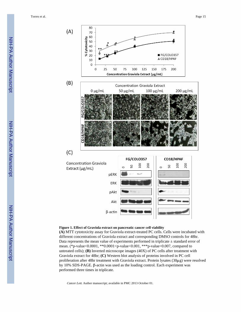
Figure 1. Effect of Graviola extract on pancreatic cancer cell viability(A) MTT cytotoxicity assay for Graviola extract-treated PC cells. Cells were incubated withdifferent concentrations of Graviola extract and corresponding DMSO controls for 48hr.Data represents the mean value of experiments performed in triplicate ± standard error ofmean. (*p-value<0.0001, **0.0001<p-value<0.001, ***p-value=0.007, compared tountreated cells); (B) Inverted microscope images (40X) of PC cells after treatment withGraviola extract for 48hr; (C) Western blot analysis of proteins involved in PC cellproliferation after 48hr treatment with Graviola extract. Protein lysates (30μg) were resolvedby 10% SDS-PAGE. β-actin was used as the loading control. Each experiment wasperformed three times in triplicate.
Torres et al. Page 15
Cancer Lett. Author manuscript; available in PMC 2013 October 01.
NIH
-PA Author Manuscript
NIH
-PA Author Manuscript
NIH
-PA Author Manuscript

Figure 2. Effect of Graviola extract on the metabolism of pancreatic cancer cells(A) Western blot analysis of HIF-1α and NF-κB expression in PC cells after treatment withGraviola extract. Protein lysates (30μg) were resolved on 10% SDS-PAGE gels. β-actin wasused as a loading control; (B) Real-time PCR-based measurement of transcript levels ofglucose transporters 1 and 4 (GLUT1, GLUT4), hexokinase II (HKII), and lactatedehydrogenase A (LDHA) in PC cells after incubation with Graviola extract. Data ispresented as the average fold difference in gene expression for the gene of interest inGraviola extract-treated cells versus untreated cells (0μg/mL) ± standard error of mean. Thehousekeeping gene β-actin was used as an internal control. (*0.01<p-value<0.05,**0.005<p-value<0.001, ***p-value<0.005); (C) Measurement of glucose uptake in PCcells after treatment with Graviola extract. Radioactive counts of cells labeled with [3H]-2-
Torres et al. Page 16
Cancer Lett. Author manuscript; available in PMC 2013 October 01.
NIH
-PA Author Manuscript
NIH
-PA Author Manuscript
NIH
-PA Author Manuscript

deoxyglucose were normalized with controls (***p-value<0.0001); (D) ATP quantificationof PC cells after treatment with Graviola extract. A Luminescent Cell Viability assay wasused to measure the ATP content in the cells. Data is presented as mean value fromexperiments performed in triplicates normalized with the protein content ± standard error ofmean. (*p-value=0.003, **p-value=0.002, ***p-value=0.0002) Data in the left panel is fromFG/COLO357 cells, whereas data in the right panel is from CD18/HPAF cells.
Torres et al. Page 17
Cancer Lett. Author manuscript; available in PMC 2013 October 01.
NIH
-PA Author Manuscript
NIH
-PA Author Manuscript
NIH
-PA Author Manuscript

Figure 3. Analysis of cytotoxic mechanism of Graviola extract in pancreatic cancer cells(A) Quantification of apoptotic and necrotic PC cells after treatment with Graviola extract(Apoptotic cells Annexin V+/PI− staining; Necrotic cells Annexin V+/PI+staining). Data ispresented as the mean value of the corresponding % cell population in duplicate samples ±standard error of mean; (B) The production of reactive oxygen species (ROS) in PC cellsafter treatment with Graviola extract was determined after incubating Graviola extract-treated PC cells with 2′,7′-Dichlorofluorescein diacetate (DCFH-DA). Cells were thenanalyzed by confocal microscopy. Scale bar represents 20μm; (C) Western blot analysis ofCaspase-3 expression in PC cells after treatment with Graviola extract. Protein lysates(30μg) were resolved on 10% SDS-PAGE gels. β-actin was used as a loading control; (D)Cell cycle analysis of FG/COLO357 PC cells after treatment with Graviola extract. Cellswere synchronized in the G1/S phase by thymidine block before adding Graviola extract.
Torres et al. Page 18
Cancer Lett. Author manuscript; available in PMC 2013 October 01.
NIH
-PA Author Manuscript
NIH
-PA Author Manuscript
NIH
-PA Author Manuscript
The effect of Graviola extract on the distribution of cells in different phases of the cell cyclewas analyzed by flow cytometry. The data is presented as the mean value of thecorresponding % cell population in duplicate samples ± standard error of mean.Representative flow cytometry histograms of cells treated with different concentrations ofGraviola extract are shown. (*p-value=0.0001; **p-value<0.0001); (E) Western blotanalysis of the expression of the cell cycle-related protein CyclinD1 in PC cells after beingincubated with Graviola extract. Protein lysates (30μg) were resolved in 10% SDS-PAGEgels. β-actin was used as the loading control. In (A), (B), and (C), data in the left panel isfrom FG/COLO357 cells, whereas data in the right panel is from CD18/HPAF cells.
Torres et al. Page 19
Cancer Lett. Author manuscript; available in PMC 2013 October 01.
NIH
-PA Author Manuscript
NIH
-PA Author Manuscript
NIH
-PA Author Manuscript

Figure 4. Effect of Graviola extract in the motility, migration, and cytoskeleton of pancreaticcancer cells(A) Wound healing assay of FG/COLO357 PC cells after treatment with Graviola extract.Microscope images (40X) of the artificially created wound in PC cells monolayer weretaken before (0hr) and after adding Graviola extract (24hr); (B) Migration of FG/COLO357PC cells after treatment with Graviola extract. The number of cells that migrated through the8μm pores of a polyethylene terephtalate (PET) membrane was quantified in 10 randomfields. Data represent the mean value of migrating cells ± standard error of mean (*p-value =0.0009; **p-value < 0.0001, compared to untreated control cells); (C) Actin filaments wereanalyzed by confocal microscopy by Rhodamine-anti-Phalloidin staining of FG/COLO357cells after treatment with Graviola extract. Nucleus was stained with DAPI. Scale bars
Torres et al. Page 20
Cancer Lett. Author manuscript; available in PMC 2013 October 01.
NIH
-PA Author Manuscript
NIH
-PA Author Manuscript
NIH
-PA Author Manuscript

represent 20μm; (D) Microtubules were analyzed by confocal microscopy after FITC-anti-βTubulin staining of FG/COLO357 cells after treatment with Graviola extract. Nucleus wasstained with DAPI. Scale bars represent 20μm; (E) Expression of proteins related tomigration/motility of PC cells after treatment with Graviola extract. Protein lysates (30μg)were resolved by 10% SDS-PAGE. β-actin was used as a loading control.
Torres et al. Page 21
Cancer Lett. Author manuscript; available in PMC 2013 October 01.
NIH
-PA Author Manuscript
NIH
-PA Author Manuscript
NIH
-PA Author Manuscript

Figure 5. Evaluation of Graviola Extract in pancreatic cancer orthotopic xenograft model(A) Pancreatic tumor weight results after treatment with Graviola extract. CD18/HPAF-Luciferase cells were injected orthotopically in the pancreas of athymic nude mice. After 1week of tumor growth, oral gavage treatment of PBS-suspended Graviola extract was givendaily for 35 days (N=8). Data is presented as box plots of the mean tumor weight of mice ineach treatment group. (*p-value = 0.006; **p-value=0.0008, compared to tumors of PBS-treated mice); (B) Major sites of metastasis in each treatment group. Results are presented asnumber of animals having metastasis out of total number of animals per group. Statisticalanalysis was done comparing Graviola extract-treated mice with untreated mice (0mg/kgGraviola extract); (C) In vivo biophotonic imaging of pancreatic tumors during the course of
Torres et al. Page 22
Cancer Lett. Author manuscript; available in PMC 2013 October 01.
NIH
-PA Author Manuscript
NIH
-PA Author Manuscript
NIH
-PA Author Manuscript

treatment with Graviola extract. Representative IVIS images of mice from differenttreatment groups are shown (D) Hematoxylin and Eosin (H&E) staining of paraffinembedded pancreatic tumors. Images on the right (20X) are magnified areas from theimages located at the left (10X). Yellow arrows in H&E sections represent necrotic areas intumors from mice treated with Graviola extract.
Torres et al. Page 23
Cancer Lett. Author manuscript; available in PMC 2013 October 01.
NIH
-PA Author Manuscript
NIH
-PA Author Manuscript
NIH
-PA Author Manuscript

Figure 6. Immunohistochemical analyses of pancreatic tumors after treatment with Graviolaextract(A) Immunohistochemical staining of MMP9 in paraffin-embedded pancreatic tumors.Representative images (20X) of tumors from different treatment groups are shown with theaverage composite score shown at the right. Data from experiments performed in triplicatesis presented as the mean value of the composite score of tumors ± standard error of mean.MMP9 expression in pancreatic tumors was also assessed by western blot analysis.Homogenized protein tumor lysates (30μg) were resolved by 10% SDS-PAGE. β-actin wasused as a loading control. (B) Immunohistochemistry staining of MUC4 in paraffin-embedded pancreatic tumors. Representative images (200X) of tumors from differenttreatment groups are shown with the average composite score shown at the right. Data fromexperiments performed in triplicate is presented as the mean value of the composite score of
Torres et al. Page 24
Cancer Lett. Author manuscript; available in PMC 2013 October 01.
NIH
-PA Author Manuscript
NIH
-PA Author Manuscript
NIH
-PA Author Manuscript

tumors ± standard error of mean. MUC4 expression in pancreatic tumors was also assessedby western blot analysis. Homogenized protein tumor lysates (30μg) were resolved by 2%agarose gels. β-actin was used as the loading control.
Torres et al. Page 25
Cancer Lett. Author manuscript; available in PMC 2013 October 01.
NIH
-PA Author Manuscript
NIH
-PA Author Manuscript
NIH
-PA Author Manuscript