NIH Common Fund | - th 5 Annual Investigators Meeting › sites › default › files › 2017...
58
5 th Annual Investigators Meeting June 29-30, 2017 Masur Auditorium, NIH, Bethesda, MD
Transcript of NIH Common Fund | - th 5 Annual Investigators Meeting › sites › default › files › 2017...

5th
Annual Investigators Meeting
June 29-30, 2017
Masur Auditorium, NIH, Bethesda, MD

THURSDAY, JUNE 29, 2017 8:00 a.m. Registration and Check-In 8:30 a.m. Welcome & Presentation of Award(s) for the NIH “Follow that Cell Challenge”
James Anderson, Ph.D., M.D., Director of the NIH Division of Program Coordination, Planning, and Strategic Initiatives (DPCPSI) Roderic Pettigrew, Ph.D., M.D., Director of the National Institute of Biomedical Imaging and Bioengineering (NIBIB) Joshua A. Gordon, Ph.D. M.D., Director of the National Institute of Mental Health (NIMH)
9:00 a.m. Presentation by Follow that Cell Winner(s) Winner(s) 9:30 a.m. Keynote Address Integrated learning across modalities, technologies, and species for single cell genomics Rahul Satija, Ph.D. (New York University) 10:00 a.m. Theories of Cellular Phenotype – Multimodal Analysis of in vivo and in vitro cells James Eberwine, Ph.D. (University of Pennsylvania) 10:20 a.m. Break 10:45 a.m. Keynote Address Having fun with single cell RNA-seq: imputation, manifolds and trajectories Dana Pe’er, Ph.D. (Memorial Sloan Kettering Cancer Center) 11:15 a.m. Multiplex and Multimodal Analysis of RNA Expression by HCR and SeqFISH Scott Fraser, Ph.D. (University of Southern California) 11:35 a.m. Single Cell Imaging of Epigenetic Dynamics Peter Yingxiao Wang, Ph.D. (University of California San Diego) 11:55 am. Single Cell Genomics: When Stochasticity Meet Precision Xiaoliang Sunney Xie, Ph.D. (Harvard University) 12:15 p.m. Lunch on Your Own 1:00 p.m. Poster Session Location: FAES Terrace 1:00 – 2:00 Presenters for odd numbered posters 2:00 – 3:00 Presenters for even numbered posters 3:00 p.m. Keynote Address Genes, cells, and behavior: lessons from C. elegans Cori Bargmann Ph.D. (The Rockefeller University; The Chan Zuckerberg Initiative)
NIH Common Fund 5th Annual Single Cell Analysis Investigators Meeting
Masur Auditorium, NIH Campus, Bethesda, MD

3:30 p.m. Breakout Sessions
Single-cell approaches to infectious disease Moderator: John Yin, Ph.D. (University of Wisconsin–Madison) Location: Classroom 1&2 Is Heterogeneity Regulated? Moderator: Suraj Bhat, Ph.D. (University of California Los Angeles) Location: Classroom 4 Immune Cell Diversity Moderator: William Lu, Ph.D. (National Cancer Institute) Location: Classroom 6
5:00 p.m. General Meeting Adjourns
FRIDAY, JUNE 30, 2017
8:00 a.m. Registration and Check-In
8:30 a.m. Keynote Address Imaging Single Molecules of mRNA in Single Living Cells Robert Singer, Ph.D. (Albert Einstein College of Medicine)
9:00 a.m. Breakout Sessions Moving single cell technologies out of the lab for wider adoption Moderator: Navin Varadarajan, Ph.D. (University of Houston) Location: Classroom 3 Public sharing of resources and data: Lessons from other trans-NIH Programs Moderator: Grace Shen, Ph.D. (National Eye Institute) Location: Classroom 6 Birds of a Feather Location: TBD
10:30 a.m. Break
11:00 a.m. Keynote Address Illuminating Cellular Diversity in the Nervous System John Ngai, Ph.D. (University of California, Berkeley)
11:30 a.m. Comprehensive and integrated DNA, RNA and protein profiling in single cells in situ with cleavable fluorescent probes Jia Guo, Ph.D. (Arizona State University)
11:50 a.m. Multimodal imaging of single cell populations by mass spectrometry, immunocytochemistry, and vibrational spectroscopy for uncovering chemical heterogeneity within the brain Elizabeth Neumann (University of Illinois at Urbana-Champaign)
12:10 p.m. Automating the Optical Manipulation of Single Cells in Complex Tissues Pavak Shah, Ph.D. (Memorial Sloan-Kettering Cancer Center)
12:30 p.m. Rare cell variability and drug-induced reprogramming as a mode of cancer drug resistance Sydney Shaffer (University of Pennsylvania)
12:50 p.m. Wrap-Up
1:00 p.m. General Meeting Adjourns

General Information The main meeting session will be held in the Masur Auditorium in the Clinical Center (Building 10) on the NIH Campus.
Details of campus access and security can be found here. You must present a valid form of ID. Expect the security check to take 20-30 minutes. Parking NIH offers visitor parking areas in parking lots, garages, and metered spaces throughout the campus. All visitor parking areas are paid areas, charged at the following rates:
• Garages and Lots: $2.00 for the first hour, $4.00 for the second hour, and $6.00 for the third hour. Any time exceeding 3 hours, will be charged the daily rate of $12.00.
• Metered Parking Spaces: $2.00 per hour. Refer to the visitor map above for lots that permit visitor parking. All visitor lots are managed by the NIH Parking Services Contractor: Mid Atlantic Parking, Inc. If you are not an NIH employee, you will need to pass through NIH security at the Gateway Center (from Rockville Pike – Route 355) before you are allowed on campus. Metrorail Visitors are strongly encouraged to use public transportation such as the Metrorail subway system which has a convenient stop (Medical Center) on the NIH campus. Visit the "Metro" site for information on fares and schedules.
5th Annual Single Cell Analysis Investigators Meeting Masur Auditorium, NIH Campus, Bethesda, MD
Meeting Information

Kiss and Ride Visitors can be dropped off and picked up from the Kiss and Ride park located at 9000 Rockville Pike, Bethesda, Maryland 20892 On-Campus Shuttle Shuttle services are provided throughout the day on the NIH Campus for employees, patients, and visitors. Click here for Shuttle routes and schedules Directions to Masur Auditorium from the Clinical Center
North lobby entrance: From the lobby, go down the right side, passing Admissions on your right. Continue straight through the sliding glass doors, following posted signs to the Masur. Continue following the “Detour” signs to the Masur. The auditorium is just past the main elevators. From the South lobby entrance: From the lobby, take either the left or right hallway up a slight incline until you come to the entrance of the Masur Auditorium. When the two hallways converge, you are standing in front of Masur Auditorium. Food & Beverages Food and beverages must be purchased. A full cafeteria is open from 6:30 a.m. - 2:30 p.m. located on the B1 level of the Clinical Center. More selections including Au Bon Pain is located on the SE side of the Clinical Center near the Main Lobby. Three concession/coffee stands are also available. The concession stand is located on the B1 level near the cafeteria and is open from 7:00 a.m. - 6:00 p.m. Two coffee stands are open from 7:00 a.m. - 4:00 p.m. and are located on the 1st floor in the CRC and the FAES corridor. Additionally, downtown Bethesda offers a fine selection of restaurants. Click Here for More Information

Meeting Information Poster Session The poster session on June 29th will be held in the Terrace located near the FAES classrooms. Directional signs will be posted in the registration area. Breakout Sessions The breakout sessions will be held on June 29th and June 30th. The breakout sessions will be in the FAES classrooms. Specific information regarding each session can be found on the meeting agenda.

WEDNESDAY, JUNE 28, 2017
12:20 pm Introductory Remarks - Robert Star (Director Division of Kidney, Urologic, and Hematologic Diseases National Institute of Diabetes and Digestive and Kidney Diseases, HuBMAP Co-Chair)
12:30 – 2:30pm Challenges in collecting and pre-analytical processing of tissue
Moderator: Robert Star (NIDDK)
There are many tissue collection and processing factors that influence data quality, from length of ischemia time to storage conditions and collection method. These factors influence the distribution and degradation of biomolecules at different rates. Therefore, it is critical to match the choice of tissue source, collection method and preservation technique with the types of biomolecules being studied by different downstream assays.
The purpose of this session is to identify some of the challenges in collecting, preserving, and annotating high quality human tissue that will be used for downstream analytical techniques in the HuBMAP program. These techniques include single cell RNAseq, FISH, immuno-fluorescence as well as emerging techniques such as MERFISH, FISSEQ, seqFISH, MIBI-TOF, and 3-dimensional high end imaging. Through the discussion, we hope to have a better understanding of the challenges HuBMAP might face in collecting and pre-analytical processing of tissue specimens and how this processing will impact the quality of data collected by different single cell, tissue, and imaging assays.
A number of components add to these challenges. One component is to record the spatial orientation of samples relative to anatomical landmarks (and build this into the sample management pipeline). A second component is the analysis, then integration and iteration of data from multiple imaging and omics assays to develop comprehensive molecular (and omic) profiles of the cells within the tissue, including location information. A third key component is to understand when sources of variability are biologically relevant (within tissue samples from same patient, across multiple tissues, and across multiple donors) or artifacts of the collection and processing of the samples.
Questions for the breakout session to consider include:
• Quality: What are practical quality measures for assessing the impact of tissue collection methods and the degree of degradation? How does the magnitude of ischemia signatures compare with collection, dissociation or storage signatures? Is there a common set of quality biomarkers that can be used across all tissues and that are compatible with downstream assays?
• Metadata: Beyond SPREC 2.0, are there common data elements describing collection and processing that are relevant to mapping DNA, RNA and proteins biomolecular distributions in tissues?
• Assay Workflow: What are best practices for assessing the impact of single cell (liberase) and tissue (LCM, super-resolution, imaging MS/MS) based tissue “dissociation” methods on assay measurements? Can tissue sections be used for multiple assays (RNA in situ, then protein, then routine stains)?
• Collection: For what assays and tissue types do tissues need to be collected from live donors? Rapid autopsy protocols?
• Staining: Do common stains (e.g. H&E, trichrome, toluidine) influence the sensitivity and specificity of downstream assays?
• Orientation: How do we preserve orientation of a tissue specimen through the processing chain?
NIH Common Fund HuBMAP / SCAP Mini Workshop Neuroscience Center, Bethesda, MD

• Fixing, clearing and embedding: Are there tissue stabilization techniques that can be used before or during collection? For current and emerging fixatives/preservatives of excised tissue, which biomolecular species do they preserve with good fidelity (not only nucleic acids and proteins, but how effective are these techniques at preserving metabolites or carbohydrates), what compatibility issues are there with different tissue types, cell types, dissociation techniques and assays? What are some of the challenges associated with clearing techniques?
• Sectioning: What are tissue-specific considerations in preparing tissue sections? How does the choice of tissue size and format influence ischemia and preservation timing and in term the quality of the tissue for different downstream assays?
• End-users: What format, quantity, and quality level is needed for: RNAseq, DropSeq, MERFISH / FISSEQ / seqFISH, immuno-florescence, MIBI-TOF and CyTOF approaches?
2:30 – 3:00pm Break
3:00 – 5:00 pm Data Analysis, Standards, and Benchmarks for Single Cell Analysis
Moderator: Junhyong Kim (University of Pennsylvania)
Because of the difficulty of obtaining measurements at the single cell scale, the field has been driven by technological advances, including various RNA/DNA sequencing technologies, high-resolution proteomics and metabolomics, multiplexing strategies, cell handling technologies, etc. Despite these technological advances, single cell measurements remain difficult and is fundamentally challenged by the fact that the units of measurement, each cell, has no replication. It has been extremely difficult to assess the efficiency of measurements, establish benchmarks or controls, agree on protocols for data analysis, and coherently define standards for reporting experiments and data analysis. An especially important challenge is placing single cell data in their organismal context, including spatial coordinates.
Questions for this breakout session to consider include:
• Is there benchmark data to compare new experimental or computational methods? • How do we establish material standards such as specific cells or spike-in RNA? • What metadata about calibration is important to know? • What information is important to collect about the sample and its preparation? • How can we work together with manufacturers to build standards into their methods? • Does an ontology need to be established for single cell analysis? • How can we associate single cells to tissue orientation information? More generally, how can data be organized
from the single cell scale to whole organism scale? • What are the common data elements between imaging and sequencing assays? Is there a common header we
can use for all data, similar to FITS or DICOM?
5:00 pm Closing Remarks

Suggested background reading for these breakouts:
• Unhale, S. A., Skubitz, A. P., Solomon, R., & Hubel, A. (2012). Stabilization of tissue specimens for pathological examination and biomedical research. Biopreservation and biobanking, 10(6), 493-500. [http://online.liebertpub.com/doi/abs/10.1089/bio.2012.0031]
• Hubel, A., Spindler, R., & Skubitz, A. P. (2014). Storage of human biospecimens: selection of the optimal storage temperature. Biopreservation and biobanking, 12(3), 165-175. [http://online.liebertpub.com/doi/abs/10.1089/bio.2013.0084]
• Hubel, A., Aksan, A., Skubitz, A. P., Wendt, C., & Zhong, X. (2011). State of the art in preservation of fluid biospecimens. Biopreservation and biobanking, 9(3), 237-244. [http://online.liebertpub.com/doi/abs/10.1089/bio.2010.0034]
• Chung, Cho H, Hewitt SM (2016). The paraffin-embedded RNA metric (PERM) for RNA isolated from formalin-fixed, paraffin-embedded tissue. Biotechniques. May 1;60(5):239-44 [http://www.biotechniques.com/BiotechniquesJournal/2016/May/The-paraffin-embedded-RNA-metric-PERM-for-RNA-isolated-from-formalin-fixed-paraffin-embedded-tissue/biotechniques-364401.html]
• Carithers, L. J., Ardlie, K., Barcus, M., Branton, P. A., Britton, A., Buia, S. A., ... & Guan, P. (2015). A novel approach to high-quality postmortem tissue procurement: the GTEx project. Biopreservation and biobanking, 13(5), 311-319. [http://online.liebertpub.com/doi/full/10.1089/bio.2015.0032]

Meeting Location
This meeting will be located in Conference Room D on the ground floor of 6001 Executive Blvd.
Visitor Parking
Located at 6001 Executive Blvd, NIH offers two convenient garages and several parking lots for visitor parking. While the NIH parking Office does not issue permits to visitors, a validation sticker will be provided upon request.
Restaurants
Executive Deli Located at 6011 Executive Blvd (adjacent to 6001 Executive Blvd), the Executive Deli offers a variety of food options to pick from. Hours of operation are 7:00am-3:30pm. Menu available at the registration table. Pike & Rose Located at 11580 Old Georgetown Rd., North Bethesda, MD 20852, the Pike and Rose offers a fine selection of dining choices. Pike and Rose offers two convenient garages, and free parking for the first two hours. Click Here for More Information
Webex Information
These breakout sessions will also be videocast through Webex:
https://nih.webex.com/nih/onstage/g.php?MTID=e8179a0e2db31cb7a2c7abc135a4a76b2

Is heterogeneity regulated? 3:30 – 5pm, Thursday June 29 Moderator: Suraj Bhat, Ph.D. (University of California Los Angeles) Location: Classroom 4 The heterogeneity of gene expression in single cells is well established however it is unclear if this heterogeneity has any relationship to the morphological and/or molecular phenotype of a tissue or an organ. In this session we will ask questions to elucidate the challenges, both technical as well as conceptual, in understanding the role of the cellular variability (as assessed by gene expression) in the context of multicellularity of tissues and organs. We will explore the possible role of cellular heterogeneity in terminal differentiation. At the current state of our knowledge, we do not have a handle on whether the variation in the abundance of a gene transcript from cell to cell is because of the fluctuations intrinsic to the gene activity or whether it is the other cellular components that determine the variability between cells. In either case the question remains – what is regulated? Some of the questions that we will address (not necessarily in the order listed):
• Is heterogeneity causal or a result of the gene activity? • Is heterogeneity functional? • What is the relationship between the tissue /organ phenotype to the single cell? • Is heterogeneity the pathway to terminal differentiation? • Do different developmental programs entail specific states (stages) of heterogeneity? • What are the deterministic sources of cellular variability and how are they maintained? • Is deterministic variability important and what purpose does it serve? • Do we understand the link between molecular variability and the phenotypic variability between individual cells?
(Technically how do we study heterogeneity in single cells?)
Immune Cell Diversity 3:30 – 5pm, Thursday June 29 Moderator: Y. William Lu, Ph.D. (National Cancer Institute) Location: Classroom 6 Immunologists usually rely on flow cytometry and other traditional tools to conduct immune-related research. In the past several years, new single-cell technologies, such as single-cell transcriptomics and mass cytometry (CyTOF), have enabled researchers to ask scientific questions that could not be addressed previously. The theme for this session is to discuss the opportunities and potential challenges in immune-related studies, such as cell type diversity and phenotypic analysis. Questions for this breakout session to consider include:
• What immunological questions can we ask from the CyTOF analysis? • Similarly, what immunological questions can we ask from the single-cell transcriptome analysis? • Several technologies are available for single-cell transcriptome analysis. What are the pros and cons for these
technologies? Which technology is suitable for immune-related studies? • What kind of data quality should we expect in order to generate reproducible results? Should we validate the data by
additional assays? • One of the potential challenges is the communication between scientists with different expertise. Sometimes the
experiment may not perform well as expected. As an immunologist, a molecular biologist or a bioinformatician, what is the challenges to communicate with scientists with other disciplines?
• What kind of community resources can we establish to help immunologists to understand and utilize new single-cell technologies?
5th Annual Single Cell Analysis Investigators Meeting Masur Auditorium, NIH Campus, Bethesda, MD
Breakout Sessions

Single-cell approaches to infectious disease 3:30 – 5:00pm, Thursday June 29 Moderator: John Yin (University of Wisconsin–Madison) Location: Classroom 1 & 2 Transmission of infectious diseases from ailing to healthy hosts occurs through a cough or sneeze, handshake, faucet handle, or mosquito’s bite. Typically, the process transfers a few bacterial cells or virus particles. Although a small number of cells or particles encounter a few susceptible host tissues or cells, the resulting infection initiates a battle --- with potentially critical outcomes. The behavior of a few host cells infected by a small number of bacteria or virus particles can give rise to large 'noise' and significant variability in gene expression by the pathogen to amplify itself or by the host to set innate immune blockades, which then influence how further cycles of host cell or tissue infection amplify or inhibit the pathogen. The result can often be a diversity of symptoms and disease severities for patients, from mild to serious, or even deadly. Questions for the breakout session to consider include: • To what extent do genetic, environmental, or other (stochastic) factors contribute to extremely heterogeneous
distributions of virus production (yield) from single cells? • What are key challenges and opportunities for advancing innovative technologies to enable routine high-throughput
single-cell measurements? • How can the intrinsic heterogeneity of single-cell readouts be exploited to extract new insights into virus-cell
interactions? • How can systems biology approaches (mathematical modeling, computer simulations) add value to enable
mechanistic interpretation of single-cell data? • What features of virus or cellular behaviors at the single-cell level most impact the severity of infection in natural
hosts? (Note: Most infections in nature are initiated by small numbers of host cells initially becoming infected. From an evolutionary perspective, transmission can create genetic bottlenecks for the pathogen.)
Public sharing of resources and data: Lessons from other trans-NIH Programs 9:00 – 10:30am, Friday June 30 Moderator: Grace Shen, Ph.D. (National Eye Institute) Location: Classroom 6 Resource and data sharing is essential to speed translation of research results into knowledge, therapies, and procedures to improve our understanding of biological processes and human health. NIH is committed to sharing data from its research and supports a variety of resources and tools for researchers. These resources include tissue banks and repositories, datasets and databases, model organisms, genome and DNA sequences, and resource libraries. The panelists in this session will describe some of the resources and data that are available from programs they are involved with across NIH. Programs that will be covered include: BRAIN, Cancer Systems Biology Consortium, the Physical Sciences-Oncology Network, ImmPort, ITN TrialShare, ImmuneSpace, IEDB, ImmGen, BD2K and NIGMS resources. Panelists for this session:
• Andrea Beckel-Mitchener, Ph.D. (National Institute of Mental Health) • Susan Gregurick, Ph.D. (National Institute of General Medical Sciences) • Shannon Hughes, Ph.D. (National Cancer Institute) • Halonna Kelly, Ph.D. (National Institute of Allergy and Infectious Diseases)

Moving single cell technologies out of the lab for wider adoption 9:00 – 10:30am, Friday June 30 Moderator: Navin Varadarajan, Ph.D. (University of Houston) Location: Classroom 3 The last few years have seen a dramatic increase in the number and complexity of single-cell technologies. Many these advances have been pioneered through individual laboratories and the scope of the biological questions interrogated has primarily been dictated by collaborations with these laboratories. For single-cell technologies to mature, they must become standardized tools that enable any biologist/clinician can access to test hypotheses. Mass cytometry is an example of one such tool that has made the successful transition. The scope of this discussion is to understand the challenges behind moving the technologies from the labs of the inventors and to make them commercially available tools/techniques. Questions for the breakout session to consider include: • Validation. One of the major challenges with single-cell technologies is cross-platform validation. While single-cell
technologies provide the ability to deliver insight that would not be available based on population analyses, it is important to be able to identify technical errors, limitations and how to implement confidence metrics in single cell results.
• Standardization of statistics, bioinformatics and visualization. Another major challenge with single-cell data is to have robust methods that perform normalization, discretization etc. in a standardized manner. Good practices in defining thresholds, how they are determined and adhering to a minimal set of standards that will be published (e.g. see Minimal Information About T-cell Assays, MIATA) comprise an essential framework. Similarly, there is a need to develop to visualization packages that better describe the complexity of multi-dimensional, time-resolved single-cell data. E.g. viSNE is good for single time points but what about time series?
• Commercialization. There are many layers to commercialization including IP framework, hurdles to manufacturing, ease of implementation and market adoption. What programs can help PIs understand these challenges: NSF ICorps? Institutional help?
Birds of a Feather 9:00 – 10:30am, Friday June 30 Moderator: You? Location: TBD Lead or attend a Birds of a Feather session! Meet with your peers! Discuss questions that have been on your mind or the next big things! This Birds-of-a-Feather session is an opportunity for very informal gatherings of people interested in a particular topic that we are not covering otherwise at the meeting. The goal is to have audience-driven discussion, grassroots participation and networking. How it will work: There will be a poster board at the registration desk for the meeting. If you are interested in leading a Birds of a Feather session, please use one of the postcards to write the topic for discussion on Thursday. If you are interested in participating in one of the sessions please add your name to the postcard. We will help each of the BOF groups find space to hold their discussion during the breakout session on Friday morning.

As part of the annual meeting we have a number of focus group meetings. These are small, invite-only meetings for NIH staff to meet with different groups of attendees at the annual meeting. If you have received an invite to one of these meetings you will have received materials for the meeting. Below are the details of the meeting rooms for each of these focus groups.
Thursday June 29
3:30 – 5:00pm: Industry Representatives (Classroom 8)
5:00 – 6:30pm: R01/R21/R33 and Supplement Grantees (Classroom 4)
Friday June 30
9:00 – 10:30am: Program Consultants (Classroom 7)
1:30 – 2:30pm: U01 Grantees (Classroom 1 & 2)
5th Annual Single Cell Analysis Investigators Meeting Masur Auditorium, NIH Campus, Bethesda, MD
Focus Group Meetings

Poster Number Authors Affiliations Poster Title
1
Prithvijit Mukherjee, Lingqian Chang, Eric Berns, S. Shiva P. Nathamgari, Milan Mrksich, Horacio D. Espinosa
Infinitesimal LLC, Skokie, IL Department of Mechanical Engineering, Northwestern University, Evanston, IL
A Localized Cell Analysis Device for Temporal Cell Analysis - Measuring Protein Tyrosine Phosphatase Activity in Live Cancer Cells
2 Yi Lu and Pak Kin Wong Department of Biomedical Engineering, The Pennsylvania State University, University Park, PA
A Multispectral Single Molecule Nanobiosensor for Dynamic Multigene Analysis during Collective Cell Migration
3
Masahiro Hitomi, Anastasia Chumakova, Stephanie Jarvis, Neha Anand, Bridget Corrigan, Peter Yoo, Upashruti Agrawal, Vid Yogeswaran, Malini Kamineni, Sunghyun Kim, and Justin D. Lathia
Dept. Cellular and Molecular Medicine, Lerner Research Institute, Cleveland Clinic, Cleveland, OH
Asymmetric cell division regulates fate decision of glioblastoma cancer stem cells
4 Pavak K. Shah, Anthony Santella, Adrian Jacobo, Kimberly Siletti, A. James Hudspeth, Zhirong Bao
Developmental Biology Program, Sloan Kettering Institute, New York, NY Howard Hughes Medical Institute and Laboratory of Sensory Neuroscience, The Rockefeller University, New York, New York
Automating the Optical Manipulation of Single Cells in Complex Tissues
5
Lin Han, Hua-Jun Wu, Haiying Zhu, Kun-Yong Kim, Sadie L. Marjani, Markus Riester, Ghia Euskirchen, Xiaoyuan Zi, Jennifer Yang, Jasper Han, Michael Snyder, In-Hyun Park, Rafael Irizarry, Sherman M. Weissman, Franziska Michor, Rong Fan, Xinghua Pan
Department of Biology Chemistry and Molecular Biology, School of Basic Medical Sciences, Southern Medical University, Guangzhou, Guangdong Province, China Department of Genetics, Yale University School of Medicine, New Haven, CT 06519
Bisulfite-independent analysis of CpG island methylation enables genome-scale stratification of single cells
6 Sachiko Sato, Ann Rancourt, Yukiko Sato, and Masahiko S. Satoh
Glycobiology and Bioimaging Laboratory of Research Center for Infectious Diseases Laboratory of DNA Damage Responses and Bioimaging, CHU de Québec, Faculty of Medicine, Laval University, 2705 Boulevard Laurier, Quebec, Quebec G1V 4G2, Canada Department of Physiology, McGill University, Montreal, Canada
Characterization of cultured cell lines using single-cell lineage tracking analysis
7 Jia Guo, Manas Mondal, Renjie Liao, Lu Xiao
Biodesign Institute & School of Molecular Sciences, Arizona State University, Tempe, AZ
Comprehensive and integrated DNA, RNA and protein profiling in single cells in situ with cleavable fluorescent probes
8
Erika P. Portero, Rosemary M. Onjiko, Sally A. Moody, and Peter Nemes
Department of Chemistry & Department of Anatomy and Regenerative Biology, The George Washington University, Washington, DC
Discovery Single-cell Mass Spectrometry Profiles Metabolic Gradients in the 16-cell Vertebrate (Frog) Embryo
NIH Common Fund 5th Annual Single Cell Analysis Investigators Meeting
Poster Session Abstracts

NIH Single Cell Analysis Investigators Meeting 2017 2
9
Tania Konry, Saheli Sarkar, Pooja Sabhachandani, Dina Stroopinksi, Kristen Palmer, Noa Cohen, Jacalyn Rosenblatt, David Avigan
Department of Pharmaceutical Sciences, Northeastern University, 360 Huntington Avenue, Boston, MA, 02115 Beth Israel Deaconess Medical Center, Harvard Medical School, Boston, MA 02115
Dynamic analysis of immune and cancer cell interaction at single cell level in microfluidic droplets
10 Tianyi Yuan, Diane S. Krause, and Oleg Denisenko
Yale University, New Haven, CT 06520 Department of Medicine, University of Washington, Seattle, WA 98109
Epigenetic analysis of gene activation in a single cell
11
P.A. Osmulski, Y.-T. Hsu, G. Huang, S.R. Polusani, C.-L. Chen, D. Mahalingam, N.B. Kirma, M.E. Gaczynska, and T. Hui-Ming Huang
Department of Molecular Medicine & Department of Medicine, University of Texas Health, San Antonio, TX
Guilty by adhesion – assessment of cells grip with atomic force microscopy
12 Stephen M. Anthony, Bryan Carson, Jerilyn A. Timlin
Bioenergy and Defense Technologies Department, Sandia National Laboratories, Albuquerque, NM
Hyperspectral Imaging Analysis of Cellular Heterogeneity Between and Across Populations
13 Peter Nemes, Rosemary M. Onjiko, Erika Portero, and Sally A. Moody
Department of Chemistry, The George Washington University, Washington, DC Department of Anatomy and Regenerative Biology, The George Washington University, Washington, DC
In Situ Optoguided Microsampling Single-cell Mass Spectrometry for Elucidating Cell Heterogeneity in the Developing Xenopus laevis (frog) Embryo
14
Dipjyoti Das, Dörthe Jülich, Jamie Schwendinger-Schreck, Andrew Lawton, Nicolas Dray, Thierry Emonet, Corey S. O’Hern, Mark D. Shattuck and Scott A. Holley
Department of Molecular, Cellular and Developmental Biology, Department of Physics, Department of Mechanical Engineering and Materials Science, Department of Applied Physics & Department of Physics, Yale University, New Haven, CT Benjamin Levich Institute, City College of the City University of New York, NY.
Long-range mechanical orchestration by the vertebrate tail organizer
15 Luke Stevens, Tanaya Pande, Hongru Hu, Aravindan Krishnan, Claudia Mizutani, Rui Sousa-Neves,
Department of Biology & Department of Genetics and Genome Sciences, Case Western Reserve University, Cleveland, OH
Multidimensional analyses of whole brain aging with single cell resolution
16
Elizabeth K. Neumann, Troy J. Comi, Stanislav S. Rubakhin, Sanghamitra Deb, Nicholas Spegazzini, Jennifer W. Mitchell, Collin Kaufman, Rohit Bhargava, Martha U. Gillette, Jonathan V. Sweedler
Department of Chemistry, Beckman Institute for Advanced Science and Technology, Department of Bioengineering, Department of Cell and Developmental Biology & Neuroscience Program, University of Illinois at Urbana-Champaign, Urbana, IL.
Multimodal imaging of single cell populations by mass spectrometry, immunocytochemistry, and vibrational spectroscopy for uncovering chemical heterogeneity within the brain
17 Scott E. Fraser, Long Cai
University of Southern California, Translational Imaging Center, Molecular and Computational Biology, Los Angeles, CA California Institute of Technology, Biology and Biological Engineering, Pasadena, CA
Multiplex and Multimodal Analysis of RNA Expression by HCR and SeqFISH
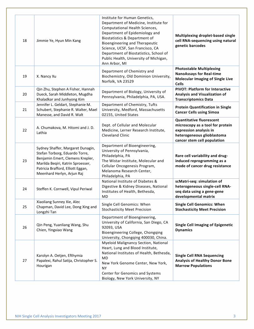
NIH Single Cell Analysis Investigators Meeting 2017 3
18 Jimmie Ye, Hyun Min Kang
Institute for Human Genetics, Department of Medicine, Institute for Computational Health Sciences, Department of Epidemiology and Biostatistics & Department of Bioengineering and Therapeutic Science, UCSF, San Francisco, CA Department of Biostatistics, School of Public Health, University of Michigan, Ann Arbor, MI
Multiplexing droplet-based single cell RNA-sequencing using natural genetic barcodes
19 X. Nancy Xu Department of Chemistry and Biochemistry, Old Dominion University, Norfolk, VA 23529
Photostable Multiplexing NanoAssays for Real-time Molecular Imaging of Single Live Cells
20 Qin Zhu, Stephen A Fisher, Hannah Dueck, Sarah Middleton, Mugdha Khaladkar and Junhyong Kim
Department of Biology, University of Pennsylvania, Philadelphia, PA, USA.
PIVOT: Platform for Interactive Analysis and Visualization of Transcriptomics Data
21 Jennifer L. Geldart, Stephanie M. Schubert, Stephanie R. Walter, Mael Manesse, and David R. Walt
Department of Chemistry, Tufts University, Medford, Massachusetts 02155, United States
Protein Quantification in Single Cancer Cells using Simoa
22 A. Chumakova, M. Hitomi and J. D. Lathia
Dept. of Cellular and Molecular Medicine, Lerner Research Institute, Cleveland Clinic
Quantitative fluorescent microscopy as a tool for protein expression analysis in heterogeneous glioblastoma cancer stem cell population
23
Sydney Shaffer, Margaret Dunagin, Stefan Torborg, Eduardo Torre, Benjamin Emert, Clemens Krepler, Marilda Beqiri, Katrin Sproesser, Patricia Brafford, Elliott Eggan, Meenhard Herlyn, Arjun Raj
Department of Bioengineering, University of Pennsylvania, Philadelphia, PA The Wistar Institute, Molecular and Cellular Oncogenesis Program, Melanoma Research Center, Philadelphia, PA
Rare cell variability and drug-induced reprogramming as a mode of cancer drug resistance
24 Steffen K. Cornwell, Vipul Periwal
National Institute of Diabetes & Digestive & Kidney Diseases, National Institutes of Health, Bethesda, MD
scMatri-seq: simulation of heterogeneous single-cell RNA-seq data using a gene-gene developmental matrix
25 Xiaoliang Sunney Xie, Alec Chapman, David Lee, Dong Xing and Longzhi Tan
Single Cell Genomics: When Stochasticity Meet Precision
Single Cell Genomics: When Stochasticity Meet Precision
26 Qin Peng, Yuanliang Wang, Shu Chien, Yingxiao Wang
Department of Bioengineering, University of California, San Diego, CA 92093, USA Bioengineering College, Chongqing University, Chongqing 400030, China.
Single Cell Imaging of Epigenetic Dynamics
27 Karolyn A. Oetjen, Efthymia Papalexi, Rahul Satija, Christopher S. Hourigan
Myeloid Malignancy Section, National Heart, Lung and Blood Institute, National Institutes of Health, Bethesda, MD New York Genome Center, New York, NY Center for Genomics and Systems Biology, New York University, NY
Single Cell RNA Sequencing Analysis of Healthy Donor Bone Marrow Populations

NIH Single Cell Analysis Investigators Meeting 2017 4
28 Yue J. Wang, Dana Avrahami-Tzfati, Klaus H. Kaestner
Department of Genetics and Institute for Diabetes, Obesity, and Metabolism, University of Pennsylvania Perelman School of Medicine, Philadelphia, PA Endocrinology and Metabolism Service, Hadassah-Hebrew University Medical Center, Jerusalem, Israel
Single-cell analyses of the endocrine pancreas from a neonatal donor
29 Camille Lombard-Banek, Aparna Baxi, Sally A. Moody, and Peter Nemes
Department of Chemistry & Department of Anatomy & Regenerative Biology, The George Washington University, Washington DC, 20052
Single-cell Proteomics in the Developing Frog (Xenopus) Embryo
30
Po-Yuan Tung, John D. Blischak, Chiaowen Joyce Hsiao, David A. Knowles, Jonathan E. Burnett, Jonathan K. Pritchard & Yoav Gilad
Department of Human Genetics, Department of Medicine, University of Chicago, Chicago, Illinois. Department of Genetics, Department of Radiology & Department of Biology, Stanford University, Stanford, CA Howard Hughes Medical Institute, Stanford University, CA
Batch effects and the effective design of single-cell gene expression studies
31
Josip Herman, Jon Penterman, Sagar, Andreas Diefenbach, Antigoni Triantafyllopoulou, Melanie A. Adams-Cioaba, Dominic Grün, and Stuart S. Levine
Max Planck Institute of Immunobiology and Epigenetics, 79108 Freiburg, Germany BioMicro Center, Massachusetts Institute of Technology, Cambridge, MA 02139, USA Institute of Medical Microbiology and Hygiene, University of Mainz Medical Center, 55131 Mainz, Germany Department of Rheumatology and Clinical Immunology, Medical Center–University of Freiburg, Faculty of Medicine, University of Freiburg, 79106 Freiburg, Germany) TTP Labtech
Miniaturization and automaton of Cel-seq2 and SMARTer-seq using the mosquito HTS liquid handler
32
James Eberwine, Jacqueline Morris, Young-Ji Na, Jaehee Lee, Hua Zhu, Eun-Hee Shim, Jinhui Wang, Kevin Miyashiro, Alexandra V. Ulyanova, Thomas Bell, John Wolf, Sean Grady, Jai Yoon Sul and Junhyong Kim
Department of Systems Pharmacology, Department of Neurosurgery & Department of Biology, University of Pennsylvania, Philadelphia, PA, USA.
Theories of Cellular Phenotype – Multimodal Analysis of in vivo and in vitro cells
33 Aaron Streets, Markita P. Landry
Department of Bioengineering, Department of Chemical and Biomolecular Engineering, California Institute for Quantitative Biosciences, QB3, University of California, Berkeley, CA Chan-Zuckerberg Biohub, San Francisco, CA
Toward Label-Free Single-Cell Profiling: Single-Molecule Detection of Protein Efflux and Raman Mapping of Intracellular Metabolites in Isolated Microorganisms and Brain Tissue
34 Juan Guan, Harrison Liu, Xiaoyu Shi, Siyu Feng, Bo Huang
Department of Pharmaceutical Chemistry, Department of Biochemistry and Biophysics & Department of Bioengineering, University of California San Francisco, San Francisco, CA
Tracking multiple genomic elements in single cell nuclei using correlative CRISPR imaging and sequential DNA FISH

NIH Single Cell Analysis Investigators Meeting 2017 5
35 Wen Zhou, Mary A. Yui, Brian A. Williams, Barbara J. Wold, Long Cai, Ellen V. Rothenberg
Division of Biology and Biological Engineering, California Institute of Technology, Pasadena, CA Division of Chemistry and Chemical Engineering, California Institute of Technology, Pasadena, CA
Transcriptional profiling with scRNAseq and SeqFISH on early T cell precursors reveals fine developmental steps
36
Robert S. Welner, Sam Wolock, Indira Krishnan, Danielle Tenen, Puneet Agarwal, Victoria McClearn, Ravi Bhatia, Daniel G Tenen, and Allon Klein
Division of Hematology/Oncology, Dept of Medicine, University of Alabama at Birmingham, Birmingham, AL Department of Systems Biology, Harvard Medical School, Boston, MA Division of Hematology/Oncology, Dept of Medicine, Beth Israel Deaconess Medical Center, Boston, MA
Unbiased Single-cell Analysis Reveals Hierarchy of the Bone Marrow Niche
37 Kushal K Dey, Chiaowen Joyce Hsiao, Matthew Stephens
Department of Statistics, University of Chicago, Chicago, Illinois 60637, USA Department of Human Genetics, University of Chicago, Chicago, Illinois 60637, USA
Visualizing the Structure of Single Cell RNA-seq Expression Data using Grade of Membership Models
38 Homero Pastrana, Alexander X. Cartagena-Rivera, Arvind Raman, Alba Avila
Departamento de Ingeniería Eléctrica y Electrónica & Centro de Microelectrónica, Universidad de los Andes (CMUA), Bogotá D.C., Colombia. School of Mechanical Engineering & Birck Nanotechnology Center, Purdue University, West Lafayette, Indiana, USA. Laboratory of Cellular Biology, Section on Auditory Mechanics, National Institute on Deafness and Other Communication Disorders (NIDCD), National Institutes of Health, Bethesda (NIH), Maryland, USA
CYTOTOXIC EFFECTS OF CARBON BASED NANOMATERIALS ON SINGLE CELL YOUNG’S MODULUS RESPONSE
39
40 Nick Trotta, Rob McLellan, Nicholas Dobes, Steve Gebhart
Cell Microsystems, Inc. Research Triangle Park, North Carolina
The CellRaft AIR™ System: Automated Imaging, Sorting and Isolation of Single Cells

NIH Single Cell Analysis Investigators Meeting 2017 6
Poster #1: A Localized Cell Analysis Device for Temporal Cell Analysis - Measuring Protein Tyrosine Phosphatase Activity in Live Cancer Cells
Prithvijit Mukherjee2, Lingqian Chang2, Eric Berns3, S. Shiva P. Nathamgari2, Milan Mrksich3,4,5, Horacio D. Espinosa1,2
1Infinitesimal LLC, Skokie, IL
2Department of Mechanical Engineering, Northwestern University, Evanston, IL
3Department of Biomedical Engineering, Northwestern University, Evanston, IL
4Department of Chemistry, Northwestern University, Evanston, IL
5Department of Cell and Molecular Biology, Feinberg School of Medicine, Northwestern University, Chicago, IL
The ability to temporally monitor intra-cellular biomarkers at the single cell level, without disrupting cellular functionality, is essential for the understanding of dynamic cellular processes such as differentiation as well as progression of disease, cellular heterogeneity and drug toxicity. Current cellular assays rely on cell lysis and thus only provide analyses of endpoints. Emerging non-destructive sampling technologies based on nanopipettes, hollow AFM probes and high aspect ratio structures such as nano-porous membranes, nano-straws and carbon nanotubes could potentially address such issues. However, significant challenges remain in terms of achieving high throughput and high sensitivity for all targets of interest in single cells.
We have developed a localized cell analysis device (LCAD) and used it in conjunction with a high-throughput, self-assembled monolayer based enzymatic assay (SAMDI) for non-destructive cellular sampling and analysis. The LCAD combines microfluidic seeding of few to single cells in microwells, followed by reversible electro-permeabilization of the plasma membrane at localized regions of substrate nano-pores. This allows for the extraction of cytosolic contents for detection and quantification at multiple time points. The SAMDI assay consists of self-assembled monolayers of alkanethiolates functionalized with peptides on gold spots. Enzymes in the cytosolic extract act on the immobilized peptide and the activity is quantified using matrix-assisted laser desorption/ionization mass spectrometry (MALDI-MS). In this work, a peptide with a phosphorylated tyrosine residue has been used to report on Protein Tyrosine Phosphatase (PTP) activity in MDA-MB 231 breast cancer cells. Using the LCAD-SAMDI system, we have detected PTP activity from cellular extracts of as few as two cells while preserving cell viability. Our current efforts are directed towards optimizing the system for single cell sensitivity and longitudinal sampling over several days. We are also working towards determining the long-term effects of disruption of the intra-cellular milieu and extending our technique to other relevant assays (such as mRNA profiling) and cell types (e.g. iPSCs). We believe that this platform would prove to be a valuable tool for answering fundamental questions pertaining to cell differentiation and single cell heterogeneity.
Acknowledgement
This project was supported in part by a grant from NIH (1R43GM110893-01)

NIH Single Cell Analysis Investigators Meeting 2017 7
Poster #2: A Multispectral Single Molecule Nanobiosensor for Dynamic Multigene Analysis during Collective Cell Migration
Yi Lu and Pak Kin Wong
Department of Biomedical Engineering, The Pennsylvania State University, University Park, PA
Collective cell migration is a fundamental multicellular activity that plays essential roles in numerous physiological and pathological processes, such as angiogenesis, tissue regeneration, and cancer metastasis. Despite its importance, the fundamental mechanisms that govern collective cell migration, such as the formation of leader cells and biomechanical coupling, remain poorly understood. To address this issue, we establish a multispectral single molecule imaging technique for dynamic single molecule detection in live cells. Single molecule tracking allows us to distinguish target molecules from autofluorescence and non-specific signal due to probe degradation as well as nanobiosensors entrapped in endosomes, lysosomes and other vesicular compartments. Furthermore, we demonstrate a FRET-based single molecule barcode scheme using multilabel locked nucleic acid LNA probes for multigene analysis. The single molecule barcode scheme can significantly increase the number of genes that can be monitored in the same cell. The single molecule barcode scheme is applicable to various fluorescence detection techniques, e.g., gold nanorod and tetrahedral DNA nanobiosensors for mRNA monitoring, gapmer aptamers for intracellular protein detection, and double-stranded locked nucleic acid probes for microRNA sensing, rendering its translational potential in a wide spectrum of biomedical applications. In this presentation, we will discuss the application of the single cell nanobiosensor for probing collective cell migration. We identify that the formation of leader cells during collective migration is dynamically regulated by Notch1-Dll4 signaling and intercellular tension. Our finding provides a molecular basis for the stochastic emergence of leader cells, which may enable novel approaches in regenerative medicine, diabetic wound healing and cancer treatment in the future.

NIH Single Cell Analysis Investigators Meeting 2017 8
Poster #3: Asymmetric cell division regulates fate decision of glioblastoma cancer stem cells
Masahiro Hitomi, Anastasia Chumakova, Stephanie Jarvis, Neha Anand, Bridget Corrigan, Peter Yoo, Upashruti Agrawal, Vid Yogeswaran, Malini Kamineni, Sunghyun Kim, and Justin D. Lathia
Dept. Cellular and Molecular Medicine, Lerner Research Institute, Cleveland Clinic, Cleveland, OH
Cancer stem cells (CSCs) are postulated to be responsible for therapeutic resistance and tumor recurrence because of their abilities to withstand therapies better than non-stem cancer cells and to initiate the tumor consisting of heterogeneous neoplastic cells including themselves. Asymmetric cell division would play a pivotal role in maintaining a CSC pool during regeneration of the tumor with cellular heterogeneity. However the biological role of this cell division mode is not well understood.
Previously we demonstrated that the incidence of CD133 asymmetrical segregation during mitosis correlated with that of asymmetric fate decision of glioblastoma CSCs. As CD133 is enriched in the lipid rafts, we hypothesized that CD133 asymmetry reports asymmetric distribution of lipid raft-enriched molecules that are critical for maintenance of CSC state. To test this hypothesis, we introduced plasma membrane-GFP (PM-GFP), a GFP fusion protein, whose expression is concentrated in the lipid rafts. As predicted, the distribution of PM-GFP between the emerging daughter cells faithfully reflected that of CD133. Time-lapse analysis and quantitative immunofluorescence at the end of the time-lapse recording revealed that asymmetrically divided daughter cells receiving more PM-GFP at the time of mitosis expressed a pluripotency stem cell transcription factor at higher levels than the ones that received less PM-GFP. This prospective study at a single cell level suggests that the inheritance of the molecules enriched in the lipid rafts dictates the fate of asymmetrically dividing CSCs.
Acknowledgement
This project was supported in part by a grant from NIH (R03CA215939).

NIH Single Cell Analysis Investigators Meeting 2017 9
Poster #4: Automating the Optical Manipulation of Single Cells in Complex Tissues
Pavak K. Shah1, Anthony Santella1, Adrian Jacobo2, Kimberly Siletti2, A. James Hudspeth2, Zhirong Bao1
1 Developmental Biology Program, Sloan Kettering Instititute, New York, NY 2 Howard Hughes Medical Institute and Laboratory of Sensory Neuroscience, The Rockefeller University, New York, New York
While in toto imaging and image analysis methods have advanced the study of multicellular phenomena in development at single-cell resolution, not much progress has been made in the design of tools to perturb complex tissues with comparable spatial and temporal resolution. Both classical single cell perturbation techniques such as laser cell ablation and newer technologies built around photoactivatable reagents offer significant promise in filling this need. Their use to-date, however, remains limited by the challenges associated with reliably identifying specific target cells for perturbation and with systematically monitoring experimental outcomes and off-target effects.
We developed ShootingStar, a platform for the real-time detection and tracking of single cells in 3D tissues to enable reproducible single cell perturbations at high throughput and without a need for cell-specific markers. ShootingStar is able track thousands of cells in real-time with sufficient accuracy to reconstruct cell lineages. Target cells are automatically identified using a user-defined set of criteria directly measured from image and tracking data. These same measurements can be used to systematically and quantitatively validate experimental results and to detect subtle off-target effects. We used ShootingStar to automate laser ablations in the embryo of Caenorhabditis elegans and the larva of Danio rerio to probe cell function in morphogenesis and polarity. Additionally, we used ShootingStar to automate cell labeling by selective photoconversion in the C. elegans embryo to capture the dynamics of process outgrowth in a single neuron. ShootingStar simplifies the conduct of a broad set of challenging experiments while simultaneously enabling their systematic validation and review.
Acknowledgement
This work was supported by NIH grant U01 HD075602 to Zhirong Bao, Hari Shroff, and Daniel Colón-Ramos.

NIH Single Cell Analysis Investigators Meeting 2017 10
Poster #5: Bisulfite-independent analysis of CpG island methylation enables genome-scale
stratification of single cells
Lin Han, Hua-Jun Wu, Haiying Zhu, Kun-Yong Kim, Sadie L. Marjani, Markus Riester, Ghia Euskirchen, Xiaoyuan Zi, Jennifer Yang, Jasper Han, Michael Snyder, In-Hyun Park, Rafael Irizarry, Sherman M. Weissman, Franziska Michor, Rong Fan, Xinghua Pan
1 Department of Genetics, Yale School of Medicine, USA 2 Department of Biomedical Engineering, Yale University, USA 3 Department of Biostatistics and Computational Biology, Dana-Farber Cancer Institute, USA 4 Department of Genetics, Yale Stem Cell Center, USA 5 Department of Genetics, Stanford University, USA 6 Department of Biochemistry and Molecular Biology, School of Basic Medical Sciences, Southern Medical University, China 7 Center for Single Cell Technology and Application, in Guangdong Province, China.
Conventional DNA bisulfite sequencing has been extended to single cell level, but the coverage consistency is insufficient for parallel comparison. Here we report a novel method for genome wide CpG island (CGI) methylation sequencing for single cells (scCGI-seq), combining methylation sensitive restriction enzyme digestion and multiple displacement amplification for selective detection of methylated CGIs. We applied this method to analyzing single cells from two types of hematopoietic cells, K562 and GM12878 and small populations of fibroblasts and induced pluripotent stem cells. The method detected 21 798 CGIs (76% of all CGIs) per cell, and the number of CGIs consistently detected from all 16 profiled single cells was 20 864 (72.7%), with 12 961 promoters covered. This coverage represents a substantial improvement over results obtained using single cell reduced representation bisulfite sequencing, with a 66-fold increase in the fraction of consistently profiled CGIs across individual cells. Single cells of the same type were more similar to each other than to other types, but also displayed epigenetic heterogeneity. The method was further validated by comparing the CpG methylation pattern, methylation profile of CGIs/promoters and repeat regions and 41 classes of known regulatory markers to the ENCODE data. Although not every minor methylation differences between cells are detectable, scCGI-seq provides a solid tool for unsupervised stratification of a heterogeneous cell population.

NIH Single Cell Analysis Investigators Meeting 2017 11
Poster #6: Characterization of cultured cell lines using single-cell lineage tracking analysis
Sachiko Sato1, Ann Rancourt1, 2, Yukiko Sato1,3, and Masahiko S. Satoh2*
1Glycobiology and Bioimaging Laboratory of Research Center for Infectious Diseases, and
2Laboratory of DNA Damage Responses and Bioimaging, CHU de Québec, Faculty of Medicine, Laval University, 2705 Boulevard Laurier, Quebec, Quebec G1V 4G2, Canada
3 Present address: Department of Physiology, McGill University, Montreal, Canada
Mammalian cell culture has been used in many biological studies on the assumption that a cell line comprises putatively homogeneous clonal cells, thereby sharing similar phenotypic features. This fundamental assumption has not yet been fully tested; therefore, we have built a custom made-to-order microscope optimized for long-term live cell imaging, and developed associated software, which can routinely control 1–3 week-long live cell imaging, make up to 16 videos simultaneously, track every single cell recorded on the videos, create cell-lineage databases (~2-3 Tb of data) and perform bio-informatics analyses in an automated manner.
We analyzed HeLa cells and found that cell fate varied significantly, indicating that, in contrast to the assumption, the HeLa cell line is composed of highly heterogeneous cells. Moreover, our results reveal that only a limited number of cells are immortal and renew themselves, giving rise to the remaining cells. These remaining cells have reduced reproductive ability, creating a functionally heterogeneous cell population. Hence, the HeLa cell line is maintained by the limited number of immortal cells (cancer stem cell-like cells). Furthermore, we fund other types of cell lines are also composed of highly heterogeneous cells. Thus, it will be important to characterize established cultured cells using the single-cell lineage tracking analysis to fully reveal the nature of the cells.

NIH Single Cell Analysis Investigators Meeting 2017 12
Poster #7: Comprehensive and integrated DNA, RNA and protein profiling in single cells in situ with cleavable fluorescent probes
Jia Guo, Manas Mondal, Renjie Liao, Lu Xiao
Biodesign Institute & School of Molecular Sciences, Arizona State University, Tempe, AZ
The ability to profile the comprehensive molecular states in single cells in situ is crucial for our understanding of cancer, neurobiology, and stem cell biology. However, existing single cell genomics and proteomics technologies are carried out on isolated and amplified biomolecules. Thus, they conceal the spatial relationships among biomolecules. Meanwhile, other in situ imaging based methods are limited by a small number of parallel analyses. To enable highly multiplexed single-cell in situ analysis, we have developed cleavable fluorescent probes (CFP) for comprehensive molecular profiling in single cells in situ. In this method, affinity probes, which can target biomolecules with high efficiency and specificity, are conjugated to fluorophores through a chemically cleavable linker. In the first analysis cycle, different probes labeled with varied fluorophores are applied to bind to their molecular targets in single cells. After fluorescence imaging and data storage, all the different fluorophores coupled to affinity probes in the whole specimen are efficiently cleaved simultaneously without loss of the integrity of any biomolecules. Upon continuous cycles of target binding, fluorescence imaging, and fluorophore cleavage, this approach enables the quantification of the identities, positions and abundances of a large number of different genomic loci, transcripts and proteins in individual cells of intact tissues. This highly multiplexed single cell in situ analysis approach will bring new insights into systems biology, cell heterogeneity studies, molecular diagnosis and cellular targeted therapy.
NIH Single Cell Analysis Investigators Meeting 2017 13
Poster #8: Discovery Single-cell Mass Spectrometry Profiles Metabolic Gradients in the 16-cell Vertebrate (Frog) Embryo
Erika P. Portero1, Rosemary M. Onjiko1, Sally A. Moody2, and Peter Nemes1
1Department of Chemistry, The George Washington University, Washington, DC
2Department of Anatomy and Regenerative Biology, The George Washington University, Washington, DC
Discovery-based profiling of the metabolome in single-cells raises the potential to enhance our understanding of basic biochemical processes during normal development of the vertebrate embryo. Recently, we developed and validated an in situ microprobe mass spectrometry (MS) technique that is minimally intrusive to the normal development of the live embryo. Here, we used this technology to explore whether metabolic cell heterogeneity exists between dorsal and ventral cells that give rise to different tissue types in the same 16-cell Xenopus laevis (frog) embryo. Microprobe MS enabled us to aspirate ~1–10 nL cellular portion from each targeted cell. Next, we extracted endogenous metabolites from these samples for their subsequent analysis using a custom-built volume-limited capillary electrophoresis electrospray ionization MS instrument. Remarkably, microprobe CE-ESI-MS revealed a comparable number of molecular features compared to whole-cell dissection procedure despite collecting ~1–10% material from single cells. Of a total of ~250 molecular features detected between cells, we identified 70 as small molecules (metabolites) based on accurate mass measurements, tandem MS, and migration time comparison to standards. Moreover, using multivariate data analysis strategies (PCA, HCA, PLSDA), we uncovered diverse metabolite gradients between four different types of cells in the 16-cell embryo. Knowledge of these metabolite cell gradients positions us to design functional studies to help understand the implications that metabolism has during early embryogenesis.
NIH Single Cell Analysis Investigators Meeting 2017 14
Poster #9: Dynamic analysis of immune and cancer cell interaction at single cell level in microfluidic droplets
Tania Konry1 Saheli Sarkar1, Pooja Sabhachandani1, Dina Stroopinksi2, Kristen Palmer2, Noa Cohen1, Jacalyn Rosenblatt2, David Avigan2
1Department of Pharmaceutical Sciences, Northeastern University, 360 Huntington Avenue, Boston, MA, 02115
2Beth Israel Deaconess Medical Center, Harvard Medical School, Boston, MA 02115
Cell-cell communication mediates immune responses to physiological stimuli at local and systemic levels. Intercellular communication occurs via direct contact between cells as well as by secretory contact-independent mechanisms. However, there are few existing methods that allow quantitative resolution of contact-dependent and –independent cellular processes in a rapid, precisely controlled and dynamic format. This study utilizes a high-throughput microfluidic droplet array platform to analyze cell-cell interaction and effector functions at single cell level. Controlled encapsulation of distinct heterotypic cell pairs was achieved in a single-step cell loading process. Dynamic analysis of dendritic cell (DC)-T cell interactions demonstrated marked heterogeneity in type of contact and duration. Non-stimulated DCs and T cells interacted less frequently and more transiently while antigen and chemokine-loaded DCs and T cells depicted highly stable interactions in addition to transient and sequential contact. The effector function of CD8+ T cells was assessed via cytolysis of multiple myeloma cell line. Variable cell conjugation periods and killing time were detected irrespective of the activation of T cells, although activated T cells delivered significantly higher cytotoxicity. T cell alloreactivity against the target cells was partially mediated by secretion of interferon gamma, which was abrogated by the addition of a neutralizing antibody. These results suggest that the droplet array-based microfluidic platform is a powerful technique for dynamic phenotypic screening and potentially applicable for preclinical evaluation of cell-based immunotherapeutic agents.

NIH Single Cell Analysis Investigators Meeting 2017 15
Poster #10: Epigenetic analysis of gene activation in a single cell
Tianyi Yuan1, Diane S. Krause1, and Oleg Denisenko2
1Yale University, New Haven, CT 06520, 2Department of Medicine, University of Washington, Seattle, WA 98109
We are interested in epigenetic mechanisms that drive cell differentiation of megakaryocytes (Mk), cells that produce blood platelets. Megakaryocyte-erythroid progenitor (MEP) cells differentiate into erythroblasts (Eb) and Mk. Previously, we have identified a link between the serum response factor (SRF) and its cofactor MKL1 and Mk differentiation. Our goal is to determine changes in the epigenetic state at SRF/MKL1 target genes, such as EGR1, as primary human MEP cells undergo Mk lineage commitment and differentiation. Specifically we are interested if MEP cells are epigenetically homogeneous or there are distinct subpopulations of cells pre-determined to become Mk and Eb.
To explore epigenetic states associated with cell differentiation in individual cells, we have developed the epigenetic visualization assay (EVA), which is based on a proximity in situ reaction that produces fluorescent signal if the epigenetic mark of interest is present at a specific gene locus. An internal signal normalization system allows for quantitative analysis. Previously we have demonstrated reliable detection of DNA methylation (meDNA) along the EGR1 locus in exponentially growing Jurkat cells and validated EVA data using the MeDIP assay. To imitate processes of Mk differentiation, we activated the SRF pathway in Jurkat cells using serum stimulation, and examined meDNA changes at the EGR1 locus. We found that cells produce three types of signal, i) both of the EGR1 alleles are meDNA-negative (-/-); ii) only one allele is meDNA-positive (+/-); and iii) both alleles are meDNA-positive (+/+). Serum treatment caused significant changes in the frequencies of these meDNA signal types, showing that transitions between the unmethylated and methylated locus states are dynamic. EGR1 RNA FISH analysis of the same cells indicate that only methylated foci were transcribed. The methylation data have been confirmed in the megakaryocytic cell line after stimulation with TPA, which promoted magekaryocytic maturation of these cells.
Thus, we have developed an approach to visualize the dynamic nature of DNA methylation in individual cells, and have shown that before serum treatment of cell culture, there are cell sub-populations with distinct epigenetic states of EGR1 gene differentially predisposed to inducible transcription.

NIH Single Cell Analysis Investigators Meeting 2017 16
Poster #11: Guilty by adhesion – assessment of cells grip with atomic force microscopy
P.A. Osmulski1, Y.-T. Hsu1, G. Huang1, S.R. Polusani1, C.-L. Chen1, D. Mahalingam2,3, N.B. Kirma1, M.E. Gaczynska1, and T. Hui-Ming Huang1,3
1Department of Molecular Medicine, University of Texas Health, San Antonio, TX
2Departments of Medicine, University of Texas Health, San Antonio, TX
3Cancer Research and Therapy Center, University of Texas Health, San Antonio, TX
Cells migrate, invade, attach to a base or associate with other cells to form a multicellular organism, respond to microbial attack or start metastasis. For all these functions cells have to execute molecular programs that control their competence to travel or settle. This capability is strongly associated with cell mechanical properties, especially, their adhesion. Formally, adhesion is defined as free energy change to separate unit areas of two media from contact to infinity. In the cell biology realm, adhesion is responsible for keeping together cell assemblies. The cell adhesiveness or “attractiveness” is a result of a complex interplay of nonspecific physical and chemical properties of surface biomolecules and specific interactions of surface receptors, the latter often being part of cancer-relevant signaling network. Unfortunately, from a physical perspective, relatively little is known about adhesiveness on a single cell level. To fill this gap we apply atomic force microscopy (AFM) that offers a unique access to distinctively rich information source on single cell adhesion. Here, we advance the power of AFM to follow epithelial-mesenchymal transition (EMT) by tracing changes in single cell adhesion to an AFM probe. This way we detected an early response of cells to drug treatment. We also demonstrated that enhanced invasiveness of model cancer cells undergoing EMT is accompanied by lowered adhesion. On the other hand, adhesion of circulating tumor cells (CTCs) isolated from metastatic prostate cancer patients increases with poor prognosis, indicating their propensity to cluster and to attach to blood vessel walls for seeding metastasis. Then, by using a specific AFM probe chemically modified with antibodies, we monitored how surface dynamics of single EpCAM molecules on a cell surface affects EMT. Moreover, by attaching a single cell to an AFM probe, we precisely measured and compared adhesion forces between different cell lines. Finally, we present a new, gentle AFM method to probe changes in cancer cell adhesiveness and simultaneously trace these alterations to specific cell surface components.
NIH Single Cell Analysis Investigators Meeting 2017 17
Poster #12: Hyperspectral Imaging Analysis of Cellular Heterogeneity Between and Across Populations
Stephen M. Anthony1, Bryan Carson,1 Jerilyn A. Timlin1
1Bioenergy and Defense Technologies Department, Sandia National Laboratories, Albuquerque, NM
Detecting host response to unknown viral pathogens without the use of specific affinity reagents is a challenging problem, complicated by the fact that not all cells will respond identically. Hyperspectral imaging is an excellent tool for exploring the intracellular heterogeneity both between uninfected and infected cells and across those populations. When combined with multivariate curve resolution (MCR), hyperspectral imaging provides detailed maps of the spatial distribution of multiple fluorescent species within individual cells. Importantly, fluorescence signals from different compounds with overlapping signals can readily be distinguished, including separately identifying autofluorescence contributions. By providing the distribution of fluorophores within individual cells, hyperspectral imaging is capable of detecting not only overall shifts in fluorophore concentration, but also alterations in fluorophore distribution throughout the cell. Observations show that while morphological and spectroscopic signatures of viral infection exist, intracellular heterogeneity blurs the separation between the two populations.
Hyperspectral confocal fluorescence microscopy was applied to both uninfected mouse macrophage-like cells and cells infected with adenovirus. A combination of morphological and spectroscopic signatures allowed most cells to be classified into uninfected and infected populations. However, significant heterogeneity was observed across both the uninfected and infected populations. As a result, a third, indeterminate classification was required to account for portions of the populations which overlapped each other. We are in the process of building a hyperspectral stimulated emission depletion (STED) microscope, which will upgrade the capabilities of our current hyperspectral confocal microscope to offer super-resolution microscopy, enhancing our ability to detect fine morphological signatures.
Acknowledgement
Sandia National Laboratories is a multimission laboratory managed and operated by National Technology and Engineering Solutions of Sandia, LLC., a wholly owned subsidiary of Honeywell International, Inc., for the U.S. Department of Energy’s National Nuclear Security Administration under contract DE-NA0003525.

NIH Single Cell Analysis Investigators Meeting 2017 18
Poster #13: In Situ Optoguided Microsampling Single-cell Mass Spectrometry for Elucidating Cell Heterogeneity in the Developing Xenopus laevis (frog) Embryo
Peter Nemes,1 Rosemary M. Onjiko,1 Erika Portero,1 and Sally A. Moody2
1Department of Chemistry, The George Washington University, Washington, DC, 2Department of Anatomy and Regenerative Biology, The George Washington University, Washington, DC
In this presentation, we summarize the development and validation of a single-cell mass spectrometry platform to enable the detection of small molecules (metabolites) in single embryonic cells directly in live embryos. Briefly, the technology combines precision microsampling using a pulled microcapillary, microextraction of metabolites, and detection and quantification using a custom-built microanalytical mass spectrometry instrument. This mass spectrometry instrument uses a microloading stage capable of injecting 1 to 10 nL of extract into a fused silica capillary, a custom-built capillary electrophoresis system to separate metabolites based on electrophoretic mobility differences, a custom-built capillary electrophoresis electrospray ionization interface to ionize molecules, and a high-resolution tandem mass spectrometer to detect and identify metabolite ions. The in situ single-cell approach has allowed us to detect ~300 different molecule signals in single cells in the Xenopus laevis embryo and to identify ~70 of these signals to various metabolites. To demonstrate the utility of the technology, we have compared the metabolic composition of identified cells in 8-, 16-, 32-, and 64-cell Xenopus laevis embryos, which give rise to distinct types of tissues. Specifically, we microsampled cells in live embryos, which develop into dorsal and ventral structures, including neural, epidermal, and gut tissues. Quantitative evaluation of the resulting data revealed previously unknown metabolic differences between these cells in space. Furthermore, the data also uncovered reorganization of the metabolome in the temporal dimension. The ability to measure broad diversity of small molecules in cells in the complex body of the live vertebrate embryo using microcapillary-sampling single-cell mass spectrometry now raises an opportunity to help better understand molecular processes underlying cell differentiation and development.
Acknowledgement
This work was supported by the National Institute of General Medical Sciences of the National Institutes of Health under Award Number R21GM114854.

NIH Single Cell Analysis Investigators Meeting 2017 19
Poster #14: Long-range mechanical orchestration by the vertebrate tail organizer
Dipjyoti Das1*, Dörthe Jülich1*, Jamie Schwendinger-Schreck1*, Andrew Lawton1, Nicolas Dray1, Thierry Emonet1,2, Corey S. O’Hern2,3,4, Mark D. Shattuck5 and Scott A. Holley1
1Department of Molecular, Cellular and Developmental Biology, Yale University, New Haven, CT,
2Department of Physics, Yale University, New Haven, CT,
3Department of Mechanical Engineering and Materials Science, Yale University, New Haven, CT,
4Department of Applied Physics, Yale University, New Haven, CT,
5Department of Physics and Benjamin Levich Institute, City College of the City University of New York, NY
Embryonic organizers are multicellular domains that govern the differentiation of adjacent cells, typically by secreting diffusible signaling molecules. The vertebrate tail organizer functions within a flux of tailbud mesodermal progenitors and ectoderm to direct the elongation of the developing spinal column. Using pharmacological and localized transgenic perturbations, 4D live confocal imaging of the zebrafish embryo, cell tracking and systematic analysis of cell motion, we characterized role of the organizer in tailbud tissue mechanics. Cells transiting the organizer express the posterior homeobox gene eve1 as well as bmp2b and bmp4 and cease expressing these genes when they exit the organizer. Surprisingly, localized perturbation of the organizer increases the heterogeneity in cell motion many cell diameters upstream of the organizer where Bmp signaling is undetectable. We find that this long-range effect is mechanical and not via cell signaling. This mechanical information, propagated via relay through local cell-cell adhesion and repulsion, can project more efficiently than diffusible signals and thus extend the organizer’s sphere of influence beyond that of a canonical morphogen gradient. Mechanical information flow may represent a general mechanism for rapid and long-range orchestration of cell behavior.
Acknowledgement
This work was supported by award R33GM114257 from the NIH Common Fund Single Cell Analysis Program.
NIH Single Cell Analysis Investigators Meeting 2017 20
Poster #15: Multidimensional analyses of whole brain aging with single cell resolution
Luke Stevens1, Tanaya Pande2, Hongru Hu1, Aravindan Krishnan1, Claudia Mizutani1, Rui Sousa-Neves2
1Department of Biology, Case Western Reserve University, Cleveland, OH.
2Department of Genetics and Genome Sciences, Case Western Reserve University, Cleveland, OH.
The expanding aging population creates a pressing public health concern, as research shows that brain aging significantly increases the risk of developing neurodegenerative diseases such as Alzheimer’s. Genomic studies have found that gene expression in the brain changes during aging in various species, suggesting an upregulation of general age-related genes (e.g. involved in oxidative stress response, inflammation) and downregulation of several genes related to neuronal function (e.g. synaptic transmission, vesicular transport). However, because these studies used homogenates of entire brains or brain parts containing thousands to billions of cells, these data represent averages of heterogeneous cell populations, and are unlikely to detect significant changes in discrete cell populations. Furthermore, these studies implicitly assume that there is no variation in cell numbers and that all variation in gene expression reflects transcriptional or proteomic regulation. To address these issues, we are developing a novel multidimensional platform to analyze whole brains with single cell resolution in Drosophila. This platform employs Geographic Information Systems (GIS) for establishing spatial and temporal relationships between individual attributes of each single cell in the brain. Using GIS, the number of dimensions analyzed such as cell numbers, gene and protein expression levels, gene inactivation and cell volumes in 3D over time, can be greatly expanded and interrogated using spatial statistics. Our results obtained with this platform suggest that aging of the brain leads to significant changes in cell density patterns, gene inactivation, cell elimination, post-mitotic gene activation, and changes in levels of expression that are otherwise hidden by the complexity of the tissue.
Acknowledgement
This work was supported by award R33AG049863 from the NIH Common Fund Single Cell Analysis Program.

NIH Single Cell Analysis Investigators Meeting 2017 21
Poster #16: Multimodal imaging of single cell populations by mass spectrometry, immunocytochemistry, and vibrational spectroscopy for uncovering chemical heterogeneity
within the brain
Elizabeth K. Neumann1,2, Troy J. Comi1,2, Stanislav S. Rubakhin1,2, Sanghamitra Deb2,3, Nicholas Spegazzini2,3, Jennifer W. Mitchell2,4, Collin Kaufman2,4,5, Rohit Bhargava1,2,3, Martha U. Gillette2,3,4,5, Jonathan V. Sweedler1,2,5.
1Department of Chemistry, 2Beckman Institute for Advanced Science and Technology, 3Department of Bioengineering, 4Department of Cell and Developmental Biology, 5Neuroscience Program, University of Illinois at Urbana-Champaign, Urbana, IL.
The brain is one of the most complex and least understood organs, partly due to its chemical, cellular, and spatial heterogeneity. To better understand the chemical differences between cells, we are creating new technologies to combine high-throughput single-cell mass spectrometry (MS) with fluorescence microscopy and vibrational spectroscopy. Matrix-assisted laser desorption/ionization MS is a highly sensitive technique capable of label-free, multiplexed detection and structural characterization of analytes. Traditional cell classification method relies on immunostaining with fluorescently-labelled antibodies; however, it is difficult to perform untargeted chemical analysis with this technique alone. Finally, vibrational spectroscopy is non-destructive with submicron spatial resolution and sensitivity to biochemical classes within complex mixtures. Combining these approaches enhances our ability to uncover chemical heterogeneity and reveal subpopulations within traditional cell classifications.
Briefly, targeted rodent brain regions were dissociated into single cells and plated onto microscopy slides, and the coordinates for the cells obtained via fluorescence microscopy. For MS, cell coordinates guide MS acquisition to only locations containing cells. Since cells stay adhered to the slide and MS consumes a fraction of the sample, the experimental approach is flexible, allowing integration of other techniques either pre- or post-analysis. For instance, vibrational spectroscopy can be performed prior to MS to provide spatial and broad chemical information for each individual cell. Additionally, the chemical matrix may be removed following MS analysis allowing for immunocytochemical profiling. Linking the spatial location of each cell allows direct correlation of mass spectral data with vibrational spectra and immunostaining profiles. Multivariate analysis enables the characterization of cell populations and subpopulations within key physiological systems, such as the hippocampus and cerebellum. The results of our work provide an unprecedented glimpse into the chemical and cellular heterogeneity of the brain at the single cell level.
Acknowledgement
This work was supported by award 1U01MH109062.

NIH Single Cell Analysis Investigators Meeting 2017 22
Poster #17: Multiplex and Multimodal Analysis of RNA Expression by HCR and SeqFISH
Scott E. Fraser1, Long Cai2
1University of Southern California, Translational Imaging Center, Molecular and Computational Biology, Los Angeles, CA, 2California Institute of Technology, Biology and Biological Engineering, Pasadena, CA
Image-based approaches represent the most direct means to embody single cell analyses, as they can place high-content and high-throughput data in their proper spatial contexts. Each imaging approach must compromise between throughput, multiplexing, quantitation and spatial context. High molecular gain technologies (branched-DNA, HCR) can moderate these tradeoffs. Emerging imaging approaches based on elemental labeling (IMC, MIBI) offer new multiplexing possibilities but suffer from sensitivity limitations. We are developing MUSE (Multimodal Universal Signal Enhancement) as a versatile molecular gain technology for multiplexed, quantitative image-based detection of RNA species, protein and peptides. MUSE employs programmable self-assembly of small DNA subunits into dendritic DNA polymers in the presence of initiators, which can be adjoined to a wide variety of detection probes. There are multiple advantages:
• Universality: the detection probes can target RNA probes, peptides and antibodies. • Scalability: polymerization is specific and orthogonal, ensuring multiplex. • Sensitivity: each dendritic polymer amplifies the signal 200X. • Versatility: each amplification polymer can carry various labels ranging from fluorophores & quantum dots to
metal chelating polymers.
SeqFISH extends such approaches to 100’s of target species, with detection efficiency of ~80% for each transcript, low false positives (<0.5%), and very large dynamic range.
This combination of technologies offers a powerful bridge between disparate single cell analysis technologies, permitting quantitative analyses of cells within their normal contexts.
NIH Single Cell Analysis Investigators Meeting 2017 23
Poster #18: Multiplexing droplet-based single cell RNA-sequencing using natural genetic barcodes
Jimmie Ye1,2,3,4, Hyun Min Kang5
1Institute for Human Genetics, Department of Medicine, UCSF, San Francisco, CA
2Institute for Computational Health Sciences, UCSF, San Francisco, CA
3Department of Epidemiology and Biostatistics, UCSF, San Francisco, CA
4Department of Bioengineering and Therapeutic Science, UCSF, San Francisco, CA
5Department of Biostatistics, School of Public Health, University of Michigan, Ann Arbor, MI
Droplet-based single-cell RNA-sequencing (dscRNA-seq) has enabled rapid, massively parallel profiling of transcriptomes from tens of thousands of cells. Multiplexing samples for single cell capture and library preparation in dscRNA-seq would enable cost-effective designs of differential expression and genetic studies while avoiding technical batch effects, but its implementation remains challenging.
Here, we introduce an in-silico algorithm demuxlet that harnesses natural genetic variation to discover the sample identity of each cell and identify droplets containing two cells. These capabilities enable multiplexed dscRNA-seq experiments where cells from unrelated individuals are pooled and captured at several folds higher throughput than standard workflows.
To demonstrate the performance of demuxlet, we sequenced 3 pools of peripheral blood mononuclear cells (PBMCs) from 8 lupus patients. Given genotyping data for each individual, demuxlet correctly recovered the sample identity of > 99% of singlets, and identified doublets at rates consistent with previous estimates. In PBMCs, we demonstrate the utility of multiplexed dscRNA-seq in two applications: characterizing cell type specificity and inter-individual variability of cytokine response from 8 lupus patients and mapping genetic variants associated with cell type specific gene expression from 23 donors. In both experiments, multiplexed dscRNA-seq achieves a reduction in library preparation cost of 8x per sample and 3x per cell. Demuxlet is fast, accurate, scalable and could be extended to other single cell datasets that incorporate natural or synthetic DNA barcodes.
NIH Single Cell Analysis Investigators Meeting 2017 24
Poster #19: Photostable Multiplexing NanoAssays for Real-time Molecular Imaging of Single Live Cells
X. Nancy Xu
Department of Chemistry and Biochemistry, Old Dominion University, Norfolk, VA 23529
Currently, fluorescence microscopy is a primary workhorse for single live cell imaging. Unfortunately, fluorescence probes (fluorophor, fluorescence protein, QD) suffer intrinsic photobleaching, making them unable to continuously capture the dynamic events of the same single live cells over a desired long period of time (e.g., minutes, hours, days, weeks). Photobleaching also makes quantitative analysis over time challenging. Separation of different excitation and emission of various fluorophores leads to complex and expensive instrumentation and restricts their capacity of multiplexing study of multiple types of molecules simultaneously. Further, the excitation beam often causes phototoxicity of live cells over time. All fluorescence imaging methods including conventional, confocal and super-resolution fluorescence microscopy suffer these same fundamental limitations. To overcome these limitations, we have developed a new molecular imaging platform including photostable multicolored and multifunctional single molecule nanoparticle optical biosensors (SMNOBS) and far-field photostable optical nanoscopy (PHOTON) for quantitative study of functions of the same single live cells over a desired long period of time with high spatiotemporal resolutions and single molecule sensitivity and selectivity. We have demonstrated that these new imaging tools can be used to: (i) quantitatively and continuously imaging dynamic cascades of apoptotic signaling pathways of the same single live cells induced by the binding of single ligand and receptor on single live cells over hours in real-time with single molecule sensitivity, nm spatial and ms temporal resolutions; and (ii) quantitatively image and molecular characterize roles and functions of single biomarker receptor molecules on rare subsets of single colorectal cancer stem cells (cCSCs) in a highly heterogeneous population of tumor cells in both cell culture and clinical human tissues.
NIH Single Cell Analysis Investigators Meeting 2017 25
Poster #20: PIVOT: Platform for Interactive Analysis and Visualization of Transcriptomics Data
Qin Zhu1, Stephen A Fisher1, Hannah Dueck1, Sarah Middleton1, Mugdha Khaladkar1 and Junhyong Kim1,*
1Department of Biology, University of Pennsylvania, Philadelphia, PA, USA.
Technologies such as RNA-sequencing and microarray measure gene expression and present them as high-dimensional expression matrixes for downstream analysis. Many R packages have been developed for transcriptome analysis but their use often requires familiarity with R and integrating results of different packages is difficult. Here we present PIVOT, an R package with a uniform user interface and graphical data management that allows non-programmers to conveniently access a multitude of bioinformatics tools and interactively explore transcriptomics data.
PIVOT is designed to aid exploratory data analysis for both single-cell and bulk RNA-Seq data. It supports a variety of normalization methods and computes multiple feature and sample statistics to facilitate the assessment of data quality. PIVOT implements a visual data management system, which allow users to create multiple datasets for different analyses (e.g., subsets of genes selected by extensive set of filters) and graphically displays the linked relationship between data variants, allowing navigation through derived data objects and automatic re-analysis. The analysis modules of PIVOT include basic summary statistics, unsupervised clustering, correlation analyses, heatmap generation, dimension reduction, differential expression (DE) analyses, network analysis and cell type classification. Each analysis module contains multiple control widgets where users can adjust the parameters and obtain updated results on the fly. PIVOT presents a unified interface for popular analysis workflows such as DESeq, edgeR, SCDE and Monocle. PIVOT further supports automatic report generation and publication-quality plots. A key feature of PIVOT is the ability to save the data and programs used as a single object, such that all analysis can be saved, shared and reproduced. PIVOT is designed to be extensible and future versions will continue to integrate popular transcriptome analysis routines as they are made available to the research community.
Acknowledgement
This work has been supported in part by NIMH grant U01MH098953 to J. Kim and J. Eberwine.

NIH Single Cell Analysis Investigators Meeting 2017 26
Poster #21: Protein Quantification in Single Cancer Cells using Simoa
Jennifer L. Geldart, Stephanie M. Schubert, Stephanie R. Walter, Mael Manesse, and David R. Walt*
Department of Chemistry, Tufts University, Medford, Massachusetts 02155, United States
Proteins are critical components of cellular pathways and their expression defines the phenotype of the cell. Due to the low copy number of proteins in individual cells, single cell protein analysis methods require high sensitivity. Additionally, protein levels can span several orders of magnitude and thus the method should also have a wide dynamic range. To overcome these limitations, we applied the Single Molecule Array (Simoa) technology, which is capable of measuring single protein molecules with high sensitivity. In this study, we used Simoa to measure prostate specific antigen (PSA) protein molecules in individual prostate cancer cells. We quantified the PSA expression levels in single cells and identified population distributions in two related cell lines. We also showed the phenotypic effect of genetic drift on prostate cancer cells. This technique for protein detection at the single cell level can also be useful for understanding fundamental biology and has applications in disease diagnostics.

NIH Single Cell Analysis Investigators Meeting 2017 27
Poster #22: Quantitative fluorescent microscopy as a tool for protein expression analysis in heterogeneous glioblastoma cancer stem cell population
A. Chumakova1, M. Hitomi1 and J. D. Lathia1
1Dept. of Cellular and Molecular Medicine, Lerner Research Institute, Cleveland Clinic
Glioblastoma cancer stem cells are responsible for tumor recurrence and therapeutic resistance, which are ultimately the two causes of patient mortality. Cellular heterogeneity is at the core of the ability of these cells to adapt to the microenvironment and it is a target for the efforts to develop effective cancer stem cell therapies. To preserve cellular heterogeneity of the tumor patient derived xenographt model was developed. It provides a great research opportunity to study the complexity of cancer biology. However, this poses a challenge for interrogating CSCs molecular biology since the current gold standard methods focus on assessing cumulative protein, RNA or DNA in the cell population.
To overcome this problem and better understand the biology of glioblastoma cancer stem cells we propose a new fluorescence microscopy based approach that allows to obtain protein expression data at single cell resolution. The approach enabled assessment of protein expression distribution in the heterogeneous cell population as well as quantification of cumulative expression. The cumulative expression data obtained using this approach correlated well with the data obtained by Western blot analyses indicating that the method is quantitative. We were also able to determine protein expression based on subcellular localization and cell cycle phase of individual cells. Utilizing public domain software, we developed an automated image analysis routine that allows for quicker and less biased data collection. This technique has a strong potential to supplement other protein analysis methods in many fields including cancer stem cell research, where cellular heterogeneity poses a significant challenge.
Acknowledgement
This work has been supported in part by NIH Grant R03CA215939.

NIH Single Cell Analysis Investigators Meeting 2017 28
Poster #23: Rare cell variability and drug-induced reprogramming as a mode of cancer drug resistance
Sydney Shaffer1, Margaret Dunagin1, Stefan Torborg1, Eduardo Torre1, Benjamin Emert1, Clemens Krepler2, Marilda Beqiri2, Katrin Sproesser2, Patricia Brafford2, Elliott Eggan1, Meenhard Herlyn2, Arjun Raj1
1Department of Bioengineering, University of Pennsylvania, Philadelphia, PA
2The Wistar Institute, Molecular and Cellular Oncogenesis Program, Melanoma Research Center, Philadelphia, PA
Therapies targeting signaling molecules mutated in cancers can often have striking short-term effects, but the emergence of resistant cancer cells is a major barrier to full cures. Often, resistance results from a secondary mutation in rare cells, but other times, there is no clear genetic cause, leaving the possibility of non-genetic rare cell variability. In this work, we adapt the experimental framework Luria and Delbruck to discover that resistance to targeted therapy in melanoma is not heritable. We next show that melanoma cells can display profound transcriptional variability at the single cell level that predicts which cells will ultimately resist drug. This variability involves infrequent, semi-coordinated transcription of a number of resistance markers at high levels in a very small percentage of cells. The addition of drug then induces an epigenetic reprogramming in these cells, converting the transient transcriptional state to a stably resistant state. This reprogramming is a progressive process consisting of a loss of SOX10-mediated differentiation followed by activation of new signaling pathways, partially mediated by activity of Jun-AP-1 and TEAD. Our work reveals the multistage nature of the acquisition of drug resistance and provides a framework for understanding resistance dynamics. We find that other cell types also exhibit sporadic expression of many of these same marker genes, suggesting the existence of a general rare-cell expression program.

NIH Single Cell Analysis Investigators Meeting 2017 29
Poster #24: scMatri-seq: simulation of heterogeneous single-cell RNA-seq data using a gene-gene developmental matrix
Steffen K. Cornwell, Vipul Periwal
National Institute of Diabetes & Digestive & Kidney Diseases, National Institutes of Health, Bethesda,
MD
The increasing popularity of single-cell RNA sequencing has been spurred by its ability to identify rare cell types and characterize cell heterogeneity. However, single-cell RNA-seq data suffers from higher amounts of noise than bulk RNA-seq data does, so many existing methods for analyzing bulk RNAseq data cannot be applied to single-cell data to receive accurate results. As a result, many new analysis methods have been developed to take into account this noise and gain insights unique to RNA-seq data. Many current single-cell RNA-seq data simulations exist to test these methods; however, none of them mimic the actual process of cellular differentiation, and hence features of a real single-cell data set such as correlations of expression patterns between genes, cell cycle effects, and variations due to cellular differentiation are not evident in these simulated data sets.
In this paper we present scMatri-seq, a Python package that simulates single-cell RNA-seq data sets using a randomly generated gene-gene interaction matrix to simulate the process of cellular differentiation and development. A cell's gene expression machinery is a dynamic system, and so a realistic simulation of a cell's gene expression patterns should use a similar dynamic system. scMatriseq is able to simulate a wide variety of scRNA-seq data cell features, such as differentiating cell types, cell development effects, and gene expression correlations. Distributions and trends are generated from the simulated machinery of the gene-gene interaction matrix rather than being imposed on the data. We hope to further extend our package in the future to deduce model parameters from real scRNA-seq data sets so that the simulated data set based on the estimated parameters reproduces the features observed.
Acknowledgement
This research was supported by the Intramural Research Program of the National Institutes of Health, NIDDK.

NIH Single Cell Analysis Investigators Meeting 2017 30
Poster #25: Single Cell Genomics: When Stochasticity Meet Precision
Xiaoliang Sunney Xie, Alec Chapman, David Lee, Dong Xing and Longzhi Tan
Department of Chemistry and Chemical Biology, Harvard University
We have developed transposase-based methods for single cell whole genome amplification, which have superseded previous methods. With the improved genome coverage, we developed a high resolution single cell chromatin conformation capture method, which allows for the first 3D genome map of a human diploid cell. We have also developed a method for single cell transcriptome with high detection efficiency and accuracy. We are able to extract transcriptional regulatory networks acting within a particular cell type from the stochastic gene expression data of many individual cells under a non-equilibrium steady state condition. Applications and implications to biology and medicine of these new approaches will be discussed.

NIH Single Cell Analysis Investigators Meeting 2017 31
Poster #26: Single Cell Imaging of Epigenetic Dynamics
Qin Peng1, 2, Yuanliang Wang2, Shu Chien1, Yingxiao Wang1
1Department of Bioengineering, University of California, San Diego, CA 92093, USA, 858-822-4502; 2Bioengineering College, Chongqing University, Chongqing 400030, China.
Epigenetic modification including histone methylation at different residues can recruit differential sets of chromatin remodeling complexes to modulate chromatin structures and guide gene regulation networks/expressions. H3K9me3 is crucial for heterochromatin formation and transcriptional repression. However, the dynamic epigenetic changes cannot be readily detected by static immunostaining using antibodies. Here, we developed a genetically-encoded H3K9me3 biosensor based on fluorescence resonance energy transfer (FRET) for the visualization and quantification of the dynamic H3K9me3 profile at single cell level during cell mitosis to understand the relationship between heterochromatin remodeling and H3K9me3 dynamics. The H3K9me3 was monitored by the biosensor in MEFs during cell division. Our results showed that the biosensor can be introduced into live cells and localize to the nucleus. Furthermore, this biosensor can be methylated similarly as the endogenous histones. The biosensor FRET level was suppressed by H3K9 methyltransferase knockout and increased by the histone demethylase inhibitor TCP, indicating a high specificity of the biosensor in reporting H3K9 methylation. Furthermore, mutations of the FRET biosensor to change H3K9 residue or disrupt its functional binding domain abolished the FRET signals, whereas mutations of H3K4 and H3K27 residues didn’t affect the FRET signals. These results suggest that the biosensor undergoes the conformational change to result in the desired FRET signals as we designed. This biosensor further allowed the monitoring of the H3K9me3 dynamic change in MEFs during cell division, with a drastic reduction when entering mitosis and a rapid recovery after division. These results indicate that our H3K9me3 FRET biosensor can specifically monitor the dynamic H3K9me3 with high sensitivity and precision during cell division. Hence, we have successfully developed a FRET biosensor for visualizing histone H3K9me3 in single living cells, which should provide a powerful tool for the spatial and temporal analysis of H3K9me3, as well as for our better understanding on how H3K9me3 modulates heterochromatin remodeling.
Acknowledgement
This work was supported in part by NIH Common Fund Single Cell Analysis Program GM106403, as well as by China Scholarship Council.

NIH Single Cell Analysis Investigators Meeting 2017 32
Poster #27: Single Cell RNA Sequencing Analysis of Healthy Donor Bone Marrow Populations
Karolyn A. Oetjen1, Efthymia Papalexi2,3, Rahul Satija2,3, Christopher S. Hourigan1
1Myeloid Malignancy Section, National Heart, Lung and Blood Institute, National Institutes of Health, Bethesda, MD
2New York Genome Center, New York, NY
3Center for Genomics and Systems Biology, New York University, NY
The bone marrow contains the entire spectrum of immature and maturing blood cells, which are responsible for the production of all circulating blood cells and many immune responses. To deeply characterize the transcriptional profile of this immensely complicated cellular environment, we performed droplet-based single-cell RNA sequencing of bone marrow aspirates from 20 healthy volunteers, comprised of 10 men and 10 women ranging in age from 24 to 84 years (median 51 years), using the 10X Genomics Chromium Single Cell platform. To further characterize the variability in bone marrow cell populations over brief periods of time, the procedure was repeated for two donors 2 or 5 months later, respectively. We obtained approximately 3,000 bone marrow cells from each individual, representing immature progenitor cells, erythroid precursors, monocytes, T cells, B cells, NK cells and dendritic cell subsets. We anticipate the transcriptional profiles of this rich dataset will become a valuable resource for research groups studying normal blood cell development and immunologic responses, as well as diseases including bone marrow failure syndromes and leukemia.
NIH Single Cell Analysis Investigators Meeting 2017 33
Poster #28: Single-cell analyses of the endocrine pancreas from a neonatal donor
Yue J. Wang1, Dana Avrahami-Tzfati2, Klaus H. Kaestner1
1Department of Genetics and Institute for Diabetes, Obesity, and Metabolism, University of Pennsylvania Perelman School of Medicine, Philadelphia, PA
2Endocrinology and Metabolism Service, Hadassah-Hebrew University Medical Center, Jerusalem, Israel
Type 1 and Type 2 diabetes are characterized by pancreatic β-cell insufficiency. β cells subjected to high glycemic stress may undergo apoptosis, dedifferentiation and/or transdifferentiation. Thus, it is important to understand the maturation and proliferation process of β cells in order to derive effective methods to reverse the β-cell shortage observed in diabetic patients. However, β-cell matures very early during childhood. Moreover, the proliferation rate of β cells declines precipitously with aging. In adults, the majority of β cells are quiescent, with Ki67+ cells less than 0.5%. In rare cases, when we receive islet samples from neonatal donors, it provides us a unique opportunity to investigate the maturation and proliferation nature of β cells.
Our capabilities to study rare and heterogeneous cell populations have been greatly advanced by the development of single cell technologies. Here, we examined pancreatic endocrine cells from an 18-day old neonatal donor by single cell RNA-seq. Pseudotemporal trajectory analysis placed β cells from this donor at an early maturation stage, compared with other β cells from juvenile, adult controls and donors with type 1 or type 2 diabetes. We also identified different group of genes that display distinct pseudo-temporal kinetics. Furthermore, we successfully captured and profiled the transcriptome of one proliferating β cell from this newborn donor. We showed that this replicating β cell has unique pathway signatures compared with other known proliferation signals.

NIH Single Cell Analysis Investigators Meeting 2017 34
Poster #29: Single-cell Proteomics in the Developing Frog (Xenopus) Embryo
Camille Lombard-Banek1, Aparna Baxi2, Sally A. Moody2, and Peter Nemes1
1Department of Chemistry and 2Department of Anatomy & Regenerative Biology, The George Washington University, Washington DC, 20052
Understanding how gene expression varies at the single cell level in the embryo promises to deepen our understanding of embryonic development. In the 16-cell embryo of the South African clawed frog (Xenopus laevis), each cell has been shown to give rise to specific tissues. We recently combined single-cell dissection with capillary electrophoresis (CE) electrospray ionization (ESI) mass spectrometry (MS) to find proteomic differences between select cells in the 16-cell embryo. Here we extended this approach to capillary microsampling to enable the proteomic analysis of identified cells directly in the embryo. We used a pulled capillary to collect ~10 nL portion of identified cells in the embryo. The collected material was then processed via bottom-up proteomics workflows before CE-ESI-MS analysis. We benchmarked microprobe CE-ESI-MS against whole cell dissection, which is the closest neighboring technology of choice for the X. laevis embryo. Interestingly, we found that microprobe sampling allowed us to identify a similar number of proteins than whole-cell dissection. Next, we applied the microsampling approach to smaller cells in the developing embryo, and we compared protein production between four embryonic stages: 16-cell, 32-cell, 64-cell, and 128-cell. While using less than 0.1% of the cell proteome content, we were able to identify 470 and quantify ~440 proteins. Additionally, using hierarchical cluster analysis (HCA) we demonstrated that for the most part the cell proteome was characteristic of cell stages and we identified trends in the evolution of different protein amounts as the embryo developed. Essentially, this technology not only enables direct measurement of proteins from cell clones in the early developing vertebrate embryo of Xenopus laevis, but it also provides new opportunities to investigate how protein changes may underlie tissue development.

NIH Single Cell Analysis Investigators Meeting 2017 35
Poster #30: Batch effects and the effective design of single-cell gene expression studies.
Po-Yuan Tung1,* , John D. Blischak1,2,* , Chiaowen Joyce Hsiao1,* , David A. Knowles3,4, Jonathan E. Burnett1, Jonathan K. Pritchard3,5,6 & Yoav Gilad1,7
1Department of Human Genetics, University of Chicago, Chicago, Illinois, USA. 2Committee on Genetics, Genomics, and Systems Biology, University of Chicago, Chicago, Illinois, USA.
3Department of Genetics, Stanford University, Stanford, CA, USA.
4Department of Radiology, Stanford University, Stanford, CA, USA.
5Department of Biology, Stanford University, Stanford, CA, USA.
6Howard Hughes Medical Institute, Stanford University, CA, USA.
7Department of Medicine, University of Chicago, Chicago, Illinois, USA.
*These authors contributed equally to this work.
Single-cell RNA sequencing (scRNA-seq) can be used to characterize variation in gene expression levels at high resolution. However, the sources of experimental noise in scRNA-seq are not yet well understood. We investigated the technical variation associated with sample processing using the single-cell Fluidigm C1 platform. To do so, we processed three C1 replicates from three human induced pluripotent stem cell (iPSC) lines. We added unique molecular identifiers (UMIs) to all samples, to account for amplification bias. We found that the major source of variation in the gene expression data was driven by genotype, but we also observed substantial variation between the technical replicates. We observed that the conversion of reads to molecules using the UMIs was impacted by both biological and technical variation, indicating that UMI counts are not an unbiased estimator of gene expression levels. Based on our results, we suggest a framework for effective scRNA-seq studies.
Acknowledgement
This work was funded by NIH grant HL092206 to Y.G. and HHMI funds to J.K.P. P.Y.T. is supported by NIH T32HL007381. J.D.B. was supported by NIH T32GM007197.

NIH Single Cell Analysis Investigators Meeting 2017 36
Poster #31: Miniaturization and automaton of Cel-seq2 and SMARTer-seq using the mosquito HTS liquid handler
Josip Herman1, Jon Penterman2, Sagar1, Andreas Diefenbach3, Antigoni Triantafyllopoulou4, Melanie A. Adams-Cioaba5, Dominic Grün1, and Stuart S. Levine2
1 Max Planck Institute of Immunobiology and Epigenetics, 79108 Freiburg, Germany
2 BioMicro Center, Massachusetts Institute of Technology, Cambridge, MA 02139, USA
3 Institute of Medical Microbiology and Hygiene, University of Mainz Medical Center, 55131 Mainz, Germany
4 Department of Rheumatology and Clinical Immunology, Medical Center–University of Freiburg, Faculty of Medicine, University of Freiburg, 79106 Freiburg, Germany)
5 TTP Labtech
Automating single cell mRNA-sequencing (scRNA-seq) protocols on robotic platforms has the potential to enable unbiased, transcriptomic analyses of individual cells at an unprecedented scale. Liquid handling steps for scRNA-seq on robotic platforms must be accurate and robust to avoid any loss of material, prevent cross-contamination, and limit the introduction of technical error into sequencing data. Furthermore, miniaturization of sample preparation results in a significantly reduced cost per cell that is essential for large scale single cell studies.
Here we present the use of the mosquito liquid handler to automate and miniaturise two scRNA-seq protocols, Cel-seq2 and SMARTer-seq, up to the point where the samples are pooled for bulk processing. The mosquito is a versatile and easy-to-use platform that is capable of a broad range of liquid handling functions, including both dispensing and aspiration. Semi-automated magnetic bead purification at low volume is also enabled on the system. This setup allows for high-throughput parallel processing of single cells on 384-well plates with a significant reduction in reaction volume and hands on time, while maintaining or improving excellent data quality compared with manual liquid handling steps.

NIH Single Cell Analysis Investigators Meeting 2017 37
Poster #32: Theories of Cellular Phenotype – Multimodal Analysis of in vivo and in vitro cells
James Eberwine#*, Jacqueline Morris#*, Young-Ji Na^, Jaehee Lee#*, Hua Zhu#*, Eun-Hee Shim#*, Jinhui Wang#*, Kevin Miyashiro#*, Alexandra V. Ulyanova@*, Thomas Bell#*, John Wolf@*, Sean Grady@*, Jai Yoon Sul#* and Junhyong Kim^
Dept of Systems Pharmacology#, Dept of Neurosurgery@ and Dept of Biology^, University of Pennsylvania
With the advent of single cell analyses many unanticipated cell biological phenomena have been discovered including, new classes of RNA, identification of RNA targeting sequences and perhaps most surprisingly the discovery of cell-to-cell variation in transcriptome abundances even in presumptively identical cells. There is large-scale single cell RNA variability for different cell types that can’t be explained as simple molecular or technical noise. These data have led to the hypothesis that there is a many-to-one relationship between transcriptome states and a cell’s phenotype. In this relationship the functional molecular ratios of the RNA are determined by the cell systems’ stoichiometric constraints, which underdetermine the transcriptome state. Because a broad set of multi-genic combinations support a particular phenotype, changes in the transcriptome state do not necessarily lead to changes in the phenotype.
Such “transcriptional equi-phenotypes” show a cell’s “potential” for exhibiting a possible phenotype but not that it necessarily does so. There is hope that simultaneous measurement of varied intracellular chemistries such as the epigenome, transcriptome, proteome, and metabolome will provide hierarchical cell descriptors that will better classify cells. Data from multimodal analysis of individual cells will be presented in support of particular cell type definitions. Data and new analysis methods will be presented using live and fixed mouse and human cells including cells in their natural microenvironment where neuronal connectivity and cellular interactions are intact. Particular emphasis will be on the dynamics of subcellular genomics of individual cells. These data provide a framework for understanding cellular heterogeneity in phenotypic response to variant conditions and provides a means for rethinking how to manipulate cellular responses so that desired outcomes are obtained including identification of molecules showing therapeutic efficacy.

NIH Single Cell Analysis Investigators Meeting 2017 38
Poster #33: Toward Label-Free Single-Cell Profiling: Single-Molecule Detection of Protein Efflux and Raman Mapping of Intracellular Metabolites in Isolated Microorganisms and Brain
Tissue
Aaron Streets1,2,3, Markita P. Landry2,3,4
1 Department of Bioengineering, University of California, Berkeley, CA
2 California Institute for Quantitative Biosciences, QB3, University of California, Berkeley, CA
3 Chan-Zuckerberg Biohub, San Francisco, CA
4 Department of Chemical and Biomolecular Engineering, University of California, Berkeley, CA
We demonstrate label-free detection of cellular efflux and uptake of a wide range of small molecules, macromolecules, and proteins for several cell types. For small molecule efflux detection: We use a platform for bio-mimetic polymer adsorption onto nanoparticles to create fluorescent probes that provide molecular recognition of any biomolecular target, including reactive oxygen and nitrogen species, neurotransmitters, vitamins, and proteins. We apply this platform to measure neurotransmitter dopamine efflux from living neurons 400 μm deep into living brain tissue. We further apply a ratiometric multiplexed version of our platform to measure both nitric oxide and hydrogen peroxide one half millimeter deep into living plant leaf tissue. We build a microfluidic nanosensor array to image unlabeled RAP1 GTPase and HIV integrase proteins selectively and with a single-protein limit of detection and demonstrate single-protein efflux imaging from various cell lines: Escherichia coli (bacteria), Pichia pastoris (yeast), and HEK293 (human embryonic kidney cells), where each cell is immobilized in a microfluidic device and monitored for protein secretion in real time, for over 75 minutes. Our results show that the process of E. coli induction, protein synthesis, and protein export is highly stochastic, yielding variability in protein secretion, with E. coli cells undergoing division under starved conditions producing 66% fewer secreted protein products than their non-dividing counterparts. We further demonstrate the detection of a unique protein product resulting from T7 bacteriophage infection of E. coli, illustrating that nanosensor arrays can enable real-time, single-cell analysis of a broad range of protein products from various cell types. Finally, we apply stimulated Raman scattering microscopy to quantify fatty-acid uptake and lipid metabolism in single cells. We take advantage of a high-throughput microfluidic cell-culture platform to systematically probe dose dependence of lipid droplet formation and morphology. The microfluidic approach allows us to rapidly and precisely probe a vast concentration space across a number of cell types in parallel. Furthermore, we demonstrate the application of SRS microscopy for label-free histology of cancer tissue samples.

NIH Single Cell Analysis Investigators Meeting 2017 39
Poster #34: Tracking multiple genomic elements in single cell nuclei using correlative CRISPR imaging and sequential DNA FISH
Juan Guan1, Harrison Liu2, Xiaoyu Shi1, Siyu Feng2, Bo Huang1,3
1Department of Pharmaceutical Chemistry, University of California San Francisco, San Francisco, CA
2Department of Bioengineering, University of California San Francisco, San Francisco, CA 3Department of Biochemistry and Biophysics, University of California San Francisco, San Francisco, CA
Live imaging of genome has offered important insights into the dynamics of the genome organization and gene expression. The demand to image simultaneously multiple genomic loci has prompted a flurry of exciting advances in multi-color CRISPR imaging, although color-based multiplexing is limited by the need for spectrally distinct fluorophores. Here we introduce an approach to achieve highly multiplexed live recording of single cell nuclei via correlative CRISPR imaging and sequential DNA fluorescence in situ hybridization (FISH). This approach first performs one-color live imaging of multiple genomic loci and then uses sequential rounds of DNA FISH to determine the loci identity. We have optimized the FISH protocol so that each round is complete in 1 min, demonstrating the identification of 7 genomic elements and the capability to sustain reversible staining and washing for up to 20 rounds. We have also developed a correlation-based algorithm to faithfully register live and FISH images. Our approach keeps the rest of the color palette open to image other cellular phenomena of interest at the single-cell level, as demonstrated by our simultaneous live imaging of genomic loci together with a cell cycle reporter. Furthermore, the algorithm to register faithfully between live and fixed imaging is directly transferrable to other single-cell analysis systems such as multiplex RNA imaging with RNA-FISH and multiplex protein imaging with antibody-staining.

NIH Single Cell Analysis Investigators Meeting 2017 40
Poster #35: Transcriptional profiling with scRNAseq and SeqFISH on early T cell precursors reveals fine developmental steps
Wen Zhou1, Mary A. Yui1, Brian A. Williams1, Barbara J. Wold1, Long Cai2, Ellen V. Rothenberg1
1Division of Biology and Biological Engineering, California Institute of Technology, Pasadena, CA
2Division of Chemistry and Chemical Engineering, California Institute of Technology, Pasadena, CA
Early T cell development spans the progression from lymphoid-primed multipotent precursors that begin differentiation in the thymic environment, to cells that have irreversibly activated the transcriptional program that confers T-cell identity and excludes other lineage possibilities. Multiparameter cell sorting based on cell surface markers and developmental assays in vivo and in vitro have provided a robust framework for understanding the outlines of this process, but the stages in the pathway still need better causal explanation in terms of sequential transcription factor actions on defined target genes. The rarity and heterogeneity of the earliest developing T cells make single cell analysis necessary to understand these developmental steps leading to commitment. However, unlike other single-cell transcriptome applications, this project demands good quantitation of transcription factor coding RNAs, which are often expressed at low copy number, as well as resolution of developmental trajectories, not just divergent developmental end states. Here, we demonstrate, using a combination of high depth Fluidigm C1 scRNAseq and SeqFISH, that we can robustly resolve fine transcriptional features for low-level expressed genes that encode regulators of the developmental processes of early T cells. The general agreement between scRNAseq and SeqFISH has made it possible to quantify expression of genes with low expression levels, such as those encoding transcription factors, from large numbers of individual cells (SeqFISH), without ignoring full transcriptome information (scRNAseq). For the first time, we could sub-divide the critical pre-commitment ETP and DN2a stages into finer steps using Multi-Dimensional Scaling (MDS), resolving the orders in which the precursor and multipotency associated genes are turned off and the first T cell specific genes are turned on. Using these analyses, we could also distinguish the transcriptional profiles of developing early T cells directly isolated from adult thymus from those differentiating through corresponding stages from prethymic precursors in a useful in vitro stromal coculture system, which mimics early T-cell development in vivo but exhibits different population dynamics. Finally, we developed the single molecule resolution RNA profiling method on thymic tissue sections with SeqFISH, and preliminary evidence indicates that we can resolve the transcriptional profiles of developing T cells in the context of their native developmental niches.
Acknowledgement
The work was supported by SCAP Supplement 3R01HD076915-02S1 to E.V.R. and L.C.

NIH Single Cell Analysis Investigators Meeting 2017 41
Poster #36: Unbiased Single-cell Analysis Reveals Hierarchy of the Bone Marrow Niche
Robert S. Welner1, Sam Wolock2, Indira Krishnan3, Danielle Tenen3, Puneet Agarwal1, Victoria McClearn1, Ravi Bhatia1, Daniel G Tenen2, and Allon Klein2
1 Division of Hematology/Oncology, Dept of Medicine, University of Alabama at Birmingham, Birmingham, AL
2 Department of Systems Biology, Harvard Medical School, Boston, MA
3 Division of Hematology/Oncology, Dept of Medicine, Beth Israel Deaconess Medical Center, Boston, MA
Within the bone marrow, the stem cell niche is responsible for protecting hematopoietic stem cells and mediates their differentiation, self-renewal and overall blood maintenance. This regulation requires the integrated function of multiple non-hematopoietic cell types, although the full complexity of the bone marrow microenvironment remains unclear. Many of these subsets can be visualized using Cre-reporter strains and fate mapping models. In fact, multipotent stromal cells (MSCs) are progenitor cells for the entire mesenchymal lineage, producing bone, cartilage, and fat cells, as well as various stromal cells with essential scaffolding roles in depositing and shaping the extracellular matrix components. However, the regulation of differentiation and hierarchy of transcription factors mediating this process remain largely unknown. Single-cell RNA-Sequencing has the potential to search the complex developmental compartments within the bone marrow niche. This unbiased and comprehensive approach enables progressive ordering of cellular and regulatory gene hierarchies without prior knowledge. Here we perform single-cell RNA-Seq (inDrop) of murine non-hematopoietic progenitors focusing on the existence of differentiation trajectories to test the utility of this approach on the bone marrow niche. From this data, we map bone, cartilage, fat, and stromal populations. We then investigated the transcriptome for unique gene-expression patterns that would predict potential regulators of lineage commitment. By using novel unsupervised clustering analysis as well as Population Balance Analysis (PBA) tools, we exploit our single-cell gene expression data to uncover new developmental relationships. By combining these analytical approaches with functional assessment, we describe transcription factors of each lineage that are necessary to establish gene networks leading to adipocytes, chondrocytes, or osteocytes. Many of these factors are validated through single cell RT-PCR and subpopulations are resolved through specific surface markers, adhesion molecules, and cytokine receptors. Thus single-cell RNA-Seq is a powerful tool to characterize hierarchical and rare cellular states along with the transcription factors that control their fate within the bone marrow microenvironment. These findings frame our future studies to understand the niche perturbations during mouse and human disease.

NIH Single Cell Analysis Investigators Meeting 2017 42
Poster #37: Visualizing the Structure of Single Cell RNA-seq Expression Data using Grade of Membership Models
Kushal K Dey1, Chiaowen Joyce Hsiao2, Matthew Stephens1,2
1Department of Statistics, University of Chicago, Chicago, Illinois 60637, USA
2Department of Human Genetics, University of Chicago, Chicago, Illinois 60637, USA
Grade of membership models, also known as admixture models", topic models" or Latent Dirichlet Allocation", are a generalization of cluster models that allow each sample to have membership in multiple clusters. These models are widely used in population genetics to model admixed individuals who have ancestry from multiple populations", and in natural language processing to model documents having words from multiple topics". Here we illustrate the potential for these models to cluster single cell samples of RNA-seq gene expression data. We also provide methods to help interpret the clusters, by identifying genes that are distinctively expressed in each cluster. By applying these methods to several example RNA-seq applications we demonstrate their utility in identifying and summarizing structure and heterogeneity. Applying to single-cell expression data from mouse preimplantation embryos, the approach highlights both discrete and continuous variation through early embryonic development stages, and highlights genes involved in a variety of relevant processes from germ cell development, through compaction and morula formation, to the formation of inner cell mass and trophoblast at the blastocyst stage. The methods are implemented in the Bioconductor package CountClust.
Acknowledgement
The project is supported by the grant U01CA198933 from the NIH BD2K program.

NIH Single Cell Analysis Investigators Meeting 2017 43
Poster #38: CYTOTOXIC EFFECTS OF CARBON BASED NANOMATERIALS ON SINGLE CELL YOUNG’S MODULUS RESPONSE
Homero Pastrana1-2, Alexander X. Cartagena-Rivera5, Arvind Raman3-4, Alba Avila1-2.
1 Departamento de Ingeniería Eléctrica y Electrónica, Universidad de Los Andes, Bogotá D.C., Colombia.
2 Centro de Microelectrónica, Universidad de los Andes (CMUA), Bogotá D.C., Colombia.
3 School of Mechanical Engineering, Purdue University, West Lafayette, Indiana, USA.
4 Birck Nanotechnology Center, Purdue University, West Lafayette, Indiana, USA.
5 Laboratory of Cellular Biology, Section on Auditory Mechanics, National Institute on Deafness and Other Communication Disorders (NIDCD), National Institutes of Health, Bethesda (NIH), Maryland, USA
Biochemical assays to identify potential cytotoxic effects are currently based on large cells populations and indirect assessment of metabolic activities. Gold standard examples of these assays are MTT, Live/Dead and XTT. These approaches provide a general information of the tested group of cells. However, individual information of the cell behavior is limited. In addition, biochemical reagents from the assays are repeatedly reported to interfere with tested nanomaterials reducing the assays reliability. Therefore, Single analysis by non-biochemical approach in large population is proposed here to unveil the nanomaterials effect on cells metabolism and viability. We proposed a structured approach for cytotoxicity assessment based on cells' mechanical responses represented as the variations of Young’s modulus, understanding the interaction of nanomaterials with the cells and their cytoskeleton. We compared the mechanical response with the actin networks measured by conventional biochemical assays (Alexa 488 phalloidin) on cells exposed to multiwall carbon nanotubes (MWCNT) and graphene flakes (GF).
We found that the estimated Young’s modulus decrease 30% for cells exposed to GF and 20% for cells exposed to MWCNT. The changes on Young’s modulus are directly correlated to the disruption of the actin fibers of each cells cytoskeleton. Applying both conventional biochemical approach and cells mechanics, we detected differences in the acting networks produced by the presence of MWCNT inside and GF near by the cells. The results contrast the monitoring at 2 h and 24 h of the elasticity dramatically reduction with the live/death which indicated viabilities greater than 80% test 24h. The analysis of the changes in the length of acting fibers confirmed the disruption of the cytoskeleton and the consequences of toxicity. We evidenced the importance of not just nanomaterials properties but also the effect of the location to assess cytotoxic effects of nanomaterials. The variation in the cellular mechanical response is an indicator of the toxicity induced by exposure to CBNs. The measurement of the mechanical properties in live fibroblast cells exposed to CBNs was demonstrated to overcome the interferences encountered with gold standard toxicity kits and will provide a complementary understanding of the routes taken by nanomaterials within cells to induce toxicity.
NIH Single Cell Analysis Investigators Meeting 2017 44
Poster #40: The CellRaft AIR™ System: Automated Imaging, Sorting and Isolation of Single Cells
Nick Trotta1, Rob McLellan1, Nicholas Dobes1, Steve Gebhart1
1 Cell Microsystems, Inc. Research Triangle Park, North Carolina
Isolating single cells is at the core of many contemporary molecular analysis workflows, with several products capable of partitioning a given sample into individual cells. Cell Microsystems has developed the Automated Isolation and Retrieval or AIR™ System, intended to address many of the unmet needs in the single cell analysis community including: 1) enabling analysis from very small samples; 2) imaging-based sorting of cells in multiple fluorescent channels and 3) employing a gentler isolation modality to reduce stress-related phenotypic artifacts. The AIR™ System and accompanying CytoSort™ Array are based on our proprietary CellRaft™ Technology. Briefly, this approach employs a microwell array where cells can be seeded and imaged using brightfield and fluorescent microscopy. During manufacturing, a polystyrene cell culture surface (i.e. a CellRaft) is fabricated at the bottom of each microwell. After selecting a cell of interest, the CellRaft, which contains paramagnetic nanoparticles, can be released from the microwell and captured using a magnetic wand. The cell, still attached to the CellRaft, can then be deposited in a collection vessel for further analysis or expansion culture. The AIR™ System employs the same core CellRaft Technology and comprises a motorized epifluorescence microscope for automated scanning of the CytoSort Array for single cells, and a graphical user interface supporting 3-channel microscopy and cytometric cell sorting. Currently, the AIR™ System supports the collection of 96 single cells in under 1 hour, with adapters for both 384 well plates and low-volume PCR strip tubes under development. During preliminary prototype testing, Early Adopter laboratories have tested the AIR™ for isolation of cells prior to both genomic analysis of single cells as well as clonal colony propagation after genome editing (i.e. CRISPR/Cas9). Future work will include validation of the AIR™ System in various single cell analysis methods including single nucleus sequencing, novel cloning methods and high-throughput genome editing.