NICE has accredited the process used by the Scottish ...€¦ · NICE has accredited the process...
158
Transcript of NICE has accredited the process used by the Scottish ...€¦ · NICE has accredited the process...


NICE has accredited the process used by the Scottish Dental Clinical Effectiveness Programme to produce its Prevention and Management of Dental Caries in Children guidance. Accreditation is valid for 5 years from 15 March 2016. More information on accreditation can be viewed at www.nice.org.uk/accreditation.
For further information about SDCEP’s accreditation, visit www.sdcep.org.uk/how-we-work/nice-accreditation.


© Scottish Dental Clinical Effectiveness Programme SDCEP operates within NHS Education for Scotland. You may copy or reproduce the information in this document for use within NHS Scotland and for non-commercial educational purposes. Use of this document for commercial purposes is permitted only with written permission. Permission to reproduce photographs in this publication should be sought by contacting SDCEP in writing or by email. ISBN 978 1 905829 32 3 First published 2010 Second Edition published 2018 Scottish Dental Clinical Effectiveness Programme Dundee Dental Education Centre, Frankland Building, Small’s Wynd, Dundee DD1 4HN Email [email protected] Tel 01382 425751 / 425771 Website www.sdcep.org.uk

Prevention and Management of Dental Caries in Children
iii
Summary of Key Recommendations vii
1 Introduction 1
1.1 Why this guidance has been provided 1
1.2 Why follow this guidance? 2
1.3 Scope of this guidance 2
1.4 Who should use this guidance? 3
1.5 How the guidance is presented 3
1.6 Supporting tools 5
1.7 Statement of Intent 5
2 Overarching Principles 6
3 Assessing the Child and Family 8
3.1 Gaining Rapport with the Child and Parent/Carer 8
3.2 Assessing Parent/Carer Motivation and Ability to Take Responsibility 9
3.3 Taking a History 11
3.4 Clinical Assessment 12
3.4.1 Helping the younger child with an examination 13
3.4.2 Assessing dental caries 13
3.4.3 Carious lesion classification 19
3.4.4 Dental caries and molar incisor hypomineralisation 21
3.4.5 Assessing for pain 22
3.4.6 Assessing for dental abscess/infection in primary teeth 22
3.4.7 Assessing the risk of pain or infection developing before exfoliation 23
3.4.8 Assessing toothbrushing 26
3.5 Caries Risk Assessment 27
4 Helping the Family Manage Dental Care 28
4.1 Dental Anxiety 28
4.2 Behaviour Management - Helping the Child to Adapt to a Dental Environment 29
4.2.1 Communication 29
4.2.2 Enhancing control 30
4.2.3 Tell, show, do 30
4.2.4 Behaviour shaping and positive reinforcement 31
4.2.5 Structured time 32
4.2.6 Distraction 32
4.2.7 Relaxation 32
4.2.8 Systematic Desensitisation 33
5 Defining Needs and Developing a Personal Care Plan 34
6 Diagnosing and Managing Dental Pain or Infection 37
6.1 Causes and Management of Dental Pain or Infection 37

Prevention and Management of Dental Caries in Children
iv
6.2 Determining a Management Strategy 38
7 Caries Prevention 41
7.1 Motivating, Action Planning and Habit Forming 42
7.2 Toothbrushing with Fluoride Toothpaste 45
7.2.1 Toothbrushing instruction technique 47
7.3 Dietary Advice 49
7.4 Fissure Sealants 52
7.4.1 Resin fissure sealant application technique 53
7.4.2 Using glass ionomer cement as a sealant material 56
7.5 Topical Fluorides 57
7.5.1 Fluoride varnish application technique 58
8 Management of Caries in Primary Teeth 59
8.1 Primary Molar Teeth with Occlusal Caries 63
8.1.1 Initial caries (occlusal) 63
8.1.2 Advanced caries (occlusal) 64
8.2 Primary Molar Teeth with Proximal Caries 65
8.2.1 Initial caries (proximal) 65
8.2.2 Advanced caries (proximal) 65
8.3 Primary Anterior Teeth with Carious Lesions 67
8.3.1 Initial caries (anterior) 67
8.3.2 Advanced caries (anterior) 67
8.4 Primary Tooth with Pain or Infection 68
8.4.1 Pulpitis – reversible symptoms 68
8.4.2 Pulpitis – irreversible symptoms 69
8.4.3 Dental abscess/periradicular periodontitis 69
8.5 Radiograph with no clear separation between carious lesion and the
dental pulp 70
8.6 Teeth Close to Exfoliation 70
8.7 Teeth with Arrested Dentinal Caries 71
8.8 Unrestorable Primary Teeth 71
9 Management of Caries in Permanent Teeth 72
9.1 Permanent Teeth with Occlusal Caries 77
9.1.1 Initial caries (occlusal) 77
9.1.2 Moderate dentinal caries (occlusal) 78
9.1.3 Extensive dentinal caries (occlusal) 78
9.2 Permanent Posterior Teeth with Proximal Caries 79
9.2.1 Initial caries (proximal) 79
9.2.2 Moderate dentinal caries (proximal) 80
9.2.3 Extensive dentinal caries (proximal) 81
9.3 First Permanent Molars of Poor Prognosis 81

Prevention and Management of Dental Caries in Children
v
9.4 Permanent Anterior Teeth with Carious Lesions 83
9.4.1 Initial caries (anterior) 83
9.4.2 Advanced caries (anterior) 84
9.5 Permanent Tooth with Pain/Infection 84
9.5.1 Pulpitis – reversible symptoms 85
9.5.2 Pulpitis – irreversible symptoms or dental abscess/periradicular
periodontitis 85
9.6 Unrestorable Permanent Teeth 86
10 Dental Techniques 87
10.1 Site-Specific Prevention 87
10.2 No Caries Removal and Seal using the Hall Technique 88
10.3 No Caries Removal and Seal with a Fissure Sealant 89
10.4 Selective Caries Removal and Restoration 91
10.4.1 Atraumatic restorative technique (ART) 93
10.5 Stepwise Caries Removal and Restoration 95
10.6 Non-Restorative Cavity Control 96
10.7 Complete Caries Removal and Restoration 98
10.8 Pulpotomy for Primary Molars (vital pulp therapy) 100
10.9 Local Measures for Control of Infection 102
10.10 Extraction of Primary or Permanent Teeth 103
10.10.1 Balancing extractions in the primary dentition 104
10.11 Avoiding Iatrogenic Damage when Preparing Multi-Surface Restorations 105
10.12 Local Anaesthesia 106
10.12.1 Intra-papillary injection technique 107
10.12.2 Wand® 107
11 Referral for Care 109
11.1 Dental Service for Children 109
11.2 Referral of Children for Dental Care 110
11.3 Referral for Sedation and General Anaesthesia 110
12 Recall 114
13 Providing Additional Support 115
13.1 General Dental Council Standards 115
13.2 Child Protection Guidelines 115
13.3 Getting It Right for Every Child 116
13.3.1 Information sharing 118
13.4 Actions for the Dental Team 119
14 Quality Improvement and Research 121
14.1 Quality Improvement 121
14.2 Research 121

Prevention and Management of Dental Caries in Children
vi
15 Evidence into Practice 122
15.1 Prevention of caries in primary and permanent teeth 122
15.2 Management of caries in primary teeth 125
15.3 Pulp therapy in primary teeth 127
15.4 Management of caries in permanent teeth 128
Appendix 1 Guidance Development 130
Appendix 2 Childsmile/National Dental Inspection Programme Dental
Health Surveillance Pathway 135
References 136
Prevention and Management of Dental Caries in Children
Summary of Key Recommendations
The key recommendations within this guidance are listed below. For a full understanding of these
recommendations, the basis for making them and other important considerations, it is necessary
to read the sections of the guidance referred to.
Prevention of Dental Caries in Children
Provide all children with personalised oral health promotion advice.
(Refer to Section 7.1)
Encourage and support all children to brush their teeth, or to have their teeth brushed for
them, at least twice a day using fluoride toothpaste, including recommending:
• the use of both an amount of toothpaste and a fluoride concentration appropriate for
the child’s age and caries risk level;
• supervised brushing until the child can brush his/her teeth effectively;
• that children do not rinse their mouths after toothbrushing (spit, don’t rinse).
(Refer to Section 7.2)
Advise all children and their parent/carers about how a healthy diet can help prevent caries, at
intervals determined by their risk of developing dental caries.
(Refer to Section 7.3)
For all children, place fissure sealants on the permanent molars as early as possible after
eruption.
(Refer to Section 7.4)
For all children aged 2 years and over, apply sodium fluoride varnish at least twice per year.
(Refer to Section 7.5)
Management of Dental Caries in Children
For a child with a carious lesion in a primary tooth, choose the least invasive feasible caries
management strategy, taking into account: the time to exfoliation, the site and extent of the
lesion, the risk of pain or infection, the absence or presence of infection, preservation of tooth
structure, the number of teeth affected, avoidance of treatment-induced anxiety.
(Refer to Section 8)
For a child in pain due to pulpitis in a vital primary tooth with irreversible symptoms and no
evidence of dental abscess, consider carrying out a pulpotomy to preserve the tooth and to
avoid the need for an extraction.
(Refer to Section 8)
For a child with a carious lesion in a permanent tooth, choose the least invasive feasible caries
management strategy taking into account: the site and extent of the lesion, the risk of pain or
infection, preservation of tooth structure and the health of the dental pulp, avoidance of
treatment-induced anxiety, lifetime prognosis of the tooth, orthodontic considerations and
occlusal development.
(Refer to Section 9)


Prevention and Management of Dental Caries in Children
1 Introduction
1
Children have a right “to the enjoyment of the highest attainable standard of health, and to
facilities for the treatment of illness and rehabilitation of health.”
United Nations Convention on the Rights of the Child, Article 241
1.1 Why this guidance has been provided
Dental caries is the world’s most common disease with 60-90% of school children worldwide
having experienced dental caries.2,3
In Scotland, significant improvements in the oral health of children have been achieved in recent
years with over two-thirds of P1 children (4-7 years old) and three-quarters of P7 children (10-13
years old) now having no obvious decay experience.4,5 However, in high deprivation areas nearly
half (45%) of P1 children and around a third (34%) of P7 children do have obvious caries
experience. Similar patterns of falling disease levels but with a higher burden of disease in
children from more deprived areas are seen in England and Wales.6,7Untreated dental caries can
result in pain and infection impacting on quality of life, school performance and development.8,9 In
the UK dental extraction remains one of the most common reasons for a child to undergo an
elective hospital admission for general anaesthetic.
Dental caries is largely preventable. Effective, evidence-based strategies are available for caries
prevention and also for management of the disease. This guidance deals with clinical
interventions that are focussed on the individual child, rather than population-based measures,
including water fluoridation.10
All members of the dental team play a vital role in both preventing and managing caries in
children. These efforts need to be supported in two ways. Firstly by broader action to address the
wider social determinants of health through wider multidisciplinary working.11 Secondly, as
dietary risk factors for caries are shared by a number of other chronic diseases, adopting a
collaborative approach in prevention with other health professionals (known as the common risk
factor approach) is more rational than one that is disease specific.12 Therefore, the dental team
needs to work collaboratively with other health professional agencies and third sector
organisations to protect children’s health and wellbeing,13 which requires effective lines of
communication to be in place. In Scotland, the national GIRFEC (Getting it right for every child)
policy supports this multiagency early year’s approach and is discussed further in Section 13.
Childsmile is a national programme for improving children’s oral health in Scotland.14 It has been
developed to deliver multidisciplinary primary caries prevention, anticipatory care and supports
appropriate management of dental caries via NHS dental services and other settings. Working
alongside Childsmile, this guidance was first published in 2010 with the aim of presenting clear
and consistent advice to support dental professionals to deliver preventive care and, where
necessary, to manage caries in children. This second edition brings this advice up to date with the

Prevention and Management of Dental Caries in Children
1 Introduction
2
most recent research evidence, national policy and legislation and provides support for the
implementation of the Scottish Government's Oral Health Improvement Plan.15
Although this guidance has been developed to support improvements in the oral health care and
oral health of children in Scotland, these recommendations are likely to be relevant in other
countries, taking local differences in the organisation of dental services into consideration. Other
programmes in the UK aimed at improving oral health in children, include Designed to Smile in
Wales, Happy Smiles in Northern Ireland and Starting Well: A Smile4Life Initiative in England.
Public Health England has also published Delivering Oral Health - an evidence based toolkit for
prevention guidance for health professionals.16
1.2 Why follow this guidance?
There is now a wealth of evidence to inform the prevention and management of dental caries in
children. Consequently, many of the recommendations in this guidance are based on research
evidence while others draw on the consensus view of expert and experienced practitioners. Each
dental team member is encouraged to follow these recommendations as their standard practice
for all their child patients. The evidence that underpins this guidance indicates that this will
significantly benefit both children’s oral health and experience of dental care.
1.3 Scope of this guidance
Prevention and Management of Dental Caries in Children is designed to assist and support primary
care practitioners and their teams in improving and maintaining the oral health of their young
patients from birth up to the age of 16 years. Based on information distilled from a range of
sources, this document provides clear guidance on what to do, when to do it and how to do it. It
includes advice on:
• assessing the child and family
• helping the family manage dental care
• delivery of preventive care based on caries risk
• choosing from the range of caries management options available
• delivery of restorative care, including how to carry out specific treatments
• referral and recall
• management of suspected dental neglect
• working with other agencies to support and safeguard the wellbeing of children and
young people
The second edition of this guidance has been updated to take account of recent evidence.
Recommendations for caries prevention in children are primarily based on the evidence on which
the Scottish Intercollegiate Guideline Network (SIGN) guideline 138 Dental Interventions to Prevent
Caries in Children is based.17 Recommendations on the management of dental caries have been

Prevention and Management of Dental Caries in Children
1 Introduction
3
expanded, with a more comprehensive approach to management of children’s primary and
permanent teeth and management techniques presented in a separate section. Further details
about the updating of this guidance are given in Appendix 1.
The complete dental management of children with bleeding disorders, or who are
immunocompromised and those at increased risk from infection, is outside the remit of this
guidance. However, there are few children for whom preventive care cannot be managed within
primary care practice. Similarly, for children with additional needs, such as those with a significant
behavioural or learning difficulty, preventive care should be provided in primary care practice.
However, as an individual child’s ability to cope with dental treatment can vary, it is recognised
that in some circumstances a child with additional needs might have to be referred for specific
items of treatment. Other important aspects of children’s oral health, including monitoring the
developing occlusion and the management of dental trauma, are also outside the remit of this
guidance and are not discussed.
1.4 Who should use this guidance?
This guidance is directed towards all members of the primary care dental team involved in
providing oral health care for children in general practice and the Public Dental Service or
Community Dental Service. This includes trainee and qualified dentists, dental hygienists, dental
therapists, dental nurses, dental health support workers and oral health educators. It is also of
relevance to the Hospital Dental Service, dental educationalists and those involved in dental and
wider public health.
1.5 How the guidance is presented
Evidence-based practice makes use of the best current research evidence, taking into account
clinical expertise and the preferences of the patient, to inform decisions about patient care. The
recommendations in this guidance have been developed to assist in clinical decision making and
are based on critical evaluation of the available body of evidence and expert opinion. Each
recommendation is considered important for the provision of high-quality dental care.
The guidance is presented in several sections. Sections 3–12 each address a specific aspect of the
prevention and management of dental caries in children. Additional action to improve and
support children’s wellbeing that includes collaborative working is discussed in Section 13.
Section 14 briefly discusses recommendations for quality improvement and future research.
Section 15 presents an overview of the evidence appraisal which underpins the guidance
recommendations.
Throughout the text, specific types of information are included as follows.
Key recommendations communicate the core messages in the guidance and are only
included in those sections specifically devoted to the prevention and management of

Prevention and Management of Dental Caries in Children
1 Introduction
4
dental caries (Sections 7–9). The strength of each key recommendation is stated directly after the
recommendation with a brief justification in the accompanying text. A strong recommendation is
one where it is considered, based on all the available information and weighing up the balance of
benefits versus risk, that almost all individual patients would choose this option. A conditional
recommendation is one where there is a finer balance between the options and it is likely that the
majority but not all would choose the recommended option. In the case of a conditional
recommendation, the dental practitioner should expect to spend more time discussing the
treatment management options so that the patient/carer can make an informed decision.
Evidence summary - an overview of the evidence which informs the recommendations
within Sections 7 – 9; further information for each key clinical question is presented in
Section 15.
Considered judgement and guidance recommendation – the Guidance Development
Group’s evaluation of the available evidence for each key clinical question in Section
15.
Other clinical practice advice in this guidance is based on consensus, expert opinion and existing
best practice as identified in the accompanying text. These advice points are indicated with
‘molar’ bullet points ( ).
In Section 7, Standard Prevention actions for all children are presented in amber boxes, with
Enhanced Prevention actions for those children assessed as at increased risk of developing caries
in red boxes.
Throughout the guidance, the term ‘clinician’ is used to mean any suitably trained dentist or
dental care professional with clinical responsibility for the oral health care of the child. A formal
tooth notation system has not been used because it has not been necessary to specify individual
teeth.
The word ‘family’ is used in this guidance to describe individuals who are close to a child and who
may have a role in his or her care. It is acknowledged that the care arrangements of children vary
considerably and that in this context the family might include unrelated individuals.
How caries is measured, described and managed is an evolving area, with no consensus on the
preferred approach. Consequently, for the purposes of this guidance a system of classifying
carious lesions based on how they can be managed has been created for both primary and
permanent teeth (see Section 3.4.3).
Further details about the Scottish Dental Clinical Effectiveness Programme (SDCEP) and the
development of this guidance are given in Appendix 1.

Prevention and Management of Dental Caries in Children
1 Introduction
5
1.6 Supporting tools
Other resources to support the implementation of this guidance, including a summary Guidance in
Brief version, can be accessed at www.sdcep.org.uk.
1.7 Statement of Intent
This guidance has resulted from careful consideration of the available evidence, expert opinion,
current legislation and professional regulations. It should be taken into account when making
decisions regarding treatment in discussion with the patient and/or parent or carer.
As guidance, the information presented here does not override the clinician’s right, and duty, to
make decisions appropriate to each patient with their valid consent. It is advised that significant
departures from this guidance, and the reasons for this, are documented in the patient’s clinical
record.

Prevention and Management of Dental Caries in Children
2 Overarching Principles
6
While at all times safeguarding the wellbeing of the child, the aims when providing dental care for
children are:
• to prevent disease in the primary and permanent dentition;
• to reduce the risk of the child experiencing pain or infection or acquiring treatment-
induced dental anxiety if dental caries does occur;
• for the child to grow up feeling positive about their oral health and with the skills and
motivation to maintain it.
To achieve these aims, the priorities for the dental team are:
• to involve both the child and their parent/carer in decisions regarding the child’s oral
health care;
• to encourage the child’s parent/carer to take responsibility for their child’s oral health,
implement preventive advice at home and meet their responsibilities to bring their child
for dental care;
• to ensure that valid consent for planned treatment is obtained from the child and/or
their parent/carer;
• to relieve pain or infection, if present;
• to apply preventive measures to the highest standard possible informed by an
assessment of the child’s risk of developing caries;
• to focus on prevention of caries in the permanent dentition before management of any
caries in the primary dentition;
• if caries in the permanent dentition does occur, to diagnose it early, and manage it
appropriately;
• to manage caries in the primary dentition using an appropriate technique that
maximises the chance of the tooth exfoliating without causing pain or infection, while
minimising the risk of treatment-induced anxiety;
• to identify as early as possible those children where there is concern about a
parent/carer’s ability to comply with dental health preventive advice, support or
treatment uptake, and to contact and work collaboratively with other professionals (e.g.
school nurse, general medical practitioner, Childsmile dental health support worker,
health visitor or social worker).
In practice, the prevention and management of dental caries in children comprises several
elements. This is illustrated in Figure 2.1, which emphasises that while some children may require
additional support and pain and/or caries management, all children need caries prevention.
Figure 2.1 also serves as a route map for this guidance.

Prevention and Management of Dental Caries in Children
2 Overarching Principles
7
Figure 2.1 Overview of the prevention and management of dental caries in children
Sections of this guidance that are concerned with each element of the prevention and
management of dental caries in children are as indicated.
Assessment & Planning
Recall Section 12
Referral Section 11
Managing Pain Section 6
Providing Care
Managing Caries
Primary teeth Section 8
Permanent teeth Section 9
Techniques Section 10
Caries Prevention Section 7
Defining Needs and Developing a Personal Care Plan
Section 5
Providing Additional
Support Section 13
Assessing the Child and Family
Section 3
if the child is in pain
if caries is present
Providing Additional Support
Section 13
Helping
the
Family
Manage
Dental
Care
Section 4

Prevention and Management of Dental Caries in Children
3 Assessing the Child and Family
8
Successful prevention and management of caries is dependent on a thorough assessment of the
child. Parents or carers have a crucial role in the prevention of dental caries in their children.
Consequently, it is important to understand the child’s family circumstances and, in particular,
the ability and willingness of the child’s parent/carer to take responsibility for the child’s oral
health. The first assessment should be carried out before the child is six months old in order that
parent/carers can be encouraged to adopt optimum caries preventive practices early. Assessment
needs to be reviewed regularly because family circumstances can change and influence the child’s
risk of developing caries. The assessment of adult and child patients is described in the SDCEP
Oral Health Assessment and Review guidance.18 Issues of particular relevance to assessing for
dental caries in the child patient are discussed in more detail in this section.
A comprehensive assessment of a child needs to include the following elements if the personal
care plan is to be effective in improving the child’s oral health.
• Parent/Carer Motivation and Responsibility
• Patient History
• Clinical Examination
• Caries Risk Assessment
Although listed as discrete items, most clinicians assess these simultaneously and it is important
that through gathering this information, the need for collaborative working with others (e.g.
health visitor, school nurse or other relevant professionals) to provide the child with any
additional support is also assessed.
An assessment of the developing occlusion is also necessary for children with mixed dentition.
This might influence a personal care plan for the prevention and management of dental caries
(e.g. when considering first permanent molars of poor prognosis or extraction of primary teeth).
However, detailed consideration of the management of occlusion is beyond the scope of this
guidance. If clinicians have concerns, the child should be referred for specialist advice following
local protocols.
3.1 Gaining Rapport with the Child and Parent/Carer
Gaining rapport with both the child and the parent/carer and maintaining effective
communication throughout all stages of delivering care, starting with assessment, is central to
establishing an effective relationship and essential to the parent/carer’s active participation in the
child’s oral health care. They might be feeling stress, apprehension or guilt. All members of the
practice team, including the receptionist and the dental nurse, play an important role in gaining
rapport.

Prevention and Management of Dental Caries in Children
3 Assessing the Child and Family
9
Agree whether the clinician or dental nurse will take
primary responsibility for welcoming the child or family into
the surgery, to avoid confusion.
Welcome the child as they enter the surgery.
• Make eye contact (crouch down if necessary).
• Greet them by their name.
• Say “Hello, my name is…” and something to make them
smile.
Gain rapport with the parent/carer and discuss how they
can support and encourage the child in the surgery.
Involve the child as much as possible in all conversations
and avoid ‘talking over’ them.
For further information about behavioural management, refer to Section 4.
3.2 Assessing Parent/Carer Motivation and Ability to Take
Responsibility
Children are dependent on their parent/carer for maintaining their oral health through applying
preventive interventions, promoting a positive attitude to oral health and for bringing the child for
regular dental care. Therefore, the parent/carer’s cooperation and active participation is essential
in the successful prevention and management of dental caries. Some parent/carers need support
and encouragement to be able to accept responsibility for their child’s oral health. They might
need additional support from other community services to achieve this. Where needs cannot be
met by the dental team alone, the clinician has a responsibility to ensure that multidisciplinary
support is sought.
However, it is important to acknowledge that there are a number of factors that can contribute to
difficulty in establishing healthy behaviours, including:
• education, family health or social issues (e.g. deprivation);
• individuals with differing life priorities;
• complex child care arrangements;
• children/families with intellectual, medical, mental health, physical, or other disabilities;
• parent/carer’s lack of knowledge or motivation regarding prevention of dental disease.
Therefore, when advising the parent/carer of their key role in improving their child’s oral health,
each dental professional needs to be aware of these factors and be empathic, non-judgemental
and supportive. Parent/carer’s ability and motivation to take responsibility for their child’s oral
health at all stages of providing dental care needs to be considered. If this is in doubt or lacking,

Prevention and Management of Dental Caries in Children
3 Assessing the Child and Family
10
engaging in multidisciplinary support may be required to improve this. Supporting people to
change their attitudes and health behaviour takes time and patience, but changing behaviour is
possible.
In some circumstances, where there is lack of compliance with preventive care and advice, or
where the advised and scheduled care is not taken up, dental neglect may be suspected either as
a standalone issue or as part of an overall picture of neglect. Dental neglect has been defined as
‘the persistent failure to meet a child’s basic oral health needs, likely to result in the serious
impairment of a child’s oral or general health or development’.19 Where suspected, the clinician
has a responsibility to the child to pursue this using established child protection procedures.20
Dental professionals should be aware of who to contact when additional support is required.
Further guidance on the provision of additional support and identifying and managing suspected
dental neglect is provided in Section 13.
Ensure that local additional support contacts are available and kept up to date in the
practice.
Take a full medical, dental and social history to help understand the ability and motivation
of the parent/carer and child to maintain oral health.
Provide appropriate information and support to enable the parent/carer to maintain and
improve the child’s ongoing oral health and ensure that they fully understand the
information given, using translation services or alternative formats if required.
Encourage compliance by initially tailoring preventive care and treatment to the situation
as it is at present, rather than how you would like it to be or think it should be. For example,
be prepared to provide care in phases over an extended period, and to negotiate planned
treatment.
If you have concerns about compliance or attendance, consider contacting other
professionals (e.g. the child’s health visitor, school nurse, general medical practitioner,
Childsmile dental health support worker, social worker) for advice and support in the future
dental health management of the child.
If after initial assessment or during subsequent management and consultation with others
you suspect dental neglect or have any other concerns about the child’s wellbeing, act to
provide additional support measures for the child and parent/carer. Give the advice and
care outlined above and also follow the advice set out in Section 13.
If you have concerns regarding the child’s immediate safety, consider the need for a child
protection referral. Follow the advice set out in Section 13.

Prevention and Management of Dental Caries in Children
3 Assessing the Child and Family
11
3.3 Taking a History
For all patients, a full medical, dental and social history provides essential information to develop
an effective personal care plan (see Section 5).
For children, knowledge of caries experience and dental-related anxiety in parent and siblings
gathered as part of the social history may help inform the caries risk assessment and in
understanding the ability and motivation of the child and parent/carer to maintain oral health. It
is particularly important to ask about toothbrushing and dietary habits as part of the dental
history. By including this at the beginning of every dental examination, the importance of brushing
and diet is emphasised to both the child and the parent/carer. This may help to assess motivation
and enable targeted prevention (see Sections 3.2 and 7).
Awareness of the child’s previous experience of dental treatment will help predict how the child
might react to treatment and whether the child is likely to accept it. Alternative methods for
completing treatment might need to be considered (see Section 4). For some parent/carers,
several visits for preventive and restorative care might present difficulties. Knowledge of all this
information will allow tailoring of a personal care plan for the individual child.
For older children, it is important to consider that they might smoke or drink alcohol.
Confirm the reason for attendance and begin to assess the oral health expectations and
motivation of the child and parent/carer.
Take a full medical and dental history, and ensure this is kept up to date.
• The SDCEP Oral Health Assessment and Review guidance18 provides further details.
Take a social history, to determine:
• which adults provide care for the child and need to be included in any caries preventive
programme (e.g. regular overnight stays with grandparents, family members,
childminders);
• which days and times are easiest for the parent/carer to bring the child for care.
• the name of the medical practice attended (to facilitate contact with GP and/or Health
Visitor) the name of the nursery or school attended (to facilitate contact with the School
Nurse or in relation to Childsmile Nursery/School Programme)
Ask about caries experience in parent and siblings.
Ask about toothbrushing habits. For example: Does the child or the parent/carer brush the
child’s teeth? How often does the child brush? What is the fluoride concentration in
toothpaste used? Is the child supervised and if so who does this? Does the child spit out
after brushing?

Prevention and Management of Dental Caries in Children
3 Assessing the Child and Family
12
Ask about dietary habits. For example: Does the child take a bottle to bed at night and if so
what is in it? How often does the child drink sugary drinks? Does the child have sugar added
to hot drinks? Does the child take regular sugar-containing medication? What does the child
eat between meals? What does the child eat at lunchtime at school? How many portions of
fruit and vegetables does the child eat each day?
Ask about previous dental experiences. For example: What treatment has been carried out?
Does the child have any experience of local anaesthesia? Is the child anxious about visiting
the dental surgery?
Consider completing an anxiety questionnaire with the child (refer to Section 4 for
behavioural management options).
Ask the parent/carer if there will be any difficulties in bringing the child for dental visits.
Use all of the information gathered to inform your assessment of the child and/or
parent/carer’s attitude towards oral health and their ability and motivation to take
responsibility for it.
3.4 Clinical Assessment
For each child, a comprehensive clinical assessment that includes a full extra- and intra-oral
examination should be carried out, including consideration of a radiographic examination (see
Section 3.4.2). Some children will not cope with a full assessment initially but this should be
introduced as early as possible.
For the majority of children, dental caries is the most common cause of oral health problems.
Early detection can greatly improve treatment outcomes for the child. Although there is a poor
correlation between plaque levels and dental caries, assessing plaque levels over time provides
valuable information about the child’s oral hygiene behaviour and compliance with toothbrushing
using fluoride toothpaste, which is one of the most effective preventive interventions. Regular
assessment of the dentition is required to accurately diagnose, manage and then monitor carious
lesions over time. Because primary teeth are shed, the clinician’s priorities when managing
carious primary teeth differ from those when managing the carious permanent dentition. This
needs to be taken into account when planning care.
Assess the child’s plaque levels and their, or the parent/carer’s, toothbrushing
skills/knowledge and discuss this with the child and parent/carer (see Section 3.4.8).
Assess the child’s primary and permanent dentition for caries (on clean and dry teeth) using
a tooth-by-tooth approach and discuss with the child and their parent/carer (see Sections
3.4.2-3.4.7). For the primary dentition assess caries, pain and infection as follows.

Prevention and Management of Dental Caries in Children
3 Assessing the Child and Family
13
Diagnose carious lesions (Sections 3.4.2, 3.4.3 and 3.4.4)
Assess for pain and abscess/infection (Sections 3.4.5 and 3.4.6)
Assess the risk of pain or infection developing before exfoliation
(Section 3.4.7)
3.4.1 Helping the younger child with an examination
This technique can be used to facilitate examination of a young child. The parent/carer sits on a
chair with the child sitting on their lap facing them (i.e. you do not use the dental chair).
In a relaxed and smiling manner, explain that
• the parent/carer will sit the child towards them and hold their hands while lowering the
child’s head into the clinician’s lap;
• the parent/carer should keep looking at the child and continue to smile;
• most young children do not like to be held still and may cry a little, but this is likely to
stop as soon as you sit him/her up and let the parent/carer cuddle them.
Help the parent/carer to lower the child’s head onto the clinician’s lap.
Have the parent/carer hold the child’s hands while you
carry out the examination. Continue to smile and to talk
gently to the child telling them how well they are doing.
As you use a mirror to examine the mouth, allow the
parent/carer to look at the teeth too.
At the end of the examination encourage the parent/carer
to give the child a cuddle.
3.4.2 Assessing dental caries
The best method for detecting caries (reducing the risk of under- and over-diagnosis) is visual
inspection on clean, dry teeth with good light. Radiographic diagnosis can supplement this.
A brief overview of each method, and how to maximise diagnostic yield from them, is given below.

Prevention and Management of Dental Caries in Children
3 Assessing the Child and Family
14
Caries on the surface of
enamel
Intact enamel with
underlying dentinal caries
A typical enamel/dentine
lesion affecting the disto-
palatine fissure of a
maxillary molar
Visual diagnosis of dental caries
Much clinically relevant
information can be gained with an
understanding of how dental caries
affects the optical properties of
enamel and, therefore, why the
appearance of caries on the outer
surface of enamel differs from
caries affecting only the inner
border of enamel, at the enamel/dentine junction. Normal healthy enamel is over 98%
mineralised and is, therefore, almost transparent. Its apparent colour is due to the colour of
whatever lies beneath it, usually healthy dentine.
Caries affected enamel has a white appearance. Acidic solutions (from cariogenic plaque biofilm,
or acid etching solution) preferentially dissolve prism sheaths in enamel, creating pores. These
pores refract the light, reflecting it back, instead of letting it pass through.
If the enamel layer is affected, the lesion is matt, opaque,
chalky white), as on the cervical region of the mandibular
molar below left. When viewing anterior lesions using
transmitted light, the lesions will appear dark compared to
adjacent healthy enamel due to the light being blocked.
Surface enamel lesions with no cavitation are very unlikely to
be associated with significant underlying infected dentine
and dentinal carious lesions.
Particularly in a proximal lesion, if caries has reached the
enamel dentine junction, the surface layer of enamel may appear unaffected and still transparent.
However, the lesion appears opalescent white (like mother of pearl, or translucent plastic), as for
Dentine seen through non-
carious enamel
Bluish grey colour, due to
dentine thinning towards the
incisal edge. The apparent
colour is due to the shadow at
the back of the mouth
Examining for lesions in anterior
teeth using transmitted light

Prevention and Management of Dental Caries in Children
3 Assessing the Child and Family
15
The abraded surface of a
clear acrylic sheet makes it
appear white, masking the
underlying dark paper.
Applying liquid restores
the transparency,
obscuring the abraded
acrylic in the same way
that a wet tooth hides
enamel caries.
the proximal lesion above centre. These tend to be associated with underlying infected dentine
and dentinal carious lesions.
The extent of dentinal lesions can be assessed based on the appearance of the overlying enamel.
In the example on the right above, the central cavitation is directly visible as dark, carious dentine.
This is surrounded by an opalescent white halo where the enamel sub-surface is partially
demineralised and reflects light back. Note, the surface of this enamel is not affected, and will
appear shiny, rather than the matt white of surface enamel caries. Beyond this, a dark halo is due
to direct visualisation of carious dentine through the (as yet) unaffected enamel. Elsewhere in the
tooth, healthy dentine is seen
through healthy enamel.
If surface enamel pores fill with water
(saliva), which has similar optical
properties to enamel, then the pores
allow light to be transmitted through
the enamel. Consequently, teeth
must be clean and dry for effective
caries diagnosis.
Ensure all teeth are completely clean and dry before assessing for the presence of caries
Examine each tooth using a bright, focussed light, and consider using magnification.
Upper permanent teeth before and after cleaning and
drying
Mandibular molar before and after
cleaning

Prevention and Management of Dental Caries in Children
3 Assessing the Child and Family
16
• Opalescent enamel adjacent to a stained
fissure indicates dentinal involvement.
• A stained pit or fissure without adjacent
white opalescent enamel, and with no
obvious radiographic signs indicates the
carious lesion is confined to the enamel
fissure, with no indication for restorative
intervention.
• Probing damages pits and fissures is not
an acceptable method for detecting the
presence of carious lesions in pits and
fissures.
• White opalescent enamel at a marginal ridge indicates a proximal lesion with dentinal
involvement. Radiographic examination will confirm the extent of the lesion (see below).
Assessment of carious lesion activity
An arrested carious lesion is one that does not progress. Assessing whether a lesion is active or
arrested requires clinical and radiographic monitoring over time, and clinical photography may
assist.
However, to inform caries prevention and management choices it is often necessary to judge
whether a lesion is likely to be arrested or active at a single point in time from its clinical
characteristics.
Assess the activity of each carious lesion clinically.
• Enamel lesions – roughness/smoothness. Arrested enamel surface lesions will usually
feel smooth to a probe lightly drawn across the surface. A ball ended probe can be used.
If the surface feels rough or the probe is felt to drag compared with adjacent sound
enamel, then the lesion is active.
• Lesions on exposed dentine – hardness/softness. The hardness of dentine, as determined
by a caries excavator lightly drawn across the surface, is indicative of lesion activity; the
softer the lesion, the more active it is likely to be. Harder lesions may also appear shiny.
Softer lesions appear more matt and are more likely to be active.
• Colour of carious dentine is not always a reliable indicator of lesion activity; some
arrested lesions are dark while some are pale.
Use radiographs to assess carious lesion progression over time.
• Film holders will improve standardisation, and therefore repeatability of radiographic
views, allowing reliable comparison of lesions over time (see below).
Assume that all carious lesions are active, unless there is evidence that they are arrested.
Demineralised
enamel adjacent
to fissure
Dentinal
radiolucency
adjacent to the
enamel-dentine
junction

Prevention and Management of Dental Caries in Children
3 Assessing the Child and Family
17
Radiographic assessment and diagnosis of carious lesions in children
In both the primary and permanent dentition, radiography can be valuable in diagnosing the
presence and extent of carious lesions and can be used for assessing caries progression. The
broad contact points of the primary
dentition make diagnosis of proximal
caries difficult using visual
examination alone. Although taking
radiographs can be difficult,
particularly with young or anxious
children, bitewing radiographs can be
an important adjunct to visual
diagnosis of caries for children aged
four and above. In view of the
increased skin dosage, a dental
panoramic radiograph should only be considered if there is a clear clinical indication, for example
as part of a pre-general anaesthetic assessment.
The frequency at which bitewing radiographs are taken should be based on an individual caries
risk assessment and revised if the child’s risk of caries changes. Smaller films are available for use
with children.21
For both primary and permanent teeth,
accurate assessment of the extent of caries is
essential to inform the management strategy.
A judgment needs to be made about whether
caries extends beyond the enamel-dentine
junction into the outer, middle or inner third
of dentine as illustrated below.
Lower left primary molars with no cavitation but enamel
changes and advanced, proximal lesions on the
radiograph.
Diagrammatic
illustration: outer,
middle and inner
thirds of dentine and
dental pulp (dark
grey).
Enamel-dentine
junction lesion
Middle third dentinal
lesion
NB. This radiograph has been digitally manipulated to illustrate the progression of an occlusal lesion
Inner third dentinal
lesion
Outer third dentinal
lesion

Prevention and Management of Dental Caries in Children
3 Assessing the Child and Family
18
To treatment plan management options
for more advanced lesions (i.e.
cavitation with visible dentine or
widespread dentinal shadow;
radiograph: inner third dentine) it is
necessary to assess whether there is a
clear band of normal looking dentine
separating the carious lesion and the
pulp. If a clear separation between
caries and pulp cannot be seen, more invasive techniques are required.
When a parent/carer expresses concern about exposing a child to X-rays, they can be reassured
that the risks from dental radiography are very low and greatly outweighed by the diagnostic
benefit. A bitewing radiograph is the equivalent of a few days' worth of background radiation.22
After clinical examination, if no previous radiographs have been taken or are available,
consider taking bitewing radiographs to enable the extent of any caries to be accurately
diagnosed.
If radiographs have been taken previously, take subsequent bitewing radiographs at the
following intervals based on the child’s risk of developing caries (see Section 3.5).
• For children at increased risk of developing caries: 6-12 months.
• For all other children: 2 years.
These frequencies are based on recommendations by the Faculty of General Dental Practice
(UK).21
Consider the use of orthodontic separators to assess for cavitation if enamel-only proximal
lesions are identified.
When examining radiographs, be aware
that some triangle-shaped radiolucencies
seen on the mesial surface of maxillary
second primary molars, and maxillary first
permanent molars (e.g. just visible on
maxillary E below) can be caused by the
Cusp of Carabelli and may be mistaken for
proximal caries.
• Such a radiolucency is more occlusally situated than is normal for a carious proximal
lesion and there will be no opalescent white enamel (indicative of enamel/dentine
caries) visible under the mesial marginal ridge and no radiolucency in the enamel.
Record all carious lesions, including white spot lesions.
Radiographs showing carious lesions with (left) and
without (right) a clear band of normal dentine visible
Cusp of Carabelli

Prevention and Management of Dental Caries in Children
3 Assessing the Child and Family
19
Ensure the justification for taking radiographs is recorded in the patient’s notes as per
radiation protection regulations.23
If there is a valid reason not to take radiographs as specified above (e.g. well-spaced
dentition where posterior contacts are examinable and no other caries is visible or pre-co-
operative child), record this in the patient’s notes.
Carious lesion management for primary and permanent teeth is discussed in Sections 8 and 9
respectively.
Taking bitewing radiographs with young children
The majority of young children are happy to have bitewing radiographs taken.24,25 If a child is
anxious, consider a Systematic Desensitisation approach (Section 4.2.8). Conventional film or
digital plate radiographs may be used with smaller films/plates available for use with children.
Using age-appropriate language. For example, explain to the child how much you would
“like to have the pictures to help in looking after their teeth”.
Use film/plate holders where possible. If this is not possible, consider using adhesive tabs.
3.4.3 Carious lesion classification
A treatment-based classification of carious lesions is provided in Table 3.1. This includes the
terminology used in this guidance and descriptions of carious lesions in primary and permanent
teeth. The section that describes the management of each type of lesion is shown in brackets.
Setting up the film holder Applying the adhesive tabs
5-year-old boy having a
bitewing radiograph
taken using a Size 0 film
with a film holder on the
left and and adhesive
tab on the right.
Holder, films and digital plates

Prevention and Management of Dental Caries in Children
3 Assessing the Child and Family
20
Table 3.1 Classification of carious lesions in primary and permanent teeth
Primary teeth Illustration
Occ
lusa
l Initial Noncavitated, dentine shadow or minimal enamel cavitation
Radiograph: outer third dentine (Section 8.1.1)
Advanced Dentine shadow or cavitation with visible dentine
Radiograph: middle or inner third dentine (Section 8.1.2)
Pro
xim
al Initial
White spot lesions or shadow
Radiograph: lesion confined to enamel (Section 8.2.1)
Advanced Enamel cavitation and dentine shadow or cavity with visible dentine
Radiograph: may extend into inner third dentine (Section 8.2.2)
An
teri
or
Initial White spot lesions but no dentinal caries (Section 8.3.1)
Advanced Cavitation or dentine shadow (Section 8.3.2)
Sp
eci
al C
ase
s
Pulpal
involvement
Any tooth with clinical pulpal exposure or no clear separation between carious lesion and
dental pulp radiographically (Section 8.5)
Near to
exfoliation
Clinically mobile
Radiograph: root resorption (Section 8.6)
Arrested caries Any tooth with arrested caries and where aesthetics is not a priority (Section 8.7)
Unrestorable Crown destroyed by caries or fractured, or pulp exposed with pulp polyp (pain/infection free)
(Section 8.8)
Permanent Teeth Illustration
Occ
lusa
l
Initial
Noncavitated enamel carious lesions: white spot lesions; discoloured or
stained fissures
Radiograph: up to the enamel-dentine junction or not visible (Section 9.1.1)
Moderate Enamel cavitation and dentine shadow or cavity with visible dentine
Radiograph: up to and including middle third dentine (Section 9.1.2)
Extensive Cavitation with visible dentine or widespread dentine shadow
Radiograph: inner third dentine (Section 9.1.3)
Pro
xim
al
Initial White spot lesions or dentine shadow. Enamel intact
Radiograph: outer third dentine (Section 9.2.1)
Moderate Enamel cavitation or dentine shadow
Radiograph: outer or middle third dentine (Section 9.2.2)
Extensive Cavitation with visible dentine or widespread dentine shadow
Radiograph: inner third dentine (Section 9.2.3)
An
teri
or
Initial White spot lesions but no dentinal caries (Section 9.5.1)
Advanced Cavitation or dentine shadow (Section 9.5.2)
Sp
eci
al
Ca
ses
Pulpal
involvement
Any tooth with clinical pulpal exposure or no clear separation between carious lesion and
dental pulp radiographically
Unrestorable Crown destroyed by caries or fractured, or pulp exposed with pulp polyp (pain/infection free)
(Section 9.6)
Prevention and Management of Dental Caries in Children
3 Assessing the Child and Family
21
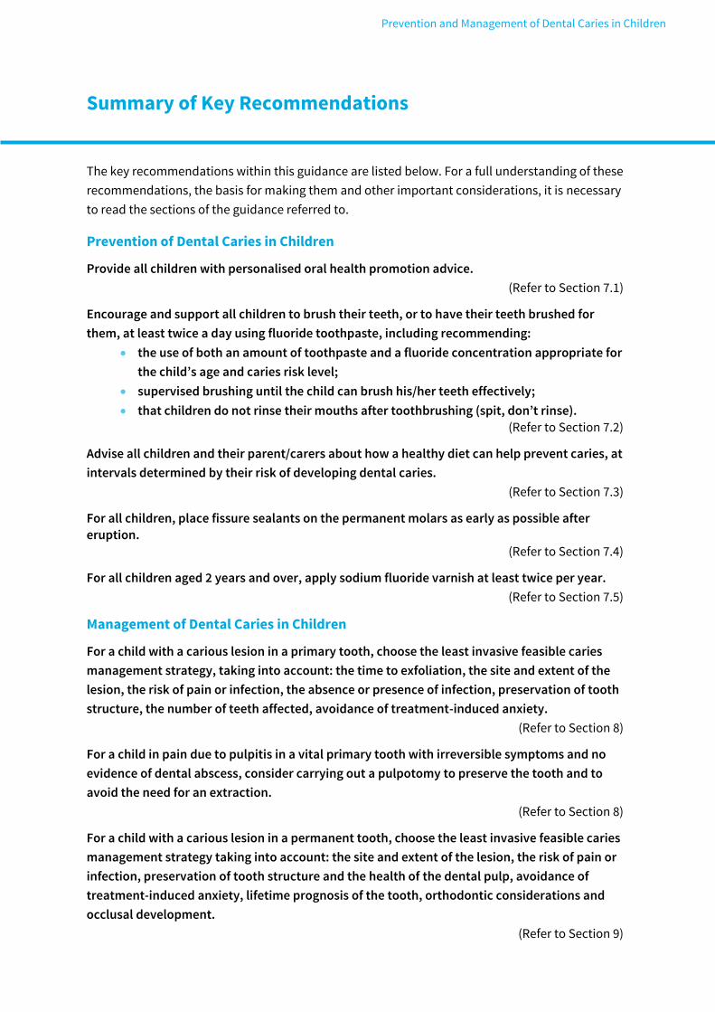
3.4.4 Dental caries and molar incisor hypomineralisation
Molar Incisor Hypomineralisation (MIH) is a common developmental
condition, defined as a “hypomineralisation of systemic origin of 1-4
permanent first molars, frequently associated with affected incisors”.26
Second primary molars can be similarly affected.27 Molars with
hypomineralisation are prone to breakdown. The poor quality of enamel
means that they are often sensitive to temperature and sometimes even
painful on toothbrushing. These factors combined with increased caries
susceptibility can lead to rapidly progressing caries. MIH enamel has an
abnormal etching and bonding pattern that compromises restorative
outcomes.
There is a wide spectrum of presentation both in the number of affected
teeth and the effect on the teeth. Even within individuals, some teeth will
be more affected than others. Lesions range from small, demarcated
discoloured areas (white opacities) with no breakdown to large, dark
(yellow to dark brown) areas that can fracture off, due to the weakness of
hypomineralised enamel, exposing underlying dentine. Patient reported
symptoms are variable, and may not necessarily match clinical
presentation.
Due to the possibility of rapid post-eruptive breakdown of the enamel (which can be of variable
quality), early diagnosis of MIH is key to avoid acute pain and delayed, complicated treatment. If
restorations have already been placed, they will often be atypical in shape. This can aid diagnosis
of MIH when the lesions are no longer visible.
Assess all hypomineralised molars independently to determine the extent of the disease
and likely prognoses. Factors to be taken into consideration when determining whether
teeth affected by hypomineralisation are of poor prognosis, include:
• enamel colour in order of severity and increasing likelihood of breakdown: white/cream,
then yellow, then brown
• location of the defects in order of severity: smooth surface, then occlusal surface/incisal
edge, then cuspal involvement
• sensitivity from brushing or to temperature
• atypically shaped restorations
• any patient reported symptoms

Prevention and Management of Dental Caries in Children
3 Assessing the Child and Family
22
3.4.5 Assessing for pain
Children are not always reliable in reporting pain, either because they have not yet developed the
necessary communication skills or because they wish to avoid dental treatment. For example, an
anxious child might not report an exfoliating tooth as painful, until reassured.
When obtaining a patient history, be aware that the child might not report pain reliably.
Include input from the parent/carer as well as the child and ask about any problems with
eating or drinking, changes to sleeping patterns and use of painkillers.
Advice on pain diagnosis and management is given in Section 6.
3.4.6 Assessing for dental abscess/infection in primary teeth
Dental abscess/infection can be difficult to diagnose because the clinical presentation can vary.
Sinuses, if present, are not always obvious, but they are usually located on the non-attached
mucosa adjacent to the attached mucosa. A slight cleft or notch may also be seen in the adjacent
gingival margin.
It is unacceptable to ignore dental infection in the mouth, even if asymptomatic.28
Look for the following indicators of established dental infection (see images below):
• tenderness to percussion
in a non-exfoliating tooth
• alveolar tenderness,
sinus or swelling
• non-physiological
mobility (compared with
the healthy contralateral
tooth) when the tooth is
gently rocked bucco-
lingually with the points
of a pair of tweezers
placed on the occlusal
surface
• radiographic signs
including inter-radicular
radiolucency
Advice on the management of infection is given in Section 6.
Sinus with associated inter-radicular radiolucency of lower D
Alveolar inflammation that, on
gentle palpation, releases
infected material from a lower D
Assessing for increased, non-
physiological mobility often
associated with infection

Prevention and Management of Dental Caries in Children
3 Assessing the Child and Family
23
3.4.7 Assessing the risk of pain or infection developing before exfoliation
When examining the primary dentition, assess the risk of each carious lesion progressing to pain
or infection to decide on the most appropriate management option. Not all carious lesions require
operative management. To make this decision consider several factors including:
• extent of the lesion
• site of the lesion
• activity of the lesion
• time to exfoliation
• number of other lesions present in the dentition
• the child’s medical status
• anticipated cooperation of the child, now and in the future
• anticipated cooperation of the parent/carer with the preventive interventions and to
attend repeat management appointments
• the range of clinical procedures the clinician has the skill to provide
With so many variables, it is not possible to clearly define specific criteria that will accurately
predict which carious lesions will result in pain or infection for the child. The clinician needs to use
their skill and judgement when carrying out this risk assessment.
Caries activity is variable, and lesions can arrest or have the potential to arrest. Carious lesions
that are slowing or arrested tend to be hard to probing and dark in colour. However, some
arrested lesions can be light in colour. Examples of teeth with different carious lesions assessed as
at high or low risk of developing pain or infection are shown in the two sets of photographs below.
These are intended as a guide only.

Prevention and Management of Dental Caries in Children
3 Assessing the Child and Family
24
Lesions in primary teeth with high risk of causing pain or infection
None of the following lesions have clinically evident signs or symptoms of pain or infection, but
are likely to be associated with pain or infection before exfoliation if left unmanaged.
Initial distal lesion, lower D in a 5-year-old child that is only evident
radiographically Cavitated lesion, lower
E in a 5-year-old child
Upper D with radiographic evidence of pulp exposure Clinical exposures of necrotic pulps in
primary molars and several years before
exfoliation

Prevention and Management of Dental Caries in Children
3 Assessing the Child and Family
25
Lesions in primary teeth at low risk of causing pain or infection
None of the following lesions has clinically evident signs or symptoms of pain or infection, and,
although the teeth do not appear ‘healthy’, it is likely that they will proceed to exfoliation without
causing further problems, provided they are closely monitored and the patient is given Enhanced
Prevention (see Section 7 for details).
Note that although a pulp polyp in a carious primary molar indicates that at least one of the root
canals is vital, the other canals may be necrotic. If there are signs or symptoms of infection, then
extraction or pulp therapy is required.
For each diagnosed carious lesion in a primary tooth, assess the risk of pain or infection
developing, prior to exfoliation of the tooth and then decide on a management option.
Upper Ds and Es with clinical exposures of vital pulps (pulp
polyps unlikely to cause infection before exfoliation)
Retained root, lower D
(dark coloured and hard)
Arrested caries lower CDE (dark coloured, hard and cleansable
cavity) Arrested caries, upper D (light
coloured and hard)

Prevention and Management of Dental Caries in Children
3 Assessing the Child and Family
26
3.4.8 Assessing toothbrushing
Gingival health is a useful indicator of tooth cleaning over time. Assessing and recording levels of
visible plaque at each examination, and sharing this information with the child and their
parent/carer, will help reinforce the importance of effective toothbrushing. An example of a quick
method of recording plaque levels, and presenting the information in terms the child will
understand, is to give marks out of 10 as follows.
The worst score in each
sextant is recorded, for
example:
8/10 6/10 8/10
8/10 6/10 8/10
perfectly clean
tooth
10/10
plaque line
around the
cervical margin
8/10
cervical third of
the crown
covered
6/10
middle third
covered
4/10
It is also important to assess the surface of open carious lesions for plaque that is visible or
evident when an instrument is gently drawn across the surface of the lesion, particularly if
considering managing the lesion with a prevention-alone approach (Section 10.1).
Assess whether the gingiva appear healthy or whether there is inflammation indicative of
poor plaque removal.
Consider recording plaque scores at each examination, particularly if the child is assessed
as at increased caries risk.
Record the presence of plaque on the surface of open carious lesions at recall visits for
lesions where the prevention-alone management strategy has previously been selected (see
Section 10.1).

Prevention and Management of Dental Caries in Children
3 Assessing the Child and Family
27
3.5 Caries Risk Assessment
All children are at risk of developing dental caries and, therefore, require preventive intervention.
However, many children are at increased risk of developing caries. Identifying these children
enables more intensive, enhanced prevention to be delivered to them. For each child, an
individualised personal care plan comprising preventive and, if necessary, restorative care can be
planned which is based on the child’s likelihood of developing caries.
Several factors are known to be associated with development of caries and, therefore, knowledge
of them can inform a prediction of the risk of a child developing caries in the future. These factors
include17:
• clinical evidence of previous disease
• dietary habits, especially frequency of
sugary food and drink consumption
• social history, especially
socioeconomic status
• use of fluoride
• plaque control
• saliva
• medical history
Although several tools for caries risk assessment exist, there is no consensus on which is most
effective. Amongst the risk factors listed above, previous caries experience (decayed, missing due
to caries or filled teeth) appears to be the more reliable predictors of caries risk. For delivery of
community prevention, area-based socioeconomic status is often used.17,29-33 Therefore, any child
who is resident in an area of relative disadvantage or who has decayed, missing or filled teeth is
considered to be at increased risk of developing caries. A child’s risk of developing caries can
change over time and therefore it is important to review the caries risk assessment regularly.
Regard any child who is resident in an area of relative disadvantage or who has any
decayed, missing (due to caries) or filled teeth as at increased risk of developing caries.
• The home postcode can be used to identify whether a child lives in a relatively
disadvantaged area. For example, in Scotland, Quintiles 1-3 of the Scottish Index of
Multiple Deprivation (SIMD) are considered to be relatively disadvantaged. A SIMD
postcode lookup is available on the NHS National Services Scotland website.34
Based on consideration of the other risk factors, your knowledge of the child and the history
taken, use your subjective clinical judgement to assess whether or not the child is at
increased risk of developing caries.
Use the caries risk assessment to inform the frequency of review radiographs (see Section
3.4.2), provision of preventive interventions (see Section 7) and frequency of recall (see
Section 12).
Reassess the child’s caries risk at each assessment.

Prevention and Management of Dental Caries in Children
4 Helping the Family Manage Dental Care
28
Children depend on their families (i.e. individuals who are close to a child and who may have a
role in his or her care. See also Section 1.5) to bring them to dental appointments and to ensure
that appropriate preventive strategies are followed. This “treatment alliance” is based on trust
between the child, accompanying adult and dental team.35 Awareness of factors that can affect
this relationship will help the dental team to establish rapport with the family.
4.1 Dental Anxiety
Dental anxiety is common and while the effects may be apparent at any age they are most obvious
in children aged under four years.36 When parents/carers are unable to hide their own anxiety it
can engender or increase anxiety in the child. A pre-appointment letter, welcoming the family to
the practice, outlining what will happen and advising them how to prepare their child can reduce
both parental and child anxiety.
Dental anxiety may begin in childhood or adolescence.37 Children with high caries are more likely
to have dental anxiety, and may require specialist input to manage them effectively. Identifying
these children and establishing preventive protocols is a priority and also allows the dental team
to modify their approach.
The Modified Child Dental Anxiety Scale (MCDAS) is a self-completion measure for children aged
eight years and older. It consists of eight questions about specific dental procedures. A five-point
Likert scale range 1 (relaxed/not worried) to 5 (very worried) can differentiate between children
with and without dental anxiety. A version using faces (MCDASf) is also available.38.39 Both versions
have been suggested as suitable for use in a dental surgery setting40 and use of an anxiety
questionnaire has been reported to alleviate anxiety to an extent.41 The aim should be to reduce
the anxiety score over time. The impact of a child’s anxiety about dental care needs to be
considered in the context of the care required. Even very anxious children can be helped to accept
preventive care.
Consider the child’s anxiety level when planning care and to determine whether use of
specific behaviour management strategies is indicated.
Consider using an anxiety questionnaire if there are concerns about dental anxiety.
Although some children respond well to coaxing, if a child becomes distressed, cease
treatment immediately. Consider a treatment compromise as a positive way of ending the
appointment (e.g. fluoride varnish application rather than no treatment) and either arrange
a later appointment to complete the planned treatment. Do not force dental treatment on a
child who is unwilling or unable to cope with it, whatever the imperative felt by the clinician,
and/or the parent/carer to complete treatment.

Prevention and Management of Dental Caries in Children
4 Helping the Family Manage Dental Care
29
4.2 Behaviour Management - Helping the Child to Adapt to a Dental
Environment
The aim of Behaviour Management (BM) is to promote a positive attitude to dental care and
facilitate ongoing prevention and care. A variety of techniques may be useful to the dental team
working with children and families to promote oral health.36 While they are described individually,
they are often used in combination, and all rely on good communication. For example, a clinician
may find that relaxation, coupled with behaviour shaping and giving control, empathy and praise,
will help the majority of children in a dental setting. The expectation is that the successful
practitioner will use appropriate behaviour management with all patients.
While BM techniques may be effective for some children and appropriate for some clinicians, the
evidence base for their effectiveness is limited. However, the following techniques may help
children adapt to a dental environment and enable a stress-free experience for the child and
dental team. Although they are often described in the context of providing restorative
management, they may be equally valuable when trying to examine a child for the first time or to
introduce a child to any new situation.
The techniques summarised below are described in more detail in the British Society of Paediatric
Dentistry Non-pharmacological behaviour management guideline.36 All these techniques can be
used with children who can communicate but the child’s ability to understand the language used
must also be considered.
Consider the use of one or a combination of the following behaviour management
strategies to facilitate provision of both preventive care and treatment:
• Communication; Enhancing control; Tell, show, do; Behaviour shaping and positive
reinforcement; Structured time; Distraction; Relaxation; Systematic desensitisation.
4.2.1 Communication
Communication with children can be more complex than the one-to-one communication that
exists with most adult patients. The child, clinician, parent/carer and dental nurse may all be
involved. Each member of the team, including the parent/carer, needs to understand their role to
create an effective treatment alliance and communicate in a consistent manner to support the
child. The dental team needs to ensure that parents/carers know how to support their child
without disrupting the appointment. This might entail discussing how to prepare the child for a
visit and negotiating ground rules on how to behave and communicate in the surgery. For very
anxious parents/carers who cannot mask their own fears it may be beneficial to discuss whether

Prevention and Management of Dental Caries in Children
4 Helping the Family Manage Dental Care
30
another adult familiar to the child should attend visits. The ability to communicate as a dental
team and to include the family in this process is critical to deliver effective care (see Section 3.1).
We communicate continuously and need to be aware of the messages we are sending consciously
and unconsciously to children and families. Communication consists of:
• Non-verbal communication – which conveys emotion and attitude
• Words – which convey information
• Tone – which conveys emotion and attitude
Non-verbal communication occurs continuously and may reinforce or contradict verbal signals. It
includes facial expressions, eye contact, gestures, body movements, posture and touch. A happy
smiling dental team from reception to the clinical staff needs to be sending the same positive
message. A child-friendly environment is also important as some posters aimed at adults might
scare a child.
If the three components of communication are not working in harmony the messages we send can
be confusing. Young children may not understand the words that we use, but will recognise tone.
They will also pick up on the body language that the dental team and any accompanying adult
exhibits. A relaxed posture, smile and gentle tone convey empathy even if the words cannot be
understood. Used well, communication is a powerful way to support a child.
4.2.2 Enhancing control
People often complain about the feeling of loss of control in dental appointments. Making the
child’s role in saying ‘stop’ and ‘go’ explicit is a very simple way of enhancing feelings of control.
This technique should be used every time for all children. It gives the child a degree of control over
the clinician’s behaviour by giving them a way to interrupt an examination or active treatment.
Saying to a child “Let me know if you want me to stop” is not adequate to give them control.
Giving control means making sure the child understands that they decide whether the clinician
continues or not by rehearsing a signal to stop, such as raising their hand.
If the child gives the signal, then stop the examination or treatment immediately. If you do not
stop treatment, the child may develop mistrust phobia which can be very difficult to resolve.
The technique is useful for children of school age and older; younger children may not understand
the concept of them having control.
4.2.3 Tell, show, do
This technique is widely used to introduce a child to a new situation. The “Tell” phase should use
age appropriate language that avoids technical and emotive terms.
Explain what you are going to do, and then show them, before actually performing the action.

Prevention and Management of Dental Caries in Children
4 Helping the Family Manage Dental Care
31
Using tell, show, do to acclimatise an apprehensive 5-year-old child to accept treatment that
involves use of a high speed handpiece for the first time will involve the following.
• Gain rapport (see Section 3.1).
• Give control (see Section 4.2.2).
• Tell the child what you would like to do, and show them the handpiece.
• Show them the high-volume aspirator and ask them if it would be all right to try it.
• If they agree, ask the nurse to put the aspirator tip into the child’s mouth, switch it on,
then off and then remove it.
• Tell the child they have done well and check that it was OK.
• Tell the child you are going to put the handpiece in their mouth using language
appropriate to the child’s age and understanding, switch it on, count to 4 (Structured
time, Section 4.2.5) then remove it. You will not touch their tooth and they can practise
staying big and wide while you wash their tooth.
• Ask them to open their mouth, then proceed as above. If the child manages this, praise
them, and tell them you would now like to touch the tooth for the count of 4.
• Continue using this technique for each new action.
Although this approach can appear laboured, after a child has accepted the procedure, it is
usually not necessary to work through all the stages at subsequent visits. The approach can be
adapted for a child with anxiety using an enhanced approach termed Systematic Desensitisation
(Section 4.2.8).
4.2.4 Behaviour shaping and positive reinforcement
Dental visits can be confusing to children who need assistance to learn how to behave in the
surgery. Behaviour shaping consists of a defined series of steps towards ideal behaviour. This is
most easily achieved by positive reinforcement of desired behaviour, increasing the probability of
that behaviour being repeated; while ignoring undesirable behaviours to avoid drawing attention
to them.
For example, if a child keeps closing his/her mouth during placement of a fissure sealant the
clinician should highlight the desired behaviour and praise the child when it occurs, thus
reinforcing it. “Can you open your mouth big and wide like a lion for me? That’s great, that makes
my job really easy!” This approach requires practice, as it is very easy to become frustrated, stop
smiling and say something like “Can you try and stop closing your mouth?” Here the clinician
reinforces the undesired behaviour and, instead of praising the child, might sound cross.
The most powerful reinforcers are social stimuli, such as facial expression, positive voice
modulation, verbal praise, and approval by parent/carer in the form of a hug. Anything that the
child finds pleasant or gratifying or is a visible sign of ‘good’ behaviour can act as a positive
reinforcer, for example, stickers or badges at the end of a successful appointment. However, it
must be clear to the child what action is being rewarded. A child-centred, empathic response

Prevention and Management of Dental Caries in Children
4 Helping the Family Manage Dental Care
32
giving specific praise, for example, “I would like you to choose a sticker for sitting nice and still
while I counted your teeth” has been shown to be more effective than a general comment such as
“Choose a sticker for being a good girl.” As with Tell, Show, Do, the use of age-specific language is
important.
4.2.5 Structured time
Children can tolerate what they may find a potentially upsetting procedure (such as counting their
teeth for the first time or use of a slow handpiece) if they know it will only continue for a finite
period of time. “Just a little bit more” for a child could mean anything from a second or two, to
eternity. Instead: break down time into units the child can understand.
• For example; “I’ll buzz your tooth while I count to three, then stop; is that OK? Good,
1…..2…..3; Well done! And again, 1….2….3 etc”.
Note that young children (less than 5 years old) may not understand the concept of numbers
above 4, despite being able to recite them.
4.2.6 Distraction
This technique aims to shift the patient’s attention from the dental setting to some other situation
or from a potentially unpleasant procedure to some other action. Cartoons have been shown to
reduce disruptive behaviours in children when combined with reinforcement; the children knew
the cartoon would be switched off if they did not behave. Audio distraction, although proven
effective for adults, has been shown to have variable success in children. Short term distracters
such as diverting attention by pulling the lip as a local anaesthetic is given or having patients raise
their legs to stop them gagging during radiography or while taking impressions may also be
useful. Verbal distraction, for example the clinician who talks while undertaking an examination,
polishing teeth or applying topical paste and administering local anaesthetic, can also be
effective.
4.2.7 Relaxation
Telling a child to relax will not help them to relax but showing them how to breathe deeply using
their diaphragm can.
• Ask the child to place a hand on their
tummy.
• Ask them to breath in slowly and deeply,
making the air “fill their tummy”.
• Watch to see if their tummy rises; if so,
praise them, and ask them to release their
breath slowly, telling them that as they
breathe slowly out, so they will become

Prevention and Management of Dental Caries in Children
4 Helping the Family Manage Dental Care
33
more relaxed.
• Ask them to do this three times, any time they feel tense and worried.
This technique is useful for children of school age and older.
4.2.8 Systematic Desensitisation
Systematic Desensitisation can be a very powerful technique for helping a child with anxieties
regarding local anaesthesia, radiography or any other aspect of dental care that they struggle to
cope with. It involves teaching the child how to relax because it is not physiologically possible to
be both anxious and relaxed at the same time. In its simplest form, the procedure is broken down
into stages, and the child is taught to relax at each stage before moving on to the next with
positive reinforcement.
A common application in dentistry is systematic needle desensitisation which is used to manage
needle phobia. Only an outline is given here. For further details refer to the British Society of
Paediatric Dentistry Non-pharmacological behaviour management guideline.36
Discuss with the child how to recognise the signs of stress and anxiety that they may be
experiencing (e.g. hyperventilation, tension).
Teach the child how to manage their anxiety, principally with breathing (see Section 4.2.7)
but also using progressive muscle relaxation and other techniques such as guided imagery.
Teach the child how to describe their level of anxiety, using a scale from 1 to 10 (where 1 is
completely relaxed, and 10 is the most anxious they have ever been).
Break the procedure down into stages, and describe all the stages to the child.
Give control, then try the first stage, asking the child at the end of it to describe their anxiety
level. If rated above 5, ask them to spend a minute going through their relaxation regime,
and try again, only proceeding to the next stage when the child has reduced their anxiety to
a manageable level and the child has given their permission to proceed.
• Giving local anaesthesia could, for example, be broken down into holding the syringe by
the side of the chair, placing it in the child’s mouth but with the cap on, then holding the
syringe in the child’s mouth with the cap off, and so on.
If a child continues to demonstrate significant anxiety despite the dental team using the
above techniques, consider referral to a specialist.
• If referring to a specialist, include in your referral letter details of treatments that have
been attempted and how successful each was found to be (see Section 11 for a referral
checklist).

Prevention and Management of Dental Caries in Children
5 Defining Needs and Developing a Personal Care Plan
34
Managing Carious lesions (and asymptomatic infection)
Sections 8,9 & 10
Caries Prevention Section 7
Managing Pain, if present Section 6
The information gathered from the assessment of the child will enable a diagnosis to be
formulated. From this, the needs to be addressed and the proposed interventions to manage
them can be listed and prioritised into a personal care plan which is agreed with the child and
parent/carer.
For any care plan to be successful, it must be tailored to a realistic assessment of the child’s ability
to accept treatment and aim to increase the likelihood of behaviours that maintain or contribute
to good oral health. In addition, the parent/carer’s support is essential in bringing their child for
care and in supervising preventive interventions. Therefore, it is crucial to fully discuss, explain
and support understanding of the treatment options with the child and parent/carer before
agreeing a proposed care plan and obtaining their consent to it. Some children have needs which
cannot be met in their usual place of care and may require referral. In such cases it is the
responsibility of the clinician to follow their local referral policy and to work in partnership to
provide shared care (see Section 11).
The accepted sequence for planning and providing care is as follows.
Although some children may require pain management or caries management, it is imperative
that all children receive caries prevention and appropriate behaviour management.
There is wide variation among clinicians when planning care for patients with similar diagnoses
and it is unlikely that there will be a single optimal plan for a particular child patient. However, the
normal sequence, following management of any pain, is to initiate an effective preventive
programme first. Then, if required, a caries management plan can be implemented to manage
caries in permanent teeth and to reduce the risk of any caries in the primary dentition causing the
child pain or infection.

Prevention and Management of Dental Caries in Children
5 Defining Needs and Developing a Personal Care Plan
35
For children with caries, many factors can influence the choice of management strategy. The
optimum strategy will vary and no single approach will suit every clinician, every child or every
carious tooth. Furthermore, not all carious lesions in primary teeth require operative management
(see Section 3.4.7) and, because of their limited lifespan, slowing caries progression may be
sufficient.
When planning care, the first priority is to keep the permanent molars free from caries, as these
teeth are more likely to experience decay than other permanent teeth in a child’s dentition.42 If
caries is diagnosed at these sites, then it must be managed appropriately (see Section 9). The next
priority is to reduce the risk of any caries in the primary dentition resulting in pain or infection
before the tooth exfoliates. This can often be achieved without using the standard adult
restorative approach, instead selecting an appropriate alternative caries management strategy
that reduces the risk of causing treatment-induced anxiety (see Section 8).
If the child has pain, ensure this is managed first.
• Try to avoid dental extractions at the child’s first visit if at all possible.
Discuss and explain caries prevention, with the child and parent/carer (see Section 7).
Discuss and explain management options for carious lesions with the child and
parent/carer, which might differ if there are multiple rather than a single carious lesion (see
Sections 8, 9 and 10).
Plan to carry out any preventive interventions for permanent teeth before treatment of the
primary teeth (e.g. fissure seal first permanent molars before restoring primary teeth).
Devise and agree an initial care plan with the child and parent/carer, which includes the
expected number, content and duration of appointments, but be prepared to modify this if
the child is unable to accept some treatments or there are changes in caries activity.
Having carefully explained the child’s oral health needs and any proposed treatment
options, check that the child and/or parent/carer clearly understand the care that they are
agreeing to.
Obtain valid consent for the agreed care plan from the child where possible and/or the
patient/carer.
• It is a legal requirement that when obtaining consent clinicians take reasonable steps to
ensure that patients are aware of any material risks involved in the proposed treatment,
and reasonable alternative.43
• The SDCEP Practice Support Manual23 provides further details about consent.
Prevention and Management of Dental Caries in Children
5 Defining Needs and Developing a Personal Care Plan
36
If a child is pre-cooperative or unable to cooperate (due to young age, a learning disability,
or where behaviour management techniques have been unsuccessful) or has multiple
affected teeth, consider referral to assess suitability for treatment under sedation or general
anaesthesia (see Section 11).
Consider dividing treatment into several stages, with a month or two between stages, if
there are concerns about child or parent/carer compliance.
If required, include in your care plan collaboration with the other professionals (e.g. the
child’s health visitor, school nurse, Childsmile dental health support worker, general
medical practitioner social worker) to offer and provide additional home and community
support for preventive interventions and to encourage attendance for treatment (see
Section 13).
Ensure complete and accurate records are kept, including advice given and the rationale for
treatment options agreed and any referrals made.
• SDCEP Practice Support Manual23 and Oral Health Assessment and Review18 guidance for
provide further information.
The use of behavioural management techniques and good clinical judgement to select treatment
options will enable most children to complete a planned course of care. However, some children
will have difficulty accepting dental treatment in general dental practice even with behavioural
management techniques to support them. They may require sedation (for young children this is
usually inhalation sedation; for older children inhalation sedation or intravenous sedation) or
general anaesthesia. Section 11 provides further guidance on referral for care, including advice
and a flowchart to aid decisions around referral for sedation or general anaesthesia.
If during treatment a child is unwilling or unable to cope, stop the procedure. Consider
alternative behaviour management and/or treatment options and agree these with the
child and the parent/carer.
• If a child resists treatment it is not appropriate to continue even if the parent/carer
wishes you to or disagrees.

Prevention and Management of Dental Caries in Children
6 Diagnosing and Managing Dental Pain or Infection
37
Dental pain is usually the principal reason for a child’s unscheduled attendance for dental care.
The treatment provided will depend on the diagnosis and child’s ability to cope. Although it is vital
that a child’s pain is managed promptly, if a dental extraction is indicated, it is generally in the
child’s best interests in the long term to avoid an extraction at the first visit. This is to minimise
future anxiety about dental treatment and increase the likelihood that the child will return to
complete a planned course of treatment. A personal care plan for the child should be agreed and
followed (see Section 5).
6.1 Causes and Management of Dental Pain or Infection
The most common reason for children to experience dental pain is pulpal pathology as a
consequence of dental caries. The immune system allows vital dental pulps to cope with some
bacterial ingress from a carious lesion through the triggering of an inflammatory response in the
region of the pulp closest to the lesion. However, if a progressing carious lesion is not managed,
then over time the increasing bacterial infection causes a more extensive and severe inflammatory
response in the dental pulp. Eventually, the bacterial infection and the associated increase in
tissue pressure overwhelms the ability of the circulatory system to keep the pulp perfused, which
will then transition from being vital (perfused), to non-vital and necrotic (no perfusion). Bacteria
can then thrive within the necrotic pulpal remnants, and the diffusion of these bacteria and their
products through the radicular foramina causes inflammation of the periodontal ligament (hence
these teeth becoming tender to percussion and mobile), and a dental abscess.
Clinically, it is useful to divide this process into three stages, pulpitis with reversible symptoms,
pulpitis with irreversible symptoms and dental abscess/periradicular periodontitis. However,
these do not refer to discrete, well separated stages of pathology, but are on a continuum of
unmanaged pulpal disease from mild inflammation through to pulpal necrosis. Clinicians must
use their clinical judgement to decide the extent of the pulpal pathology and the appropriate
management strategy.
• Pulpitis, reversible symptoms – pain is provoked by a stimulus (e.g. cold, sweet) and
relieved when it is removed. The pain is intermittent, difficult for the child to localise and
does not tend to affect the child’s sleep. The pulp is still vital and the tooth is not tender
to percussion. Management of the carious lesion alone may be enough to resolve the
inflammation, and allow pulpal healing.
• Pulpitis, irreversible symptoms – pain can occur spontaneously but if provoked by a
stimulus is typically not relieved when the stimulus is removed. The pain may last for
several hours, may be difficult for the child to localise and may keep the child awake at
night. The pain may be dull and throbbing, worsened by heat and may be alleviated by
cold. There are no signs and symptoms of infection such as sinuses or abscesses or
periradicular pathology and the pulp is still vital, although inflamed. Usually the tooth is
not tender to percussion. Management of the carious lesion alone is unlikely to resolve
the inflammation, and pulp therapy (for primary teeth a pulpotomy, where

Prevention and Management of Dental Caries in Children
6 Diagnosing and Managing Dental Pain or Infection
38
instrumentation is confined to the pulp chamber only; in permanent teeth a root canal
therapy) or extraction is necessary.
• Dental abscess/periradicular periodontitis – when acute, pain is likely to be
spontaneous, will keep the child awake at night and can be easily localised by the child.
The tooth may show increased mobility and may be tender to percussion. There may be
clinical evidence of a sinus, abscess or swelling or radiographically evidence of
periradicular pathology. When chronic, the child may not report pain but other signs and
symptoms may be present. The infected, necrotic remnants of the dental pulp will
continue to cause problems unless managed by pulp therapy (in this case for both
primary and permanent teeth a pulpectomy/root canal therapy, where instrumentation
also includes the root canal system) or extraction.
Signs and symptoms to assist the clinician in diagnosing these stages are given in Figure 6.1.
However, the following points should be noted.
• It is possible for pulpal disease to progress from mild inflammation to complete pulpal
necrosis without any obvious signs, and without the child reporting symptoms. This is
probably a factor of host resistance, which can increase or decrease over time, resulting
in previously aymptomatic teeth with pulpal pathology becoming symptomatic.
• For multi-rooted teeth it is not uncommon to encounter different pathology in individual
roots. The root canal with the most advanced pathology will dictate the management
strategy.
6.2 Determining a Management Strategy
Diagnose the cause of pain and determine a suitable management strategy. The flow
diagram in Figure 6.1 illustrates this process for a child with no medical complications.
• Each patient should also receive an oral health assessment which may be carried out
before or after diagnosis and management of pain, depending on its severity.
For primary molars, it may be possible to retain those with pulpitis with irreversible
symptoms using pulpotomy rather than extraction. Therefore, consider this treatment
option if the child is cooperative.
For primary molars, where a radiograph shows no clear separation between the carious
lesion and the dental pulp, consider carrying out a pulpotomy (see Section 10.8).
• It is possible that the carious lesion has encroached significantly on the dental pulp
causing irreversible pulpal disease.

Prevention and Management of Dental Caries in Children
6 Diagnosing and Managing Dental Pain or Infection
39
For both primary and permanent teeth, where there are symptoms of pain that may be due
to food packing or pulpitis with reversible symptoms but the diagnosis is uncertain, place a
temporary dressing and review the patient 3-7 days later to check symptoms.
• Resolution of the symptoms at review will indicate that the pulpitis was reversible and a
suitable restoration or crown can be placed.
• If symptoms do not resolve, worsen or an abscess develops then for primary teeth
consider extraction or appropriate pulp therapy (see Sections 8.4.2 and 8.4.3) and for
permanent teeth a root canal treatment or an extraction.
Do not leave dental infection untreated.
• In some cases, local measures to bring the infection under control may be appropriate
(see Section 10.9).
• Antibiotics are not recommended unless there are signs of spreading infection or
systemic symptoms.
• In primary teeth, extraction may be the best option, but in certain circumstances
consider referral for pulpectomy (Section 8.4.3).
If dental infection is asymptomatic and the child is presently unable to cope with an
extraction but judged to be likely to accept treatment with acclimatisation, then, allow up
to three months for acclimatisation visits using active behaviour management techniques.
• This approach is not suitable for medically compromised children.
If a child is pre-cooperative or unable to cooperate (due to young age, a learning disability
or where behaviour management techniques have been unsuccessful), has multiple
affected teeth or the dental infection becomes symptomatic, consider referral to assess
suitability for extractions under sedation or general anaesthesia (GA) (see Section 11).

Prevention and Management of Dental Caries in Children
6 Diagnosing and Managing Dental Pain or Infection
40
Figure 6.1 Diagnosis and management of caries-related dental pain or infection in a
child with no medical complications
† Antibiotics should only be prescribed if there is evidence of spreading infection (swelling,
cellulitis, lymph node involvement) or systemic involvement (fever, malaise).
* Refer to the SDCEP Drug Prescribing for Dentistry guidance.44
Abbreviation: GA General anaesthesia.
Note that each patient should receive an oral health assessment which may be carried out before
or after diagnosis and management of pain, depending on its severity.
Pulpitis, irreversible symptoms?
- Spontaneous pain wakens child at night
- Does not resolve on removal of stimulus
- Pain to hot/cold
- Does not resolve with placement of a
temporary dressing
Pre-cooperative child?
Pre-cooperative child or
multiple abscessed teeth?
Restore, or place
a dressing and
restore later
Try to dress with sub-
lining of corticosteroid-
antibiotic paste.
Prescribe pain relief*
Primary: Refer for treatment/
extraction with sedation or GA
(see Section 11).
Permanent: Carry out root
canal therapy or extraction. If
the child remains
uncooperative, refer for
specialist care.
No
Yes
No
Yes
Yes
No
No
Yes
Primary: Refer for extraction
with sedation or GA (see
Section 11).
Permanent: Carry out root
canal therapy or extraction.
May require referral for
specialist care.
Primary: Carry out
extraction or appropriate
pulp therapy (see Sections
8.4.2 and 8.4.3).
Permanent: Carry out
root canal therapy or
extraction.
Dental abscess/periradicular periodontitis
- Spontaneous pain wakens child at night
- Tooth mobile and tender to percussion
- Swelling
- Malaise
Consider local measures to
control infection.
If indicated, first prescribe
antibiotics† and pain relief*
Pulpitis, reversible symptoms?
- Pain to cold/sweet
- Tooth not tender to percussion
- Resolves on removal of stimulus
- Tooth difficult to localise

Prevention and Management of Dental Caries in Children
7 Caries Prevention
41
Dental caries is not inevitable and can be prevented. There are several effective evidence-based
caries preventive interventions, including giving advice and instruction about toothbrushing with
fluoride toothpaste, giving dietary advice, placement of fissure sealants, and use of topical
fluorides. Similarly, there are evidence-based strategies that can be employed by health care
professionals in a primary care setting and that can help parents/carers, who have responsibility
for the oral health of their child, to follow preventive advice.
Encouraging parent/carers to begin attending the dental practice with infants will assist the
child’s acclimatisation to the practice environment and enable preventive advice to be given to
the parent/carer at an early stage.
As every child is at risk of developing caries, preventive interventions are required for all children.
In Sections 7.2–7.5 of this guidance, Standard Prevention actions for all children are presented in
amber boxes. For children assessed to be at increased risk of developing caries, additional
preventive interventions are recommended and are presented in red boxes as Enhanced
Prevention.
Encourage parents/carers to register their child with a dentist as early as possible, before or
as soon as the first tooth appears, and to attend regularly.
Ensure that all children receive Standard Prevention appropriate to their age.
If the child is at increased risk of developing caries, in addition to Standard Prevention,
ensure they receive Enhanced Prevention, unless there is valid reason not to. In this case,
ensure this is documented in the patient’s notes.
When giving preventive advice, ensure that both the child and the parent/carer who is
responsible for supervision are included in the discussion.
After relief of any pain, carry out preventive interventions for permanent teeth before
treatment of the primary teeth (e.g. fissure seal first permanent molars before managing
carious primary teeth).
In addition to the crucial preventive interventions provided by the dental team, in Scotland the
Childsmile programme also promotes multidisciplinary prevention and oral health care for
children in other settings. Young children who attend nursery or school within the most deprived
areas are targeted to receive specific additional preventive interventions through Childsmile.14
Dental teams can also refer families who require additional support to local Childsmile dental
health support workers.
The preventive interventions described in this section can also be employed as part of a strategy
for the management of certain carious lesions, as discussed in Sections 8, 9 and 10.
Prevention and Management of Dental Caries in Children
7 Caries Prevention
42
7.1 Motivating, Action Planning and Habit Forming
KEY RECOMMENDATION
Provide all children with personalised oral health promotion advice.
(Strong recommendation; moderate quality evidence)
EVIDENCE
Based on a review of the literature, SIGN guideline 13817 recommended that oral health
promotion strategies should:
• facilitate daily toothbrushing with fluoride toothpaste;
• be based on recognised oral health behaviour theory and models such as
motivational interviewing;
• be specific to individuals, and tailored to their particular needs and
circumstances.
The social history gathered during the assessment gives an understanding of the child’s current
oral health practice, the parent/carer’s ability and attitude towards maintaining oral health and
their motivation to take responsibility for it (Section 3.2).
As knowledge does not readily translate into behaviour change, providing caries prevention
advice alone has limited success in influencing patients. However, there is evidence that using a
brief motivational interviewing (MI) approach and forming an action plan increases the likelihood
of parents/carers or older children adopting desired oral health behaviours.45 This approach is
summarised in Figure 7.1.
To use brief motivational interviewing successfully as part of an approach to supporting patients
to adopt healthy behaviours ideally requires understanding of the theory gained through training
and experience.

Prevention and Management of Dental Caries in Children
7 Caries Prevention
43
Figure 7.1 Route map of health behaviour change using motivational interviewing
Developing an individualised action plan to encourage the child’s habit formation
Identify a convenient time and place for the preventive behaviour to occur (e.g.
toothbrushing after breakfast and last thing at night), a date for when the task is to be
started (ideally from the day of the appointment) and who is to carry it out.
• If difficulties are reported, alternatives may be necessary. For example, if the child is
often too tired for toothbrushing last thing at night, agree an earlier time.
Identify a trigger as a reminder for the child or parent/carer to carry out the preventive
behaviour (e.g. when the child gets ready for bed).
Agree a date to review progress (e.g. assess oral hygiene at the next visit).
Agree the action plan with the child and parent/carer and write this down for them if
necessary, possibly on a copy of a food and drink diary or toothbrushing chart.
Step 5:
Repeat at
each recall
visit
Step 1: Explore current practice and
attitudes using a Motivational
Interviewing approach
Gain empathy
Seek permission
Open questions
Affirmations
Reflective listening
Summarising
Develop discrepancy, roll with
resistance
Elicit change talk
Step 2: Educational intervention
Improve Knowledge and Skills.
Step 3: Action planning
Set time, date and place to start
Step 4: Encouraging habit formation
Achieve sufficient repetition.
The situation
as it is now
The situation
as we would
like it to be
Making it
happen
Keeping it
that way

Prevention and Management of Dental Caries in Children
7 Caries Prevention
44
Record the action plan in the child's notes so that it can be referenced at subsequent visits.
At subsequent visits, encourage, give further support and review the action plan and revise
it, if necessary.
While discussing the action plan, assess the parent/carer or child’s ability and motivation to
comply and if there is doubt about this, discuss collaboration with other healthcare
professionals (e.g. health visitor, school nurse, Childsmile dental health support worker) as
a source of community/home support for the child and include this in the action plan.
Examples of action plans
• For a 6-year-old child, the parent/carer will supervise the child’s brushing at home every
morning after breakfast and brush the child’s teeth for them every evening after dinner,
when the child gets ready for bed.
• When preparing the child’s packed lunch, the parent/carer will give the child water
instead of a sugar-containing drink every day.
• A child will note on a toothbrushing chart every time they brush their teeth at home and
bring the chart back at the next visit to receive a sticker.

Prevention and Management of Dental Caries in Children
7 Caries Prevention
45
7.2 Toothbrushing with Fluoride Toothpaste
KEY RECOMMENDATION
Encourage and support all children to brush their teeth, or to have their teeth brushed
for them, at least twice a day using fluoride toothpaste, including recommending:
• the use of both an amount of toothpaste and a fluoride concentration
appropriate for the child’s age and caries risk level;
• supervised brushing until the child can brush his/her teeth effectively;
• that children do not rinse their mouths after toothbrushing (‘spit, don’t rinse’).
(Strong recommendation; high quality evidence)
EVIDENCE
Toothbrushing with fluoride toothpaste is one of the most effective methods for
preventing caries. SIGN guideline 138 extensively reviewed the evidence related to
toothbrushing including use of fluoride toothpaste, fluoride concentration, toothpaste
composition, frequency and duration of brushing, age of commencement of brushing and
toothbrushing practice.17 Figure 7.2 summarises advice on fluoride toothpaste use based on age
and caries risk. Further discussion of the evidence on which this section of the guidance is based
is provided in Section 15.1.
Figure 7.2 Fluoride toothpaste use based on age and caries risk.
Caries Risk
Assessment
Standard prevention
use 1000-1500 ppm F
Enhanced prevention
use 1350-1500 ppm F
or for Age 10+
consider 2800 ppm F
Age 3 years and over
use a pea-
sized
amount
Age under 3 years
use a
smear
Standard risk
Increased risk
Based on Recommendations on the use of fluoride toothpaste and fluoride supplements in Scotland 201746

Prevention and Management of Dental Caries in Children
7 Caries Prevention
46
Standard Prevention for all children
At least once a year, advise or remind the child and/or parent/carer to:
Brush thoroughly twice daily, including last thing at night
• Brushing is best done in the morning and last thing at night before bed with nothing to
eat or drink after brushing at night, apart from water. Brushing last thing at night is likely
to be particularly effective due to retention of fluoride in the mouth.
• In Scotland, toothbrushing at school or nursery as part of Childsmile is in addition to
these two occasions.
Use the age-appropriate amount of a toothpaste containing 1000 to 1500 ppm fluoride
• See Figure 7.2 for details.
‘Spit, don’t rinse’
• Children who spit out and do not rinse after brushing experience significantly less caries
compared with those who rinse their mouth out with water after brushing.47,48
Supervise children until they can brush their teeth effectively (see Section 7.2.1)
• As younger children do not have the manual dexterity to brush their own teeth, the
parent/carer must take responsibility.
Demonstrate brushing on the child (~3 minutes) annually.
• By modelling the desired behaviour, facilitating practice of the desired behaviour and
giving reassurance that the behaviour is being done correctly, you can increase the child
or parent/carer’s confidence and so the likelihood that the toothbrushing behaviour will
be done at home.
Use action planning to encourage toothbrushing.
• For example, ask what routine habits exist each day (e.g. getting changed in the morning
and night, bedtime story) that could be used as a reminder to brush teeth immediately
afterwards. Being very specific about what, when, and where something is done (action
planning increases the likelihood of it happening. The best way to establish a new habit
(i.e. toothbrushing) is to add it to something that is already a habit. For example, add
toothbrushing to the evening bath time regime or just before the usual bed/story time.
Advise the parent/carer to start brushing as soon as the first primary tooth erupts.
Advise the parent/carer not to allow the child to eat or lick toothpaste.
Emphasise to teenagers the importance of regular brushing for oral health and the
additional positive effect on appearance and general health.
• Providing information about others’ approval, providing information about the
behaviour-health link/of positive consequences can prompt self-motivating statements
and evaluations and reduce resistance to change.

Prevention and Management of Dental Caries in Children
7 Caries Prevention
47
Enhanced Prevention for children at increased risk of caries
At each recall visit provide Standard Prevention toothbrushing advice as detailed above.
Give hands-on brushing instruction (~3 minutes) to the child and parent/carer at each
recall visit (see Section 7.2.1).
Consider providing additional preventive interventions depending on the child’s
circumstances, for example:
• Recommend the use of 1350-1500 ppm fluoride toothpaste for children up to 10 years of
age.
• Prescribe 2800 ppm fluoride toothpaste for children aged 10 - 16 years for a limited
period (see SDCEP Drug Prescribing for Dentistry guidance44 for details). Regular review is
required46
Utilise any community/home support for toothbrushing that is available locally (e.g. health
visitor, school nurse, Childsmile dental health support worker).
7.2.1 Toothbrushing instruction technique
This technique incorporates both evidence based actions for toothbrushing and behaviour change
strategies (providing information, modelling, rehearsal, reinforcement, increasing control to build
confidence and motivate, prompts, cues, incentives) to encourage regular effective toothbrushing.
Empathise with the parent/carer about how difficult it can be with small children to brush
properly, but give advice (as described above) and reinforce the importance of following it.
Ask the child or parent/carer whether they would prefer to: (1) brush all surfaces of a section
of the mouth, before moving on to the next section; or (2) the same surface of each
complete arch, before moving
onto the next surface (all
cheek, all palate, all biting
surfaces).
If the parent/carer is brushing
the child’s teeth, ask them if
they would prefer to stand
behind their child, sit or kneel
in front of the child.
Demonstrate the chosen technique on the child, and then encourage the child or
parent/carer, or both, as appropriate for the age of the child, to show you.

Prevention and Management of Dental Caries in Children
7 Caries Prevention
48
• For example, the dental team member brushes one arch and then encourages the
parent/carer to brush the other arch.
Instruct the use of a short, scrubbing motion. Teaching the child to recognise the ‘shh-shh’
sound of the correct scrubbing motion may be helpful.
Advise that it is likely to take at least two minutes brushing to clean all tooth surfaces and
gums. Using a timer (e.g. watch or app) might help motivate the child.
Advise that after eating or drinking something acidic (e.g. fruit juice) it is preferable to wait
at least 30 minutes before brushing.
When first or second molars are only partially erupted,
show the child and parent/carer how brushing in line
with the arch can miss the vulnerable occlusal surface of
these teeth, and emphasise the need to brush the
occlusal surface from the side of the mouth.
Assess the likely benefits for the child of:
• disclosing plaque at the brushing instruction visits and either recommending use of
disclosing tablets or giving the parent/carer some to use at home.
• providing toothbrushing charts on which the child or parent/carer can record each time
the child’s teeth are brushed. This will act as a reminder about brushing frequency.
Consider providing a free toothbrush and free toothpaste to children at increased risk.
• In Scotland, Childsmile can supply practices with loose toothbrushes for demonstration
and brush and toothpaste packs to give to children at increased caries risk.
Various resources to support parents, including a video about toothbrushing are available on the
Childsmile website.49

Prevention and Management of Dental Caries in Children
7 Caries Prevention
49
7.3 Dietary Advice
KEY RECOMMENDATION
Advise all children and their parent/carers about how a healthy diet can help prevent
caries, at intervals determined by their risk of developing dental caries.
(Strong recommendation; moderate quality evidence)
EVIDENCE
There is moderate quality evidence that lower sugar consumption can prevent caries50
and low quality evidence that dietary advice in a dental setting encourages a change in
dietary behaviour.51 Consequently, and in support of the common risk factor approach, a
comprehensive approach to caries prevention in children should include advice about diet and
the benefits of restricting sugar intake for both oral health and general health and be informed by
current national guidance.52,53 Further discussion of the evidence on which this section of the
guidance is based is provided in Section 15.1.
Standard prevention for all children
At least once a year, advise or remind the child and/or parent/carer about how a healthy
diet can help prevent caries, including the following points.
Limit consumption of food and drinks containing sugar
• By confining sugar-containing foods and drinks to meal times, the time that teeth are
exposed to acid attack is reduced. Therefore ‘grazing’ sugar-containing foods between
meals and sipping soft drinks containing sugar or acid over prolonged periods should be
avoided.
Drink only water or milk between meals
• Cow’s milk is virtually non-cariogenic.
• Drinks containing free sugars such as sugary drinks, sweetened milk, soy formula milk
and fruit juices increase the risk of caries.
• Exclusive breastfeeding is recommended for the first six months of life with continued
breastfeeding along with appropriate complementary foods up to 2 years of age or
beyond.54
• Full-fat cow’s milk may be introduced from 12 months and, after the age of 2 years,
children can gradually move to semi-skimmed milk provided they are eating a varied diet
and growing well.
• Skimmed milk is only suitable for children over 5 years of age as it contains too few
calories and minimal vitamin A.

Prevention and Management of Dental Caries in Children
7 Caries Prevention
50
Snack on healthier foods, which are low in sugar, such as fresh fruit, carrot, peppers,
breadsticks, oatcakes and occasionally a small amount of lower fat cheese.
• Although fresh fruit does contain natural sugars, at normal consumption levels there is
no evidence that it is cariogenic at recommended levels.
Do not place sugary drinks, fruit juices, sweetened milk or soy formula milk in feeding
bottles or pacifiers
• If a child has a bottle at bedtime, this should only contain water.
Do not eat or drink, apart from tap water, after brushing at night
Be aware of hidden sugars in food
• Many processed foods (e.g. some yoghurts, cereals and crisps) contain free sugars, which
may have been added, including, sucrose, glucose, maltose, dextrose, fructose,
hydrolysed starch, honey, corn or maize syrup, molasses, raw/brown sugar, treacle and
concentrated fruit juice.55
• Note that labels should show values for sugars; a high sugar content is >22.5g/100g in
food and >11.25g/100 ml in drinks and may be coloured red on labelling; a low sugar
content is ≤5.0g/100g in food and ≤2.5 g/100 ml in drinks.56
Be aware of the acid content of drinks and restrict carbonated (fizzy), drinks to meal
times, choosing low or zero sugar varieties.
There are numerous sources of further advice and resources to encourage a healthy diet, including
Drinks for Babies and Young Children;57 Oral Health and Nutrition for Professionals;52 The Eatwell
Guide;53 Change for Life.58
Enhanced prevention for children at increased risk of caries
Provide standard prevention dietary advice at each recall visit, as detailed above.
• Patients and parent/carers in this group may require more in-depth support to change
dietary habits, such as motivational interviewing.
Based on your assessment of the child’s circumstances, if likely to be beneficial, use
additional preventive interventions as follows:
• A food and drink diary: the child or their parent/carer keeps a record of all food and drink
consumed over a three to five-day period, with at least one of the days being at the
weekend. A member of the dental team reviews the diary and offers advice if necessary.
Alternatively, a 24 hour recall food and drink diary can be completed by the parent/carer
whilst in the surgery.59
• Action planning to encourage change.
Utilise any community/home support for dietary change that is available locally (e.g. Health
Visitor, School Nurse, Childsmile Dental Health Support Worker).

Prevention and Management of Dental Caries in Children
7 Caries Prevention
51
Advice for general health
In addition to the dietary advice described for standard and enhanced prevention,
whenever possible, provide the following dietary advice for general health.
• Reduce the amount of fat, saturated fat, salt and sugar in the diet, and try to choose
foods labelled
o Low in sugar
o Low in salt (sodium)
o Low in fat
o Low in saturates
• Base meals on starchy foods, such as bread, rice, potatoes, pasta and choose wholegrain
varieties where possible.
• Eat at least five portions of a variety of vegetables and fruit every day.
• Eat some beans, pulses fish, eggs, meat and other proteins, including two portions of fish
each week (one of which should be oil-rich)
• Choose unsaturated oils and spreads and eat these in small amounts.
• Drink 6-8 cups/glasses of fluid a day.
• If consuming foods and drinks that are high in fat, salt or sugar, have these less often and
in small amounts.
Support and promote breast feeding.
There are various sources of further information about nutrition for healthy teeth and general
health. In Scotland these include:
• Oral Health and Nutrition Guidance for Professionals52
• The Eatwell Guide49
• Snack Ideas on the Childsmile website60
• Parents and Carers section of the Childsmile website61
• The Take life on, one step at a time website62
Prevention and Management of Dental Caries in Children
7 Caries Prevention
52
7.4 Fissure Sealants
KEY RECOMMENDATION
For all children, place fissure sealants on the permanent molars as early as possible
after eruption.
(Strong recommendation; moderate quality evidence)
EVIDENCE
Evidence for the effectiveness of fissure sealants was reviewed for SIGN guideline 138.17
Both resin based and glass ionomer sealants are effective in preventing caries
(moderate and low to very low quality of evidence respectively). Resin based sealants showing
better retention. Further discussion of the evidence on which this section of the guidance is based
is provided in Section 15.1.
Standard Prevention for all children
Place sealants in all pits and fissures of permanent molars as soon as possible after
eruption.
• Resin-based sealants are the first choice of material (see technique in Section 7.4.1).
• Ensure the buccal pits of lower first permanent molars and the palatal fissures of upper
first permanent molars are sealed.
• On fully erupted teeth where the child is uncooperative, use glass ionomer fissure
sealants and ensure that fluoride varnish application is optimal.
Check existing sealants for wear and integrity/leakage at every recall visit.
‘Top up’ worn or damaged sealants.
Enhanced Prevention for children at increased risk of caries
Provide Standard Prevention as detailed above.
If unable to provide fissure sealants (e.g. due to the child being pre-cooperative or having a
learning disability), then ensure that fluoride varnish application is optimal and attempt
again as cooperation improves.
Consider using glass ionomer as a temporary sealant on partially erupted first and second
permanent molars until the tooth is fully erupted.
Fissure seal palatal pits on upper lateral permanent incisors, and the occlusal and palatal
surfaces of Ds, Es, first and second permanent molars, if assessed as likely to be beneficial.

Prevention and Management of Dental Caries in Children
7 Caries Prevention
53
7.4.1 Resin fissure sealant application technique
Fissure sealants must be placed with careful attention to technique if they are to show good long-
term retention. However, even well-placed sealants wear or are lost over time. Therefore, they
must be monitored and topped up with additional resin if they wear sufficiently to expose fissures.
Clean the tooth to ensure it is free from obvious debris. This
can involve wiping the tooth with a cotton wool pledget,
cleaning with toothbrush with no paste, using a bristle
brush with prophy paste or gently pulling a probe through
the fissures to remove debris – DO NOT push into the
fissures as this will cause irreversible damage to the
enamel. Use of a 3-in-1 syringe alone is usually insufficient
to clean fissures if debris is present.
Check the air line is free from water by blowing air onto the mirror surface to reveal any
water contamination.
Isolate the tooth using cotton wool rolls, mouth mirror and
saliva ejector and use dry guard if appropriate.
Etch the tooth
Dry the tooth to avoid
diluting the etch.
Apply the phosphoric acid
etch (not self-etch products)
for 30 seconds.
Wash the etch, positioning
the high-volume aspirator so
that the water will flow off
the tooth into the aspirator.

Prevention and Management of Dental Caries in Children
7 Caries Prevention
54
Apply the sealant.
Check the sealant.
Avoid moisture
contamination of the tooth
when changing cotton wool
rolls. Dry the tooth surface,
until the entire surface is
frosty.
Light cure the sealant. Apply resin to etched enamel,
ensuring the resin flows
without air inclusions to
cover approximately a third
of the incline of the cusp.
Do not allow resin to overflow
into gingival sulcus as this
might compromise the seal.
Wipe the air-inhibited layer
from the surface of the
sealant as children find the
taste distressing.
Completed sealant. Check for flash and the
integrity of the sealant with a
probe.
If the sealant can be picked
off with a probe, then it is
almost certainly leaking and
needs to be removed.

Prevention and Management of Dental Caries in Children
7 Caries Prevention
55
Monitoring fissure sealants over time
A fissure sealant is only effective when all the fissures are fully covered by resin. Fissures should be
monitored at each recall visit and fissure sealants maintained.63
Visually check fissure
sealants.
Physically check fissure sealants with a probe.
Top up any fissure sealants as required.
An apparently sound fissure
sealant at recall visit.
A stained fissure is revealed. Probe inserted under palatal
extension, which lifts away.
Worn sealant where exposed
fissures are now carious.
Failure to monitor and
maintain sealants can allow
caries to develop that could
have been prevented.
With clear sealants,
opalescence visible at the
sealant/tooth interface
usually indicates leakage and
demineralisation. This
sealant should be removed.

Prevention and Management of Dental Caries in Children
7 Caries Prevention
56
7.4.2 Using glass ionomer cement as a sealant material
Placing a resin sealant can be difficult on a partially erupted tooth, or with a child whose
cooperation is limited. In these situations, a glass ionomer material can be used as a temporary
measure, but the retention rates of this material are poorer over the long term.
Consider using glass ionomer sealant material:
• when the child is pre-cooperative, or
• when resin sealant is indicated but there are concerns about
moisture control63
or
• on a partially erupted tooth.
For a child not yet able to tolerate the resin sealant procedure,
consider placing glass ionomer sealants with the following “press
finger” technique
Note that the placing of fissure sealants can also be used in the management of certain carious
lesions, as discussed in Sections 8, 9 and 10.
Lower first permanent
molar to be sealed.
Place the second finger
in the mouth, and
rapidly switch fingers,
to allow coverage of
glass ionomer with
petroleum jelly before
moisture
contamination.
In this example, Fuji
Triage™ was used.
If possible, wipe the
tooth surface with a
cotton wool roll.
Firmly apply the
finger tip with glass
ionomer to the tooth
surface to be sealed.
Keep finger in place
for two minutes.
Place a small amount
of glass ionomer on
one finger tip, and
petroleum jelly on the
adjacent finger.
Partially erupted
lower first
permanent molar

Prevention and Management of Dental Caries in Children
7 Caries Prevention
57
7.5 Topical Fluorides
KEY RECOMMENDATION
For all children aged 2 years and over, apply sodium fluoride varnish at least twice per
year.
(Strong recommendation; moderate quality evidence)
EVIDENCE
In addition to fluoride toothpaste, there is a range of topical fluoride delivery systems
that can be used to help prevent caries in children. Evidence for the effectiveness of
topical fluorides was reviewed for SIGN guideline 13817 and it was found that fluoride varnish is the
most effective additional topical fluoride agent for reducing caries in both primary and permanent
teeth. Further discussion of the evidence on which this section of the guidance is based is
provided in Section 15.1.
Standard Prevention for all children
Apply sodium fluoride varnish (5%) twice a year to children aged 2 years and over (see note
below and application technique in Section 7.5.1).
• Although a child might additionally receive fluoride varnish twice a year from the
targeted component of Childsmile in nursery or school, it is acceptable for children to
have varnish applied up to four times per year.
• If residual varnish is visible or the child has had varnish applied in the past 24 hours (e.g.
from Childsmile), leave application until the next visit.
Enhanced Prevention for children at increased risk of caries
Ensure that sodium fluoride varnish is applied 4 times per year to children aged 2 years and
over (see note and application technique below).
• Two applications of fluoride varnish may be provided by Childsmile and this should be
included in the total number of varnish applications per year.
• If recommending use of an alcohol-free sodium fluoride mouthwash for children from 7
years of age in addition to fluoride varnish application, advise that this should be used at
a different time from toothbrushing (see SDCEP Drug Prescribing for Dentistry
guidance44).
• It is particularly important to ensure that fluoride varnish application is optimal when
placing fissure sealants is not possible.64

Prevention and Management of Dental Caries in Children
7 Caries Prevention
58
Note Many varnishes contain colophony (e.g. Duraphat®). A child who has been hospitalised due
to severe asthma or allergy in the last 12 months or who is allergic to sticking plaster may be at
risk of an allergic reaction to colophony. In these cases, consider using a colophony-free varnish
(licenced for caries prevention in the UK) or suggest the use of alternative age-appropriate fluoride
preparations (e.g. fluoride mouthwash or higher concentration fluoride toothpaste).
7.5.1 Fluoride varnish application technique
Fluoride varnishes contain high concentrations of fluoride and
therefore it is important not to exceed the manufacturer’s
recommendations. For Duraphat® varnish which contains
22,600 ppm fluoride, 0.25 ml is used for children in nursery and
Primary 1 (approximate age 2-5 years) and 0.4 ml for children
in Primary 2 (approximate age 5-7 years) and above.65
As proximal surfaces of primary teeth are particularly prone to caries, it is particularly important
to include these areas when applying varnish to tooth surfaces.
Isolate and thoroughly dry the teeth a quadrant at a time
to optimise adhesion of the varnish to the tooth.
Apply a small amount of fluoride varnish using a small
brush. Interproximal application prior to occlusal
application is shown.
Advise that soft foods and liquids may be consumed from
30 minutes after fluoride varnish application and that the
child should wait at least four hours before brushing their
teeth or chewing hard food.
0.25 ml 0.4 ml
Prevention and Management of Dental Caries in Children
8 Management of Caries in Primary Teeth
59
KEY RECOMMENDATIONS
For a child with a carious lesion in a primary tooth, choose the least invasive, feasible
caries management strategy, taking into account: the time to exfoliation, the site and
extent of the lesion, the risk of pain or infection, the absence or presence of infection,
preservation of tooth structure, the number of teeth affected, avoidance of treatment-
induced anxiety.
(Strong recommendation; low quality evidence)
For a child in pain due to pulpitis in a vital primary tooth with irreversible symptoms
and no evidence of dental abscess, consider carrying out a pulpotomy to preserve the
tooth and to avoid the need for an extraction.
(Conditional recommendation; low quality evidence)
EVIDENCE
There is increasing evidence that less invasive approaches to caries management
based on altering the environment of the caries/plaque biofilm are effective in
managing caries in the primary dentition. These methods reduce pulp exposure and may reduce
treatment-induced anxiety. Further discussion of the evidence on which this section of the
guidance is based is provided in Sections 15.2 and 15.3.
Based on this evidence, the principal strategies for managing caries in the primary dentition are:
• no caries removal, seal with a crown using the Hall Technique (Section 10.2)
• no caries removal and fissure seal (Section 10.3)
• selective caries removal and restoration (i.e. walls prepared to hard dentine with adequate
depth for restorative material, previously known as partial caries removal) (Section 10.4)
• pulpotomy (Section 10.8)
Other options which are less supported by evidence are:
• site-specific prevention (no caries removal, active prevention) (Section 10.1)
• non-restorative cavity control (i.e. no caries removal, make cavity and lesion cleansable
and apply fluoride) (Section 10.6)
• complete caries removal and restoration (Section 10.7)
• extraction, or review with extraction if pain or infection develops (Section 10.10)
Involving the child and parent/carer in treatment decision making will encourage their
engagement in their care. By adopting a minimally invasive approach to caries management, the
risk of upsetting the child and causing treatment-induced anxiety will be minimised. Non-invasive
management of early carious lesions avoids the child entering the restorative cycle.
The terminology used in this guidance to describe and define carious lesions in primary and
permanent teeth is given in Section 3.4.3. The flow diagram in Figure 8.1 is a guide to assist in

Prevention and Management of Dental Caries in Children
8 Management of Caries in Primary Teeth
60
making decisions about management options. The ranges of lesions for which these strategies can
be considered are illustrated in Table 8.1. There will usually be more than one treatment strategy
for each lesion and the dentist must use clinical judgement to select the most appropriate option
for each clinical situation. It is essential that every carious lesion is actively managed.
Section 10 details the clinical techniques for these management options, including advice on how
to avoid iatrogenic damage to adjacent teeth when preparing multi-surface cavities, (Section
10.11) and use of local anaesthesia (Section 10.12). Advice on referral for sedation and general
anaesthesia is provided in Section 11.
Taking all relevant factors into account, establish which management options are
appropriate and in the best interests of the child.
• Figure 8.1 and Table 8.1 can be used to inform management decisions for caries in the
primary dentition. Sections 8.1-8.8 provide further information about each type of
carious lesion and management options.
• Use of dental amalgam in primary teeth should be avoided.66
Consider using bitewing radiographs for treatment planning (see Section 3.4.2).
Discuss potential management options with the child and the parent/carer.
Agree a caries treatment plan, staging care as necessary (see Section 5).
Avoid operative interventions involving local anaesthetic until the child can cope.
Use a minimally invasive approach to caries management whenever possible.
Manage a primary tooth which is associated with infection (signs or symptoms of abscess,
sinus, inter-radicular radiolucency, non-physiological mobility) either by extraction (see
Section 10.10) or, in certain circumstances consider referral for pulpectomy (see Section
8.4.3).
• In some cases, local measures to bring infection under control may be appropriate (see
Section 10.9).
Avoid iatrogenic damage to the proximal surface of the adjacent tooth when preparing
multi-surface cavities. When restorations involve the distal of Es, take particular care to
avoid damage to the first permanent molar. The Hall Technique may be indicated.
Obtain valid consent from the child or their parent/carer, depending on the age of the child
(see Section 5).
Carry out the treatment.
• Section 4 provides further information about helping the family accept treatment.
• Section 10 provides further information about each technique.
Do not leave infection untreated.
Do not leave caries in primary teeth unmanaged.

Prevention and Management of Dental Caries in Children
8 Management of Caries in Primary Teeth
61
Figure 8.1 Decision-making for managing the carious primary tooth in a child with no
medical complications
This flow diagram illustrates the key decisions to be made in forming an appropriate caries
management plan that takes into account the factors that influence treatment provision. If a child
is pre-cooperative or unable to co-operate (due to young age, a learning disability or where
behaviour management techniques have been unsuccessful) or has multiple affected teeth,
referral to assess suitability for extractions under sedation or general anaesthesia may be
necessary (see Section 11).
For descriptions of initial and advanced lesions in primary teeth, see Section 3.4.3.
Refer to Pain
diagnosis and
management
flowchart in
Figure 6.1
Molar, Occlusal
Initial lesion: Fissure
sealant or Site-specific
prevention (Sections
10.3 & 10.1)
Advanced lesion:
Selective caries
removal or Hall
Technique (Sections
10.4 & 10.2)
Anterior tooth
Initial lesion: Site-
specific prevention
(Section 10.1)
Advanced lesion:
Selective caries removal,
Complete caries removal
or Non-restorative cavity
control (Sections 10.4,
10.7 & 10.6)
Yes
No
Yes
Is the tooth close to exfoliation?
Is the tooth restorable?
Based on the history and
clinical examination of the
tooth, including radiographs
Are there signs
and symptoms
of infection/
abscess?
Molar, Proximal
Initial lesion: Site-
specific prevention or a
Sealant/infiltration
(Sections 10.1 & 10.3)
Advanced lesion:
Hall Technique or
Selective caries
removal (Sections 10.2
& 10.4)
Explain uncertain
prognosis and
consider
management
options (Section
8.5)
No
Yes
No
Yes
Is there caries-related dental pain?
• Swelling (intra- or extra-oral), redness,
lymphadenopathy
• Sinus or abscess
• Pathological mobility or tooth tender to
percussion
• Interradicular pathology radiographically
Consider local
measures to
control infection
(Section 10.9)
Extract the
tooth or
pulpectomy
Extract tooth or try Non-
restorative cavity control and
review (Sections 10.9 & 10.6)
On a radiograph, is there
clear separation between
carious lesion and pulp?
Non-restorative cavity
control or Site-specific
prevention
(Sections 10.6 & 10.1)
No
Has caries arrested?
Enamel: smooth; Dentine: hard and
lesion likely to be dark in colour
Yes
No
Yes
No

Prevention and Management of Dental Caries in Children
8 Management of Caries in Primary Teeth
62
Table 8.1 Management options for carious primary teeth when there are no clinical
or radiographic signs of pulpal involvement
In a child with no medical complications, for each type of lesion when there are no clinical or
radiographic signs of pulpal involvement, the preferred treatment option(s) are indicated ✓.
Alternative options that may be appropriate in certain circumstances are indicated (✓) with
explanation in the footnotes. Section 10 provides further details on each caries treatment
technique. For a description of each lesion type, see Section 3.4.3.
Technique
Tooth/Lesion
Site-
specific
prevention
No caries
removal
and seal
using the
Hall
Technique
No caries
removal
and seal
with fissure
sealant (or
infiltration)
Selective
caries
removal
and
restoration
Non-
restorative
cavity
control
Complete
caries
removal
and
restoration
Extraction
Section 10.1 Section 10.2 Section 10.3 Section 10.4 Section 10.6 Section 10.7 Section 10.10
Tooth near to
exfoliation ✓ ✓
Any tooth with
arrested caries
-aesthetics not
a prioritya
✓ ✓
Occ
lusa
l Initial ✓ (✓)b ✓
Advanced
(✓)b ✓ (✓)c
Pro
xim
al
Initial ✓ (✓)d
Advanced
✓ (✓)c (✓)c
An
teri
or Initial ✓
Advanced
✓ (✓)c ✓
Unrestorable
tooth (pain/
infection free)
✓ ✓
a Caries is considered to have arrested when there is demonstrable evidence of non-progression of
lesions over several months using a recording system, such as photographs or ICDAS codes.
b For these lesions, other options are considered preferable.
c Due to a lack of supporting evidence, this approach is only appropriate for these types of lesions
if no alternative is feasible. Document use of this approach and rationale in the patient’s record.
d An emerging technique with a developing supporting evidence base.

Prevention and Management of Dental Caries in Children
8 Management of Caries in Primary Teeth
63
8.1 Primary Molar Teeth with Occlusal Caries
8.1.1 Initial caries (occlusal)
Description: Visual diagnosis - teeth with noncavitated
lesions (white spot lesions, discoloured or stained
fissures; see Section 3.4.2). There may be dentine
shadowing or minimal cavitation where enamel is
beginning to break down but no dentine is visible.
Radiographic diagnosis – caries may be visible in the
outer third of dentine (see Section 3.4.2).
Aim: To use a minimally invasive approach to slow or arrest caries and reduce the risk of a tooth
requiring a restoration.
There are alternative approaches for the management of an initial occlusal lesion in a primary
tooth, the choice of which needs to be based on an assessment of the individual child.
Seal the lesion by placing a fissure sealant (see Section 10.3). Alternatively, carry out site-
specific prevention (see Section 10.1).
If a sealant is placed, monitor at each recall visit, top up the sealant if it is worn or fractured,
and, if the lesion is progressing, adopt an alternative management strategy.
• Careful long-term monitoring, maintenance and repair of fissure sealants are essential to
avoid the lesion progressing.
• Monitoring involves checking the sealant visually and physically with a probe at each
visit, and radiographically at risk-based intervals (see Section 3.4.2).
If the child is unable to accept a resin sealant, consider placing a glass ionomer sealant
using the press finger technique (see Section 7.4.2).
If the child is unable to accept any type of fissure sealant, consider sealing using the Hall
Technique (see Section 10.2).
Only continue with the selected approach if caries has arrested and there is no evidence of
progression.
For these noncavitated lesions, fissure sealing is preferable. Resin fissure sealants are preferable
to glass ionomer but the child’s cooperation may mean that a temporary glass ionomer sealant
has to be used until the child is able to cope with a resin sealant (see Section 7.4 for application
techniques).

Prevention and Management of Dental Caries in Children
8 Management of Caries in Primary Teeth
64
8.1.2 Advanced caries (occlusal)
Description: Visual diagnosis – teeth with cavitation or dentine shadow and visible dentine.
Radiographic diagnosis - on a bitewing radiograph,
these lesions are visible within dentine and may
extend into the inner third. There should be a clear
band of dentine visible that separates the carious
lesion and the pulp (see Section 3.4.2).
Aim: To use a minimally invasive approach to slow or
arrest caries, avoiding pulpal exposure and pain or infection before exfoliation.
If the caries is only present on the occlusal surface, carry out selective caries removal
(Section 10.4) and restore using composite, resin modified glass ionomer, compomer or
glass ionomer.
If the child is not cooperative enough for selective caries removal with a good adhesive
restoration, seal in the caries using the Hall Technique (see Section 10.2).
If a proximal lesion is also present, seal using the Hall Technique (see Section 10.2).
If there is extensive cavitation or the tooth is not restorable, consider a non-restorative
cavity control approach (see Section 10.6).
The first choice of treatment for these lesions is selective caries removal and sealing with a
restoration. The Hall Technique should only be used where the child is not cooperative enough for
selective caries removal and for a good seal to be achieved with an adhesive restoration or if the
proximal surface is affected.
Complete caries removal should not be carried out on these teeth because of the significantly
higher risk of pulp exposure compared with carrying out selective caries removal.
Carious lesions which involve dentine can range from minimal to quite extensive. These lesions
can still be managed minimally using a sealing in approach but care must be taken to detect
lesions where there is no clear band of dentine visible radiographically that separates the pulp
and the lesion. In these cases, the uncertain prognosis should be explained and treatment options
discussed (Section 8.5).

Prevention and Management of Dental Caries in Children
8 Management of Caries in Primary Teeth
65
8.2 Primary Molar Teeth with Proximal Caries
These lesions are difficult to detect where there is an adjacent tooth. Separation using
orthodontic separators can be used but requires an extra appointment. On visible surfaces (such
as the distal of E before a first permanent molar erupts or a surface next to where a tooth has been
extracted) there might be early enamel changes, with a white spot lesion only detectable upon
drying the enamel. More established white spot lesions will be visible even when the tooth surface
is wet. Cavitation or shadowing indicate that the lesion has extended into dentine and is an
indication to check radiographically to assess the extent of the lesion and adjacent teeth.
8.2.1 Initial caries (proximal)
Description: Visual diagnosis - teeth with white spot
lesions or shadowing.
Radiographic diagnosis - there may be enamel lesions
but these do not extend into dentine.
Aim: To use a minimally invasive approach to slow or
arrest caries and reduce the risk of a tooth requiring a restoration.
Carry out site-specific prevention as described in Section 10.1, monitor at each recall visit
and if the lesion is progressing, adopt an alternative management strategy.
• Only continue with this approach if caries has arrested and there is no evidence of
progression.
Alternatively, consider sealing the lesion by placing a sealant or resin infiltration (see
Section 10.3) and monitor at each recall visit, repairing and replacing as necessary.
• Careful long-term monitoring, maintenance and repair of sealants are essential to avoid
the lesion progressing.
8.2.2 Advanced caries (proximal)
Description: Visual diagnosis - teeth with enamel cavitation and dentine shadow or a cavity with
visible dentine.
Radiographic diagnosis - on a bitewing radiograph,
these lesions are visible within dentine and may extend
as far as the inner third. There should be a clear band
of dentine visible that separates the pulp and the
carious lesion (see Section 3.4.2). Where the radiograph

Prevention and Management of Dental Caries in Children
8 Management of Caries in Primary Teeth
66
shows there is no clear band of dentine, it is likely that the carious lesion has encroached
significantly on the dental pulp and a pulpotomy is necessary (see Section 8.5).
Aim: To use a minimally invasive restoration to completely seal the carious lesion from the oral
environment so that the ecosystem of the plaque biofilm is altered sufficiently to slow or even
arrest caries progression.
Without removing the caries, seal in the caries using the Hall Technique (see Section 10.2).
Alternatively, carry out selective caries removal and restore using composite, resin modified
glass ionomer or compomer (see Section 10.4).
• Where there are symptoms of pain that may be due to food packing or pulpitis with
reversible symptoms but the diagnosis is uncertain, a temporary dressing can be placed
into the cavity and the patient reviewed 3-7 days later to check symptoms. Resolution of
the symptoms at review will indicate that the pulpitis was reversible and a Hall crown or
suitable restoration can then be placed. If symptoms do not resolve, or they worsen,
then extraction or pulpotomy (Section 10.8) should be considered.
If neither of these approaches is feasible or the tooth is unrestorable, consider non-
restorative cavity control (see Section 10.6). However, there is a lack of supporting
evidence, and use of, and rationale for, this approach should be documented in the
patient’s records.
Using the Hall Technique avoids the
possibility of iatrogenic damage to the
mesial of the first permanent molar from
rotary instruments. If restoring, take extreme
care (see Section 10.11).
Complete caries removal should not be carried out on these teeth because of the significantly
higher risk of pulp exposure compared with carrying out selective caries removal.
Carious lesions which involve dentine can range from minimal to quite extensive. These lesions
can still be managed minimally using a sealing in approach but care must be taken to detect
lesions where there is no clear band of dentine visible radiographically that separates the pulp
and the lesion. In these cases, the uncertain prognosis should be explained and treatment options
discussed (Section 8.5).
Proximal distal dentinal lesion
on upper left E

Prevention and Management of Dental Caries in Children
8 Management of Caries in Primary Teeth
67
8.3 Primary Anterior Teeth with Carious Lesions
8.3.1 Initial caries (anterior)
Description: Visual diagnosis - teeth with white spot lesions/areas
of demineralisation confined to the enamel.
Aim: To use a preventive approach to slow or arrest caries and
reduce the risk of a tooth requiring a restoration.
Carry out site-specific prevention as described in Section 10.1.
Monitor at each recall visit and only continue with this approach if caries has arrested and
there is no evidence of progression
If the lesion is progressing, adopt an alternative management strategy.
Some white spot lesions are only detectable on dry enamel, while other more established lesions
are visible on wet enamel. Non-invasive management of early carious lesions is the preferred
approach. However, careful long-term monitoring of enamel lesions is required to enable more
intensive prevention or restoration if the lesion is progressing.
8.3.2 Advanced caries (anterior)
Description: Visual diagnosis - teeth with cavitation or dentinal
shadow.
Aim: To actively manage caries with minimal use of local
anaesthesia and dental handpieces to reduce the risk of causing
treatment-induced anxiety.
Depending on the cooperation of the child, the preferences of the child and their parent/carer and
the extent of the lesion, one of the following approaches may be used.
Carry out selective caries removal and restore using composite, resin modified glass
ionomer, compomer, glass ionomer or strip crowns (Section 10.4).
Or
Prevention and Management of Dental Caries in Children
8 Management of Caries in Primary Teeth
68
Completely remove caries and restore (Section 10.7).
Or
Carry out non-restorative cavity control (Section 10.6).
The preferred treatment is selective caries removal and restore to avoid iatrogenic damage to the
pulp and to reduce the need for local anaesthesia. However, carious lesions which involve the
dentine can range from minimal to quite extensive. It may be necessary to remove some tooth
substance to provide a good margin on which to place the restoration and to allow placement of
adequate bulk of material for stability of the restoration. Care must be taken to detect lesions that
have extended into the pulp. In these cases, the tooth will need to be extracted unless a pulp
therapy can be carried out.
8.4 Primary Tooth with Pain or Infection
Clinically, it is useful to divide this process into three stages, pulpitis with reversible symptoms,
pulpitis with irreversible symptoms, and dental abscess/periradicular periodontitis. However,
these do not refer to discrete, well separated stages of pathology, but are on a continuum of
unmanaged pulpal disease from mild inflammation through to pulpal necrosis. Clinicians must
use their clinical judgement to decide the extent of the pulpal pathology and the appropriate
management strategy.
8.4.1 Pulpitis – reversible symptoms
Description: Pain is provoked by a stimulus (e.g. cold, sweet) and relieved when it is removed. The
pain is intermittent and does not tend to affect the child’s sleep. The pulp is still vital and the
tooth is not tender to percussion.
Aim: To remove pain and avoid the disease progressing to pulpitis with irreversible symptoms.
Place a crown using the Hall Technique (see Section 10.2) or if an occlusal lesion, carry out
selective caries removal, avoiding the pulp, and restore using composite, resin modified
glass ionomer, compomer or glass ionomer (Section 10.4).
• Where there are symptoms of pain that may be due to food packing or pulpitis with
reversible symptoms but the diagnosis is uncertain, a temporary dressing can be placed
into the cavity and the patient reviewed 3-7 days later to check symptoms. Resolution of
the symptoms at review will indicate that the pulpitis was reversible and a Hall crown or
suitable restoration can then be placed. If symptoms do not resolve or worsen then
extraction or pulpotomy (Section 10.8) should be considered.
Prevention and Management of Dental Caries in Children
8 Management of Caries in Primary Teeth
69
• Where a radiograph shows no clear separation between the carious lesion and the dental
pulp (see Section 3.4.2), it is likely that the carious lesion has encroached significantly on
the dental pulp and a pulpotomy will be necessary.
If the tooth is close to exfoliation, consider applying a dressing.
8.4.2 Pulpitis – irreversible symptoms
Description: Pain that can occur spontaneously but if provoked by a stimulus is typically not
relieved when the stimulus is removed. The pain may last for several hours and may keep the child
awake at night. The pain may be dull and throbbing, worsened by heat and may be alleviated by
cold. There are no signs and symptoms of infection such as sinuses or abscesses or periradicular
pathology and the pulp is still vital, although inflamed. Usually the tooth is not tender to
percussion.
Aim: To relieve pain.
If the child is anxious, and/or it is their first visit, gently remove gross debris from the cavity,
and apply corticosteroid antibiotic paste under a temporary dressing. Ideally, if cooperation
permits, open the pulp chamber under local anaesthesia and apply corticosteroid paste
directly to the pulp, then place a dressing. Prescribe pain relief then carry out a pulpotomy
(Section 10.8) or extract the tooth (Section 10.10) at a later date.
Alternatively, if co-operation allows, carry out pulpotomy (Section 10.8), or extract the tooth
(Section 10.10).
8.4.3 Dental abscess/periradicular periodontitis
Description: Pain, if present, may be spontaneous, will keep the child awake at night and can be
easily localised by the child. The tooth will show increased mobility and will be tender to
percussion. Clinical evidence of a sinus, abscess or swelling or radiographic evidence of
interradicular pathology may be present.
Aim: To remove the source of infection and avoid or relieve pain.
If the child is cooperative, extract the tooth, even if the infection is asymptomatic. In
exceptional circumstances if the tooth is restorable, consider a pulpectomy, which may
require referral.

Prevention and Management of Dental Caries in Children
8 Management of Caries in Primary Teeth
70
• In some cases, local measures to bring infection under control may be appropriate (see
Section 10.9).
If the child is uncooperative refer to a specialist for treatment.
8.5 Radiograph with no clear separation between carious lesion and
the dental pulp
Description: The radiograph shows a carious lesion
that extends into the inner third of dentine and there is
no clear band of normal looking dentine visible that
separates the carious lesion and the dental pulp.
Aim: To avoid pain and infection.
Where there are no signs or symptoms of pulpal pathology and a degree of uncertainty
around separation between the lesion and the pulp, consider sealing using the Hall
technique.
• There should be discussion with the parent/carer around the uncertain prognosis for the
tooth.
Alternatively, or when there are signs and symptoms of pulpal pathology, carry out a
pulpotomy (see Section 10.8).
8.6 Teeth Close to Exfoliation
Description: Teeth which are clinically mobile or radiographically show evidence of root
resorption.
Aim: To avoid unnecessary dental interventions.
Carry out site-specific prevention (see Section 10.1) or non-restorative cavity control (see
Section 10.6) on teeth close to exfoliation.
• For first primary molars, exfoliation is usually around 9 to 11 years of age and for 2nd
primary molars, around 10 to 12 years.
If the tooth is likely to exfoliate without causing pain or infection, then treatment is unnecessary.

Prevention and Management of Dental Caries in Children
8 Management of Caries in Primary Teeth
71
8.7 Teeth with Arrested Dentinal Caries
Description: The surface of the tooth will be hard when a ball
ended probe is drawn across it.
Aim: To maintain the arrested state of the tooth.
Any carious lesion, at any stage, can arrest. However, as there
have been conditions in the mouth previously that have led to the
initiation and progression of caries, maintaining the state of arrest
relies on proactive intervention.
Carry out site-specific prevention (see Section 10.1) or non-restorative cavity control (see
Section 10.6).
These teeth often appear black but may also have a honey yellow appearance. The key is the
hardness of the dentine. Teeth with active dentinal caries will be soft, moist and friable to the
touch. The non-restorative cavity control approach to these teeth must involve the parent/carer
and it is important that the tooth/lesion is a cleansable shape.
8.8 Unrestorable Primary Teeth
Description: Much of the crown of the tooth has been destroyed by caries
or has fractured off making restoration impossible, or the dental pulp is
exposed and has formed a pulp polyp.
Aim: To avoid pain or infection by either arresting caries progression and
maintaining the arrested state, or by extraction.
Either carry out non-restorative cavity control (see Section 10.6) or extract the tooth
(Section 10.10).
• Try to avoid extractions at the child’s first visit, if possible.
If the lesion is judged to be unlikely to cause symptoms before exfoliation, and there is no
associated signs and symptoms of infection, the preferred option is non-restorative cavity control.
However, if at review the caries has not arrested, then extraction is likely to be necessary. If there
is associated infection, the tooth should be extracted.

Prevention and Management of Dental Caries in Children
9 Management of Caries in Permanent Teeth
72
KEY RECOMMENDATION
For a child with a carious lesion in a permanent tooth, choose the least invasive,
feasible caries management strategy taking into account: the site and extent of the
lesion, the risk of pain or infection, preservation of tooth structure and the health of
the dental pulp, avoidance of treatment-induced anxiety, lifetime prognosis of the
tooth, orthodontic considerations and occlusal development.
(Strong recommendation; low quality evidence)
EVIDENCE
There is increasing evidence to support the use of less invasive approaches to caries
management in permanent teeth (as in the primary dentition) based on altering the
environment of the caries/plaque biofilm. These methods reduce pulp exposure and help
maintain tooth structural integrity. Further discussion of the evidence on which this section of the
guidance is based is provided in Section 15.4.
The principal evidence-based strategies for managing caries in the permanent dentition are:
• site-specific prevention (Section 10.1)
• selective caries removal and restoration (i.e. walls prepared to hard dentine with adequate
depth for restorative material, previously known as partial caries removal) (Section 10.4)
• stepwise caries removal and restoration (i.e. walls prepared to hard dentine, temporary
restoration, re-entry and permanent restoration after 6-12 months) (Section 10.5)
• complete caries removal, and restoration (Section 10.7)
Another option which is less supported by evidence is:
• no caries removal and fissure seal (Section 10.3)
Additional treatments that might be required for managing carious permanent teeth are:
• root canal therapy
• extraction (Section 10.10)
The permanent teeth most vulnerable to decay in childhood and adolescence are the permanent
molars.67 Caries most commonly develops at just two sites on permanent molars: at the base of
pits and fissures, and on the proximal surfaces, just below the contact point. Both these sites
present challenges to the clinician in terms of caries diagnosis and caries management.
Children may present with first permanent molars with advanced caries. In addition,
approximately 15% of children will be affected by molar incisor hypomineralisation (MIH) to some
degree.68 If a first permanent molar is assessed as having a poor life-time prognosis (whether from

Prevention and Management of Dental Caries in Children
9 Management of Caries in Permanent Teeth
73
caries or MIH), and the second permanent molar is not yet erupted, then it may be in the child’s
best long-term interests to extract the first permanent molar, allowing the second permanent
molars to erupt into its place (see Section 9.3).
The terminology used in this guidance to describe and define carious lesions in primary and
permanent teeth is given in Section 3.4.3. The flow diagram in Figure 9.1 is provided to assist in
making decisions about management options. The range of lesions for which these strategies can
be considered are illustrated in Table 9.1.
It is essential that every carious lesion has some active management. In children and young
people, carious lesion activity is more changeable than in adults because they are less likely to
have established oral health-related behaviours. Consequently, lesion activity is not one of the
main influences in determining treatment. In this guidance, all lesions in permanent teeth have
been considered to be active or likely to become active.
Section 10 details the clinical techniques for these management options, including advice on how
to avoid iatrogenic damage to adjacent teeth when preparing multi-surface cavities (Section
10.11), and use of local anaesthesia (Section 10.12). Advice on referral for sedation and general
anaesthesia is also provided in Section 11.
Develop the child’s personal care plan to prioritise keeping permanent teeth caries free.
• The primary dentition is transient, while the permanent dentition must last the child for
life.
With a high index of suspicion for caries, thoroughly examine all first and second permanent
molars, focusing on the base of pits and fissures and the proximal surfaces just below the
contact points.
Taking all relevant factors into account, establish which treatment options are appropriate
and which are in the best interests of the child.
• Figure 9.1 and Table 9.1 can be used to inform management decisions for caries in the
permanent dentition. Sections 9.1-9.6 provide further information about each type of
carious lesion and management options.
• Dental amalgam should not be used in the permanent teeth of a child or young person
under 15 years of age unless exceptional circumstances can be justified.66
Avoid iatrogenic damage to the proximal surface of the adjacent tooth when preparing
multi-surface cavities.
When managing a dentinal lesion, choose a technique that reduces the likelihood of pulpal
exposure and maintains the structural integrity of the tooth.

Prevention and Management of Dental Caries in Children
9 Management of Caries in Permanent Teeth
74
When caries or MIH involves the first permanent molars, consider prognosis and planned
loss (see Section 9.3).
If a first permanent molar is assessed as needing a restoration, consider temporising it until
prevention is established and the child’s cooperation is sufficient to cope with the planned
treatment.
For first permanent molars with MIH:
• if there are carious lesions which are not severe, are not sensitive, do not require
restoration and are unlikely to in the future, provide enhanced prevention, including
fissure sealants, and monitor.
• if there is good quality enamel with small defects that require restoration, use adhesive
restorative materials. Indirect restorations extending onto sound enamel have better
longevity, and it may be necessary to modify the cavity shape to achieve this.
• if the molars are sensitive, use glass ionomer cement as a fissure sealant (see Section
7.4).
Discuss the potential management options with the child and the parent/carer.
Agree a caries treatment plan, staging care as necessary (see Section 5).
Obtain valid consent from the child or their parent/carer depending on the age of the child.
Carry out the treatment.
• Section 4 provides further information about helping the family accept treatment.
• Section 10 provides further information about each technique.
When restoring permanent teeth in children, ensure this is done to the same high standard
as for adults to maximize the longevity of restorations and to minimise the amount of
treatment required later in life.
Do not leave infection untreated.
Do not leave caries in permanent teeth unmanaged.

Prevention and Management of Dental Caries in Children
9 Management of Caries in Permanent Teeth
75
Figure 9.1 Decision-making for managing the carious permanent tooth in a child
with no medical complications
This flow diagram illustrates the key decisions to be made in forming an appropriate caries
management plan that takes into account the factors that influence treatment provision.
For descriptions of initial, moderate and extensive lesions in permanent posterior teeth and initial
and advanced lesions in permanent anterior teeth, see Section 3.4.3.
Refer to Pain
diagnosis and
management
flowchart in
Figure 6.1
Molar, Occlusal
Initial lesion: Seal with a fissure
sealant or composite as
appropriate to the lesion and seal
remaining fissures (Section 10.3)
Moderate lesion: Selective or
Complete caries removal and
restore (Sections 10.4 or 10.7)
Extensive lesion: Stepwise caries
removal and restore (Section 10.5)
Anterior tooth
Initial lesion: Site-
specific prevention
(Section 10.1)
Advanced lesion:
Complete caries
removal and restore
(Section 10.7)
No
Yes
Is the tooth restorable?
Based on the history and
clinical examination of the
tooth, including radiographs
Has the caries
extended to the pulp
or are there signs
and symptoms of
infection?
Molar, Proximal
Initial lesion: Site-specific
prevention or Seal
(Sections 10.1 or 10.3)
Moderate lesion: Selective
or Complete caries
removal and restore
(Sections 10.4 or 10.7)
Extensive lesion: Stepwise
caries removal and restore
(Sections 10.5)
No
Yes
Is there caries-related dental pain?
• Sinus or abscess
• Swelling
• Periradicular
pathology
radiographically
Consider local measures to
control infection (Section 10.9)
Manage first permanent
molars of poor prognosis
(Section 9.3)
Extract the tooth
(Section 10.10) Carry out root
canal treatment or
extract the tooth
Is the tooth a first permanent molar
in a child 10 years old or younger?
No
Yes
No
Yes

Prevention and Management of Dental Caries in Children
9 Management of Caries in Permanent Teeth
76
Table 9.1 Management options for carious permanent teeth when there are no
clinical or radiographic signs of pulpal involvement
In a child with no medical complications, for each type of lesion when there are no clinical or
radiographic signs of pulpal involvement the preferred treatment option(s) are indicated ✓.
Alternative options that may be appropriate in certain circumstances are indicated (✓) with
explanation in the footnote. Further details on each caries treatment technique are provided in
Section 10. For a description of each lesion type, see Section 3.4.3.
Technique
Tooth/Lesion
Site-specific
prevention
No caries
removal and
seal with
fissure
sealant (or
infiltration)
Selective
caries
removal and
restoration
Stepwise
caries
removal and
restoration
Complete
caries
removal and
restoration
Extraction
Section 10.1 Section 10.3 Section 10.4 Section 10.5 Section 10.7 Section 10.10
Occ
lusa
l
Initial ✓
Moderate
✓ ✓
Extensive
✓
Pro
xim
al
Initial ✓ ✓
Moderate
✓ ✓
Extensive
✓
An
teri
or Initial ✓
Advanced
(✓)a ✓
Unrestorable
tooth ✓
a May be appropriate in some circumstances for anterior teeth, although evidence derived from
studies on posterior teeth.

Prevention and Management of Dental Caries in Children
9 Management of Caries in Permanent Teeth
77
9.1 Permanent Teeth with Occlusal Caries
9.1.1 Initial caries (occlusal)
Description: Visual diagnosis - teeth with noncavitated
enamel carious lesions: white spot lesions; discoloured
or stained fissures.
Radiograph - lesion up to the enamel-dentine junction
or not visible.
Aim: To use a minimally invasive approach to slow or arrest caries and reduce the risk of a
permanent molar or premolar requiring an occlusal restoration.
Place a resin fissure sealant (see Sections 10.3 and 7.4).
• If early occlusal dentinal caries is inadvertently sealed in, provided
the sealant is maintained, the caries is unlikely to progress.
Clinically review sealant for wear and check integrity at every recall
visit physically with a probe.
• If the sealant is worn, top it up.
• If the sealant is not adherent to the tooth, remove it and replace.
• If the lesion has progressed, adopt an alternative management
strategy.
Radiographically review in line with current recommendations (see
Section 3.4.2).
• If the lesion has progressed into the middle third of dentine,
manage as described in Sections 9.1.2 or 9.1.3.
If the tooth is only partially erupted, or the child’s cooperation is
insufficient for placement of a resin fissure sealant or a restoration, consider the use of a
glass ionomer material as a temporary sealant or restoration (see Section 7.4).
Worn sealant
where exposed
fissures are now
carious. Failure to
monitor and
maintain sealants
can allow caries to
develop that could
have been
prevented.

Prevention and Management of Dental Caries in Children
9 Management of Caries in Permanent Teeth
78
9.1.2 Moderate dentinal caries (occlusal)
Description: Visual diagnosis - teeth with enamel
cavitation and dentine shadow or a cavity with visible
dentine.
Radiographic diagnosis - on a bitewing radiograph
these lesions are visible within dentine and may extend
into the middle third of dentine (see Section 3.4.2).
Aim: To remove sufficient caries to allow a long-lasting restoration to be placed.
Carry out selective caries removal (see Section 10.4) or, if necessary to allow sufficient depth
and surface area for the restorative material, carry out complete caries removal (see Section
10.7) prior to restoration.
Seal the remaining fissures.
There is little risk of pulp exposure and, as the caries is not deep, it may be necessary to remove all
of it to provide clear walls with enough depth for a sound restoration.
9.1.3 Extensive dentinal caries (occlusal)
Description: Visual diagnosis - teeth with cavitation (this may be extensive) with visible dentine, or
widespread dentinal shadow.
Radiographic diagnosis - on a bitewing radiograph, these lesions will extend into the inner third of
dentine but there should still be a clear band of dentine that separates the pulp and the carious
lesion (see Section 3.4.2).
Aim: To remove caries, avoiding pulpal exposure and to provide a long-lasting restoration.
Carry out stepwise caries removal (see Section 10.5), temporise with an obvious temporary
material and restore with a permanent restoration after 6 to 12 months.
Seal the remaining fissures.

Prevention and Management of Dental Caries in Children
9 Management of Caries in Permanent Teeth
79
To avoid exposing the dental pulp, a stepwise approach to caries removal can be used where
selective caries removal is carried out as a first step. After a period long enough to allow
reactionary dentine to be laid down by the pulp in response to the irritant stimulus of caries, the
remaining decay is removed. A permanent restoration is not provided at the first stage because
there is a lack of evidence to support this and some concern that wet dentine does not provide a
sound base for a permanent restoration. “Drying out” of the lesion may occur in the intervening
period.
If the caries has extended to the pulp, a root canal therapy is required. The long-term prognosis of
the tooth should be considered when treatment planning.
9.2 Permanent Posterior Teeth with Proximal Caries
Proximal caries is particularly difficult to diagnose visually,
and radiographic examination is recommended at regular
intervals based on an individual caries risk assessment. On
visible surfaces (such as the mesial of a first permanent molar
where an E has been extracted) there might be early enamel
changes, with a white spot lesion only detectable upon drying
the enamel or more established with the white spot lesion
visible even when the tooth surface is wet. Care should always
be taken to avoid iatrogenic damage to the adjacent proximal
surface (see Section 10.11).
Orthodontic separators may be used to allow visualisation,
but this requires the child to re-attend after five days. Early
diagnosis of lesions before they cavitate may allow them to be managed without operative
intervention.
9.2.1 Initial caries (proximal)
Description: Visual diagnosis - teeth with white spot lesions or shadowing.
Enamel is intact, though this may be difficult to detect visually.
Radiographic diagnosis - caries may be visible in the outer third of
dentine.
Aim: To use a preventive or minimally invasive approach to slow or arrest caries and reduce the
risk of a permanent molar or premolar requiring a multisurface restoration.
If uncertain
whether
cavitated, a
separator can
be applied.
Separator
removed five
days later,
allowing
visualisation.

Prevention and Management of Dental Caries in Children
9 Management of Caries in Permanent Teeth
80
Identify and arrest early enamel-only lesions paying particular attention to the mesial
surface of first permanent molars.
Carry out site specific prevention (see Section 10.1) and monitor with bitewing radiographs
(see Section 3.4.2).
• Ensure that the parent/carer is fully aware of the potential impact on their child’s oral
health.
Alternatively, seal the lesion (see Section 10.3).
9.2.2 Moderate dentinal caries (proximal)
Description: Visual diagnosis - There will be enamel cavitation, though this may be difficult to
detect visually. There may be dentine shadowing.
Radiographic diagnosis - on a bitewing radiograph these lesions are visible within dentine and
may extend into the outer or middle third of dentine (see Section 3.4.2).
Aim: To remove enough caries to provide a long-lasting restoration.
Carry out selective caries removal (see Section 10.4) or, if necessary to allow sufficient depth
and surface area for the restorative material, carry out complete caries removal (see Section
10.7) prior to restoration. Seal the remaining fissures.
Early stages of enamel breakdown (cavitation) are very difficult to detect on the proximal surfaces
of the teeth. Separation using orthodontic separators can be used but requires an extra
appointment. Even where there is no dentinal caries visible radiographically, if there is any
cavitatation in the enamel, these teeth are not suitable to be managed preventively as there is an
area (within the cavitation) where the biofilm cannot be easily removed regularly with brushing.
The presence of cavitation also means that resin infiltration techniques are not suitable. If during
caries removal the lesion is found to be extensive with risk of pulp exposure, stepwise caries
removal should be used (see Section 9.2.3).

Prevention and Management of Dental Caries in Children
9 Management of Caries in Permanent Teeth
81
9.2.3 Extensive dentinal caries (proximal)
Description: Visual diagnosis – teeth with cavitation (this may be extensive) with visible dentine,
or widespread dentinal shadow.
Radiographic diagnosis - on a bitewing radiograph, these lesions may extend into the inner third
of dentine but do not reach the pulp.
Aim: To remove caries, avoiding pulpal exposure and provide a long-lasting restoration.
Carry out stepwise caries removal (see Section 10.5), temporise with an obviously
temporary material and restore with a permanent restoration after 6 to 12 months.
9.3 First Permanent Molars of Poor Prognosis
Description: Teeth with moderate to severe molar incisor
hypomineralisation (MIH), advanced or unrestorable caries, pulpitis
with reversible or irreversible symptoms, dental infection, pulpal
involvement radiographically or periradicular pathology.
Aim: To ensure all relevant factors are considered in the decision
about how to manage first permanent molars of poor prognosis and
that if extraction is indicated, the timing is chosen to enable second
permanent molars to occupy their spaces to give an optimal occlusion.
Assess whether first permanent molars are of poor prognosis, including those with:
• advanced occlusal or proximal lesions or recurrent caries around existing restorations
• hypomineralisation that has caused breakdown and cavitation of enamel
• lingual decalcification, with cavitation
• pulpal signs or symptoms
• dental infection
Taking into account your assessment of all four molars, the child’s occlusion and stage of
dental development (whether second permanent molars have erupted) determine whether
to request an orthodontic assessment or make a definitive treatment plan to extract or
restore the affected teeth.

Prevention and Management of Dental Caries in Children
9 Management of Caries in Permanent Teeth
82
Obtain a good quality dental panoramic radiograph to determine whether all teeth are
present, in good condition and are well placed for eruption before extracting any first
permanent molars.
With the possible exception of the third permanent molars, if any of the remaining
permanent teeth are missing (hypodontia), poorly placed, have signs of generalised
developmental defects or there is significant skeletal discrepancy, refer for specialist
paediatric dental or orthodontic opinion before undertaking extractions.
If there is pain or infection and the child accepts local anaesthesia, if extraction is necessary,
consider extracting only the affected tooth before referring for specialist paediatric dental
or orthodontic opinion.
• Management of MIH teeth is complicated by children’s heightened experience of
hypersensitivity, and affected first permanent molars can be difficult to fully
anaesthetise.69 Diagnosis of MIH related hypersensitivity should help avoid potentially
traumatic experiences with associated increase in dental fear.
In young children, to keep first permanent molars of poor prognosis free from symptoms
until the optimal age for extractions is reached, consider temporising, possibly using a Hall
Technique approach (no preparation and no diseased tooth tissue removal) following
separator placement.
• Adhesive restorative materials are not very successful for maintaining first permanent
molars of poor prognosis due to MIH/caries.
• The correct size of crown for first permanent molars
(whether partially or fully erupted) is likely to need to
be trimmed to reduce the coronal height. Trimming
can be most easily carried out using crown scissors.
Stones (blue and brown) are then used to polish away
sharp edges of the crown.
• As it can be difficult to remember the exact status of
the tooth/teeth after they are restored, photographic records can be useful for future
treatment planning.
If in doubt at any stage, temporise the teeth, continue prevention and refer the child for
specialist paediatric dental or orthodontic opinion (see Section 11).

Prevention and Management of Dental Caries in Children
9 Management of Caries in Permanent Teeth
83
In some situations, extraction of first permanent molars of poor long-term prognosis at the correct
time can allow the development of a caries free dentition in the young person, without spacing.
However, extraction of permanent molar teeth from young
children, which is demanding for the child, may require general
anaesthesia, with associated risks.
Extraction of first permanent molars of poor prognosis at 8-10
years of age can allow the second permanent molars to erupt
into an acceptable occlusion with the second premolars.
Factors that might influence optimal outcomes include:
• bifurcation of the second permanent molars is seen
to be forming on a full mouth panoramic radiograph,
usually around the age of 8½–10 years of age
• second premolars and third molars are all present on
a full mouth panoramic radiograph
• mild buccal segment crowding is present
• class I incisor relationship is present
When deciding on extractions, each first permanent molar
should be considered on its own merit. It is not necessary to
balance extractions (extraction of the contralateral tooth), and
as evidence supporting the benefit of compensating extractions (extraction of the same tooth in
the opposing arch) is weak, this should be considered on an individual basis. The Royal College of
Surgeons of England guideline provides more detailed advice on planned extraction of first
permanent molars.70
9.4 Permanent Anterior Teeth with Carious Lesions
9.4.1 Initial caries (anterior)
Description: Visual diagnosis - teeth with white spot lesions but no dentinal
caries.
Aim: To use a preventive approach to slow or arrest caries and reduce the
risk of teeth requiring a restoration.
Molar with advanced occlusal
lesion
Radiograph showing bifurcation
of lower second permanent
molars forming

Prevention and Management of Dental Caries in Children
9 Management of Caries in Permanent Teeth
84
Carry out site-specific prevention as described in Section 10.1.
Monitor at each recall visit and if the lesion is progressing, adopt an alternative
management strategy.
• Only continue with this approach if caries has arrested and there is no evidence of
progression.
Some white spot lesions are only detectable on dry enamel, while other more established lesions
are visible on wet enamel. Non-invasive management of early carious lesions is the preferred
approach. However, careful long-term monitoring of enamel lesions is required to enable more
intensive prevention or restoration if the lesion is progressing.
9.4.2 Advanced caries (anterior)
Description: Visual diagnosis - teeth with cavitation or dentinal shadow.
Aim: To remove caries and provide a long-lasting restoration
Completely remove caries and restore (see Section 10.7) or consider selective caries
removal and restore (see Section 10.4).
Evidence to support selective caries removal to reduce the risk of pulp exposure is derived from
studies of posterior teeth. However, in principle, if space permits this approach may also be
applied to advanced lesions in anterior teeth.
9.5 Permanent Tooth with Pain/Infection
Clinically, it is useful to divide this process into three stages, pulpitis with reversible symptoms,
pulpitis with irreversible symptoms, and dental abscess/periradicular periodontitis. However,
these do not refer to discrete, well separated stages of pathology, but are on a continuum of
unmanaged pulpal disease from mild inflammation through to pulpal necrosis. Clinicians must
use their clinical judgement to decide the extent of the pulpal pathology and the appropriate
management strategy.

Prevention and Management of Dental Caries in Children
9 Management of Caries in Permanent Teeth
85
9.5.1 Pulpitis – reversible symptoms
Description: Pain is provoked by a stimulus (e.g. cold, sweet) and relieved when it is removed.
The pain is intermittent and does not tend to affect the child’s sleep. The pulp is vital.
Aim: To remove pain and avoid the disease progressing to a pulpitis with irreversible
symptoms.
Carry out stepwise or complete caries removal, taking care to avoid the pulp, and place a
restoration (see Sections 10.5 and 10.7).
• It may be necessary to provide a temporary dressing and review the tooth before placing
a permanent restoration later.
9.5.2 Pulpitis – irreversible symptoms or dental abscess/periradicular
periodontitis
Description: Pain can occur spontaneously but if provoked by a stimulus, is typically not relieved
when the stimulus is removed. The pain may last for several hours and may keep the child awake
at night. The pain may be dull and throbbing, worsened by heat and may be alleviated by cold.
The affected tooth may be readily identified by the patient, and tender to percussion. There may
be clinical evidence of a sinus, abscess or swelling or radiographic evidence of radicular
pathology.
Aim: To relieve pain and/or the source of infection.
Either carry out a root canal therapy or extract the tooth (see Section 10.10).
To relieve symptoms, and to allow time for long term treatment planning, consider root
canal therapy and dressing of the root canals, before deciding on extraction of a permanent
tooth.
• In some cases, local measures to bring infection under control may be appropriate (see
Section 10.9).
Several factors need to be considered when making the decision to carry out root canal therapy or
to extract, including the long-term prognosis of the tooth (see Section 9.3) and whether avoiding
extraction may reduce the risk of treatment-induced anxiety.

Prevention and Management of Dental Caries in Children
9 Management of Caries in Permanent Teeth
86
9.6 Unrestorable Permanent Teeth
Description: Much of the crown of the tooth has been destroyed by caries
or has fractured off making restoration impossible, or the dental pulp is
exposed and has formed a pulp polyp.
Aim: To avoid pain.
Extract the tooth.
• Try to avoid extractions at a child’s first visit if at all possible.
If the child is unable to cope with the extraction (due to a learning disability or where
behaviour management techniques have been unsuccessful), temporise the tooth, continue
prevention and refer the child for specialist paediatric dental or orthodontic opinion (see
Section 11).

Prevention and Management of Dental Caries in Children
10 Dental Techniques
87
Techniques used for the management of specific types of lesions are described in Section 10.1 to
10.10. Techniques to assist in the delivery of treatment are described in Sections 10.11 and 10.12.
10.1 Site-Specific Prevention
Suitable for:
• a primary tooth with an initial lesion in an occlusal or proximal surface
• a primary anterior tooth with an initial lesion
• a primary tooth with arrested caries or when the tooth is close to exfoliation
• a permanent tooth with an initial lesion in a proximal surface
• a permanent anterior tooth with an initial lesion
Aim: To stop enamel caries progressing and promote remineralisation of early lesions.
The evidence that this approach can be effective is mainly for permanent teeth. Children are likely
to find this more acceptable than more invasive techniques but success will be reliant on the
parent/carer and child changing their oral health behaviours.
Show the parent/carer and child the carious lesions and explain the proposed treatment
and the important role that they have in its success.
Ensure that the child or young person, or in the case of younger children their parent/carer,
are made fully aware of their role and responsibility for the success of this approach.
If this approach is acceptable and agreed, provide site-specific prevention as follows.
• Demonstrate effective brushing of the lesion (e.g. to brush a multi-surface lesion may
require the brush to be moved laterally).
• Give dietary advice.
• Apply fluoride varnish to the lesion four times per year.
Keep a record of the site and extent of the lesion to enable monitoring and an alteration of
the treatment plan if the lesion does not arrest, for example, via radiographs or
photography (see SDCEP Oral Health Assessment and Review guidance18 for further details).
Record details of the agreed treatment in the patient’s notes. At each visit, assess for the
presence or absence of plaque biofilm on the surface of the lesion and consider recording
plaque scores (see Section 3.4.8). If the child or parent/carer cannot keep the lesion free
from plaque, consider an alternative management strategy.
Review the lesion after three months and if active lesions are not arrested (or showing signs
of arresting) or previously inactive lesions have become active, then consider an alternative
strategy.

Prevention and Management of Dental Caries in Children
10 Dental Techniques
88
• Although radiographs are necessary for monitoring of proximal lesions, the minimum
recommended interval for taking bitewing radiographs is six-monthly.21
Continue to provide Enhanced Prevention as described in Section 7.
10.2 No Caries Removal and Seal using the Hall Technique
Suitable for:
• a primary tooth with an advanced lesion in an occlusal or proximal surface
Aim: To completely seal a carious lesion so that the environment of the plaque biofilm is altered
sufficiently to slow or even arrest caries progression.
There is evidence, including some research from primary care, that this approach can be effective,
and is preferred to complete caries removal techniques by children, their parent/carer and
dentists.71
This technique involves sealing caries into primary
molars with a preformed metal crown (PMC). No local
anaesthesia, tooth preparation or caries removal is used
and there is no risk of iatrogenic damage to adjacent
teeth. This technique is only suitable when there is
unaffected dentine between the lesion and the pulp and
its success is dependent on the quality of the seal. If the
seal fails, caries will progress.
Only an outline of the technique is given below. Before
using it, refer to the Hall Technique User’s Manual.72
Early detection of multi-surface lesions with radiographs before there is marginal ridge breakdown
will facilitate their management with the Hall Technique because PMCs can be more difficult to
satisfactorily fit if the mesio-distal width has been reduced due to mesial migration of the tooth
behind.
Outline of the Hall Technique
Ensure the child is sitting upright.
Assess whether separators are required.
• Placing separators requires a second visit 3-5 days later to remove them and to fit the
crown, but some clinicians find they ease the fitting of a crown fitted using the Hall
Technique.
The Hall Technique: caries in a lower E
sealed in with a PMC

Prevention and Management of Dental Caries in Children
10 Dental Techniques
89
• Separators might be required where there is a broad tight contact between adjacent
teeth.
If there is any possibility of the crown endangering the airway during fitting, make a ‘handle’
for it with a strip of sticking plaster, or ensure the airway is protected with gauze.
Select the correct size of PMC.
• Do not seat the crown through contacts prior to cementation as it might be difficult to
remove.
Ensure the PMC is well filled with a glass ionomer luting cement.
Seat the PMC over the tooth.
• Seating can be assisted by the child biting on the crown or on a cotton wool roll placed
on the crown.
Ask the child to open, check the crown is seated evenly over the tooth and through the
contacts and ask them to bite down hard again (or press hard to complete the seating).
Remove excess cement and clear the contacts using floss.
Avoid excess cement reaching the tongue because it has a very bitter taste that children
dislike.
10.3 No Caries Removal and Seal with a Fissure Sealant
Suitable for:
• a primary tooth with an initial occlusal or proximal lesion
• a permanent tooth with an initial occlusal or proximal lesion
Aim: To completely seal a noncavitated carious lesion from the oral environment to slow or even
arrest caries progression.
The evidence that this approach can be effective is mainly for permanent teeth.73-75 No local
anaesthesia, tooth preparation or caries removal is used and there is no risk of iatrogenic damage
to adjacent teeth.
If the seal fails, caries will progress.

Prevention and Management of Dental Caries in Children
10 Dental Techniques
90
Technique for occlusal surface
Place a fissure sealant over noncavitated pit or
fissure caries, to completely seal the fissure.
If using this approach on a pre-cooperative child,
consider using the press finger technique with a
glass ionomer material as a temporary measure
(see Section 7.4.2).
Technique for proximal surface
Separate the teeth. This can be done using orthodontic separators which are left in place for
2-5 days or with progressive wedging apart of the teeth at the appointment. Topical
anaesthetic may be required.
Isolate the teeth (ideally with rubber dam).
Protect the adjacent tooth which is not to be etched by placing a matrix strip against it.
Etch the surface of tooth and rinse well.
Place a fresh, dry matrix strip and apply the resin sealant to the tooth surface to be sealed.
Check there is no excess pooling around the gingivae.
Light cure and use floss to check the contact area.
Technique for proximal surface using Icon®
An alternative method for placing resin infiltrations into proximal enamel lesions to stop the
demineralisation from progressing is to use a specially designed kit (Icon®). This involves isolating
the tooth, etching, using ethanol to dry out the pores that have been created and then flowing
unfilled, low viscosity resin into the pores, blocking them.
Clean the teeth with toothbrush or prophy brush/cup with pumice/prophy paste. Wash and
dry the tooth.
Place an interdental wedge to create an interdental space.
Place Icon-Etch syringe (green side towards tooth) between teeth, apply etch. Leave 2
minutes.
Noncavitated carious lesion before and
after fissure sealing

Prevention and Management of Dental Caries in Children
10 Dental Techniques
91
Remove syringe and dry for 30 seconds.
Use Icon-Dry for 30 seconds and dry.
Place Icon-Infiltration syringe between teeth. Apply material and leave for 3 minutes.
Remove application syringe, remove excess material with floss and light cure for 40
seconds.
Repeat the last two steps.
Remove wedge and polish if necessary with a polishing strip.
Aftercare
For all sealants, use radiography to monitor if the lesion is progressing at intervals informed
by caries risk assessment.
For occlusal sealants, check the integrity of the sealant with a probe at each recall visit.
When an occlusal fissure sealant that has been applied over a carious lesion has worn
enough to expose some parts of the fissure system, apply a fresh fissure sealant.
10.4 Selective Caries Removal and Restoration
Suitable for:
• a primary posterior tooth with an advanced occlusal or proximal lesion
• a primary anterior tooth with an advanced lesion
• a permanent tooth with a moderate occlusal or proximal lesion
• a permanent anterior tooth with an advanced lesion
Aim: To remove sufficient carious tooth tissue to enable an effective marginal seal to be obtained
with a bonded adhesive restorative material, inhibiting further progression of residual caries while
minimising the risk of iatrogenic pulpal damage.
The evidence that this approach can be effective for both primary and permanent teeth is largely
from secondary care and private practice.76
For both primary and permanent teeth, selective caries removal and restoration reduces the risk
of pulpal exposure and the time required for cavity preparation. While the principles of selective
caries removal are common to both permanent tooth management, and also the first stage of
stepwise caries removal (see Section 10.5), there are some notable differences.

Prevention and Management of Dental Caries in Children
10 Dental Techniques
92
• Local anaesthesia may not be necessary for primary teeth unless removing sound
dentine, but is likely to be required for treating permanent teeth.
• Hand excavation (Atraumatic Restorative Technique, ART) may be useful for cavity
preparation in primary teeth (see Section 10.4.1).
• Obtaining a marginal seal to arrest caries is essential and dependent on good cavity
preparation, which is particularly important for the long-term effectiveness of
restorations in permanent teeth.
• For primary molars, the use of plastic adhesive materials is likely to be most successful
on occlusal lesions, while a crown placed using the Hall Technique is the preferred
option for multi-surface lesions where there can be difficulty achieving a complete
peripheral seal (see Section 10.2).
Technique
Gain access to carious tissue, if necessary using a high-speed handpiece and using local
anaesthesia if indicated.
Remove superficial caries with a slow-speed handpiece or excavators, until there is no
obvious caries visible at the enamel-dentine junction and the cavity depth allows an
adequate thickness of restorative material to be placed.
Clear the cavity walls to hard (“scratchy”) dentine to provide a
good surface for bonding.
• Stained but hard (“scratchy”) dentine may be left unless it
causes an aesthetic problem with anterior restorations.
Pulpally, remove enough carious tissue to give adequate depth
for a durable restoration, avoiding pulp exposure.
• The consistency of dentine reached pulpally is likely to differ
depending on lesion depth. For shallow to moderately deep
lesions, caries is likely to be removed until leathery or firm
(i.e. feeling of resistance to a hand excavator). For deep
lesions, it is likely that some soft dentine caries will be left (i.e. deforming when a hand
excavator is pressed on to it and could be easily lifted).
• It is important to avoid iatrogenic damage to adjacent teeth if cutting a multi-surface
cavity (see Section 10.11). Placing a matrix band around the adjacent tooth may help.
• Be aware of the pulp chamber anatomy to reduce the risk of pulpal exposure.
Remove any unsupported or undermined enamel.
Place the restoration, using adhesive material and a bonding system. Do not use
conventional glass ionomer materials for restoration of a multi-surface cavity.
Premolar with cavity
walls cleared to hard
dentine and soft carious
tissue remaining on
pulpal floor

Prevention and Management of Dental Caries in Children
10 Dental Techniques
93
Fissure seal unprotected pits and fissures and as many of the restoration margins as
possible (see Section 7.4).
Monitor for any caries progression using radiographs where appropriate.
Technique for primary incisors
Thoroughly clean the teeth
with prophy paste.
• Caries removal will be
minimal so local
anaesthesia is not required.
Clean the margins of the cavity
to ensure that the whole
perimeter of the restoration material will be placed on sound tooth substance.
Acid etch the entire crown; wash, dry and apply a bonding system.
Place the composite restoration, either by hand building or using strip crowns.
10.4.1 Atraumatic restorative technique (ART)
Suitable for:
• a primary tooth with a single surface lesion
Aim: To prepare a cavity and carry out a restoration with minimal stress for the child
The atraumatic restorative technique (ART) may not require local anaesthesia (as sound dentine is
not removed) and uses hand instrumentation to prepare the cavity and then place a restoration
using glass ionomer. ART has been found to be less stressful for children than using conventional
techniques and may be advantageous for anxious children. However, it is a difficult technique that
must be performed well using sharp hand instruments with the correct technique, prior to glass
ionomer placement, to give a high-quality result.
As and Bs managed by selective caries removal and restoration

Prevention and Management of Dental Caries in Children
10 Dental Techniques
94
Technique
The technique relies on the use
of very sharp hand instruments:
enamel chisels to cleave off
unsupported enamel, and then
excavators to remove carious
dentine.
Ensure excavators and enamel chisels/gingival margin trimmers are sharp.
Advise the child that it will sound “scratchy” or “picky”.
Use a firm finger rest.
If the entrance to the cavity is too small with no access to the carious dentine, enlarge it by
placing an enamel access cutter, dental hatchet or sharp small spoon excavator into the
breach in the enamel/cavity and rotating the instrument. This cleaves off thin and
unsupported enamel that might fracture when the restoration is placed enabling access to
carious dentine.
Remove caries and prepare the cavity walls and floor as described in Section 10.4.
• Using a cutting movement across the carious lesion minimises pain during
instrumentation, which occurs when pressure is applied in a pulpal direction increasing
dentinal tubule fluid pressure that is then transmitted to the pulp.
Clean the cavity with a wet cotton pellet.
Dry the cavity using a dry cotton pellet. Do not use the 3-in-1 syringe as this will overly dry
the dentine.
Ensure proper isolation and maintain an environment uncontaminated by saliva or blood.
Use an encapsulated material.
Use high-viscosity glass ionomer. Do not use conventional glass ionomer materials for
restoration of a multi-surface cavity due to the unacceptably high failure rate.
Use the finger press technique to cover the rest of the surface and hold there until the glass
ionomer cement has set.
Immediately rub some petroleum jelly over your index finger and press again for 20 seconds
(or follow manufacturer’s instructions).
Advise the patient to avoid eating for an hour after treatment.
Excavator and
enamel margin
trimmer
Using a sharpening
stone to sharpen
instruments
Excavator sharp
enough to cut
paper

Prevention and Management of Dental Caries in Children
10 Dental Techniques
95
10.5 Stepwise Caries Removal and Restoration
Suitable for:
• a permanent tooth with an extensive lesion in occlusal or proximal surfaces
Aim: To avoid pulp exposure in teeth with deep caries lesions by a two-step removal technique.
The evidence favours stepwise caries removal for lesions with risk of pulpal involvement.77 Stage
one is to gain access, and removal of superficial caries - enough to allow an effective marginal seal
to be obtained with a bonded adhesive restorative material. This temporary restoration will
inhibit further progression of residual caries whilst allowing reactionary dentine to be laid down.
Stage two is typically carried out 6-12 months later by accessing the cavity and restoring the
tooth. The increased distance of the pulp from the caries lesion reduces the chance of pulpal
exposure when removing all caries and providing a permanent restoration.
Technique
Place local anaesthesia and gain access to carious tissue using a high-speed handpiece.
Remove carious tissue with a slow-speed handpiece or
excavators, until the cavity walls are
cleared to hard dentine.
Pulpally, selectively remove carious
dentine until soft dentine is reached.
Remove enough tissue to place a durable
restoration avoiding pulp exposure.
• If the lesion involves the proximal surface, take extra care not to cause iatrogenic
damage to adjacent teeth if cutting a multi-surface cavity (see Section 10.11). Placing a
matrix band around the adjacent tooth may help.
• Be aware of the pulp chamber anatomy to reduce the risk of
pulpal exposure.
Place the restoration, using adhesive material and a bonding
system or a glass ionomer based material.
• It is preferable to use a coloured restoration (such as Fuji
Triage® in pink). This makes it easier to identify the temporary
restoration and avoid unnecessarily removing tooth structure
when removing the temporary restoration at stage 2.
Wait 6-12 months.
Place local anaesthesia and remove the temporary restoration.
Premolar with cavity
walls cleared to hard
dentine and soft
carious tissue
remaining on pulpal
floor
A permanent molar
with a coloured glass
ionomer temporary
restoration

Prevention and Management of Dental Caries in Children
10 Dental Techniques
96
• A gap might be found beneath the restoration where the
dentine has dried out.
Remove any remaining carious tissue until hard dentine is
reached.
• Again, be aware of the pulp chamber anatomy to reduce the
risk of pulpal exposure. If concerned about pulpal exposure,
place an indirect pulp cap.
Place a permanent restoration.
• Consider fissure sealing over occlusal restorations to improve
the seal.
10.6 Non-Restorative Cavity Control
Suitable for:
• a primary tooth with arrested caries or when the tooth is unrestorable or close to
exfoliation
• a primary tooth with an advanced lesion, where alternative methods are not feasible
Aim: To reduce the cariogenic potential of the lesion by altering the environment of the plaque
biofilm overlying the carious lesion through brushing and dietary advice.
For lesions that are not readily cleansable (e.g. where dentine is not exposed or there are enamel
overhangs) the lesion shape should be altered. Making the lesion cleansable may aid plaque
control but there is no evidence of the effectiveness of this approach. Children might find this
more acceptable than more invasive techniques, although success is wholly dependent on the
child and patent/carer changing oral health behaviours. Therefore, the decision to use this
management technique must be made on an individual patient basis, assessing the risks and
potential implications of caries progression.
Show the parent/carer and child the carious lesions and explain the proposed treatment
and the important role that they have in its success. Confirm that the parent/carer is in
agreement with this approach.
Ensure that the child or young person, or in the case of younger children their parent/carer,
are made fully aware of their role and responsibility for the success of this approach.
If necessary, make the lesion cleansable (refer to technique below).
If this approach is acceptable and agreed, provide site-specific prevention as follows.
A permanent molar at
Stage 2 with cavity
being prepared to
hard dentine.

Prevention and Management of Dental Caries in Children
10 Dental Techniques
97
This carious lower E has been managed with a
prevention alone strategy. This has not been
successful as plaque is visible four months later
and the caries appears active rather than dark,
hard and inactive (arrested). Therefore, a more
restorative based approach is now required.
• Demonstrate effective brushing of the lesion (e.g. to brush a multi-surface lesion may
require the brush to be moved laterally).
• Give dietary advice.
• Apply fluoride varnish to the lesion four times per year.
Keep a record of the site and extent of the lesion to enable monitoring and an alteration of
the treatment plan if the lesion does not arrest, for example, via radiographs or
photography (see SDCEP Oral Health Assessment and Review guidance18 for further details).
Record details of the agreed treatment in the patient’s notes. At each visit, assess for the
presence or absence of plaque biofilm on the surface of the lesion and consider recording
plaque scores (see Section 3.4.8). If the child, or parent/carer cannot keep the lesion free
from plaque, consider an alternative management strategy.
Review the lesion after three months and if active lesions are not arrested (or showing signs
of arresting) or previously inactive lesions have become active, consider an alternative
strategy.
• For non-restorable teeth, this will be an extraction (see Section 10.10).
• For restorable teeth, see Figure 8.1.
Continue to provide Enhanced Prevention as described in Section 7.
Technique for making a lesion cleansable
As only enamel and carious dentine are removed, the use of a local anaesthetic should not be
necessary unless subgingival tooth preparation is required.
Using a high-speed handpiece, or hand
instruments, remove undermined enamel
adjacent to the carious lesion making the
surface of the lesion accessible to
toothbrushing.
• The resulting cavity form will vary in shape
depending on the lesion. Extreme care is required to avoid iatrogenic damage to the
mesial of the first permanent molar when rotary instruments are used (see Section
10.11).

Prevention and Management of Dental Caries in Children
10 Dental Techniques
98
10.7 Complete Caries Removal and Restoration
Suitable for:
• a primary tooth with an advanced lesion in occlusal or proximal surfaces
• a primary anterior tooth with an advanced lesion
• a permanent tooth with a moderate lesion in occlusal or proximal surfaces
• a permanent anterior tooth with an advanced lesion
Aim: To remove all infected carious tooth tissue, and restore the tooth to function.
There is evidence that this technique can be effective. However, for primary teeth other
techniques are preferable due to the risk of pulp exposure and the demanding nature of the
procedure for child and clinician, as it involves use of local anaesthesia and high-speed
handpieces, and requires good moisture control. For permanent teeth, this technique is not
suitable for extensive lesions in premolars and molars, where stepwise caries removal should be
used.
Technique for plastic restorative material
Example shown: restoration of a mesial
cavity, upper left E.
Give local anaesthetic before
commencing cavity preparation as this
technique requires sound dentine to be
cut.
Consider the use of rubber dam.
Gain access to caries using a high-speed handpiece, leaving a
wall of enamel to protect the adjacent tooth.
Remove caries with a slow-speed handpiece and excavators.
• Be aware of pulp chamber anatomy to reduce the risk of
pulpal exposure.
To prevent iatrogenic damage, prepare proximal cavity margins
using a wedge, gingival margin trimmers or a matrix band on the
adjacent tooth.

Prevention and Management of Dental Caries in Children
10 Dental Techniques
99
Place the restoration.
• If at risk of pulpal exposure, place an
indirect pulp cap.
• Do not use conventional glass
ionomer materials for restoration of a
multi-surface cavity due to the
unacceptably high failure rate.
Technique for conventional preformed metal crown (PMC) preparation
Example shown: crown preparation for lower
right E.
Give local anaesthetic
Protect the airway.
Consider the use of rubber dam.
Remove caries.
• If at risk of pulpal exposure, place an indirect
pulp cap, before placing a glass ionomer
cement dressing. Note the use of a distal
wedge prior to preparing the distal slice.
Carry out occlusal reduction sufficient to allow a
straight probe to be passed across the tooth
surface when the teeth are in occlusion.
Complete before cutting mesial and distal slices.
• The bur should pass through the crown
cervically in order to avoid creation of a
cervical ledge, as this will impede the seating of the crown. The wedge protects the
gingivae during distal preparation.
• Note the wall of enamel left while cutting the slice to ensure there is no iatrogenic
damage to the adjacent tooth. The wall will then fall away as the
cut is completed cervically.
• Once the wedge is removed a probe can pass freely from buccal
to lingual through the contact.
Select the correct size of PMC, and adjust to fit with crown
contouring pliers as shown, or with root/ tooth forceps.

Prevention and Management of Dental Caries in Children
10 Dental Techniques
100
Cement the PMC in place with glass ionomer
cement, remove excess cement and clear
contacts using floss.
10.8 Pulpotomy for Primary Molars (vital pulp therapy)
Suitable for:
• pulpitis with irreversible symptoms (vital pulp)
• a primary molar with an advanced carious lesion with no clear band of dentine visible
radiographically that separates the lesion and pulp
Aim: To enable a vital primary molar with pulpal disease to be retained free from pain and
infection until exfoliation.
Where there are symptoms of pain that may be due to food packing or pulpitis with reversible
symptoms but the diagnosis is uncertain, a temporary dressing can be placed into the cavity and
the patient reviewed 3-7 days later to check symptoms. Resolution of the symptoms at review will
indicate that the pulpitis was reversible and a pulpotomy is not necessary. When symptoms
resolve, a permanent restoration or Hall crown can then be placed. If symptoms do not resolve or
worsen then a pulpotomy should be carried out.
These techniques can be demanding both for the child and the clinician as they require local
anaesthesia and immediate placement of a preformed metal crown (PMC) to maximise
effectiveness. Contraindications for pulp therapy include teeth that are close to exfoliation or are
unrestorable, children who are pre-cooperative or immunocompromised and cases requiring
multiple pulp therapies where extraction is indicated. As roots of primary teeth resorb,
conventional endodontics is contraindicated. Instead, removal of irreversibly diseased pulp tissue
from the pulp chamber alone, followed by placement of a PMC to achieve a good coronal seal can
Clinical view
Radiographic view
Pulp morphology of
upper D
Perforated pulp
chamber floor
Symptomatic upper left D in a 5-year-
old which requires pulp therapy

Prevention and Management of Dental Caries in Children
10 Dental Techniques
101
resolve symptoms.
Note from the radiograph and model above:
• how much higher the pulp horns are relative to the central part of the pulp chamber roof
in primary molars;
• how divergent the root canals are when leaving the pulp chamber.
Care is needed to avoid perforating the floor of the pulp chamber, which is very thin in primary
molars (photo above right).
Technique
Example shown: pulpotomy of a symptomatic upper left D in a 5-year-
old.
Give local anaesthetic and consider the use of rubber dam.
Cut a large access cavity using a high-speed handpiece, ensuring the
entire roof of the chamber is cleared.
Remove the contents of the pulp chamber using a
slow-speed handpiece, or sharp excavator.
Thoroughly irrigate the pulp chamber with water
from the 3-in-1 syringe.
• Avoid the use of compressed air, which could cause surgical emphysema.
Identify entrances to root canals, which will be in the corners of the pulp chamber.
• Maxillary primary molars have three canals (two buccal and one palatal).
• Mandibular primary molars have just two canals (mesial and distal).
If still bleeding, arrest haemorrhage by placing a
pledget of cotton wool dampened in ferric
sulphate (commonly found as the haemostatic
agent in gingival retraction kits) into the pulp
chamber, place another pledget on top, and then
have the child bite on a cotton wool roll placed
over the tooth for ~2 minutes.
• Formocresol should not be used due to concerns about its safety.77
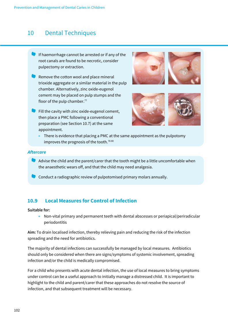
Prevention and Management of Dental Caries in Children
10 Dental Techniques
102
If haemorrhage cannot be arrested or if any of the
root canals are found to be necrotic, consider
pulpectomy or extraction.
Remove the cotton wool and place mineral
trioxide aggregate or a similar material in the pulp
chamber. Alternatively, zinc oxide-eugenol
cement may be placed on pulp stumps and the
floor of the pulp chamber.77
Fill the cavity with zinc oxide-eugenol cement,
then place a PMC following a conventional
preparation (see Section 10.7) at the same
appointment.
• There is evidence that placing a PMC at the same appointment as the pulpotomy
improves the prognosis of the tooth.78-80
Aftercare
Advise the child and the parent/carer that the tooth might be a little uncomfortable when
the anaesthetic wears off, and that the child may need analgesia.
Conduct a radiographic review of pulpotomised primary molars annually.
10.9 Local Measures for Control of Infection
Suitable for:
• Non-vital primary and permanent teeth with dental abscesses or periapical/periradicular
periodontitis
Aim: To drain localised infection, thereby relieving pain and reducing the risk of the infection
spreading and the need for antibiotics.
The majority of dental infections can successfully be managed by local measures. Antibiotics
should only be considered when there are signs/symptoms of systemic involvement, spreading
infection and/or the child is medically compromised.
For a child who presents with acute dental infection, the use of local measures to bring symptoms
under control can be a useful approach to initially manage a distressed child. It is important to
highlight to the child and parent/carer that these approaches do not resolve the source of
infection, and that subsequent treatment will be necessary.

Prevention and Management of Dental Caries in Children
10 Dental Techniques
103
For primary teeth, consider using gentle hand excavation of carious tissue to drain infection
without local anaesthesia to achieve an open communication with the necrotic pulp
chamber.
• If drainage is achieved by this method, it is advisable not to place any form of dressing
that would inhibit further drainage.
• For teeth that become tender prior to achieving drainage, a corticosteroid dressing may
be placed. When placing such a dressing a temporary restorative material than can
easily be removed by hand instruments should be used. This allows easy removal of the
dressing should it become necessary at future visits.
• Incision into the soft tissues to achieve drainage is rarely indicated in the primary
dentition, and would require suitable anaesthesia to be administered.
For permanent teeth, consider accessing the pulp chamber to remove the necrotic pulp
and/or achieve drainage.
• In circumstances where a larger fluctuant intraoral swelling is present, it may be
appropriate to undertake incision of the swelling undertaken under local anaesthesia.
10.10 Extraction of Primary or Permanent Teeth
Suitable for:
• a primary tooth that is unrestorable or of poor prognosis
• a permanent tooth that is unrestorable or of poor prognosis
Aim: To relieve or avoid pain or infection when alternative management approaches are not
feasible or are not in the child’s best interests.
For primary teeth with associated pain (pulpitis with irreversible symptoms) or infection and when
alternative management options have been excluded, an extraction should be considered.
However, dental extractions with local anaesthesia are potentially traumatic for the child and can
leave them with a negative perception of dental care, especially in children where this is an early
experience of dental care for them. It is better to avoid an extraction in an emergency situation
where a child has little positive experience of dental treatment, and consider delaying until the
child’s confidence has been built and they are able to cope with the procedure. The possibility of
the child not being brought back for treatment once out of pain should be considered.
Avoid dental extractions with local anaesthesia on a child’s first visit if at all possible.
If the child is in pain (pulpitis with irreversible symptoms), consider dressing the tooth with
corticosteroid-antibiotic paste and a temporary dressing.

Prevention and Management of Dental Caries in Children
10 Dental Techniques
104
Where there are signs or symptoms of dental abscess/infection and an extraction is either
not possible or better delayed, build up the child’s ability to cope to allow the extraction to
be carried out.
• In some cases, local measures to bring infection under control may be appropriate (see
Section 10.9).
• If there are signs or symptoms of systemic involvement or spreading infection,
antibiotics may be prescribed.
10.10.1 Balancing extractions in the primary dentition
A balancing extraction is the extraction of a contralateral tooth, performed in order to minimise
centre-line shift and maintain symmetry of the developing occlusion. Over the last decade
however, orthodontic treatment has moved away from techniques based on extraction of
premolars and the use of removable appliances to the use of fixed appliance therapy, with less
reliance on extractions. Therefore, there can be more flexibility in the guidance on the indications
for balancing extractions, the benefits of which have to be weighed against the risk of causing
treatment-induced anxiety through the additional extraction.81
Note that if caries is identified on one side of the mouth, it is quite likely to also be present on the
other side. Therefore, it is particularly important to examine contralateral teeth for disease with
bitewing radiographs, if indicated.
Consider balancing extractions when:
• one C is to be extracted
• one C has exfoliated prematurely due to eruption of the permanent lateral incisor
• centre-line shift is developing following extraction of one D
Be aware that balancing extractions are not usually necessary in the following situations:
• loss of primary incisors
• loss of Ds unless a centre-line shift is developing
• loss of Es
If in doubt, arrange an orthodontic assessment.
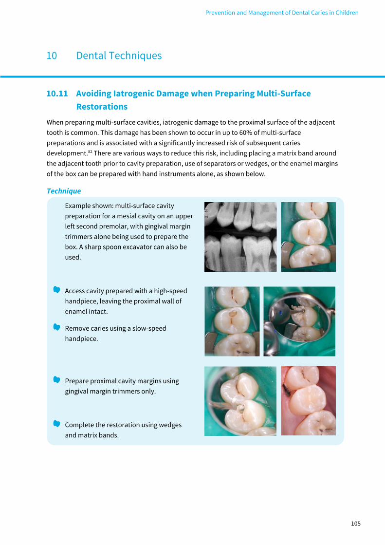
Prevention and Management of Dental Caries in Children
10 Dental Techniques
105
10.11 Avoiding Iatrogenic Damage when Preparing Multi-Surface
Restorations
When preparing multi-surface cavities, iatrogenic damage to the proximal surface of the adjacent
tooth is common. This damage has been shown to occur in up to 60% of multi-surface
preparations and is associated with a significantly increased risk of subsequent caries
development.82 There are various ways to reduce this risk, including placing a matrix band around
the adjacent tooth prior to cavity preparation, use of separators or wedges, or the enamel margins
of the box can be prepared with hand instruments alone, as shown below.
Technique
Example shown: multi-surface cavity
preparation for a mesial cavity on an upper
left second premolar, with gingival margin
trimmers alone being used to prepare the
box. A sharp spoon excavator can also be
used.
Access cavity prepared with a high-speed
handpiece, leaving the proximal wall of
enamel intact.
Remove caries using a slow-speed
handpiece.
Prepare proximal cavity margins using
gingival margin trimmers only.
Complete the restoration using wedges
and matrix bands.

Prevention and Management of Dental Caries in Children
10 Dental Techniques
106
10.12 Local Anaesthesia
The greatest challenge to most dentists when treating children is to ensure the child has a pain-
free positive experience whilst carrying out efficient good quality dentistry. Local anaesthesia (LA)
is perhaps the procedure most likely to provoke anxiety but it is also key to providing pain-free
dentistry. Use of the following techniques can help children accept local anaesthetic and ensure a
stress-free experience for the child, parent/carer and dental team.
LA is recommended for any cavity preparation that involves cutting sound dentine in both primary
and permanent teeth. Dentine in primary teeth is as sensitive as that of permanent teeth. LA can
be used successfully in children as young as 4 years of age. While infiltrations are effective for
most treatments on primary teeth, including extractions, for mandibular primary molars an IDB
injection with lignocaine may be necessary.
Most children will be apprehensive about receiving LA. However, the use of “sleight of hand”
techniques when giving LA may lead to “mistrust” phobias, which may be difficult to resolve at a
later time. Of the Behaviour Management techniques described previously, distraction (Section
4.2.6) is the most appropriate for use while giving LA.
Distraction aims to shift the patient’s attention from a potentially unpleasant procedure such as
LA to some other action. Short term distracters such as diverting attention by pulling the lip as LA
is given can work well. In addition to ensuring the tissues are taut as the needle penetrates for a
buccal infiltration, thereby decreasing pain, the clinician can use verbal distraction specifically
directing the child’s attention to the lip for example “You will feel me pulling your lip up to get a
better look.” Verbal distraction is also useful while applying topical anaesthetic.
Ask the child if they want to see what you would like to use to make
their tooth (and not them!) go to sleep. If they do, then show them
the syringe, emphasising how fine the needle is (like a cat’s whisker),
and that only a tiny part of it will go into their gum.
To reduce the discomfort of LA use:
• topical anaesthesia;
• distraction;
• a very slow injection technique, taking at least 60 seconds for an infiltration;
• intra-papillary injections rather than palatal injections (see below).

Prevention and Management of Dental Caries in Children
10 Dental Techniques
107
10.12.1 Intra-papillary injection technique
Intra-papillary injections are useful for achieving palatal or lingual anaesthesia without any
discomfort. However, it does take several minutes to complete.
Apply topical anaesthesia.
Give a buccal infiltration injection adjacent to the tooth
you want to anaesthetise.
Draw an imaginary line across the base of one of the
interdental papilla, and drop a perpendicular down onto
the line. Where the lines intersect, insert the needle
horizontally, so as to pass between the teeth on either side.
Advance the needle 1-2 mm and gently inject a drop or two
of LA solution. Ensure the needle remains in the correct
plane and does not become obstructed on the interseptal
bone or emerge from the gingivae.
Advance another 1-2 mm, and inject another drop of LA
solution.
Continue to do this, while observing the palatal aspect of the mucosa in your mirror.
After blanching is seen, withdraw the needle and insert it into the blanched area on the
palatal side. The child will not feel this, and the needle may then be advanced further
apically, if necessary, until complete anaesthesia is achieved.
10.12.2 Wand®
The Wand® is a computerised local anaesthetic delivery system
consisting of a microprocessor/drive unit that accommodates a
local anaesthetic cartridge. This is linked by lightweight disposable
tubing to a pen-like handle with a Luer lock needle attached. It
allows a constant slow flow-rate of anaesthetic solution irrespective
of tissue resistance. The delivery of anaesthetic is activated by a
foot control and allows two rates of flow. The slow rate is used for
needle insertion to the target area. The faster rate is used once the correct location is achieved. It
may be used for conventional blocks, buccal infiltrations and intra-ligamental techniques.

Prevention and Management of Dental Caries in Children
10 Dental Techniques
108
There is limited evidence that the WAND may be less painful than conventional techniques
particularly when used by inexperienced clinicians. Further, as it does not look like a conventional
syringe it may be useful for children who have had negative experiences with LA.83-86 However, it is
slower than conventional techniques and more expensive.
Prevention and Management of Dental Caries in Children
11 Referral for Care
109
11.1 Dental Service for Children
Dental services for children generally provide care up to 16 years of age, though in certain
circumstances this may be extended to an older age before transition to adult dental services
occurs.
Dental care for children is provided as follows.
Independent Practitioners
• General dental practitioners (GDPs) are responsible for the routine care of the great
majority of children and young people (approximately 90%).87
• Dental hygienists and therapists usually provide care following a treatment plan
prescribed by a dentist. Since 2013, they may also treat patients without a prescription
from a dentist, but consequently must have in place appropriate referral pathways for
provision of care that is beyond their scope of practice.
• In England and Wales, some independent practitioners have been commissioned to
provide additional/specialist services such as treatment under sedation.
Public Dental Service or Community Dental Service
• May provide routine care for children in areas where independent practice is not readily
available, or emergency care for children not registered with a GDP.
• On referral, supply care for children unable to be treated in a general practice setting e.g.
pre-cooperative children, or children with additional needs.
• Provide additional/specialist services which are not available in general dental practice,
such as, sedation and/or dental general anaesthesia for patients with anxiety or
behavioural problems.
• In some areas, paediatric dental specialists or consultants work in the service delivering
advice and treatment within the community setting.
• Coordinate and support provision of multidisciplinary programmes provided through
NHS dental and community settings (e.g. the Childsmile programme in Scotland).
• Work with other local services to organise dental care for particularly vulnerable
children. For example, children with complex additional needs or looked after children.
• Participate in screening programmes for dental epidemiology.
Hospital Dental Service
• Includes specialist and consultant paediatric dentists in the dental institutions.
• Undertakes treatment for more complex cases, including those requiring multi-
disciplinary care.
• Coordinates care for children with complex medical backgrounds, including those
undergoing inpatient treatment.
• Provide teaching, training and research facilities. In some areas of the country, care for a
small number of children is delivered within University Dental Outreach Centres.

Prevention and Management of Dental Caries in Children
11 Referral for Care
110
11.2 Referral of Children for Dental Care
As it is unacceptable to leave caries in children unmanaged, when the care of an individual child is
not feasible within your service, it is imperative that the child is referred to the appropriate service
to receive care. In such cases it is the responsibility of the clinician to follow their local referral
policy and to work in partnership to provide shared care. It remains the referring clinician’s
responsibility to provide the referred child’s continued regular dental care, including appropriate
emergency care, during and after this period.
Only refer cases after appropriate local treatments have been exhausted, using the
strategies outlined in Section 4.
Be aware of the referral options available locally and the agreed referral procedures.
When referring, ensure that this is to the appropriate service and that the agreed local
procedures are followed.
• The 2017 report from the Scottish Dental Needs Assessment Programme Oral Health and
Dental Services for Children88 provides a useful outline in Appendix 1 of where different
types of referral should be directed in Scotland.
For children who live in a different locality than your practice, beware that you should refer
to the service local to the child. This may be different to the service you routinely refer too.
If a child is referred for care, ensure that you provide their continued dental care.
Increasingly, electronic referral systems are being implemented. However, in situations
where you are writing a referral letter ensure all the relevant information is included. The
checklist in Figure 11.1 can be used as a reminder.
11.3 Referral for Sedation and General Anaesthesia
Taking the time to use behavioural management techniques together with good clinical
judgement regarding selection of appropriate treatment options will enable most children to
complete a planned course of care. However, some children will have difficulty accepting dental
treatment without sedation (for young children usually inhalation sedation; for older
children/young people inhalation sedation or intravenous sedation) or general anaesthesia (GA).
Inhalation sedation is a safe and effective form of sedation. It is useful for children aged 5 years or
older, but is of limited effectiveness for pre-school children. Intravenous sedation may be useful
for teenagers (young people). All forms of sedation can only be provided by clinicians who have
undertaken appropriate postgraduate training.89 While in Scotland, sedation services for children
are usually only available in the Public Dental Services or Hospital Dental Service elsewhere in the
UK, sedation may be provided in general dental practice or the Community Dental Service.90 The

Prevention and Management of Dental Caries in Children
11 Referral for Care
111
various types of care available locally for children often stems from different historical
commissioning patterns.
Compared with inhalation sedation, GA is generally believed to be associated with greater
morbidity and mortality (risk of death from GA for healthy children having minor or moderate non-
emergency surgery is less than <1:100,000, i.e. very rare91). In view of this, it is generally
recommended that GA is only used after less invasive management options, such as inhalation
sedation, have at least been properly considered, if not tried. For very young children (under 5
years of age) who may be unable to comprehend the required behaviour for treatment, young
school children who need extensive treatment, or for other potentially traumatic procedures GA
may be the preferred management option. A meeting of GA providers in Scotland in 2003 agreed
that the use of GA was a reasonable first option for 9 or 10 year-old children requiring extraction of
all first permanent molars (unpublished).
The final decision about whether inhalation sedation or GA is justified for dental care, and the
planning of the dental care to be provided in this way, must be made by an appropriately
experienced clinician.
The flow diagram in Figure 11.2 may be helpful in deciding whether or not to refer a child for
treatment under sedation or GA.
Referral for sedation or GA is not a management option for acute dental pain. It is the referring
dentist’s responsibility to relieve the child of dental pain (see Section 6).
Before referring a child for treatment with sedation or GA, first relieve pain, provide
prevention and attempt caries treatment using behavioural management techniques and
local anaesthesia if indicated.
If referring a child for sedation or GA, follow your local protocol if there is one in place.
Consider the need for temporary dressings to reduce the chance of further pain.
Advise the parent/carer and child that their first visit to the centre will probably be for
assessment only.
Include all relevant information in the referral letter, such as radiographs if available, and
state why in your opinion, sedation or GA is required (see Referral Checklist in Figure 11.1).
Do not promise the child and parent/carer that either sedation or GA will be provided; this
decision must be made by the clinician at the referral centre.
For further information consult the Guideline for the Use of General Anaesthesia (GA) in
Paediatric Dentistry.92
After a child has received care with sedation or GA, ensure that ongoing preventive care is
provided.

Prevention and Management of Dental Caries in Children
11 Referral for Care
112
Figure 11.1 Checklist of information to include in a referral letter for dental care
Referral Checklist
☐ Your details - name, address, telephone number and email address
☐ Patient details - name, address, telephone number, date of birth, CHI number (or equivalent patient
identification number)
☐ Patient’s presenting complaint - if a young child, parent/carer’s concerns and expectations
☐ Clinician’s concerns
• Why are you referring the patient?
• Is the referral urgent or routine?
• Has the child been in pain or had swelling?
☐ Medical history – including current medications (and where applicable dosages, e.g. prednisone)
☐ Dental history
• How anxious is the child about dental treatment? – include details of anxiety assessment (this
might include a modified dental anxiety scale)
• What treatment has been attempted in this course of treatment before the referral was made?
• How has the child coped with treatment?
• Has local anaesthesia been used?
• Has the patient received sedation or general anaesthesia previously?
• Have antibiotics been prescribed in the past six months? (if so, type, dose, frequency)
• Does the child attend for scheduled care appointments?
☐ Social history
• Who is the principal carer/legal guardian? Be mindful of the child’s social circumstances, and if
necessary include details of who has parental rights for the child and how they can be contacted.
• Who does the child live with?
• What school or nursery does the child attend?
• Has the child received dental health home or community support or intervention from other
agencies?
• If English is not the family’s first language, clearly state what language is spoken by the family so
that an interpreter can be organised.
☐ Summary of oral health status (e.g. caries and whether or not the child has good oral hygiene)
☐ Details of your request
• Advice only
• Care plan
• Treatment by the referral service, but clearly indicate the care that you can provide locally e.g.
prevention
☐ Enclosures
• Details of any relevant records/correspondence (e.g. radiographs, study models, photographs,
correspondence from orthodontist or other healthcare professional).
• Include these with the referral. Failure to include relevant records/correspondence may result in
your referral being returned or unnecessarily delay the patient’s assessment with the referral
service.

Prevention and Management of Dental Caries in Children
11 Referral for Care
113
Figure 11.2 Assessing management options for the child with carious primary teeth
This flow diagram illustrates decisions to be made when considering referral for treatment after
first attempting to provide care using good behavioural management techniques. It is assumed
that if the child is in pain, this has been relieved and that there are no medical complications.
Continue
Enhanced
Prevention,
monitor
lesions and
review the
need for
extractions.
Prioritise treatment of
teeth most likely to
cause pain or infection.
If necessary, refer for
sedation or GA and
continue Enhanced
Prevention and caries
management.
Provide Enhanced
Prevention and caries
management.
If unable to carry out
treatment, consider
referral for sedation or
GA and continue
Enhanced Prevention.
No
Yes
No
Yes
Are the teeth
close to
exfoliation?
Could teeth be managed
with a prevention alone
approach until the child is
able to accept treatment?
(factors to consider include:
likelihood of pain; extent of
caries; parent/carer's
support; likelihood of
cooperation developing)
Is parent/carer willing to
attend an adequate
number of appointments
for a preventive and
restorative programme
and likely to return for
follow up appointments?
Provide
Enhanced
Prevention and
monitor. Build on
cooperation and
reassess child’s
ability to accept
treatment.
Consider
referral for
inhalation
sedation
or GA and
continue
Enhanced
Prevention
.
No
Yes
No
Yes
Is the child likely to
accept treatment?
(e.g. some pre-school
children may not)

Prevention and Management of Dental Caries in Children
12 Recall
114
Once the child’s planned course of care is completed, a recall interval can be assigned. Based on
the clinician’s knowledge of the child and an assessment of disease levels and the overall risk of or
from dental disease, the National Institute for Health and Care Excellence (NICE) recommends
that a recall interval of between 3 and 12 months is assigned for children.93 In areas where the
incidence of dental caries in children is high, as is the case in many parts of UK, assigning a recall
interval of 12 months is only likely to be appropriate for a relatively small proportion of child
patients. In addition to considering dental caries, other factors will influence the recall interval,
including the child’s occlusal development and the need to provide any further care. It is
important to reassess a child’s caries risk at each recall visit as this may change over time.
Assign a recall interval that is based on caries risk and specific to the oral health needs of
the child.
• The SDCEP Oral Health Assessment and Review18 guidance provides further details on
assigning a risk-based recall interval.
At each recall visit:
Carry out a focused oral health review18, including asking again about toothbrushing
practice and dietary habits (see Section 3).
Enquire about compliance with agreed action plans.
Closely monitor lesions managed with prevention alone (see Section 10.1). Consider
recording plaque scores on the surface of the lesions, recording caries progression via
radiographs or photography and ensure the parent/carer is made fully aware of their
responsibility. If caries progresses, consider another option.
Check the condition of fissure sealants: visually for wear and physically for integrity or
leakage (see Section 7.4).
Reassess the child’s caries control and caries risk (see Section 3.5).
If caries is not being effectively controlled by the parent/carer or the child, consider the
possibility of dental neglect and the need for additional support (see Section 13).
If the child is assessed as at increased risk of developing caries, provide Enhanced
Prevention at 3 monthly intervals. Otherwise, provide Standard Prevention at 6 monthly
intervals, or exceptionally at longer intervals (e.g. for an older child if justified and recorded)
(see Section 7).
Ensure comprehensive records are maintained and create a new personal care plan as
required.

Prevention and Management of Dental Caries in Children
13 Providing Additional Support
115
Members of the dental team need to act as soon as they recognise a concern with a child or young
person’s wellbeing, and before crisis point is reached. This may be due to a lack of ability or
motivation to attend or to comply with dental preventive care, advice and treatment offered (e.g. an
appointment for a significant problem, such as pain or abscess, has been scheduled and missed).
13.1 General Dental Council Standards
The General Dental Council (GDC) Standards for the Dental Team20 set out the responsibilities of
dental team members to ensure they take action where there is concern over possible abuse or
neglect of children and vulnerable adults. The standards go on to state:
• You must raise any concerns you may have about the possible abuse or neglect of
children or vulnerable adults. You must know who to contact for further advice and how
to refer concerns to an appropriate authority such as your local social services
department.
• You must find out about local procedures for the protection of children and vulnerable
adults. You must follow these procedures if you suspect that a child or vulnerable adult
might be at risk because of abuse or neglect.
Not only is there a responsibility to take action but there is also a responsibility to ensure you have
ascertained and followed local procedures.
13.2 Child Protection Guidelines
The Scottish Government’s Child Protection Guidance for Health Professionals94 highlights the role
of health professionals working in a variety of settings and illustrates how concerns over wellbeing
can be addressed through a more structured approach.
The Child Protection and the Dental Team95 website is a source of tools and guidance to help dental
teams develop child protection procedures. It describes a tiered approach to managing dental
concerns with three stages of intervention, implemented according to the level of concern:
• Preventive dental team (single agency) response
• Preventive multi-agency response
• Child protection referral
The following questions are suggested as a starting point for ascertaining whether a child
protection referral may be required.
1. Has there been delay in seeking dental advice, for which there is no satisfactory
explanation?
2. Does the history change over time or not explain the injury or illness?
3. When you examine the child, are there any injuries that cannot be explained?
4. Are you concerned about the child’s behaviour and interaction with the parent/carer?
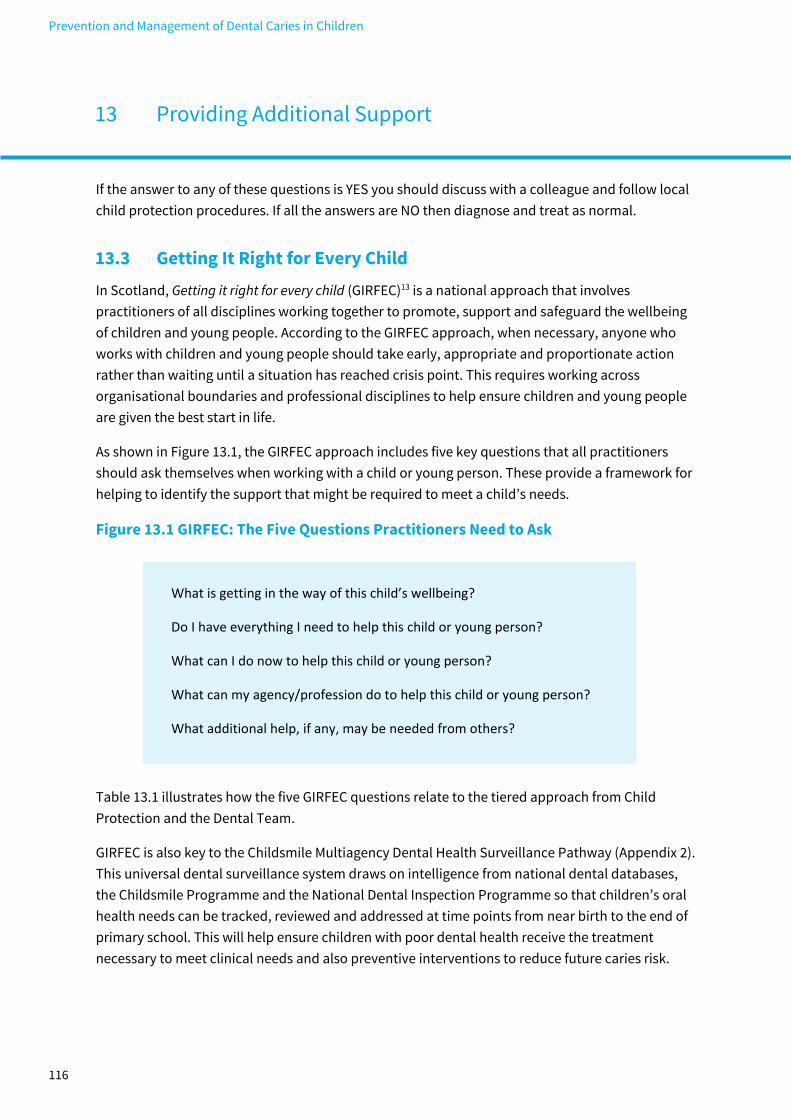
Prevention and Management of Dental Caries in Children
13 Providing Additional Support
116
If the answer to any of these questions is YES you should discuss with a colleague and follow local
child protection procedures. If all the answers are NO then diagnose and treat as normal.
13.3 Getting It Right for Every Child
In Scotland, Getting it right for every child (GIRFEC)13 is a national approach that involves
practitioners of all disciplines working together to promote, support and safeguard the wellbeing
of children and young people. According to the GIRFEC approach, when necessary, anyone who
works with children and young people should take early, appropriate and proportionate action
rather than waiting until a situation has reached crisis point. This requires working across
organisational boundaries and professional disciplines to help ensure children and young people
are given the best start in life.
As shown in Figure 13.1, the GIRFEC approach includes five key questions that all practitioners
should ask themselves when working with a child or young person. These provide a framework for
helping to identify the support that might be required to meet a child’s needs.
Figure 13.1 GIRFEC: The Five Questions Practitioners Need to Ask
What is getting in the way of this child’s wellbeing?
Do I have everything I need to help this child or young person?
What can I do now to help this child or young person?
What can my agency/profession do to help this child or young person?
What additional help, if any, may be needed from others?
Table 13.1 illustrates how the five GIRFEC questions relate to the tiered approach from Child
Protection and the Dental Team.
GIRFEC is also key to the Childsmile Multiagency Dental Health Surveillance Pathway (Appendix 2).
This universal dental surveillance system draws on intelligence from national dental databases,
the Childsmile Programme and the National Dental Inspection Programme so that children’s oral
health needs can be tracked, reviewed and addressed at time points from near birth to the end of
primary school. This will help ensure children with poor dental health receive the treatment
necessary to meet clinical needs and also preventive interventions to reduce future caries risk.

Prevention and Management of Dental Caries in Children
13 Providing Additional Support
117
Table 13.1 GIRFEC and Child Protection and the Dental Team Example
Alignment of the five GIRFEC questions with the tiered approach to managing dental neglect
based on an example from Child Protection and the Dental Team.95
GIRFEC
question
Guide for
action Action required
Suggested team
member(s)
responsible
What is getting
in the way of
this child’s
wellbeing?
and
Do I have
everything I
need to help
this child or
young person?
Gather
information
• Gather a thorough social, medical and dental history
• Complete a thorough clinical examination
Dental
receptionist,
dental nurse,
dentist
Keep accurate
records
• Keep accurate clinical records
Dentist and/or
other team
members
• Keep accurate administrative records of
appointments and attendance Dental receptionist
What can I do
now to help this
child or young
person?
Raise concerns
with parent/
carers
• Explain clinical findings, the possible impact on the
child, and why you are concerned Dentist
Explain what
changes are
required
• Explain treatment needed and expectation of
attendance Dentist
• Give advice on changes needed in diet, fluoride use
and oral hygiene
Dentist, therapist,
hygienist or dental
nurse
What can my
agency/
profession do
to help this
child or young
person?
Offer support
• Consider giving free fluoride toothpaste and a tooth
brush
Dentist, therapist,
hygienist or dental
nurse
• Offer the parent/carer a choice of appointment times Dental receptionist
• Listen for indications of a breakdown in
communication or parental worries about the
planned care, and offer to discuss again or to
arrange a second opinion
All team members
Continue to
liaise with
parent/carers
• Keep up open communication with the parent/carer
and repeat advice, so that they know what is
expected of them
All team members
Monitor
progress • Arrange a recall visit and keep accurate records of
attempted contacts
Dentist and dental
receptionist
What additional
help, if any,
may be needed
from others?
If necessary,
involve other
agencies
• Discuss and offer to arrange community/home
based dental health support via the child’s Health
Visitor, School Nurse, General Medical Practitioner or
Childsmile Dental Health Support Worker
All team members
• Agree and liaise with the child’s Health
Visitor/School Nurse to ensure preventive advice is
being reinforced in the community/home setting
All team members
• If concerns for the child’s wellbeing continue or
increase, consider a child protection referral All team members
• If at any stage there is concern for a child’s
immediate safety, or that the child is suffering
significant harm, follow local procedures to make a
child protection referral
All team members

Prevention and Management of Dental Caries in Children
13 Providing Additional Support
118
13.3.1 Information sharing
Practitioners should be reassured that appropriate information sharing is an important part of
their role in meeting children’s needs.
Where a family (i.e. the child and/or the patent/carer) gives consent, information can be shared.
Practitioners should inform a family what they plan to share, with whom and for what purpose.
This should be done prior to sharing or as soon as possible afterwards. Table 13.2 provides
guidance for sharing information.
If there are concerns that are not at the level of child protection (e.g. lack of motivation or ability
to attend or comply with treatment and advice) but the family withholds consent to share
information, practitioners should strive to continue to work with the family, providing advice and
support. If circumstances fail to improve, consider whether any more can be done by you. If not,
consider escalating the situation to a child protection referral.
Table 13.2 Sharing information
Key points when considering sharing information
When to share When sharing is necessary, fair and proportionate
When you have consent or there is another legal basis for sharing
What to share Share information which is relevant; necessary; legitimate; appropriate and
proportionate
Share information relating only to your worry or concern – reduce or remove unnecessary
information or data
Who to share
with
Share information on a need to know basis only
Share information with your line manager/supervisor or a trusted colleague; lead
professional or other key workers
How to share Share information verbally, face-to-face, at meetings, written reports or assessments
Share information by secure methods, ensure you record information sharing accurately
Information to include
Your details Name, address, telephone number and e-mail address
Child’s details Full name, address, post code, telephone number, date of birth, details of parent/carer,
CHI number (or equivalent) if you have it, child’s school or nursery
Description of
clinician’s
concerns
Concerns may include:
- failure to attend for the first time following referral to your service
- missed scheduled recall appointments
- missed scheduled care appointments
- attendance for emergency pain relief
- extraction/care under general anaesthesia required
- concerns over parent/carer in relation to supporting the child’s wellbeing
Social history Where applicable: ethnicity, religion, first language (especially if not English)

Prevention and Management of Dental Caries in Children
13 Providing Additional Support
119
13.4 Actions for the Dental Team
Preparation
Be aware of your responsibilities as described in the General Dental Council Standards for
the Dental Team.20
Ensure that child protection procedures are in place to address immediate concerns for a
child’s welfare or safety.
Be aware of the responsibility to share information when there is a concern about a child,
following local protocols and procedures.
In Scotland, ensure that all members of the dental team are familiar with the GIRFEC
approach and the five key GIRFEC questions.
Managing dental neglect or another concern about a child or young person’s wellbeing
The features that give cause for particular concern after parent/carers have been made aware of
dental problems, and acceptable treatment offered, are summarised in Child Protection and the
Dental Team as:
• severe untreated dental disease, particularly that which is obvious to a layperson or
other non-dental health professional
• dental disease resulting in a significant impact on the child
• parents or carers persistently failing to access treatment for the child, as may be
indicated by:
o irregular attendance and repeated missed appointments
o failure to complete planned treatment
o returning in pain at repeated intervals
o requiring repeated general anaesthesia for dental extractions
Failure to attend for treatment when the child has persistent pain or signs of infection is also
indicative of dental neglect.
When a child or young person presents with dental neglect*, which might be indicative of more
general neglect, or there is another concern about his/her wellbeing:
Follow the actions presented in Table 13.1, which are structured around the 5 key GIRFEC
questions and the Child Protection and the Dental Team tiered response.
Continue to offer and provide the child or young person with appropriate prevention, advice
and treatment.

Prevention and Management of Dental Caries in Children
13 Providing Additional Support
120
If at any stage there is concern for a child or young person’s immediate safety, or that
he/she is suffering significant harm, follow local procedures to make a child protection
referral.
* Dental neglect has been defined as ‘the persistent failure to meet a child’s basic oral health
needs, likely to result in the serious impairment of a child’s oral or general health or
development’.19
Prevention and Management of Dental Caries in Children
14 Quality Improvement and Research
121
14.1 Quality Improvement
Quality improvement activity in dental practice is requirement of NHSScotland terms and conditions
of service. There are various forms of quality improvement activity, which will be of most value if
based on a real need for improvement that has been identified through appraisal of day-to-day
work.96
Topics for quality improvement activities relevant to this guidance include:
• the accuracy and completeness of records (e.g. recording of caries risk assessment,
taking of radiographs);
• for children under regular care, relating causes of attending with pain and infection with
treatment previously provided;
• compliance with guidance on provision of fluoride varnish and fissure sealants;
• outcomes of treatment provided, including adhesive restorations in primary molars, Hall
Technique, and pulp therapies;
• effectiveness of biological methods of caries management for the primary dentition,
including fissure sealing over caries, selective caries removal and restoration and non-
restorative cavity control.
14.2 Research
Clinically important aspects of caries prevention and management in children for which research is
required to improve the evidence base include:
• factors that increase parental and child compliance with brushing advice;
• utility of food and drink diaries;
• effectiveness of combining preventive strategies;
• cost effectiveness of preventive strategies;
• effectiveness of caries risk assessment;
• children’s perceptions of dental treatment, including extractions, sedation and general
anaesthesia;
• effectiveness of sealing moderate carious lesions in occlusal surfaces;
• effectiveness of interventions to treat extensive carious lesions.
Prevention and Management of Dental Caries in Children
15 Evidence into Practice
122
A summary of the evidence relevant to each clinical question and the considered judgement on
which key recommendations were based are provided in Sections 15.1 to 15.4. Further details
about the development of this guidance are given in Appendix 1.
15.1 Prevention of caries in primary and permanent teeth
What evidence is there that delivery of brief interventions by the dental team lead to health
behaviour changes/healthy dental behaviours?
Evidence Summary
SIGN guideline 138 reviewed the evidence for delivery of brief interventions in the
practice setting, including the effectiveness and format of dental brief interventions.17
There is evidence of moderate quality in systematic reviews that brief interventions to
promote good oral health behaviours, including toothbrushing, can be effective. There is evidence
that theoretically based strategies to encourage health behaviour change can be effective, with
motivational interviewing favoured. However, further research is required to compare
interventions.
Considered judgement
SIGN guideline 138 supports use of oral health promotion strategies to facilitate daily
toothbrushing with fluoride toothpaste.17 These should be based on recognised oral
health behaviour theory and models such as motivational interviewing and should be
specific to individuals, and tailored to their particular needs and circumstances.
What factors influence the effectiveness of toothbrushing for the prevention of dental caries in
children?
Evidence Summary
SIGN guideline 138 extensively reviewed the evidence related to toothbrushing
including use of fluoride toothpaste, fluoride concentration, toothpaste composition,
frequency and duration of brushing, age of commencement of brushing and
toothbrushing practice.17
High quality evidence from systematic reviews indicates that there is a dose-response relationship
between toothpaste fluoride concentration and level of caries reduction.97-105 Toothbrushing with
fluoride toothpaste can also arrest early carious lesions.75 Although there is evidence to support
twice daily brushing with a fluoride toothpaste,98 there is insufficient evidence to recommend a
specific duration for each episode of toothbrushing.17 Brushing should start as soon as the first
tooth erupts, should be supervised and the amount of toothpaste restricted, particularly in
children under the age of three years. Rinsing with water after brushing reduces the caries-

Prevention and Management of Dental Caries in Children
15 Evidence into Practice
123
preventive effect of fluoride and can increase caries incidence. The evidence for the use of floss in
addition to toothbrushing is insufficient to support its use.17
Considered judgement
Toothbrushing with fluoride toothpaste is one of the most effective methods of
preventing caries. For standard prevention, toothpastes in the range 1000 to 1500 ppm
fluoride are recommended for use by children up to the age of 18 years. Higher dose
toothpaste may be beneficial for older children at increased caries risk.
The amount of toothpaste should be appropriate to the age of the child – a smear if under the age
of three years, a pea size amount for children over three years of age. Brushing should be
supervised. Professionals often advise brushing for two minutes, though the main point is to
ensure that sufficient time is taken for all tooth surfaces to be cleaned effectively.
What evidence is there for the effectiveness of giving dietary advice for the prevention of dental
caries in children?
Evidence Summary
Since the 1980s, epidemiological data has noted an association between sugar
consumption and level of dental caries. Countries with sugar consumption of less than
18 kg/person per year have consistently low disease levels.106,107
A survey of sugar intake of 1,700 Scottish children published in 2008108 found the average non-milk
extrinsic sugars (NMES) consumption was 17.4% of calorie intake. NMES intakes were higher in
older children; sugar consumption was 15.8% of calories in three to seven year olds and up to
19.1% in 12 to 17 year olds. A systematic review,50 which was conducted to inform a new World
Health Organization (WHO) guideline on dietary recommendations for populations, concluded
that there was evidence of moderate quality to show that dental caries is lower when free sugars
intake is less than 10%.
Reflecting recommendations from the independent Scientific Advisory Committee on Nutrition
(SACN) in 2015, revised Scottish dietary goals were issued in 2016, indicating that free sugars (also
known as NMES) should not exceed 5% of total energy in adults and children over 2 years.109
A Cochrane systematic review found tentative evidence that one-to-one dietary interventions
delivered in a dental setting aimed at promoting general rather than oral health, are effective at
changing dietary behaviour.51
NHS Health Scotland’s Oral Health and Nutrition Guidance (2012)52 gives oral health and nutrition
advice for the whole population with a focus on under fives. The guidance includes advice on: diet
and nutrition (a healthy balanced diet); diet and tooth decay (sugars; non-milk extrinsic sugars;
labelling); oral health advice (key oral health messages; diet/sugar; dental visits; dental erosion);
Prevention and Management of Dental Caries in Children
15 Evidence into Practice
124
oral health and between-meals snacks and drinks as a reference to enable professionals to give
practical, consistent advice to all age groups. More recent UK-wide guidance on healthy eating is
provided in the Eatwell Guide (2016).53
Considered judgement
Although there is limited evidence of the effectiveness of dietary advice for the
prevention of caries, the association between sugar and caries incidence and the
national drive to reduce the consumption of sugar justifies a strong recommendation
to encourage children to eat a healthy diet and restrict sugar intake in food and drinks.
What is the evidence for effectiveness of sealants in preventing dental caries in children?
Evidence Summary
Evidence for the effectiveness of fissure sealants was reviewed for SIGN guideline 138.17
Fissure sealants have been shown to reduce pit and fissure caries in primary and
permanent teeth73 and are more effective in reducing decay in occlusal surfaces than
fluoride varnish.110 Both resin-based and glass ionomer sealants are effective (moderate and low
to very low quality evidence respectively).
There is no clear evidence to suggest which sealant material is more effective at preventing caries
but resin-based sealants have been shown to be better retained than glass ionomer sealants.73
This is consistent with the most recent systematic review and recommendations of the American
Dental Association.111,112 Fissure sealants are also used in the management of carious lesions (see
Sections 8 and 9).
Considered judgement
The evidence from two Cochrane systematic reviews and a systematic review by the
American Dental Association supports the use of fissure sealants. Resin-based sealants
may be preferable based on their superior retention. However, glass ionomer sealants
are effective and may be particularly useful for application to newly erupted teeth. In agreement
with SIGN guideline 138, recommending the application of fissure sealants to the permanent
molars of all children in Scotland to prevent dental caries is considered likely to be beneficial.
Some children may also benefit from sealant application to other teeth.
What is the evidence for effectiveness of topical fluoride interventions in preventing dental
caries in children and what are the adverse effects?
Evidence Summary
In addition to fluoride toothpaste, there is a range of topical fluoride delivery systems
that can be used to help prevent caries in children. Evidence for the effectiveness of
Prevention and Management of Dental Caries in Children
15 Evidence into Practice
125
topical fluorides was reviewed for SIGN guideline 138.17 There is moderate quality evidence that
fluoride varnish is the most effective additional topical fluoride agent and that it significantly
reduces caries increment in both primary and permanent teeth.113
There is little evidence that other topical fluoride delivery systems (gels, beads, drops, tablets,
lozenges) are effective,17 although fluoride mouthwash may be useful as an alternative to or in
addition to varnish,114,115 for example for those who are at risk of an allergic reaction or for
enhanced protection for those at increased risk of caries.
Considered judgement
In agreement with SIGN guideline 138, all children (over 2 years of age) should receive
fluoride varnish application at least twice a year as part of standard prevention. More
frequent applications of fluoride varnish to children assessed at higher risk of caries is
also recommended.
15.2 Management of caries in primary teeth
In children, what evidence is there for the effectiveness of various methods for the treatment or
management of caries in primary teeth?
Evidence Summary
Six systematic reviews address various aspects of operative management of caries in
primary teeth.71,76,116-119 In those that examined the extent of caries removal before
restoration, much of the evidence is considered to be of low quality. However, the
Cochrane systematic review,76 which included studies assessed as of moderate quality, concluded
that stepwise and selective/partial caries removal are preferred to complete caries removal in
vital symptom-free primary or permanent teeth. This is consistent with earlier systematic
reviews.117,118 In primary teeth, the evidence available does not indicate a preferred restorative
material.116 However, there is moderate quality evidence that crowns placed on primary molar
teeth with carious lesions or following pulp treatment reduce the risk of pain or infection in the
long term compared to restorations.71
In addition, although limited, the available evidence does support no caries removal and sealing
with a stainless steel crown in primary teeth.71,76
The American Dental Association (ADA) has made clinical recommendations for use of pit and
fissure sealants on occlusal surfaces that are based on a recent systematic review of 23 studies, all
but one of which focussed on permanent teeth.112 Based on this, the ADA recommends use of
fissure sealants on noncavitated occlusal lesions to prevent their progression in both children and
adolescents.111 An earlier systematic review75 also supports the use of fissure sealants to slow the
progress or reverse noncavitated carious lesions. Of the six studies included, one evaluated

Prevention and Management of Dental Caries in Children
15 Evidence into Practice
126
lesions on primary teeth; this was of moderate quality and found that progression of fissure sealed
lesions on first primary molars was significantly reduced.
A recent Cochrane systematic review examined the effectiveness of microinvasive interventions
(lesion sealing or infiltration) for managing proximal enamel and initial dentinal caries lesions and
found moderate quality evidence that these techniques are more effective in reducing lesion
progression than non-invasive methods or no treatment.74 This is supportive of the consideration
of these emerging techniques when managing noncavitated proximal lesions in primary teeth,
taking into account clinical indications and the feasibility of different techniques.
One systematic review75 focussed on various non-surgical caries prevention methods to arrest or
reverse the progression of noncavitated carious lesions in primary and permanent teeth. Evidence
was generally of low quality. This review found that fluoride interventions (varnishes, gels, and
toothpaste) seem to have the most consistent benefit in decreasing the progression and incidence
of noncavitated lesions, though only one of the thirteen studies assessed primary teeth and this
was of low quality. Two more recent systematic reviews found that professionally applied 5%
sodium fluoride varnish can arrest and reverse/remineralise early enamel caries in primary and
permanent teeth, though the quality of evidence was low.120,121
A European regulation, which aims to limit the use of mercury, states “from 1 July 2018, dental
amalgam shall not be used for dental treatment of deciduous teeth…except where deemed
strictly necessary by the dental practitioner based on the specific medical needs of the patient”.66
Considered judgement
There are several options for managing caries in primary teeth, including complete,
selective or stepwise caries removal and restoration, sealing over caries using the Hall
Technique, sealant or infiltration and preventive only interventions. There is evidence
to indicate that the less invasive approaches, which are based on altering the environment of the
caries/plaque biofilm, can be effective and although the evidence from studies of primary teeth is
relatively scarce, it is believed reasonable to consider applying these methods to primary teeth.
Dental amalgam should not be used in primary teeth, for which there are no indications.
Unlike permanent teeth, pulpal health and restoration survival are not a major priority for primary
teeth. Consequently, although stepwise caries removal has been shown to be successful in
primary teeth, this method offers no advantage over selective caries removal and is not
recommended.
If there is no dentine involvement, it is recommended that site-specific prevention (application of
fluoride varnish, oral hygiene instruction, brushing with fluoride toothpaste) is provided or that
caries is not removed but is treated with a fissure sealant or resin infiltration.
If there is dentine involvement, the recommended treatment for an occlusal lesion is selective
caries removal and restoration and for a proximal lesion, sealing using the Hall Technique.

Prevention and Management of Dental Caries in Children
15 Evidence into Practice
127
Complete caries removal is an alternative, less preferred option. For anterior teeth, the
recommended treatment options are selective or complete caries removal and restoration or non-
restorative cavity control.
A variety of factors specific to primary teeth need to be taken account when determining a
suitable management strategy for each carious lesion.
15.3 Pulp therapy in primary teeth
In children, what evidence is there for the effectiveness of specific methods for pulp therapy for
primary teeth? Consider: Mineral trioxide aggregate/formocresol/ferric sulphate; Preformed
metal/stainless steel crown.
Evidence Summary
The evidence comprises one guideline from the British Society of Paediatric Dentistry
(BSPD)122 and five recent systematic reviews.77,123-126 The evidence base and the way that
evidence informed recommendations within the BSPD guideline is not clearly stated.
The guideline is supportive of the use of indirect pulp treatment, pulpotomy and, with sufficient
experience and appropriate patient selection, pulpectomy. The Cochrane systematic review77
included 47 studies assessed overall to be of low quality. It reported no significant differences
between a variety of pulpotomy medicaments though favoured ferric sulphate (FS) or mineral
trioxide aggregate (MTA) due to concern about potential harms and significantly greater
radiological failure with other agents. While this is consistent with another review126 and the BSPD
recommendations, other less well conducted reviews found that the long-term outcomes using
MTA were better than for FS in pulpotomy of primary molars.124,125 A more recent and thorough
systematic review supports the use of several pulpotomy medicaments with MTA and formocresol
giving the highest success based on high quality evidence.123
Considered judgement
Extraction of carious primary teeth can be distressing for the child and has the
potential to cause treatment-induced anxiety. To avoid the need for an extraction,
various pulp therapies are available. The evidence indicates that these can be
successful but does not provide clear insight into which technique is preferred. However,
pulpotomy is the most widely reported to date for treating deep carious lesions in primary teeth
that are still vital and pulpectomy for those that are non-vital. It is recommended that pulp
therapy is considered to preserve primary teeth in cases of pulpal involvement or dental infection.

Prevention and Management of Dental Caries in Children
15 Evidence into Practice
128
15.4 Management of caries in permanent teeth
In children, what evidence is there for the effectiveness of various methods for the treatment or
management of caries in permanent teeth?
Evidence Summary
Five systematic reviews76,116,118,119,127 address various aspects of operative management
of caries in permanent teeth. In those that examined the extent of caries removal
before restoration, much of the evidence is considered to be of low quality. However,
the Cochrane systematic review,76 which included studies assessed as of moderate quality,
concluded that stepwise and selective/partial caries removal are preferred to complete caries
removal in vital symptom-free primary or permanent teeth. This is consistent with two other
systematic reviews.118,127 One systematic review reported that glass ionomer cement has a higher
caries-preventive effect than amalgam for restoration of permanent teeth.116
The American Dental Association (ADA) has made clinical recommendations for use of pit and
fissure sealants that are based on a recent systematic review of 23 studies, all but one of which
were concerned with permanent teeth.112 The ADA recommends use of fissure sealants on
noncavitated occlusal lesions to prevent their progression in both children and adolescents.111 An
earlier systematic review75 also supports the use of sealing to slow the progress or reverse
noncavitated carious lesions. Of the six studies included, four assessed fissure sealants and two
assessed resin infiltration.
A recent systematic review based on eight studies (six for permanent teeth) examined the
effectiveness of microinvasive interventions (lesion sealing or infiltration) for managing proximal
enamel and initial dentinal caries lesions and found moderate quality evidence that these
techniques are more effective in reducing lesion progression than non-invasive methods or no
treatment.74 Although there is insufficient evidence to favour a particular technique, this review is
supportive of the consideration of these emerging techniques when managing noncavitated
proximal lesions in permanent and primary teeth, taking into account clinical indications and the
feasibility of different techniques.
One systematic review75 focussed on various non-surgical caries prevention methods to arrest or
reverse the progression of noncavitated carious lesions in primary and permanent teeth. Evidence
was generally of low quality. This review found that fluoride interventions (varnishes, gels, and
toothpaste) seem to have the most consistent benefit in decreasing the progression and incidence
of noncavitated lesions. Two more recent systematic reviews found that professionally applied 5%
sodium fluoride varnish can arrest and reverse/remineralise early enamel caries in primary and
permanent teeth, though the quality of evidence was low.120,121
A European regulation, which aims to limit the use of mercury, states “from 1 July 2018, dental
amalgam shall not be used for dental treatment of…children under 15 years…except where

Prevention and Management of Dental Caries in Children
15 Evidence into Practice
129
deemed strictly necessary by the dental practitioner based on the specific medical needs of the
patient”.66
Considered judgement
There are several options for managing caries in permanent teeth, including complete
caries removal, selective or stepwise caries removal and restoration, sealing over
caries with sealant or infiltration and preventive interventions only. There is evidence
to indicate that the less invasive approaches that are based on altering the environment of the
caries/plaque biofilm can be effective in permanent teeth. Use of dental amalgam should be
avoided in the permanent teeth of a child or young person under 15 years of age unless
exceptional circumstances can be justified.
In permanent teeth, stepwise caries removal and selective caries removal are clearly supported by
evidence for deep lesions with risk of pulp involvement. For less deep lesions, selective caries
removal or complete caries removal are preferable. Complete caries removal and restoration is
also the preferred option for anterior teeth although in some circumstances selective caries
removal may be achievable.
If there is no cavitation, it is recommended that caries is not removed but is either sealed with a
fissure sealant or resin infiltration or, for a proximal lesion or a lesion on an anterior tooth, site-
specific prevention (application of fluoride varnish, oral hygiene instruction, brushing with
fluoride toothpaste) may be provided.
A variety of factors specific to permanent teeth need to be taken account when determining a
suitable management strategy for each carious lesion.

Prevention and Management of Dental Caries in Children
Appendix 1 Guidance Development
130
The Scottish Dental Clinical Effectiveness Programme
The Scottish Dental Clinical Effectiveness Programme (SDCEP) is an initiative of the National
Dental Advisory Committee (NDAC) and operates within NHS Education for Scotland (NES).
The NDAC comprises representatives of all branches of the dental profession and acts in an
advisory capacity to the Chief Dental Officer. It considers issues that are of national importance in
Scottish dentistry and also provides feedback to other bodies within the Scottish Government on
related, relevant healthcare matters.
SDCEP was established in 2004 under the direction of the NDAC to give a structured approach to
providing clinical guidance for the dental profession. The programme’s primary aim is to develop
guidance that supports dental teams to provide quality patient care. SDCEP brings together the
best available information that is relevant to priority areas in dentistry, and presents guidance on
best practice in a form that can be interpreted easily and implemented. The guidance
recommendations may be based on a variety of sources of information, including research
evidence, guidelines, legislation, policies and expert opinion as appropriate to the subject. SDCEP
guidance takes a variety of forms to suit the diverse topics being addressed.
Recognising that publication of guidance alone is likely to have a limited influence on practice,
SDCEP also contributes to the research and development of interventions to enhance the
translation of guidance recommendations into practice through its participation in the TRiaDS
(Translation Research in a Dental Setting) collaboration (www.triads.org.uk).
All of SDCEP’s activities are overseen by a steering group that includes representatives of guidance
development groups and the dental institutions in Scotland. Up-to-date membership of this
steering group is available at www.sdcep.org.uk.
SDCEP is funded by NHS Education for Scotland and has made important contributions to the
implementation of the Scottish Government’s Dental Action Plan, which aimed to both modernise
dental services and improve oral health in Scotland. The views and opinions of the funders have
not in any way influenced the recommendations made in this guidance update.

Prevention and Management of Dental Caries in Children
Appendix 1 Guidance Development
131
The Guidance Development Group
The Guidance Development Group (GDG) for the updating of this guidance comprised individuals
from a range of relevant branches of the dental profession and two patient representatives.
Barbara Chadwick (Co-Chair) Professor of Paediatric Dentistry, Vice-Dean, School of
Dentistry, Cardiff University
Nicola Innes (Co-Chair) Professor of Paediatric Dentistry, School of Dentistry,
University of Dundee
Paul Ashley Consultant in Paediatric Dentistry, UCL Eastman Dental
Institute, London
Sarah-Louise Blackwood Patient Representative
Dafydd Evans Honorary Senior Lecturer in Paediatric Dentistry, University
of Dundee
Timothy Cooke Senior Dental Officer, Public Dental Service, Nairn, NHS
Highland
Brett Duane Associate Professor in Dental Public Health, Trinity College,
Dublin
David Conway Professor of Dental Public Health, University of Glasgow
Martin Foster Specialist in Paediatric Dentistry, Children’s Dental Service,
NHS Lothian
Alex Keightley Consultant and Honorary Senior Clinical Lecturer in
Paediatric Dentistry, Edinburgh Dental Institute
Nicole Kettles General Dental Practitioner
Peter King Childsmile Programme Manager (West Region)
Maxine Lee BSc Programme Lead, Oral Health Sciences, Dundee Dental
Hospital
Gillian Nevin General Dental Practitioner; Assistant Director for
Postgraduate GDP Education, NHS Education for Scotland
Derek Richards Consultant in Dental Public Health, South East Scotland
Maguerite Robertson Patient Representative
Margaret Ross Senior Lecturer for Dental Care Professionals, University of
Edinburgh
The GDG would like to thank Anne Littlewood, Trials Search Co-ordinator, Cochrane Oral Health
Group, for performing literature searches. The GDG would also like to acknowledge Udit
Bhatnagar, Clement Seeballuck and Emily McDougall for provision of clinical images and
illustrations. SDCEP is grateful to all those who provided feedback through consultation and to
peer reviewers

Prevention and Management of Dental Caries in Children
Appendix 1 Guidance Development
132
The Programme Development Team
The Programme Development Team operates within NHS Education for Scotland and is
responsible for the methodology of guidance development. Working with members of the
Guidance Development Group, the team facilitates all aspects of guidance development. The
following members of the Programme Development Team were directly involved in the
development of this edition of Prevention and Management of Dental Caries in Children.
Jan Clarkson
Programme Director and Professor of Clinical Effectiveness,
University of Dundee, SDCEP Director
Douglas Stirling Programme Manager, Guidance and Programme
Development
Linda Young Programme Manager, Evaluation and Implementation
Claire Scott Specialist Research Lead
Elizabeth Payne Programme Administrator
Guidance Development Methodology
SDCEP endeavours to use a methodology for guidance development that reflects that used to
develop high quality guidelines. It aims to be transparent, systematic and to adhere as far as
possible to international standards set out by the AGREE (Appraisal of Guidelines for Research and
Evaluation) Collaboration (www.agreetrust.org). The updating of Prevention and Management of
Dental Caries in Children followed the NICE accredited methodology described in the SDCEP
Guidance Development Manual (Version 1.3, February 2016). Details of SDCEP guidance
development methodology are available at www.sdcep.org.uk/how we work/.
In 2010, SDCEP first published guidance entitled Prevention and Management of Dental Caries in
Children. Much of the guidance on prevention was based on two previously published SIGN
guidelines. In 2014, these guidelines were withdrawn and replaced by SIGN 138: Dental
Interventions to Prevent Caries in Children.17 This and other advancements in the evidence-base
relevant to the scope prompted a review and update of the SDCEP guidance.
Prior to updating this guidance, SDCEP conducted a survey to ascertain dentists’ attitudes
towards the guidance and to obtain feedback on how they felt it could be improved. Suggestions
for improvements were considered during the updating of the guidance.
For this guidance, evidence to inform recommendations for prevention of dental caries was
largely derived from systematic reviews used in the updating of the SIGN guideline.17 Some of
these systematic reviews have since been updated and consequently when referred to in this
guidance the most recent versions are cited. For management of dental caries, comprehensive
searches of MEDLINE, EMBASE and the Cochrane Library, were first conducted by the Trials Search

Prevention and Management of Dental Caries in Children
Appendix 1 Guidance Development
133
Coordinator of the Cochrane Oral Health Group in January 2014 and updated on 3 October 2017.
Potentially eligible articles were identified from the list of titles and abstracts retrieved. An article
was considered potentially eligible if it met all of the following criteria:
1. The article was a systematic review or a guideline. An article was included as a systematic
review, if it included a methods section, a search of one or more electronic databases and
a table of included studies. An article was included as a guideline if it made
recommendations for clinical practice.
2. The article was concerned with a relevant aspect of management of dental caries in
children (primary or permanent teeth).
Additional manual searching of guideline repositories and other resources, and follow up of
citations from relevant articles found through the systematic searching was also carried out. Other
sources of evidence identified by GDG members were also considered, taking relevance and
quality into account.
A list of clinical questions related to the scope of the guidance was compiled and eligible articles
which were potentially relevant to each question were identified. The methodological quality of
each systematic review was assessed using the AMSTAR checklist.128,129 For guidelines, the AGREE II
instrument was used to assess the methodological quality (www.agreetrust.org).130
Evidence summaries were distributed to the GDG to inform and facilitate the development of the
recommendations for the guidance. Where authoritative evidence was unavailable, the GDG was
asked to make recommendations based on current best practice and expert opinion, reached by
consensus. When making recommendations the quality, consistency, generalisability and
applicability of evidence were considered together with other factors as appropriate, such as the
balance of risks and benefits, the values and preferences of the patients, and the limitations and
inconveniences of the treatment. The impact of potential barriers to implementation of the
recommendations, which were identified during guidance updating and through stakeholder
involvement and external consultation, was also considered.
The guidance was subject to open consultation for a twelve-week period initiated in May 2017.
Notification was sent to a wide range of individuals and organisations across the UK with a
particular interest in this topic, including all dentists in Scotland via the NES Portal, in addition to
professional bodies and charities representing patient groups. During this period the draft
guidance was available on the SDCEP website for comment. Targeted external peer review and
implementation interviews with potential end-users of the guidance also took place at this time.
Comments received through the consultation process were reviewed, the feedback considered by
the GDG, and the guidance was amended accordingly prior to publication.
An assessment of the potential impact of this guidance on equality target groups was conducted.

Prevention and Management of Dental Caries in Children
Appendix 1 Guidance Development
134
Further information about SDCEP and the methodology used for the development of this
guidance is available at www.sdcep.org.uk.
For this guidance, a review of the topic will take place five years after publication and, if this has
changed significantly, the guidance will be updated accordingly.
Conflict of Interests
All contributors to SDCEP are required to declare their financial, intellectual and other relevant
interests. At each group meeting, participants are asked to confirm whether there are any changes
to these. Should any potential conflicts of interest arise, these are discussed and actions for their
management agreed. All declarations of interest and decisions about potential conflicts of interest
are available on request.

Prevention and Management of Dental Caries in Children
Appendix 2 Childsmile/National Dental Inspection
Programme Dental Health Surveillance Pathway
135
Health visitor 6-8
week assessment
Health visitor 27-
30 month review
Childsmile nursery
fluoride varnish
application
Primary 1 basic
NDIP inspection
o health visitor refers
to Childsmile
dental health
support worker
o family supported
with oral health
needs, and dental
registration and
attendance
o health visitor
discusses child oral
health status
o dental registration
and attendance in
the last 12 months
is checked
o further oral health
support is offered
o two applications
per year in targeted
nurseries
o a letter is sent to
the family where
there is a concern
o support is provided
for registration and
attendance with
GDP
o near universal
inspection of
Primary 1 children
o 'A' letter children
and 'not seen'
followed up by
local Childsmile
teams
o support is provided
for dental
registration where
needed
Targeted Childsmile
nursery fluoride
varnish application
age 3 & 4 years
Primary 1
basic NDIP
inspection
6-8 week
assessment
27-30 month
review
The diagram below shows the key points on the Childsmile Early Years Pathway in Scotland, which
supports access to general dental services (GDS). The aim is for children to be registered with a
primary care dental practitioner as early as possible. Health visitors provide universal reviews for
all children, currently at 6-8 weeks and 27-30 months after birth. At 27-30 months, health visitors
also receive information on a child’s registration status. Childsmile nursery fluoride varnish
sessions include: offer of help with dental registration and a letter home advising a dental visit if
concerns have been identified. Lastly, as part of the National Dental Inspection Programme
(NDIP), each ‘A’ (high risk) letter sent to parent/carers about a child in Primary 1 after their dental
inspection will be followed up to support dental registration and attendance. These links to GDS
support the increasing focus within GDS on preventive children’s interventions.

Prevention and Management of Dental Caries in Children
References
136
1 Office of the High Commissioner for Human Rights. United Nations Convention on the Rights
of the Child. 1989; www.ohchr.org/EN/ProfessionalInterest/Pages/CRC.aspx. Accessed 23
December 2016.
2 Kassebaum NJ, Bernabe E, Dahiya M, Bhandari B, Murray CJ, Marcenes W. Global burden of
untreated caries: a systematic review and metaregression. Journal of Dental Research.
2015;94:650-658.
3 Marcenes W, Kassebaum NJ, Bernabe E, et al. Global burden of oral conditions in 1990-2010:
a systematic analysis. Journal of Dental Research. 2013;92:592-597.
4 Information Services Division, NHS National Services Scotland. Report of the 2016 Detailed
National Dental Inspection Programme of Primary 1 children and the Basic Inspection of
Primary 1 and Primary 7 children. 2016; http://ndip.scottishdental.org/wp-
content/uploads/2016/10/2016-10-25-NDIP-Report.pdf. Accessed 23 November 2017.
5 Information Services Division, NHS National Services Scotland. Report of the 2017 Detailed
National Dental Inspection Programme of Primary 7 Children and the Basic Inspection of
Primary 1 and Primary 7 children. 2017; http://ndip.scottishdental.org/wp-
content/uploads/2017/10/ndip_scotland2017-P7.pdf. Accessed 23 November 2017.
6 Jones CM, Davies GM, Monaghan N, Morgan MZ, Neville JS, Pitts NB. The caries experience of
5 year-old children in Scotland in 2013-2014, and in England and Wales in 2014-2015.
Reports of cross-sectional dental surveys using BASCD criteria. Community Dental Health.
2017;34:157-162.
7 Monaghan N, Davies GM, Jones CM, Neville JS, Pitts NB. The caries experience of 5-year-old
children in Scotland, Wales and England in 2011-2012: reports of cross-sectional surveys
using BASCD criteria. Community Dental Health. 2014;31:105-110.
8 Blumenshine SL, Vann WF, Jr., Gizlice Z, Lee JY. Children's school performance: impact of
general and oral health. Journal of Public Health Dentistry. 2008;68:82-87.
9 Seirawan H, Faust S, Mulligan R. The impact of oral health on the academic performance of
disadvantaged children. American Journal of Public Health. 2012;102:1729-1734.
10 Iheozor-Ejiofor Z, Worthington HV, Walsh T, et al. Water fluoridation for the prevention of
dental caries. The Cochrane Database of Systematic Reviews. 2015;6:CD010856.
11 Watt RG. Social determinants of oral health inequalities: implications for action. Community
Dentistry and Oral Epidemiology. 2012;40 Suppl 2:44-48.
12 Sheiham A, Watt RG. The common risk factor approach: a rational basis for promoting oral
health. Community Dentistry and Oral Epidemiology. 2000;28:399-406.

Prevention and Management of Dental Caries in Children
References
137
13 Scottish Government. Getting It Right For Every Child. 2017;
www.gov.scot/Topics/People/Young-People/gettingitright. Accessed 16 April 2017.
14 Childsmile Prevention Programme. 2018; www.child-smile.org.uk. Accessed 16 April 2016.
15 Scottish Government. Oral Health Improvement Plan. 2018;
www.gov.scot/Publications/2018/01/9275. Accessed 26 January 2018.
16 Public Health England. Delivering better oral health: an evidence-based toolkit for
prevention. 2017; www.gov.uk/government/publications/delivering-better-oral-health-an-
evidence-based-toolkit-for-prevention. Accessed 16 April 2018.
17 Scottish Intercollegiate Guidelines Network (SIGN). Dental interventions to prevent caries in
children. 2014; www.sign.ac.uk/assets/sign138.pdf. Accessed 16 April 2018.
18 Scottish Dental Clinical Effectiveness Programme (SDCEP). Oral Health Assessment and
Review: Guidance in Brief. 2011; www.sdcep.org.uk/wp-
content/uploads/2013/03/SDCEP+OHAR+Guidance+in+Brief.pdf. Accessed 16 April 2018.
19 Harris JC, Balmer RC, Sidebotham PD. British Society of Paediatric Dentistry: a policy
document on dental neglect in children. 2009; http://bspd.co.uk/Resources/Policy-
Statements. Accessed 16 April 2018.
20 General Dental Council. Standards for the Dental Team. 2013; www.gdc-
uk.org/professionals/standards/team. Accessed 16 April 2018.
21 Horner K, Eaton KA. Selection Criteria for Dental Radiography 3rd Edition. Faculty of General
Dental Practice (UK). 2013 (updated 2018); www.fgdp.org.uk/publication/selection-criteria-
dental-radiography. Accessed 28 February 2018.
22 NHS Inform. X-Ray Risks. 2018; www.nhsinform.scot/tests-and-treatments/scans-and-x-
rays/x-ray. Accessed 8 May 2018.
23 Scottish Dental Clinical Effectiveness Programme (SDCEP). Practice Support Manual. 2010;
www.psm.sdcep.org.uk. Accessed 21 December 2017.
24 Bader JD, Rozier RG, Lohr KN, Frame PS. Physicians' roles in preventing dental caries in
preschool children: a summary of the evidence for the U.S. Preventive Services Task Force.
American Journal of Preventive Medicine. 2004;26:315-325.
25 Pitts NB, Rimmer PA. An in vivo comparison of radiographic and directly assessed clinical
caries status of posterior approximal surfaces in primary and permanent teeth. Caries
Research. 1992;26:146-152.
26 Weerheijm KL, Jalevik B, Alaluusua S. Molar-incisor hypomineralisation. Caries Research.
2001;35:390-391.

Prevention and Management of Dental Caries in Children
References
138
27 Elfrink ME, ten Cate JM, Jaddoe VW, Hofman A, Moll HA, Veerkamp JS. Deciduous molar
hypomineralization and molar incisor hypomineralization. Journal of Dental Research.
2012;91:551-555.
28 Pine C, Harris R, Burnside G, Merrett M. An investigation of the relationship between
untreated decayed teeth and dental sepsis in 5-year-old children. British Dental Journal.
2006;200:45-47.
29 Mejare I, Axelsson S, Dahlen G, et al. Caries risk assessment. A systematic review. Acta
odontologica Scandinavica. 2014;72:81-91.
30 Tellez M, Gomez J, Pretty I, Ellwood R, Ismail AI. Evidence on existing caries risk assessment
systems: are they predictive of future caries? Community Dentistry and Oral Epidemiology.
2013;41:67-78.
31 Senneby A, Mejare I, Sahlin NE, Svensater G, Rohlin M. Diagnostic accuracy of different
caries risk assessment methods. A systematic review. Journal of Dentistry. 2015;43:1385-
1393.
32 Twetman S. Caries risk assessment in children: how accurate are we? European Archives of
Paediatric Dentistry: Official Journal of the European Academy of Paediatric Dentistry.
2016;17:27-32.
33 Fontana M. The Clinical, Environmental, and Behavioral Factors That Foster Early Childhood
Caries: Evidence for Caries Risk Assessment. Pediatric Dentistry. 2015;37:217-225.
34 NHS National Services Scotland. Scottish Index of Multiple Deprivation (SIMD)
https://nhsnss.org/services/practitioner/dental/scottish-index-of-multiple-deprivation-
simd/. Accessed 21 February 2018.
35 Freeman R. The case for mother in the surgery. British Dental Journal. 26 1999; 186:610-613.
36 Campbell C, Soldani F, Busuttil-Naudi A, Chadwick B. Non-pharmacological behaviour
management. 2011; http://bspd.co.uk/Resources/BSPD-Guidelines. Accessed 16 April 2018.
37 Locker D, Liddell A, Dempster L, Shapiro D. Age of onset of dental anxiety. Journal of Dental
Research. 1999;78:790-796.
38 Wong HM, Humphris GM, Lee GT. Preliminary validation and reliability of the Modified Child
Dental Anxiety Scale. Psychological Reports. 1998;83:1179-1186.
39 Howard KE, Freeman R. Reliability and validity of a faces version of the Modified Child
Dental Anxiety Scale. International Journal of Paediatric Dentistry. 2007;17:281-288.

Prevention and Management of Dental Caries in Children
References
139
40 Porritt J, Buchanan H, Hall M, Gilchrist F, Marshman Z. Assessing children's dental anxiety: a
systematic review of current measures. Community Dentistry and Oral Epidemiology.
2013;41:130-142.
41 Dailey YM, Humphris GM, Lennon MA. Reducing patients' state anxiety in general dental
practice: a randomized controlled trial. Journal of Dental Research. 2002;81:319-322.
42 Batchelor PA, Sheiham A. Grouping of tooth surfaces by susceptibility to caries: a study in 5-
16 year-old children. BMC Oral Health. 2004;4:2.
43 Royal College of Surgeons of England. Consent: Supported Decision-Making - a good
practice guide. 2016; https://www.rcseng.ac.uk/library-and-publications/rcs-
publications/docs/consent-good-practice-guide/. Accessed 21 December, 2017.
44 Scottish Dental Clinical Effectiveness (SDCEP). Drug Prescribing For Dentistry: Dental
Clinical Guidance 3rd Edition. 2016; www.sdcep.org.uk/wp-
content/uploads/2016/03/SDCEP-Drug-Prescribing-for-Dentistry-3rd-edition.pdf. Accessed
16 April 2018.
45 Sniehotta F, Soares VA, Dombrowski S. Randomized controlled trial of a one-minute
intervention changing oral self-care behavior. Journal of Dental Research. 2007;86:641-645.
46 Scottish Consultants in Dental Public Health Group. Recommendations on the use of
fluoride toothpaste and fluoride supplements in Scotland 2017;
http://www.scottishdental.org/library/recommendations-on-the-use-of-fluoride-
toothpaste-and-fluoride-supplements-in-scotland-2017/. Accessed 21 December 2017.
47 Sjogren K, Birkhed D, Rangmar B. Effect of a modified toothpaste technique on approximal
caries in preschool children. Caries Research. 1995;29:435-441.
48 Chestnutt IG, Schafer F, Jacobson AP, Stephen KW. The influence of toothbrushing
frequency and post-brushing rinsing on caries experience in a caries clinical trial.
Community Dentistry and Oral Epidemiology. 1998;26:406-411.
49 Childsmile. Toothbrushing. 2011; http://www.child-smile.org.uk/parents-and-
carers/toothbrushing/index.aspx. Accessed 16 April 2018.
50 Moynihan PJ, Kelly SA. Effect on caries of restricting sugars intake: systematic review to
inform WHO guidelines. Journal of Dental Research. 2014;93:8-18.
51 Harris R, Gamboa A, Dailey Y, Ashcroft A. One-to-one dietary interventions undertaken in a
dental setting to change dietary behaviour. The Cochrane Database of Systematic Reviews.
2012;3:CD006540.
https://www.rcseng.ac.uk/library-and-publications/rcs-publications/docs/consent-good-practice-guide/

Prevention and Management of Dental Caries in Children
References
140
52 NHS Scotland. Oral Health and Nutrition Guidance for Professionals. 2012;
www.healthscotland.com/documents/5885.aspx. Accessed 21 December 2017.
53 Public Health England in association with the Welsh Government, Food Standards Scotland
and the Food Standards Agency in Northern Ireland. The Eatwell Guide. 2016;
www.foodstandards.gov.scot/publications-and-research/eatwell-guide-booklet. Accessed 8
May 2018.
54 World Health Organisation. Health Topics: Breastfeeding. 2017;
www.who.int/topics/breastfeeding/en/. Accessed 21 December 2017.
55 Food Standards Scotland. Sugar. 2018; www.foodstandards.gov.scot/consumers/healthy-
eating/nutrition/sugar. Accessed 29 January 2018.
56 Food Standards Scotland. Guide to creating front of pack (FoP) nutrition label for pre-
packed products sold through retail outlets 2016;
www.foodstandards.gov.scot/publications-and-research/guide-to-creating-a-front-of-pack-
nutrition-label-for-pre-packed-products-s. Accessed 21 December 2017.
57 NHS Scotland. Drinks for Babies and Young Children. 2018;
www.healthscotland.com/documents/5064.aspx. Accessed 16 April 2018.
58 Public Health England. Change 4 Life. 2017; www.nhs.uk/change4life. Accessed 21
December 2017.
59 Childsmile. Food and Drink Diary. 2017; www.child-
smile.org.uk/professionals/resources/downloads.aspx. Accessed 16 April 2018.
60 Childsmile. Snack Ideas. 2018; www.child-smile.org.uk/documents/22388.aspx. Accessed 16
April 2018.
61 Childsmile. Parents and carers. 2018; www.child-smile.org.uk/parents-and-
carers/index.aspx. Accessed 16 April 2018.
62 Scottish Government Take life on, one step at a time. 2008-18; www.takelifeon.co.uk/.
Accessed 16 April 2018.
63 Beauchamp J, Caufield PW, Crall JJ, et al. Evidence-based clinical recommendations for the
use of pit-and-fissure sealants: a report of the American Dental Association Council on
Scientific Affairs. Journal of the American Dental Association. 2008;139:257-268.
64 Chestnutt IG, Playle R, Hutchings S, et al. Fissure Seal or Fluoride Varnish? A Randomized
Trial of Relative Effectiveness. Journal of Dental Research. 2017;96:754-761.
65 Childsmile. Fluoride Varnish Application Procedure. 2016; www.child-
smile.org.uk/professionals/fluoridevarnish1app.aspx. Accessed 16 April 2018.

Prevention and Management of Dental Caries in Children
References
141
66 European Parliament and Council of the European Union. Regulation (EU) 2017/852 of the
European Parliament and of the Council of 17 May 2017 on mercury, and repealing
Regulation (EC) No 1102/2008 (Text with EEA relevance) 2017;
https://publications.europa.eu/en/publication-detail/-/publication/687ef0ed-4045-11e7-
a9b0-01aa75ed71a1/language-en. Accessed 21 December 2017.
67 Pitts N, Nugent Z, Smith P. Report of the 1998/99 Survey of 14 Year Old Children. Scottish
Health Boards' Dental Epidemiological Programme (SHBDEP). 1999.
www.dundee.ac.uk/dhsru/publications/shbdep98/#s61.
68 Zhao D, Dong B, Yu D, Ren Q, Sun Y. The prevalence of molar incisor hypomineralization:
evidence from 70 studies. International Journal of Paediatric Dentistry. 2018;28:170-179.
69 Jalevik B, Klingberg GA. Dental treatment, dental fear and behaviour management
problems in children with severe enamel hypomineralization of their permanent first
molars. International Journal of Paediatric Dentistry. 2002;12:24-32.
70 Cobourne M, Willians A, Harrison M. A Guideline for the Extraction of First Permanent Molars
in Children. 2014; www.rcseng.ac.uk/-/media/files/rcs/fds/publications/a-guideline-for-the-
extraction-of-first-permanent-molars-in-children-rev-sept-2014.pdf. Accessed 16 April 2018.
71 Innes NP, Ricketts D, Chong LY, Keightley AJ, Lamont T, Santamaria RM. Preformed crowns
for decayed primary molar teeth. The Cochrane Database of Systematic Reviews.
2015;12:CD005512.
72 Innes N, Evans D, Stewart M, Keightley A. Hall Technique Users Manual. 2015; 4th Edition:
https://en.wikipedia.org/wiki/Hall_Technique. Accessed 8 April 2016.
73 Ahovuo-Saloranta A, Forss H, Walsh T, Nordblad A, Makela M, Worthington HV. Pit and
fissure sealants for preventing dental decay in permanent teeth. Cochrane Database of
Systematic Reviews. 2017;7:CD001830. *
74 Dorri M, Dunne SM, Walsh T, Schwendicke F. Micro-invasive interventions for managing
proximal dental decay in primary and permanent teeth. The Cochrane Database of
Systematic Reviews. 2015;11:CD010431.
75 Tellez M, Gomez J, Kaur S, Pretty IA, Ellwood R, Ismail AI. Non-surgical management
methods of noncavitated carious lesions. Community Dentistry and Oral Epidemiology.
2013;41(1):79-96.
76 Ricketts D, Lamont T, Innes NP, Kidd E, Clarkson JE. Operative caries management in adults
and children. The Cochrane Database of Systematic Reviews. 2013;3:CD003808.

Prevention and Management of Dental Caries in Children
References
142
77 Smail-Faugeron V, Courson F, Durieux P, Muller-Bolla M, Glenny AM, Fron Chabouis H. Pulp
treatment for extensive decay in primary teeth. The Cochrane Database of Systematic
Reviews. 2014;8:CD003220.
78 Farooq N, Coll J, Kuwabara A, Shelton P. Success rates of formocresol pulpotomy and
indirect pulp therapy in the treatment of deep dentinal caries in primary teeth. Pediatric
Dentistry. 2000;22:278-286.
79 Gruythuysen RJ, Weerheijm KL. Calcium hydroxide pulpotomy with a light-cured cavity-
sealing material after two years. ASDC Journal of Dentistry for Children. 1997;64:251-253.
80 Guelmann M, Fair J, Bimstein E. Permanent versus temporary restorations after emergency
pulpotomies in primary molars. Pediatric Dentistry. 2005;27:478-481.
81 Rock WP. UK National Clinical Guidelines in Paediatric Dentistry. Extraction of primary teeth
-- balance and compensation. International Journal of Paediatric Dentistry. 2002;12:151-153.
82 Qvist V, Johannessen L, Bruun M. Progression of approximal caries in relation to iatrogenic
preparation damage. Journal of Dental Research. 1992;71:1370-1373.
83 Tahmassebi JF, Nikolaou M, Duggal MS. A comparison of pain and anxiety associated with
the administration of maxillary local analgesia with Wand and conventional technique.
European Archives of Paediatric Dentistry: Official Journal of the European Academy of
Paediatric Dentistry. 2009;10:77-82.
84 Ram D, Peretz B. The assessment of pain sensation during local anesthesia using a
computerized local anesthesia (Wand) and a conventional syringe. Journal of Dentistry for
Children. 2003;70:130-133.
85 Kuscu OO, Akyuz S. Children's preferences concerning the physical appearance of dental
injectors. Journal of Dentistry for Children. 2006;73:116-121.
86 Kuscu OO, Akyuz S. Is it the injection device or the anxiety experienced that causes pain
during dental local anaesthesia? International Journal of Paediatric Dentistry. 2008;18:139-
145.
87 Information Services Division, NHS National Services Scotland. Dental Statistics – NHS
Registration and Participation (26 January 2016) 2016; www.isdscotland.org/Health-
Topics/Dental-Care/Publications/2016-01-26/2016-01-26-Dental-Report.pdf. Accessed 21
December 2017.
88 Scottish Dental Needs Assessment Programme (SDNAP). Oral Health and Dental Services for
Children: Needs Assessment Report. 2017; www.scottishdental.org/wp-
content/uploads/2017/03/SDNAP_Oral-Health-Dental-Services-for-Children-2017.pdf.
Accessed 21 December 2017.

Prevention and Management of Dental Caries in Children
References
143
89 Scottish Dental Clinical Effectiveness Programme (SDCEP). Conscious Sedation in Dentistry:
Dental Clinical Guidance 3rd Edition. 2017; www.sdcep.org.uk/wp-
content/uploads/2017/07/SDCEP-Conscious-Sedation-Guidance.pdf. Accessed 21 December
2017.
90 Office of the Chief Dental Officer of England. Commissioning Dental Services: Service
standards for Conscious Sedation in a primary care setting. 2017;
www.england.nhs.uk/publication/commissioning-dental-services-service-standards-for-
conscious-sedation-in-a-primary-care-setting/. Accessed 20 July 2017.
91 Royal College of Anaesthetists. Your child’s general anaesthetic: Information for parents and
guardians of children 5th Edition. 2017; https://www.rcoa.ac.uk/system/files/07-
ChildsAnaesthetic.pdf. Accessed 21 December 2017
92 Davies C, Harrison M, Roberts G. Guideline for the Use of General Anaesthesia (GA) in
Paediatric Dentistry. 2008; www.rcseng.ac.uk/-/media/files/rcs/fds/publications/guideline-
for-the-use-of-ga-in-paediatric-dentistry-may-2008-final.pdf?la=en. Accessed 21 December
2017.
93 National Institute for Health and Care Excellence Clinical Guideline 19. Dental recall – Recall
interval between routine dental examinations. 2004. www.nice.org.uk/guidance/cg19 .
Accessed 21 November 2017.
94 Scottish Government. Child Protection Guidance for Health Professionals. 2013;
www.gov.scot/Resource/0041/00411543.pdf. Accessed 16 April 2018.
95 Harris J, Sidebotham P, Welbury R. Child Protection and the Dental Team. 2013;
www.bda.org/childprotection. Accessed 164 April 2018.
96 NHS Education for Scotland. Quality Improvement Activity. 2017.
www.nes.scot.nhs.uk/education-and-training/by-discipline/dentistry/areas-of-
education/professional-development/quality-improvement-activity.aspx. Accessed 21
December 2017.
97 Marinho VC, Higgins JP, Logan S, Sheiham A. Topical fluoride (toothpastes, mouthrinses,
gels or varnishes) for preventing dental caries in children and adolescents. The Cochrane
Database of Systematic Reviews. 2003;4:CD002782.
98 Marinho VC, Higgins JP, Sheiham A, Logan S. Fluoride toothpastes for preventing dental
caries in children and adolescents. The Cochrane Database of Systematic Reviews.
2003;1:CD002278.
99 Wong MC, Glenny AM, Tsang BW, Lo EC, Worthington HV, Marinho VC. Topical fluoride as a
cause of dental fluorosis in children. The Cochrane Database of Systematic Reviews.
2010;1:CD007693.

Prevention and Management of Dental Caries in Children
References
144
100 Twetman S. Caries prevention with fluoride toothpaste in children: an update. European
Archives of Paediatric Dentistry: Official Journal of the European Academy of Paediatric
Dentistry. 2009;10:162-167.
101 Twetman S, Axelsson S, Dahlgren H, et al. Caries-preventive effect of fluoride toothpaste: a
systematic review. Acta Odontologica Scandinavica. 2003;61:347-355.
102 Walsh T, Worthington HV, Glenny AM, Appelbe P, Marinho VC, Shi X. Fluoride toothpastes of
different concentrations for preventing dental caries in children and adolescents. The
Cochrane Database of Systematic Reviews. 2010;1:CD007868.
103 Steiner M, Helfenstein U, Menghini G. Effect of 1000 ppm relative to 250 ppm fluoride
toothpaste. A meta-analysis. American Journal of Dentistry. 2004;17:85-88.
104 dos Santos AP, Nadanovsky P, de Oliveira BH. A systematic review and meta-analysis of the
effects of fluoride toothpastes on the prevention of dental caries in the primary dentition of
preschool children. Community Dentistry and Oral Epidemiology. 2013;41:1-12.
105 Twetman S. Prevention of early childhood caries (ECC): review of the literature published
1998-2007. European Archives of Paediatric Dentistry. 2007;9:12-18.
106 Sreebny LM. Sugar availability, sugar consumption and dental caries. Community Dentistry
and Oral Epidemiology. 1982;10:1-7.
107 Woodward M, Walker AR. Sugar consumption and dental caries: evidence from 90 countries.
British Dental Journal. 23 1994;176:297-302.
108 Sheehy C, McNeill G, Masson L, et al. Survey of sugar intake among children in Scotland
Summary report. 2008; www.foodstandards.gov.scot/sites/default/files/607-1-
1098_sugarintakescot2008summ.pdf. Accessed 21 December 2017
109 Scottish Government. Revised Dietary Goals for Scotland. 2016;
www.gov.scot/Resource/0049/00497558.pdf. Accessed 21 December 2017.
110 Ahovuo-Saloranta A, Forss H, Hiiri A, Nordblad A, Makela M. Pit and fissure sealants versus
fluoride varnishes for preventing dental decay in the permanent teeth of children and
adolescents. The Cochrane Database of Systematic Reviews. 2016;1:CD003067. *
111 Wright JT, Crall JJ, Fontana M, et al. Evidence-based clinical practice guideline for the use of
pit-and-fissure sealants: A report of the American Dental Association and the American
Academy of Pediatric Dentistry. Journal of the American Dental Association. 2016;147:672-
682 e612.
112 Wright JT, Tampi MP, Graham L, et al. Sealants for preventing and arresting pit-and-fissure
occlusal caries in primary and permanent molars: A systematic review of randomized

Prevention and Management of Dental Caries in Children
References
145
controlled trials-a report of the American Dental Association and the American Academy of
Pediatric Dentistry. Journal of the American Dental Association. 2016;147:631-645 e618.
113 Marinho VC, Worthington HV, Walsh T, Clarkson JE. Fluoride varnishes for preventing dental
caries in children and adolescents. The Cochrane Database of Systematic Reviews.
2013;7:CD002279.
114 Marinho VC, Chong LY, Worthington HV, Walsh T. Fluoride mouthrinses for preventing dental
caries in children and adolescents. The Cochrane Database of Systematic Reviews.
2016;7:CD002284. *
115 Twetman S, Petersson L, Axelsson S, et al. Caries-preventive effect of sodium fluoride
mouthrinses: a systematic review of controlled clinical trials. Acta Odontologica
Scandinavica. 2004;62:223-230.
116 Mickenautsch S, Yengopal V. Absence of carious lesions at margins of glass-ionomer cement
and amalgam restorations: An update of systematic review evidence. BMC Research Notes.
2011;4:58.
117 Ferreira JM, Pinheiro SL, Sampaio FC, de Menezes VA. Caries removal in primary teeth--a
systematic review. Quintessence International. 2012;43:e9-15.
118 Schwendicke F, Dorfer CE, Paris S. Incomplete caries removal: a systematic review and
meta-analysis. Journal of Dental Research. 2013;92:306-314.
119 Schwendicke F, Meyer-Lueckel H, Dorfer C, Paris S. Failure of incompletely excavated teeth--
a systematic review. Journal of Dentistry. 2013;41:569-580.
120 Lenzi TL, Montagner AF, Soares FZ, de Oliveira Rocha R. Are topical fluorides effective for
treating incipient carious lesions?: A systematic review and meta-analysis. Journal of the
American Dental Association. 2016;147:84-91.e81.
121 Gao SS, Zhang S, Mei ML, Lo EC, Chu CH. Caries remineralisation and arresting effect in
children by professionally applied fluoride treatment - a systematic review. BMC Oral Health.
2016;16:12.
122 Rodd H, Waterhouse P, Fuks AB, Fayle SA, Moffat MA. Pulp therapy for primary molars
International Journal of Paediatric Dentistry. 2006;16 Suppl 1:15-23. Available at
www.rcseng.ac.uk/-/media/files/rcs/fds/publications/pulptherapy.pdf?la=en. Accessed 18
March 2018.
123 Coll JA, Seale NS, Vargas K, Marghalani AA, Al Shamali S, Graham L. Primary Tooth Vital Pulp
Therapy: A Systematic Review and Meta-analysis. Pediatric Dentistry. 2017;39:16-123.

Prevention and Management of Dental Caries in Children
References
146
124 Stringhini Junior E, Vitcel ME, Oliveira LB. Evidence of pulpotomy in primary teeth
comparing MTA, calcium hydroxide, ferric sulphate, and electrosurgery with formocresol.
European Archives of Paediatric Dentistry: Official Journal of the European Academy of
Paediatric Dentistry. 2015;16:303-312.
125 Asgary S, Shirvani A, Fazlyab M. MTA and ferric sulfate in pulpotomy outcomes of primary
molars: a systematic review and meta-analysis. Journal of Clinical Pediatric Dentistry.
2014;39:1-8.
126 Anthonappa RP, King NM, Martens LC. Is there sufficient evidence to support the long-term
efficacy of mineral trioxide aggregate (MTA) for endodontic therapy in primary teeth?
International Endodontic Journal. 2013;46:198-204.
127 Hoefler V, Nagaoka H, Miller CS. Long-term survival and vitality outcomes of permanent
teeth following deep caries treatment with step-wise and partial-caries-removal: A
Systematic Review. Journal of Dentistry. 2016;54:25-32.
128 Shea BJ, Bouter LM, Peterson J, et al. External validation of a measurement tool to assess
systematic reviews (AMSTAR). PloS One. 2007;2:e1350.
129 Shea BJ, Grimshaw JM, Wells GA, et al. Development of AMSTAR: a measurement tool to
assess the methodological quality of systematic reviews. BMC Medical Research
Methodology. 2007;7:10.
130 Brouwers MC, Kho ME, Browman GP, et al. AGREE II: advancing guideline development,
reporting and evaluation in health care. Canadian Medical Association Journal.
2010;182:E839-842.
* Note that previous versions of references 73, 110 and 114 were cited in SIGN guideline 138.
These systematic reviews have since been updated and the most recent versions are listed
in here.


The Scottish Dental Clinical Effectiveness Programme (SDCEP) is an initiative of the National Dental Advisory Committee (NDAC) and operates within NHS Education for Scotland. The Programme provides user-friendly, evidence-based guidance on topics identified as priorities for oral health care.
SDCEP guidance supports improvements in patient care by bringing together, in a structured manner, the best available information that is relevant to the topic, and presenting this information in a form that can be interpreted easily and implemented.
The second edition of Prevention and Management of Dental Caries in Children aims to support dental teams to improve and maintain the oral health of their younger patients through the delivery of preventive care and, when necessary, effective management of dental caries.
Scottish Dental Clinical Effectiveness Programme Dundee Dental Education Centre, Frankland Building, Small’s Wynd, Dundee DD1 4HN
Email: [email protected] | Tel: 01382 425751 / 425771 Website: www.sdcep.org.uk
This resource may be made available, in full or in summary form, in alternative formats and community languages.
Please contact NHS Education for Scotland on 0131 656 3200 or email [email protected] to discuss how we can best meet your requirements.