NHS Bowel Cancer Screening Programme Guidance on …
51
NHS Bowel Cancer Screening Programme Guidance on reporting lesions Public Health England leads the NHS Screening Programmes
Transcript of NHS Bowel Cancer Screening Programme Guidance on …

NHS Bowel Cancer Screening Programme Guidance on reporting lesions
Public Health England leads the NHS Screening Programmes

NHS Bowel Cancer Screening Programme: guidance on reporting lesions
2
About Public Health England
Public Health England (PHE) exists to protect and improve the nation’s health and
wellbeing, and reduce health inequalities. We do this through world-leading science,
knowledge
and intelligence, advocacy, partnerships and the delivery of specialist public health
services. We are an executive agency of the Department of Health, and a distinct
delivery organisation with operational autonomy to advise and support government,
local authorities and the NHS in a professionally independent manner.
Public Health England, Wellington House, 133-155 Waterloo Road, London SE1 8UG
Tel: 020 7654 8000 www.gov.uk/phe
Twitter: @PHE_uk Facebook: www.facebook.com/PublicHealthEngland
About PHE screening
Screening identifies apparently healthy people who may be at increased risk of a disease
or condition, enabling earlier treatment or better informed decisions. National population
screening programmes are implemented in the NHS on the advice of the UK National
Screening Committee (UK NSC), which makes independent, evidence-based
recommendations to ministers in the four UK countries. The Screening Quality Assurance
Service ensures programmes are safe and effective by checking that national standards
are met. PHE leads the NHS Screening Programmes and hosts the UK NSC secretariat.
PHE Screening, Floor 2, Zone B, Skipton House, 80 London Road, London SE1 6LH
www.gov.uk/topic/population-screening-programmes
Twitter: @PHE_Screening Blog: phescreening.blog.gov.uk
Prepared by: [this line can be deleted if not required]
For queries relating to this document, please contact: [email protected]
© Crown copyright 2017
You may re-use this information (excluding logos) free of charge in any format or
medium, under the terms of the Open Government Licence v3.0. To view this licence,
visit OGL or email [email protected]. Where we have identified any third
party copyright information you will need to obtain permission from the copyright
holders concerned.
Published January 2018
PHE publications PHE supports the UN
gateway number: 201XXXX Sustainable Development Goals

NHS Bowel Cancer Screening Programme: guidance on reporting lesions
3
Contents
About Public Health England 2
About PHE screening 2
Preface 4
1. Executive summary 6
2. Introduction 7
3. Standards 8
3.1 Reporting requirements 8 3.2 Standards relating to BCSP pathology 9 3.3 Standards relating to colorectal cancer pathology 10
3.4 Endoscopy requirements 10
4. Specimen handling 11
4.1 Measurement 11 4.2 Sectioning 11
4.3 Margins 12
5. Reporting non-invasive lesions 12
5.1 Data 12
5.2 Classical adenomas 13 5.3 Serrated lesions 26
5.4 Inflammatory polyps 31
6. Reporting invasive neoplasia 32
6.1 Definition of invasion 32 6.2 Reporting of diagnostic biopsy specimens 32 6.3 Stage pT1 adenocarcinoma 33
6.4 Histological tumour type 34 6.5 Histological differentiation 34
6.6 Extent of local invasion 35 6.7 Lymphatic, venous and perineural invasion 36 6.8 Presence of a precursor lesion 37
6.9 Margin assessment 37 6.10 Tumour budding 40
References 42
Appendix 1 Bowel Cancer Screening System (BCSS) polyp dataset (2017) 47
Appendix 2 Recommended SNOMED codes 48
Appendix 3 TNM 8 classification of colorectal tumours 51

NHS Bowel Cancer Screening Programme: guidance on reporting lesions
4
Preface
This guidance is produced in collaboration with the pathology committees of the United
Kingdom and Ireland Bowel Cancer Screening Programmes.
Writing committee:
Dr Maurice B. Loughrey, Royal Victoria Hospital, Belfast Trust, Queen’s University
Belfast, Belfast, Northern Ireland
Dr Adrian C. Bateman, University Hospital Southampton NHS Foundation Trust,
Southampton, England
Professor Neil A. Shepherd, Gloucestershire Cellular Pathology Laboratory,
Cheltenham, England
Professor Philip Quirke, Leeds Teaching Hospitals NHS Trust and Leeds University,
Leeds, England
Contributions by:
Lead Pathology Irish Bowel Cancer Screening Programme
Professor Kieran Sheahan, St Vincent’s Hospital, Dublin, Ireland
Lead Pathology Northern Ireland Bowel Cancer Screening Programme
Dr Maurice B. Loughrey, Royal Victoria Hospital, Belfast Trust,
Queen’s University Belfast, Belfast, Northern Ireland
Lead Pathology Scotland Bowel Cancer Screening Programme
Professor Frank Carey, Ninewells Hospital, Dundee, Scotland
Lead Pathology Welsh Bowel Cancer Screening Programme
Dr Namor Williams, Abertawe Bro Morgannwg University Local Health Board, Wales
Professor Geraint Williams, University Hospital of Wales, Cardiff (2006-11)
English Pathology Bowel Cancer Screening Committee 2006 – present, in
alphabetical order
Dr Adrian C. Bateman, University Hospital Southampton NHS Foundation Trust,
Southampton
Dr Ann Buxton, Harrogate and District NHS Foundation Trust, Harrogate (2014-16)
Dr Philip DaCosta, Airedale Hospital NHS Foundation Trust, Keighley (2006-13)
Dr James Henry, Gateshead Health NHS Foundation Trust, Gateshead
Dr Nic Mapstone, Manchester Royal Infirmary, Manchester
Professor Philip Quirke, Leeds Teaching Hospitals and Leeds University, Leeds
Dr Manuel Rodriguez-Justo, University College Hospital NHS Foundation Trust,
London

NHS Bowel Cancer Screening Programme: guidance on reporting lesions
5
Dr Scott Sanders, South Warwickshire, NHS Foundation Trust, Warwick (2006-2016)
Professor John Schofield, Maidstone and Tunbridge Wells NHS Trust, Maidstone
Professor Neil A. Shepherd, Gloucestershire Cellular Pathology Laboratory,
Cheltenham
Professor Kevin West, Leicester Royal Infirmary, Leicester
Public Health England, Screening Division
John Davy (in memoriam) Programme Manager (2015-2017), NHS Bowel Cancer
Screening Programme
Billie Moores, National Quality Assurance Lead, NHS Bowel Cancer Screening
Programme
These guidelines have been endorsed by the:
Royal College of Pathologists
Association for Coloproctology of Great Britain and Ireland
British Society of Gastroenterology – Pathology Section
British Division of the International Academy of Pathology

NHS Bowel Cancer Screening Programme: guidance on reporting lesions
6
1. Executive summary
This document is NHS Bowel Cancer Screening Programme (BCSP) guidance and
updates the original 2007 version (NHS BCSP Publication No. 1). It is effective from 1
January 2018, and aims to support consistent practice and data collection as well as
developing the evidence base for future recommendations regarding routine practice.
The NHS BCSP Pathology Committee engages with pathologists across the United
Kingdom (UK) and Ireland. The standards mentioned within this document refer
specifically to the English programme. Different arrangements may apply elsewhere but
most aspects of this guidance will be common to all.
These guidelines are consistent with the guidance and dataset produced by the Royal
College of Pathologists (UK) (RCPath) for reporting colorectal cancer (CRC) (2). They
are also consistent with the quality assurance guidelines commissioned by the
European Union in 2010 (3).
The main updates to the 2007 version of this publication are:
elaboration of standards applicable to pathologists reporting BCSP specimens
(section 3.1)
expectations required of endoscopists submitting BCSP specimens for histology
(section 3.4)
careful consideration of adenoma sizing, as this is important for colonoscopic
surveillance pathways (section 5.2.2)
elaboration of descriptors and minimum diagnostic criteria for villousness, in an
attempt to improve reproducibility (section 5.2.3)
assessment of polypectomy margin – distinguishing margin involvement by
dysplasia from incomplete excision of adenocarcinoma (section 5.2.5)
detailed discussion of epithelial misplacement within adenomas versus
adenocarcinoma (section 5.2.6)
update in terminology to be applied to serrated lesions (section 5.3)
minimum criteria for diagnosing adenocarcinoma on endoscopic biopsy
specimens (section 6.2)
reporting of stage pT1 CRC in line with recommendations in the
RCPathguidance (2), to include depth and width of invasion (in mm) and
separate assessment of lymphatic, neural and venous invasion (section 6.3)
reference to National Institute for Health and Care Excellence (NICE)
recommendation on performing mismatch repair (MMR) immunohistochemistry
or microsatellite instability (MSI) testing in all cases of colorectal cancer (4)
(section 6.5)

NHS Bowel Cancer Screening Programme: guidance on reporting lesions
7
Changes as detailed in the RCPath guidance (2) to the surgical resection dataset
proforma for the move from TNM5 to TNM8 (Appendix 3) are:
changes to T, N and M staging categories
the Dukes and Bussey classification of CRC staging is no longer to be reported,
as it is not compatible with TNM 8 staging
the number of tumour deposits (use the precise number up to 5, or use ‘>5’)
should be recorded in node negative cases only (stage pN1c in TNM 8)
depth of venous invasion is now recorded as extramural or intramural
(comprising submucosal and intramuscular)
the presence (L1) or absence (L0) of lymphatic (small vessel) infiltration should
be recorded, with an indication of greatest depth of invasion (extramural or
intramural)
the presence (Pn1) or absence (Pn0) of perineural infiltration should be
recorded, with an indication of greatest depth of invasion (extramural or
intramural)
assessment of tumour regression following preoperative therapy has been
modified slightly and a tumour regression score (0–3) added
the section describing separate abnormalities in the specimen (aside from the
tumour) has been simplified
recording of additional immunohistochemical and molecular data
2. Introduction
The aim of this document is to help pathologists report specimens which derive from
BCSP practice. The vast majority of such specimens will be endoscopic in origin, either
polypectomies or mucosal biopsies. A minority will be other forms of local excision
specimen, such as endoscopic mucosal resection (EMR), or surgical resection
specimens for colorectal neoplasms deemed unsuitable for local resection.
Most BCSP colonoscopy specimens are diagnosed as adenomatous polyps, with
adenocarcinoma and inflammatory conditions each comprising fewer than 10% of
specimens in the BCSP setting. Flexible sigmoidoscopy screening generates fewer
cancers, fewer large adenomas and more hyperplastic polyps due to the anatomical
segment examined in this screening method. Within screen-detected CRCs, a higher
proportion are early stage (stage pT1) compared to symptomatic CRCs, and frequently
these can be treated by polypectomy or a form of local excision only.
Given this typical distribution of BCSP specimens, the emphasis within this guidance is
on reporting non-malignant polyps and on polypectomy or other local excision
specimens which contain adenocarcinoma. It is beyond the scope of this document to

NHS Bowel Cancer Screening Programme: guidance on reporting lesions
8
discuss reporting of surgical resection specimens or colorectal inflammatory conditions
in detail. You are referred to the guidance and dataset produced by the RCPath, major
gastrointestinal pathology textbooks and elsewhere in this regard (2, 5, 6).
Standardisation of pathology reporting is emphasised. This includes which data items
are reported and the approach to, and nomenclature used for, each data item. This
guidance aims to standardise BCSP pathology reporting further, and to facilitate
regional, national and international comparisons for audit and research purposes. We
strongly encourage the use of standardised agreed datasets and electronic reporting
systems, to aid collation of data by appropriate registries.
3. Standards
3.1 Reporting requirements
Pathologists who wish to start reporting BCSP (including BowelScope) cases should:
inform their Regional Screening Quality Assurance Service (SQAS) to ensure
they receive relevant quality assurance (QA) information
typically be in a substantive consultant post and this will be confirmed by the
regional pathology clinical advisor via the SQAS
register for the national BCSP external quality assurance (EQA) scheme and
participate in the next available round
During a QA visit, the regional pathology clinical advisor will check that all reporting pathologists:
participate in the national EQA scheme, defined as participation in two of the last
three circulations
have attended an educational event, relevant to their screening work, in at least
one of the past three years.
Pathologists who do not fulfil these requirements should not report BCSP cases. SQAS
will hold a list of BCSP pathologists whilst these requirements are embedded into
routine practice.

NHS Bowel Cancer Screening Programme: guidance on reporting lesions
9
3.2 Standards relating to BCSP pathology
1. To report BCSP cases, pathologists must fulfil the criteria specified in section 3.1.
In addition, they should participate in local QA visits and audit their own reporting
practice alongside colleagues.
2. Pathologists must complete either the screening programme proforma or its
computerised version for each BCSP case and each polyp reported. These should
be returned to the screening centre administrator for pathology data to be entered
onto the central bowel cancer screening system (BCSS). A copy of the latest
version of the BCSP proforma is provided in Appendix 1. Pathologists may also
wish to provide a free text report directly to the clinician.
3. Turnaround time of pathology reports should allow patients who have had lesions
removed at endoscopy to be managed appropriately and given a timely
appointment to be seen at a follow-up clinic if required. Current QA standards
relating to turnaround times should be adhered to, with day zero being receipt of
sample in the laboratory and the end point being the day the report is authorised.
Interim reports are encouraged if cases are referred for second opinions.
4. Between 3% and 10% of adenomas reported in the setting of a faecal occult blood
(FOB)/faecal immunochemical test (FIT) based screening programme are expected
to be classified as having high grade dysplasia (section 5.2.4). Between 1% and
5% of adenomas reported in the setting of flexible sigmoidoscopy-based screening
(such as bowel scope screening) are expected to be classified as having high
grade dysplasia.
5. Appropriate SNOMED codes should be applied to all cases (Appendix 2).
6. Cancers should be reported according to the latest Royal College of Pathologists
(UK) and BCSP guidelines.
Double reporting of all pT1 cancers is required to minimise overdiagnosis of
adenocarcinoma. Both reporting pathologists should be BCSP registered and named
on the pathology report. If there is any doubt about a diagnosis of cancer, then referral
is required for a further opinion. It is recommended that a local opinion is sought first
with a further regional opinion or opinion from the national ‘Expert Board’ should the
diagnosis still be in doubt.
These standards should be monitored by regular departmental audit, for all BCSP
pathologists involved. Other audits which may be informative could address measuring
size of adenomas and interobserver variation in frequencies of classifying adenomas as
tubular or villous/tubulovillous.

NHS Bowel Cancer Screening Programme: guidance on reporting lesions
10
3.3 Standards relating to colorectal cancer pathology
These relate to the RCPath dataset (2).
It is recommended that multidisciplinary teams and/or pathology departments audit their
CRC resection pathology reports at regular intervals. Three standards are
recommended, and should be audited for each BCSP pathologist. These should be
evaluated on a series of at least 50 resection specimens from symptomatic patients
(not screen-detected cancers) who have not undergone pre-operative therapy.
1. The number of lymph nodes examined should be as high as possible with at least a
median of 12 per case and many centres reporting a median of 15-25 per case.
2. The frequency of serosal involvement should be at least 20% for colonic cancers.
3. The frequency of venous invasion, including intramural (submucosal and
intramuscular) and extramural, should be at least 30%.
It is also worthwhile auditing frequencies of reporting of important features within stage
pT1 CRC polypectomy specimens, such as lymphatic invasion, venous invasion and
poor differentiation.
3.4 Endoscopy requirements
It is essential that the histopathology request form accompanying any BCSP specimen
includes all the relevant information to allow thorough and accurate pathology reporting.
The responsible endoscopist should include:
patient demographic details, including NHS/Health and Care number or other
unique identifier
date of endoscopy
relevant clinical information
endoscopic findings
The form should clearly indicate, by means of a suitable label or stamp, that the
specimen has resulted from BCSP endoscopy and must therefore be reported by a
BCSP pathologist. Bowel scope specimens should be clearly identified as such.
A copy of the endoscopy report should be submitted with the request form, and if
possible, accompanied by an endoscopic photographs of any lesions identified. As a
minimum, the request form should specify:
the precise number and respective sites of any polyps submitted for histology
any clinical suspicion of malignancy at endoscopy
endoscopic polyp sizes
NHS Bowel Cancer Screening Programme: guidance on reporting lesions
11
Each individual polyp should be submitted in separate specimen pots appropriately
labelled. The label should clearly state if:
each polyp has been endoscopically removed (intact or piecemeal) or only
biopsied or partially removed
a polyp has been biopsied multiple times or removed piecemeal (to avoid
misinterpretation as multiple polyps)
4. Specimen handling
BCSP specimens are no different to those originating from non-BCSP endoscopic or
surgical practice and should be handled according to the routine laboratory procedure
for such specimens. Most are received in the laboratory in formalin, and local excision
specimens may be pinned to cork and orientated with sutures. After adequate fixation,
for at least 4 hours but typically overnight, endoscopic biopsy specimens are
transferred to cassettes for processing, with the number of mucosal fragments in each
specimen recorded.
4.1 Measurement
Polypectomy specimens should be macroscopically measured in 3 dimensions in
millimetres (mm). Rounding up or down (terminal digit preference) should be avoided. It
is recommended that these measurements are recorded in a systematic fashion, for
example length x breadth x height, with an indication of any stalk present (with its
measurement). This conveys the maximum macroscopic information to the reporting
pathologist.
Given the clinical significance of adenoma size, notably around the 10mm diameter cut-
off, it is important that larger polyps received intact are sectioned along their longer axis
if possible, so that the maximum dimension can be presented on the glass slide for
microscopic measurement (7). If the largest axis within an intact polypectomy specimen
is not presented on the glass slide, it is important that this information is conveyed to
the reporting pathologist by the dissector to avoid potential underestimation of overall
adenoma size.
4.2 Sectioning
Sectioning should be perpendicular to the polyp base excision margin if this is
identifiable. Inking of the polyp base is not considered necessary on a routine basis as

NHS Bowel Cancer Screening Programme: guidance on reporting lesions
12
the excision margin is usually readily identifiable microscopically, often through the
presence of diathermy artefact.
Polyps with a narrow stalk should be trimmed to keep the stalk intact and orientated to
allow clear microscopic visualisation of the polyp base margin, through multiple levels if
necessary. Polyps with a broader stalk (‘semi-pedunculated’) or sessile polyps should
be serially sectioned at 3mm intervals, perpendicular to the base margin if this is
identifiable. All tissue should be processed for histological evaluation.
Careful liaison between dissector and reporting pathologist is vital so that the
dimensions of the specimen and the mode of dissection are understood. This approach
enables accurate assessment at microscopy of the maximum dimension of any
adenomatous component of the polyp (section 5.2.2). This measurement of adenoma
sizes will be used to inform individual risk stratification and recommend the surveillance
interval for any follow-up colonoscopy (7).
4.3 Margins
If there is any suspicion of malignancy within a local excision specimen at endoscopic
procurement or at dissection, it is advisable to paint any identifiable resection margins
(mucosal and/or deep) to ensure subsequent microscopic identification. If any mucosal
lesion has a surrounding mucosal margin of normal tissue that macroscopically
measures less than 3mm, this margin should be examined perpendicularly by taking
sections of the margin at right angles from a thicker slice. Macroscopic images are
helpful to illustrate margin status and block sampling.
5. Reporting non-invasive lesions
5.1 Data
Unpublished histopathology data derived from 558,637 non-invasive lesions diagnosed
at screening colonoscopy since inception of the English programme revealed that:
just over 70% were classical adenomas (55% tubular adenoma, 15%
tubulovillous adenoma, <1% villous adenoma)
19% were hyperplastic polyps
<1% were reported as sessile serrated lesions, although this percentage is likely
to grow as this entity was likely under-recognised in the early years of the
programme
all other diagnoses comprise <3% of all lesions (Table 1)

NHS Bowel Cancer Screening Programme: guidance on reporting lesions
13
Other diagnoses included traditional serrated adenoma, inflammatory and post-
inflammatory polyps, a wide range of benign mesenchymal polyps, and juvenile type,
Peutz-Jeghers type and other hamartomatous polyps. Individually, these specific
diagnoses are all rare in BCSP practice.
Table 1. Frequencies of common histopathological diagnoses from non-invasive
lesions detected during screening colonoscopy since inception of the English Bowel
Cancer Screening Programme to April 2006 - May 2017
Lesion Percentage
Tubular adenoma 54.7%
Tubulovillous adenoma 15.2%
Villous adenoma 0.8%
Hyperplastic polyp 19.2%
Mixed hyperplastic polyp/adenoma 0.3%
Serrated adenoma 0.7%
Sessile serrated lesion 0.5%
Traditional serrated adenoma 0.1%
Inflammatory polyp 1.2%
Not polyp 5.7%
Lipoma 0.1%
Lymphoid polyp 0.1%
Other polyp 0.5%
Not recorded 0.9%
Total 100%
5.2 Classical adenomas
5.2.1 General comments
The World Health Organisation (WHO) classification of premalignant epithelial lesions
or adenomatous polyps is recommended (8). By definition, adenomas must show
dysplasia (intraepithelial neoplasia). Classical adenomas are divided into tubular,
tubulovillous and villous types. Demarcation between them is based on the relative
proportions of tubular and villous components.
The term ‘advanced adenoma’ (mainly employed in United States screening literature)
most commonly describes any adenoma that is ≥10mm, has a villous component >25%

NHS Bowel Cancer Screening Programme: guidance on reporting lesions
14
(villous or tubulovillous) or has high grade dysplasia (9). This term should not be used
without a clear indication of its meaning, so as to avoid confusion with terms used in
risk stratification based on number and size of adenomas only (7).
5.2.2 Sizing
As discussed in section 4, accurate polyp sizing requires careful macroscopic and
microscopic correlation. The current available evidence indicates that the pathology
size of adenomas is more accurate and reliable than the endoscopy size. It is
mandated that the pathology size of adenomas is used for clinical decision making if
both sizes are available (10,11,12).
For adenomas, the aim of the reporting pathologist should be to provide the single
maximum dimension of the adenomatous component of the polyp. In many cases, this
will equate to the maximum macroscopic dimension of the formalin-fixed polyp, if all or
almost all of the polyp is adenomatous.
In some adenomas, large size or unusual configuration may preclude representation on
the glass slide of the largest polyp axis. In such cases, if microscopy demonstrates that
the entire polyp is adenomatous then the largest macroscopic dimension of the polyp,
after formalin fixation, can be safely recorded as the maximum adenoma diameter. If
the polyp includes a non-adenomatous component, then the maximum microscopic
dimension is recorded (Figure 1).
Figure 1: measuring adenomas
A. Bisected polypectomy specimen in which only part of the specimen is adenomatous. In such cases, the maximum
microscopic dimension of the adenomatous component should be recorded as the adenoma size. In this case, the
adenoma size (arrow) is 4mm, compared to 11mm maximum macroscopic dimension of the fixed polypectomy
specimen.
A
B

NHS Bowel Cancer Screening Programme: guidance on reporting lesions
15
B. Occasionally, the maximum microscopic dimension of an adenoma may be a vertical measurement. Correlation
with macroscopic assessment of overall polyp dimensions is important and care should be taken not to include any
non-adenomatous polyp stalk or non-adenomatous epithelial misplacement in the adenoma measurement. In this
case, the adenoma size (arrow) is 6mm, compared to 10mm maximum (vertical) dimension of the entire
polypectomy specimen.
Measurement should be via the BCSP graticule or an ISO accredited graticule if
measuring on the glass slide. Size should be measured to the nearest millimetre and
not rounded up or down to the nearest 5 or 10mm (terminal digit preference bias).
If a polyp is received piecemeal, or if a non-excision biopsy only is received, then
pathology size is not assessable and should be recorded as such. Recording sizes of
individual piecemeal fragments received is potentially confusing and not recommended.
In these circumstances, the endoscopic size should be used to determine follow-up.
Endoscopic sizing may take place in vivo assisted by size comparison to open biopsy
forceps, or after excision from within the specimen container. As audit of endoscopy
versus pathology size is an endoscopic quality measurement, care should be taken not
to record the endoscopy size under the pathology size.
5.2.3 Villousness
Villousness (a tubulovillous or villous morphology) has long been used as one of the 3
criteria for designating an adenoma as ‘advanced’ in nature; in other words, associated
with an increased risk of synchronous or subsequent CRC. The other 2 ‘defining’
features in this regard are:
adenoma size 10mm or greater
the presence of high grade dysplasia (13)
Associations have been demonstrated between all 3 of these features. Larger
adenomas also tend to show greater villousness and a higher incidence of high grade
dysplasia. Increasing villousness below the threshold required for a diagnosis of
tubulovillous adenoma has been associated with the acquisition of molecular alterations
(for example KRAS mutations) that are characteristic of tubulovillous adenomas (14). It
is less clear whether villousness alone is an independent risk factor for the presence of
synchronous or metachronous CRC (regardless of the size of the adenoma and the
presence or absence of high grade dysplasia).
Data derived from BCSP in England (2006 to 2017) indicates that around 8% of all
adenomas reported in BCSP are tubulovillous adenomas measuring up to 10mm in
diameter and showing low grade dysplasia. These lesions would be assessed as
‘advanced’ in nature based solely on their villousness (section 5.2.1). This data also

NHS Bowel Cancer Screening Programme: guidance on reporting lesions
16
indicates that in adenomas up to 10mm in size, high grade dysplasia is identified in
0.6% of those assigned as tubular but in 3.7% of those assessed as tubulovillous.
Accurate and reproducible identification of ‘advanced’ adenomas (those with size
≥10mm, a villous component >25% or high grade dysplasia) has become relevant in
the UK with the introduction of flexible sigmoidoscopy-based screening (bowel scope)
within the NHS BCSP, since the finding of such an adenoma in this setting will trigger
consideration for full colonoscopy within this programme (15). Inter-observer agreement
during the assessment of polyp features, particularly villousness, is poor (16,17,18).
It is recommended not to use the term ‘advanced adenoma’ in pathology reporting but
instead to detail each of the relevant pathology features (see section 5.2.1).
One potential solution to the difficulty of reproducibility might be to remove villousness
from the list of features that define an adenoma. Since increasing adenoma size is
closely linked with the presence of high grade dysplasia, size may be the most practical
determinant, within an individual lesion, of subsequent CRC risk (19). However, there
is not currently enough data relating to the risk for synchronous or metachronous CRC
that may be conferred by villousness when this feature is the only criterion of
‘advanced’ adenoma that is present. Until more data is available regarding its potential
independent biological significance, villousness should remain one of the criteria
assessed in reporting adenomas, but improved reproducibility of its assessment is
needed.
It is difficult to arrive at a definition of villousness that would facilitate recognition of this
feature with a high degree of inter-observer agreement. However, the following
descriptors and minimum criteria are recommended.
Descriptors of villousness
Classical villi are finger-like up growths of neoplastic epithelium with a stromal
core comprising lamina propria.
The sides of classical villi are often parallel but the tips may be bulbous.
Palmate villi have a more complex, branched and/or leaf-like architecture.
Tangential cutting may result in palmate villi having the appearance of tubular
glands.
Villi may extend down to the muscularis mucosae or, in the case of an adenoma
with a mixed tubular and villous architecture, protrude from the otherwise smooth
contour that is imparted by the tubular component of the lesion.
The invaginations that are sometimes seen within adenomas that have an
otherwise tubular morphology do not constitute villi if the overall surface contour
of the lesion is smooth.

NHS Bowel Cancer Screening Programme: guidance on reporting lesions
17
Minimum criteria for categorisation
1. At least 25% of an adenoma is required to possess a villous architecture in order
for the lesion to be categorised as a tubulovillous adenoma (8). Adenomas in
which a villous component comprises less than 25% of the lesion are designated as
tubular adenomas. Adenomas in which a villous component comprises more than
75% of the lesion are designated as villous adenomas. As villous adenomas are
rare, tubulovillous and villous adenomas are usually grouped together (any
adenoma with a significant (>25%) adenomatous component).
2. Within biopsies of larger lesions, any degree of villousness should raise the
possibility that the lesion is tubulovillous or villous in nature and result in the
categorisation of a lesion as a tubulovillous (or villous) adenoma.
As a guide, no more than 25% of colorectal adenomas should be designated as
tubulovillous in nature. Unpublished data derived from BCSP in England (2006 to 2017)
indicates that 75% of more than 265,000 colorectal adenomas reported were classified
as tubular in nature. Pure villous adenomas are rare in the screening programme.
Images designed to illustrate the above descriptors are shown in Figure 2. These
images are included to support improvement in consistency of reporting of villousness
in colorectal adenomas and facilitate the future determination of the relative importance
of this feature in the designation of adenomas as ‘advanced’.

NHS Bowel Cancer Screening Programme: guidance on reporting lesions
18
Figure 2: Features of villousness
NHS Bowel Cancer Screening Programme: guidance on reporting lesions
19
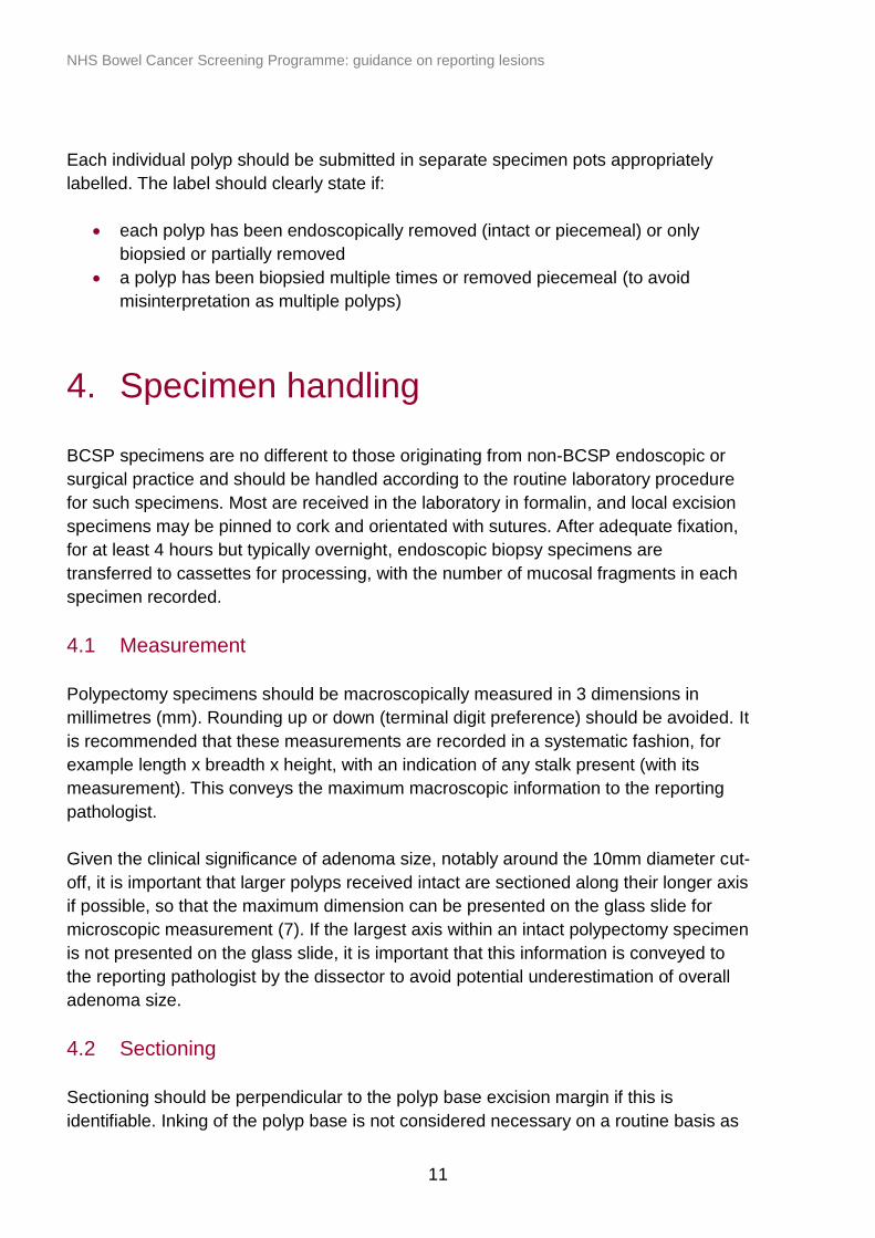
(Figure 2 cont.)
A. Typical slender villi, with parallel edges and a core of lamina propria.
B. Villi with a slightly more complex, branched growth pattern.
C. Villi containing ectopic crypts, leading to a more complex appearance that should not be mistaken for high-grade
dysplasia. Although considered an important feature of traditional serrated adenoma, ectopic crypt foci are also
commonly seen in tubulovillous adenomas
D. Tangential cutting leading to accentuation of the complex nature of this villiform area, with the juxtaposition of
villous and tubular structures.
E. A villiform growth pattern is present centrally and on the left side of this image. Despite the relatively smooth
overall contour of the polyp within this area, there is sufficient evidence of villousness for this to be assessed as
villous in nature.
F. An adenoma that is fragmented, with some clefting. However, villousness is not present and this should be
assessed as a tubular adenoma, noting the overall smooth surface contour.
G. Clefting is present on the left side of this image, leading to borderline villousness within a polyp that still shows a
smooth overall contour. These features are insufficient to be assessed as villous.
H. An area of established villousness, showing several villous structures that appear to ‘originate’ approximately
halfway between the muscularis mucosae and the polyp surface, but with one such structure on the right side of this
image appearing to extend almost from the level of the muscularis mucosae.
5.2.4 Grading dysplasia
A 2-tier stratification of adenomatous dysplasiais now widely accepted. The terms used
are:
low grade
high grade
This affords greater reproducibility and provides a uniform system for integrating global
(particularly Western and Japanese) histopathology grading data (8). The terms mild,
moderate or severe dysplasia should not be used.
The changes of high grade dysplasia should usually involve more than just one or 2
glands (except in tiny biopsies of polyps), enough to be identified at low power
examination. Caution should be exercised in overinterpreting isolated surface changes
that may be due to trauma, erosion or prolapse. Similarly, crush artefact should not be
interpreted as glandular complexity. High grade dysplasia is primarily diagnosed on
architecture, supplemented by cytology. This means its presence is nearly always
suspected by the appearance under low power of complex architectural abnormalities
in structures whose epithelium looks thick, blue, disorganised and ‘dirty’. The
architectural features of high grade dysplasia are:
complex glandular crowding and irregularity
a cribriform appearance and ‘back to back’ glands

NHS Bowel Cancer Screening Programme: guidance on reporting lesions
20
prominent intraluminal papillary tufting
Although many of these features often coexist in high grade dysplasia, individually they
are neither necessary nor usually sufficient. Indeed, they may occasionally occur in
lower grades of dysplasia, which is why it is also necessary to scrutinise the cytological
features for signs of high grade dysplasia. The cytological features are:
loss of cell polarity or nuclear stratification to the extent that the nuclei are
approximately equally, though haphazardly, distributed within all 3 thirds of the
height of the epithelium
markedly enlarged nuclei, often with a dispersed chromatin pattern and a
prominent nucleolus
atypical mitotic figures
prominent apoptosis, giving the epithelium of the lesion a ‘dirty’ appearance
Again, these features usually coexist in high grade dysplasia, and caution must be
exercised in using just one feature to make the diagnosis. It should be emphasised that
these cytological features should occur in a background of complex architectural
abnormality to allow classification as high grade dysplasia. Marked loss of polarity and
nuclear stratification sometimes occur on the surface of small, architecturally regular,
tubular adenomas that otherwise have features of low grade dysplasia (probably as a
result of trauma or ulceration) and must not be used to classify a lesion as high grade.
The only potential exception to the rule is when the specimen consists of a small biopsy
from the surface of a polyp – when there is insufficient tissue to assess the architecture
properly. In this situation, it is permissible to regard marked cytological abnormalities
alone as high grade dysplasia, but this will usually lead to excision of the whole polyp
so it will become possible to assess the whole lesion properly.
Examples of low and high grade dysplasia are illustrated in Figure 3.

NHS Bowel Cancer Screening Programme: guidance on reporting lesions
21
Figure 3: Low and high grade dysplasia
A. An adenoma with predominantly villous architecture; even on low power magnification, the lack of any complex
architectural features suggests low grade dysplasia throughout.
B. higher magnification of the same adenoma confirms architectural and cytological features of low grade dysplasia.
C and D. Two separate adenomas, each demonstrating focal high grade dysplasia, with complex glandular crowding
and irregularity, a cribriform appearance and luminal necrosis. Higher magnification to confirm the corresponding
cytological features is rarely necessary given these low power appearances.
5.2.5 Assessment of polypectomy margin
The endoscopist should remove all adenomatous polyps completely so as to prevent
progression to adenocarcinoma. Ideally, removal should be in one piece to optimise
histopathological assessment of the lesion. Larger polyps may require removal in
multiple steps, resulting in a piecemeal polypectomy specimen.
Regardless of the nature of the specimen, the endoscopic impression of completeness
of excision should be conveyed on the pathology request form. The endoscopist’s
impression is typically more valuable than that of the pathologist, who can comment
only on any involvement of a diathermied margin by dysplasia (and specify high or low
grade) within an intact polypectomy specimen. This does not equate to incomplete

NHS Bowel Cancer Screening Programme: guidance on reporting lesions
22
excision as diathermy may destroy a zone (up to several millimetres) of normal tissue,
creating the impression of incomplete excision. Therefore, the phrase ‘involvement of
diathermied margin by dysplasia’ is preferred when diathermy artefact is seen.
It should be emphasised that the vast majority of small polypectomy specimens are not
oriented and residual margin involvement by dysplasia is not assessable. Similarly, no
useful comment can typically be made on margin involvement by dysplasia within a
piecemeal polypectomy specimen unless a fragment specified as the margin has been
clearly indicated.
It is not considered necessary to comment on the polypectomy margin for hyperplastic
polyps.
5.2.6 Epithelial misplacement
Epithelial misplacement of adenomatous epithelium into the submucosa of a polyp is a
well-recognised phenomenon, particularly common in large prolapsing polyps in the
sigmoid colon. Distinction of epithelial misplacement, or so-called ‘pseudoinvasion’,
from invasive adenocarcinoma is perhaps the single most difficult area of pathological
diagnostic practice in BCSP pathology assessment. Large sigmoid colonic polyps are
particularly prone to inflammation and ulceration, features which tend to enhance the
dysplastic changes. When associated with epithelial misplacement, the potential for
misdiagnosis of early adenocarcinoma (stage pT1) increases and the overall diagnostic
difficulties become greater.
Double reporting by BCSP pathologists of all cases of endoscopically resected stage
pT1 CRC is now mandatory in BCSP pathology practice. This is because misdiagnosis
of adenocarcinoma may lead to an inappropriate major surgical intervention. Despite
increased awareness of the problem and enhanced recognition of the classical features
of epithelial misplacement versus adenocarcinoma (Table 2), many cases are highly
problematic as the features presented are overlapping. Figure 4 illustrates some
examples.
NHS Bowel Cancer Screening Programme: guidance on reporting lesions
23
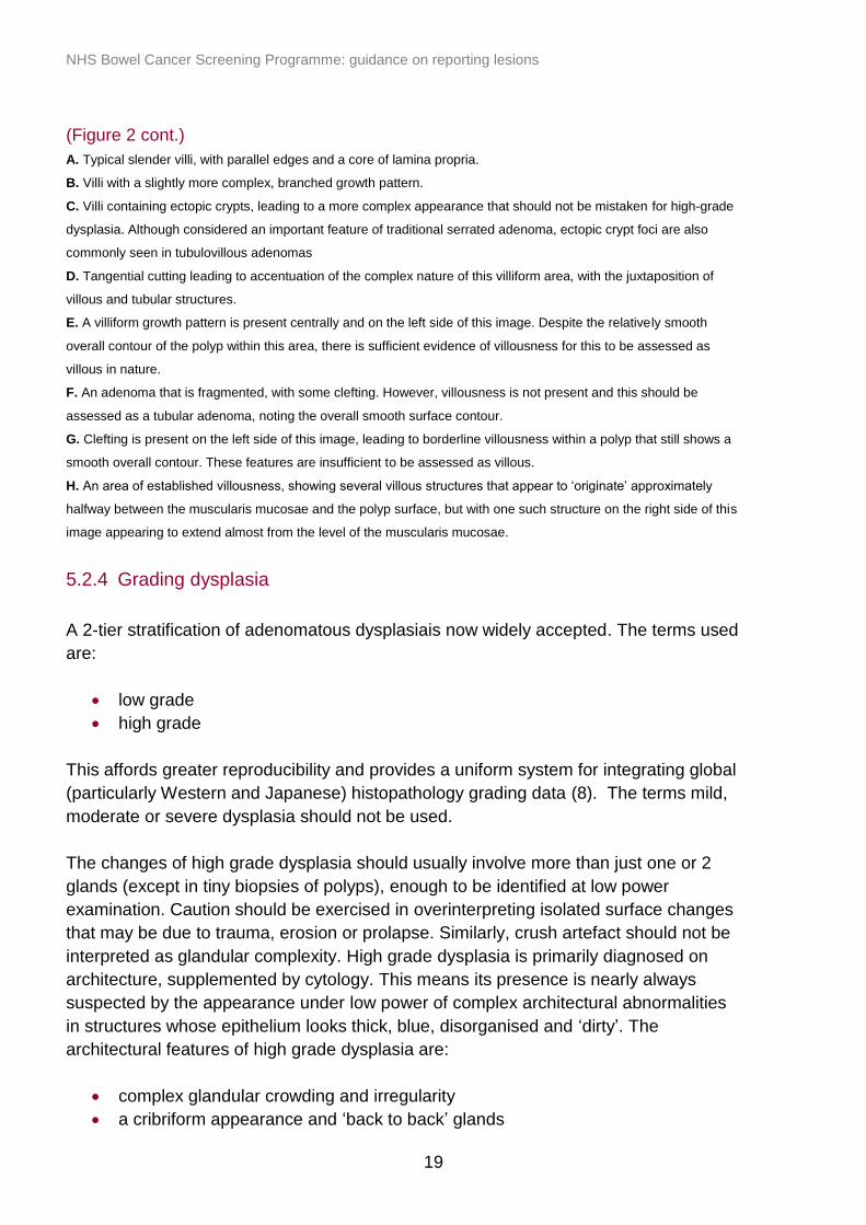
Figure 4: Epithelial misplacement versus early adenocarcinoma
Low power views of four polyps are presented to illustrate the important features evident at this magnification.
A. Classical features of epithelial misplacement with a lobulated glandular arrangement, surrounding lamina propria
rather than desmoplastic stroma, dysplastic change in continuity with surface adenoma and vascular congestion,
haemorrhage and mucin extravasation resulting from gland rupture.
B. Well differentiated adenocarcinoma featuring haphazardly infiltrating glands eliciting a desmoplastic stromal
reaction.
C. A focal area of submucosal invasion within a tubulovillous adenoma. Note the thin band of intact muscularis
mucosae towards the lateral aspects but central destruction of muscularis mucosae associated with small infiltrating
glands.
D. A difficult case featuring glands with high grade dysplasia located within the submucosa of the polyp head. A
lobular architecture is retained and there is probable gland rupture with adjacent abscess formation favouring
benignity, but confident distinction from an early adenocarcinoma is not possible.

NHS Bowel Cancer Screening Programme: guidance on reporting lesions
24
Polyps demonstrating the classical misplacement features of lobulated glands,
surrounding lamina propria, haemosiderin deposition and prolapse-type muscular
stroma usually provide no diagnostic problems. However, many cases show only some
of these appearances, often accompanied by more concerning morphological features,
such as apparent desmoplastic stroma, haphazard glandular ‘pseudoinfiltrative’ pattern,
glandular angulation or single cells lying within stroma. The most difficult cases often
include misplaced glands showing high grade dysplasia, associated with gland rupture,
mucin extravasation and secondary inflammatory changes. The correct diagnosis in
such cases may be almost impossible to determine. Previous biopsy or partial
polypectomy may also induce mucosal necrosis, ulceration with regenerative atypia
and/or a desmoplastic stromal reaction, strongly mimicking malignancy. Any previous
intervention should always be conveyed on the pathology specimen request form.
BCSP pathologists can access an ‘expert board’ whereby these difficult diagnostic
problems of epithelial misplacement versus polyp cancer are assessed by 3 specialist
gastrointestinal pathologists. The current coordinator is Professor Neil A Shepherd at
Cheltenham (address given at the front of this document) and all such cases should be
sent initially to him. PHE currently fund the board and consequently there is no charge
for this specialist diagnostic service. The results for the first 5 years of these
assessments have been published (20).
Overall judgment must be based foremost on appropriate clinical management. If
surgical intervention would not be warranted in a given case, regardless of a diagnosis
of epithelial misplacement or adenocarcinoma (stage pT1 with no adverse features and
clear margins), it is considered prudent to reserve a diagnosis of malignancy for those
cases with considerable certainty in the diagnosis.

NHS Bowel Cancer Screening Programme: guidance on reporting lesions
25
Table 2. A comparison of the pathological features for differentiating epithelial
misplacement from invasive adenocarcinoma (reproduced with permission from (21) )
Epithelial misplacement
(EM)
Adenocarcinoma
Epithelial ‘differentiation’ Usually similar to that of the
surface adenomatous
component
Variable and usually different to
the surface adenomatous
component
Lamina propria
accompaniment
Characteristic but may be
lacking when there is secondary
inflammation and epithelial
destruction
Usually absent. Can be present in
rare, very well-differentiated
carcinoma
Accompaniment by non-
adenomatous epithelium
Characteristically seen when EM
is due to previous intervention
Absent
Haemosiderin deposition Characteristic and indicative of
previous necrosis and/or
haemorrhage
Usually absent
Mucosal prolapse
changes
Often present Usually absent
Mucus cysts Characteristic. They probably
represent epithelial
misplacement that has become
‘detached’ from the more
superficial components
Only present, usually, in
mucinous tumours
Continuity with surface
adenomatous component
Characteristic but often only
appreciated in multiple levels
and/or 3D reconstruction studies
Usually absent but some cases
do show continuity, even in 3D
reconstruction studies.
Involvement of
muscularis
propria (MP)
Usually absent. Can be seen
very rarely, especially after
previous intervention
Present if at least pT2
Budding Usually absent but a similar
phenomenon can be seen as a
result of epithelial destruction
and/or inflammation
Often present
Desmoplastic reaction to
glands
Usually absent but fibromuscular
stromal proliferation can
accompany EM
Usually present
Lymphatic and/or
vascular invasion
Absent
Diagnostic of cancer

NHS Bowel Cancer Screening Programme: guidance on reporting lesions
26
5.3 Serrated lesions
The histopathological assessment of serrated colorectal lesions can be a problematic
area. The terminology used to describe lesions within this spectrum is variable and the
suggested minimum diagnostic criteria for some lesions differ between authorities. One
of the most difficult areas is the nomenclature of, and diagnostic criteria for, sessile
serrated lesions (SSL) (also termed sessile serrated polyp, sessile serrated adenoma
or sessile serrated adenoma/polyp). This is particularly important as these lesions,
while bearing histological resemblance to hyperplastic polyps (HP), may be associated
with the earlier development of epithelial dysplasia and adenocarcinoma. This topic is
the subject of a recent review, an edited version of which forms the basis of the advice
given within this document (22).
Lesions with serrated morphology should be given one of the following names
according to their morphological features:
hyperplastic polyp (HP)
sessile serrated lesion (SSL)
sessile serrated lesion with dysplasia
traditional serrated adenoma (TSA)
mixed polyp
5.3.1 Hyperplastic polyps (HPs)
HPs are small serrated lesions showing no features that would allow categorisation as
SSL and no evidence of dysplasia. We use the term ‘dysplasia’ in this context to refer
to the morphological appearances of epithelial neoplasia within the mucosa of the colon
and rectum – for example, the epithelial changes recognised by histopathologists as
characteristic of ‘classical’ adenomas.
HPs are usually small (less than 5mm diameter) and may occur anywhere within the
colon and the rectum. However they are particularly common in the distal colon and
rectum and are often multiple. There are 2 common morphological forms of HPs.
1. Microvesicular HP. These demonstrate vesicular mucin-containing epithelial cells
and goblet cells are decreased in number compared with normal crypts. Goblet cell-
rich HPs account for about a third of all hyperplastic polyps and these, too, almost
always occur in the left colon and rectum. Unsurprisingly, they show numerous
goblet cells. Microvesicular HPs tend to demonstrate BRAF mutations whereas
KRAS mutations are predominant in the goblet cell-rich variant.
2. Mucin-poor HP variant. This type is rare and is similar to the microvesicular HP but
contains less microvesicular mucin and less goblet cells.

NHS Bowel Cancer Screening Programme: guidance on reporting lesions
27
The cancer risk associated with small HPs is very low.
5.3.2 Sessile serrated lesions (SSLs)
SSLs are synonymous with the sessile serrated adenoma/polyp in the current WHO
classification and the latter terminology is in common use, particularly in North America
(23). When ‘pure’, these lesions show no evidence of dysplasia but, in comparison to
HPs, they contain one or more of the following histopathological features:
irregular distribution of crypts
dilatation of crypt bases
serration present at crypt bases
branched crypts
horizontal extension of crypt bases
herniation of crypts through the muscularis mucosae (Figure 5)
SSLs also show ‘dysmaturation’. This is a disorganised arrangement of proliferating
cells and goblet cells within the lower half of the crypts, with subtle cytological
abnormalities that are more pronounced than in hyperplastic polyps. Some pathologists
believe that ‘dysmaturation’ represents a form of dysplasia but these changes are
distinct from those that are recognised as dysplasia within ‘classical’ adenomas.
Opinion in the UK is that the term ‘adenoma’ is inappropriate for a lesion in which
morphological dysplasia is not demonstrable and hence we would not use the term
sessile serrated adenoma for such a lesion. Use of the WHO diagnostic criteria (for
sessile serrated adenoma/polyp) is recommended – the presence of 3 (or 2 adjacent)
characteristic crypts, as a minimum diagnostic requirement (24).

NHS Bowel Cancer Screening Programme: guidance on reporting lesions
28
Figure 5: Histology of sessile serrated lesions

NHS Bowel Cancer Screening Programme: guidance on reporting lesions
29
Figure 5 (cont.)
A. Branched crypts.
B. Pronounced serration.
C. Horizontal extension of crypt bases.
D. Surface villousness.
E. Crypt dilatation.
F. Herniation of crypts through muscularis mucosae.
G and H. Crypt bases showing ‘dysmaturation’, evidenced by nuclear enlargement, hyperchromasia, stratification
and apposition of goblet and non-goblet cells.
HPs and SSLs share many morphological features and both are associated with
mutations in the BRAF gene (25). It is therefore possible that they represent part of the
same ‘spectrum’ of serrated lesions, with small HPs at one end and larger (10mm+)
SSLs at the other. The condition originally termed ‘hyperplastic polyposis’ is now
termed ‘serrated polyposis’ after the morphological features of SSLs were identified in
this setting. In this model, it is unclear why tiny, often distal, HPs do not appear to be
associated with a significant risk of CRC development, while larger lesions with the
features of SSLs can be associated with the development of dysplasia and
adenocarcinoma.
5.3.3 Sessile serrated lesions with dysplasia
SSLs may contain a focus of dysplasia as defined above. This dysplasia may be low or
high grade in degree and is almost invariably present within a lesion that shows
background features of a sessile serrated lesion without dysplasia. It has been
suggested that these lesions may be associated with faster progression to
adenocarcinoma than ‘classical’ adenomas. The term ‘mixed polyp’ has previously
been used to describe this lesion (see below). Dysplasia arising in the context of an
SSL commonly shows loss of immunohistochemical expression of the DNA mismatch
repair enzyme hMLH-1, as part of a genetic signature that includes BRAF mutation and
widespread DNA methylation (the ‘CpG island methylator phenotype’ - CIMP) (26).
Most so-called mixed polyps, especially in the right colon, are likely to represent various
stages in the serrated neoplasia pathway, namely the presence of an SSL within which
dysplasia has arisen.
5.3.4 Traditional serrated adenomas (TSAs)
TSAs are distinct from SSLs. They most commonly occur in the distal colon and rectum
and may have a pronounced villiform or even filiform architecture. They are
characterised by the presence of dysplasia (often subtle) together with a variable
proportion of the lesion showing eosinophilic cytoplasm, pencillate nuclei and ectopic
crypts. The serration in TSAs is imparted by a combination of undulations in the crypt
epithelium and crypt budding. TSAs almost always comprise a mixture of foci showing

NHS Bowel Cancer Screening Programme: guidance on reporting lesions
30
the above characteristic features with areas showing a ‘classical’ adenoma growth
pattern, in which obvious dysplasia is present. The proportion of areas showing the
characteristic TSA features and ‘classical’ adenoma features is variable and the
minimum criteria for a diagnosis of TSA are not well defined. Molecular analysis has
revealed that TSAs more commonly possess KRAS mutations and less commonly
harbour BRAF mutations than SSLs (25). For these reasons, TSAs appear to be more
closely related to ‘classical’ adenomas than SSLs.
TSAs are characterised by a disruption of the signalling pathways involved in stem cell
control and cell fate determination. This results in the expansion of a progenitor cell
population from the crypt base into the ectopic crypt foci or lateral buds that
morphologically characterise this condition. These progenitor cells actively proliferate
and accumulate somatic mutations with resultant dysplasia arising from outside the
crypt base stem cell niche (27). This may be why TSAs seem to have a more rapid
malignant potential compared to ‘classical’ adenomas, as the ectopic crypt foci/lateral
buds act like extra crypt cell niches and are subject to additional mutations.
5.3.5 Mixed polyps
It is likely that the majority of ‘mixed’ polyps, especially in the right colon, represent
SSLs with and without dysplasia. However polyps may rarely be encountered,
particularly in the left colon, that appear more likely to have arisen due to a true
‘collision’ event between a HP and a ‘classical’ adenoma. Furthermore, TSAs in which
a significant component shows the features of a ‘classical’ adenoma are also seen. The
minimum proportion of a TSA that is required to show features of a ‘classical’ adenoma
in order for the polyp as a whole to be considered ‘mixed’ has yet to be defined.
Occasionally polyps showing a combination of SSL and TSA-like features are
encountered, with or without areas with a ‘classical’ adenoma appearance. Another
variant of the mixed polyp is the combination of HP changes and serrated low grade
dysplasia with features of a TSA. These lesions are more unusual and are seen
typically in the sigmoid colon and rectum. While a collision lesion is possible, we
believe that the latter form of mixed polyp most likely represents different stages in the
traditional serrated neoplasia sequence with serrated dysplasia deriving from a pre-
existing HP.
Due to the existence of lesions such as these, it is sensible to retain the term ‘mixed
polyp’ within the recommended terminology list, even if they may represent different
serrated entities and different serrated neoplasia pathways.
Use of the term ‘mixed polyp’ should always be accompanied by a detailed description
of the mixed features present and relative contributions to overall polyp composition.
Where a lesion is showing a mixed type the management should be based on the most
clinically important component.

NHS Bowel Cancer Screening Programme: guidance on reporting lesions
31
5.3.6 Serration in other situations
It is now recognised that serration may be seen as a complication of chronic
inflammatory bowel disease. The significance of isolated epithelial serration in
ulcerative colitis in particular is currently uncertain. Serration may also be seen in
dysplasia arising in the context of chronic idiopathic inflammatory bowel disease.
It also appears that epithelial serration, in the colon and rectum, can occur in other
settings. For instance, epithelial serration is associated with stromal lesions, particularly
small stromal polyps. Previously, this has been documented in colonic neurofibromas,
perineuriomas and so-called ‘benign fibroblastic polyp of the colon’. Further, particularly
in the right colon, one may also see mucosal serration overlying submucosal lipomas.
Although this might represent the coincidence of a sessile serrated lesion and an
underlying lipoma, there is recent unpublished literature to suggest that this may
represent divergent differentiation in the same lesion. Evidence for the latter has
certainly accrued for the combination of serrated pathology and perineuriomatous
proliferations (28,29,30). If in doubt please consider carefully whether any serration
found is a distinct lesion or secondary to an underlying pathology.
5.4 Inflammatory polyps
Inflammatory-type polyps are relatively common in BCSP practice and represent a
heterogeneous group. Although they are most usually seen as a complication of
chronic idiopathic inflammatory bowel disease (particularly chronic ulcerative colitis)
they are also frequently encountered in association with diverticulosis and/or mucosal
prolapse. Furthermore, sporadic single inflammatory-type polyps are well described in
the colorectum. As the reporting pathologist may not know the clinical context of such
polyps, specifically the full colonoscopic appearances, we recommend that all such
polyps are classified as ‘inflammatory polyps’.
The morphological features may vary depending on the clinical context, but there is
considerable overlap. Features may be sufficiently characteristic to allow diagnosis of a
specific entity such as an inflammatory myoglandular polyp or colonic mucosubmucosal
elongated polyp (CMSEP), entities reported to be distinct from other more common
inflammatory and mesenchymal polyps (31,32). For the purposes of data recording
such entities should also be included under the umbrella term ‘inflammatory polyp’.

NHS Bowel Cancer Screening Programme: guidance on reporting lesions
32
6. Reporting invasive neoplasia
6.1 Definition of invasion
The WHO recommended definition of colorectal adenocarcinoma is the one in everyday
use within the UK, namely: “invasion of neoplastic glandular epithelial cells through the
muscularis mucosae into the submucosa of the bowel wall” (8). This definition does not
allow comparison with Japanese series, in which a diagnosis of carcinoma can be
made on purely cytological grounds or in cases with invasion into the lamina propria but
not beneath the muscularis mucosae. However it is consistent with United States and
European literature and is also recommended in European guidelines for CRC
pathology reporting (3).
This definition does not allow for the diagnosis of intramucosal carcinoma or stage pT
in situ (pTis). Such terms are discouraged, to avoid overtreatment of lesions considered
to have minimal or no risk of metastatic spread. The term high grade dysplasia should
be used for such cases.
6.2 Reporting of diagnostic biopsy specimens
The definitive identification of adenocarcinoma in endoscopic biopsy specimens is one
of the most difficult tasks faced by diagnostic pathologists reporting BCSP specimens.
The diagnostic requirement to demonstrate submucosal invasion is problematic as
submucosal tissue may not be represented in endoscopic biopsy material. Biopsies
from endoscopically suspicious colorectal tumours therefore often fail to demonstrate
clear-cut submucosal invasion. The presence of a desmoplastic stromal response to
neoplastic glands is considered acceptable for a diagnosis of adenocarcinoma in most
clinical circumstances, with the notable caveat to exercise caution in the context of
previous endoscopic biopsies or partial polypectomy from the same site. Also
juxtaposition of neoplastic glands to submucosal structures such as larger blood
vessels, nerves and other neural structures may be sufficiently convincing to signify
adenocarcinoma.
In the clinical context of a suspicious colonic mass on endoscopy and/or imaging which
is locally unresectable, a histological diagnosis of primary glandular neoplasia – high
grade dysplasia suspicious of adenocarcinoma or adenocarcinoma – is usually
sufficient to proceed to surgery. More caution should be exercised with rectal lesions,
given the greater number of therapeutic options for local excision and the higher
morbidity of rectal surgery. Further, as rectal cancer is often treated with preoperative
radiotherapy or chemoradiotherapy, it may be prudent to try to obtain a diagnostic

NHS Bowel Cancer Screening Programme: guidance on reporting lesions
33
sample should any form of molecular assay subsequently be requested. Such assays
are best performed on treatment-naïve tissue specimens.
In summary, it is recommended to report the features that are evident microscopically,
and to determine clinical management at a multidisciplinary team meeting discussion. A
histological diagnosis of CRC requires, as a minimum, either definite histological
evidence of submucosal invasion or a desmoplastic reaction to neoplastic glands in the
setting of a clinically evident malignancy.
6.3 Stage pT1 adenocarcinoma
It is beyond the scope of this document to discuss in detail reporting of CRC surgical
resection specimens. Guidance produced by the Royal College of Pathologists (UK) for
reporting CRC (including local excision) specimens is recommended (2). However,
within screen-detected CRCs, a high proportion (up to 20%) are of early stage (stage
pT1 or ‘polyp cancers’) compared to symptomatic CRCs.
While the principles of reporting local excision specimens are the same as in reporting
major surgical resections, a number of features require special attention in reporting
local excisions of presumed early cancers. This is because they are used to evaluate
the need for further surgical intervention. Areas of attention include:
an assessment of margins to indicate completeness of excision
measurement of parameters that are considered to predict the presence of
lymph node metastatic disease, specifically:
o tumour size
o differentiation
o extent of submucosal invasion
o presence of lymphatic or venous invasion
The core data items recommended in the RCPath dataset (2) for recording are:
specimen type (polypectomy, endoscopic mucosal resection, endoscopic
submucosal dissection, transanal endoscopic microsurgical excision, local
surgical excision, major surgical excision stating the type of operation and
specimen removed)
site of tumour
overall specimen (usually polyp) size
histological tumour type
histological differentiation
extent of local invasion
lymphatic invasion
venous invasion

NHS Bowel Cancer Screening Programme: guidance on reporting lesions
34
perineural invasion
presence of a precursor lesion (or rarely other polyp type)
margin involvement by carcinoma (deep/peripheral)
minimum deep margin clearance of the invasive carcinoma (in millimetres)
pT stage
pN stage
MMR/MSI tumour status, with an indication if the patient needs to undergo
further testing for possible Lynch syndrome
6.4 Histological tumour type
Tumours should be reported using the WHO classification of 2010 (8). The vast
majority of malignant colorectal tumours are adenocarcinomas. Variants worthy of
recognition are:
mucinous adenocarcinoma (adenocarcinoma with >50% composed of
extracellular mucin)
signet ring cell adenocarcinoma (adenocarcinoma with >50% signet ring cells)
medullary carcinoma
serrated adenocarcinoma
cribriform comedo-type adenocarcinoma
micropapillary adenocarcinoma
Some of these histological variants are associated with characteristic biological and/or
clinical features. Mucinous, signet ring and medullary carcinoma, when associated with
mismatch repair (MMR) deficiency, have an excellent clinical prognosis. Cribriform and
micropapillary variants of adenocarcinoma tend to behave aggressively and, in the
setting of early stage CRC, have been reported in multiple series to have a significantly
increased risk of regional lymph node metastatic disease (28 to 31). All other
carcinoma types are rare in the colon and rectum.
6.5 Histological differentiation
Differentiation of CRCs is based primarily on architecture and specifically gland or
tubule formation (8). Poorly differentiated tumours demonstrate either irregularly folded,
ill-formed, small tubules or no tubule formation at all. Poor differentiation in early stage
CRC is a significant risk factor for regional lymph node metastatic disease, and
therefore a potential indicator for surgical intervention. However most publications when
assessing pT1 cancers fail to indicate whether poor differentiation is based on the
predominant area or the worst area of differentiation within the tumour (37, 38).
As it is likely that most have used the worst area, in line with other guidance it is
currently recommended that, in pT1 cancers only, poor differentiation should be based

NHS Bowel Cancer Screening Programme: guidance on reporting lesions
35
on the presence of any area of definite poor differentiation until the situation is clarified
by further research (2, 3).
This approach helps minimise the risk of patient under-treatment. However, tumour
‘budding’ alone is not considered morphological evidence of poor differentiation
(section 6.10). It is emphasised that for stage pT2 CRCs and above, the predominant
area should be used for grading differentiation as recommended by the RCPath
guidance, based upon the work of Halvorsen and others (39).
Poor differentiation and mucinous or signet ring cell morphology are among the
morphological features suggesting involvement of the microsatellite instability (MSI) or
mismatch repair (MMR)-deficient pathway to carcinogenesis (40). Other features
include a medullary or solid architecture and prominent tumour-infiltrating lymphocytes.
These features raise the possibility of underlying Lynch syndrome or, more likely in the
age range of BCSP, sporadic MSI/MMR-deficient pathway CRC (40). There is
considerable evidence that MMR-deficient CRCs have a better prognosis than MMR-
proficient tumours (41,42). It is unclear if MMR status influences the significance of
poor differentiation on lymph node metastatic risk in stage pT1 CRC, but it is possible
that poor differentiation is only an adverse feature in MMR-proficient tumours, and
should not be used as a factor triggering surgical intervention in MMR-deficient
tumours. Further studies are required in this regard.
NICE recommends (4) that MMR immunohistochemistry/MSI testing is undertaken in all
new diagnoses of CRC. Where this is not yet implemented, consider its use in cases of
stage pT1 CRC diagnosed in the BCSP setting which demonstrate poor differentiation
or other features suggestive of MMR deficiency (section 6.5). If MMR deficiency/MSI-
high status is identified further testing (BRAF and hMLH1 methylation status) performed
outside of the BCSP will be required to evaluate possible Lynch syndrome.
6.6 Extent of local invasion
A variety of descriptive or quantitative methods have been proposed for stratifying
stage pT1 CRCs, mainly with the purpose of determining the risk of regional lymph
node metastatic disease when the cancer has been removed by local excision. In stage
pT1 CRCs, the frequency of lymph node metastasis in sessile tumours that involve the
superficial, middle and deep thirds of the submucosa (so-called Kikuchi levels sm1,
sm2 and sm3 respectively) has been reported to be 2%, 8% and 23% respectively
(43,44).
In polypoid lesions, Haggitt and others identified the level of invasion into the stalk of
the polyp as being important in predicting outcome and found that ‘level 4’ invasion, in
which tumour extended beyond the stalk of the polyp into the submucosa, but did not
invade the muscularis propria, was an adverse factor (45). However, neither the

NHS Bowel Cancer Screening Programme: guidance on reporting lesions
36
Kikuchi nor Haggitt systems are easy to use in practice. Haggitt level is particularly
difficult in polypoid specimens lacking a clearly defined stalk (‘sub-pedunculated’) or if
the specimen is poorly orientated. Kikuchi level cannot be evaluated accurately without
representation of muscularis propria in the specimen, to allow division of the
submucosa into thirds. Such representation is rare in local excision specimens, with the
exception of some transanally derived specimens.
Despite these limitations which result in a limited clinical utility of Haggitt and Kikuchi
levels in routine practice, they should still be recorded where possible in the absence of
strong evidence to recommend alternative approaches.
Ueno and others have proposed that the depth of invasion of tumour beyond the
muscularis mucosae and width of the invasive tumour provide more objective measures
of potential for lymph node metastasis (46). Both of these quantitative measurements
should also be recorded, in millimetres, in line with the recommendation of the RCPath
dataset (2). Depth (or thickness) of invasive tumour should be measured from the
muscularis mucosae where it is intact and identifiable. If the muscularis mucosae is
obscured or destroyed by tumour, tumour thickness should be measured from the
surface of intact mucosa or ulcer (47).
It is hoped that further evidence will be forthcoming to indicate which of these methods
of assessment of extent of local invasion will be most useful for clinical management
decisions. This is subject to ongoing study within the BCSP.
6.7 Lymphatic, venous and perineural invasion
Submucosal lymphovascular invasion, defined as tumour infiltration of endothelium-
lined spaces in the submucosa, is regarded as a significant risk factor in local excision
specimens for lymph node or distant metastatic disease. Two recent meta-analyses
examining studies of stage pT1 CRC concluded that lymphatic invasion and, to a much
lesser extent, venous invasion, are powerful predictors of lymph node metastatic
disease (37, 38). Therefore it is now considered appropriate to attempt to evaluate
lymphatic and venous invasion separately if possible.
It is important to distinguish lymphatic invasion from retraction artefact, and this may be
assisted by application of D2-40 immunohistochemistry. This specifically identifies
lymphatic channel endothelium and not venous channel endothelium (48, 49). Venous
invasion is defined as tumour lying within an endothelium-lined space that is either
surrounded by a rim of muscle or contains red blood cells (50). Elastic stains may
highlight the rounded structure of a vein wall if tumour has obliterated the vein lumen
(51). The greatest depth of lymphatic and venous invasion (intramural (comprising
submucosal and intramuscular) or extramural) should be recorded, although this will
almost always be submucosal (intramural) within local excision specimens. Lymphatic

NHS Bowel Cancer Screening Programme: guidance on reporting lesions
37
channels lack the muscular wall evident in veins and usually, though not always,
contain no red blood cells. Confidently identifying thin-walled submucosal vessels as
lymphatic channels or post-capillary venules may be extremely difficult, and application
of D2-40 immunohistochemistry in selected cases is recommended given this
potentially important distinction (52). Immunohistochemistry may also help distinguish
lymphatic and/or venous invasion from retraction artefact or tumour ‘budding’.
Lymphatic and/or venous invasion should only be recorded as positive if the features
are considered definitive.
The significance of perineural invasion has only been demonstrated for CRC in surgical
resection specimens not local resections. However for consistency the presence and
deepest level (intramural (intramuscular or submucosal) or extramural) of perineural
invasion should be reported for all CRC resection specimens (2).
6.8 Presence of a precursor lesion
Invasive carcinoma may destroy any precursor non-invasive lesion but, if any residual
precursor is identified, the nature of this should be recorded. This will usually be a
‘classical’ adenoma, but may on occasion be some other polyp such as a SSL or TSA.
6.9 Margin assessment
Both peripheral (mucosal) and deep margins need to be assessed. The peripheral
margin of a local excision may be involved by invasive carcinoma, by non-invasive
adenoma, or clear of both. The precise measurement of the closest proximity of the
deep margin from invasive tumour should be recorded.
Most large polyps detected in BCSP practice are removed by diathermy snare or
similar devices. Diathermy resection produces a zone of diathermy burn which can be
up to several millimetres thick. Due to coagulation of tissue, and this introduces a
number of secondary artefactual changes (Figure 6A). These include:
the diathermied plane of resection is drawn back into the stalk of the polyp and
may be buried beneath the less affected mucosal rim
coagulated blood vessels may stand proud of the rest of the retracted diathermy
plane as they do not shrink to the same degree as the surrounding stroma
possible marked clefting alongside the coagulated blood vessels because of the
differential shrinkage of vessels and stroma - the coagulation zone is brittle and
so may split or fragment during dissection
the loose submucosal stroma may appear markedly disrupted beyond the zone
of diathermy burn, probably due to a vaporisation effect

NHS Bowel Cancer Screening Programme: guidance on reporting lesions
38
Care should be taken to take account of artefact which could give rise to a false
assessment of distance of tumour to the margin. Drawing a straight line to join the two
edges of the retracted plane and using that as a putative plane will give an erroneous
measurement if either the tumour is close to the margin and also retracts back into the
polyp, or the lesion is sessile but develops a curved shape due to diathermy and
fixation. For this reason, it is advised that the outer edge of the diathermy zone is used
for assessment of the margin (Figures 6B & 6C).
1. Starting from the muscularis mucosae on one side, draw a smooth line following the
outer edge of the diathermy burn to run to the muscularis mucosae of the opposite
side. Include any indentations, but ignore any artefactual splits and clefts.
2. Measure the distance of invasive carcinoma to the notional line. Distances should
be recorded in millimetres to one decimal point.
Cancer in whatever context (for example in blood vessels or present as pools of
mucinous carcinoma) should be considered when assessing tumour proximity to a
margin. If there is coexisting epithelial misplacement care should be taken to ensure
benign elements such as mucin lakes are not included. If there is infiltration by
malignant glands into the diathermy zone and this is associated with morphological
distortion of tissue to the extent that it is not possible to confidently identify tumour
clearance from the outer margin (Figure 6D), then this should be regarded as margin
involvement and a distance of 0mm of clearance recorded. Cytokeratin
immunohistochemistry may help assessment in this situation by identifying neoplastic
glands within the diathermy zone and their relationship with the outer edge of the zone
of diathermy.

NHS Bowel Cancer Screening Programme: guidance on reporting lesions
39
Figure 6: Diathermy artefact and margin assessment
Diathermy snare resection introduces a zone of secondary artefactual changes that makes margin assessment
difficult.
A. Stromal vaporisation, clefting and withdrawal of the resection plane is most marked adjacent to thick submucosal
blood vessels.
B. Measurement of tumour to resection margin should avoid such artefacts and a best attempt made to measure to
the true resection margin (arrow). A distance of <1 mm from tumour to margin is considered margin involvement (R1
resection status).
C. The zone of diathermy artefact can be several millimetres thick depending on the excision technique employed
(short arrow = 2.1 mm); tumour is well clear of the outer (true) resection margin here (long arrow = 3.9 mm).
Measurement of clearance should not be to the inner aspect of the diathermy zone, in this event.
D. However, any infiltration by malignant glands into the diathermy zone (short arrows) is regarded as margin
involvement (0 mm distance recorded), as it is not possible to confidently determine the true extent of infiltration in
this situation.

NHS Bowel Cancer Screening Programme: guidance on reporting lesions
40
Inking the diathermy line is not recommended because of the artefactual clefting and
the disruption of loose connective tissue stroma within the polyp stalk, both of which
can ‘wick’ marking ink for a considerable distance into the stalk. The zone of diathermy
burn should provide adequate evidence of the true margin. Problems in assessment
may also arise because the axis of section does not include the diathermy line or
because of cross cut elements in convoluted lesions.
In general:
when dissecting polyps ensure that the plane of section includes the diathermy
line
when assessing polyps be aware of their three-dimensional configuration and
their orientation
sections cut at deeper levels may assist assessment, particularly if the lesion is
convoluted or the artefact is marked
in cases of doubt adopt a conservative approach and only measure to a margin
about which you are confident
Involvement of a peripheral margin may indicate the need for repeat endoscopy and
further local excision. Involvement of the deep resection margin by invasive tumour has
traditionally been an indication for considering surgical intervention. There has been
considerable discussion and controversy in the literature over the degree of clearance
that might be regarded as acceptable in tumours which extend close to the deep
submucosal margin. Most existing guidance considers a clearance of <1 or 1 mm as
needing consideration of further therapy (2, 3, 11). However there is recent evidence to
suggest that for pT1 tumours only tumour present at the true or estimated resection
margin, or within the diathermy burn zone, should be considered for further treatment.
This may be local re-excision if no other adverse pathology features are identified
(34,35,53). However it is considered that more substantive evidence is required before
a change of protocol is made, and a margin of 1mm or less remains the indicator for
incomplete excision until this is forthcoming.
If tumour is close to the excision margin, multiple levels (at least three and preferably
more) should be examined in the assessment of possible margin involvement, with
adjunctive immunohistochemistry if required. Careful follow-up of the local excision site
of carcinomas less than 1mm from the margin is recommended if the MDT decide not
to undertake a further excision or resection.
6.10 Tumour budding
There is considerable evidence emerging that identification of so-called ‘tumour
budding’ in local excision specimens may predict outcome or indicate increased risk of
nodal metastatic disease (35,36,54,55). However studies reporting the significance of

NHS Bowel Cancer Screening Programme: guidance on reporting lesions
41
tumour budding employ a wide variety of methods of assessment, some requiring
cytokeratin immunohistochemistry, and there remain concerns over methodologies, cut-
offs and reproducibility given the wide ranges of budding reported in CRC (56). For
these reasons, routine reporting of tumour budding is currently not recommended.
International standards have been agreed (57) but these await validation in a screening
context.

NHS Bowel Cancer Screening Programme: guidance on reporting lesions
42
References
1 Schreuders EH, Ruco A, Rabeneck L, et al. Colorectal cancer screening: a global overview of existing programmes. Gut 2015;64:1637-49 doi:10.1136/gutjnl-2014-309086 (doi).
2 Loughrey MB, Quirke P, Shepherd NA. Standards and datasets for reporting cancers: Dataset for colorectal cancer histopathology reports. Version 4 London: Royal College of Pathologists 2017.
3 Quirke P, Risio M, Lambert R, et al. Quality assurance in pathology in colorectal cancer screening and diagnosis-European recommendations. Virchows Arch 2011;458:1-19 doi:10.1007/s00428-010-0977-6 (doi).
4 National Institute for Clinical Excellence. Molecular testing strategies for Lynch syndrome in people with colorectal cancer. 22 February 2017 https://www.nice.org.uk/guidance/dg27 (accessed 16/12/2017)
5 Feakins RM, British Society of Gastroenterology. Inflammatory bowel disease biopsies: updated British Society of Gastroenterology reporting guidelines. J Clin Pathol 2013;66:1005-26 doi:10.1136/jclinpath-2013-201885 (doi).
6 Shepherd NA, Warren BF, Williams GT, et al. Morson & Dawson’s Gastrointestinal Pathology. Oxford: Wiley-Blackwell 2013.
7 Cairns SR, Scholefield JH, Steele RJ, et al. Guidelines for colorectal cancer screening and surveillance in moderate and high risk groups (update from 2002). Gut 2010;59:666-89 doi:10.1136/gut.2009.179804 (doi).
8 Hamilton SR, Bosman FT, Boffetta P, et al. Carcinoma of the colon and rectum. In: Bosman FT, Carneiro F, Hruban RH, et al., eds. WHO classification of tumours of the digestive system. Lyon: IARC Press 2010.
9 Heitman SJ, Ronksley PE, Hilsden RJ, et al. Prevalence of adenomas and colorectal cancer in average risk individuals: a systematic review and meta-analysis. Clin Gastroenterol Hepatol 2009;7:1272-8 doi:10.1016/j.cgh.2009.05.032 (doi).
10 Eichenseer PJ, Dhanekula R, Jakate S, et al. Endoscopic mis-sizing of polyps changes colorectal cancer surveillance recommendations. Dis Colon Rectum 2013;56:315-21 doi:10.1097/DCR.0b013e31826dd138 (doi).
11 Williams JG, Pullan RD, Hill J, et al. Management of the malignant colorectal polyp: ACPGBI position statement. Colorectal Dis 2013;15 Suppl 2:1-38 doi:10.1111/codi.12262 (doi).
12 Taylor JL, Coleman HG, Gray RT, Kelly PJ, Cameron RI, O'Neill CJ, Shah RM, Owen TA, Dickey W,Loughrey MB A comparison of endoscopy versus pathology sizing of colorectal adenomas and potential implications for surveillance colonoscopy. Gastrointest Endosc. 2016 Aug;84(2):341-51.
13 Atkin WS, Morson BC, Cuzick J. Long-term risk of colorectal cancer after excision of rectosigmoid adenomas. N Engl J Med 1992;326:658-62 doi:10.1056/NEJM199203053261002 (doi).
https://www.ncbi.nlm.nih.gov/pubmed/?term=Coleman%20HG%5BAuthor%5D&cauthor=true&cauthor_uid=27102832
https://www.ncbi.nlm.nih.gov/pubmed/?term=Cameron%20RI%5BAuthor%5D&cauthor=true&cauthor_uid=27102832

NHS Bowel Cancer Screening Programme: guidance on reporting lesions
43
14 Ishii T, Notohara K, Umapathy A, et al. Tubular adenomas with minor villous changes show molecular features characteristic of tubulovillous adenomas. Am J Surg Pathol 2011;35:212-20 doi:10.1097/PAS.0b013e318205df20 (doi).
15 Atkin WS, Edwards R, Kralj-Hans I, et al. Once-only flexible sigmoidoscopy screening in prevention of colorectal cancer: a multicentre randomised controlled trial. Lancet 2010;375:1624-33 doi:10.1016/S0140-6736(10)60551-X (doi).
16 Terry MB, Neugut AI, Bostick RM, et al. Reliability in the classification of advanced colorectal adenomas. Cancer Epidemiol Biomarkers Prev 2002;11:660-3.
17 Turner JK, Williams GT, Morgan M, et al. Interobserver agreement in the reporting of colorectal polyp pathology among bowel cancer screening pathologists in Wales. Histopathology 2013;62:916-24 doi:10.1111/his.12110 (doi).
18 Osmond A, Li-Chang H, Kirsch R, et al. Interobserver variability in assessing dysplasia and architecture in colorectal adenomas: a multicentre Canadian study. J Clin Pathol 2014;67:781-6 doi:jclinpath-2014-202177 (pii).
19 Konishi F, Morson BC. Pathology of colorectal adenomas: a colonoscopic survey. J Clin Pathol 1982;35:830-41.
20 Griggs RKS, Novelli MR, Sanders DSA, Warren BF, Williams GT, Quirke P, Shepherd NA. Challenging diagnostic issues in adenomatous polyps with epithelial misplacement in bowel cancer screening: five years’ experience of the BCSP Expert Board. Histopathology 2017; 70: 466–472.
21 Loughrey MB, Shepherd NA. The pathology of bowel cancer screening. Histopathology 2015;66:66-77 doi:10.1111/his.12530 (doi).
22 Bateman AC, Shepherd NA. UK guidance for the pathological reporting of serrated lesions of the colorectum. J Clin Pathol 2015;68:585-91 doi:10.1136/jclinpath-2015-203016 (doi).
23 Snover DC, Ahnen DJ, Burt RW, et al. Serrated polyps of the colon and rectum and serrated polyposis. In: Bosman FT, Carneiro F, Hruban RH, et al., eds. WHO classification of tumours of the digestive system. Lyon: IARC Press 2010.
24 Bosman F, Carneiro F, Hruban R, et al., eds. WHO classification of Tumours of the Digestive System. Lyon: IARC Press 2010.
25 Carr NJ, Mahajan H, Tan KL, et al. Serrated and non-serrated polyps of the colorectum: their prevalence in an unselected case series and correlation of BRAF mutation analysis with the diagnosis of sessile serrated adenoma. J Clin Pathol 2009;62:516-8 doi:10.1136/jcp.2008.061960 (doi).
26 Bateman AC. Pathology of serrated colorectal lesions. J Clin Pathol 2014;67:865-74 doi:jclinpath-2014-202175 (pii).
27 Davis H, Irshad S, Bansal M, et al. Aberrant epithelial GREM1 expression initiates colonic tumorigenesis from cells outside the stem cell niche. Nat Med 2015;21:62-70 doi:10.1038/nm.3750 (doi).Matake H, Matsui T,

NHS Bowel Cancer Screening Programme: guidance on reporting lesions
44
28 Agaimy A, Stoehr R, Vieth M, Hartmann A. Benign serrated colorectal fibroblastic polyps/intramucosal perineuriomas are true mixed epithelial-stromal polyps (hybrid hyperplastic polyp/mucosal perineurioma) with frequent BRAF mutations. Am J Surg Pathol 2010; 34: 1663-71.
29 Pai R, Mojtahed A, Rouse R, Soetikno R, Kaltenbach T, Ma L, Arber D, Plesec T, Goldblum JR, Pai, R. Histologic and molecular analyses of colonic perineurial-like proliferations in serrated polyps: perineurial-like stromal proliferations are seen in sessile serrated adenomas. Am J Surg Pathol 2011; 35: 1373-80.
30 Groisman G, Hershkovitz D, Vieth, M, Sabo E. Colonic perineuriomas with and without crypt serration: a comparative study. Am J Surg Pathol 2013; 37: 745-51.
31 Yao T, et al. Long pedunculated colonic polyp composed of mucosa and submucosa: proposal of a new entity, colonic muco-submucosal elongated polyp. Dis Colon Rectum 1998;41:1557-61.
32 Nakamura S, Kino I, Akagi T. Inflammatory myoglandular polyps of the colon and rectum. A clinicopathological study of 32 pedunculated polyps, distinct from other types of polyps. Am J Surg Pathol 1992;16:772-9.
33 Xu F, Xu J, Lou Z, et al. Micropapillary component in colorectal carcinoma is associated with lymph node metastasis in T1 and T2 Stages and decreased survival time in TNM stages I and II. Am J Surg Pathol 2009;33:1287-92 doi:10.1097/PAS.0b013e3181a5387b (doi).
34 Brown IS, Bettington ML, Bettington A, et al. Adverse histological features in malignant colorectal polyps: a contemporary series of 239 cases. J Clin Pathol 2015 doi:jclinpath-2015-203203 (pii).
35 Ueno H, Mochizuki H, Hashiguchi Y, et al. Risk factors for an adverse outcome in early invasive colorectal carcinoma. Gastroenterology 2004;127:385-94 doi:S0016508504007176 (pii).
36 Egashira Y, Yoshida T, Hirata I, et al. Analysis of pathological risk factors for lymph node metastasis of submucosal invasive colon cancer. Mod Pathol 2004;17:503-11 doi:10.1038/modpathol.3800030.
37 Beaton C, Stephenson BM, Williams GL. Risk of lymph node metastasis in malignant colorectal polyps. Colorectal Dis 2014;16:67 doi:10.1111/codi.12404 (doi).
38 Bosch SL, Teerenstra S, de Wilt JH, et al. Predicting lymph node metastasis in pT1 colorectal cancer: a systematic review of risk factors providing rationale for therapy decisions. Endoscopy 2013;45:827-34 doi:10.1055/s-0033-1344238 (doi).
39 Halvorsen TB, Seim E. Degree of differentiation in colorectal adenocarcinomas: a multivariate analysis of the influence on survival. J Clin Pathol 1988;41:532-7.
40 Young J, Simms LA, Biden KG, et al. Features of colorectal cancers with high-level microsatellite instability occurring in familial and sporadic settings: parallel pathways of tumorigenesis. Am J Pathol 2001;159:2107-16 doi:10.1016/S0002-9440(10)63062-3.
41 Braun MS, Richman SD, Quirke P, et al. Predictive biomarkers of chemotherapy efficacy in colorectal cancer: results from the UK MRC FOCUS trial. J Clin Oncol 2008;26:2690-8 doi:10.1200/JCO.2007.15.5580; 10.1200/JCO.2007.15.5580.

NHS Bowel Cancer Screening Programme: guidance on reporting lesions
45
42 Hutchins G, Southward K, Handley K, et al. Value of mismatch repair, KRAS, and BRAF mutations in predicting recurrence and benefits from chemotherapy in colorectal cancer. J Clin Oncol 2011;29:1261-70 doi:10.1200/JCO.2010.30.1366 (doi).
43 Kikuchi R, Takano M, Takagi K, et al. Management of early invasive colorectal cancer. Risk of recurrence and clinical guidelines. Dis Colon Rectum 1995;38:1286-95.
44 Nascimbeni R, Burgart LJ, Nivatvongs S, et al. Risk of lymph node metastasis in T1 carcinoma of the colon and rectum. Dis Colon Rectum 2002;45:200-6.
45 Haggitt RC, Glotzbach RE, Soffer EE, et al. Prognostic factors in colorectal carcinomas arising in adenomas: implications for lesions removed by endoscopic polypectomy. Gastroenterology 1985;89:328-36.
46 Ueno H, Mochizuki H, Hashiguchi Y, et al. Risk factors for an adverse outcome in early invasive colorectal carcinoma. Gastroenterology 2004;127:385-94.
47 Watanabe T, Itabashi M, Shimada Y, et al. Japanese Society for Cancer of the Colon and Rectum (JSCCR) guidelines 2010 for the treatment of colorectal cancer. Int J Clin Oncol 2012;17:1-29 doi:10.1007/s10147-011-0315-2 (doi).
48 Barresi V, Reggiani Bonetti L, Vitarelli E, et al. Immunohistochemical assessment of lymphovascular invasion in stage I colorectal carcinoma: prognostic relevance and correlation with nodal micrometastases. Am J Surg Pathol 2012;36:66-72 doi:10.1097/PAS.0b013e31822d3008; 10.1097/PAS.0b013e31822d3008.
49 Ishii M, Ota M, Saito S, et al. Lymphatic vessel invasion detected by monoclonal antibody D2-40 as a predictor of lymph node metastasis in T1 colorectal cancer. Int J Colorectal Dis 2009;24:1069-74 doi:10.1007/s00384-009-0699-x; 10.1007/s00384-009-0699-x.
50 Talbot IC, Ritchie S, Leighton M, et al. Invasion of veins by carcinoma of rectum: method of detection, histological features and significance. Histopathology 1981;5:141-63.
51 Roxburgh CS, McMillan DC, Anderson JH, et al. Elastica staining for venous invasion results in superior prediction of cancer-specific survival in colorectal cancer. Ann Surg 2010;252:989-97 doi:10.1097/SLA.0b013e3181f1c60d; 10.1097/SLA.0b013e3181f1c60d.
52 Ervine AJ, McBride HA, Kelly PJ, et al. Double immunohistochemistry enhances detection of lymphatic and venous invasion in early-stage colorectal cancer. Virchows Arch 2015;467:265-71 doi:10.1007/s00428-015-1792-x (doi).
53 Gill MD, Rutter MD, Holtham SJ. Management and short-term outcome of malignant colorectal polyps in the north of England(1). Colorectal Dis 2013;15:169-76 doi:10.1111/j.1463-1318.2012.03130.x (doi).
54 Ishikawa Y, Akishima-Fukasawa Y, Ito K, et al. Histopathologic determinants of regional lymph node metastasis in early colorectal cancer. Cancer 2008;112:924-33 doi:10.1002/cncr.23248; 10.1002/cncr.23248.
55 Wang LM, Kevans D, Mulcahy H, et al. Tumor budding is a strong and reproducible prognostic marker in T3N0 colorectal cancer. Am J Surg Pathol 2009;33:134-41 doi:10.1097/PAS.0b013e318184cd55; 10.1097/PAS.0b013e318184cd55.
NHS Bowel Cancer Screening Programme: guidance on reporting lesions
46
56 Graham RP, Vierkant RA, Tillmans LS, et al. Tumor Budding in Colorectal Carcinoma: Confirmation of Prognostic Significance and Histologic Cutoff in a Population-based Cohort. Am J Surg Pathol 2015;39:1340-6 doi:10.1097/PAS.0000000000000504 (doi).
57 Lugli A, Kirsch R, Ajioka Y, Bosman F, Cathomas G, Dawson H, El Zimaity H, Fléjou JF, Hansen TP, Hartmann A, Kakar S, Langner C, Nagtegaal I, Puppa G, Riddell R, Ristimäki A, Sheahan K, Smyrk T, Sugihara K, Terris B, Ueno H, Vieth M, Zlobec I, Quirke P. Recommendations for reporting tumor budding in colorectal cancer based on the International Tumor Budding Consensus Conference (ITBCC) 2016. Mod Pathol. 2017;30:1299-1311.
58 Brierley JD, Gospodarowicz MK, Wittekind C (eds). UICC, Union for International Cancer Control, TNM Classification of Malignant Tumours (8th edition). Oxford, UK: Wiley-Blackwell, 2017.

NHS Bowel Cancer Screening Programme: guidance on reporting lesions
47
Appendix 1
Bowel Cancer Screening System (BCSS)
polyp dataset (2017)
Polyp type: Adenoma Subtype: Tubular adenoma Tubulovillous adenoma Villous adenoma Serrated lesion Subtype: Hyperplastic polyp
Sessile serrated lesion Sessile serrated lesion with dysplasia Traditional serrated adenoma Mixed polyp
Inflammatory polyp Peutz-Jeghers polyp Juvenile polyp
Other polyp Subtype: Neuroendocrine tumour (carcinoid) Lymphoid polyp Lipoma Stromal Other polyp
Normal mucosa Polyp margin clear: Yes No Polyp size (mm): Polyp dysplasia: Low grade High grade Not applicable Polyp carcinoma: Yes No
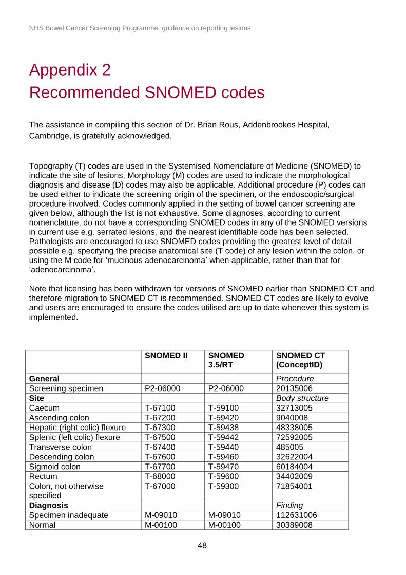
NHS Bowel Cancer Screening Programme: guidance on reporting lesions
48
Appendix 2
Recommended SNOMED codes
The assistance in compiling this section of Dr. Brian Rous, Addenbrookes Hospital,
Cambridge, is gratefully acknowledged.
Topography (T) codes are used in the Systemised Nomenclature of Medicine (SNOMED) to indicate the site of lesions, Morphology (M) codes are used to indicate the morphological diagnosis and disease (D) codes may also be applicable. Additional procedure (P) codes can be used either to indicate the screening origin of the specimen, or the endoscopic/surgical procedure involved. Codes commonly applied in the setting of bowel cancer screening are given below, although the list is not exhaustive. Some diagnoses, according to current nomenclature, do not have a corresponding SNOMED codes in any of the SNOMED versions in current use e.g. serrated lesions, and the nearest identifiable code has been selected. Pathologists are encouraged to use SNOMED codes providing the greatest level of detail possible e.g. specifying the precise anatomical site (T code) of any lesion within the colon, or using the M code for ‘mucinous adenocarcinoma’ when applicable, rather than that for ‘adenocarcinoma’.
Note that licensing has been withdrawn for versions of SNOMED earlier than SNOMED CT and therefore migration to SNOMED CT is recommended. SNOMED CT codes are likely to evolve and users are encouraged to ensure the codes utilised are up to date whenever this system is implemented.
SNOMED II SNOMED 3.5/RT
SNOMED CT (ConceptID)
General Procedure
Screening specimen P2-06000 P2-06000 20135006
Site Body structure
Caecum T-67100 T-59100 32713005
Ascending colon T-67200 T-59420 9040008
Hepatic (right colic) flexure T-67300 T-59438 48338005
Splenic (left colic) flexure T-67500 T-59442 72592005
Transverse colon T-67400 T-59440 485005
Descending colon T-67600 T-59460 32622004
Sigmoid colon T-67700 T-59470 60184004
Rectum T-68000 T-59600 34402009
Colon, not otherwise specified
T-67000 T-59300 71854001
Diagnosis Finding
Specimen inadequate M-09010 M-09010 112631006
Normal M-00100 M-00100 30389008

NHS Bowel Cancer Screening Programme: guidance on reporting lesions
49
SNOMED II SNOMED 3.5/RT
SNOMED CT (ConceptID)
Polyps Morphologic abnormality
Tubular adenoma M-82110 M-82110 19665009
Tubulovillous adenoma M-82630 M-82630 61722000
Villous adenoma M-82611 M-82611 128859003
Hyperplastic polyp M-72042 M-72042 62047007
Sessile serrated lesion M-76801 M-76801 103679000
Traditional serrated adenoma M-82130 M-82130 128653004
Inflammatory polyp M-76820 M-76820 76235005
Mucosal prolapse M-31050 M-31050 29696001
Juvenile polyp M-75662 M-75662 110448004
Neuroendocrine tumour (carcinoid) M-82403 M-82403 81622000
Leiomyoma M-88900 M-88900 44598004
Schwannoma M-95600 M-95600 985004
Neurofibroma M-95400 M-95400 89084002
Ganglioneuroma M-94900 M-94900 53801007
Gastrointestinal stromal tumour M-89361 M-89361 128755003
Lipoma M-88500 M-88500 46720004
Cancers Morphologic abnormality
Adenocarcinoma M-81403 M-81403 35917007
Adenocarcinoma within a polyp M-82103 M-82103 43233001
Suspicious of adenocarcinoma M-67060 M-67060 44085002
Mucinous adenocarcinoma M-84803 M-84803 72495009
Signet ring cell adenocarcinoma M-84903 M-84903 87737001
Adenosquamous carcinoma M-85603 M-85603 59367005
Squamous cell carcinoma M-80703 M-80703 28899001
Neuroendocrine carcinoma M-82463 M-82463 55937004
Small cell carcinoma M-80413 M-80413 74364000
Goblet cell carcinoid tumour M-82433 M-82433 31396002
Mixed adenocarcinoma-neuroendocrine carcinoma (MANEC)
M-82443 M-82443 51465000
Undifferentiated carcinoma M-80203 M-80203 38549000
Carcinoma, other M-80103 M-80103 68453008

NHS Bowel Cancer Screening Programme: guidance on reporting lesions
50
SNOMED II SNOMED 3.5/RT
SNOMED CT (ConceptID)
Dysplasia grade Morphologic abnormality
Low grade dysplasia M-74001 M-74001 43185009
High grade dysplasia M-74003 M-74003 55237006
Other pathology Disorder
Ulcerative colitis D5-41110 D5-41110 64766004
Crohn's disease D5-41000 D5-41000 34000006
Chronic idiopathic inflammatory bowel disease, unclassified
D5-409A0 D5-409A0 359664009
Inflammation, unspecified M-40000 M-40000 23583003

NHS Bowel Cancer Screening Programme: guidance on reporting lesions
51
Appendix 3
TNM 8 classification of colorectal tumours
pT Primary tumour
pTX Primary tumour cannot be assessed
pT0 No evidence of primary tumour
pT1 Tumour invades submucosa
pT2 Tumour invades muscularis propria
pT3 Tumour invades into subserosa or into non-peritonealised pericolic or perirectal
tissues
pT4 Tumour perforates visceral peritoneum (4a) and /or directly invades other organs
or structures (4b)
pN Regional lymph nodes
pNX Regional lymph nodes cannot be assessed
pN0 No regional lymph node metastatic disease
pN1 Metastatic disease in 1 to 3 regional lymph nodes
pN1a Metastasis in 1 regional lymph node
pN1b Metastases in 2 to 3 regional lymph nodes
pN1c Tumour deposit(s), i.e. satellites,* in the subserosa, or in non‐
peritonealised pericolic or perirectal soft tissue without regional lymph
node metastatic disease
pN2 Metastatic disease in 4 or more regional lymph nodes
pN2a Metastases in 4–6 regional lymph nodes
pN2b Metastases in 7 or more regional lymph nodes
pM Distant metastatic disease
pM1 Distant metastatic disease
pM1a Metastasis confined to one organ without peritoneal metastases
pM1b Metastases in more than one organ
pM1c Metastases to the peritoneum with or without other organ involvement
*Tumour deposits or satellites are discrete macroscopic or microscopic nodules of
cancer in the pericolorectal adipose tissue’s lymph drainage area of a primary
carcinoma that are discontinuous from the primary and without histological evidence of
residual lymph node or identifiable vascular or neural structures.