
NGHIÊN CӿU NUãI TҷO TһM BIәU Mã Tԁ Tӕ BÀO GӧC BIәU Mã … · tự thân hoặc của...
55
BỘ GIÁO DỤC VÀ ĐÀO TẠO BỘ Y TẾ TRƢỜNG ĐẠI HỌC Y HÀ NỘI ĐÀO THỊ THÚY PHƢỢNG NGHIÊN CỨU NUÔI TẠO TẤM BIỂU MÔ TỪ TẾ BÀO GỐC BIỂU MÔ NIÊM MẠC MIỆNG Chuyên ngành : Mô - Phôi thai học Mã số : 62720103 TÓM TẮT LUẬN ÁN TIẾN SĨ Y HỌC HÀ NỘI - 2016
Transcript of NGHIÊN CӿU NUãI TҷO TһM BIәU Mã Tԁ Tӕ BÀO GӧC BIәU Mã … · tự thân hoặc của...
BỘ GIÁO DỤC VÀ ĐÀO TẠO BỘ Y TẾ
TRƢỜNG ĐẠI HỌC Y HÀ NỘI
ĐÀO THỊ THÚY PHƢỢNG
NGHIÊN CỨU NUÔI TẠO TẤM BIỂU MÔ
TỪ TẾ BÀO GỐC BIỂU MÔ
NIÊM MẠC MIỆNG
Chuyên ngành : Mô - Phôi thai học
Mã số : 62720103
TÓM TẮT LUẬN ÁN TIẾN SĨ Y HỌC
HÀ NỘI - 2016
CÔNG TRÌNH ĐƯỢC HOÀN THÀNH TẠI:
TRƢỜNG ĐẠI HỌC Y HÀ NỘI
THẦY HƢỚNG DẪN KHOA HỌC:
PGS.TS. NGUYỄN THỊ BÌNH
Phản biện 1: PGS.TS. Quản Hoàng Lâm
Phản biện 2: PGS.TS. Nguyễn Hồng Giang
Phản biện 3: PGS.TS. Trần Vân Khánh
Luận án được bảo vệ trước Hội đồng đánh giá luận án cấp
Trường họp tại Trường Đại học Y Hà Nội.
Vào hồi giờ ngày tháng năm
Có thể tìm hiểu luận án tại:
- Thư viện quốc gia
- Thư viện thông tin Y học Trung ương
- Thư viện Trường Đại học Y Hà Nội
1
ĐẶT VẤN ĐỀ
Tổn thương bề mặt nhãn cầu (BMNC) do nhiều nguyên nhân
khác nhau thường để lại di chứng là hội chứng suy giảm tế bào gốc
vùng rìa giác mạc (limbal stem cell deficiency-LSCD) và hậu quả là
suy giảm thị lực. Trên thế giới, phương pháp hiện đại nhất để điều trị
hội chứng LSCD toàn bộ cả hai bên mắt là ghép tấm biểu mô nuôi
cấy từ tế bào gốc biểu mô niêm mạc miệng tự thân. Niêm mạc miệng
sở dĩ được lựa chọn do có cùng nguồn gốc phôi thai và cấu trúc mô
học với biểu mô trước giác mạc. Đã có nhiều công trình nghiên cứu
trên thế giới công bố về sự thành công của phương pháp này. Tuy
nhiên, cho đến nay ở Việt Nam chưa có một nghiên cứu nào về vấn
đề này. Vì vậy, chúng tôi tiến hành “Nghiên cứu nuôi tạo tấm biểu
mô từ tế bào gốc biểu mô niêm mạc miệng” với các mục tiêu sau:
1. Xác định vị trí, kích thước mảnh mô niêm mạc miệng và môi
trường nuôi cấy phù hợp cho nuôi tạo tấm biểu mô.
2. Xác định phương pháp phù hợp nuôi tạo tấm biểu mô niêm
mạc miệng.
NHỮNG ĐÓNG GÓP MỚI CỦA LUẬN ÁN
1. Đã tìm ra được phương pháp nuôi tạo tấm biểu mô niêm mạc
miệng hoàn toàn mới đó là sử dụng mảnh biểu mô và lớp tế bào đồng
nuôi cấy là nguyên bào sợi tự thân. Trên thực nghiệm, ở giai đoạn
đầu quá trình nghiên cứu, phương pháp này cho tỷ lệ nuôi tạo thành
công cao hơn phương pháp mảnh mô và thấp hơn phương pháp dịch
treo nhưng sự khác biệt không có ý nghĩa thống kê. Ở giai đoạn 2,
chúng tôi đã nuôi 30 mẫu trên thỏ với tỷ lệ nuôi tạo thành công là
100%. Phương pháp mới này đơn giản, hiệu quả, khắc phục được
nhược điểm và tận dụng được ưu điểm của cả hai phương pháp nuôi
cấy hiện đang sử dụng trên thế giới. Nguyên bào sợi tự thân được sử
dụng thay thế cho 3T3 là loại tế bào có nguồn gốc từ chuột. Áp dụng
phương pháp này để nuôi tạo tấm biểu mô từ tế bào gốc biểu mô
niêm mạc miệng trên người, tỷ lệ thành công là 90%.

2
2. Sử dụng nhiều phương pháp khác nhau để định danh các tế bào của
tấm biểu mô nuôi cấy. Kết quả: các tế bào của tấm biểu mô nuôi cấy có
cấu trúc hình thái và hóa học giống với biểu mô trước giác mạc.
3. Ghép các tấm biểu mô bằng phương pháp này cho 15 mắt thỏ bị
suy giảm tế bào gốc vùng rìa giác mạc toàn bộ, 60 ngày sau ghép
giác mạc thỏ trong và tấm biểu mô dính sát vào lớp chân bì. 17 bệnh
nhân được ghép tấm biểu mô có 9 bệnh nhân cải thiện thị lực, số còn
lại không còn hiện tượng tăng sinh xơ mạch vào giác mạc.
CẤU TRÚC CỦA LUẬN ÁN
Luận án gồm 122 trang, 4 chương, 5 bảng, 55 hình, 124 tài liệu
tham khảo với 5 tài liệu tiếng Việt, 119 tài liệu nước ngoài.
Phần đặt vấn đề: 02 trang; chương 1: tổng quan tài liệu 34 trang;
chương 2: đối tượng và phương pháp nghiên cứu 18 trang; chương 3:
kết quả nghiên cứu 38 trang; chương 4: bàn luận 28 trang; kết luận: 1
trang; khuyến nghị: 01 trang; danh mục bài báo liên quan; tài liệu
tham khảo; phụ lục.
CHƢƠNG 1: TỔNG QUAN TÀI LIỆU
1.1. Cấu trúc của bề mặt nhãn cầu
BMNC là vùng được giới hạn bởi hai đường xám của mi trên và
mi dưới, bao gồm biểu mô giác mạc, biểu mô kết mạc và ranh giới là
biểu mô vùng rìa giác mạc và là nơi có các tế bào gốc của giác mạc.
1.1.1. Giác mạc
Biểu mô giác mạc: là biểu mô lát tầng không sừng hoá, gồm 4-6
hàng tế bào, chiếm khoảng 10% bề dày của giác mạc. Biểu mô được
chia thành 3 lớp: lớp đáy, lớp tế bào hình cánh, lớp bề mặt.
1.1.2. Kết mạc
Kết mạc là một bộ phận phụ thuộc nhãn cầu, trải từ vùng rìa
củng giác mạc đến đường xám của bờ mi, được chia thành 3 phần:
kết mạc mi, kết mạc nhãn cầu, kết mạc cùng đồ.
1.1.3. Vùng rìa củng-giác mạc
Vùng rìa là vùng tiếp nối giữa củng mạc với giác mạc, ở đây có
sự chuyển tiếp từ biểu mô giác mạc thành biểu mô kết mạc nhãn cầu.
3
1.1.4. Các yếu tố liên quan đảm bảo sự toàn vẹn của BMNC
Mắt cần có hệ thống thần kinh toàn vẹn và sự toàn vẹn của
BMNC giúp cho hình ảnh nhìn thấy có thể tới điểm hội tụ trên võng
mạc. Mi mắt, phim nước mắt, các tuyến lệ, sự toàn vẹn của hai cung
phản xạ điều tiết nước mắt, chức năng của tế bào biểu mô BMNC
được hỗ trợ bởi nguyên bào sợi, nhu mô và chất cơ bản là các yếu tố
đảm bảo toàn vẹn của bề mặt nhãn cầu.
1.2. Cấu trúc biểu mô bề mặt khoang miệng
Niêm mạc miệng có cấu tạo gồm hai phần chính: biểu mô và lớp
đệm. Biểu mô niêm mạc miệng là loại biểu mô tầng.
Biểu mô niêm mạc má thuộc loại lát tầng không sừng hóa. Tế
bào biểu mô ở lớp đáy gồm 2-3 lớp sát màng đáy có hình trụ hoặc
hình đa diện, có khả năng phân chia để duy trì quần thể tế bào biểu
mô ổn định. Các tế bào phân chia thường tạo thành từng cụm, nhìn
thấy nhiều hơn ở chỗ sâu nhất của lõm biểu mô.
Khi tế bào rời lớp đáy và bước vào quá trình biệt hóa, tế bào lớn hơn
dẹt dần và tích lũy xơ keratin và lipid trong bào tương ngày một nhiều.
Ở niêm mạc miệng, khi tế bào gốc phân chia, nó sẽ tạo ra một tế
bào con giữ nguyên đặc tính của tế bào gốc là khả năng phân chia vô
hạn định và cho ra một tế bào con khác bước vào quá trình biệt hoá.
Có rất nhiều yếu tố quyết định số phận tế bào sẽ trở thành gốc hay tế
bào tăng sinh chuyển tiếp.
Các tương tác giữa tế bào biểu mô và mô liên kết đóng vai trò
quan trọng trong việc điều khiển sự phát triển mô.
1.3. Hội chứng suy giảm tế bào gốc vùng rìa giác mạc
Hội chứng suy giảm tế bào gốc vùng rìa có thể nguyên phát,
hoặc có thể là hậu quả thứ phát. Suy giảm vùng rìa có thể xảy ra toàn
bộ hoặc chỉ ở một góc của vùng rìa. Biểu hiện lâm sàng có thể chỉ
là giác mạc mờ đục, bề mặt gồ ghề không đều, có tân mạch nông
hoặc sâu trong bề dày giác mạc hoặc kết mạc hóa giác mạc. Nặng
hơn nữa có thể là ổ loét giác mạc khó hàn gắn, bờ ổ loét ranh giới
4
rõ và gồ lên, xung quanh ổ loét có thể tồn tại tổ chức xơ tân mạch,
BMNC gồ ghề với biểu hiện của một quá trình viêm mãn tính,
nhuyễn giác mạc, giác mạc mỏng hoặc thủng giác mạc có thể xảy
ra trong trường hợp nặng.
1.4. Những nghiên cứu về nuôi tạo tấm biểu mô niêm mạc miệng
Yếu tố quan trọng để nuôi tạo tấm biểu mô là lựa chọn giá đỡ
chính xác về tính phù hợp sinh học, độ xốp, ổn định sinh học và đặc
tính vật lí. Các giá đỡ sử dụng trong nuôi cấy tế bào gốc biểu mô
niêm mạc miệng gồm nhiều loại khác nhau, nhưng màng ối đã được
nhiều tác giả sử dụng.
Việc chuẩn bị mẫu mô niêm mạc miệng và xử lý miếng mô cho
nuôi cấy cũng có vai trò rất quan trọng. Sau khi sát khuẩn kỹ khoang
miệng, bệnh nhân được gây tê tại chỗ, dùng dao tròn trích thủ mảnh
niêm mạc miệng. Kích thước mảnh mô trích thủ thay đổi tuỳ từng tác
giả và phụ thuộc vào yêu cầu và phương pháp nuôi cấy. Mảnh mô
được xử lý qua nhiều công đoạn và phương pháp xử lý mảnh mô
khác nhau. Có hai phương pháp chính nuôi cấy đó là nuôi bằng mảnh
mô hoặc dịch treo tế bào.
Trong nuôi cấy, việc sử dụng nguyên bào sợi chuột bất hoạt 3T3
làm lớp tế bào nuôi cũng gây nhiều lo ngại bởi đây là sản phẩm có
nguồn gốc động vật. Đã có nhiều nghiên cứu cho thấy thành công của
nuôi tạo tấm biểu mô mà không cần tới sự có mặt của tế bào này.
Môi trường nuôi cấy tế bào biểu mô niêm mạc miệng thông
thường là sự kết hợp của Ham’s F12 và DMEM với tỉ lệ 1:1. Ngoài
ra cần có các yếu tố bổ sung khác: Insulin, hydrocortisone, T3,
isoproterenol, choleratoxin, các hormon tăng trưởng... Các thành
phần bổ sung thông thường có mặt đầy đủ trong huyết thanh. Huyết
thanh bào thai bò (FBS) được rất nhiều tác giả sử dụng, nhưng đây là
sản phẩm có nguồn gốc động vật. Vì thế, có tác giả dùng huyết thanh
tự thân hoặc của người có nhóm máu AB để thay thế FBS. Một số tác
giả đã sử dụng môi trường không huyết thanh.
5
Có nhiều cách khác nhau để định danh tế bào của tấm biểu mô:
(1) Quan sát hình thái tấm biểu mô sống bằng kính hiển vi soi nổi.
(2) Nhuộm trypan blue: để xác định tỷ lệ tế bào sống và chết của tế
bào. (3) Nhuộm giemsa: để quan sát bề mặt của tấm biểu mô nuôi
cấy. (4) Nhuộm Hematoxylin-Eosin (H.E) nhằm đánh giá cấu trúc vi
thể của tấm biểu mô theo chiều dọc. (5) Kỹ thuật hiển vi điện tử: để
nghiên cứu cấu trúc của tế bào và tấm biểu mô. (6) Kỹ thuật khuyếch
đại chuỗi PCR (polymerase chain reaction): dùng phát hiện các
marker của tế bào biểu mô niêm mạc miệng. (7) Kỹ thuật hoá mô
miễn dịch: Đây là kỹ thuật hiện đại và được sử dụng phổ biến khi
đánh giá đặc điểm của tấm biểu mô niêm mạc miệng nuôi cấy.
Nhuộm hoá mô miễn dịch để xác định các marker: K3, K12,
connexin-43 (Cx-43), p63, p75, MCSP, β1 intergrin, PPARγ, Ki67,
Pax 6, occludin, ZO1, ABCG2, desmoplakin... (8) Test tạo cụm: Khi
tách và phát triển ngoài cơ thể, một số đặc điểm của tế bào gốc được
giữ lại và phản ánh ở kiểu của các cụm mà nó hình thành.
Tấm biểu mô niêm mạc miệng được ứng dụng: (1) Trong
nhãn khoa, (2) Điều trị các trường hợp bỏng da rộng, cho các phẫu
thuật đường niệu, tạo hình âm đạo, thực quản hoặc tái thiết mi mắt.
CHƢƠNG 2: ĐỐI TƢỢNG VÀ PHƢƠNG PHÁP NGHIÊN CỨU
2.1.Đối tƣợng nghiên cứu
2.1.1.Đối tượng và vật liệu nghiên cứu:
-Mảnh mô niêm mạc miệng của thỏ chủng Orytolagus Cuniculus
khoẻ mạnh.
-Mảnh mô niêm mạc miệng người.
-3T3 do bộ môn Tế bào-Mô Phôi và Lý sinh, Đại học Khoa học
tự nhiên cung cấp.
-Màng ối người đã xử lý theo quy trình của bộ môn Mô-Phôi,
trường Đại học Y Hà Nội.
6
2.1.2. Mô hình nghiên cứu
Nghiên cứu tiến hành qua hai giai đoạn:
Giai đoạn 1: Tiến hành thực nghiệm trên thỏ.
-Nghiên cứu quy trình trích thủ và xử lý tấm biểu mô niêm mạc
miệng, lựa chọn môi trường nuôi cấy, phương pháp nuôi cấy phù hợp
-Đánh giá chất lượng tấm biểu mô
-Ghép thực nghiệm trên thỏ đã gây bỏng tổn thương toàn bộ
vùng rìa.
Giai đoạn 2:
Dựa trên những kết quả trên thỏ, giai đoạn 2 sẽ tiến hành trên bệnh
nhân suy giảm tế bào gốc vùng rìa toàn bộ với quy trình tương tự.
2.2. Quy trình nuôi cấy
2.2.1. Chuẩn bị trang thiết bị cần thiết cho nuôi cấy
2.2.2. Thực nghiệm trên thỏ
2.2.2.1. Chuẩn bị màng ối: Sử dụng màng ối được xử lí theo quy
trình của bộ môn Mô-Phôi, trường Đại học Y Hà Nội, tức là màng ối
đã loại bỏ biểu mô bằng ammonia 10%.
2.2.2.2. Chuẩn bị lớp 3T3 làm nền nuôi cấy: Chuẩn bị lớp 3T3 (sử
dụng mẫu 3T3 đã qua xử lý mitomycin)
2.2.2.3. Chuẩn bị mảnh mô niêm mạc miệng cho nuôi cấy
- Gây mê thỏ bằng đường tĩnh mạch rìa tai, sát trùng khoang miệng
- Trích thủ: (1) mặt trong niêm mạc má phần trung tâm, (2) mặt
trong niêm mạc má cách góc miệng 2mm và vuông góc, (3) mặt
trong niêm mạc môi dưới, phần trung tâm.
- Dùng dao tròn trích thủ các mảnh niêm mạc với kích thước:
đường kính 3mm, 6mm, 8mm.
+ Kiểm tra cấu trúc vi thể của mảnh niêm mạc miệng
+ Mảnh niêm mạc miệng được rửa bằng PBS có bổ sung kháng
sinh, kháng nấm.
(1) Nuôi bằng phương pháp mảnh mô:
- Cắt mảnh niêm mạc miệng thành các mảnh nhỏ 1x1mm
- Ủ miếng mô trong dung dịch dispase II, rửa lại bằng PBS,
ngâm EDTA, sau đó miếng mô được rửa lại sạch bằng môi trường
nuôi cấy.
7
- Nuôi cấy các mảnh mô đã được xử lý trên nền màng ối.
(2) Nuôi cấy bằng dịch treo:
- Cắt mảnh niêm mạc miệng thành các mảnh nhỏ 0,5x0,5mm.
- Ủ mảnh niêm mạc miệng trong dispase II, bóc mảnh biểu mô
ra khỏi mô nền, ngâm mảnh biểu mô trong Trypsin-EDTA, sau đó
rửa lại mảnh biểu mô và bằng DMEM+Ham’s F12 có kháng sinh,
kháng nấm và 10% FBS.
- Nạo lấy các tế bào lớp đáy biểu mô và li tâm lấy các tế bào
biểu mô.
- Tạo dịch treo có mật độ tế bào 1x10⁶ tế bào/ml.
- Nuôi cấy trong lồng nuôi cấy ở điều kiện 37oC, 5% CO2. Thay
môi trường đều đặn 2 ngày 1 lần.
- Nếu sử dụng lớp 3T3: 03 ngày thay 3T3 một lần.
(3) Nuôi cấy bằng phương pháp mảnh biểu mô:
Đây là phương pháp hoàn toàn mới, chưa có tác giả nào trên thế
giới sử dụng.
- Ủ mảnh niêm mạc miệng đã cắt nhỏ 0,5x0,5mm trong dispase
II, bóc rời mảnh biểu mô khỏi mô nền, ngâm mô sau bóc vào trysin-
EDTA 0,05%, sau đó rửa lại bằng DMEM+Ham’s F12 có kháng
sinh, kháng nấm và 10% FBS.
- Dán mảnh biểu mô lên trên nền màng ối để mặt biểu mô hướng
lên trên.
- Dán mô nền xuống đáy giếng nuôi cấy với tỉ lệ 3 mảnh biểu
mô/2 mảnh mô nền.
- Nuôi cấy trong điều kiện 370C, 5% CO2, thay môi trường 2
ngày/lần.
Ở các phương pháp nuôi cấy khác nhau, theo dõi liên tục sự phát
triển của tấm biểu mô. Khi tế bào biểu mô mọc kín đáy lồng nuôi
cấy, tiến hành tạo tầng cho tấm biểu mô, đánh giá chất lượng của tấm
biểu mô nuôi cấy sau thu hoạch.
2.2.2.3. Môi trường nuôi cấy, quy trình nuôi cấy và theo dõi
- Môi trường nuôi cấy SHEM 1: gồm DMEM/F12 tỉ lệ 1:1
(Gibco-Mỹ), có bổ sung: FBS 10% (Gibco), insulin 5µg/ml (Gibco),
8
EGF 10ng/ml (Gibco), penicillin 100UI/ml (Wako), streptomycin
100µg/ml (Wako), amphotericin B 0,25µg/ml (Gibco).
- Môi trường nuôi cấy SHEM 2: gồm DMEM/F12 tỉ lệ 1:1 (có
bổ sung: FBS 10%, insulin 5µg/ml, EGF 10ng/ml, triiodothyronin
1,3ng/ml, isoproterenol 0,25µg/ml, hydrocortisone 0,5µg/ml, penicillin
100UI/ml, streptomycin 100µg/ml, amphotericin B 0,25µg/ml.
2.2.2.4. Thu hoạch và định danh tế bào nuôi cấy
Sau khi nuôi cấy được tấm biểu mô kích thước 4 cm2, mẫu nuôi
cấy được ghép lại cho thỏ thực nghiệm, phần còn lại tiến hành định
danh tế bào của tấm biểu mô nuôi cấy bằng các kỹ thuật hiển vi
quang học, hiển vi điện tử, hoá mô, hoá mô miễn dịch.
2.2.3. Thử nghiệm trên bệnh nhân tự nguyện.
Tiến hành sau khi có kết quả định hướng của thực nghiệm trên thỏ.
2.3. Chỉ tiêu nghiên cứu
Tỷ lệ nuôi tạo thành công tấm biểu mô.
Thời gian nuôi cấy.
Cấu trúc vi thể, siêu vi thể và hóa học của tấm biểu mô nuôi cấy.
2.4. Địa điểm và thời gian nghiên cứu
Nghiên cứu tiến cứu được tiến hành tại bộ môn Mô-Phôi và khoa
Kết-Giác mạc Bệnh viện Mắt trung ương, từ tháng 10/1010 đến
10/2013.
2.5. Thiết kế nghiên cứu
Xử lí số liệu theo phần mềm SPSS 16.0.
2.6. Đạo đức nghiên cứu
Đề tài là một phần của đề tài độc lập cấp nhà nước “Nghiên cứu
quy trình sử dụng tế bào gốc để điểu trị một số bệnh cả bề mặt nhãn
cầu”đã được thông qua hội đồng đạo đức Y học, Trường Đại học Y
Hà Nội.
9
CHƢƠNG 3: KẾT QUẢ NGHIÊN CỨU
3.1. Kết quả nghiên cứu về nuôi tạo tấm biểu mô trên thỏ thực nghiệm.
3.1.1. Lựa chọn vị trí sinh thiết và kích thước mảnh mô để nuôi cấy
Sinh thiết niêm mạc miệng trên 5 thỏ chủng Orytolagus
Cuniculus ở 3 vị trí khác nhau, chúng tôi nhận thấy:
Ở mặt trong niêm mạc má phần trung tâm: Biểu mô là loại lát
tầng không sừng hóa. Biểu mô dày, gồm 18-20 hàng tế bào, chia làm
3 lớp, lớp tế bào đáy gồm 2-3 hàng tế bào có kích thước nhỏ, nhân
hình trứng, sẫm màu, bào tương rất ưa base. Trên các tiêu bản nhuộm
p63, nhân các tế bào lớp đáy bắt màu rất đậm. Mô liên kết của lớp
đệm tạo thành các nhú chân bì rất cao.
Mặt trong niêm mạc má cách góc miệng 2mm và vuông góc, mặt
trong niêm mạc môi dưới phần trung tâm:Biểu mô là loại lát tầng
không sừng hóa, mỏng, gồm 4-5 hàng tế bào. Các tế bào lớp đáy có
nhân hình trứng, sẫm màu, bào tương ưa base. Ranh giới giữa biểu
mô và mô liên kết bên dưới tương đối bằng phẳng, không có các nhú
chân bì.
Chúng tôi lựa chọn vị trí lấy mảnh mô dùng cho nuôi cấy là mặt
trong, phần trung tâm niêm mạc má.
Khi trích thủ mẫu để làm nghiên cứu, chúng tôi nhận thấy, để
nuôi tạo được 2 tấm biểu mô, mảnh mô trích thủ cần phải có kích
thước: (1) đường kính 6mm ở phương pháp nuôi bằng mảnh mô. (2)
đường kính 8mm mới đủ lượng tế bào tạo được 2 ml dịch treo có mật
độ 1x10⁶ tế bào/ml ở phương pháp dịch treo. (3) đường kính 3mm ở
phương pháp nuôi bằng mảnh biểu mô.
3.1.2. Lựa chọn môi trường nuôi cấy
Giai đoạn đầu tiến hành thực nghiệm chúng tôi nuôi 18 mẫu
mảnh mô niêm mạc miệng bằng môi trường SHEM1, chỉ có 30%
mẫu mọc và các mẫu mọc đều không kín đáy sau 28 ngày nuôi cấy.
Sau đó, nghiên cứu chuyển sang sử dụng môi trường SHEM2, và
10
toàn bộ kết quả nghiên cứu tiến hành nuôi cấy trong môi trường
SHEM2 với tỉ lệ nuôi tạo thành công tấm biểu mô là 76,47-95%.
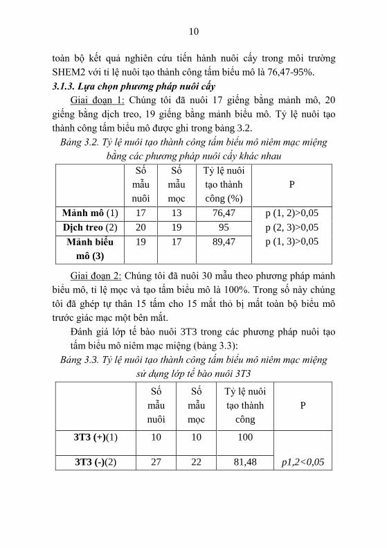
3.1.3. Lựa chọn phương pháp nuôi cấy
Giai đoạn 1: Chúng tôi đã nuôi 17 giếng bằng mảnh mô, 20
giếng bằng dịch treo, 19 giếng bằng mảnh biểu mô. Tỷ lệ nuôi tạo
thành công tấm biểu mô được ghi trong bảng 3.2.
Bảng 3.2. Tỷ lệ nuôi tạo thành công tấm biểu mô niêm mạc miệng
bằng các phương pháp nuôi cấy khác nhau
Số
mẫu
nuôi
Số
mẫu
mọc
Tỷ lệ nuôi
tạo thành
công (%)
P
Mảnh mô (1) 17 13 76,47 p (1, 2)>0,05
Dịch treo (2) 20 19 95 p (2, 3)>0,05
p (1, 3)>0,05 Mảnh biểu
mô (3)
19 17 89,47
Giai đoạn 2: Chúng tôi đã nuôi 30 mẫu theo phương pháp mảnh
biểu mô, tỉ lệ mọc và tạo tấm biểu mô là 100%. Trong số này chúng
tôi đã ghép tự thân 15 tấm cho 15 mắt thỏ bị mất toàn bộ biểu mô
trước giác mạc một bên mắt.
Đánh giá lớp tế bào nuôi 3T3 trong các phương pháp nuôi tạo
tấm biểu mô niêm mạc miệng (bảng 3.3):
Bảng 3.3. Tỷ lệ nuôi tạo thành công tấm biểu mô niêm mạc miệng
sử dụng lớp tế bào nuôi 3T3
Số
mẫu
nuôi
Số
mẫu
mọc
Tỷ lệ nuôi
tạo thành
công
P
3T3 (+)(1) 10 10 100
3T3 (-)(2) 27 22 81,48 p1,2<0,05
11
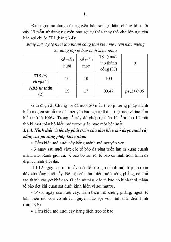
Đánh giá tác dụng của nguyên bào sợi tự thân, chúng tôi nuôi
cấy 19 mẫu sử dụng nguyên bào sợi tự thân thay thế cho lớp nguyên
bào sợi chuột 3T3 (bảng 3.4):
Bảng 3.4. Tỷ lệ nuôi tạo thành công tấm biểu mô niêm mạc miệng
sử dụng lớp tế bào nuôi khác nhau
Số mẫu
nuôi
Số mẫu
mọc
Tỷ lệ nuôi
tạo thành
công (%)
p
3T3 (+)
chuột(1) 10 10 100
NBS tự thân
(2) 19 17 89,47 p1,2>0,05
Giai đoạn 2: Chúng tôi đã nuôi 30 mẫu theo phương pháp mảnh
biểu mô, có sự hỗ trợ của nguyên bào sợi tự thân, tỉ lệ mọc và tạo tấm
biểu mô là 100%. Trong số này đã ghép tự thân 15 tấm cho 15 mắt
thỏ bị mất toàn bộ biểu mô trước giác mạc một bên mắt.
3.1.4. Hình thái và tốc độ phát triển của tấm biểu mô được nuôi cấy
bằng các phương pháp khác nhau
Tấm biểu mô nuôi cấy bằng mảnh mô nguyên vẹn:
- 3 ngày sau nuôi cấy: các tế bào đã phát triển lan ra xung quanh
mảnh mô. Ranh giới các tế bào bò lan rõ, tế bào có hình tròn, hình đa
diện và hình thoi dài.
-10-12 ngày sau nuôi cấy: các tế bào tạo thành một lớp phủ kín
đáy của lồng nuôi cấy. Bề mặt của tấm biểu mô không phẳng, có chỗ
tạo thành các gờ khá cao. Ở các gờ này, các tế bào có hình thoi, nhân
tế bào dẹt khi quan sát dưới kính hiển vi soi ngược.
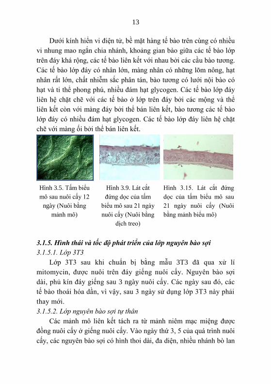
- 14-16 ngày sau nuôi cấy: Tấm biểu mô không phẳng, ngoài tế
bào biểu mô còn có nhiều nguyên bào sợi với hình thái điển hình
(hình 3.5).
Tấm biểu mô nuôi cấy bằng dịch treo tế bào
12
- 2 ngày sau nuôi cấy: có khá nhiều các tế bào tròn bám vào đáy,
sau đó, các tế bào này xoè rộng với các nhánh bào tương khá dài
- 12-14 ngày sau nuôi cấy: tế bào biểu mô phủ kín lồng nuôi cấy.
- Sau khi tạo tầng, nhuộm H.E. thấy: tấm biểu mô phẳng, gồm 5-7
hàng tế bào, các lớp tế bào trên có xu hướng dẹt dần. Khoảng gian bào
của tấm biểu mô rộng. Không thấy các tế bào hình thoi xen lẫn (hình
3.9).
Trên kính hiển vi điện tử, khoảng gian bào giữa các tế bào ở lớp
giữa của tấm biểu mô khá rộng. Các tế bào ở đây liên kết với nhau
bằng các mộng bào tương và thể liên kết. Trong bào tương các tế
bào, các bào quan rất phát triển, có các hạt glycogen, các bó xơ
trương lực, lưới nội bào có hạt.
Tấm biểu mô nuôi cấy bằng mảnh biểu mô
- 3-4 ngày sau nuôi cấy: Các tế bào có hình tròn, một số có hình
đa diện với các nhánh bào tương dài bò lan.Tế bào lan rộng dần, lúc
đầu các tế bào có hình đa diện lớn, khoảng gian bào rộng, khi các tế
bào phát triển kín đáy lồng nuôi cấy vào khoảng ngày thứ 10-12, các
tế bào nằm sát nhau, kích thước nhỏ đi, khoảng gian bào hẹp tuy
nhiên vẫn quan sát thấy rõ ranh giới giữa các tế bào với nhau, nhiều
hình ảnh tế bào đang phân chia.
- Sau khi tạo tầng: tấm biểu mô phẳng, gồm 5-7 hàng tế bào, các
lớp tế bào trên có xu hướng dẹt dần. Khoảng gian bào của tấm biểu
mô nuôi bằng mảnh biểu mô hẹp hơn so với nuôi dịch treo. Không
thấy các tế bào hình thoi xen lẫn (hình 3.15).
Khi quan sát trên bề mặt tấm biểu mô nuôi cấy vào ngày thứ 14
bằng phương pháp nhuộm giemsa: tấm biểu mô được phủ kín. Ở
phương pháp mảnh biểu mô, khoảng gian bào hẹp và đều nhau trên toàn
đáy lồng nuôi cấy, ngược lại với kết quả của phương pháp dịch treo có
khoảng gian tế bào rộng hơn và có những khoảng gian tế bào với kích
thước không đều nhau. Không thấy các tế bào hình thoi xen lẫn.
13
Dưới kính hiển vi điện tử, bề mặt hàng tế bào trên cùng có nhiều
vi nhung mao ngắn chia nhánh, khoảng gian bào giữa các tế bào lớp
trên đáy khá rộng, các tế bào liên kết với nhau bởi các cầu bào tương.
Các tế bào lớp đáy có nhân lớn, màng nhân có những lõm nông, hạt
nhân rất lớn, chất nhiễm sắc phân tán, bào tương có lưới nội bào có
hạt và ti thể phong phú, nhiều đám hạt glycogen. Các tế bào lớp đáy
liên hệ chặt chẽ với các tế bào ở lớp trên đáy bởi các mộng và thể
liên kết còn với màng đáy bởi thể bán liên kết, bào tương các tế bào
lớp đáy có nhiều đám hạt glycogen. Các tế bào lớp đáy liên hệ chặt
chẽ với màng ối bởi thể bán liên kết.
Hình 3.5. Tấm biểu
mô sau nuôi cấy 12
ngày (Nuôi bằng
mảnh mô)
Hình 3.9. Lát cắt
đứng dọc của tấm
biểu mô sau 21 ngày
nuôi cấy (Nuôi bằng
dịch treo)
Hình 3.15. Lát cắt đứng
dọc của tấm biểu mô sau
21 ngày nuôi cấy (Nuôi
bằng mảnh biểu mô)
3.1.5. Hình thái và tốc độ phát triển của lớp nguyên bào sợi
3.1.5.1. Lớp 3T3
Lớp 3T3 sau khi chuẩn bị bằng mẫu 3T3 đã qua xử lí
mitomycin, được nuôi trên đáy giếng nuôi cấy. Nguyên bào sợi
dài, phủ kín đáy giếng sau 3 ngày nuôi cấy. Các ngày sau đó, các
tế bào thoái hóa dần, vì vậy, sau 3 ngày sử dụng lớp 3T3 này phải
thay mới.
3.1.5.2. Lớp nguyên bào sợi tự thân
Các mảnh mô liên kết tách ra từ mảnh niêm mạc miệng được
đồng nuôi cấy ở giếng nuôi cấy. Vào ngày thứ 3, 5 của quá trình nuôi
cấy, các nguyên bào sợi có hình thoi dài, đa diện, nhiều nhánh bò lan
14
ra xung quanh mảnh mô liên kết, chỉ ngày thứ 6, khoảng 1/2 diện tích
của đáy giếng nuôi cấy đã được phủ bởi nguyên bào sợi và tới
khoảng ngày thứ 10 thì toàn bộ đáy giếng được phủ kín.
3.1.6. Kết quả định danh tế bào tấm biểu mô nuôi cấy bằng hóa mô
miễn dịch
Trên các tiêu bản nhuộm hóa mô miễn dịch phát hiện p63, nhân
các tế bào của tấm biểu mô bắt màu nâu sẫm, đặc biệt là nhân các tế
bào lớp đáy.
Nhuộm phát hiện K3 và K12: K3 và K12 thể hiện yếu ở các tế
bào lớp trên đáy.
Nhuộm P.A.S.: Trong bào tương các tế bào lớp dưới có ít
glycogen. Không thấy các tế bào tiết nhày.
3.1.7. Kết quả ghép tấm biểu mô niêm mạc miệng nuôi cấy cho thỏ
gây bỏng thực nghiệm
Tổng số thỏ sống trong quá trình làm thực nghiệm là 21, trong
đó 15 thỏ được ghép tấm biểu mô niêm mạc miệng nuôi cấy ở các
thời điểm khác nhau.
Tất cả các thỏ ở các lô đều có kết quả tốt: giác mạc trong, biểu
mô liền tốt, nhẵn bóng, không còn tân mạch. Chỉ có 1 thỏ có kết quả
trung bình: tân mạch qua rìa vào chu biên ở thời điểm 60 ngày nhưng
không vào đến trung tâm giác mạc.
3.2. Kết quả nuôi cấy tấm biểu mô niêm mạc miệng từ tế bào gốc
niêm mạc miệng trên ngƣời
Dựa trên kết quả phân tích trên thỏ, chúng tôi lựa chọn vị trí sinh
thiết trên người là mặt trong trung tâm niêm mạc má. Kết quả cho
thấy biểu mô gồm khoảng 10-15 hàng tế bào, không dày như niêm
mạc vùng tương ứng của thỏ. Tuy vậy, lớp đáy dày và gồm khoảng
3-4 lớp gồm các tế bào có kích thước nhỏ, bào tương bắt màu base
đậm, lớp Malpighi gồm nhiều hàng (7-10 hàng), kích thước tế bào
lớp này lớn hơn của thỏ ở vị trí tương ứng, ranh giới giữa các tế bào
khá rõ. Trên cùng là khoảng 2-3 hàng tế bào dẹt, chứa nhân dẹt. Trên
15
tiêu bản nhuộm p63, nhân tế bào đặc biệt là ở lớp đáy bắt màu đậm.
Các nhú chân bì cũng có kích thước lớn, chia nhánh rõ. Mô đệm lỏng
lẻo, ít tế bào. Cấu trúc niêm mạc miệng vùng giữa má ở nam và nữ
đều giống nhau.
Sau khi cân nhắc về độ phức tạp của quy trình và kết quả thành
công nuôi tạo của hai phương pháp dịch treo và mảnh biểu mô, cùng
với kích thước trích thủ mảnh mô, phương pháp mảnh biểu mô đã
được lựa chọn trong nghiên cứu ứng dụng trên người của chúng tôi.
Kích thước mảnh mô được lựa chọn là đường kính 3mm, vị trí sinh
thiết ở mặt trong vùng giữa má. Tỷ lệ nuôi thành công là 90%.
Dựa trên kết quả nghiên cứu thực nghiệm trên thỏ, chúng tôi đã
tiến hành nuôi cấy niêm mạc miệng của 17 bệnh nhân (4 bệnh nhân
nuôi 2 lần) bằng môi trường SHEM2, với phương pháp nuôi cấy là
mảnh biểu mô. Số tấm biểu mô nuôi được là 54 (tỷ lệ nuôi thành
công là 90%, 3 bệnh nhân nuôi cấy không thành công). Số tấm biểu
mô được ghép lại cho bệnh nhân là 22. Với thời gian nuôi cấy các tế
bào biểu mô là 16-28 ngày. Tấm biểu mô nuôi cấy có khoảng 4-5
hàng tế bào, hàng tế bào trên cùng dẹt và có nhân dẹt.
Bề mặt tế bào tấm biểu mô nuôi cấy trên người có những vi
nhung mao, giống với hình ảnh của tế bào bề mặt ở giác mạc người
bình thường. Song kích thước của các vi nhung mao lớn hơn, và số
lượng ít hơn so với tấm biểu mô nuôi cấy ở thỏ, tế bào của tấm biểu
mô liên kết với nhau bằng các cầu bào tương dài (dài hơn so với cầu
bào tương khi nuôi cấy trên thỏ thực nghiệm) và các thể liên kết
trong bào tương tế bào có nhiều lưới nội bào có hạt, ti thể, hạt
glycogen bộ Golgi nằm gần nhân với các túi dẹt và không bào, ranh
giới giữa các tế bào rộng, tuy nhiên hẹp hơn so với khoảng gian bào
của tấm biểu mô khi nuôi cấy trên thỏ. Bào tương tế bào biểu mô
nuôi cấy có nhiều ti thể dài, mào rõ, chất nền sẫm màu và lưới nội
bào có hạt phát triển mạnh, lưới nội bào có lòng hẹp và ít ribosom
bám ngoài, khoảng gian bào rất hẹp.
16
Trên các tiêu bản nhuộm hóa mô miễn dịch phát hiện p63, nhân
các tế bào của tấm biểu mô bắt màu nâu sẫm, đặc biệt là nhân các tế
bào lớp đáy.
Nhuộm phát hiện K3: K3 thể hiện yếu ở các tế bào lớp đáy và rõ
ở các tế bào lớp trên.
Tấm biểu mô niêm mạc miệng nuôi cấy được ghép lại cho bệnh
nhân và được đánh giá kết quả phẫu thuật dựa trên 3 yếu tố: độ
trong và áp của tấm biểu mô, tình trạng biểu mô bề mặt nhãn cầu,
tăng sinh tân mạch nông, sâu hoặc tổ chức xơ trên giác mạc.
Phẫu thuật ghép thành công ở 12 ca. Trong đó 9 ca có thị lực cải
thiện, chúng tôi nhận thấy ở các bệnh nhân này có sự cải thiện rõ rệt
về thị lực nhìn gần, trong khoảng 10–30 cm.
CHƢƠNG 4: BÀN LUẬN
4.1. Về lựa chọn nền nuôi cấy
Nghiên cứu của chúng tôi sử dụng màng ối đã nạo sạch biểu mô
bằng ammonia 10% theo quy trình chuẩn của bộ môn Mô-Phôi,
trường Đại học Y Hà Nội. Hiện nay, các tác giả trên thế giới chưa
thống nhất được sử dụng nền nuôi cấy nào và nếu dùng màng ối thì
sử dụng như thế nào? Nếu dùng màng ối, loại bỏ biểu mô trước khi
nuôi cấy được sử dụng rộng rãi hơn (Kim và cs. 2008, Lim và cs.
2009, Nakamura và cs. 2004). Bên cạnh đó, có những nghiên cứu lại
khẳng định sự ưu việt của màng ối để nguyên biểu mô (Fukuda và cs.
1999).
Tuy nhiên, nếu không sử dụng màng ối (sử dụng màng polymer
nhạy cảm nhiệt hoặc hồ fibrin), bề mặt giác mạc sau ghép sẽ trong
hơn khi quan sát trên thỏ (Higa K. và cs. 2007, Higa K. và cs. 2012,
Hayashida và cs. (2005), Kokaba V. và cs. (2014), Nishida K. và cs.
(2004), Hori Y. và cs. (2007), Hori Y. và cs. 2008, Oie Y. và cs.
(2010), Hayashida Y. và cs. (2005)).
17
Nghiên cứu của Higa K. tiến hành trên mô hình thỏ bị nạo bỏ
biểu mô giác mạc và vùng rìa, nhưng không gây thương tổn gì mô
đệm, vì vậy, kết quả ghép tấm biểu mô nuôi cấy trên nền fibrin cho
kết quả tốt. Tuy nhiên, trong trường hợp tổn thương mô đệm, hay gặp
do bỏng nặng, thì sử dụng nền màng ối là sự lựa chọn phù hợp hơn.
Chính tác giả Kocaba V. và cs. (2014) sử dụng màng polymer nhạy
cảm nhiệt, song không thấy sự tái tạo trở lại của màng Bowman do bị
hủy trước khi ghép, vậy sử dụng màng ối hợp với sinh lý hơn.Sudha
B. và cs. (2009) cũng khẳng định ưu điểm của màng ối.
Trong nghiên cứu của chúng tôi, bệnh nhân Võ Vũ Ngọc Y. (31
tuổi) sau ghép tấm biểu mô niêm mạc miệng nuôi cấy 12 tháng, tấm
biểu mô còn nguyên vẹn, điều này chứng tỏ trong tấm biểu mô nuôi
cấy vẫn có tế bào gốc.
4.2. Về vị trí và kích thƣớc của mảnh niêm mạc miệng dùng cho
nuôi cấy
Khi nghiên cứu cấu trúc vi thể niêm mạc miệng thỏ chúng tôi
thấy: niêm mạc vùng má thỏ có lớp biểu mô khá dày, đường ranh
giới với mô liên kết bên dưới có các nhú cao. Khoảng 2-3 hàng tế bào
sát đáy có kích thước nhỏ, nhân lớn, biểu hiện dương tính với marker
p63. Ở bệnh nhân, niêm mạc vùng mặt trong má cũng có kết quả
tương tự. Kết quả của chúng tôi phù hợp với nhiều công bố trên thế
giới Priya C. G. và cs. 2011, Squier C. A. và cs. 2001, Hori Y. và cs.
2007, Inatomi T. và cs 2006, Dua và cs 2003, Ma D. H. và cs. 2009,
Hayashida Y. và cs. 2005.
Để có được hai tấm biểu mô có kích thước 4cm2, khi nuôi cấy
bằng dịch treo, mảnh niêm mạc cần có đường kính 8mm. Một số tác
giả trích thủ mảnh mô nhỏ hơn, song chỉ nuôi 01 tấm: Nishida K. và
cs. (2004) sử dụng đường kính 3mm, Ma D. H. và cs. (2009) là
6x6mm. Khi nuôi bằng mảnh mô, chúng tôi trích thủ mảnh có đường
kính 6mm. Phương pháp nuôi bằng mảnh biểu mô, đường kính của
mảnh niêm mạc miệng là 3mm. Ở phương pháp nuôi cấy bằng mảnh
18
biểu mô, diện tiếp xúc của các tế bào gốc lớp đáy được bộc lộ tối đa,
loại bỏ được hoàn toàn nguyên bào sợi nên tấm biểu mô nuôi cấy
phẳng đẹp.
4.3. Về môi trƣờng nuôi cấy
Mỗi tác giả có một công thức riêng cho môi trường nuôi cấy.
Giai đoạn đầu, chúng tôi sử dụng môi trường SHEM1, tỉ lệ các mẫu
mọc chỉ là 30%, các tấm biểu mô có tế bào thưa thớt.
Khi dùng SHEM2 là môi trường SHEM1 có bổ sung insulin,
hydrocortisone, T3, isoproterenol, các tế bào gốc biểu mô niêm mạc
miệng đã tăng sinh tạo thành tấm biểu mô với tỉ lệ từ 89,5%-95%.
Mặc dù so với nhiều tác giả khác trên thế giới, môi trường SHEM2
của chúng tôi thiếu choleratoxin. Tuy nhiên, với tỉ lệ nuôi tạo thành
công tấm biểu mô, môi trường SHEM2 sử dụng tốt để nuôi tạo tấm
biểu mô từ tế bào gốc niêm mạc miệng.
Trong điều kiện nuôi cấy tại Việt Nam, việc sử dụng kháng
sinh và kháng nấm trong môi trường nuôi cấy tế bào, đặc biệt là
biểu mô niêm mạc miệng là cần thiết, đặc biệt với các mẫu nuôi
cấy trên thỏ. Các tác giả khác nhau sử dụng các loại kháng sinh
khác nhau, ở những nồng độ hoàn toàn khác nhau. Madhira S. L.
và cs. (2008), Satake Y. và cs. (2008) có sử dụng gentamicin trong
thành phần môi trường nuôi cấy. Amphotericin B là chất được hầu
hết các tác giả sử dụng, tuy nhiên do tính độc nên một số nghiên
cứu không sử dụng Trong môi trường nuôi cấy, huyết thanh bào
thai của bò vẫn được sử dụng trong nhiều nghiên cứu (Nakamura
và cs. 2003, Nakamura và cs. 2004, Picot 2005). Tuy nhiên, việc
sử dụng huyết thanh bào thai của bò có những bất lợi, đặc biệt là
vấn đề protein dị loài. Để giải quyết vấn đề này, cho tới nay, có
nhiều nghiên cứu sử dụng huyết thanh tự thân ở nhiều nồng độ
khác nhau cho kết quả nuôi cấy tốt (Nakamura T. và cs. (2006),
Ang L. P. và cs. (2006). Priya C. G. và cs. (2011). Satake Y. và cs.
(2011). Hirayama M. và cs. (2012)). Bên cạnh việc sử dụng huyết
19
thanh tự thân để mong muốn loại bỏ các thành phần có nguồn gốc
động vật khỏi thành phần nuôi cấy, huyết thanh người có nhóm
máu AB cũng được sử dụng (Zakaria N. và cs. (2014)).
Môi trường không huyết thanh là một lựa chọn trong nhiều
nghiên cứu, tuy nhiên để áp dụng rộng rãi loại môi trường này cần
có nhiều nghiên cứu sâu hơn và hiện tại giá thành của môi trường
khá (Ilmarinen T. và cs. 2012, Hayashi I. và cs. 1976). Rõ ràng
rằng, việc sử dụng môi trường có huyết thanh cho kết quả tốt hơn
khi không có nó trong nuôi cấy. Trong điều kiện thực tại ở Việt
Nam và trong nghiên cứu của chúng tôi có sử dụng huyết thanh bào
thai của bò ở nồng độ 10%.
Các hormon insulin, triiodothyronin, EGF, hydrocortisone,
isoproterenol, choleratoxin cũng được hầu hết các tác giả trên thế
giới sử dụng khi nuôi cấy tế bào biểu mô niêm mạc miệng với các
nồng độ khác nhau.
4.4. Về phƣơng pháp nuôi cấy
4.4.1. Phương pháp nuôi cấy bằng mảnh mô:
Khi sử dụng phương pháp này, trừ các tế bào ở chu vi mảnh mô
được dán vào màng ối sẽ dễ bò lan, các tế bào khác bị hạn chế bởi
mô đệm để có thể tiếp xúc với nền nuôi cấy.
Tấm biểu mô nuôi cấy của chúng tôi không bằng phẳng, có các
gờ nổi lên, đó là nguyên bào sợi. Theo chúng tôi, niêm mạc miệng có
lớp mô liên kết lỏng lẻo, đàn hồi và mềm dẻo hơn so với mô liên kết
ở các vùng khác. Vì vậy, khi cắt gọt mẫu bằng tay không thể loại bỏ
hoàn toàn được lớp mô liên kết này. Những nguyên bào sợi trong mô
liên kết còn sót lại đã tăng sinh, thậm chí ở một số mẫu nuôi cấy
chúng còn chiếm ưu thế hơn so với tế bào biểu mô. Điều này sẽ ảnh
hưởng tới hiệu quả khi ghép lại cho bệnh nhân. Sự có mặt của các
nguyên bào sợi là điều kiện thuận lợi cho tổ chức xơ mạch bò vào
giác mạc. Khi nuôi cấy tấm biểu mô giác mạc từ tế bào gốc vùng rìa
giác mạc trong các đề tài trước, chúng tôi cũng nuôi bằng phương
20
pháp mảnh mô nhưng không thấy hiện tượng này xảy ra. Kanayama
S. và cs. (2007) khi nghiên cứu tấm biểu mô giác mạc và niêm mạc
miệng nuôi cấy để xác định các yếu tố gây ra hiện tượng kết mạc hóa
sau ghép, kết quả là FGF là nguyên nhân dẫn tới hiện tượng tân mạch
và kết mạc hóa giác mạc xảy ra ở tấm biểu mô niêm mạc miệng nuôi
cấy mà đối với tấm biểu mô nuôi cấy từ tế bào gốc vùng rìa giác mạc
không gặp hiện tượng này.
Mặc dù phương pháp nuôi cấy bằng mảnh mô đơn giản và cũng
được nhiều tác giả trên thế giới sử dụng, nhưng trong nghiên cứu của
chúng tôi, chất lượng của tấm biểu mô nuôi cấy không đạt yêu cầu đề
ra nên chúng tôi không sử dụng phương pháp này nữa.
4.4.2. Phương pháp nuôi bằng dịch treo:
Khi xử lý tạo dịch treo tế bào và mảnh biểu mô, chúng tôi dùng
dispase.Đâylà enzym có tác dụng cắt các mối liên kết giữa tế bào và
màng đáy nhờ đó lớp tế bào biểu mô được tách rời ra khỏi mô đệm.
Kiểm tra cấu trúc của mô nền sau bóc tách lớp biểu mô thấy: toàn bộ
lớp biểu mô đã được lột bỏ, không còn tế bào biểu mô nào sót lại trên
bề mặt mô nền, cũng không thấy có mặt các tế bào của mô liên kết ở
mảnh biểu mô. Sau đó sử dụng enzyme trypsin-EDTA để li giải lớp
tế bào biểu mô thành những tế bào riêng rẽ để nạo lấy những tế bào
lớp đáy. Kiểm tra cấu trúc vi thể của phần còn lại của lớp biểu mô
thấy rằng toàn bộ tế bào ở các lớp sát đáy đã được lấy vào trong dịch
treo nuôi cấy.
Như vậy, với kỹ thuật xử lý tạo dịch treo các tế bào đầu dòng có
mặt ở các lớp sát đáy đều đã được tận dụng triệt để. Đối với phương
pháp nuôi tạo tấm biểu mô niêm mạc miệng bằng dịch treo, nguồn gốc
của các tế bào ở tấm này là tế bào dòng biểu mô được khẳng định khi
nhuộm K3 dương tính ở bào tương của các tế bào lớp trên đáy. Kết
luận này của chúng tôi cũng phù hợp với kết luận của Ma D. H. và cs.
(2009), Nakamura T. và cs. (2003), Hayashida Y. và cs. (2005).
21
4.4.3. Phương pháp nuôi bằng mảnh biểu mô:
So sánh hiệu quả của nuôi bằng kỹ thuật tạo dịch treo và kỹ thuật
nuôi mảnh biểu mô thấy rằng: tỷ lệ mọc, tốc độ mọc và cấu trúc vi
thể của hai tấm biểu mô hầu như là tương đồng với nhau. Tỷ lệ nuôi
tạo thành công tấm biểu mô từ ba phương pháp mảnh mô, dịch treo
và mảnh biểu mô bóc là 76,47%, 95% và 89,47%, phương pháp
mảnh biểu mô có tỉ lệ nuôi tạo thành công thấp hơn dịch treo, nhưng
sự khác biệt này không có ý nghĩa thống kê. Như vậy, đánh giá một
cách toàn diện, kỹ thuật xử lý tạo mảnh biểu mô thể hiện có nhiều ưu
điểm nổi bật hơn so với kỹ thuật xử lý tạo dịch treo bởi các lý do:
giảm kích thước của mẫu mô cần cho nuôi cấy, rút ngắn thời gian xử
lý mẫu và giảm thiểu thời gian tiếp xúc của tế bào nuôi cấy với
enzyme trypsin-EDTA sẽ làm tăng khả năng sống sót của tế bào nuôi
cấy, giảm thiểu trang thiết bị của phòng lab, không cần 3T3 và một lý
do nữa chúng tôi cho rằng có liên quan tới hiệu quả tăng sinh của tế
bào đó là sự duy trì được mối liên hệ giữa tế bào-tế bào.
Kết quả nghiên cứu của chúng tôi cho thấy nuôi cấy sử dụng
nguyên bào sợi tự thân cho tỉ lệ mọc cao hơn khi không sử dụng lớp này
(89,47% so với 81,48%). Nuôi cấy có sử dụng 3T3 chuột cho tỉ lệ mọc
cao hơn khi không sử dụng với p<0,05 (100% so với 81,48%).
Theo tổng quan, chưa có nhóm nghiên cứu nào trên thế giới sử
dụng kỹ thuật xử lý tạo mảnh biểu mô giống như chúng tôi. Đây là một
kỹ thuật hoàn toàn mới, thể hiện tất cả các lợi thế hơn các phương pháp
nuôi cấy tấm biểu mô niêm mạc miệng hiện nay trên thế giới đang áp
dụng: (1) kích thước mảnh mô trích thủ nhỏ, (2) quy trình nuôi cấy đơn
giản, (3) sử dụng nguyên bào sợi tự thân làm nền nuôi cấy, (4) tấm biểu
mô thu được về hình thái rất đẹp, mảnh ghép tốt.
4.5. Về chất lƣợng tấm biểu mô nuôi cấy.
Sau 16-28 ngày nuôi cấy trong tủ 370C, 5% CO2, chúng tôi thu
được tấm biểu mô là biểu mô lát tầng không sừng hóa gồm 4-5 hàng
tế bào, hàng trên cùng dẹt và vẫn còn nhân. Trên tiêu bản nhuộm
22
giemsa ở trước giai đoạn cho biểu mô tiếp xúc với không khí thấy
hình ảnh các tế bào có kích thước nhỏ, tỉ lệ nhân/bào tương lớn. Về
mặt hình thái, đây được cho là những tế bào gốc (Izumi K. và cs.
(2007), Priya C. G. và cs. (2011)).
Tế bào của tấm biểu mô thu hoạch thể hiện các cấu trúc của biểu
mô điển hình. Trong bào tương của các tế bào lớp dưới, các bào quan
như lưới nội bào, ti thể, bộ Golgi phong phú. Kết quả nhuộm hoá mô
miễn dịch cho thấy: các tế bào đặc biệt là các tế bào lớp đáy dương
tính mạnh với p63; các tế bào lớp trên dương tính mạnh với K3-K12,
cấu trúc của tấm biểu mô niêm mạc miệng của chúng tôi giống với cấu
trúc của biểu mô trước giác mạc bình thường và tấm biểu mô giác mạc
nuôi cấy và cũng được nhiều tác giả trên thế giới mô tả (Nakamura và
cs. 2003, Madhira và cs. 2008, Moharamzadeh và cs. 2007, Nakamura
và cs. 2010, Ang và cs. 2010, Sekiyama và cs. 2006).
Các tấm biểu mô mà chúng tôi nuôi cấy được đã ghép lại trên
thực nghiệm cho thỏ và cho bệnh nhân bị hội chứng suy giảm tế bào
gốc vùng rìa cả hai mắt với kết quả khá tốt. Vấn đề quan trọng là sự
tồn tại của mảnh ghép về lâu dài thế nào? Trên thực nghiệm, chúng
tôi đã theo dõi thỏ được ghép tấm biểu mô niêm mạc miệng nuôi cấy
lâu nhất là 180 ngày, tấm biểu mô sống và áp sát vào mô nền giác
mạc. Ở mảnh gọt bề mặt giác mạc của bệnh nhân Võ Vũ Ngọc Y. (31
tuổi) sau ghép tấm biểu mô niêm mạc miệng nuôi cấy được 12 tháng,
chúng tôi vẫn thấy một phần của tấm biểu mô tồn tại. Trên thế giới
cũng có những bằng chứng về sự tồn tại lâu dài của tấm biểu mô sau
ghép trên bệnh nhân (Kocaba V. và cs. (2014), Sangwan V. S. và cs.
(2014)).
4.6. Vấn đề tồn tại cần nghiên cứu tiếp để hoàn thiện quy trình
nuôi cấy tấm biểu mô niêm mạc miệng
Để giảm thiểu tối đa sự tiếp xúc với các sản phẩm của động vật,
trong quy trình thu hoạch tấm biểu mô chúng tôi đã rửa tấm biểu mô
23
nuôi cấy bằng DMEM:Ham’s F12 (không FBS), sau đó chuyển phẫu
thuật trong điều kiện vô khuẩn ở 370C.
Sử dụng nguyên bào sợi tự thân trong nuôi cấy tấm biểu mô theo
chúng tôi là một phương pháp có ưu điểm hơn so với sử dụng 3T3
chuột. Tuy nhiên, trong quá trình lấy tấm biểu mô ra khỏi lồng nuôi
cấy đôi khi gặp khó khăn.
Khi nghiên cứu trên người, tổng số lần tiến hành sinh thiết niêm
mạc miệng là 26, do có 4 lần nuôi cấy tế bào biểu mô không mọc
hoặc mọc rất thưa không dùng để ghép được hoặc có chỉ định phẫu
thuật thêm nên phải sinh thiết để nuôi cấy lần 2. Như vậy, có 22 lần
nuôi cấy mọc thành tấm biểu mô hoàn chỉnh, phủ kín đáy giếng sau
16-28 ngày nuôi cấy trên nềnmàng ối. Chúng tôi đã tiến hành được
22 phẫu thuật ghép tấm biểu mô cho bệnh nhân. Ở 22 ca này chúng
tôi đều có 1 tấm dùng để ghép cho bệnh nhân và 1 tấm dùng để làm
tiêu bản mô học. Cấu trúc vi thể của các tấm được khẳng định hoàn
toàn bình thường. Trong khi tách tấm biểu mô khỏi đáy lồng nuôi
cấy, một số tấm dính đáy lồng nhiều nên làm mất lớp biểu mô từng
đám nhỏ (2-4mm) và 4 tấm rách trong khi tách.
Ở các tấm khó bóc sau khi nuôi cấy chúng tôi thấy nguyên bào
sợi đã phát triển và bám ở dưới đáy lồng nuôi cấy. Hiện tượng này
chúng tôi chưa thấy tác giả nào mô tả. Đây là vấn đề tồn tại cần
nghiên cứu tiếp để hoàn thiện quy trình nuôi cấy.
Theo nhận định của chúng tôi, có thể trong quá trình nuôi cấy
mảnh mô nền dùng cho việc tạo ra lớp nguyên bào sợi có kích thước
quá lớn tạo điều kiện cho nguyên bào sợi phát triển, thông qua các lỗ
màng đã tạo được mối liên hệ giữa tế bào sợi và màng ối làm cho quá
trình bóc tách gặp khó khăn. Để hạn chế vấn đề này, theo chúng tôi,
mảnh mô nền tạo lớp nguyên bào sợi cần phải giảm kích thước và khi
nguyên bào sợi phủ kín khoảng 2/3 diện tích đáy lồng nuôi cấy
(khoảng ngày thứ 8 hoặc 9) sẽ nhấc bỏ mảnh mô ra khỏi đáy giếng
nuôi cấy.
24
KẾT LUẬN
Qua nghiên cứu thực nghiệm trên thỏ và thử nghiệm trên bệnh
nhân, chúng tôi có kết luận như sau:
1. Đã xác định được vị trí, kích thước mảnh niêm mạc miệng và
môi trường dùng để nuôi cấy:
- Vị trí sinh thiết mảnh niêm mạc miệng dùng nuôi cấy tấm biểu
mô là vùng trung tâm má, mảnh niêm mạc miệng có đường kính
3mm đủ để nuôi tạo hai tấm biểu mô.
- Môi trường nuôi cấy tấm biểu mô niêm mạc miệng: Môi trường
SHEM2: phối hợp DMEM/Ham’s F12 tỉ lệ 1:1, bổ sung thêm các yếu
tố khác: EGF, insulin, hydrocortisone, isoproterenol, T3, FBS, kháng
sinh, kháng nấm.
2. Xác định được phương pháp mới nuôi tạo tấm biểu mô niêm
mạc miệng: quy trình nuôi tạo tấm biểu mô bằng phương pháp mảnh
biểu mô, sử dụng lớp tế bào nuôi là nguyên bào sợi tự thân.
KHUYẾN NGHỊ
(1) Cần có những nghiên cứu tiếp theo để hoàn thiện quy trình
thu hoạch tấm biểu mô niêm mạc miệng.
(2) Cần có những nghiên cứu sâu hơn nữa về môi trường nuôi
cấy để loại trừ hoàn toàn các sản phẩm của động vật khỏi môi trường
nuôi cấy sử dụng trên người.
(3) Cần có những theo dõi dài hơn và quy mô hơn để đánh giá
chất lượng tấm biểu mô nuôi cấy sau cấy ghép.
DANH MỤC NHỮNG CÔNG TRÌNH NGHIÊN CỨU
ĐÃ ĐƢỢC CÔNG BỐ CỦA TÁC GIẢ LIÊN QUAN
ĐẾN LUẬN ÁN
1. Đào Thị Thúy Phượng, Nguyễn Thị Bình và cộng sự (2013).
Nghiên cứu cấu trúc hình thái tấm biểu mô niêm mạc miệng
nuôi cấy trên nền màng ối người. Tạp chí Y-Dược học Quân
sự số chuyên đề Mô - Phôi, 4, 65-69.
2. Đỗ Thùy Hương, Đào Thị Thúy Phượng, Nguyễn Khang Sơn,
Nguyễn Thị Bình và cộng sự (2012). Nghiên cứu phương
pháp nuôi tạo tấm biểu mô niêm mạc miệng để điều trị tổn
thương bề mặt nhãn cầu. Tạp chí Y học Thực hành, 818-819,
560-563.
MINISTRY OF EDUCATION MINISTRY OF HEALTH
AND TRAINING
HANOI MEDICAL UNIVERSITY
DAO THI THUY PHUONG
RESEARCH TO CREATE THE CULTURED
ORAL EPITHELIAL CELLS SHEET FROM
ORAL EPITHELIAL STEM CELLS
Speciality: Histology and Embryology
Code: 62720103
ABREVIATION OF DOCTORAL THESIS
HANOI - 2016
The thesis is completed at:
Hanoi Medical University
Advisor:
Ass. Prof. Nguyen Thi Binh
Criticizer 1: Ass. Prof. Quan Hoang Lam
Criticizer 2: Ass. Prof. Nguyen Hong Giang
Criticizer 3: Ass. Prof. Tran Van Khanh
This thesis will be presented at the Hanoi Medical University’s
doctoral degree granting committee as a fulfillment of the
Doctor of Science degree in Medicine.
This session will be held at Hanoi Medical University.
Time: Date:
The thesis is placed at:
- National library
- National Medicine library

- Hanoi Medical University library
1
BACKGROUND
The injuries of the eyes’s surface are the consequences of many
different causes. That leads to the limbal stem cell deficiency
syndrome (LSCD) resulting in visual loss. Worldwide, one of the
most modern methods to treat LSCD is using cultivated autologous
oral epithelial cells sheet from oral mucosa epithelial stem cells. Oral
mucosa is selected because it has the same embryonic origin and
similar histological structure to corneal epithelium. There have been
many studies around the world announced the success of this
approach. However, so far in Vietnam, therehave not yet had any
research on this issue. Therefore, we conducted a "Research to create
the cultured oral epithelial cells sheet from oral epithelial stem cells"
with the following objectives:
1. Specify the location, the size of the oral mucosa tissue and
culture medium suitable for the culture of epithelial sheets.
2. Determine the appropriate method to create the oral mucosa
epithelial sheet.
NEW CONTRIBUTIONS OF THE THESIS
1. Find out the completely new method to create the cultured
oral mucosal epithelial sheet: using epithelial fragment only and co-
cultured with autologous fibroblasts. In phase 1 of experimental
period, the proportion of successful creation of cultured epithelial
sheet of epithelial fragment method were higher than explant, and
lower than suspension method but the difference were not significant.
In phase 2: We have adopted 30 samples by the epithelial fragment
method, the success growth rate is 100%. This new method is simple,
effective and has many advatantages over and take full of pros of
current methods in the world. Autologous fibroblasts were co-
cultured instead of mouse 3T3 (animal derived product). On human,
the success growth rate was 90% when using this new finding
method.
2. Using many different methods to characterize the cells of
cultured epithelial sheets, the results showed that the morphological
2
and chemical characteristics of the cultured cells are similar to
corneal epithelial cells.
3. Fifteen cultured oral epithelial sheets created by our new
method were transplanted for 15 eyes of 15 rabbits with total LSCD
in one eye, clear corneal surfaces were observed and the grafts were
firmly attached to the underlying connective tissue after 60 days
of transplantation. After grafting, among 17 total limbal stem cell
deficiency patients, the visual acuity was improved in 9 patients,
no fibrovascular tissue invasion was found on the corneal surface
in the rest.
STRUCTURE OF THE THESIS
The thesis consists of 122 pages, 4 chapters, 5 tables, 55 figures,
124 references: Vietnamese: 5, foreign language: 119.
Background: 02 pages; Chapter 1: Overview: 34 pages; Chapter
2: Objects and Methods: 18 pages; Chapter 3: Results: 38 pages; Chapter 4:
Discussion: 28 pages; Conclusions: 1 page; Recommendations: 01
page; lists of related articles; references; appendix.
CHAPTER 1: OVERVIEW
1.1. The structure of the eye’s surface
The surface of the eye is the area that is bounded by two gray
lines on upper and lower eyelids, including corneal and conjunctival
and epithelium and at the boundary is the limbal epithelium where
the stem cells of the cornea are located.
1.1.1. The cornea
Corneal epithelium isnon-keratinized stratified squamous
structure and is comprised of 4-6 cell lines, accounting for about 10
% of the corneal thickness. Epithelium is divided into three layers:
the basal, prickle and the superficial layer.
1.1.2. The conjunctiva
It spreads from the periphery of the cornea to gray linesof the
eyelids, and is divided into 3 parts: palpebral, fornical and bulbal
conjunctiva.
3
1.1.3. The limbus
Limbus is located between the cornea and the conjunctiva, where
has the transition from the corneal to the conjunctivalepithelium.
1.1.4. The relevant factors to ensure the integrity of the eyes
Eyes need nervous system and surfaceintegrity to see. Eyelids,
tear film, the lacrimal glands, the integrity of the two regulatory
reflex arcsof tears and function of eye surface epithelial cells
supported by fibroblasts, connective tissue are required to ensure the
integrity of the ocular surface.
1.2. The Structure of the oral mucosal surface
Oral mucosa consists of two main parts: the epithelium and the
connective tissue. The mucosal epithelium of the mouth is stratified
squamous, may keratinized, nonkeratinized or parakeratinized
depending on different regions and has thesame proliferative pattern
of skin.
Epithelial cells in the basal layer of the thin epithelium or 2-3
layers close to the basement membrane in thick epithelium possess
cylindal or cubal shapes. They are capable of division to maintain
epithelial cell population. The dividing cells usually exist in clusters,
see more in the deepest recess of the epithelium.When leaving the
basal layer, they enter the progress of differentiation, become larger,
flatter, accumulate the keratin fibers and lipid in the cytoplasm.
In the buccal mucosa, when the stem cell divides, it create a new
daughter cell retain the ability to divide indefinitely of stem cells, and
produces a different daughter cells enter differentiation process. Lots
of factors being found in many studies decide the fate of cells will
become stem or transit amplifying cell.
The interaction between epithelial cells and connective tissue
plays an important role in the growth of the epithelium.
1.3. Limbal stem cell deficiency syndrome
Limbal stem cell deficiency syndrome may be primary or
secondary consequences and may be total or partial. Clinical
manifestationsmay be corneal opacity, roughness surface, shallow or
4
deep neovascularization in the thickness of the cornea or
conjunctivalization of the cornea. It may be difficult in corneal ulcer
healing. Thin cornea or corneal perforation can occur in severe cases.
There are many ways to treat the LSCD. The most effective and
modern method is using the cultured corneal epithelium sheet. In the
case of total stem cell deficiency, there has no limbal stem cells, so
using allograft or autologous epithelial cells are compulsory
solutions. So grafting autologous cultured mucosa epithelium sheet is
the top choice to prevent using immunosuppressive drug.
1.4. Studies on culturing the oral mucosa epithelium sheet.
Selection the suitable culture substrate on biologcal and physical
properties, porosity, stablility is one of the most important factors to
make epithelial sheet. There are a lot of kinds of substrates using in
culturing the oral epithelial sheet, amniotic membrane is selected by
most of the authors.
The preparation and handling of the oral mucosal tissue plays a
significant role in culture. After oral antiseptic, the patient is under
local anesthesia, special kind of knife manually use to extract the oral
mucosa. The size of the extracted tissue manually change depending
on the author and methods of culturing. Biosy tissue is processed
through several stages and methods of handling vary depending on
the authors. There are two main culture methods: explants culture and
cell-suspension.
Besides, the use of inactivated mouse fibroblast 3T3 cell is
controversial, fibroblast must be inactivated by irradiation or
mitomycin before use to avoid further reproduction of cells. 3T3 has
many pros in co- cultures but has many cons because it is the product
of animal. There are also many success studies without using 3T3.
Oral mucosa culture media is a combination of Ham 's F12 and
DMEM with ratio 1:1 and additional factors: insulin, hydrocortisone,
T3, growth hormone... The additional components normally present
in serum. Fetal bovine serum (FBS) is chosen by most of the authors,
5
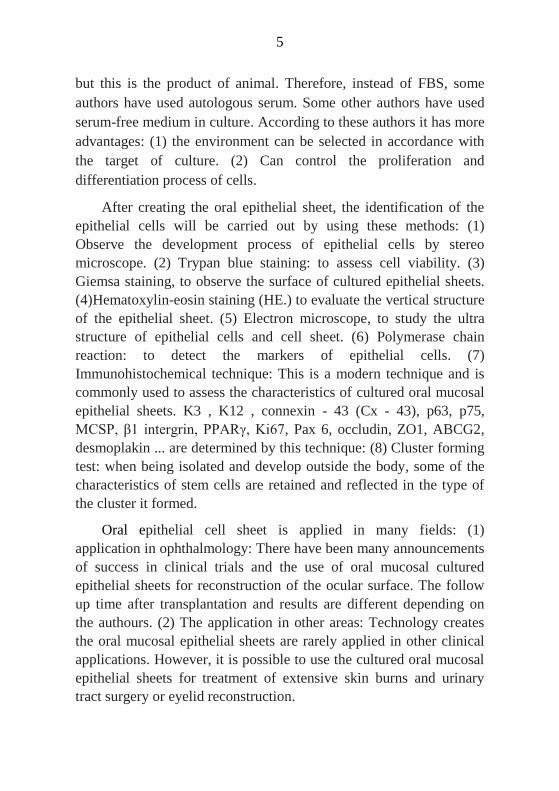
but this is the product of animal. Therefore, instead of FBS, some
authors have used autologous serum. Some other authors have used
serum-free medium in culture. According to these authors it has more
advantages: (1) the environment can be selected in accordance with
the target of culture. (2) Can control the proliferation and
differentiation process of cells.
After creating the oral epithelial sheet, the identification of the
epithelial cells will be carried out by using these methods: (1)
Observe the development process of epithelial cells by stereo
microscope. (2) Trypan blue staining: to assess cell viability. (3)
Giemsa staining, to observe the surface of cultured epithelial sheets.
(4)Hematoxylin-eosin staining (HE.) to evaluate the vertical structure
of the epithelial sheet. (5) Electron microscope, to study the ultra
structure of epithelial cells and cell sheet. (6) Polymerase chain
reaction: to detect the markers of epithelial cells. (7)
Immunohistochemical technique: This is a modern technique and is
commonly used to assess the characteristics of cultured oral mucosal
epithelial sheets. K3 , K12 , connexin - 43 (Cx - 43), p63, p75,
MCSP, β1 intergrin, PPARγ, Ki67, Pax 6, occludin, ZO1, ABCG2,
desmoplakin ... are determined by this technique: (8) Cluster forming
test: when being isolated and develop outside the body, some of the
characteristics of stem cells are retained and reflected in the type of
the cluster it formed.
Oral epithelial cell sheet is applied in many fields: (1)
application in ophthalmology: There have been many announcements
of success in clinical trials and the use of oral mucosal cultured
epithelial sheets for reconstruction of the ocular surface. The follow
up time after transplantation and results are different depending on
the authours. (2) The application in other areas: Technology creates
the oral mucosal epithelial sheets are rarely applied in other clinical
applications. However, it is possible to use the cultured oral mucosal
epithelial sheets for treatment of extensive skin burns and urinary
tract surgery or eyelid reconstruction.
6
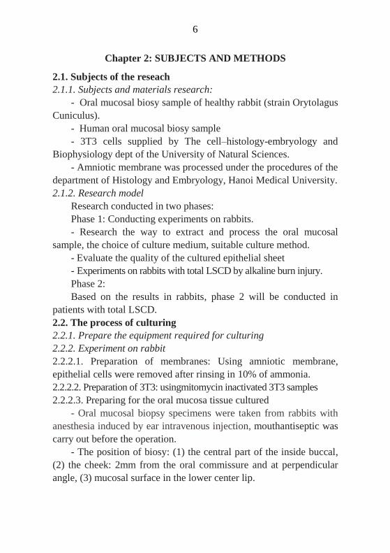
Chapter 2: SUBJECTS AND METHODS
2.1. Subjects of the reseach
2.1.1. Subjects and materials research:
- Oral mucosal biosy sample of healthy rabbit (strain Orytolagus
Cuniculus).
- Human oral mucosal biosy sample
- 3T3 cells supplied by The cell–histology-embryology and
Biophysiology dept of the University of Natural Sciences.
- Amniotic membrane was processed under the procedures of the
department of Histology and Embryology, Hanoi Medical University.
2.1.2. Research model
Research conducted in two phases:
Phase 1: Conducting experiments on rabbits.
- Research the way to extract and process the oral mucosal
sample, the choice of culture medium, suitable culture method.
- Evaluate the quality of the cultured epithelial sheet
- Experiments on rabbits with total LSCD by alkaline burn injury.
Phase 2:
Based on the results in rabbits, phase 2 will be conducted in
patients with total LSCD.
2.2. The process of culturing
2.2.1. Prepare the equipment required for culturing
2.2.2. Experiment on rabbit
2.2.2.1. Preparation of membranes: Using amniotic membrane,
epithelial cells were removed after rinsing in 10% of ammonia.
2.2.2.2. Preparation of 3T3: usingmitomycin inactivated 3T3 samples
2.2.2.3. Preparing for the oral mucosa tissue cultured
- Oral mucosal biopsy specimens were taken from rabbits with
anesthesia induced by ear intravenous injection, mouthantiseptic was
carry out before the operation.
- The position of biosy: (1) the central part of the inside buccal,
(2) the cheek: 2mm from the oral commissure and at perpendicular
angle, (3) mucosal surface in the lower center lip.
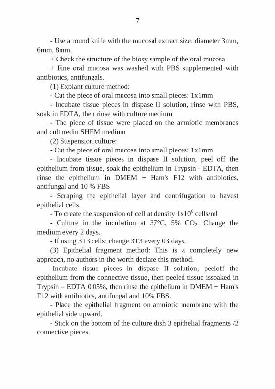
7
- Use a round knife with the mucosal extract size: diameter 3mm,
6mm, 8mm.
+ Check the structure of the biosy sample of the oral mucosa
+ Fine oral mucosa was washed with PBS supplemented with
antibiotics, antifungals.
(1) Explant culture method:
- Cut the piece of oral mucosa into small pieces: 1x1mm
- Incubate tissue pieces in dispase II solution, rinse with PBS,
soak in EDTA, then rinse with culture medium
- The piece of tissue were placed on the amniotic membranes
and culturedin SHEM medium
(2) Suspension culture:
- Cut the piece of oral mucosa into small pieces: 1x1mm
- Incubate tissue pieces in dispase II solution, peel off the
epithelium from tissue, soak the epithelium in Trypsin - EDTA, then
rinse the epithelium in DMEM + Ham's F12 with antibiotics,
antifungal and 10 % FBS
- Scraping the epithelial layer and centrifugation to havest
epithelial cells.
- To create the suspension of cell at density 1x10⁶ cells/ml
- Culture in the incubation at 37°C, 5% CO2. Change the
medium every 2 days.
- If using 3T3 cells: change 3T3 every 03 days.
(3) Epithelial fragment method: This is a completely new
approach, no authors in the worth declare this method.
-Incubate tissue pieces in dispase II solution, peeloff the
epithelium from the connective tissue, then peeled tissue issoaked in
Trypsin – EDTA 0,05%, then rinse the epithelium in DMEM + Ham's
F12 with antibiotics, antifungal and 10% FBS.
- Place the epithelial fragment on amniotic membrane with the
epithelial side upward.
- Stick on the bottom of the culture dish 3 epithelial fragments /2
connective pieces.
8
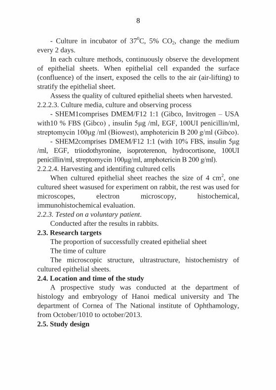
- Culture in incubator of 37
0C, 5% CO2, change the medium
every 2 days.
In each culture methods, continuously observe the development
of epithelial sheets. When epithelial cell expanded the surface
(confluence) of the insert, exposed the cells to the air (air-lifting) to
stratify the epithelial sheet.
Assess the quality of cultured epithelial sheets when harvested.
2.2.2.3. Culture media, culture and observing process
- SHEM1comprises DMEM/F12 1:1 (Gibco, Invitrogen – USA
with10 % FBS (Gibco) , insulin 5μg /ml, EGF, 100UI penicillin/ml,
streptomycin 100μg /ml (Biowest), amphotericin B 200 g/ml (Gibco).
- SHEM2comprises DMEM/F12 1:1 (with 10% FBS, insulin 5μg
/ml, EGF, triiodothyronine, isoproterenon, hydrocortisone, 100UI
penicillin/ml, streptomycin 100μg/ml, amphotericin B 200 g/ml).
2.2.2.4. Harvesting and identifing cultured cells
When cultured epithelial sheet reaches the size of 4 cm2, one
cultured sheet wasused for experiment on rabbit, the rest was used for
microscopes, electron microscopy, histochemical,
immunohistochemical evaluation.
2.2.3. Tested on a voluntary patient.
Conducted after the results in rabbits.
2.3. Research targets
The proportion of successfully created epithelial sheet
The time of culture
The microscopic structure, ultrastructure, histochemistry of
cultured epithelial sheets.
2.4. Location and time of the study
A prospective study was conducted at the department of
histology and embryology of Hanoi medical university and The
department of Cornea of The National institute of Ophthamology,
from October/1010 to october/2013.
2.5. Study design
9
Statistical analysis was performed using the SPSS 16.0 statistical
software. The rate was compared with T test, P<0.05 was considered
statistically significant.
2.6. Ethics of research
The thesis is part of the National independent study, "Research
on the use of stem cells for the treatment of ocular surface diseases",
code DTDL.2010T/15 have adopted ethical council of Medicine,
Hanoi medical University.
Chapter 3: FINDINGS
3.1. Results of studies to create the epithelial sheet on rabbits.
3.1.1. Selecting the location and the size of biopsy samples
Biopsy of oral mucosa conducted in 5 rabbits (strain Orytolagus
Cuniculus) in 3 different positions, we found that:
On the mucosa in the central part of the inside buccal:
Epithelium is stratified nonkeratinized and very thick, composed of
18-20 cell lines, divided into three layers, the basal cell layer consists
of 2-3 rows of small size cells with the egg-shaped, deep nuclear,
very basophilic cytoplasm. Using P63 staining, the cells on the
bottom layer are strongly expressed. The connective tissue under the
epithelial layer creates very high papillary.
Inside mucosal surface of the cheek, in a perpendicular angle and
2mm from the oral commissure, inside mucosal surface in the central
part of the bottom lip: Epithelium is thin, stratified and non-
keratinized, consisting of 4-5 cell lines. The cells on the basal layer
possess the egg-shaped, deep nuclear, very basophilic cytoplasm.The
boundary between epithelial and connective tissue beneath is
relatively flat, without the papillary.
We selected the place to take the sample for culture is the central
part of the inside buccal.

10
When extract samples for research, we found that, to form 2
epithelial sheets, the size of biosy sample should be: (1) diameter
6mm in tissue culture method by explant. (2) 8mm is enough for 2 ml
suspension at density of 1x10⁶
cells /ml in suspension method. (3)
3mm in the method adopted by the epithelium fragment.
3.1.2. Selection of culture medium
In the beginning, we conducted experimental animal models: 18
pieces were cultured by SHEM1 medium, only 30% of the sample
developed and were not confluentafter 28 days of culture. Then,
SHEM2 was applied, and all research results conducted in SHEM2
with the rate of success is 76.47 to 95%.
3.1.3. Selection culture method
Phase 1: We cultured 17 wells with tissue fragments method, 20
wells with suspension, 19 wells with epithelial fragments. The
proportion of successful creation of epithelial sheet was listed in
Table 3.2.
Table 3.2. The proportion of successful creation the oral mucosa
epithelial sheets with different culture methods
Culture
sample
Successful
sheet
Success
culture
rate (%)
p
Explant(1) 17 13 76,47 p(1, 2)>0,05
Suspension(2) 20 19 95 p(2, 3)>0,05
p(1, 3)>0,05 Epithelial
fragment(3)
19 17 89,47
Phase 2: We have adopted 30 samples by the epithelial fragment
method, the rate of growth and success culture rate is 100%. Among
these, 15 cultured epithelial sheets were grafted for 15 eyes of 15
rabbits losing the entire corneal epithelium in one eye by alkaline
burn.
11
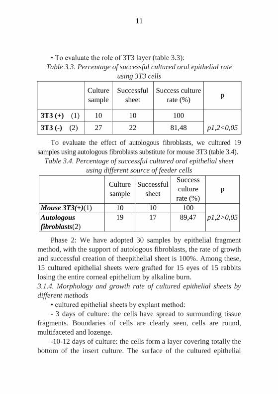
• To evaluate the role of 3T3 layer (table 3.3):
Table 3.3. Percentage of successful cultured oral epithelial rate
using 3T3 cells
Culture
sample
Successful
sheet
Success culture
rate (%) p
3T3 (+) (1) 10 10 100
3T3 (-) (2) 27 22 81,48 p1,2<0,05
To evaluate the effect of autologous fibroblasts, we cultured 19
samples using autologous fibroblasts substitute for mouse 3T3 (table 3.4).
Table 3.4. Percentage of successful cultured oral epithelial sheet
using different source of feeder cells
Culture
sample
Successful
sheet
Success
culture
rate (%)
p
Mouse 3T3(+)(1) 10 10 100
Autologous
fibroblasts(2)
19 17 89,47 p1,2>0,05
Phase 2: We have adopted 30 samples by epithelial fragment
method, with the support of autologous fibroblasts, the rate of growth
and successful creation of theepithelial sheet is 100%. Among these,
15 cultured epithelial sheets were grafted for 15 eyes of 15 rabbits
losing the entire corneal epithelium by alkaline burn.
3.1.4. Morphology and growth rate of cultured epithelial sheets by
different methods
• cultured epithelial sheets by explant method:
- 3 days of culture: the cells have spread to surrounding tissue
fragments. Boundaries of cells are clearly seen, cells are round,
multifaceted and lozenge.
-10-12 days of culture: the cells form a layer covering totally the
bottom of the insert culture. The surface of the cultured epithelial
12
sheet is not flat, high edges were observed on the surface. In this
edge, there are a lot of long cells with flat nucleus when using
inverted microscope.
- 14-16 days of culture: epithelial sheets are not flat, many
fibroblasts co-exist in the epithelial sheet (figure 3.5).
• cultured epithelial sheets by cell suspension
- 2 days of culture: there are plenty of round cells stick to the
bottom, then, they spread with long cytoplasmic branches
- 12-14 days of culture: epithelial cells are confluent.
- After air-lifting, using H.E. staining and Giemsa staining
method: the surface of the cultured epithelial sheet was flat,
consisting of 5-7 rows of cells, the the shape of the higher cell is
flatter. The intercellular space is wide, the lozenge cells were not
existed in the cultured sheet (figure 3.9).
On electron microscopy, intercellular space between the cells in
the supra-basal layer of the epithelial sheet is quite wide. The cells
here are closely attached to neighbouring cells by numerous
desmosomal andintercellular junctions. In the cytoplasm of cells,
there are a lot of organelles, glycogen particles, the bundle of fiber,
rough endoplasmic reticulum.
• Cultured epithelial sheets by epithelial fragments
- 3-4 days of culture: The cells with round shape, polyhedral
shape with long cytoplasmic branches spread. Cell spreading
gradually, beginning with polyhedral shape cells, wide intercellular
space. When the cells are confluent all the surface of the bottom of
insert culture dish in 10-12 days, the cells close together, the size of
the cells are smaller with narrower intercellular space but still clearly
observed boundaries, dividing cells appeared crowdedly.
- After air-lifting: We harvested the flat surface cultured
epithelial sheet, consisting of 5-7 rows of cells, the higher cell
position the flatter shape they become. The intercellular space is
narrower than the space in the culture sheet by suspension method.
Spindle cell could not observed by this method (figure 3.15).
13
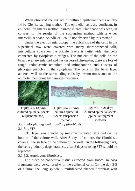
When observed the surface of cultured epithelial sheets on day
14 by Giemsa staining method: The epithelial cells are confluent. In
epithelial fragments method, narrow intercellular space was seen, in
contrast to the results of the suspension method with a wider
intercellular space. Spindle cell could not observed by this method.
Under the electron microscope: the apical side of the cells in the
superficial row were covered with many short-branched villi,
intercellular space on the prickle layers is quite wide, the cells
connected by cytoplasmic bridges. The nucleus of the cells on the
basal layer are enlarged and has dispersed chromatin, there are lots of
rough endoplasmic reticulum and mitochondria and clusters of
glycogen particles in the cytoplasm. The cells on the basal layer
adhered well to the surrounding cells by desmosomes and to the
amniotic membrane by hemi-desmosomes.
Figure 3.5. 12 days
cultured epithelial sheets
(explant method)
Figure 3.9. 21 days
cultured epithelial
sheets (suspension
method)
Figure 3.15.21 days
cultured epithelial sheets
(epithelial fragment
method)
3.1.5. Morphology and growth of fibroblasts
3.1.5.1. 3T3
3T3 layer was created by mitomycin-treated 3T3, fed on the
bottom of the culture well. After 3 days of culture, the fibroblasts
cover all the surface of the bottom of the well. On the following days,
the cells gradually degenerate, so, after 3 days of using 3T3 should be
replaced.
3.1.5.2. Autologous fibroblasts
The piece of connective tissue extracted from buccal mucosa
fragments were co-cultured with the epithelial cells. On the day 3-5
of culture, the long spindle - multifaceted shaped fibroblast with
14
many branches spread around the connective tissue, on the 6th day of
culture, about half of the area of the bottom are covered by fibroblasts
and to about day 10 of culture, the entire bottom are covered.
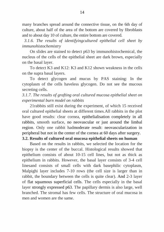
3.1.6. The results of identifyingcultured epithelial cell sheet by
immunohistochemistry
On slides are stained to detect p63 by immunohistochemical, the
nucleus of the cells of the epithelial sheet are dark brown, especially
on the basal layer.
To detect K3 and K12: K3 and K12 shown weakness in the cells
on the supra basal layers.
To detect glycogen and mucus by PAS staining: In the
cytoplasm of the cells haveless glycogen. Do not see the mucous
secreting cells.
3.1.7. The results of grafting oral cultured mucosa epithelial sheet on
experimental burn model on rabbits
21rabbits still exist during the experiment, of which 15 received
oral cultured epithelial sheets at different times.All rabbits in the plot
have good results: clear cornea, epithelialisation completely in all
rabbits, smooth surface, no neovascular or just around the limbal
region. Only one rabbit hadmoderate result: neovascularization in
peripheral but not in the center of the cornea at 60 days after surgery.
3.2. Results of cultured oral mucosa epithelial sheets on human
Based on the results in rabbits, we selected the location for the
biopsy is the center of the buccal. Histological results showed that
epithelium consists of about 10-15 cell lines, but not as thick as
epithelium in rabbits. However, the basal layer consists of 3-4 cell
linesand consists of small cells with dark basephilic cytoplasm,
Malpighi layer includes 7-10 rows (the cell size is larger than in
rabbit, the boundary between the cells is quite clear). And 2-3 layer
of flat squamous superficial cells. The cells especially in the basal
layer strongly expressed p63. The papillary dermis is also large, well
branched. The stromal has few cells. The structure of oral mucosa in
men and women are the same.
15
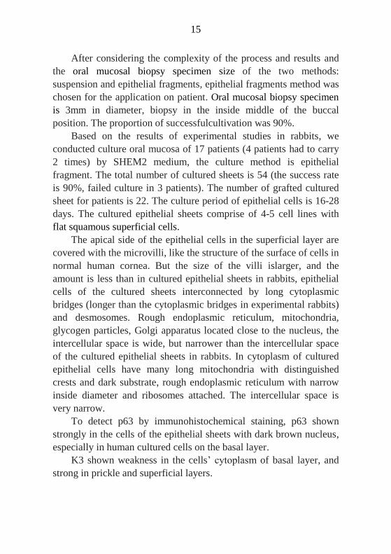
After considering the complexity of the process and results and
the oral mucosal biopsy specimen size of the two methods:
suspension and epithelial fragments, epithelial fragments method was
chosen for the application on patient. Oral mucosal biopsy specimen
is 3mm in diameter, biopsy in the inside middle of the buccal
position. The proportion of successfulcultivation was 90%.
Based on the results of experimental studies in rabbits, we
conducted culture oral mucosa of 17 patients (4 patients had to carry
2 times) by SHEM2 medium, the culture method is epithelial
fragment. The total number of cultured sheets is 54 (the success rate
is 90%, failed culture in 3 patients). The number of grafted cultured
sheet for patients is 22. The culture period of epithelial cells is 16-28
days. The cultured epithelial sheets comprise of 4-5 cell lines with
flat squamous superficial cells.
The apical side of the epithelial cells in the superficial layer are
covered with the microvilli, like the structure of the surface of cells in
normal human cornea. But the size of the villi islarger, and the
amount is less than in cultured epithelial sheets in rabbits, epithelial
cells of the cultured sheets interconnected by long cytoplasmic
bridges (longer than the cytoplasmic bridges in experimental rabbits)
and desmosomes. Rough endoplasmic reticulum, mitochondria,
glycogen particles, Golgi apparatus located close to the nucleus, the
intercellular space is wide, but narrower than the intercellular space
of the cultured epithelial sheets in rabbits. In cytoplasm of cultured
epithelial cells have many long mitochondria with distinguished
crests and dark substrate, rough endoplasmic reticulum with narrow
inside diameter and ribosomes attached. The intercellular space is
very narrow.
To detect p63 by immunohistochemical staining, p63 shown
strongly in the cells of the epithelial sheets with dark brown nucleus,
especially in human cultured cells on the basal layer.
K3 shown weakness in the cells’ cytoplasm of basal layer, and
strong in prickle and superficial layers.
16
Cutivated oral mucosa epithelium was used for transplant
patients. The evaluation criteria include the transparence degree of
cornea, the integrity of the ocular surface and corneal
neovascularization. Successful transplantation was noted in 12 cases
which have visual improvement were noted in 9 cases, especially at
short distance, about 10-30 cm.
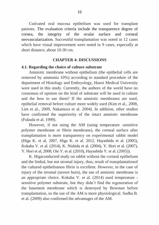
CHAPTER 4: DISCUSSIONS
4.1. Regarding the choice of culture substrate
Amniotic membrane without epithelium (the epithelial cells are
removed by ammonia 10%) according to standard procedure of the
department of Histology and Embryology, Hanoi Medical University
were used in this study. Currently, the authors of the world have no
consensus of opinion on the kind of substrate will be used in culture
and the how to use them? If the amniotic membranes are used,
epithelial removal before culture more widely used (Kim et al., 2008,
Lim et al., 2009, Nakamura et al. 2004). In addition, other studies
have confirmed the superiority of the intact amniotic membrane
(Fukuda et al. 1999).
However, if not using the AM (using temperature -sensitive
polymer membrane or fibrin membrane), the corneal surface after
transplantation is more transparency on experimental rabbit model
(Higa K. et al, 2007, Higa K. et al. 2012, Hayashida et al. (2005),
Kokaba V. et al. (2014), K. Nishida et al. (2004), Y. Hori et al. (2007),
Y. Hori et al, 2008, Oie Y. et al. (2010), Hayashida Y. et al. (2005)).
K. Higaconducted study on rabbit without the corneal epithelium
and the limbal, but not stromal injury, thus, result of transplantationof
the cultured epitheliumon fibrin is excellent. However, in the case of
injury of the stromal (severe burn), the use of amniotic membrane is
an appropriate choice. Kokaba V. et al. (2014) used temperature -
sensitive polymer substrate, but they didn’t find the regeneration of
the basement membrane which is destroyed by Bowman before
transplantation, so the use of the AM is more physiological. Sudha B.
et al. (2009) also confirmed the advantages of the AM.
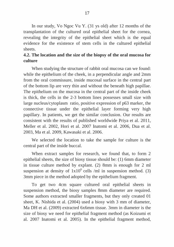
17
In our study, Vo Ngoc Vu Y. (31 ys old) after 12 months of the
transplantation of the cultured oral epithelial sheet for the cornea,
revealing the integrity of the epithelial sheet which is the equal
evidence for the existence of stem cells in the cultured epithelial
sheets.
4.2. The location and the size of the biopsy of the oral mucosa for
culture
When studying the structure of rabbit oral mucosa can we found:
while the epithelium of the cheek, in a perpendicular angle and 2mm
from the oral commissure, inside mucosal surface in the central part
of the bottom lip are very thin and without the beneath high papillae.
The epithelium on the mucosa in the central part of the inside cheek
is thick, the cells in the 2-3 bottom lines possesses small size with
large nucleus/cytoplasm ratio, positive expression of p63 marker, the
connective tissue under the epithelial layer forming very high
papillary. In patients, we get the similar conclusion. Our results are
consistent with the results of published worldwide Priya et al. 2011,
Meller et al. 2002, Hori et al. 2007 Inatomi et al. 2006, Dua et al.
2003, Ma et al. 2009, Kawasaki et al. 2006.
We selected the location to take the sample for culture is the
central part of the inside buccal.
When extract samples for research, we found that, to form 2
epithelial sheets, the size of biosy tissue should be: (1) 6mm diameter
in tissue culture method by explant. (2) 8mm is enough for 2 ml
suspension at density of 1x10⁶
cells /ml in suspension method. (3)
3mm piece in the method adopted by the epithelium fragment.
To get two 4cm square cultured oral epithelial sheets in
suspension method, the biosy samples 8mm diameter are required.
Some authors extracted smaller fragments, but they only created 01
sheet, K. Nishida et al. (2004) used a biosy with 3 mm of diameter,
Ma DH et al. (2009) extracted 6x6mm tissue. 3mm in diameter is the
size of biosy we need for epithelial fragment method (as Koizumi et
al. 2007 Inatomi et al. 2005). In the epithelial fragment method,
18
exposed area of stem cells in the basal layer is maximum, completely
eliminate fibroblasts leads to good quality and flat surface cultured
sheets.
4.3. About culture medium
Each author has a specific formula for the culture medium. In the
early period of reseach, we use SHEM1, the percentage of success
culture rate only 30%, epithelial sheets are not good with sparse cell
density.
When SHEM2 were used (SHEM1 added with insulin,
hydrocortisone, T3, isoproterenol), the epithelial stem cell
proliferation was excellent, the success culture rate are 89.5% - 95%.
Although when compared to many other authors in the world,
choleratoxin is not included in our culture medium. However, the rate
of success of creating epithelial sheet in our results suggest that
SHEM2 is good enough to culture epithelial sheets from oral mucosa
stem cells.
The use of antibiotics and antifungals in cell culture media,
especially the oral mucosa in rabbits in Vietnam is essential. The
authors used different types of antibiotics, at different concentrations.
Madhira S. L. et al. (2008), Y. Satake et al. (2008) have used
gentamicin as culture medium components. Most authors used
Amphotericin B, but due to its toxicity, some studies eliminated
(Hirayama M. 2012 didn’t use both amphotericin B and penicillin).
Currently, fetal bovine serum (FBS) is thepriority choice of
nearly all authors in the world (Nakamura et al. 2003, Nakamura et
al. 2004, Picot 2005). However, FBS has many cons, especially
heterologous protein. To solve this problem, there have been many
studies using autologous serum at different concentrations and the
results are good (T. Nakamura et al. (2006), Ang LP et al. (2006).
Priya CG et al. (2011). Y. Satake et al. (2011). Hirayama M. et al.
(2012)). Besides the use of autologous serum to remove unwanted
components from animal derived product, AB blood type human
serum is also used (Zakaria N. et al. (2014)).
19
Serum free culture medium are the choice in many studies,
however, to widespread the application of this type of medium
requires a lot ofmore researches and reduction of costs (Ilmarinen et
al. 2012, Hayashi and et al. 1976). Clearly, the use of serum in
culture brings better results than serum free medium. In the real
condition in Vietnam and in our research, fetal bovine serum was
added at a concentration of 10%.
The insulin hormone, triiodothyronine, EGF, hydrocortisone,
isoproterenol, choleratoxin are used by most authors in the world at
different concentrations.
4.4. Methods of culture
4.4.1. Explant culture
When using this method, in the perimeter of the tissue, the cells
are capable of contact with AM and have opportunity to develop
around, the other cells is limited by the under connective tissue to be
exposed to the culture substrate.
Our epithelial sheets by this method is not flat, have high edges
which are formed by the fibroblasts. In our opinion, the oral mucosa
has more loose, elastic and flexible connective tissue than in other
regions. So, we can’t eliminate completely the connective tissue by
hand. The fibroblasts in the connective tissue can proliferate with
high speed and become more dominant than the epithelial cells. This
will affect the effectiveness of transplantation for patients. The
presence of fibroblasts is a favorable condition for the invasion of
conjuctival tissue in to the cornea surface. When creating the cultured
corneal epithelial sheet from stem cells from the limbal region in our
previous researchs, we also applied the explants culture method, but
didn’t see the same phenomenon. S. Kanayama et al. (2007) study in
culture of corneal epithelial sheet from limbal stem cells and cultured
oral epithelial sheet to specify the factors that cause
conjuctivalization after transplantation, they concluded that: FGF is
the cause of the phenomenon of neovascularization and
conjunctivalization that occur in the cultured oral epithelial sheet
after transplantation.
20
Although, explants culture method are used by many authors in
the world and seems to be very simple, but our result show no
support to this way.
4.4.2. Suspension method
Dispase was the enzyme to process the biosy sample in this
study, it helps to cut the junctions between the cells and the basement
membrane. Check the structure of the stromal tissue: the entire
epithelial layer was eliminated, we also found no presence of
connective tissue cells in epithelial fragments. Then trypsin - EDTA
was used for separate the epithelial cells into individual cells. The
cells in some bottom cell layers are taken by scrapping the fragment.
The microscopic structure of the rest of the fragment showed that all
cells in the bottom layers were eliminated for suspension culture.
Thus, all stem cells in the layer close to the basement
membraneare fully utilized. The origin of the cells in cultivated cell
sheet created by this method was confirmed by immunohistochemistry
(K3 are strongly expressed in the cytoplasm of the cells on the bottom
layer). This conclusion is also consistent with our conclusion of Ma
DH. et al. (2009), T. Nakamura et al. (2003), Hayashida Y. et al.
(2005).
4.4.3. Epithelial fragment culture method
Comparing the effectiveness of the culture technique suspension
and epithelial fragment, we found that: the culturesuccess rates, the
growth ratesand the microstructure of sheets arenearly similar. The
success culture rates of three different methods are 76.47%, 95% and
89.47%.The culture success rate in epithelial fragment method is
lower than the rate of suspension method, but this difference was not
statistically significant. Overall, epithelial fragment method has much
more prominent advantages compared with suspension because of
these reasons: to reduce the size of the biosy sample size, shorten the
tissue processing time and tissue enzyme exposure time of cultured
cells with trypsin - EDTA that increases the survival of cultured cells,
21
reducing the lab equipment. And maintainance the link between cell
– cell is not excluded that leads to the success of the technique.
Besides, when autologous fibroblasts obtained from tissue
samples, this has created a favourable physiological condition for the
development of the cells, epithelial cells can still receive signals from
stromal cells.
Currently, in the suspension culture technique, most authors still
use mouse 3T3 (animal derived product). The results of our study
show that using cultured autologous fibroblasts gets higher success
culture rate than without this layer (89.47% versus 81.48%). Using
mouse 3T3 gets higher success culture rate than without 3T3 with p <
0.05 (100% versus 81.48%).
According to the review, no team in the world using epithelial
fragment culture method. This is a completely new technique,
showing all the advantages over the other methods are being applied
now: (1) small biosy (2) simple culture process (3) using autologous
fibroblasts (3T3 was eliminated) (4) the morphology of the cultured
epithelial sheet is good.
4.5. The quality of the cultured epithelial sheet
After 16-28 days of culture in 370C, 5% CO2, we obtained
stratified, non-keratinized epithelial cells sheet, the sheet consists of
4-5 rows, the cells on the superficial layer are flat and have nuleus.
Images of cells with small size, high nucleus/cytoplasm are found by
Giemsa staining at confluent period (before air-lifting). In terms of
morphology, these are stem cells (K. Izumi et al. (2007), Priya C. G.
et al. (2011)).
The cells of the cultured sheet represents the typical structure of
epithelium. In the cytoplasm of the cells on lower layers, organelles
such as endoplasmic reticulum, mitochondria, Golgi apparatus are
rich. The results of immunohistochemical staining showed that the
cells (especially the basal cells) are strongly positive for p63; Using
22
K3 - K12 staining, cells in the suprabasal layers are weakly
expressed. The structure of the cultured epithelial sheets of us are the
same with the structure of the normal corneal epithelium and cultured
corneal epithelial sheet and like other authors in the world described
(Nakamura et al. 2003, Madhira et al., 2008, Moharamzadeh et al.,
2007, Nakamura et al., 2010, Ang et al., 2010, Sekiyama et al. 2006).
The cultured epithelial sheet were grafted on experimental
rabbits and patients with LSCD in both eyes gets pretty good results.
The important thing is how long does the graft exist? In experiments,
we have monitored grafted rabbits with the longest time is 180 days,
the epithelial sheets were attached well to the corneal stromal tissue.
The cultured oral epithelial graft from the cornea surface of the
patient Vo Ngoc Vu Y. (31) was extracted after 12 months of
transplantation, we still observe part of the oral epithelial sheet.
There are many publications in the world about the long-term
survival of epithelial sheets after transplantation in patients (Kocaba
V. et al. (2014), Sangwan VS. et al. (2014)).
4.6. The remaining issues to be studied to improve the cultured
oral epithelial sheet
To minimize using the animal product, in the havesting process, the
cultured epithelial sheets are immersed in DMEM: Ham's F12 (without
FBS), then send to operating room in aseptic conditions at 370C.
The use of autologous fibroblasts in culture has more advantages
over the use of mouse 3T3. However, the process of harvesting
cultured sheets is sometimes difficult.
In human, the total number of biopsies is 26, because of 4 times
of failure in cuture, so they should not be used for transplantation or
the patient need more operation. Thus, there are 22 times of success
in culture. The cells of the epithelial sheets are confluent at the day of
16-28 of culture. We have conducted transplantation for 22 patients.
In 22 cases, we have one sheet for transplantation and the other for
examining histological characteristics. Microstructure of the sheets
was confirmed to be completely normal. While epithelial sheets
23
separated from the bottom of the insert culture, we meet a lot of
difficulties, that leads to the lost of small groups of epithelium (2-
4mm) and 4 sheets are torn during separation.
At the hard separation sheets, the fibroblasts developed and
attached well at the bottom of of the insert. We have not seen any
author describes the same phenomenon. These are problems to be
studied to improve the process of culture.
According to us, the connective tissue used for creating the
feeder layer was too big, it created a favourable condition for the
development of the fibroblast, and contact with the epithelial cells
through pores of membranes and makes the dissection difficult. To
mitigate this problem, the size of the connective tissue to form
autologous fibroblast layer must be reduced, and it must be picked
out when the fibroblasts covered about 2/3 of the insert bottom
(about 8 or 9 days).
CONCLUSIONS
Through experimental study in rabbits and tested on patients, we
conclude:
1. We are successful in specifing the exact location, the size of the
oral mucosa and the culture medium for culture:
-Central part of the inside buccal is the biosy position, the 3mm
diameter is the size of the biosy sample for culture the two sheets of
the epithelium.
- Culture medium for oral epithelial sheet: SHEM2:
DMEM/Ham's F12 with ratio1:1, other elements are added: EGF,
insulin, hydrocortisone, isoproterenol, FBS, T3, antibiotic,
antifungus.
2. We are successful in specifing the completely new culturing
method to create the oral mucosa epithelial sheets: epithelial
fragment method, co-culture with autologous fibroblasts. The steps
are as follows:
(i) Obtain the 3mm diameter biosy samplein the central part of
inside buccal;
24
(ii) Cut into small pieces of 0.5 mm x 0.5 mm in size;
(iii) The chopped pieces were incubated in dispase II solution 1.2
UI/ml at 37°C, 5% CO2 from 35 to 45 minutes;
(iv) Washed in SHEM2 to inactivate the action of dispase II;
(v) The epithelialfragments are detached from the connective tissue;
(vi) Immerse the epithelial fragmentsand connective tissue in the
solution of trypsin - EDTA 0.05% (in PBS) for 30sec;
(vii) Wash the epithelial and connective tissue twice with SHEM2;
(viii) Place the epithelial fragments on the surface of the
stretched amniotic membranes.
(ix) Place the stromal tissue on the surface of the culture dish
with a ratio of 3 pieces of epithelial / 2 pieces of connective tissue;
(x) Put the insert culture in to the well;
(xi) 1ml of SHEM2 is added in to insert culture, 1,5ml SHEM2
are poured into the culture wells and change the medium every 2
days;
(xii) Remove the connective tissue when fibroblasts covered 2/3
of the well surface;
(xiii) Remove the epithelial fragments when epithelial cells get
confluence;
(xiv) Air-lifting will be carried out to created 4 to 5 cell line of
thickness;
(xiv) Havest the epithelial sheet.
RECOMMENDATIONS
(1) There should be further research to improve the process of
harvesting the oral mucosa epithelial sheet.
(2) There should be further research on the culture medium to
completely eliminate animal products from the culture medium in
human.
(3) It should have a longer follow-up to assess the quality of
cultured epithelial sheets after transplantation.
THE LIST OF PUBLISHED ARTICLES RELATED
TO THE THESIS
1. Dao Thi Thuy Phuong, Nguyen Thi Binh et al (2013).
Characterization of the cultured oral mucosal epithelial
cell sheet on amniotic membrane. Journal of military
Phamaco-medicine, 4, April, 65-69.
2. Do Thuy Huong, Dao Thi Thuy Phuong, Nguyen Khang
Son, Nguyen Thi Binh et al (2012). Culture and
application of oral epithelial cell sheet for ocular surface
disease. Journal of medical practice, 818-819, 560-563.


