Role of Glycosylation in Transport of Vesicular Stomatitis Virus ...

Newly Synthesized G Protein of Vesicular
Stomatitis Virus Is Not Transported
to the Golgi Complex in Mitotic Cells
CAROL FEATHERSTONE, GARETH GRIFFITHS, and GRAHAM WARREN European Molecular Biology Laboratory, 6900 Heidelberg, Federal Republic of Germany. G. Warren's present address is Department of Biochemistry, University of Dundee, Dundee DD1 4HN, Scotland, U.K.
ABSTRACT Newly synthesized G protein of vesicular stomatitis virus is not transported to the surface of cultured mammalian cells during mitosis (Warren et al., 1983, J. Celt Biol. 97:1623- 1628). To determine where intracellular transport is inhibited, we have examined the post- translational modifications of G protein, which are indicators of specific compartments on the transport pathway. G protein in mitotic cells had only endo H-sensitive oligosaccharides containing seven or eight mannose residues, but no terminal glucose, and was not fatty acylated. These modifications were indicative of processing only by enzymes of the endo- plasmic reticulum (ER). Quantitative immunocytochemistry was used as an independent method to confirm that transport of G protein out of the ER was inhibited. The density of G protein in the ER cisternae was 2.5 times greater than in infected G1 cells treated similarly. Incubation of infected mitotic cells with cycloheximide, which inhibits protein synthesis without affecting transport, did not result in a decrease in the density of G protein in the ER cisternae, demonstrating that G protein cannot be chased out of the ER. These results suggest that intracellular transport stops at or before the first vesicle-mediated step on the pathway.
Transport of proteins between the membrane-bound organ- elles of the cell appears to be mediated by budding and fusion of carrier vesicles. Vesicular traffic occurs continuously in all animal cells, except for cells undergoing division. During mitosis a number of specific examples of membrane transport have been shown to be inhibited. These include endocytosis (Fawcett, 1965; Berlin et al., 1978; Berlin and Oliver, 1980), receptor recycling (Warren et al., 1984; Sager et al., 1984), and stimulated secretion (Hesketh et al., 1984).
The examples outlined above all concern transport to or from the plasma membrane. In a previous study, we showed that a model plasma membrane protein, the glycoprotein of vesicular stomatitis virus (VSV), j is not transported to the surface of mitotic cells (Warren et al., 1983), but in this case it was not clear whether transport was inhibited at the level of the plasma membrane, or earlier in the transport pathway.
J Abbrev ia t ions used in this paper. CHO, Chinese hamster ovary; endo H, endo/3-N-acetylglucosaminidase H; ER, endoplasmic retic- ulum; GM, growth medium; MEM, minimal essential medium; PMSF, phenylmethylsulfonyl fluoride; VSV, vesicular stomatitis vi- rus.
2036
Proteins destined for the plasma membrane must pass through a series of compartments from their site of synthesis in the rough endoplasmic reticulum (ER), through the cisternae of the Golgi complex, to the cell surface (Bergmann et al., 1981; Green et al., 1981). The first vesicle-mediated step on this pathway is probably the step between the transitional elements of the ER and the Golgi complex (Jamieson and Palade, 1968). If inhibition of membrane transport acts on a common feature of all vesicle-mediated transport, then one would expect that all vesicular traffic, not only that to and from the plasma membrane, would be inhibited in mitotic cells. A prediction from this hypothesis is that a newly synthesized plasma membrane protein would not be transported out of the ER. In this study we tested this prediction by attempting to identify the first step in transport to the plasma membrane that is inhibited during mitosis.
We have studied mitotic Chinese hamster ovary (CHO) cells, infected with VSV. This virus directs the synthesis of five viral proteins, one of which, the glycoprotein (G protein), is an integral membrane protein. This protein undergoes a well-defined temporal sequence of post-translational modifi-
THE JOURNAL OF CELL BIOLOGY . VOLUME 101 DECEMBER 1985 2036-2046 © "Yhe Rockefeller University Press . 0021-9525/8511212036 t $1.00
on March 21, 2018
jcb.rupress.orgD
ownloaded from

cations, including oligosaccharide processing (Hubbard and lvatt, 1981) and fatty acylation (Schmidt and Schlesinger, 1980), which provide biochemical markers for the compart- ments through which G protein must pass en route to the plasma membrane. Furthermore, since G protein is synthe- sized in large amounts in infected cells, it can be easily detected by quantitative immunocytochemical techniques (Bergmann et al., 1981). The results reported here support the notion that G protein does not leave the ER in the mitotic cell.
MATERIALS AND METHODS
Preparation of Mitotic CHO Cells Infected with VSV: The CHO cell line, obtained from Dr. Stuart Kornfeld (Washington University, St. Louis, MO), was grown and maintained as monolayers on 75-cm 2 plastic flasks in growth medium (GM) comprising minimal essential medium (MEM) (Gibeo Europe, Glasgow) supplemented with 10% (vol/vol) fetal calf serum and 100 U/ml each of penicillin and streptomycin, in an atmosphere of 5% CO~/95% air.
Mitotic cell populations were isolated from monolayer cultures grown in 850-cm z plastic roller bottles (Falcon Labware, Oxnard, CA). Each bottle was seeded with half of the cells from a confluent 75-cm z flask in 100 ml GM. The bottles were gassed with 5% CO2/95% air for 3 min and then rotated at 0.2- 0.3 rpm at 37°C. They were used to prepare mitotic cells 2 d later when the monolayers were ~50% confluent.
Bottles were subjected to two spins at 300 rpm for 5 min at 37"C, separated by an interval of 20 min, to remove debris. The cells were then incubated for 20 min with GM containing 0.04 ug/ml Nocodazole (Sigma Chemic GmbH, Taufkirchen, FRG) (Zieve et al., 1980), before being spun again at 200 rpm for 3 min to remove interphase cells that had become loosely attached as a result of the Nocodazole treatment. The cells that remained were washed twice with infection medium (MEM, 100 U/ml penicillin, 100 U/ml streptomycin, 0.2% (wt/vol) bovine serum albumin (BSA), 10 mM HEPES [final pH 6.8]) contain- ing 0.04 ~g/ml Nocodazole and infected with 10-20 infecting units (defined below) of VSV per cell (0.85-1.7 x 10⁹ infecting units per bottle) in a total volume of 10 ml for 30 min at 37*C and 0.2-0.3 rpm. The infection medium was carefully removed and replaced with 50 ml GM that contained 0.04 ug/ml Nocodazole. Mitotic cells were selectively detached from the bottle by rotation at 200 rpm (Klevecz, 1975) for 3 rain at 37"C and recovered by centrifugation at 400 g for 5 min at room temperature. 1-3 x 10⁴ cells were routinely obtained from one bottle by this procedure. Of these, usually >90% were mitotic, as determined by staining with Hoechst dye 33258 (Berlin et al., 1978), and 95% infected, as determined by immunofluorescence microscopy (see below).
G1 Cells: To obtain G~ cells, infected, mitotic cells were washed once with 50 ml GM to remove Nocodazole, recovered by centrifugation al 400 g for 5 min at room temperature, resuspended in 2 ml GM, and incubated at 37"C for 30 rain to allow the cells to complete mitosis.
Asynchronous Cells: CHO cells fur"inter-phase" controls were grown on 3.5-cm-diam plastic Petri dishes in GM. They were infected using the same protocol as described for roller bottle cultures.
Preparation of Virus and Radioactive Virus: Indiana M4 strain of VSV was grown in baby hamster kidney cells as described by Marlin et al. (1982). ts045 VSV was prepared by essentially the same method, but the cells were grown on 24 x 24 cm plates and infected for 30 h at 31 °C. [3H]mannose- labeled virus was prepared as described by Mattila et al. (1976) for Semliki Forest virus.
Estimation of VSV Infecting Units: We usedimmunofluorescence microscopy rather than plaque assay to determine the tiler of our stock virus preparation, because we found it to be a simpler, more rapid and reliable method.
A suspension of VSV in phosphate-buffered saline containing 0.2% (wt/vol) BSA, 0.5 mM EDTA (pH 7.0), was serially diluted 102-107 fold in infection medium. Baby hamster kidney cells grown on coverslips were washed three times with 2 ml infection medium and incubated with 0.6 ml of diluted virus for 60 min at 37"C. This was replaced with 2 ml GM containing 0.1 mM chloroquine to prevent further infection (Helenius et al., 1980) and the incu- bation was continued at 37"C for 4 h. The cells were fixed and processed for immunofluorescence microscopy as described below, using a rabbit anti-G protein antiserum followed by a rhodamine-conjugated sheep anti-rabbit sec- ond antibody, and then 1 ug/ml Hoechst dye 33258 in Dulbecco's PBS. Cells within a field of known area ( 1.1 × 10 -4 cm 2) were scored as positive or negative for G protein immunofluorescence, and the total number of cells per field was
estimated from the number of Hoechst-stained nuclei visible. The percentage of infected cells was plotted against dilution of virus, and the dilution at which 50% of cells were infected was determined. Assuming that each infected cell was infected by only one virus particle, and knowing the number of cells per unit area and the total area of the well in which the cells on the coverslip was infected, the number of virus particles contained in 0.6 ml infection medium was calculated. Thus, the number of infecting particles per milliliter of virus suspension was determined. This method usually gives about 10-fold fewer infecting units than plaque-forming units determined by plaque assay of the same preparation.
Labeling with [3SS]Methionine: VSV-infected, mitotic cells were washed twice with methionine-free M EM containing 100 units/ml of penicillin and streptomycin, 2 mM glutamine, 0.2% (wt/vol) BSA, 10 mM HEPES (pH 7.3), nonessential amino acids, and 0.5 ug/ml unlabeled methionine, in the presence or absence of Nocodazole. Cells were recovered after each wash by centrifugation at 400 g for 5 min at room temperature. The washed cells were resuspended in 2 ml (0.5-1.5 x 10⁴ cells/ml) of the same medium containing 100 taCi L-[3~S]methionine (7-800 Ci/mmol, Amersham Buchler GmbH, Braunschweig, FRG) and incubated at 37°C for 60 min before chasing with 0.1 mM unlabeled methionine in GM for 60 min. At the end of the chase period, the cells were transferred onto ice, resuspended, and recovered by centrifugation at 400 g and 4"C. The cells were extracted with 250 tal 1% (wt/vol) Triton X- 114 in PBS containing 0.23 mM phenylmethylsulfonyl fluoride (PMSF) and 2.8 ug/ml Trasylol, and extracted on ice for 10 min. Insoluble material was removed by centrifugation at full speed (10,000 g) in a microfuge at 4"C. Integral membrane proteins were isolated by cloud-point precipitation, essen- tially as described by Bordier (1981). The supernatant was warmed to 30°C for 3 rain and centrifuged at 2,500 g in a microfuge for 3 rain at room temperature. The detergent pellet was washed with ice-cold PBS by three successive rounds of warming to 30"C and resedimentation.
endo H Digestion and SDS PAGE: Proteins extractedwith Triton X-114 were digested with endo 13-N-acetylglucosaminidase H (endo H) from Staphylococcus plicatus (Miles Laboratories Inc., Elkhart, IN) by the addition of an equal weight of Triton X-100 to the detergent pellet on ice, then of 250 ul ice-cold 0.2 M sodium citrate pH 5.5, 0.23 mM PMSF, and 5 mU endo H per sample. The mixture was rotated at 37"C for 16-20 h, and the protein was recovered by the addition of trichloroacetic acid (TCA) to a final concentration of 10% (wt/vol) and precipitation on ice for 2 h. TCA precipitates were recovered by centrifugation at ~5,000 g for 5 min at 4"C in a microfuge, washed twice with I ml ethanol/ether (1:1, vol/vol) and ether, then dissolved in SDS sample buffer and electrophoresed in a 10% (wt/vol) polyacrylamide gel as described by Maizel (1969) with the modifications described by Green et al. (1981). Gels were fixed, soaked in EN3HANCE (New England Nuclear, Dreieich, FRG), dried, and fluorographed at -70"C using preflashed Kodak X- OMAT x-ray film as described by Laskey and Mills (1975).
Labeling with [3H]Mannose: VSV-infected mitotic cells were iso- lated as described above and resuspended in 1.5 ml glucose-free MEM contain- ing 2 mM glutamine, 15 mM HEPES (pH 7.3), 100 U/ml penicillin and streptomycin, 0.2% (wt/vol) BSA, nonessential amino acids, 10 mM sodium pyruvate, 1 mCi D-[23H]mannose (10-20 Ci/mmol, Amersbam Buchler GmbH) containing 0.04 ~g/ml Nocodazole. The cells were pulsed at 37"C for 60 rain and chased by washing with the same medium containing 10 mM unlabeled mannose and incubating for 60 rain more at 37"C. Pulsed and chased cells were recovered by centrifugation at 400 g for 5 min, washed once with ice-cold PBS, and extracted with Triton X-114 as described above. Glycopep- tides were prepared from the detergent extracts and analyzed on a Biogel P4 (200-400 mesh) column as described below. 2 h after infection with VSV, interphase CHO cells were labeled with 1 mCi [3H]mannose in the presence or absence of Nocodazole, as described for mitotic and Gt cells. The labeled cells were chased for 60 min and processed in the same way as mitotic and G~ cells.
Preparation of GIycopeptides: Glycopeptideswere preparedeither in the manner described by Tabas and Kornfeld (1978) or by a simpler and quicker procedure described below which gave essentially the same results.
[3H]Mannose-labeled cells were extracted with Triton X-114 as described above, and the pellet containing G protein and dolichol-linked oligosaccharides was washed to remove the excess, water-soluble [3H]mannose. G protein represented >50% of the total [3H]mannose-labeled protein in these extracts as determined by SDS gel electrophoresis. Glycopeptides were separated from the dolichol-linked oligosaccharides by the addition of 500 ul 50 mM HEPES (pH 7.0) containing l0 mg/ml proteinase K, and rotation at 37"C for 60 rain. This released all [3H]mannose-labeled glycopeptides into the supernatant after phase separation, leaving dolichol-linked oligosaccharides in the pellet, which could then be released by digestion with endo H. The glycopeptides were washed by increasing the concentration of Triton X-114 to 2% (wt/vol) and repeating the phase separation. SDS was added to the supernatant to a final concentration
FEATHERSTONE ET AL. Membrane Protein Transport in Mitosis 2037
on March 21, 2018
jcb.rupress.orgD
ownloaded from
of 0.1% (wt/vo]) and sodium azide to 0.2% (wt/vol), and the mixture was incubated at 56"C for 16-24 h to complete digestion. Proteinase K was inactivated by heating to 95"C for 3 min, cooling, and adding 0. l mM PMSF. Debris was removed by centrifugation at 10,000 g in a microfuge for I min, and the supernatant was lyophilized.
Digestion of Gtycopeptides with endo H and a-Mannosi- dase: Lyophilized glycopeptides (1-2 x 10⁴ cpm) were either dissolved in 100 mM Tris-HCl (pH 7.0), 0.1% (wt/vol) SDS, and 0.1% (wt/vol) sodium azide and applied directly to a Biogel P4 (200-400 mesh) column equilibrated in the same buffer (Green et al., 1981), or first digested with endo H by the addition of 0.5 ml 200 mM sodium citrate (pH 6.0) containing 0. l mM PMSF. 2.8 vg/ml Trasylol, 5 mU endo H and incubation for 48 h at 37"C. 0.7-ml fractions were collected. The column was calibrated as described by Rosner et al. (1982) using [t4C]-BSA and [14C]mannose as markers for the excluded and included volumes respectively. Markers ranging in size from MansGlcNAc to GIc3MangGlcNAc were provided by Martin Snider, Carnegie Institution of Washington, Baltimore, MD.
The ManT.sGIcNAc peak obtained from mitotic cells was desalted on a Biogel P2 column equilibrated in 0.1% (wt/vol) sodium azide, the product was lyophilized and treated with jack bean a-mannosidase as described by Pesonen et al. (1982), and the products ( I-2 x I 0 a cpm) were analyzed using the Biogel P4 column. 0.7-ml fractions were collected.
Labeling with [3H]Palmitic Acid and [3SS]Methionine: VSV- infected, mitotic CHO cells, isolated from 10 roller bottles (1-3 x 10⁷ cells), were washed once with 100 ml methionine-free MEM containing 1% (vol/vol) fetal calf serum, nonessential amino acids, penicillin and streptomycin, and 0.04 ug/ml Nocodazole. They were recovered by centrifugation at 400 g for 5 rain at room temperature and resuspended in 10 ml of the same medium. 9 ml of the cell suspension was plated onto a 6-cm-diam dish and labeled for 60 min at 37°C with 2.25 mCi [3H]palmitic acid (25-30 Ci/mmol) (New England Nuclear) added dropwise from a 50 uCi/ul solution in 100% ethanol. The remaining I ml was plated onto a 3.5-cm-diam dish and labeled with 50 uCi [3~S]methionine for 60 rain at 37"C. lnterphase cell controls were CHO clone 15B cells (obtained from Dr. Stuart Kornfeld and maintained as for the wild type), growing on 3.5-cm-diam dishes. They were washed twice with 2 ml of the medium described above and labeled for 60 rain at 37"C either with 125 #Ci [3H]palmitic acid or with 50 ~tCi [35S]methionine. After labeling, the cells were chased for 60 rain by the addition of an equal volume of MEM containing 20% (vol/vol) fetal calf serum, 2 mM unlabeled methionine, 100 ~tM unlabeled palmitic acid, and 0.04 ~tg/ml Nocodazole. At the end of the chase the dishes were transferred to metal trays on ice, mitotic cells were resuspended in 4 vol of ice-cold PBS and centrifuged at 400 g for 5 rain at 4"C, while the interphase cells on dishes were washed with 2 ml ice-cold PBS. [3H]Palmitate-labeled mitotic cell pellets were extracted with 2.25 ml 1% (wt/vol) Triton X-114 in PBS, containing 0.23 mM PMSF and 2.8 ~tg/ml Trasylol. The [3~S]methionine- labeled mitotic cell pellet, and cells on dishes, were extracted with 250 ~1 of the same Triton X-114 solution. The detergent extracts were processed, digested with endo H, and run on an SDS polyacrylamide gel as described above.
Irnmunofluorescence Microscopy: Normal rat kidney cells were processed for immunofluorescence microscopy following the general procedure of Wang et al. (1982), with modifications described by Warren et al. (1984). The anti-(] protein antibody was a rabbit antiserum raised against Triton X- 114 extracts of VSV virions. The second antibody was an affinity-purified rabbit anti-mouse IgG conjugated to rhodamine as described by Brandtzaeg (1973).
Immunoelectron Microscopy: VSV-infected cells were fixed for immunoelectron microscopy using formaldehyde because low concentrations of glutaraldehyde were found to abolish the reaction of G protein with the antibody. The cells were centrifuged at 4,000 g for 1 rain at room temperature in a microfuge to form a pellet l -2 -mm thick. The supernatant was discarded and 100 gl of 4% (wt/vol) formaldehyde in 100 mM PIPES (pH 7.0) was carefully layered onto the pellet without disturbing it. At 10-rain intervals 8% (wt/vol) formaldehyde, 100 mM PIPES, (pH 7.0) was added to the supernatant to bring the final volume to 1.5 ml after 30-40 rain.
Small pieces (<1 mm 3) of pellet were infused for 15 rain with 2.1 M sucrose, frozen in liquid nitrogen, and sectioned at -110*C (Griffiths et al., 1983) using a tungsten-coated glass knife (Griffiths et al., 1984). The sections were thawed and labeled with a rabbit ant i-G protein antibody, affinity purified on VSV (a gift from Dr. Kai Simons), and then protein A conjugated to 5-nm gold, as described by Warren et al. (1983), The sections were dried and contrasted as described by Griffiths et al. (1984).
Gold-labeling of ER was quantitated by a simple, relative procedure. The volume density of ER was estimated stereologically by point counting (Weibel, 1979) on micrographs at 55,000 magnification. Gold particles on or within 10 nm of the ER membrane were attributed to the ER.
2038 THE JOURNAL OF CELL BIOLOGY . VOLUME 10t, 1985
RESULTS
Processing of G Protein Oligosaccharides in Mitotic Cells
G P R O T E I N IS S E N S I T I V E TO D I G E S T I O N W I T H E N D O
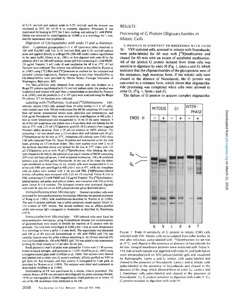
H: VSV-infected cells, arrested in mitosis with Nocodazole, were pulse-labeled for 60 min with ['S]methionine and chased for 60 min with an excess of unlabeled methionine. All of the labeled G protein isolated from these cells was sensitive to digestion by endo H (Fig. 1, lanes a and b), which indicates that the oligosaccharides of the glycoprotein were of the immature, high mannose form. If the mitotic cells were chased in the absence of Nocodazole, the G protein was converted to a resistant form, which shows that oligosaccha- ride processing was completed when cells were allowed to enter Gl (Fig. l, lanes c and d).
The failure of G protein to acquire complex oligosaccha-
FIGURE 1 Endo H sensitivity of G protein in mitotic CHO cells infected with VSV. Mitot ic cells were isolated from roller bottles 30 min after infection, pulse-labeled with [35S]methionine for 60 min at 37°C, and chased in the presence or absence of Nocodazole for 60 min. Integral membrane proteins were extracted with Triton X- 114, half of each sample was digested with endo H, and both halves were electrophoresed on SDS polyacrylamide gels and visualized by fluorography. Lanes a and b, mitotic cells pulse-labeled and chased in the presence of Nocodazole. Lanes c and d, mitotic cells pulse-labeled in the presence of Nocodazole and chased in the absence of the drug, which al lowed them to enter G1. Lanes e and f, Interphase cells pulse-labeled and chased in the presence of Nocodazole. Gs, G protein sensitive to digestion with endo H. GR, G protein resistant to digestion with endo H.
on March 21, 2018
jcb.rupress.orgD
ownloaded from

rides in cells arrested in mitosis was probably not due to a side effect of Nocodazole for the following three reasons:
(a) Complex oligosaccharides were formed on G protein in infected interphase cells, pulse-labeled for 60 minutes, and chased for 60 min in the presence of Nocodazole (Fig. l, lanes e and f ) .
(b) The time course of the conversion of endo H-resistant to endo H-sensitive G protein in infected interphase cells was unaffected by the presence of Nocodazole (Fig. 2).
(c) Transport of G protein to the surface of interphase cells in the presence of 100 times more Nocodazole than was used to arrest cells in mitosis had no effect on the appearance of G protein at the cell surface (Fig. 3). This was determined using a temperature-sensitive mutant of VSV, ts045, in which G protein remains in the ER at the nonpermissive temperature (39.5°C) but is transported to the plasma membrane at 3 l°C. After 4 h of infection with ts045 at the nonpermissive tem- perature, in the presence or absence of Nocodazole, normal rat kidney cells were either fixed immediately (Fig. 3, A and E) or transferred to the permissive temperature for 60 min (Fig. 3, B-D, F-H). Surface G protein was detected by im- munofluorescence microscopy (Fig. 3, A-D). No obvious differences between surface fluorescence intensity of cells treated with 100-fold excess Nocodazole (Fig. 3 B) or 1% (vol/ vol) dimethylsulfoxide (Fig. 3D), and control cells without Nocodazole (Fig. 3 C) could be detected.
The endo H-sensitive form of G protein predominated in cells held in mitosis with Nocodazole for at least 2.25 h after infection with VSV (Fig. 4 b). Between 3 and 5 h after infection this G protein gradually became resistant to digestion by endo H. This correlated with a drop in the mitotic index of the
100 ~ e
80
.~ 6O
Y_ N
30 ~ i
I I I I I I 0 I I I I I I ~0 2o ~.0 50 6o
Chase time (mini
FiGure 2 The effect of Nocodazole on the rate at which G protein in infected interphase cells acquires oligosaccharides resistant to endo H. CHO cells were infected with VSV for 30 min and all further treatments were performed in the presence or absence of Nocodazole. After 4 h of incubation at 37°C, the cells were pulse- labeled for 5 min with [3SS]methionine and chased for various times with an excess of unlabeled methionine. The cells were extracted with Triton X-114, the detergent pellets were digested with endo H, and the samples were fractionated by SDS PAGE. The endo H- resistant and -sensitive G protein bands, detected by fluorography, were cut out and counted. Closed symbols, with Nocodazole; open symbols, without Nocodazole; squares, endo-H-sensitive G pro- tein; circles, endo H-resistant G protein.
FIGURE .3 Transport of G protein to the surface of interphase cells infected with ts045 VSV, in the presence of Nocodazole. Normal rat kidney cells growing on coverslips were infected at the nonper- missive temperature (39.5°C) in the presence of 4 #g/ml Nocoda- zole (A, B, E, F), or 1% (vol/vol) dimethylsulfoxide (D, H) to control for the effect of solvent in the stock Nocodazole solution, or in the absence of both (C, G). 4 h after infection the cells were fixed (A, E) or transferred to the permissive temperature (31 °C) (B-D, F-H) and incubated for 60 rain more before fixation and processing for surface immunofluorescence. (A-D) Surface immunofluorescence. (E and F) Nomarski images of the same field. Bar, 20 i~m.
cells from >90% to ~30%. Almost 70% of cells had divided and returned to an interphase condition after 5 h of infection, as judged by staining with Hoechst 33258, despite the contin- ued presence of Nocodazole (Fig. 4a). For this reason the following experiments were completed within 3 h of infection when ~90% of the cells were still in the mitotic state.
THE O L I G O S A C C H A R I D E S C O N T A I N EIGHT OR NINE
S U G A R R E S I D U E S : TO determine the structure of the oli- gosaccharides on G protein, VSV-infected CHO cells, held in
FEATHERSTONE ET AL. Membrane Protein Transport in Mitosis 2039
on March 21, 2018
jcb.rupress.orgD
ownloaded from

FIGURE 4 The effect of incubation t ime on the endo H sensitivity of G protein in mitotic cells. Mitot ic CHO cells were isolated from roller bottles 30 min after infection and pulse-labeled with [~SS]methionine for 60 min at 37°C in the presence of Nocodazole. 2.25 h after infection half of the cells were al lowed to divide by washing with Nocodazole-free medium, and the incubation was continued. At various times sam- ples were stained with Hoechst dye 33258 to deter- mine the mitotic index (a), or extracted with Triton X- 114 and digested with endo H. The products were separated by SDS PAGE and visualized by fluorogra- phy (b). O, mitotic cells. O, mitotic cells washed into medium without Nocodazole after 2.25 h of infection. Cs and GR as in Fig. 1.
V '7 MITOTI[ [ELLS
8 -endoH- I / ! \ j * e n d 0 H
8
N 6 v,ra[ J l L 1 gtyco- ~ , peptidesJ ~:, A
& ~ I I I I ,a , , , / I 2 ,I L ;
t --
60 70 80 90 100 110 120 Fraction number
FIGURE 5 Gel filtration of [3H]mannose-labeled glycopeptides and oligosaccharides from infected mitotic and G~ cells. Mitotic CHO cells, isolated 30 rain after infection with VSV, were pulse-labeled with [3H]mannose for 60 min at 37°C and chased for 60 rain with an excess of unlabeled mannose in the presence or absence of Nocodazole. Membrane proteins were extracted with Triton X-114, and proteinase K glycopeptides were prepared and analyzed by gel filtration before and after digestion with endo H. (a) [3H]mannose- labeled glycopeptides from mitotic cells (broken line), and oligosac- charides resulting from endo H digestion of these glycopeptides (solid line). (b) [3H]mannose-labeled glycopeptides and oligosac- charides from G~ cells after digestion with endo H (solid line), and glycopeptides from VSV virions labeled with [3H]mannose (broken line). V0, void volume. D, position of elution of Glc3MangGlcNAc. MsN, MaN, positions of elution of MansGlcNAc and ManaGlcNac standards.
2040 THE JOURNAL OF CELL BIOLOGY - VOLUME 10l, 1985
mitosis with Nocodazole, were pulse-labeled for 60 min with [3H]mannose and chased for 60 rain with an excess of unla- beled mannose. G protein was isolated from the cells by Triton X- 114 extraction, and glycopeptides were prepared by proteinase K digestion. The glycopeptides were analyzed by gel filtration on Biogel P4 before and after digestion with endo H. Those isolated from cells in mitosis were completely sensitive to endo H, detected as a decrease in molecular weight of [3H]mannose-labeled material on gel filtration after diges- tion (Fig. 5 a). In the experiment shown here the major peak of endo H-sensitive oligosaccharide eluted in the same posi- tion as markers containing eight sugar residues, with minor amounts equivalent to those of oligosaccharides containing seven and six sugar residues. In similar experiments a peak containing nine sugar residues was also often observed. When mitotic cells were allowed to enter G~ by removal of Nocod- azole during the chase period, endo H-resistant glycopeptides were found (Fig. 5 b). That these eluted in the same position as glycopeptides prepared from VSV virions confirms that the inhibition of oligosaccharide processing in mitotic cells is reversible upon entry into G~. The lower molecular weight peak that appeared in the G~ extracts after digestion with endo H eluted in the same position as an oligosaccharide containing at least nine sugar residues, slightly larger than the major peak found in mitotic cells. This perhaps indicates that some oligosaccharide trimming occurs in mitotic cells during the second hour of mitotic arrest, that is, during the chase period.
THE OLIGOSACCHARIDES LACK GLUCOSE RESI- DUES: Oligosaccharides containing eight or nine sugar res- idues after digestion with endo H would normally result from trimming of the three outer glucose residues plus one or two mannose residues. Alternatively, one could envisage that in mitotic cells aberrant trimming of four or five outer mannose residues might also occur, leaving the outer glucose residues on the oligosaccharides. To distinguish between these two possibilities the endo H-sensitive oligosaccharides were fur- ther digested with jack bean a-mannosidase, which can trim outer mannose residues but not those blocked by glucose. The
on March 21, 2018
jcb.rupress.orgD
ownloaded from

Vc
13
12 11 I0
7 ~ 6 K s ~ 4
1 N ,
70
GIM~ N, G~M4 N~ M~N, M
• / •
80 90 10D 110 120 130 I~Q 150 160 170 180 190 200
FrQction number
Gel f i l t ra t ion o f o l igosacchar ides f r om VSV- in fec ted , mi- FIGURE 6 to t i c C H O cells a f ter t r e a t m e n t w i t h jack bean (~-mannosidase. The major peak of endo H-sensitive oligosaccharides from the column eluate shown in Fig. 5a was pooled, desalted, and lyophilized before digestion with o(-mannosidase and gel filtration on the same column as in Fig. 5. Vo, void volume. M1N~, G3M4N1, G3MsN~, positions of elution of GIcNAc /~l-4Man disaccharide, GIc3Man4- GIcNAc and GIc3MansGIcNAc, derived from a standard curve. M, position of elution of [3H]mannose.
sensitive to endo H (Fig. 8, lane b), whereas the [3H]palmitate was only found in the endo H-resistant fraction of the protein (Fig. 8, lane a). To show that this band was truly resistant to endo H and not the result of incomplete digestion due to the presence of fatty acid on the protein, interphase CHO clone 15B cells, in which oligosaccharides are not processed to the complex form, were labeled similarly and treated in parallel with endo H. The data shown in Fig. 8, lanes c-f, demonstrate that endo H digestion of fatty acylated G protein went to completion and that the distribution of 35S- and 3H-labeled G protein before and after endo H digestion was similar. In interphase CHO cells >90% of [35S]methionine and [3H]- palmitic acid were incorporated into endo H-resistant G protein under the conditions of the experiment (data not shown). We know from labeling with [aH]mannose or [3sS]- methionine that G protein in mitotic cells is >90% endo H sensitive (Figs. 1 and 5); therefore the simplest interpretation of these data is that [3H]palmitic acid is only incorporated into G protein in those cells that are not mitotic, due to division of mitotic cells as they begin to overcome the Nocod- azole block.
products of the digestion were again analyzed by P4 column chomatography (Fig. 6). The major peak obtained was coin- cident with [14C]mannose used to calibrate the column, and the small peak of slightly higher molecular weight eluted at the position calculated for the Man #1--4 GIcNAc disaccha- ride, which cannot be hydrolyzed by ~-mannosidase. The ratio of counts in these two peaks varied between 1:8.3 and 1:12.3 in three experiments, somewhat lower than the ex- pected ratio of 1:6 or 1:7. The reasons for this are unknown. The absence of any species of higher molecular weight, for example Glc3Man4GlcNAc or Glc3MansGlcNAc (Fig. 6), im- plies that the structure of the endo H-sensitive oligosaccha- rides was ManT_sGlcNAc, and that they were processed by the ER glucosidases. The oligosaccharides were also trimmed by a mannosidase, probably the E R a 1-2 mannosidase that normally trims high mannose oligosaccharides from Man9 to Mans (Bischoffand Kornfeld, 1983; Atkinson and Lee, 1984).
It is most unlikely that Nocodazole itself affects oligosac- charide processing, since the elution patterns ofglycopeptides and oligosaccharides isolated from infected interphase cells treated with or without Nocodazole were essentially the same (Fig. 7).
Fatty Acylation of G Protein in Mitotic Cells
Fatty acylation of O protein probably occurs in the late ER or early Golgi complex, at a point before the protein becomes resistant to endo H (Schmidt and Schlesinger, 1980). We could detect fatty acylation of G protein in mitotic and Gt cells only if we used more than 2 mCi [3H]palmitic acid to label cells isolated from 10 roller bottles, but, because of the time required to handle this number of bottles, we could not complete these experiments within the 3 h that guarantees a high mitotic index (see Fig. 4). In experiments in which the mitotic index ranged from 42-84% at the end of the experi- ment, fatty acylation of G protein was observed, but the [3H]palmitic acid was always incorporated into the endo H- resistant form of G protein. When a preparation of mitotic cells was divided in two, and one-half was labeled with [3H]- palmitic acid and the other with [35S]methionine under iden- tical conditions (Fig. 8), 50% of the 35S-labeled protein was
Location of G Protein in Mitotic Cells by Immunocytochemistry
Using quantitative i'mmunocytochemistry that employed a polyclonal antibody to G protein and protein A conjugated to gold, we demonstrated that the surface of mitotic cells, 3 h after infection, had 6% of the G protein found on the surface of Gl cells treated similarly (Warren et al., 1983). Because similar amounts of G protein were synthesized in mitotic and
o
E
o-.e
D INTERPHASE ~, * Nocodozo Le
V0 '/!
i i i i i ~ t
v0 {3 INTERPHASE
\/i ~o 7~ 60 9o 1oo 11o I~o
Froction number
FIGURE 7 Gel filtration of [3H]mannose-labeled glycopeptides from infected interphase cells, labeled in the presence or absence of Nocodazole. VSV-infected CHO cells were pulse-labeled with [3H]mannose for 60 min at 37°C and chased for 60 min with an excess of unlabeled mannose. Triton X-114 extracts were digested with proteinase K, and the glycopeptides released were digested with endo H and analyzed by gel filtration. (a) Cells pulsed and chased in the presence of Nocodazole. (b) Cells washed into Nocodazole-free medium during the chase period. V0 and D, as in Fig. 5.
FEATHERSTONE ET AL. Membrane Protein Transport in Mitosis 2041
on March 21, 2018
jcb.rupress.orgD
ownloaded from

test whether transport between the ER and the Golgi complex could occur during mitosis, we took mitotic CHO cells that had been infected with VSV for 2.5 h at 37"C, and divided them in half. One was fixed immediately, and the other was treated with 10 tzg/ml cycloheximide for 30 rain at 37"C before fixation and processing for immunocytochemistry as before. The density of G protein in the ER of mitotic cells was found to be similar before and after treatment with cycloheximide (Fig. 9 and Table I, experiment 2), as would be predicted if the transport step between the ER and Golgi complex was inhibited in mitosis. The small proportion of interphase cells in the mitotic preparations served as internal controls for the effective inhibition of protein synthesis. This is seen most clearly for labeling of the Golgi complex in interphase cells. Fig. l0 shows clearly the decrease in G protein in the interphase Golgi stacks after treatment with cyclohex- imide for 30 rain.
FIGURE 8 Comparison of [3H]palmitate and [355]methionine label- ing of G protein in infected mitotic cells. Mitotic CHO cells isolated 30 min after infection were labeled for 60 min at 37°C either with [3H]palmitic acid (lane a) or with [3SS]methionine (lane b), and chased in the presence o f excess un labe led meth ion ine and pal- mitic acid for 60 min more. Interphase C l i O c lone 15B cells were infected and labeled similarly wi th [3H]palmit ic acid (lanes c and d) or [3SS]methionine (lanes e and f). All samples were extracted with Tr i ton X-114, and the washed detergent pel lets f rom mitot ic cells and half o f the pel lets f rom interphase cells (lanes a, b, c, and e) were digested wi th endo H before SDS gel e lect rophores is and f luorography. Gsand GR, as in Fig. 1.
G~ cells, the diminished expression on the cell surface implied that G protein was retained in intracellular membranes. Al- though the ER is partially fragmented in mitotic CHO cells, considerable amounts of ER cisternae could be seen in the cytoplasm. Because >90% of the gold particles observed could be attributed to these ER membranes, we were confident that the gold particles we quantitated were representative of most of the G protein in the cell. The density of G protein in these cisternae, 3 h after infection with VSV, was more than 2.5 times greater than that found in the ER of G~ cells treated similarly but released from Nocodazole after 30 min of infec- tion (Table I, experiment 1), which suggests that G protein accumulates in the ER during mitosis.
Small cisternae that might be remnants of the Golgi com- plex were occasionally observed in the periphery of mitotic cells. These structures did not label above the background level, but the highly fragmented nature of the Golgi complex in mitosis makes if difficult to quantitate G protein in Golgi- derived elements using immunocytochemical techniques. To
DISCUSSION
Our observation that transport of the VSV glycoprotein to the surface of CHO cells is quite dramatically inhibited during mitosis (Warren et al., 1983) is the first example of a physio- logical inhibition of membrane protein transport through the pathway from ER to the plasma membrane. In this paper we have extended this observation by attempting to assess which step in the transport pathway is inhibited during mitosis. This has been possible by making use of the posttranslational modifications ofVSV G protein known to occur as the protein passes through the ER and Golgi complex.
All of the posttranslational processing of G protein that we have observed in mitotic cells is consistent with the retention of the protein within the ER. We have shown that [35S]- methionine-labeled G protein, and [3H]mannose-labeled oli- gosaccharides from VSV-infected, mitotic cells are digested by endo H. These high mannose oligosaccharides were shown to have a ManT_sGlcNAc2 structure. High mannose oligosac- charides are transferred en bloc from dolichol phosphate- linked precursors to the nascent polypeptide as it is inserted into the membrane of the ER (see Hubbard and Ivatt [1981] for a review). Very soon after, three glucose residues and one outer mannose residue are trimmed from the oligosaccha- rides. ER glucosidases (Grinna and Robbins, 1979) and an al,2-mannosidase (Bischoff and Kornfeld, 1983) have been identified that can perform these trimming reactions. Further modifications of G protein oligosaccharides occur in the Golgi complex. These include trimming of mannose residues, the addition of N-acetylglucosamine, and then further trimming
TABLE I. Quantitation of G Protein in the ER of Mitotic and G1 Cells by Immunocytochemistry
Cyclo- Nocoda- hex imide
Cell zole chase G Protein*
Exper iment 1 Mi to t ic + - 52.4 +-- 5.8 G1 - - 19.0 +_ 2.7
Exper iment 2 Mi to t ic + - 17.4 + 1.6 Mi to t ic + + 25.0 _+ 2.2
* Gold particles per square micrometer of membrane on the micrograph. Results are expressed as the mean + SEM of values from 20 micrographs. Background, counted as gold particles over mitochondria, was 1.3 +- 0.4- 1.8 + 0.6 per/~m 2. Gold particles not attributable to ER membranes were 7% of the total number of particles counted.
2042 THE JOURNAL OF CELL BIOLOGY - VOLUME 101, 1985
on March 21, 2018
jcb.rupress.orgD
ownloaded from

FIGURE 9 Immunocytochemical location of G protein in infected mitotic cells. VSV-.infected mitotic CHO cells were incubated at 37°C until 2 h after infection. The cells were then halved, one-half was fixed immediately, and the second half was incubated for 30 min more in the presence of cycloheximide before fixation. Thin, frozen sections of both were stained with rabbit anti-G protein antibody, and then protein A conjugated to 5 nm gold. Labeling of ER in a mitotic cell is shown before (a) and after (b) cycloheximide treatment. Arrows, gold particles attributed to ER. Arrowheads, gold particles not attributable to membranes. P, plasma membrane. Bar, 100 nm.
FEATHERSTONE ET AL. Membrane Protein Transport in Mitosis 2043
on March 21, 2018
jcb.rupress.orgD
ownloaded from

FIGURE 10 Clearance of G protein from the Golgi complex in interphase cells after treatment with cycloheximide. The samples described in the legend to Fig. 9 contained contaminating interphase cells that served as internal controls for the inhibition of protein synthesis by cycloheximide. The Golgi stacks in interphase cells in the mitotic cell preparation were heavily labeled before treatment with cycloheximide (a), but no labeling was found over similar stacks in cells after treatment with cycloheximide (b). C, Golgi stack. Arrow, trans Golgi reticulum. Bar, 100 nm.
of mannose residues, which renders the structure resistant to digestion by endo H. The structure of the oligosaccharides isolated from mitotic cells implies that the G protein was processed by the ER glucosidases I and II as well as an a 1-2 mannosidase, probably the ER enzyme. Although usually only one outer mannose residue is trimmed in the ER (Atkin- son and Lee, 1984), it has been shown that the al-2 mannos- idase of rat liver ER has activity in vitro towards high mannose oligosaccharides containing from six to nine mannose resi-
2044 THE JOURNAL OF CELL BIOLOGY • VOLUME 101, 1985
dues, with preference towards Man9 (Bischoff and Kornfeld, 1983). In contrast, the Golgi al-2 mannosidase is known to trim high mannose oligosaccharides efficiently to Mans. Therefore, G protein in mitotic cells probably had access only to the ER mannosidase. Similar trimming has been observed in other instances in which a glycoprotein was retained within the ER; for example, bovine thyroglobulin (Godelaine et al., 1981) and the Z variant of human plasma a~-antitrypsin (Hercz and Harpaz, 1980) accumulate in the rough ER of
on March 21, 2018
jcb.rupress.orgD
ownloaded from

liver with from five to seven mannose residues per oligosac- charide chain.
Although fatty acylation of G protein was always observed in mitotic cell preparations, this label was incorporated into an endo H-resistant form of G protein. We attribute this labeling to contamination of the mitotic cell preparations with interphase cells, which could not be avoided for technical reasons. This suggests that the G protein that accumulates in an endo H-sensitive form in mitotic cells does not become fatty acylated, indicating that transport of G protein is inhib- ited at a step before the compartment in which fatty acylation occurs. The proposed location of this enzyme is based on the kinetic relationship of fatty acylation to complex oligosaccha- ride formation, endo H-resistant oligosaccharides are con- structed in the trans Golgi cisternae (Roth and Berger, 1982), and G protein becomes labeled with [3H]palmitic acid 3-6 min before it acquires resistance to endo H (Schmidt and Schlesinger, 1980). This time is very similar to the time taken for G protein to move from the cis to the trans side of the Golgi stack (Bergmann and Singer, 1983). Therefore, fatty acylation appears from this evidence to be a cis Golgi func- tion. This is supported by work on Semliki Forest virus- infected cells treated with monensin, where the spike glyco- proteins become labeled with [3H]palmitic acid in or before transport to the medial Golgi cisternae (Quinn et al., 1983). If fatty acylation occurs in the cis part of the Golgi stack, our data would suggest that the first vesicle-mediated step of intracellular transport is inhibited during mitosis. However, as the rates of transport through different compartments on the pathway are unknown, such a precise morphological interpretation of the kinetic evidence may be unjustified. Genetic evidence suggests that fatty acylation occurs in the ER in yeast secretory mutants blocked in transport between the ER and Golgi complex (Wen and Schlesinger, 1984). This would be consistent with the kinetic evidence only if the rate of transport from the ER to the Golgi complex was relatively fast, and if fatty acylation occurred at a late stage after protein synthesis, perhaps in the transitional elements of the ER. This is supported by the evidence that G protein of the ts045 mutant of VSV, which accumulates throughout the ER at the nonpermissive temperature, is not fatty acylated (Zilberstein et al., 1980), which suggests that acylation is not a function of the rough ER. If this interpretation is correct, the absence of endo H-sensitive, fatty acylated G protein in mitotic cells suggests that the rough ER and transitional elements are functionally, and possibly also physically, separated during mitosis. In this instance we cannot draw any conclusions about inhibition of vesicular transport between the ER and Golgi complex, and will only be able to do so when the fatty acylation enzyme can be located by subeellular fractionation using a direct assay such as that described by Berger and Schmidt (1984).
Neither complex oligosaccharide formation (Schlesinger et at., 1976; Robertson et al., 1978), trimming by mannosidase (Burke et al., 1984), nor fatty acylation (Rose et al., 1984) is essential for G protein transport. Our observations therefore, could be explained if some processing enzymes performing posttranslational modifications were inactivated during mi- tosis but transport continued as usual. This seemed unlikely since the data presented here showed that the ER glucosidases and 1,2-mannosidase were active in mitotic cells. Also, Quin- tart et al. (1979) showed galactosyl transferase activity in mitotic cell extracts. Because we could not detect mannosidase
activity in homogenates of interphase or mitotic CHO cells, we used quantitative immunocytochemistry as an independ- ent method to confirm that G protein accumulated in the ER in mitotic cells. The observation that G protein could not be chased out of the mitotic ER during incubation with cyclo- heximide provides additional evidence that the inhibited step of transport occurs before, rather than within, the Golgi complex at a point before mannosidase action and endo H resistance are conferred. G protein was probably not retained in the ER during cycloheximide treatment due to the limited capacity of the Golgi cisternae for viral glycoproteins, since viral glycoproteins have been shown to accumulate in the Golgi of cells infected with a temperature-sensitive mutant of Semliki Forest virus, ts7 (Saraste et al., 1980) and in Semliki Forest virus-infected cells treated with monensin (Quinn et at., 1983). Given the limitations of morphometry, especially at early times of infection when labeling density is rather low, these results provide good evidence that G protein does not leave the ER in mitotic cells.
We thank Sabine Zimmerman for technical assistance, Joyce de Bruyn, Petra Riedinger, Alan Summerfield, and Annie Steiner for help in preparing the manuscript, and Drs. Brian Burke, Martyn Darby, and David Meyer for their critical comments.
C. Featherstone was the recipient of a Royal Society European Exchange Programme fellowship and a European Molecular Biology Organization fellowship.
Received for publication 13 June 1985, and in revised form 31 July 1985.
REFERENCES
Atkinson, P. H., and J. T. Lee. 1984. Co-translational excision of a-glucose and a-mannosc in nascent vesicular stomatitis virus G protein. J. Cell Biol. 98:2245-2249.
Berger, M., and M. F. G. Schmidt. 1984. Cell-free fatty acid acylation of Semliki Forest viral polypcptides with microsomal membranes from eukaryotic cells. J. Biol. Chem. 259:7245- 7252.
Bergmann, J. E., and S. J. Singer. 1983. Immunoelectron microscopic studies of the intracellular transport of the membrane glycoprotein (G) of vesicular stomatitis virus in infected Chinese Hamster ovary cells. J. Cell Biol. 97:1777-1787.
Bergmann, J. E., K. T. Tokuyasu, and S. J. Singer. 1981. Passage of an integral membrane protein, the vesicular stomatitis virus glycoprotein, through the Golgi apparatus en route to the plasma membrane. Proc. Natl. Acad. Sci. USA. 78:1746-1750.
Berlin, R. D., and J. M. Oliver. 1980. Surface functions during mitosis. II. Quantitation of pinocytosis and kinetic characterization of the mitotic cycle with a new fluorescence technique. J. Cell Biol. 85:660--671.
Berlin, R. D., J. M. Oliver, and R. J. Walter. 1978. Surface functions during mitosis. I. Phagocytosis, pinocytosis and mobility of surface-bound Con A. Cell. 15:327-341.
Bischoff, J., and R. Kornfeld. 1983. Evidence for an a-mannosidase in endoplasmic reticu- lure of rat liver. J. Biol. Chem. 258:7907-7910.
Bordier, C. 198 I. Phase separation of integral membrane proteins in Triton X-114 solution. .L Biol. Chem. 256:1604-1607.
Brandtzaeg, P. 1973. Conjugates of immunoglobulin G with different fluorochromes. I. Characterization of anionic exchange chromatography. ScanvL J. lmmunol. 2:273-290.
Burke, B., K. Matlin, E. Bause, G. Legler, N. Peyrieras, and H. Ploegh. 1984. Inhibition of N-linked oligosaccharide trimming does not interfere with surface expression of certain integral membrane proteins. EMBO (Eur. Mol. Biol. Organ.) 3:551-556.
Fawcett, D. W. 1965. Surface specializations of absorbing cells. J. Histochem. Cytochem. 13:75-91.
Godelaine, D., M. J. Spiro, and R. G. Spiro. 1981. Processing of the carbohydrate units of thyroglobulin. J. Biol. Chem. 256:10161-10168.
Green, J., G. Griffiths, D. Louvard, P. Quinn, and G. Warren. 1981. Passage of viral membrane proteins through the Golgi complex. J. Mol. Biol. 152:663~98.
Griffiths, G., A. McDowell, R. Back, and J. Dubochet. 1984. On the preparation of cryosections for immunocytochemistry. J. UItrastruct. Res. 89:65-78.
Griffiths, G., P. Quinn, and G. Warren. 1983. Dissection of the Gogi complex. L Monensin inhibits the transport of viral membrane proteins from medial to trans Golgi cistemae in baby hamster kidney cells infected with Semliki Forest virus. J Cell Biol. 96:835-850.
Grinna, L. S., and P. W. Robbins. 1979. Glycoprotein biosynthesis. Rat liver microsomal glucosidases which process oligosaccharides. J. Biol. Chem. 254:8814-8818.
Helenius, A., J. Kanenbeck, K. Simons, and E. Fries. 1980. On the entry of Semliki Forest virus into BHK-21 cells. J. Cell Biol. 84:404--420.
Hesketh, T. R., M. A. Beaven, J. Rogers, B. Burke, and G. B. Warren. 1984. Stimulated release of histamine by a rat mast cell line is inhibited during mitosis. J. CellBioL 98:2250- 2254.
Herez, A., and N. Harpaz. 1980. Characterization of the oligosaccbarides of liver Z variant at-antitrypsin. Can. J. Biochem. 58:644-648.
Hubbard, S. C., and R. J. Ivatt. 1981. Synthesis and processing of asparagine-linked oligosaccharides. Annu. Rev. Biochem. 50:555-583.
FEATHERSTONE ET AL. Membrane Protein Transport in Mitosis 2045
on March 21, 2018
jcb.rupress.orgD
ownloaded from

Jamieson, J. D. and G. E. Palade. 1968. lntracellular transport of secretory proteins in the pancreatic exocrine cell. IV. Metabolic requirements. Z Cell BioL 39:589-603.
Klevecz, R. R. 1975. Automated cell cycle analysis. Methods Cell. Biol. 10:157-172. Laskey, R. A., and A. D. Mills. 1975. Quantitative film detection of 3H and 14C in
polyacrylamide gels by fluorography. Eur..L Biochem. 56:335-341. Maizel, J. V. 1969. Acrylamide gel electrophoresis of proteins and nucleic acids. In Funda-
mental Techniques in Virology. K. Habel and N. P. Salzman, editors. Academic Press Inc., New York. 334-362.
Matlin, K. S., H. Reggio, A. Helenius, and K. Simons. 1982. Pathway of vesicular stomatitis virus entry leading to infection. ,L Mol. BioL 156:609-631.
Mattila, K., A. Luukkonen, and O. Renkonen. 1976. Protein-bound oligosaccharides of Semliki Forest virus. Biochim. Biophys. Acta. 419:435--444.
Pesonen, M. R. Ronnholm, E. Kuismanen, and R. F. Pettersson. 1982. Characterization of the oligosaccharides of lnkoo virus envelope glycoproteins. J. Gen. Virol. 63:4252,34.
Quinn, P., G. Griftiths, and G. Warren. 1983. Dissection of the Golgi complex. II. Density separation of specific Golgi functions in virally infected cells treated with monensin. J. Cell Biol 96:851-856.
Quintart, J., J. Bartholeyns, and P. Baudhuin. 1979. Characterization of subcellular com- ponents in synchronized hepatoma cells as a function of the cell cycle. Biochem Z 184:133-141.
Robertson, M. A., J. R. Etchison, J. S. Robertson, D. F. Summers, and P. Stanley. 1978. Specific changes in oligosaccharide moieties of VSV grown in different lectin-resistant CHO cells. Cell. 13:515-526.
Rose. J. K., G. A. Adams, and C. J. Gallione. 1984. The presence of cysteine in the cytoplasmic domain of the vesicular stomatitis virus glycoprotein is required for palmitate addition. Proe. Natl. Acad Sci. USA. 81:2050-2054.
RosneL M. R., S. C. Hubbard, R. J. lvatt, and P. W. Robbins. 1982. N-Asparagine-linked oligosaccharides: biosynthesis of the lipid-linked oligosaccharides. Methods EnzymoL 83:399-408.
Roth, J., and E. G. Berger. 1982. Immunocytochemical localization of galactosyltransferase in HeLa cells: codistribution with thiamine pyrophosphatase in trans Golgi cisternae..L Cell Biol. 92:223-229.
Sager, P. R., P. A. Brown, and R. D. Berlin. 1984. Analysis of transferrin recycling in mitotic and interphase Hela cells by quantitative fluorescence microscopy. Cell. 39:275-282.
Saraste, J., C.-H. von Bonsdorff, K. Hashimoto, L. Kaariainen, and S. Keranen. 1980. Semliki Forest virus mutants with temperature-sensitive transport defect of envelope proteins. Virology. 100:229-245.
Schlesinger, S., C. Gottlieb, P. Fiel, N. Gelb, and S. Kornfeld. 1976. Growth of enveloped RNA viruses in a line of Chinese hamster ovary cells with deficient N-acetyl-glucosami- nyltransferase activity. J. Virol. 17:239-246.
Schmidt, M. F. G., and M. J. Schlesinger. 1980. Relation of a fatty acid attachment to the translation and maturation of vesicular stomatitis virus and Sindbis virus membrane glycoproteins. J. BioL Chem. 255:3334-3339.
Tabas, l., and S. Kornfeld. 1978. The synthesis of complex-type oligosaccharides. IIl. Identification of an a-mannosidase activity involved in a late stage of processing of complex-type oligosaccharides. £ Biol. Chem. 253:7779-7786.
Wang, K., J. R. Feramisco, and J. F. Ash. 1982. Fluorescent localization of contractile proteins in tissue culture cells. Methods En2ymol. 85:514-562.
Warren, G., J. Davoust, and A. Cockcroft. 1984. Recycling of transferrin receptors in A431 cells is inhibited during mitosis. EMBO (Eur. Mol. BioL Organ.) J. 3:2217-2225~
Warren, G., C. Fcatherstone, G. Griffiths, and B. Burke. 1983. Newly synthesized G protein of vesicular stomatitis virus is not transported to the cell suface during mitosis. J. Cell Biol. 97:1623-1628.
Weibel, E. R. 1979. Stereological methods. I. Practical methods for biologmal morphometry. Academic Press Inc., New York. 26-30.
Wen, D., and M. J. Schlesinger. 1984. Fatty acid-acylated proteins in secretory mutants of Saccharomyces cerevisiae. Mol. Cell. Biol. 4:688-694.
Zieve, G. W., D. Turnbul[, J. M. Multins, and J. R. Mclntosh. 1980. Production of large numbers of mitotic mammalian cells by use of the reversible microtubule inhibitor Nocodazole. Exp. Cell Res. 126:3972"05.
Zilberstein, A., M. D. Snider, M. Porter, and H. F. Lodish. 1980. Mutants of vesicular stomatitis virus blocked at different stages in maturation of the viral glycoprotein. Cell. 21:417-427.
2046 THE JOURNAL OF CELL BIOLOGY . VOLUME 101, 1985
on March 21, 2018
jcb.rupress.orgD
ownloaded from