Role of Epigenetic Modification, Epigenetic Biomarkers and ...

RESEARCH ARTICLE
Newborn blood DNA epigenetic variations
and signaling pathway genes associated with
Tetralogy of Fallot (TOF)
Uppala Radhakrishna1*, Sangeetha Vishweswaraiah1, Avinash M. Veerappa2, Rita Zafra3,
Samet Albayrak4, Prajna H. Sitharam2, Nazia M. Saiyed5, Nitish K. Mishra6,
Chittibabu Guda6, Ray Bahado-Singh1
1 Department of Obstetrics and Gynecology, Oakland University William Beaumont School of Medicine,
Royal Oak, Michigan, United States of America, 2 Department of Studies in Genetics and Genomics,
Laboratory of Genomic Sciences, University of Mysore, Mysore, Karnataka, India, 3 Department of
Obstetrics and Gynecology, Icahn School of Medicine at Mount Sinai, New York, New York, United States of
America, 4 Department of Obstetrics and Gynecology, Wayne State University School of Medicine, Detroit,
Michigan, United States of America, 5 Biotechnology, Nirma Institute of Science, Nirma University,
Ahmedabad, Gujarat, India, 6 Department of Genetics, Cell Biology & Anatomy College of Medicine,
University of Nebraska Medical Center Omaha, Omaha, Nebraska, United States of America
Abstract
Tetralogy of Fallot (TOF) is the most common Critical Congenital Heart Defect (CCHD).
The etiology of TOF is unknown in most cases. Preliminary data from our group and others
suggest that epigenetic changes may play an important role in CHD. Epidemiologically, a
significant percentage of CHD including TOF fail to be diagnosed in the prenatal and early
newborn period which can negatively affect health outcomes. We performed genome-wide
methylation assay in newborn blood in 24 non-syndromic TOF cases and 24 unaffected
matched controls using Illumina Infinium HumanMethylation450 BeadChips. We identified
64 significantly differentially methylated CpG sites in TOF cases, of which 25 CpG sites
had high predictive accuracy for TOF, based on the area under the receiver operating char-
acteristics curve (AUC ROC)� 0.90). The CpG methylation difference between TOF and
controls was�10% in 51 CpG targets suggesting biological significance. Gene ontology
analysis identified significant biological processes and functions related to these differen-
tially methylated genes, including: CHD development, cardiomyopathy, diabetes, immuno-
logical, inflammation and other plausible pathways in CHD development. Multiple genes
known or plausibly linked to heart development and post-natal heart disease were found
to be differentially methylated in the blood DNA of newborns with TOF including: ABCB1,
PPP2R5C, TLR1, SELL, SCN3A, CREM, RUNX and LHX9. We generated novel and highly
accurate putative molecular markers for TOF detection using leucocyte DNA and thus pro-
vided information on pathogenesis of TOF.
PLOS ONE | https://doi.org/10.1371/journal.pone.0203893 September 13, 2018 1 / 19
a1111111111
a1111111111
a1111111111
a1111111111
a1111111111
OPENACCESS
Citation: Radhakrishna U, Vishweswaraiah S,
Veerappa AM, Zafra R, Albayrak S, Sitharam PH, et
al. (2018) Newborn blood DNA epigenetic
variations and signaling pathway genes associated
with Tetralogy of Fallot (TOF). PLoS ONE 13(9):
e0203893. https://doi.org/10.1371/journal.
pone.0203893
Editor: Leonard Eisenberg, New York Medical
College, UNITED STATES
Received: May 23, 2018
Accepted: August 29, 2018
Published: September 13, 2018
Copyright: © 2018 Radhakrishna et al. This is an
open access article distributed under the terms of
the Creative Commons Attribution License, which
permits unrestricted use, distribution, and
reproduction in any medium, provided the original
author and source are credited.
Data Availability Statement: All data is appended
in the manuscript text.
Funding: The present study was conducted using
the internal funding and no external support was
received. The funder had no role in study design,
data collection and analysis, decision to publish, or
preparation of the manuscript.
Competing interests: All the authors have declared
that no competing interests exist.

Introduction
Tetralogy of Fallot (TOF) is marked by a constellation of anatomical findings, including ven-
tricular septal defect (VSD), pulmonary stenosis (PS), right ventricular hypertrophy and an
overriding aorta. In the United States, the prevalence of TOF is estimated at 3.9 per 10,000 live
births [1] and it accounts for 7–10% of congenital heart defect (CHD) cases. Further, TOF the
most common critical congenital heart defect (CCHD) defined as CHDs that require surgical
or other significant medical intervention before one year of age.[2]
Early prenatal and newborn diagnosis is, pivotal to reducing morbidity and mortality in
CHD. Most cases of CHD occur in pregnancies with no identifiable risk factors for the disor-
der. A recent population-based screening study reported that nearly half of major CHD cases
are missed on prenatal screening [3], this is consistent with other population- based studies
reported in the literature. Effective prenatal screening will require markers with high diagnos-
tic accuracy. In addition, a significant percentage of CHD continue to be missed in the new-
born period. Indeed, over 50% of reported deaths in CCHD cases are due to missed or late
diagnosis [4]. The current standard of care requires pulse oximetry screening of all newborns
to detect CCHDs [5–7]. Despite official recommendations by the American Academy of Pedi-
atrics and the Secretary of Health and Human Services among others, utilization of pulse
oximetry is far from universal.[8] Further, simulation models suggest only modest detection
rates for TOF using pulse oximetry screening.[9]
The etiology of TOF is unknown in most cases, however the disorder is believed to have a
significant genetic component [10]. There is a strong association between TOF and 22q11
deletions [11] and aneuploidies, such as trisomy 21, 18 and 13. Mutations in a few genes have
also been reported in TOF, including NKX2-5,GATA4, ZFPM2, GATA6, GDF1, JAG1, TBX20,
HAND1, HAND2, PITX2 and TBX1. [12–18] However, these mutations appear to be responsi-
ble for only a small fraction of TOF cases.
It is now clear that focusing on individual genes and gene mutations cannot provide a com-
prehensive picture of CHD pathogenesis [19] given the complexity of cardiogenesis which is
impacted by an extensive list of environmental influences. The study of epigenetic contribu-
tion to CHD development is still relatively novel but has the potential to make significant con-
tributions to our understanding of CHD pathogenesis. Epigenetic changes are largely tissue
specific and the inaccessibility of the heart in living subjects is a monumental challenge to
studying cardiac development in general. Work by our group [20, 21] and others [22] have
reported evidence that a significant minority of epigenetic marks in blood DNA mirror similar
changes in inaccessible organs such as the heart and the brain respectively. This opens the pos-
sibility of ongoing epigenetic analysis of such organs.
The objective of the current study was to analyze cytosine methylation marks in leucocyte
DNA to investigate the pathogenesis of and for the detection of TOF in newborns. The study
was limited to non-syndromic TOF cases.
Methods
IRB approval was provided by the Michigan Department of Community Health (MDCH)
and Wayne State University. The ethics committee waived the requirement for informed
consent. The specimens were completely de-identified to the researchers prior to the analy-
sis. DNA was extracted from archived dried neonatal blood spots collected for the newborn
screening program run by the MDCH. The study subject consisted of 24 TOF cases and 24
controls matched for gestational age, gender, and maternal ethnic origin. Specimens were
collected at 48 hours on average (range 24 and 79 hours) after birth. Demographic and other
information was available for each subject including postnatal day of sample collection,
Epigenetics markers for Tetralogy of Fallot
PLOS ONE | https://doi.org/10.1371/journal.pone.0203893 September 13, 2018 2 / 19
maternal age and race, gestational age at birth and newborn sex. The average age of mothers
was 29.8 years, average gestational age at birth was 38.8 weeks. Cases of known or suspected
genetic syndromes, e.g. chromosomal or Mendelian or with extra-cardiac defects were
excluded. An equal number of unaffected individuals were used as controls. To reduce
potential variability due to race only Caucasian, non-Hispanic mothers were included in this
pilot study.
Epigenome-wide methylation scan using the Infinium methylation 450
array BeadChips
Multiple prior studies have reported genome-wide DNA methylation profiles from archived
dried blood spots using Infinium HumanMethylation450 BeadChips. [20] [21]. The Infinium
HumanMethylation450 BeadChips array for methylation (Illumina, Inc., California, USA)
contains >485,000 CpGs per sample in enhancer regions, coding regions, promoters and CpG
islands at a single-nucleotide resolution and requires only 500 ng of genomic DNA per assay.
These sites include 96% of RefSeq genes and 95% of CpG islands. DNA samples for this study
were bisulfite converted using the EZ DNA Methylation-Direct Kit (Zymo Research, Orange,
CA), and fluorescently-stained BeadChips imaged by the Illumina iScan. Data was analyzed
with Illumina’s Genome Studio methylation analysis package program. Prior to bioinformatic
and statistical analysis, data preprocessing and quality control was performed including
examination of the background signal intensity of negative controls, the methylated and
unmethylated signals, and the ratio of the methylated and unmethylated signal intensities. The
processing was done according to manufacturer’s protocol, and 99% of the CpG loci are deter-
mined unequivocally.
To avoid potentially confounding factors including sex-specific methylation bias, CpG sites
on the sex chromosomes and those containing SNPs in the probe sequence (listing dbSNP
entries near or within the probe sequence, i.e., within ten bp of the CpG site) were excluded
from further analysis.[23–25] CpG targets associated with SNPs near or within the probe
sequence may influence corresponding methylated probes.[26]
Statistical and bioinformatics analysis
Following the preprocessing described above, a DNA methylation level or ß-value was
assigned to each remaining CpG site. Differential methylation was assessed by comparing the
ß-values per individual nucleotide at each evaluated CpG locus between CHD subjects and
controls. The p-values for methylation differences between case and control groups at each
locus were calculated as previously described.[27] Filtering criteria for p-values was set at
<0.001 to enhance for the most discriminating cytosine loci. P-Values were computed with
and without False Discovery Rate (FDR) correction for multiple testing (Benjamini-Hochberg
test). Data were normalized using the Controls Normalization Method.
The most significant CpG sites based on p<value threshold was further sub-selected based
on the pre-set cutoff criteria of>2.0-fold methylation increase or >2.0-fold decrease. The
CpG sites with the highest (hypermethylated) and lowest (hypomethylated) degree of methyla-
tion were further analyzed. The significantly differentially methylated CpG sites in TOF cases
were subsequently used to generate a heatmap using the Complex Heatmap (v1.6.0) R package
(v3.2.2).[28, 29] We used ward distance for the hierarchical clustering of samples. Subse-
quently, the ß-value thresholds of each CpG locus was used to calculate the area under the
receiver operating characteristics curve (AUC) and 95% confidence intervals for TOF detec-
tion. All data cleaning and analysis were performed using R version 3.2.3.
Epigenetics markers for Tetralogy of Fallot
PLOS ONE | https://doi.org/10.1371/journal.pone.0203893 September 13, 2018 3 / 19

Principal Component Analysis (PCA)
To analyze larger number of variables to a smaller number of factors between TOF and con-
trols, we performed Principal Component Analysis. We removed all probes which have miss-
ing ß-value, remaining beta value of CpG targets were used for PCA. We used R function
“prcomp” to compute principal component (PC), and used PC1 and PC2 for the plot. The 3D
plot was generated using R package “ggplot2”.
Gene ontology analysis and functional enrichment
Literature data mining for co-occurrence of gene names and keywords of interest were per-
formed using Chilibot (www.chilibot.net). Only genes for which Entrez identifiers are available
were further analyzed. Genes found to be differentially methylated (at an FDR p-value thresh-
old�0.01) were processed through QIAGEN’S Ingenuity Pathway Analysis (IPA) software
(Ingenuity Systems, www.ingenuity.com) to identify biological functions or interacting regula-
tory networks. This provides information on the pathogenesis of the CHD. All CpGs without
mapping IDs in IPA (HG19, release 2009) were excluded from analysis, which included gene
ID conversion, bio-pathways analysis, and determination of the molecular functions of meth-
ylated and unmethylated regions. Over-represented canonical pathways, biological processes,
and molecular processes were identified.
Validation of methylation status by bisulfite sequencing
To further validate the array data, we used pyrosequencing to analyze the DNA methylation
status of the most significant CpG sites. We performed pyrosequencing analysis of bisulphite-
converted DNA with appropriate oligos using the PyroMark Q24 System and advanced CpG
Reagents (Qiagen1). Briefly, 500 ng of genomic DNA from each sample was bisulfite-treated
using the EZ methylation kit (Zymo Research) as per the manufacturer’s instructions and
amplified by a bisulfite polymerase chain reaction, followed by Quantitative DNA methylation
analysis of each CpG.
Analysis of correlation of cytosine methylation with gene expression and
chromatin conformation of CpG sites
A limitation of the study was that we were unable to perform corresponding gene expression
analysis to determine how DNA methylation correlated with gene expression given the fact
that only archived blood spots were available to us. We therefore looked at whether methyla-
tion status of the CpG loci of interest is known to correlate with gene expression levels in other
tissues. The GDAC FIREHOSE series of databases have been developed by the Broad Institute
using RNA-Seq data from over 20 types of malignant tissue (diffuse large B-cell lymphoma,
prostate adenocarcinoma-primary tumor, pancreatic adenocarcinoma-primary tumor, acute
myeloid leukemia, ovarian cancer, etc.) and include hypermethylation data at the nucleotide
level correlated with the expression mean of the gene in which the CpG site is located (gdac.
broadinstitute.org). Each of the identified differentially methylated CpG sites was examined
for inclusion in GDAC Firehose and its coordinates (correlation coefficient, p-value, methyla-
tion mean, and expression mean) were noted. Each CpG site was also examined in the
ENCODE databases using the UCSC genome browser for its location, type of location (e.g.,
promoter, intron, etc.), presence or lack of H3K27AC laying, and transcription factor occu-
pancy (e.g., PolR2a, PHF8 etc.) which indicate a potential role in gene expression.
Epigenetics markers for Tetralogy of Fallot
PLOS ONE | https://doi.org/10.1371/journal.pone.0203893 September 13, 2018 4 / 19

Results
Identification of differentially methylated CpG sites
Sixty-Four CpG sites, corresponding to 64 genes, were differentially methylated either in the
coding and/or promoter regions. This included 9 hypomethylated and 55 hypermethylated
CpG sites. The identified CpG sites, associated genes and chromosome locations are listed in
Table 1. The methylation levels of cases and controls, fold change and FDR p-value of the dif-
ference in methylation levels are also provided. The AUC (95% CI) for predicting TOF based
on methylation level at each of the cytosine loci is included. An AUC�0.75 generally indicates
a screening marker that could potentially have clinical utility. High predictive accuracy for
TOF detection was achieved at multiple CpG targets (Fig 1). In addition, the ROC curves
for the genes RUNX1 and CREMwith low methylation difference also given (S1 Fig). The
FDR p-values for the methylation difference between TOF subjects and controls were highly
significant. Overall, a total of 25 CpG loci in 25 genes had excellent predictive accuracy
(AUC� 0.90) for the detection of TOF. Principal Component Analysis (PCA) results showed
that there is a clear variance between two components. Majority of TOF components fall away
from controls (S2 Fig). Based on PCA, a subset analysis was performed using 8 TOF cases and
24 controls with a clear separation (S1 Table). This subset analysis identified a total of 2390 tar-
gets including 57 CpGs targets initially identified using 24 cases and 24 controls. A boxplot
with clear methylation differences over all the candidate CpGs is provided in S3 Fig.
Cluster analysis of differentially methylated targets
Of these 64 differentially methylated genes, unsupervised hierarchical clustering analysis dem-
onstrated their distribution as 4 major clusters (Fig 2). The methylation level of each CpG clus-
ter visually differentiated TOF versus controls. In cluster 1, we identified 8 significant novel
CpG sites (and genes) [cg02645710 (TSPAN19), cg04868078 (LHX9), cg21364560 (MYOF),cg11641791 (KRT222), cg17030055 (CDH10), ch.1.3587792F (SMG7), cg19021985 (PPP3CC),and cg04838988 (PRDM14)] associated with TOF. Most of the related CpG sites had�10%
including (N = 51) methylation difference between TOF and controls indicating a higher bio-
logical likelihood for being biologically significant.
Chromatin conformation and transcription factor occupation
Transcription initiation or regulatory sites often present an open chromatin conformation
that permits binding of regulatory transcription factors. These sites may reside in the promoter
of the gene, 1st exon, and intron or even outside of the gene. We observed that 40% (n = 28) of
the 39 CpG sites were in the promoter region of the gene, and 16% (n = 11) were in the non-
coding area of the 1st exon. A modest number of CpG sites were in an intron (29%, n = 20). A
detailed analysis of each CpG site with the ENCODE databases suggested that 81% of 64 CpG
sites (high 40% plus moderate 41%) were layered with H3K27Ac (Fig 3A), an open chromatin
conformation that is accessible to transcription factors during transcription initiation. Further
analysis showed that 57% (n = 39) of these CpG sites were occupied by RNA PolR2a (RNA
polymerase), a transcription initiator. Other transcription factors (TFs), such as PHF8, ERBB2,
MYC, YY1, EGR2, etc. were also present. We have identified 37 TFs overlap the candidate
CpGs. Among these, 25 TFs are cardiac TFs (S2 Table, Fig 3B). A smaller number of methyl-
ated CpG sites were in coding exon and intergenic sequences (S2 Table) (Fig 3C).
In some hypermethylated CpG sites (n = 10, 14%), non-Pol2R2A-dependent transcription
initiation or other accessory transcription mechanisms could possibly be involved, such as
DNA looping. However, some CpG sites are not occupied by any transcription factors
Epigenetics markers for Tetralogy of Fallot
PLOS ONE | https://doi.org/10.1371/journal.pone.0203893 September 13, 2018 5 / 19

Table 1. Differentially methylated CpG loci and genes. Target ID, Gene ID, chromosome location, % methylation change and FDR p-value for each gene methylated.
CpG sites with significant False Detection Rate p-value indicating methylation status and area under the receiving operator characteristic curve�0.75.
Target ID Gene CHR % Methylation Fold Change FDR p-Val AUC CI_upper CI_lower
Cases Controls
cg27120934 LAMA2 6 8.07 19.08 0.42 9.04E-26 0.98 1.00 0.95
cg02645710 TSPAN19 12 36.68 17.11 2.14 7.31E-45 0.98 1.00 0.94
cg04868078 LHX9 1 34.02 13.36 2.55 1.81E-45 0.97 1.00 0.93
cg23680282 LRRIQ1 12 17.31 6.17 2.81 1.12E-40 0.97 1.00 0.92
cg25697769 JOSD1 22 4.01 8.63 0.46 4.97E-09 0.97 1.00 0.93
cg21364560 MYOF 10 37.07 15.97 2.32 1.03E-45 0.96 1.00 0.90
cg24132989 C6orf162 6 22.32 45.01 0.50 1.17E-29 0.96 1.00 0.90
cg14905634 TRHDE 12 15.37 32.00 0.48 3.66E-31 0.95 1.00 0.88
cg08821669 COX6A1 12 3.80 8.49 0.45 1.97E-09 0.95 1.00 0.88
cg02062326 TMED10 14 24.46 12.16 2.01 3.35E-41 0.94 1.00 0.87
cg11641791 KRT222 17 27.02 12.77 2.12 4.02E-42 0.94 1.00 0.87
cg11792281 NLK 17 11.65 23.41 0.50 1.05E-23 0.94 1.00 0.87
cg12414181 SCAMP5 15 7.54 18.61 0.40 8.16E-27 0.94 1.00 0.87
cg17030055 CDH10 5 26.43 11.55 2.29 1.96E-42 0.94 1.00 0.86
cg17728974 LIN28B 6 17.38 7.24 2.40 3.15E-40 0.94 1.00 0.87
cg13114458 KRR1 12 12.43 5.54 2.25 7.47E-39 0.93 1.00 0.85
cg18803079 EFCAB7 1 11.34 24.22 0.47 1.12E-27 0.93 1.00 0.86
cg23274377 BPNT1 1 15.19 6.87 2.21 1.92E-39 0.93 1.00 0.85
cg01311718 IKZF2 2 14.54 6.83 2.13 3.45E-39 0.92 1.00 0.84
cg02609279 ITGA4 2 16.61 8.09 2.05 1.56E-39 0.92 1.00 0.83
cg02071276 FBXO28 1 20.60 9.15 2.25 8.18E-41 0.91 1.00 0.82
cg15946310 TTC1 5 16.42 6.48 2.53 3.82E-40 0.91 1.00 0.83
cg18295068 SCN3A 2 21.99 10.96 2.01 1.27E-40 0.91 1.00 0.83
cg09365677 CHRM3 1 24.05 9.68 2.49 3.49E-42 0.90 1.00 0.81
ch.11.319992F USP47 11 19.88 9.22 2.16 1.85E-40 0.90 1.00 0.81
cg12092090 CACNA1A 19 18.09 7.95 2.28 3.12E-40 0.89 1.00 0.79
cg19533977 CLTC 17 15.05 7.47 2.01 3.89E-39 0.89 1.00 0.79
cg20101489 SCG2 2 23.03 10.35 2.23 2.22E-41 0.89 1.00 0.80
cg01400516 NETO2 16 7.05 3.03 2.33 1.65E-08 0.89 1.00 0.79
cg23134869 ZFHX4 8 12.44 6.19 2.01 1.60E-13 0.89 1.00 0.80
cg08264335 SELL 1 15.67 7.72 2.03 2.75E-39 0.88 1.00 0.77
cg25477497 ABCB1 7 14.42 6.98 2.07 4.46E-39 0.88 1.00 0.79
cg25947619 AKAP13 15 18.92 8.64 2.19 2.72E-40 0.88 1.00 0.78
cg14534336 JMJD1C 10 9.75 4.78 2.04 2.70E-10 0.88 1.00 0.78
cg07002382 MFAP1 15 13.01 5.98 2.17 6.61E-39 0.88 1.00 0.78
cg12273284 CAMK1D 10 13.19 4.99 2.65 2.12E-39 0.87 1.00 0.77
cg10225640 ANAPC5 12 10.02 4.55 2.20 2.41E-12 0.86 1.00 0.75
ch.1.3587792F SMG7 1 25.97 12.81 2.03 1.32E-41 0.86 1.00 0.76
cg03814610 FAM13A 4 13.86 5.99 2.32 2.93E-39 0.84 1.00 0.72
cg10558887 SPG20 13 16.51 7.12 2.32 6.67E-40 0.84 1.00 0.73
cg18469624 PRKG1 10 19.24 9.52 2.02 4.75E-40 0.84 1.00 0.73
cg19579903 PPP1R10 6 14.04 5.79 2.42 2.05E-39 0.84 1.00 0.73
cg17616217 KY 3 3.29 7.05 0.47 5.42E-07 0.84 1.00 0.73
cg03547245 MSI2 17 10.26 4.40 2.33 5.14E-14 0.83 1.00 0.71
cg12129209 PPP2R5C 14 12.68 6.00 2.11 2.42E-15 0.83 1.00 0.71
(Continued)
Epigenetics markers for Tetralogy of Fallot
PLOS ONE | https://doi.org/10.1371/journal.pone.0203893 September 13, 2018 6 / 19

suggesting that either the ENCODE catalog of transcription factors may not be sufficiently
comprehensive or these CpG sites may not have a major role in regulating transcription.
Modulation of gene expression
To assess whether the observed methylation changes in each CpG in our study are likely to
modulate gene expression, we reviewed the Broad Institute Firehose methylation-expression
correlation databases, where CpG methylation changes in the genome have been correlated
with gene expression using RNA sequencing data. This would provide evidence as to whether
the observed variabilities in methylation changes in these CpGs indeed influence gene expres-
sion changes.
We found in all cases (S3 Table) that the correlation coefficient of mean methylation
changes vs mean expression changes was negative, indicating that hypermethylation in these
sites was associated with a reduction of expression, and hypomethylation with an increase in
expression. Thirty-nine of 64 of our observed differentially methylated CpG sites are present
in the database, with the extent of methylation changes correlated with the extent of expression
changes of the corresponding genes. Among these, 28 (44%) of the 64 CpG sites have alter-
ations in cytosine methylation located within the promoter of the gene. Additional CpG sites
were found to reside in other regions besides the promoter, including noncoding regions or
coding 1st exons (n = 11, 17%), which are also assumed to regulate gene expression [30] and
are also correlated with expression changes. Thus, evidence that reduction of methylation in
these 39 CpG sites increases the gene expression in tissues analyzed with RNA sequencing in
these databases, suggests that similar alterations may take place in the blood cells that we used
in our study, which could have implications in the development of TOF phenotypes.
As there were no available RNA samples from the study subjects, we have used already
available RNA-seq data from heart tissues of TOF patients and normal heart controls. The
Table 1. (Continued)
Target ID Gene CHR % Methylation Fold Change FDR p-Val AUC CI_upper CI_lower
Cases Controls
cg23404012 MED6 14 23.20 11.40 2.03 5.68E-41 0.83 1.00 0.71
cg03846926 C10orf140 10 16.64 7.20 2.31 6.36E-40 0.82 0.90 0.70
cg04254487 TBPL1 6 23.75 11.35 2.09 3.01E-41 0.82 0.90 0.69
cg08757862 TLR1 4 16.97 7.39 2.30 5.51E-40 0.82 0.90 0.69
cg26800788 PDE4D 5 18.92 8.92 2.12 3.60E-40 0.82 0.90 0.69
cg10944144 ADAMTS6 5 13.66 5.77 2.37 2.90E-39 0.81 0.90 0.69
cg02981003 GPR123 10 15.51 7.72 2.01 3.19E-39 0.81 0.90 0.69
cg26401673 ANO10 3 16.56 7.78 2.13 1.22E-39 0.80 0.90 0.67
ch.2.800013F BIRC6 2 11.69 5.53 2.12 6.69E-14 0.80 0.90 0.67
cg06237608 CHD2 15 7.92 3.49 2.27 1.53E-09 0.80 0.90 0.68
cg02558537 CWF19L2 11 10.03 4.97 2.02 2.08E-10 0.78 0.90 0.64
cg11378242 FAM36A 1 18.40 8.56 2.15 4.22E-40 0.78 0.90 0.65
cg17485454 MAPK10 4 16.60 8.24 2.01 1.84E-39 0.78 0.90 0.65
cg04838988 PRDM14 8 26.05 8.50 3.06 8.83E-44 0.77 0.90 0.63
cg19021985 PPP3CC 8 36.66 18.23 2.01 3.05E-44 0.76 0.90 0.62
ch.1.659794R UBR4 1 13.09 6.08 2.15 2.15E-16 0.76 0.90 0.63
cg27509202 CREM 10 9.96 4.96 2.01 3.24E-10 0.76 0.90 0.62
cg00994804 RUNX1 21 13.65 6.17 2.21 4.28E-39 0.76 0.90 0.62
cg22664298 ADAMTS19 5 18.76 9.24 2.03 5.81E-40 0.75 0.90 0.61
https://doi.org/10.1371/journal.pone.0203893.t001
Epigenetics markers for Tetralogy of Fallot
PLOS ONE | https://doi.org/10.1371/journal.pone.0203893 September 13, 2018 7 / 19

Fig 1. Receiver operating characteristic (ROC) curve analysis of methylation profiles for four specific markers associated with Tetralogy of Fallot. We
identified 64 differentially-methylated CpG sites in 64 genes that have an area under the ROC curve�0.75 for TOF prediction. At each locus, the False Detection
Rate p-value for the methylation difference between TOF subjects and controls was highly significantly different. Due to figure resolution concerns, we have
included only four markers (chr 12; cg02645710) (chr 1; cg04868078) (chr 10; cg21364560) (chr 5; cg17030055). AUC: Area Under the Receiver Operating
Characteristics Curve; 95% CI: 95% Confidence Interval. Lower and upper Confidence Intervals are given in parentheses.
https://doi.org/10.1371/journal.pone.0203893.g001
Epigenetics markers for Tetralogy of Fallot
PLOS ONE | https://doi.org/10.1371/journal.pone.0203893 September 13, 2018 8 / 19
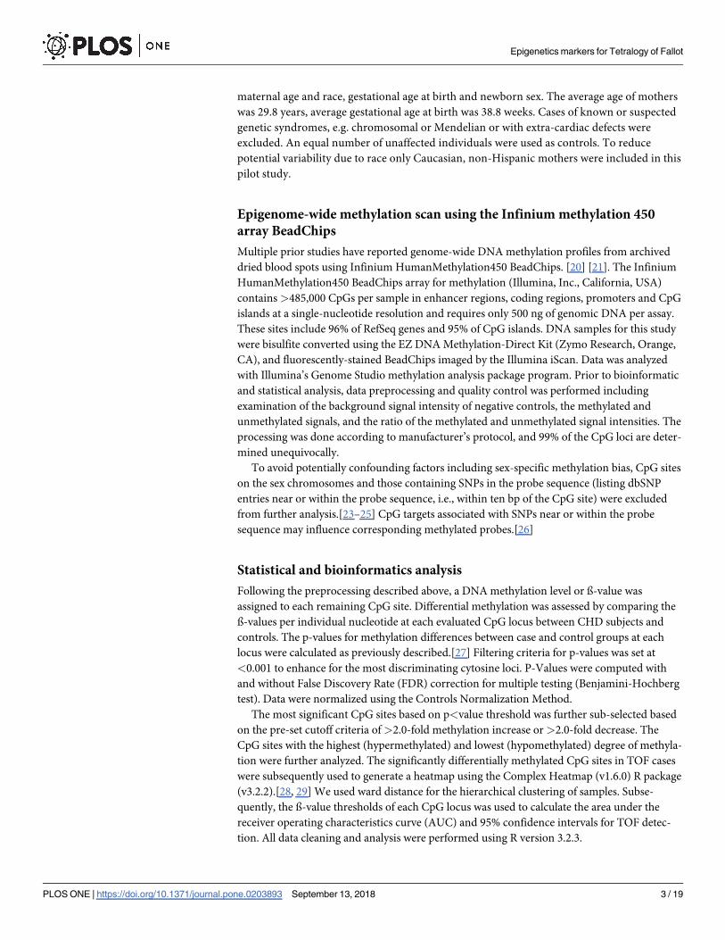
Fig 2. Heatmap of 26 highly differentially methylated loci. DNA methylation profiling based on unsupervised hierarchical clustering identified four unique
clusters that have distinct methylation signatures. These 26 targets represent the most highly differentially methylated loci (False Detection Rate< 0.000001). Only
CpG sites shown are those with either 2.0-fold hypomethylated or 2.0-fold hypermethylated in the disease TOF cases (red colored squares) compared to control
subjects (blue colored squares). The figure also displays direction, fold change in disease, probe relationship and probe annotation of differentially methylated CpG
sites. The red color in the heatmap indicates hyper DNA-methylation and blue hypo DNA-methylation values.
https://doi.org/10.1371/journal.pone.0203893.g002
Epigenetics markers for Tetralogy of Fallot
PLOS ONE | https://doi.org/10.1371/journal.pone.0203893 September 13, 2018 9 / 19

Fig 3. Open chromatin and transcription factor occupancy in functional CpG sites. Pie charts depicting H3K27Ac
histone mark layering, location of CpG sites and transcription factor binding sites. A) The layering of H3K27Ac
histone mark conferring an open chromatin status was seen in 87% of sites. B) 57% of the 64 sites were occupied by
RNA polymerase II subunit RPB1. C) 57% of 64 differentially methylated functional CpG sites were in the promoter
region of genes. See S2 Table for confirmation data per each gene.
https://doi.org/10.1371/journal.pone.0203893.g003
Epigenetics markers for Tetralogy of Fallot
PLOS ONE | https://doi.org/10.1371/journal.pone.0203893 September 13, 2018 10 / 19

data published in Grunert et al., 2014 and 2016 [31, 32] was compared with our data. Interest-
ingly we have identified ADAMTS6 gene matched with their data and was found to be differen-
tially expressed. Additionally, we have matched our differentially methylated genes with the
Grunert et al., 2016 and identified 25 genes (S4 Table).
Association with known cardiovascular disease pathways
Genes were further grouped per their Gene Ontology (GO)-characterized function. GO analy-
sis identified biological processes and roles for these genes including immunological pathways,
toxicity pathways, expression target pathways, nociception pathways, metabolic pathways,
receptor signaling, cell signaling and inflammation pathways (Fig 4). The Ingenuity Pathway
Analysis (IPA) identified important genes associated with these CpG sites that are currently
known or suspected to be associated with cardiac disease either congenital or developing in
postnatal life. The genes are associated with various postnatal cardiovascular disorders such as
Type 1 and type 2 diabetes, stroke, atherosclerosis, congenital heart defects, ischemia, coronary
artery diseases, high blood pressure, myocardial infarction, and vascular thrombosis.
Discussion
In the present study, we identified significant differences in methylation levels of multiple
CpG loci in TOF versus controls. We found 64 CpG sites in 64 genes that were significantly
differentially methylated in TOF versus controls. Among 64 differentially methylated CpGs, 55
were hypermethylated and only 9 were found to be hypomethylated. We have used top 26
Fig 4. Pathways analysis of significant DNA methylation variations and network analysis. Ingenuity pathway analysis (IPA) results for gene sets that were most
highly differentially methylated in association with TOF. IPA results indicated the gene network is relevant to immunological, toxicity, nociception, metabolic,
receptor, cell signaling, and inflammation pathways.
https://doi.org/10.1371/journal.pone.0203893.g004
Epigenetics markers for Tetralogy of Fallot
PLOS ONE | https://doi.org/10.1371/journal.pone.0203893 September 13, 2018 11 / 19

hypermethylated CpGs to generate heatmap (Fig 2). Many of these CpG loci are in genes that
are already known or suspected to be involved in CHD development or postnatal cardiovascu-
lar disorders. Some of the genes we identified have not however been previously reported to be
associated with TOF and CHD and require further evaluation. The difficulty of accurate pre-
natal and newborn diagnosis of CHD is well established in the literature [3,4,9]. Using DNA
methylation, we identified many important CpGs that preliminarily demonstrate high diag-
nostic accuracy for TOF detection (Table 1). In the future, these CpGs could have clinical util-
ity for TOF detection.
Leenen et al. [33] suggested that even relatively small differences in the methylation level,
e.g. of<10%, could be associated with changes in gene expression and phenotype. In the pres-
ent study, we have observed methylation variation between TOF and controls in 51 CpG tar-
gets with�10%.
We did not have access to fresh blood samples to perform DNA methylation at CpG sites
and gene expression correlation from the same patient. Instead we interrogated the Broad
Institute Firehose database to see whether methylation in CpG sites have been shown to corre-
late with expression. This database uses cancer tissue from various organs, e.g. ovary, breast
and colon. We found that 39 of the 64(56.5%) CpG sites, methylation levels correlate with
gene expression in these tissues. Also, using the ENCODE database, which was created using
normal tissues/cell lines, we found that 81% of differentially methylated CpG sites reported
conformed with H3K27AC occupancy at the same site. The H3K27AC binding reflects an
open chromatin formation permitting the binding of transcription factors and gene transcrip-
tion. The CpG sites with significant methylation changes in TOF are not only located within
the CpG islands in the promoter region of genes, but were also distributed throughout the
gene including transcription start site(s) (TSS), TSS200 (region from TSS to 200 bp upstream
of TSS), TSS1500 (200–1,500 bp upstream of TSS), 1st Exon, Body (coding), 5’ UTR, and
3’UTR regions. Moreover, when we compared our data with previously studied differentially
expressed and methylated regions using the myocardial biopsies, 25 genes were found to be
reproduced with differential methylation and one gene with expression variation in our study
[32]. We conclude from the above results that altered expression of one or a combination of
the 64 CpG sites in 64 genes is likely to be associated with altered expression of critical cardiac
developmental genes and possibly linked with TOF development.
Although genome- wide DNA methylation tends to be highly tissue specific, significant cor-
relation in DNA methylation profiles across tissues are now being found in a small but impor-
tant minority of CpG loci e.g. between blood and brain [22, 34]. The cause of these so called
‘mirror’ sites could be the result of epigenetic reprogramming that affected germ lines and
embryogenesis. In addition, blood leucocytes could be modified epigenetically as they circulate
through an affected primary organ. Further, genetic polymorphisms that can alter methylation
profile in more than one tissue and disease causing environmental factors that alter the epige-
netic profile in several tissues simultaneously are all potential explanations for synchronized
DNA methylation changes that have been observed when different tissues have been compared.
[35]. Our prior studies suggest that in similar fashion, leucocyte DNA might reflect cardiac epi-
genetic changes in CHD including TOF [20, 21]. Our analysis suggests a novel, non-invasive
and potentially highly impactful approach for the study of cardiac development and CHD.
Genes associated with congenital heart defects and postnatal cardiovascular
disorders
As noted, prior work by our group found highly significant alterations in CpG methylation
in blood leucocyte DNA in multiple different types of CHD including TOF. Sheng et al.
Epigenetics markers for Tetralogy of Fallot
PLOS ONE | https://doi.org/10.1371/journal.pone.0203893 September 13, 2018 12 / 19

conducted a targeted promoter region methylation study using MassARRAY-based quantita-
tive analysis in the myocardium of patients with TOF and reported DNA methylation changes.
Both studies confirm that TOF is associated with significant DNA methylation changes.
In the current study, the genes identified are involved in DNA damage response, toxicity
pathways, expression targets pathways, Notch signaling pathways, metabolic pathways, recep-
tor signaling, cell signaling and inflammation pathways. Pathways over-representation analysis
suggests that these genes could play a significant role in TOF development. All identified CpG
methylation sites are within or in close vicinity to genes whose functions have been associated
with cardiac development. These genes include ANO10,ABCB1,ARRB2,CLTC, LAMA2,
RUNX1, CHD2, LHX9, PRKG1, PDE4D, CAMK1D, SELL, SCG2,CHRM3, TLR1, LIN28B,
MAPK10, ITGA4 and CACNA1A.
ABCB1 (OMIM 171050) also known as P-glycoprotein 1 (PGY1) and Multidrug Resistance
1 (MDR1), is a gene encoding for P-glycoprotein, a large transmembrane protein. Significant
correlations between ABCB1 gene polymorphism and increased risk of congenital heart
defects,[36] hypertension,[37] stroke, and thrombosis [38] have been previously reported.
The PPP2R5C (OMIM 601645) gene on 14q32.31 is a phosphatase 2A regulatory subunit B-
family gene involved in negative control of cell growth and cell adhesion in the endothelial lin-
ing, and cardiac local signaling.[39] The protein also plays a critical role in cardiomyocyte
development thus a role in cardiac and TOF abnormalities is highly plausible.[40]
Genes associated with diabetes
The PPP2R5C (OMIM 601645) gene on 14q32.31 also plays an important role in diabetes
development.[41]. Two TLR1 (OMIM 601194) or Toll-like receptor (TLR) on 4p14 and SELL(OMIM 153240) or L-Selectin on chromosome 1q24.2 are candidate genes for the develop-
ment of type 1 diabetes. The CAMK1D (OMIM 607957) gene on 10p13 and CACNA1A(OMIM 601011) on 19p13.13 are also associated with diabetes. Maternal pregestational diabe-
tes is a potent risk factor for the development of congenital heart defects in offspring.[42, 43]
The SCN3A (OMIM 18239) gene on 2q24.3 is a sodium-dependent channel protein that
plays a major role in pulmonary artery smooth muscle and cardiomyocyte excitation.[44]
Moreover, this protein acts as a coupler of protein-protein interactions in a centrosome-cilium
formation that is distinctly related to congenital heart abnormalities.[45] The role of embry-
onic cilia in developing congenital heart malformation is well established, making a role in
TOF development plausible.
The CREM (OMIM 123812) gene on chromosome 10p11.21 is a transcription factor that
binds to a cAMP-responsive element in the promoter. Overexpression of cAMP-response ele-
ment modulator causes abnormal growth and development of the atrial myocardium and
leads to atrial fibrillation in mice.[46] and affects cardiac remodeling in mice[47] and the
development of myocardial infarction in rats.[48] CREM is involved in the switching of ß-
adrenergic receptor signaling in the decision for cell survival or death in cardiomyocytes. [49]
The LIM homeobox gene 9 (LHX9; OMIM 606066) is on 1q31.3. Lhx9 is essential for the
formation of the epicardium and heart development. Smagulova et al. demonstrated that the
mouse Lhx9 gene is the direct target of the GATA4/FOG2 repressor complex important in the
developing mouse heart.[50]. Further study will be needed to establish whether the differential
expression this homeobox gene could play a major role in developing TOF.
The ITGA4 gene (OMIM 192975) that maps to chromosome 2q31.3 is known to play a dis-
tinct role in epicardial and coronary vessel formation.[1] In Itga4-/- mouse, the epicardium
detaches from the myocardium and degrades. ITGA4 acts as fibronectin receptor and ITGA4influences epicardial Fn1 polymerization. ITGA4 overexpression alters Fibronectin/ integrins
Epigenetics markers for Tetralogy of Fallot
PLOS ONE | https://doi.org/10.1371/journal.pone.0203893 September 13, 2018 13 / 19

interactions and disrupts fibronectin deposition leading to epicardial dysmorphology and cor-
onary malformation.
Laminin Alpha-2 (LAMA2) (OMIM 156225 0) on chromosome 6q22.33 is a laminin pro-
tein that is conspicuously present in cardiomyocytes and plays a major role in cardiac develop-
ment. A mutation in the laminin gene causes dilated cardiomyopathy and a spectrum of heart
abnormalities.[51, 52]. Myoferlin (MyoF) is a ferlin family member protein located in the
plasma and nuclear membrane that plays a major role in VEGFA secretion and causes cardiac
muscle weakness in diabetes.[53]
The RUNX1 gene (OMIM 151385) on chromosome 21q22.12 is a transcription factor that
binds with promoters and enhancers in many genes. Runx1 copy number variation is associ-
ated with congenital heart malformation[54] including TOF[55] and thus differential methyla-
tion leading to aberrant expression of this gene in TOF is significantly plausible.
TOF is well known to be linked to chromosome 22q11.2 deletions [56]. However, in the
present analysis, we have not observed any gene variation(s) in this region except in the JOSD1gene. Given the relatively small number of cases used in this study, however, and the high
threshold used for significance that was used, more subtle changes in other CpG loci of other
genes could have escaped detection. Known or suspected cases with syndromic heart defects
including 22q deletions, chromosomal abnormalities, or extra cardiac abnormalities—which
increase the likelihood of genetic abnormalities were excluded from this study.
This manuscript does not posit that epigenetic changes are the only molecular changes that
are occurring in these non-syndromic CHD cases. Indeed, recent evidence suggests that single
nucleotide polymorphisms (SNP) induce and are associated with DNA methylation changes in
neighboring cytosine nucleotides. In addition, copy number variations are known to be associ-
ated with alteration of DNA methylation profiles.[57] It is therefore to likely that there is sig-
nificant overlap between DNA methylation and molecular pathologies. Thus, while a genetic
basis has been established in about a third of CHD cases,[58] this could be an underestimation.
Our cases excluded known or suspected genetic causes of TOF such as chromosomal anomaly.
In addition, our study group consisted of sporadic cases of TOF reducing the influence of
genetic causes. Given the very high frequency of epigenetic changes in our isolated CHD cases
in comparison to controls it is possible that a significant percentage could have had only epige-
nomic changes inducing the CHD. Taken together these data would suggest a further layer of
complexity where DNA methylation can cause, or result from, or occur in concert with other
molecular pathologies. While sequencing and microarray analysis of the DNA would be desir-
able to measure the extent of overlap between epigenomic and sequence and CNV changes,
this was not the objective of our study and we did not have IRB approval to perform such
analyses.
Blood is heterogenous and consists of different (leucocyte) cell types which are likely to
have different epigenetic profiles. Likewise, cardiac tissue has multiple cell types with different
methylation profiles, thus the correlation between methylation status of different leucocyte
and cardiac cell subtypes is likely to vary, adding another potential layer of complexity. It is
reasonable however to assume that unless the presence of cardiac defect alters the composition
of leucocytes in the blood (no evidence of this exists to our knowledge) that the observed dif-
ferences in the average methylation status of leucocytes between TOF and controls as assessed
in our study, was not in fact due to differences in methylation status of the different leucocyte
sub-populations but rather due to or associated with the TOF itself.
A limitation of our study was that expression analysis could not be performed since we
used archived blood spots. Analysis of Broad Institute Firehose and ENCODE databases sug-
gest that the methylation levels of most of the important CpG sites identified in our study likely
correlates with gene expression levels. We plan RNA-seq experiments in future studies to
Epigenetics markers for Tetralogy of Fallot
PLOS ONE | https://doi.org/10.1371/journal.pone.0203893 September 13, 2018 14 / 19

determine the correlation between DNA methylation and gene expression in CHD samples.
Another limitation is that the relatively small sample size means that the results, involved
genes, pathways and diagnostic accuracy for TOF detection are not definitive.
Conclusions
The use of epigenetics to understand the mechanisms of heart defects is in its relative infancy
and promises to help advance our understanding of these malformations. The identification of
the causative mechanisms in CHD will not only improve understanding of disease mecha-
nisms but could in the future contribute to the development of disease prophylaxis and
therapy.
The present study provides new target genes and cellular pathways potentially involved in
TOF development based on DNA methylation analysis based altered DNA methylation analy-
sis. Although not definitive, our results highlight the potential importance of epigenetics in the
pathogenesis of TOF. Finally, cardiac tissue is largely inaccessible in living fetuses and children
so analysis using surrogate tissue such as blood could dramatically change our ability to detect
and evaluate CHD.
Supporting information
S1 Fig. Receiver operating characteristic (ROC) curve analysis of methylation profiles for
two specific markers (RUNX1 and CREM) with low methylation difference associated with
Tetralogy of Fallot.
(TIF)
S2 Fig. Three dimensional PCA (PCA 3D) TOF and control samples.
(TIFF)
S3 Fig. A boxplot with clear methylation differences over all the candidate CpGs.
(TIFF)
S1 Table. Subset analysis of 8 cases and 24 controls based on PCA clear differentiation.
(PDF)
S2 Table. Open chromatin conformation and transcription factor binding in differentially
methylated CpG sites indicated their role in transcription initiation. ENCODE data show-
ing the H3K27Ac layering on each CpG site presenting an open chromatin conformation.
These CpG targets were also occupied with various transcription initiation factors, mostly
PolR2A. The position of each CpG site was also noted in respect to the gene in which it resided.
Some of the CpG sites that were differentially methylated resided in intronic or 1st exonic
regions, signifying their essential function in modulating transcription.
(PDF)
S3 Table. Correlation of Methylation mean with the expression mean in various human tis-
sues from GDAC data. 39 differentially methylated CpG targets were correlated with expres-
sion (RNA-seq) data. A bar chart was generated for each CpG target showing the proportion
of methylation and mean of expression of the gene in which the CpG target resided.
(PDF)
S4 Table. Differentially methylated CpG overlap with DMRs of Grunert et al., 2016.
(PDF)
Epigenetics markers for Tetralogy of Fallot
PLOS ONE | https://doi.org/10.1371/journal.pone.0203893 September 13, 2018 15 / 19

Acknowledgments
We thank Rose Callahan, Beaumont Health, for critical manuscript review and editing.
Web resources
The URLs for data presented herein are as follows:
Illumina: http://www.illumina.com/
Genome Studio: http://www.solexa.com/gsp/genomestudio_software.ilmn
Ensemble: http://www.ensembl.org/
UCSC: http://genome.ucsc.edu/
Web Gestalt: http://genereg.ornl.gov/webgestalt/
Chilibot: www.chilibot.net
Ingenuity Systems: www.ingenuity.com).
Online Mendelian Inheritance in Man (OMIM): http://www.ncbi.nlm.nih.gov/Omim/
Author Contributions
Conceptualization: Uppala Radhakrishna, Rita Zafra, Samet Albayrak, Ray Bahado-Singh.
Data curation: Uppala Radhakrishna, Sangeetha Vishweswaraiah, Avinash M. Veerappa, Nit-
ish K. Mishra, Chittibabu Guda.
Formal analysis: Uppala Radhakrishna, Sangeetha Vishweswaraiah, Avinash M. Veerappa,
Prajna H. Sitharam, Nazia M. Saiyed, Nitish K. Mishra, Chittibabu Guda.
Funding acquisition: Ray Bahado-Singh.
Investigation: Ray Bahado-Singh.
Methodology: Uppala Radhakrishna, Rita Zafra, Samet Albayrak, Nitish K. Mishra, Chittibabu
Guda.
Project administration: Uppala Radhakrishna, Ray Bahado-Singh.
Resources: Uppala Radhakrishna, Ray Bahado-Singh.
Supervision: Uppala Radhakrishna, Ray Bahado-Singh.
Validation: Uppala Radhakrishna.
References1. Centers for Disease C and Prevention. Improved national prevalence estimates for 18 selected major
birth defects—United States -. MMWR Morb Mortal Wkly Rep. 2006; 54(8):1301–5.
2. Simeone RM, Oster ME, Cassell CH, Armour BS, Gray DT, Honein MA. Pediatric inpatient hospital
resource use for congenital heart defects. Birth Defects Res A Clin Mol Teratol. 2014; 100(12):934–43.
https://doi.org/10.1002/bdra.23262 PMID: 24975483.
3. Jorgensen DE, Vejlstrup N, Jorgensen C, Maroun LL, Steensberg J, Hessellund A, et al. Prenatal detec-
tion of congenital heart disease in a low risk population undergoing first and second trimester screening.
Prenat Diagn. 2015; 35(4):325–30. https://doi.org/10.1002/pd.4525 PMID: 25352400.
4. Chang RK, Gurvitz M, Rodriguez S. Missed diagnosis of critical congenital heart disease. Arch Pediatr
Adolesc Med. 2008; 162(10):969–74. https://doi.org/10.1001/archpedi.162.10.969 PMID: 18838650.
5. Pinto NM, Keenan HT, Minich LL, Puchalski MD, Heywood M, Botto LD. Barriers to prenatal detection
of congenital heart disease: a population-based study. Ultrasound Obstet Gynecol. 2012; 40(4):418–
25. https://doi.org/10.1002/uog.10116 PMID: 21998002.
6. Quartermain MD, Pasquali SK, Hill KD, Goldberg DJ, Huhta JC, Jacobs JP, et al. Variation in Prenatal
Diagnosis of Congenital Heart Disease in Infants. Pediatrics. 2015; 136(2):e378–85. https://doi.org/10.
1542/peds.2014-3783 PMID: 26216324.
Epigenetics markers for Tetralogy of Fallot
PLOS ONE | https://doi.org/10.1371/journal.pone.0203893 September 13, 2018 16 / 19

7. Hom LA, Martin GR. U.S. international efforts on critical congenital heart disease screening: can we
have a uniform recommendation for Europe? Early Hum Dev. 2014; 90 Suppl 2:S11–4. https://doi.org/
10.1016/S0378-3782(14)50004-7 PMID: 25220118.
8. Studer MA, Smith AE, Lustik MB, Carr MR. Newborn pulse oximetry screening to detect critical congeni-
tal heart disease. J Pediatr. 2014; 164(3):505–9 e1-2. https://doi.org/10.1016/j.jpeds.2013.10.065
PMID: 24315501.
9. Ailes EC, Gilboa SM, Honein MA, Oster ME. Estimated number of infants detected and missed by criti-
cal congenital heart defect screening. Pediatrics. 2015; 135(6):1000–8. https://doi.org/10.1542/peds.
2014-3662 PMID: 25963011.
10. Harold JG. Cardiology patient page. Screening for critical congenital heart disease in newborns. Circu-
lation. 2014; 130(9):e79–81. PMID: 25156919.
11. Lu JH, Chung MY, Betau H, Chien HP, Lu JK. Molecular characterization of tetralogy of fallot within
Digeorge critical region of the chromosome 22. Pediatr Cardiol. 2001; 22(4):279–84. https://doi.org/10.
1007/s002460010230 PMID: 11455393.
12. De Luca A, Sarkozy A, Ferese R, Consoli F, Lepri F, Dentici ML, et al. New mutations in ZFPM2/FOG2
gene in tetralogy of Fallot and double outlet right ventricle. Clin Genet. 2011; 80(2):184–90. https://doi.
org/10.1111/j.1399-0004.2010.01523.x PMID: 20807224.
13. Goldmuntz E, Geiger E, Benson DW. NKX2.5 mutations in patients with tetralogy of fallot. Circulation.
2001; 104(21):2565–8. PMID: 11714651.
14. Lin X, Huo Z, Liu X, Zhang Y, Li L, Zhao H, et al. A novel GATA6 mutation in patients with tetralogy of
Fallot or atrial septal defect. J Hum Genet. 2010; 55(10):662–7. https://doi.org/10.1038/jhg.2010.84
PMID: 20631719.
15. Huang RT, Wang J, Xue S, Qiu XB, Shi HY, Li RG, et al. TBX20 loss-of-function mutation responsible
for familial tetralogy of Fallot or sporadic persistent truncus arteriosus. Int J Med Sci. 2017; 14(4):323–
32. https://doi.org/10.7150/ijms.17834 PMID: 28553164.
16. Wang J, Hu XQ, Guo YH, Gu JY, Xu JH, Li YJ, et al. HAND1 Loss-of-Function Mutation Causes Tetral-
ogy of Fallot. Pediatr Cardiol. 2017; 38(3):547–57. https://doi.org/10.1007/s00246-016-1547-8 PMID:
27942761.
17. Lu CX, Gong HR, Liu XY, Wang J, Zhao CM, Huang RT, et al. A novel HAND2 loss-of-function mutation
responsible for tetralogy of Fallot. Int J Mol Med. 2016; 37(2):445–51. https://doi.org/10.3892/ijmm.
2015.2436 PMID: 26676105.
18. Sun YM, Wang J, Qiu XB, Yuan F, Xu YJ, Li RG, et al. PITX2 loss-of-function mutation contributes to
tetralogy of Fallot. Gene. 2016; 577(2):258–64. https://doi.org/10.1016/j.gene.2015.12.001 PMID:
26657035.
19. Gittenberger-de Groot AC, Calkoen EE, Poelmann RE, Bartelings MM, Jongbloed MR. Morphogenesis
and molecular considerations on congenital cardiac septal defects. Ann Med. 2014; 46(8):640–52.
https://doi.org/10.3109/07853890.2014.959557 PMID: 25307363.
20. Radhakrishna U, Albayrak S, Alpay-Savasan Z, Zeb A, Turkoglu O, Sobolewski P, et al. Genome-Wide
DNA Methylation Analysis and Epigenetic Variations Associated with Congenital Aortic Valve Stenosis
(AVS). PLoS One. 2016; 11(5):e0154010. https://doi.org/10.1371/journal.pone.0154010 PMID:
27152866.
21. Bahado-Singh RO, Zaffra R, Albayarak S, Chelliah A, Bolinjkar R, Turkoglu O, et al. Epigenetic markers
for newborn congenital heart defect (CHD). J Matern Fetal Neonatal Med. 2016; 29(12):1881–7. https://
doi.org/10.3109/14767058.2015.1069811 PMID: 26429603.
22. Aberg KA, McClay JL, Nerella S, Clark S, Kumar G, Chen W, et al. Methylome-wide association study
of schizophrenia: identifying blood biomarker signatures of environmental insults. JAMA Psychiatry.
2014; 71(3):255–64. https://doi.org/10.1001/jamapsychiatry.2013.3730 PMID: 24402055.
23. Liu Y, Aryee MJ, Padyukov L, Fallin MD, Hesselberg E, Runarsson A, et al. Epigenome-wide associa-
tion data implicate DNA methylation as an intermediary of genetic risk in rheumatoid arthritis. Nat Bio-
technol. 2013; 31(2):142–7. https://doi.org/10.1038/nbt.2487 PMID: 23334450.
24. Chen YA, Lemire M, Choufani S, Butcher DT, Grafodatskaya D, Zanke BW, et al. Discovery of cross-
reactive probes and polymorphic CpGs in the Illumina Infinium HumanMethylation450 microarray. Epi-
genetics. 2013; 8(2):203–9. https://doi.org/10.4161/epi.23470 PMID: 23314698.
25. Wilhelm-Benartzi CS, Koestler DC, Karagas MR, Flanagan JM, Christensen BC, Kelsey KT, et al.
Review of processing and analysis methods for DNA methylation array data. Br J Cancer. 2013; 109
(6):1394–402. https://doi.org/10.1038/bjc.2013.496 PMID: 23982603.
26. Daca-Roszak P, Pfeifer A, Zebracka-Gala J, Rusinek D, Szybinska A, Jarzab B, et al. Impact of SNPs
on methylation readouts by Illumina Infinium HumanMethylation450 BeadChip Array: implications for
Epigenetics markers for Tetralogy of Fallot
PLOS ONE | https://doi.org/10.1371/journal.pone.0203893 September 13, 2018 17 / 19

comparative population studies. BMC Genomics. 2015; 16(1):1003. https://doi.org/10.1186/s12864-
015-2202-0 PMID: 26607064.
27. Altorok N, Tsou PS, Coit P, Khanna D, Sawalha AH. Genome-wide DNA methylation analysis in dermal
fibroblasts from patients with diffuse and limited systemic sclerosis reveals common and subset-specific
DNA methylation aberrancies. Annals of the rheumatic diseases. 2014. https://doi.org/10.1136/
annrheumdis-2014-205303 PMID: 24812288.
28. Gu Z, Eils R, Schlesner M. Complex heatmaps reveal patterns and correlations in multidimensional
genomic data. Bioinformatics. 2016; 32(18):2847–9. https://doi.org/10.1093/bioinformatics/btw313
PMID: 27207943.
29. Gu. Z. ComplexHeatmap: Making Complex Heatmaps. R package version 1.6.0. https://githubcom/
jokergoo/ComplexHeatmap. 2015.
30. Mercer TR, Edwards SL, Clark MB, Neph SJ, Wang H, Stergachis AB, et al. DNase I-hypersensitive
exons colocalize with promoters and distal regulatory elements. Nat Genet. 2013; 45(8):852–9. https://
doi.org/10.1038/ng.2677 PMID: 23793028.
31. Grunert M, Dorn C, Schueler M, Dunkel I, Schlesinger J, Mebus S, et al. Rare and private variations in
neural crest, apoptosis and sarcomere genes define the polygenic background of isolated Tetralogy of
Fallot. Hum Mol Genet. 2014; 23(12):3115–28. https://doi.org/10.1093/hmg/ddu021 PMID: 24459294.
32. Grunert M, Dorn C, Cui H, Dunkel I, Schulz K, Schoenhals S, et al. Comparative DNA methylation and
gene expression analysis identifies novel genes for structural congenital heart diseases. Cardiovasc
Res. 2016; 112(1):464–77. https://doi.org/10.1093/cvr/cvw195 PMID: 27496870.
33. Leenen FA, Muller CP, Turner JD. DNA methylation: conducting the orchestra from exposure to pheno-
type? Clin Epigenetics. 2016; 8:92. https://doi.org/10.1186/s13148-016-0256-8 PMID: 27602172.
34. Fan S, Zhang X. CpG island methylation pattern in different human tissues and its correlation with gene
expression. Biochem Biophys Res Commun. 2009; 383(4):421–5. https://doi.org/10.1016/j.bbrc.2009.
04.023 PMID: 19364493.
35. Aberg KA, Xie LY, McClay JL, Nerella S, Vunck S, Snider S, et al. Testing two models describing how
methylome-wide studies in blood are informative for psychiatric conditions. Epigenomics. 2013; 5
(4):367–77. https://doi.org/10.2217/epi.13.36 PMID: 23895651.
36. Wang C, Xie L, Zhou K, Zhan Y, Li Y, Li H, et al. Increased risk for congenital heart defects in children
carrying the ABCB1 Gene C3435T polymorphism and maternal periconceptional toxicants exposure.
PLoS One. 2013; 8(7):e68807. https://doi.org/10.1371/journal.pone.0068807 PMID: 23874772.
37. Zuo XC, Zhang WL, Yuan H, Barrett JS, Hua Y, Huang ZJ, et al. ABCB1 polymorphism and gender
affect the pharmacokinetics of amlodipine in Chinese patients with essential hypertension: a population
analysis. Drug Metab Pharmacokinet. 2014; 29(4):305–11. PMID: 24522199.
38. Luo M, Li J, Xu X, Sun X, Sheng W. ABCB1 C3435T polymorphism and risk of adverse clinical events in
clopidogrel treated patients: a meta-analysis. Thromb Res. 2012; 129(6):754–9. https://doi.org/10.
1016/j.thromres.2011.12.003 PMID: 22209339.
39. Zhou XW, Mudannayake M, Green M, Gigena MS, Wang G, Shen RF, et al. Proteomic studies of
PP2A-B56gamma1 phosphatase complexes reveal phosphorylation-regulated partners in cardiac local
signaling. J Proteome Res. 2007; 6(9):3433–42. https://doi.org/10.1021/pr060619l PMID: 17663574.
40. Donofrio MT, Massaro AN. Impact of congenital heart disease on brain development and neurodevelop-
mental outcome. Int J Pediatr. 2010;2010. https://doi.org/10.1155/2010/359390 PMID: 20862365.
41. Cheng YS, Seibert O, Kloting N, Dietrich A, Strassburger K, Fernandez-Veledo S, et al. PPP2R5C Cou-
ples Hepatic Glucose and Lipid Homeostasis. PLoS Genet. 2015; 11(10):e1005561. https://doi.org/10.
1371/journal.pgen.1005561 PMID: 26440364.
42. Simeone RM, Devine OJ, Marcinkevage JA, Gilboa SM, Razzaghi H, Bardenheier BH, et al. Diabetes
and congenital heart defects: a systematic review, meta-analysis, and modeling project. Am J Prev
Med. 2015; 48(2):195–204. https://doi.org/10.1016/j.amepre.2014.09.002 PMID: 25326416.
43. Correa A, Gilboa SM, Besser LM, Botto LD, Moore CA, Hobbs CA, et al. Diabetes mellitus and birth
defects. Developmental dynamics: an official publication of the American Association of Anatomists.
2008; 199(3):237 e1–9. https://doi.org/10.1016/j.ajog.2008.06.028 PMID: 18674752.
44. Platoshyn O, Remillard CV, Fantozzi I, Sison T, Yuan JX. Identification of functional voltage-gated Na
(+) channels in cultured human pulmonary artery smooth muscle cells. Pflugers Arch. 2005; 451
(2):380–7.
45. Gupta GD, Coyaud E, Goncalves J, Mojarad BA, Liu Y, Wu Q, et al. A Dynamic Protein Interaction
Landscape of the Human Centrosome-Cilium Interface. Cell. 2015; 163(6):1484–99. https://doi.org/10.
1016/j.cell.2015.10.065 PMID: 26638075.
46. Kirchhof P, Marijon E, Fabritz L, Li N, Wang W, Wang T, et al. Overexpression of cAMP-response ele-
ment modulator causes abnormal growth and development of the atrial myocardium resulting in a
Epigenetics markers for Tetralogy of Fallot
PLOS ONE | https://doi.org/10.1371/journal.pone.0203893 September 13, 2018 18 / 19
substrate for sustained atrial fibrillation in mice. Int J Cardiol. 2013; 166(2):366–74. https://doi.org/10.
1016/j.ijcard.2011.10.057 PMID: 22093963.
47. Feng J, Lucchinetti E, Fischer G, Zhu M, Zaugg K, Schaub MC, et al. Cardiac remodelling hinders acti-
vation of cyclooxygenase-2, diminishing protection by delayed pharmacological preconditioning: role of
HIF1 alpha and CREB. Cardiovasc Res. 2008; 78(1):98–107. https://doi.org/10.1093/cvr/cvn016 PMID:
18218685.
48. Ma D, Fu L, Shen J, Zhou P, Gao Y, Xie R, et al. Interventional effect of valsartan on expression of
inducible cAMP early repressor and phosphodiesterase 3A in rats after myocardial infarction. Eur J
Pharmacol. 2009; 602(2–3):348–54. https://doi.org/10.1016/j.ejphar.2008.11.002 PMID: 19027736.
49. Shin SY, Kim T, Lee HS, Kang JH, Lee JY, Cho KH, et al. The switching role of beta-adrenergic receptor
signalling in cell survival or death decision of cardiomyocytes. Nat Commun. 2014; 5:5777. https://doi.
org/10.1038/ncomms6777 PMID: 25517116.
50. Smagulova FO, Manuylov NL, Leach LL, Tevosian SG. GATA4/FOG2 transcriptional complex regu-
lates Lhx9 gene expression in murine heart development. BMC Dev Biol. 2008; 8:67. https://doi.org/10.
1186/1471-213X-8-67 PMID: 18577233.
51. Jones KJ, Morgan G, Johnston H, Tobias V, Ouvrier RA, Wilkinson I, et al. The expanding phenotype of
laminin alpha2 chain (merosin) abnormalities: case series and review. J Med Genet. 2001; 38(10):649–
57. https://doi.org/10.1136/jmg.38.10.649 PMID: 11584042.
52. Carboni N, Marrosu G, Porcu M, Mateddu A, Solla E, Cocco E, et al. Dilated cardiomyopathy with con-
duction defects in a patient with partial merosin deficiency due to mutations in the laminin-alpha2-chain
gene: a chance association or a novel phenotype? Muscle Nerve. 2011; 44(5):826–8. https://doi.org/10.
1002/mus.22228 PMID: 22006699.
53. Fahmy K, Gonzalez A, Arafa M, Peixoto P, Bellahcene A, Turtoi A, et al. Myoferlin plays a key role in
VEGFA secretion and impacts tumor-associated angiogenesis in human pancreas cancer. Int J Cancer.
2016; 138(3):652–63. https://doi.org/10.1002/ijc.29820 PMID: 26311411.
54. Melis D, Genesio R, Cappuccio G, MariaGinocchio V, Casa RD, Menna G, et al. Mental retardation,
congenital heart malformation, and myelodysplasia in a patient with a complex chromosomal rearrange-
ment involving the critical region 21q22. Am J Med Genet A. 2011; 155A(7):1697–705. https://doi.org/
10.1002/ajmg.a.33976 PMID: 21671372.
55. Tomita-Mitchell A, Mahnke DK, Struble CA, Tuffnell ME, Stamm KD, Hidestrand M, et al. Human gene
copy number spectra analysis in congenital heart malformations. Physiol Genomics. 2012; 44(9):518–
41. https://doi.org/10.1152/physiolgenomics.00013.2012 PMID: 22318994.
56. Maeda J, Yamagishi H, Matsuoka R, Ishihara J, Tokumura M, Fukushima H, et al. Frequent association
of 22q11.2 deletion with tetralogy of Fallot. Am J Med Genet. 2000; 92(4):269–72. PMID: 10842294.
57. Brahmachary M, Guilmatre A, Quilez J, Hasson D, Borel C, Warburton P, et al. Digital genotyping of
macrosatellites and multicopy genes reveals novel biological functions associated with copy number
variation of large tandem repeats. PLoS Genet. 2014; 10(6):e1004418. https://doi.org/10.1371/journal.
pgen.1004418 PMID: 24945355.
58. Zaidi S, Brueckner M. Genetics and Genomics of Congenital Heart Disease. Circ Res. 2017; 120
(6):923–40. https://doi.org/10.1161/CIRCRESAHA.116.309140 PMID: 28302740.
Epigenetics markers for Tetralogy of Fallot
PLOS ONE | https://doi.org/10.1371/journal.pone.0203893 September 13, 2018 19 / 19