NEW POSSIBILITIES FOR THE TAILORED THERAPY OF BREAST ... · q21, next to the HER2 gene, and its...
118
NEW POSSIBILITIES FOR THE TAILORED THERAPY OF BREAST CANCER PATIENTS Ph.D. Thesis Dr Nikolényi Aliz Supervisor: Prof. Zsuzsanna Kahán M.D., Ph.D. Department of Oncotherapy Faculty of Medicine, University of Szeged Szeged, Hungary Szeged 2011
Transcript of NEW POSSIBILITIES FOR THE TAILORED THERAPY OF BREAST ... · q21, next to the HER2 gene, and its...

NEW POSSIBILITIES FOR THE TAILORED
THERAPY OF BREAST CANCER PATIENTS
Ph.D. Thesis
Dr Nikolényi Aliz
Supervisor:
Prof. Zsuzsanna Kahán M.D., Ph.D.
Department of Oncotherapy
Faculty of Medicine, University of Szeged
Szeged, Hungary
Szeged
2011

1
List of full papers that served as the basis of the Ph.D. thesis
I. Nikolényi A., Sükösd F., Kaizer L., Csörgő E., Vörös A., Uhercsák G.,
Ormándi K., Lázár G., Thurzó L., Brodowicz T., Kahán Z. Tumor topoisomerase II
alpha status and response to anthracycline-based neoadjuvant chemotherapy in
breast cancer. Oncology – accepted for publication
IF: 2.390
II. Nikolényi A., Uhercsák G., Csenki M., Hamar S., Csörgő E., Tánczos E.,
Thurzó L., Brodowicz T., Wagnerova M., Kahán Z. Tumor topoisomerase II alpha
protein expression and outcome after adjuvant dose-dense anthracycline-based
chemotherapy. Pathology and Oncology Research – under review
IF:1.152
III. Varga Z., Hideghéty K., Mező T., Nikolényi A., Thurzó L., Kahán Zs.
Individual positioning: a comparative study of adjuvant breast radiotherapy in the
prone vs. the supine position. Int. J. Radiation Oncology Biol. Phys. 2009; 75:94-
100.
IF: 4.639
IV. Kahán Z, Nikolényi A, Uhercsák G, Thurzó L. Neoadjuvant systemic
therapy in breast cancer. Orv Hetil. 2009; 150:65-71.
Related article
I. Varga L, Baradnay G, Hohn J, Simonka Z, Hideghéthy K, Maráz A,
Nikolényi A, Veréb B, Tiszlavicz L, Németh I, Mán E, Lázár G. Klinikai és
hisztopatológiai eredmények előrehaladott rectumtumorok neoadjuváns kezelése
után. Magy Onkol. 2010; 54:129-135.

2
Table of contents
List of abbreviation……………………………………………………………….. 4
1. Introduction .......................................................................................................... 6
2. Aims ....................................................................................................................... 7
3. Patients and methods ........................................................................................... 8
3.1. TUMOR TOPOISOMERASE II ALPHA STATUS AND RESPONSE TO
ANTHRACYCLINE-BASED NEOADJUVANT CHEMOTHERAPY IN BREAST CANCER ........ 8
3.1.1. Patients ...................................................................................................... 8
3.1.2. Methods ..................................................................................................... 8
3.2. TUMOR TOPOISOMERASE II ALPHA PROTEIN EXPRESSION AND OUTCOME
AFTER ADJUVANT DOSE-DENSE ANTHRACYCLINE-BASED CHEMOTHERAPY ............ 11
3.2.1. Patients .................................................................................................... 11
3.2.2. Methods ................................................................................................... 11
3.3. THE EFFECT OF INDIVIDUAL POSTIONING ON THE RADIATION EXPOSURE
OF THE RISK ORGANS .............................................................................................. 12
3.3.1. Patients .................................................................................................... 12
3.3.2. Methods ................................................................................................... 12
4. Results .................................................................................................................. 15
4.1. TUMOR TOPOISOMERASE II ALPHA STATUS AND RESPONSE TO
ANTHRACYCLINE-BASED NEOADJUVANT CHEMOTHERAPY IN BREAST CANCER ...... 15
4.2. TUMOR TOPOISOMERASE II ALPHA PROTEIN EXPRESSION AND OUTCOME
AFTER ADJUVANT DOSE-DENSE ANTHRACYCLINE-BASED CHEMOTHERAPY ............ 21
4.3. THE EFFECT OF INDIVIDUAL POSTIONING ON THE RADIATION EXPOSURE
OF THE RISK ORGANS .............................................................................................. 27
4.3.1. General statistics ..................................................................................... 27
4.3.2. Radiation plans for the prone vs. the supine position ............................. 28
4.3.3. Implementation of breast radiotherapy in the prone position ................. 29
5. Discussion ............................................................................................................ 32
5.1. TUMOR TOPOISOMERASE II ALPHA STATUS AND RESPONSE TO
ANTHRACYCLINE-BASED NEOADJUVANT CHEMOTHERAPY IN BREAST CANCER ...... 32
5.2. TUMOR TOPOISOMERASE II ALPHA PROTEIN EXPRESSION AND OUTCOME
AFTER ADJUVANT DOSE-DENSE ANTHRACYCLINE-BASED CHEMOTHERAPY ............ 35

3
5.3. THE EFFECT OF INDIVIDUAL POSTIONING ON THE RADIATION EXPOSURE
OF THE RISK ORGANS .............................................................................................. 37
6. Summary, conclusions ........................................................................................ 41
7. Acknowledgement ............................................................................................... 42
8. References ........................................................................................................... 43
9. Appendix ............................................................................................................. 52

4
List of abbreviation
2D two-dimensional
3D three-dimensional
A adriamycin
ADC adriamycin (A)-docetaxel (D)-cyclophosphamide (C)
ANOVA analysis of variance
ATC adriamycin (A)-paclitaxel (T)-cyclophosphamide (C)
BMI body mass index
C cyclophosphamide
CECOG Central European Cooperative Oncology Group
CEP17 centromere of the chromosome 17
CI confidence interval
CLD central lung distance
CR complete regression
CT computed tomography
D docetaxel
DCIS ductal cancer in situ
DRR digitally reconstructed radiograph
DVH dose-volume histogram
E epirubicin
ED epirubicin-docetaxel
EDC epirubicin-docetaxel-capecitabine
ER estrogen receptor
FEC 5-fluorouracil-epirubicin-cyclophosphamide chemotherapy
FISH fluorescence in situ hybridisation
GCSF granulocyte colony stimulating factor
H202 hydrogen peroxide
IHC immunohistochemistry
LVI lymphovascular invasion
MLD mean lung dose
MRI magnetic resonance imaging
OAR organ at risk
OS overall survival

5
pCR patologically complete regression
PTV planning target volume
PgR progesterone receptor
PR partial regression
RFS recurrence-free survival
RNA ribonucleic acid
SD stable disease
T paclitaxel
TMA tissue micro array
TOP2A topoisomerase II alpha
TRG tumor regression grade
V5Gy volume receiving more than 5 Gy
V20Gy volume receiving more than 20 Gy
V25Gy volume receiving more than 25 Gy
V30Gy volume receiving more than 30 Gy
V95%-107% volume receiving at least 47.5 Gy, but less than 53.5 Gy

6
1. Introduction
Breast cancer mortality shows a decline which is attributed in part to the wide-
spread use of adjuvant treatments including systemic therapy and postoperative
irradiation (1). The selection of the most appropriate individualized therapy is
extremely important considering the efficiency and the long-term side-effects of the
adjuvant and neoadjuvant treatments. The adjuvant systemic therapy reduces the
risk of systemic relapse with 20-40%. (2). The long-term effect of the neoadjuvant
systemic therapy are equivalent to that of adjuvant therapy (3). The anthracyclines
have been widely used during the past 30 years for the adjuvant therapy of breast
cancer, and have proved superior efficacy to non-anthracycline-containing regimens
(4). The use of anthracyclines, however, involves a higher risk of long-term toxicity
such as cardiac failure and myeloproliferative disease, and restriction of their use
was suggested in view of the results of the adjuvant BCIRG006 Trial (5). There is
clearly a need for the identification of predictive factors and the selection of cancers
likely to benefit most from the use of anthracyclines. Many experimental and
clinical data support the possible role of the topoisomerase II alpha (TOP2A) status
of the tumor in the prediction of anthracycline sensitivity. TOP2A is an enzyme that
plays a pivotal role in DNA replication and cell proliferation (6-8). Targeted
inhibition of this enzyme at a molecular level is responsible for the cytotoxic effect
of the TOP2A inhibitor anthracyclines. TOP2A is located on chromosome 17 q12-
q21, next to the HER2 gene, and its aberrations (amplification or deletion) have
been demonstrated mostly (6,7), but not exclusively (9), in HER2-positive breast
cancers. Around one-third of all HER2-positive breast tumours, and at least one-
tenth of all breast cancers, present with TOP2A gene amplifications, and 4-13%
with deletion of the gene (5,7,9-16). The protein expression of TOP2A does not
depend on the presence of gene aberrations (14,15,17-20), and is highly regulated at
the RNA level (21). Both TOP2A gene abnormalities (5,9-14,17,22) and high
TOP2A expression (15,22,23) have been related to the greater efficiency of
anthracycline-based chemotherapy.
Adjuvant radiotherapy is the standard treatment after breast-conserving surgery and
in well-determinated cases after mastectomy, too. Modern CT-based 3D conformal
radiotherapy is able to reduce the radiation dose to the organs at risk, and to
improve dose homogenity within the target volume. The simplest possibility to

7
protect the OARs is individual patient positioning during breast radiotherapy. Prone
positioning has been shown to reduce the dose to the ipsilateral lung and the heart
during breast radiotherapy.
2. Aims
2.1. We aimed at performing a retrospective study of the presence of gene
abnormalities and the expression of TOP2A in a cohort of breast cancers treated
with neoadjuvant anthracycline-based chemotherapy.
2.2. We set out to perform a retrospective study of the expression of TOP2A in
3 cohorts of breast cancers treated with adjuvant dose-dense anthracycline-based
chemotherapy, with the aim of an analysis of the TOP2A status in relation to other
tumour features and the outcome.
2.3. We initiated a prospective study to compare radiotherapy in the prone
position with our usual technique in the supine position with excellent repositioning
accuracy. The identification of those patients who benefit from prone positioning by
means of dosimetry (dose homogeneity and protection of the OARs) and feasibility.

8
3. Patients and methods
All the clinical studies had been approved by the Institutional Review Board of the
University of Szeged, and all the enrolled patients gave their written informed
consent before being registered as participating in the study.
3.1. Tumor topoisomerase II alpha status and response to anthracycline-based
neoadjuvant chemotherapy in breast cancer
3.1.1. Patients
Patients with operable T2≥3 cm or T3-4 and/or N1-2 and M0 breast cancer were
eligible (if clinically node-positive, T1 tumor size was permitted). Through physical
examination, mammography, ultrasonography and breast MRI, the initial
local/regional tumor status and that after six cycles of chemotherapy were
evaluated. Via core needle biopsy, 3 tissue cylinders were taken in each case
preoperatively with a 16 G core needle for histopathological examinations.
3.1.2. Methods
Chemotherapy
All patients received docetaxel 75 mg/m2 and epirubicin 75 mg/m
2 on day 1 (ED
regimen), which in the case of tumor stage T1-T3 was supplemented with
capecitebine 2x1000 mg/m2 daily on days 1-14 (EDC regimen), irrespective of the
nodal status.
Tissue micro array (TMA) construction
From the biopsied tissues or postsurgical specimens, an experienced pathologist
selected the most cellular region. A tissue core 2 mm in diameter was punched for
the TMA and embedded in an acceptor block. Slides for TOP2A FISH and IHC
examinations were made from every block.
Fluorescent in situ hybridization (FISH)
The TMA slides were subjected to triplet color FISH assay (LSI TOP2A spectrum
Green /HER2 spectrum Orange/ CEP 17 Spectrum Aqua, Vysis, Downers Grove,
IL, USA) for simultaneous evaluation of TOP2A and HER2 genes and chromosome
17-copy number according to the manufacturer’s instructions. A ZEISS Axioimager
Z2 fluorescence microscope and the Mark and Find System (Carl Zeiss, AxioVision
4.8) were used to identify every spot, in each of which 20 cells were counted and
the number of gene copies was assessed. The numbers of the green signals of

9
TOP2A and the orange signals of the HER2 gene and centromere 17 (CEP17) were
recorded for each nucleus, and the ratios of the numbers of signals for the gene
probes TOP2A and HER2 divided by the number of signals for CEP17 were
calculated. TOP2A/CEP17 and HER2/CEP17 ratios >2.2 were defined as gene
amplification, and those of <0.8 as deletion. Polysomy was taken as 5 or more copy
numbers of centromeres for chromosome 17 per cell. (Fig.1.)
Figure 1. Simultaneous evaluation of TOP2A and HER2 genes and chromosome
17-copy number with triplet color FISH assay (TOP2A: green, HER2: orange, CEP
17: aqua). A case of TOP2A amplification.
Immunohistochemistry (IHC)
IHC was done on paired tumor samples taken from the pretreatment biopsies and
surgical specimens. All samples were formalin-fixed and paraffin-embedded. If the
complete disappearance of the cancer was obtained, only the pre-chemotherapy
value could be determined.
ER, PgR, HER2 and Ki67 IHC was carried out with an automatic staining system
applying the peroxidase-streptavidin-biotin technique (Dako Autostainer). A
peroxydase-based detection system was used according to the manufacturer’s
instructions. For HER2 IHC, the HercepTest (Dako Glostrup, Denmark) was used.
The Ki67 labeling index was assessed with the MIB1 monoclonal antibody. The
threshold for ER or PgR positivity was 10%. HER2 expression was scored
semiquantitavely according to the ASCO/CAP guidelines.
TOP2A IHC involved use of the primary specific monoclonal antibody
Topoisomerase II alpha Ki-s1 (Lab Vision, Fremont, CA, USA). Antigen retrieval

10
was achieved by autoclaving in citrate buffer, pH 6.0, for 10 min at 121°C, and an
EnVision + System (Dako) was applied as the detection system. Immunostained
sections were evaluated by two independent pathologists who had no prior
knowledge of the clinicopathologic variables. Each pathologist counted at least 50
cells within randomly selected and outlined areas on each slide, and the percentage
of immunostained cells was determined. Disagreement between the pathologists
prompted reassesment of the results and a consensus was reached by a joint re-
evaluation of the slide.
A cut-off value of 15% separated negative (≤15%) and positive cases (>15%).
For HER2 IHC, the standard method was used. HER2 expression was scored
semiquantitatively with scores 0-3+, following the accepted criteria; HER2 2+ was
regarded as indeterminate and required HER2 FISH examination. We used Ki67
labeling indices as continuous variables, thus no threshold was used in the analyses.
(Fig.2.).
Figure 2. TOP2A protein expression in the nuclei of the tumor cell (20 x
magnification)
Evaluation of the tumor response, the relapse free survival and the overall survival
The tumor characteristics were determined with standardized methods. Tumor
regression was graded via the semiquantitative scoring system developed by Sinn et
al. A pCR was taken as the absence of any invasive or in situ tumor in the breast or
the axilla. Analyses were carried out on the associations of the tumor response, the
RFS and the OS with the tumor characteristics, such as the histological type, the
pathological stage, the grade, the ER, PgR and HER2 status, the Ki67 and TOP2A

11
protein expressions and the amplification of the TOP2A gene, and the relation
between the tumor characteristics before and after chemotherapy.
Statistical analysis
The associations between the binary or multiple versus the continuous variables
were analyzed by the independent sample t-test or one-way ANOVA, respectively.
The relationships of the qualitative data were tested by chi-square tests. To examine
the changes in the tumor markers after chemotherapy, the paired sample t-test and
McNemar test were used for the continuous and categorical variables, respectively.
The relationship between the continuous variables was examined by correlational
analysis. The effects of the tumor markers on RFS and OS were analyzed with the
linear regression model. SPSS version 17.0 for Windows (SPSS Inc., Chicago, IL,
USA) was applied for statistical analysis.
3.2. Tumor topoisomerase II alpha protein expression and outcome after
adjuvant dose-dense anthracycline-based chemotherapy
3.2.1. Patients
Data from 3 phase II clinical studies with adjuvant dose-dense anthracycline-based
chemotherapy were collected. In the dose-dense sequential adriamycin (A)-
paclitaxel (T)-cyclophosphamide (C) chemotherapy study (ATC group), 55 high-
risk breast cancer patients were treated. In the very similar dose-dense sequential
adriamycin (A)-docetaxel (D)-cyclophosphamide (C) chemotherapy study (ADC
group), 34 breast cancer patients were treated. Of the 34 patients enrolled, 33 (97%)
completed all 12 cycles, whereas one was excluded after the first 7 cycles because
of disease progression. In the dose-dense FEC study (CECOG group), most of the
enrolled 51 patients completed the study, but the clinical data and the tumour
samples were accessable in only 43 cases treated at the Hungarian and the
Slovakian centres.
3.2.2. Methods
Chemotherapy
Patients received 60 mg/m2
A for 4 cycles, 200 mg/m2 T for 4 cycles, and 800
mg/m2 C for 4 cycles, all chemotherapy cycles 2 weeks apart with GCSF support in
the A-T-C group (24). Patients received 60 mg/m2
A for 4 cycles, 75 mg/m2 D for 4
cycles, and 800 mg/m2 C for 4 cycles, all chemotherapy cycles 2 weeks apart, with
GCSF support In the A-D-C group. Patients were randomized to 6 cycles of FEC75

12
or FEC90 (fluorouracil 500 mg/m2, epirubicin 75 or 90 mg/m
2, respectively and
cyclophosphamide 500 mg/m2) with pegfilgasrtim support in the CECOG group
(25)
Evaluation of the tumor features, the relapse free survival and the overall survival
Analyses were carried out on the associations of the RFS and the OS with the
tumour characteristics.
3.3. The effect of individual postioning on the radiation exposure of the risk
organs
3.3.1. Patients
Early breast cancer patients after surgery requiring only radiotherapy of the
operated breast were included in the study. In the first phase of the study (n=20),
although radiotherapy planning was performed in both positions, all patients
received radiotherapy in the supine position. The 41 patients enrolled in the second
phase were randomized to radiotherapy in the prone vs. the supine position, but the
position for radiotherapy randomized to the patient was blinded to the physician
who performed the contouring.
3.3.2. Methods
Radiotherapy
The patients were positioned on the supine thorax and the prone breast modules of
the AIO (All In One) SolutionTM
(ORFIT, Belgium) system, which contains special
cushion sets fixed to a universal baseplate. In the supine position, the patient was
laid on a 15° thorax wedge cushion with both arms elevated, resting on an arm
support, and held on an adjustable grip pole. The head was placed in the head
support secured to a supplementary baseplate attached to the thorax cushion. In the
prone position, the head was resting on a pillow, both arms were placed
superolaterally, supported by the cranial part of the prone breast cushion, and the
target breast was hanging across the semicircular aperture of the platform. The
patient was rotated slightly so as to allow the ipsilateral chest wall to extend into the
aperture. A thermoplastic mask (5-point fixation, breast precut; ORFIT, Belgium)
was applied in the supine position, moulded around the chin, the neck, the thorax
(excluding the target breast) and the abdomen. The opposite breast was covered
with the mask and carefully positioned away from the radiation fields. Mask
fixation was not used in the prone position, but a polyfoam wedge was placed under

13
the contralateral breast in order to displace it. Based on the experience gained
during the first phase of the study, in the second 41 patients, a different polyfoam
wedge was applied as a new development of the AIO system, for better protection
of the opposite breast (Fig. 3).
Figure 3. Typical prone and supine positioning during breast radiotherapy
Positioning landmarks were drawn on the skin or the mask, using two lateral lasers
and one overhead laser. All patients were scanned on a Somatom Emotion 6 CT
simulator (Siemens, Germany) in both positions. The planning target volume (PTV)
and OARs were contoured on the CT slices throughout the entire planning volume
in the XIOTM
(CMS) treatment planning system, according to the local protocol (26)
The PTV was defined as the entire breast delineated on the CT data set, extending
to within 4 mm of the skin surface. Individual conformal radiotherapy plans were
generated. A mean dose to the PTV of 50 Gy, and a uniform distribution (±10%) of
the prescribed dose to 95% of the PTV, were aimed at. Dose homogeneity within
the PTV was characterized by the volume of the breast receiving at least 47.5 Gy,
but less than 53.5 Gy (V95-107%). The radiation exposure of the OARs (the volume of
the ipsilateral lung receiving more than 20 Gy [V20Gy], the mean lung dose [MLD],
the mean dose to the heart [MHD], the volume of the heart receiving more than 25
or 30 Gy [V25Gy and V30Gy], the volume of the contralateral breast receiving more
than 5 Gy [V5Gy] and the mean dose to the contralateral breast) was registered in
both positions. The central lung distance (CLD) and breast separation were
determined in the supine position as measures of the patient anatomy.

14
Evaluation of repositioning accuracy
The objectives in the second phase of the study were patient adherence to the
protocol and repositioning accuracy and toxicity during radiotherapy. Prior to the
commencement of radiotherapy, the position of the isocenter in the patient was
checked under the CT simulator. The necessary displacement in 3D was registered
as the first datum of the repositioning accuracy. The radiotherapy was delivered
with a linear accelerator (Primus, Siemens) in 5 fractions per week. The accuracy of
patient repositioning during radiotherapy was checked 3 times per week with an
electronic portal imaging device (BeamviewTM
vs. 2.2, Siemens), with the help of
radiopaque markers placed on the skin/mask as reference markers. One portal
image for one of the tangentional beams was recorded, and compared with the
corresponding beam’s eye view digitally reconstructed radiograph generated from
the planning system. The need to correct the position of the table in 2D was
established and recorded. Analysis of each port image involved determination of the
distances between the radiopaque skin markers, and measurements of the CLD, the
lung area included in the field, the central flash distance and the inferior central
margin (27,28). The action level was set at 3 mm. Systematic and random errors
generated from the 3D vector of displacement during the CT simulation and the 2D
vector of displacement during the radiotherapy were calculated according to
conventional definitions (29,30). Acute skin reactions (graded by the CTC AE vs.
3.0) were compared in 41 patients randomized to radiotherapy the prone vs. the
supine position, at the end of the whole breast irradiation.
Statistical analysis
The relations between the data obtained by analysis of the radiotherapy plans and
repositioning accuracy vs. the patient characteristics were analyzed with the aid of
the Student t-test, the chi-square test, regression analysis, ANOVA and logistic
regression. Statistical analysis was performed with SPSS 11.0 for Windows (SPSS
Inc., Chicago, IL).

15
4. Results
4.1. Tumor topoisomerase II alpha status and response to anthracycline-based
neoadjuvant chemotherapy in breast cancer
Between 12/2003 and 08/2010, 43 patients (with 45 tumors) received neoadjuvant
anthracycline-based chemotherapy: 12 patients with the ED regimen (one with
bilateral tumor) and 31 patients with the EDC regimen (one with bilateral tumor).
The mean age (±SD) of the patients was 47.2 (±12.8) years. Forty-two patients
(97.7%) completed 6 cycles of chemotherapy, while one patient received only 5
cycles of ED because of disease progression. Complete regression (CR) was
revealed by the imaging methods in 15 cases (33.3%), and partial regression (PR) in
26 cases (57.8%); 3 cases (6.7%) did not indicate any significant change (stable
disease, SD), while 1 case (2.2%) progressed. Most patients participated in
mastectomy (62.2%) and axillary block dissection (97.8%) after the chemotherapy,
but 1 patient did not undertake surgery. The initial and post-chemotherapy clinical
tumor stages are included in Table 1.
T N Stage
T0 T1 T2 T3 T4 N0 N1 N2 N3 0 I II III
Pre-
chemotherapy
(n=45)
0 3 17 18 7 12 9 21 3 0 0 14 31
Post-
chemotherapy
(n=44)
12 17 13 2 0 25 12 5 2 10 11 15 9
Table 1 Pre-chemotherapy (clinical) and post-chemotherapy (pathological) tumor
and lymph node status before and after neoadjuvant chemotherapy according to the
UICC/AJCC TNM classification. Note that one patient did not undergo surgery
after neoadjuvant chemotherapy.

16
About half of the tumors were ER-positive, and one-third of them PgR-positive.
HER2 positivity was demonstrated by HER2 IHC and/or FISH in 18% of all
samples (Table 2).
Table 2 Pathological features of breast cancers before neoadjuvant chemotherapy
Tumor feature N %
Histological type
IDC 38 84.4
ILC 3 6.7
other 4 8.9
Histologic grade
1 0 0
2 11 24.4
3 33 73.3
unknown 1 2.2
ER
negative 25 55.6
positive 20 44.4
PR negative 28 62.2
positive 17 37.8
HER2 negative 37 82.2
positive 8 17.8
Proportion (±SD) of Ki67-positive
cells (%)
56.1±23.6
TOP2A
Negative
(≤15%)
6 15.8
Positive
(>15%)
32 84.2
unknown 7
Proportion (±SD) of TOP2A-
positive cells (%)
41.0±27.9

17
No significant change was observed in the ER, PgR or HER2 status of the tumors
after chemotherapy. The proportion of Ki67-positive tumor cells was significantly
reduced by the chemotherapy (56.1±23.6 vs. 19.0±27.7%, p=0.004).
The pathological tumor responses to chemotherapy are listed in Table 3.
Histological tumor regression
grade (TRG)
Overall n=44
(%)
EDC n=31
(%)
EC n=13
(%)
TRG0 1 (2.3) 0 (0.0) 1 (7.7)
TRG1 20 (45.5) 16 (51.6) 4 (30.8)
TRG2 10 (22.6) 6 (19.4) 4 (30.8)
TRG3 1 (2.3) 1 (3.2) 0 (0.0)
TRG4 12 (27.3) 8 (25.8) 4 (30.7)
Table 3 Pathological tumor response after neoadjuvant ED or EDC chemotherapy
(p=0.50)
Although complete disappearance of the primary tumor (TRG 4) was detected in 12
cases, the axillary lymph nodes were still involved in 3 of these cases, and 9 (20%)
cases were therefore classified as pCR. No significant difference existed between
tumor response according to the chemotherapy regimen (p=0.50): the proportions of
major tumor responses (TRG3-4) were 29% (n=9) and 30.7% (n=4) among the
patients treated with the EDC or the ED regimens, respectively (Table 3), while the
respective rates of pCR were 22.6% (n=7) and 15.4% (n=2). In an additional lymph
node-negative case, only a small DCIS focus remained. The association between the
clinical and pathological tumor responses proved to be statistically significant
(p<0.001).
TOP2A FISH/IHC
For technical reasons, the TOP2A FISH and TOP2A IHC results were assessable in
only 25 and 38 cases, respectively. With FISH, 23 tumors (92%) exhibited a normal
TOP2A gene copy number, while in 2 (8%), the TOP2A gene was amplified; both
were HER2-positive by means of IHC and FISH. Despite the fact that the median
proportion of IHC-stained cells was 50%, in view of the reference data in the
literature (31-33), we used >15% as a cut-off value for the definition of TOP2A
positivity (Table 2). Thirty-two (84.2%) tumors were classified as TOP2A-positive

18
and 6 (15.8%) as TOP2A-negative from the core biopsy. No significant correlation
was found between the TOP2A status as determined by FISH and IHC (p=0.52).
The average (±SD) proportion of TOP2A-positive cells in the evaluable samples
was 41.0±27.9% before, and 12.7±24.8% after the chemotherapy (p<0.001).
The expression of TOP2A showed a strong correlation with that of Ki67 (R=0.743,
p<0.001), and was negatively correlated with ER (R=0.404, p=0.012) and PgR
(R=0.430, p=0.007) (Fig. 4), irrespective of the HER2 status (data not shown).
Figure 4. Correlation between the expression of TOP2A and Ki67
19
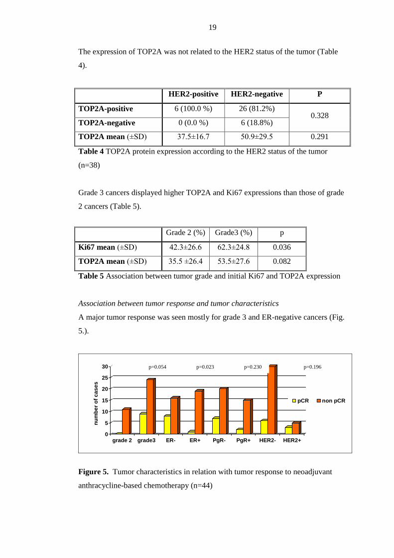
The expression of TOP2A was not related to the HER2 status of the tumor (Table
4).
HER2-positive HER2-negative P
TOP2A-positive 6 (100.0 %) 26 (81.2%) 0.328
TOP2A-negative 0 (0.0 %) 6 (18.8%)
TOP2A mean (±SD) 37.5±16.7 50.9±29.5 0.291
Table 4 TOP2A protein expression according to the HER2 status of the tumor
(n=38)
Grade 3 cancers displayed higher TOP2A and Ki67 expressions than those of grade
2 cancers (Table 5).
Grade 2 (%) Grade3 (%) p
Ki67 mean (±SD) 42.3±26.6 62.3±24.8 0.036
TOP2A mean (±SD) 35.5 ±26.4 53.5±27.6 0.082
Table 5 Association between tumor grade and initial Ki67 and TOP2A expression
Association between tumor response and tumor characteristics
A major tumor response was seen mostly for grade 3 and ER-negative cancers (Fig.
5.).
0
5
10
15
20
25
30
nu
mb
er
of
ca
se
s
grade 2 grade3 ER- ER+ PgR- PgR+ HER2- HER2+
pCR non pCR
Figure 5. Tumor characteristics in relation with tumor response to neoadjuvant
anthracycline-based chemotherapy (n=44)
p=0.054 p=0.023 p=0.230 p=0.196

20
The development of pCR was related to high grade (grade 3) (p=0.054) and ER
negativity (p=0.027). While the mean (±SD) pre-chemotherapy TOP2A expression
was 66.9±26.3% in cases with pCR, it was 41.8±26.6% in cases without pCR
(p=0.037). Eight pCRs (21%) occurred among those cases that were assessed for
TOP2A IHC, and all the pCRs occurred in TOP2A-positive cancers. Although no
association was found with TOP2A amplification, both TOP2A-amplified tumors
gave a major response: pCR in one, and a reduction in tumor size from 70 to 15 mm
in the other. Ki67 was not predictive of the tumor response in univariate analysis
(OR=1.027, 95% CI: 0.992-1.062, p=0.167). In the logistic regression model
including the grade, ER, the expression of TOP2A was an independent predictor of
pCR (OR=1.460, for every 10% increase, 95% CI: 1.016-2.096, p=0.041).
Survival
The median follow-up time was 31.0 months. Fourteen patients developed local or
distant recurrence, and 3 died. The median RFS and OS were 23.7 and 31.0
months, respectively (Fig. 6).
Figure 6. Survival (DFS and OS) according to the TOP2A status of the breast
cancer
RFS was shorter in cases with PgR-negative than in those with PgR-positive
cancers (23.0 vs. 32.6 months, p=0.07, linear regression: R=0.350, p=0.018), but
OS did not depend on any of the tumor features. The RFS and OS were not related
to the tumor response or the decrease of TOP2A protein expression.
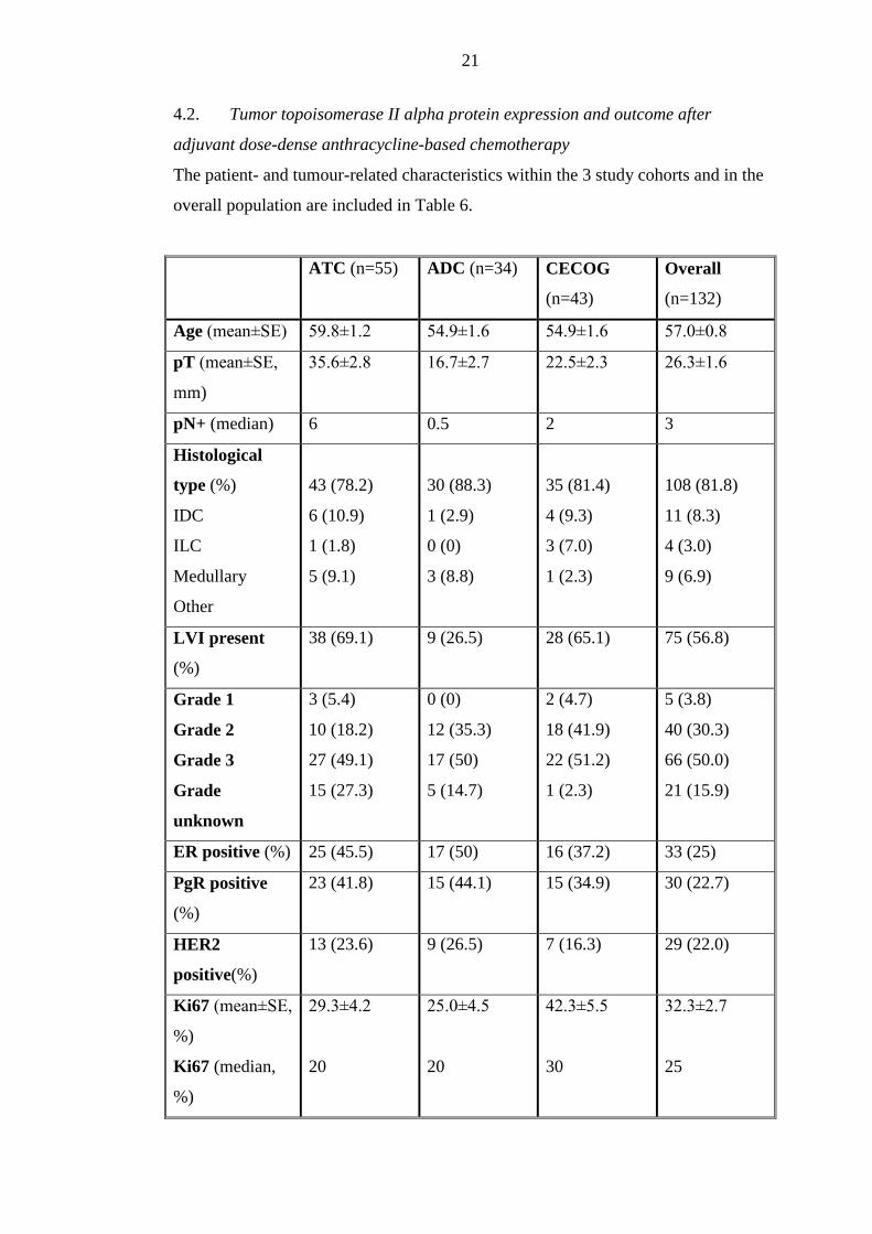
21
4.2. Tumor topoisomerase II alpha protein expression and outcome after
adjuvant dose-dense anthracycline-based chemotherapy
The patient- and tumour-related characteristics within the 3 study cohorts and in the
overall population are included in Table 6.
ATC (n=55) ADC (n=34) CECOG
(n=43)
Overall
(n=132)
Age (mean±SE) 59.8±1.2 54.9±1.6 54.9±1.6 57.0±0.8
pT (mean±SE,
mm)
35.6±2.8 16.7±2.7 22.5±2.3 26.3±1.6
pN+ (median) 6 0.5 2 3
Histological
type (%)
IDC
ILC
Medullary
Other
43 (78.2)
6 (10.9)
1 (1.8)
5 (9.1)
30 (88.3)
1 (2.9)
0 (0)
3 (8.8)
35 (81.4)
4 (9.3)
3 (7.0)
1 (2.3)
108 (81.8)
11 (8.3)
4 (3.0)
9 (6.9)
LVI present
(%)
38 (69.1) 9 (26.5) 28 (65.1) 75 (56.8)
Grade 1
Grade 2
Grade 3
Grade
unknown
3 (5.4)
10 (18.2)
27 (49.1)
15 (27.3)
0 (0)
12 (35.3)
17 (50)
5 (14.7)
2 (4.7)
18 (41.9)
22 (51.2)
1 (2.3)
5 (3.8)
40 (30.3)
66 (50.0)
21 (15.9)
ER positive (%) 25 (45.5) 17 (50) 16 (37.2) 33 (25)
PgR positive
(%)
23 (41.8) 15 (44.1) 15 (34.9) 30 (22.7)
HER2
positive(%)
13 (23.6) 9 (26.5) 7 (16.3) 29 (22.0)
Ki67 (mean±SE,
%)
Ki67 (median,
%)
29.3±4.2
20
25.0±4.5
20
42.3±5.5
30
32.3±2.7
25

22
Table 6 Patient- and tumour-related characteristics within the study groups and the
overall population
The median follow-up time for the entire population was 64.5 months, and for the
ATC, ADC and CECOG cohorts was 103, 44.5 and 60 months, respectively.
Altogether 31 relapses (23.5%) and 23 deaths (17.4%) occurred. The OS differed
significantly in the 3 cohorts: the ATC cohort exhibited the worst, and the ADC
cohort the best survival (p<0.01). Among the standard prognostic factors, the
pathological tumor size (pT) and the number of positive lymph nodes were
associated with the RFS in the overall study population (p<0.05), while the
presence of LVI was related to the RFS in the ADC cohort.
TOP2A IHC
For technical reasons, the TOP2A IHC results were assessable in only 106 cases. In
the overall population, the average and median proportions of the TOP2A-positive
cells were 21% and 10%, respectively. With a cut-off value of 15%, 48% of the
tumours were classified as TOP2A-positive (Table 7).
ATC ADC CECOG Overall
TOP2 A IHC
(n)
40 27 39 106
TOP2A
(mean±SE, %)
18.3±3.4
17.33±5.0
24.5±5.0
21.02±2.3
TOP2A
(median, %)
10 15 10 10
TOP2A+
(n)(%)
16 (40) 14 (51.9) 21 (53.8) 51 (48.1)
Table 7 TOP2A IHC status in the study groups and the overall population
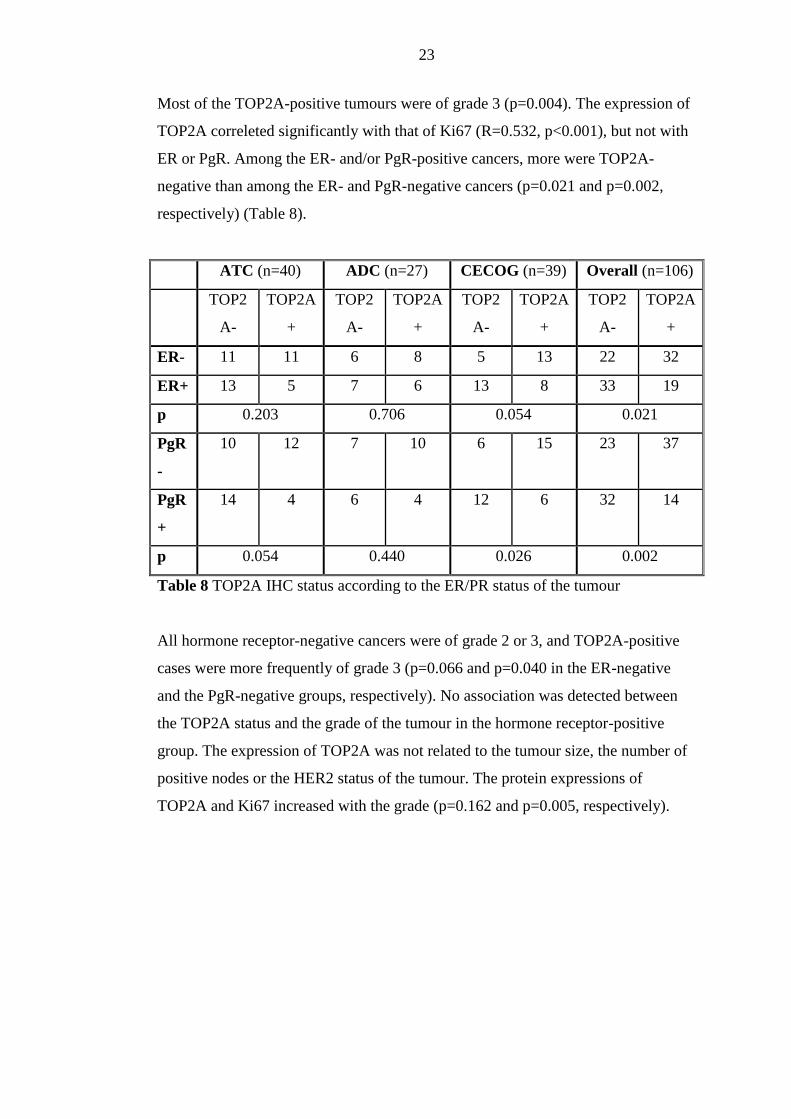
23
Most of the TOP2A-positive tumours were of grade 3 (p=0.004). The expression of
TOP2A correleted significantly with that of Ki67 (R=0.532, p<0.001), but not with
ER or PgR. Among the ER- and/or PgR-positive cancers, more were TOP2A-
negative than among the ER- and PgR-negative cancers (p=0.021 and p=0.002,
respectively) (Table 8).
ATC (n=40) ADC (n=27) CECOG (n=39) Overall (n=106)
TOP2
A-
TOP2A
+
TOP2
A-
TOP2A
+
TOP2
A-
TOP2A
+
TOP2
A-
TOP2A
+
ER- 11 11 6 8 5 13 22 32
ER+ 13 5 7 6 13 8 33 19
p 0.203 0.706 0.054 0.021
PgR
-
10 12 7 10 6 15 23 37
PgR
+
14 4 6 4 12 6 32 14
p 0.054 0.440 0.026 0.002
Table 8 TOP2A IHC status according to the ER/PR status of the tumour
All hormone receptor-negative cancers were of grade 2 or 3, and TOP2A-positive
cases were more frequently of grade 3 (p=0.066 and p=0.040 in the ER-negative
and the PgR-negative groups, respectively). No association was detected between
the TOP2A status and the grade of the tumour in the hormone receptor-positive
group. The expression of TOP2A was not related to the tumour size, the number of
positive nodes or the HER2 status of the tumour. The protein expressions of
TOP2A and Ki67 increased with the grade (p=0.162 and p=0.005, respectively).

24
Association between outcome and tumour TOP2A status
In the overall population, more relapses and more deaths occurred among the
TOP2A-negative cases than among the TOP2A-positive cases, and the RFS and OS
were longer accordingly (Table 9, Fig. 7.).
TOP2A IHC number of
deaths (%)
OS (mean±SE)
(months)
number of
relapses (%)
RFS
(mean±SE)
(months)
Negative 14/55 (25.5) 93.3±6.0 14/55 (25.5) 93.7±6.1
Positive 6/51 (11.8) 103.8±4.3 8/51 (15.7) 96.8±5.9
p (Mantel-Cox) 0.081 0.229
Table 9 Survival (OS and RFS) according to the TOP2A status of the tumour
Figure 7. Survival (OS and RFS) according to the TOP2A status of the tumour

25
The outcome in the hormone receptor-positive and hormone receptor-negative
subgroups was analysed separately (Table 10, Fig. 8).
ER-negative ER-positive PgR-negative PgR-positive
TOP2
A-
TOP2
A+
TOP2
A-
TOP2
A+
TOP2
A-
TOP2
A+
TOP2
A-
TOP2
A+
number of
deaths
6/22 2/32 8/33 4/19 7/23 2/37 7/32 4/14
OS
(mean±SE)
(months)
93.1±
9.3
109.3±
3.9
92.9±
7.6
82.7±
6.8
89.0±
9.6
110.5±
3.1
95.7±
7.4
81.3±
10.2
p (Mantel-
Cox)
0.035 0.916 0.005 0.494
number of
relapses
7/22 5/32 7/33 5/19 7/23 6/37 7/32 4/14
RFS(mean
±SE)
(months)
87.2±
10.3
97.1±
7.5
97.2±
7.3
77.6±
8.1
87.3±
10.3
97.2±
6.7
97.7±
7.1
79.5±
11.2
p (Mantel-
Cox)
0.176 0.774 0.169 0.639
Table 10 Survival (OS and RFS) according to the TOP2A and ER/PR status of the
tumor

26
Figure 8. a,c Survival (OS and RFS) according to the tumor TOP2A IHC status in
the ER and/or PR positive tumors
Figure 8 b,d Survival (OS and RFS) according to the tumor TOP2A IHC status in
the ER and/or PR negative tumors
27
While there was no difference in the number of events, or in the OS and the RFS in
the ER- and the PgR-positive subgroups according to the TOP2A status, the OS and
RFS were significantly improved in the ER- or PgR-negative and TOP2A-positive
cases as compared with the TOP2A-negative cases (Table 10, Fig. 8). Figure 7
presents the RFS and OS as functions of the TOP2A expression status in ER/PR-
negative cases.
In order to estimate the dependence of the OS and the RFS on the tumour TOP2A
and Ki67 status, the tumour grade and the nodal status in ER- and/or PgR-negative
cancer, these variables were studied in a Cox proportional hazards model. In grade
3 cases, the risk of death was decreased, with HR= 0.216 (95% CI: 0.047-0.990,
p=0.048) as compared with grade 2 cases. In the TOP2A-positive cases, the risk of
death was decreased, with HR=0.211 (95% CI: 0.042-1.05, p=0.056). In
multivariate analysis, no interaction was detected between these variables.
4.3. The effect of individual postioning on the radiation exposure of the risk
organs
4.3.1. General statistics
The first phase of the study and the second, feasibility phase involved 20 and 41
patients, respectively. The mean (±SD) age of the overall study population was
56.0±9.6 (29.3-73.9), and that in the second phase was 56.6±9.9 (29.3-73.6) years.
Twenty-seven patients needed right-sided, and 34 underwent left-sided breast
irradiation. The age, weight, waist, hip size and breast separation did not differ
significantly between the patients randomized to radiotherapy in the prone or the
supine position (Table 11).

28
Age (years) Weight (kg) Height
(cm)
BMI
(kg/cm2)
Waist size
(cm)
Hip size
(cm)
Breast
separation
(cm)
Supine
n=21
59.1±9.3
(42.1-75.0)
71.6±12.4
(52.0-96.0)
162.1±7.7
(150-175)
27.2±3.9
(20.9-33.2)
93.3±14.4
(78-145)
107.4±12.1
(95-150)
21.1±2.7
(16.4-26.9)
Prone
n=20
56.9±10.7
(30.7-72.4)
69.9±12.4
(50.0-102.0)
161.0±4.3
(152-168)
27.1±5.3
(17.7-38.9)
89.3±10.6
(69-108)
104.4±9.9
(87-124)
20.7±3.1
(14.2-26.9)
p 0.49 0.66 0.56 0.94 0.32 0.40 0.64
Table 11 Patient characteristics (mean±SD) among patients randomized to
radiotherapy in the prone vs. the supine position
Tumor bed boost irradiation and systemic treatments did not differ significantly
between the two groups.
4.3.2. Radiation plans for the prone vs. the supine position
The radiotherapy plans were first analyzed in the overall population. The mean
(±SD) percentage PTV covered by 47.5-53.5 Gy (V95-107%) in the prone vs. the
supine position was 85.1±4.2% and 89.2±2.2%, respectively (p<0.0001). The dose
homogeneity did not depend on the PTV or the breast separation. The irradiated
volume of and the dose to the ipsilateral lung determined in terms of the MLD and
the V20Gy were dramatically lower in the prone position than in the supine position
(Table 12).
Lung (n=61) Heart (n=34)
MLD
(Gy)
V20Gy
(%)
Mean dose
(Gy)
V25Gy
(%)
V30Gy
(%)
Supine 7.45±2.62 14.3±5.4 3.51±2.33 4.7±4.6 4.1±4.3
Prone 2.02±1,23 3.3±2.5 3.18±1.31 3.6±2.5 3.0±2.2
p <0.0001 <0.0001 0.413 0.171 0.152
Table 12 Radiation doses to the ipsilateral lung and the heart in the overall study
population. The mean values±SD are shown.

29
No significant difference was detected in the mean dose to the heart and the
volumes of the heart receiving at least 25 Gy or 30 Gy in 34 left-sided breast cancer
patients according to their position during radiotherapy (Table 12). The first 20
pairs of treatment plans revealed significantly higher doses to the contralateral
breast in the prone position than in the supine position. In the second phase of the
study (n=41), as a consequence of the more complete displacement of the opposite
breast due to the use of a new polyfoam wedge, there was no longer any significant
difference (Table 13).
First phase n=20 Second phase n=41 p for first vs. second phase
Mean dose
(Gy)
V5Gy
(%)
Mean dose
(Gy)
V5Gy
(%)
Mean dose
V5Gy
Supine 0.85±0.47 2.7±2.0 0.61±0.73 1.7±2.8 0.096 0.073
Prone 1.26±0.78 4.5±3.4 0.74±0.44 2.2±2.0 0.00092 0.001
p for supine
vs. prone
0.0038 0.0057 0.162 0.159
Table 13 Radiation dose to the opposite breast in the 2 consecutive cohorts of the
study
We hoped to identify those parameters related to the patient anatomy which indicate
high lung doses if radiotherapy is given in the supine position, in order to select
those patients who would benefit most from radiotherapy in the prone position. As
regards the volume of the target breast, the breast separation and the CLD, only the
CLD was significantly associated with the MLD (r=0.843, p<0.0001) and the V20Gy
(r=0.733, p<0.0001).
4.3.3 Implementation of breast radiotherapy in the prone position
In the second phase of the study, the adherence to the study protocol, the
repositioning accuracy and the early skin reactions were analyzed. The protocol was
tolerated well by all the patients; only one patient treated in the prone position
needed a 1-week break because of radiodermatitis. It was necessary to correct the
location of the isocenter in the simulator or the position of the table during
radiotherapy in 20.3% (61/301) and 20.3% (62/306) of all the checks in the prone

30
and the supine position, respectively (p=0.999). The mean length of the
displacement vector was 8.06±4.66 (3.00–22.56) mm and 6.60±3.05 (3.00–21.19)
mm in the prone and supine position, respectively (p=0.021). The population
random errors were 17.39 mm and 13.63 mm, while the population systematic
errors were 0.86 mm and 0.82 mm, for the prone and the supine position,
respectively. The random errors in the two groups are shown in Table 14.
Mean ± SE (mm) Median (mm)
Supine 2.75 ± 0.27 2.58
Prone 3.46 ± 0.37 3.48
P 0.061
Table 14 Random errors for repositioning in the prone and supine positions
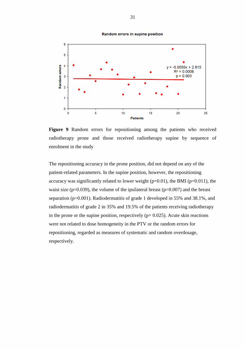
A trend was detected for better overall repositioning accuracy in the supine position
(p=0.061). We analyzed whether the repositioning accuracy changed from patient to
patient during the study period. The individual random errors for repositioning in
the prone position decreased with time, while no change was detected in the group
randomized to radiotherapy in the supine position (Fig. 9).
31
Figure 9 Random errors for repositioning among the patients who received
radiotherapy prone and those received radiotherapy supine by sequence of
enrolment in the study
The repositioning accuracy in the prone position, did not depend on any of the
patient-related parameters. In the supine position, however, the repositioning
accuracy was significantly related to lower weight (p=0.01), the BMI (p=0.011), the
waist size (p=0.039), the volume of the ipsilateral breast (p=0.007) and the breast
separation (p=0.001). Radiodermatitis of grade 1 developed in 55% and 38.1%, and
radiodermatitis of grade 2 in 35% and 19.5% of the patients receiving radiotherapy
in the prone or the supine position, respectively (p= 0.025). Acute skin reactions
were not related to dose homogeneity in the PTV or the random errors for
repositioning, regarded as measures of systematic and random overdosage,
respectively.
32
5. Discussion
5.1 Tumor topoisomerase II alpha status and response to anthracycline-based
neoadjuvant chemotherapy in breast cancer
The amplification of the TOP2A gene is a rare abnormality, and is restricted to
HER2 positive tumors (5,10-12,15,22,23,34). The deletion of the gene is less
frequent, and its role in anthracycline-sensitivity is controversial (5,10,12,16,34-
37). In our cohort, 2 cases with TOP2A gene amplification exhibited HER2
amplification and high TOP2A expression (data not shown), and showed excellent
response to the therapy, however, the small number of cases limits the interpretation
of the findings.
The cut-off point for defining TOP2A-positivity by IHC varied in different studies.
The most often used threshold value was 10-15% (range 5-25%) (11, 23,38, 39-42),
and the rate of TOP2A-positive tumors varied between 5-45% (15, 23, 42). In a
series of 245 tumors, the median proportion of TOP2A-positive cells was 27%, and
about half of the tumors rated positive (11). For the TOP2A-positive category, we
used the cut-off value of >15%. In our cohort, the median proportion of TOP2A-
stained cells was relatively high (50%), and the majority of tumors classified as
TOP2A-positive. The first reason for this finding is the selected nature of our study
population: only patients with rapidly proliferating tumors (based on the knowledge
of standard tumor characteristics), likely chemosensitive were chosen for
neoadjuvant chemotherapy. Second, the methods applied (the tumor regions with
the highest cellularity were used; TOP2A-positive cells were counted regardless of
the intensity of the staining) also favored high TOP2A values. Of note is that using
the same methods, the median TOP2A IHC value in another cohort of our patients
selected for adjuvant chemotherapy and in the whole population irrespective of
tumor characteristics was 20 (range: 0-90) and 5 (range: 0-80), respectively (data
not shown).
In our study, TOP2A protein expression was found an independent predictor of
pCR after neoadjuvant docetaxel-epirubicin chemotherapy, and the probability of
pCR increased by almost 50% with every 10% increase of the TOP2A positive
tumor cells. The predictive role of the tumor TOP2A status for anthracycline-based
chemotherapy has been investigated with different methods, in different settings.
The correlation between the efficiency of adjuvant anthracycline-based

33
chemotherapy and the presence of TOP2A gene amplification (5, 16, 43) or the
amplification and deletion of the gene (9,10,12,35) in the breast tumor is well
demonstrated. In those randomized trials which compared an anthracycline-
containing chemotherapy with a non-anthracycline containing regimen, the benefit
of the former was limited to tumors with the presence of the amplification (5, 43),
or the amplification or the deletion of the TOP2A gene (9,12,35). Some studies
demonstrated that the presence of TOP2A gene amplification is predictive for the
benefit of the dose elevation of the anthracylines (16, 44). In contrast, a
retrospective analysis of the CALGB 8541 study did not support a difference of
benefit if doxorubicin was administered at different doses in tumors with TOP2A
gene aberrations, but in that study, standard dose was compared with suboptimal
doses (34). In the neoadjuvant setting, the retrospective study of 350 cases showed
that the amplification of the TOP2A gene involves a 3 times higher probability of
pCR in patients treated with neoadjuvant anthracycline-based chemotherapy (22).
Likewise, the amplification of the TOP2A gene or the polysomy of chromosome 17
indicated a more than 4-times increased probability to obtain pCR in HER2-positive
and ER-, PgR-negative tumors (36). Park et al. found that the chance of obtaining a
response including pCR after neoadjuvant doxorubicin was associated with TOP2A
and HER2 coamplification (34). In a relatively small neoadjuvant study, in
consistence with our findings, serial TOP2A IHC determination showed a trend for
better response to anthracyclines in tumors with higher TOP2A expression, and a
significant decrease after therapy, in responders (38). Mukherjee et al. found the
extent of TOP2A protein expression predictive of the pCR in 91 patients treated
with preoperative FEC chemotherapy (45). The robust study of Press et al. provides
evidence that the coamplification of the TOP2A and HER2 genes is a clinically
useful predictive marker of an enhanced response to anthracycline-based
chemotherapy in metastatic breast cancer (46). In a phase III study in advanced
breast cancer, the TOP2A IHC status was predictive for response to doxorubicin
monotherapy, and every 10% increase in TOP2A expression was associated with a
9% increase in the probability of response, while no such effect was demonstrated
for the treatment with docetaxel monotherapy (23).
The main outcome and novelty of our study is the demonstration that a simple tool
such as TOP2A IHC is a useful, semiquantitative predictive marker of the benefit of
neoadjuvant anthracycline-based chemotherapy in breast cancer. The design of our

34
study is not appropriate to answer whether TOP2A IHC staining is a specific
marker of anthracycline sensitivity or that of chemosensitivity only. Other studies,
however, support the hypothesis that high TOP2A protein expression is predictive
of anthracycline sensitivity. In an early study of Di Leo et al., it seemed that the
>10% expression of TOP2A protein favors the benefit of both the choice and the
higher dose of an adjuvant EC regimen (42). Likewise, Durbecque et al. in a
retrospective analysis of the TAX 303 randomized study, demonstrated that
although docetaxel is more efficient than doxorubicin in the population of advanced
breast cancer patients overall, increasing TOP2A expression is associated with a
higher chance to obtain a response in the doxorubicin arm, but not in the docetaxel
arm (23).
Many studies examined the correlation between the TOP2A gene status and the
TOP2A protein expression (11, 15, 39, 41, 47, 48). Although gene amplification
favored high protein expression, the presence of the enzyme was not dependent on
the gene abnormality. Jarvinen et al. found that TOP2A protein expression
correlated well with TOP2A mRNA (48). Brase et al., analyzed TOP2A at the
levels of gene amplification, RNA expression and protein expression, and studied
their correlations. No correlation was found between gene amplification and RNA
or protein expression, but a strong correlation existed between TOP2A RNA and
protein levels (21). The conclusion was drawn that unlike in the case of the HER2
status, TOP2A protein expression is highly regulated at the RNA level. In our
cohort, most tumors exhibited high TOP2A expression without the presence of
TOP2A gene alteration, as a function of high proliferative activity. Schindlbeck et
al. based on their study on patients treated with adjuvant anthracycline-based
chemotherapy, concluded that it is the TOP2A IHC and not the gene status that
predicts benefit of the treatment (15). The relevance of TOP2A expression in the
prediction of anthracycline-sensitivity merits further studies, however, the
reconsideration of the optimal method is needed.
We did not find association between outcome and TOP2A expression or the fall of
TOP2A expression after neoadjuvant chemotherapy. This result is limited by the
relatively short follow-up time. Survival was neither different in other studies by the
TOP2A status among patients treated with anthracyclines (11,37,40). Of note is,
however, that the administration of anthracyclines did improve outcome in HER2-

35
and TOP2A-coamplified tumors to a level that was obtained without such
chemotherapy, but with the addition of Herceptin (5).
In conclusion, our findings suggest that the IHC determination of the TOP2A
protein is a useful tool for the estimation of the sensitivity of breast cancer to
anthracycline-based chemotherapy.
5.2 Tumor topoisomerase II alpha protein expression and outcome after
adjuvant dose-dense anthracycline-based chemotherapy
The TOP2A status in breast cancer has been studied as a prognostic and predictive
factor by different methods in multiple studies. Most investigators agree that the
amplification or the deletion of the TOP2A gene is restricted to HER2-positive
cancers (5,6,14,22). Co-amplification of the HER2 and TOP2A genes indicated an
increased anthracycline sensitivity in most (5,6,14,22,43), but not all studies
(49,13). The design of these retrospective studies, however, was not always
appropriate for detection of the benefit of anthracycline therapy according to the
presence of TOP2A gene abnormality (13,37). In those randomized trials which
compared anthracycline-containing chemotherapy with a non-anthracycline-
containing regimen, the benefit of the former was limited to tumours with an
abnormal TOP2A gene status (5,9,11,12,43). Some studies have demonstrated that
the presence of a TOP2A gene alteration is predictive of the benefit of an elevation
of the anthracyline dose (17,44). Deletion of the gene is less frequent, and its role in
anthracycline sensitivity seems rather controversial (5,10-12,16,22,37). In line with
the contradictory results, it is noteworthy that, although TOP2A gene abnormalities
have been observed exclusively in HER2-positive breast cancers, high
anthracycline sensitivity is not limited to this special group (50).
Investigations of whether the expression of TOP2A is a specific marker of
anthracycline sensitivity gave more concordant results. The early study by Di Leo et
al. led to the conclusion that a finding of TOP2A positivity by means of IHC
determination favoured the benefit of both the choice and a higher dose of an
adjuvant EC regimen (42). Likewise, in a retrospective analysis of the TAX 303
randomized study, Durbecque et al. demonstrated that, although docetaxel is more
efficient than doxorubicin in the population of advanced breast cancer patients
overall, increase of the TOP2A protein expression is associated with a higher
chance of obtaining a response in the doxorubicin arm, but not in the docetaxel arm

36
(23). The greater sensitivity to anthracycline-based adjuvant chemotherapy of
ER/PgR-negative breast cancers as compared with ER- or PgR-positive tumours has
been well demonstrated (50). Our own study suggests that one of the related key
factors is the more frequent TOP2A positivity among the ER/PgR-negative
tumours, and we advocate TOP2A IHC as a tool to select those hormone receptor-
negative cases which would benefit from adjuvant anthracyclines. In a patient
population treated with adjuvant anthracycline-containing chemotherapy,
Schindlbeck et al. retrospectively examined the TOP2A status. About 50% of the
cases proved to be TOP2A-positive, and after a median survival time of 42 months,
the survival was significantly poorer among the TOP2A-negative cases (15). Brase
et al. demonstrated the strong negative prognostic power of an elevated TOP2A
RNA level in 782 untreated breast cancer patients, which remained significant after
further analyses in the ER-positive and the HER2-negative and triple-negative
subgroups. In the same paper, complete tumour regression to chemotherapy with
EC was reported to be related to the high TOP2A and low ER RNA levels, results
which support our finding that anthracyclines result in a favourable outcome in ER-
negative and TOP2A-positive cancers (21). Rody et al.followed up more than 1300
patients, and found that the TOP2A expression was the strongest indicator of a poor
prognosis among hormone receptor-positive cases, while no such effect was
detected among the ER-negative cases (51). Although the prognostic effect of
TOP2A positivity was found to be independent of the systemic therapy, the nature
of the chemotherapy given in about half of the patients, was not reported. It may be
speculated that the similar outcome in the TOP2A-positive and -negative cases in
the ER-negative group may be due to the higher chemosensitivity of the TOP2A-
positive cases.
The expression of TOP2A seems to be regulated most strongly at the RNA level,
and its gene status is probably less determinative of its functional capacity. Jarvinen
et al. and Brase et al. found no correlation between gene amplification and protein
expression, but there was a strong correlation between the TOP2A RNA and protein
levels (21,48). Accordingly, although gene amplification favoured a high protein
expression in those studies that examined the correlation between the TOP2A gene
status and the TOP2A protein expression, the presence of the enzyme was not
dependent on the gene abnormality (14,15,18-20). Their findings led Brase et al. to
recommend determination of the RNA expression, while Schindlbeck et al.

37
suggested determination of the protein expression of TOP2A for patient selection,
rather than examination of the gene status (15,21).
5.3 The effect of individual postioning on the radiation exposure of the risk
organs
We evaluated our initial experience regarding the dosimetry and feasibility of
conformal breast radiotherapy in the prone position, and identified its place in
everyday practice. Our results indicate that its primary advantage is the significantly
reduced radiation exposure of the ipsilateral lung. The doses to the heart and the
contralateral breast are similar in the prone and supine positions. Special practice in
and attention to accurate repositioning are needed if the prone position is applied,
and the dose inhomogeneity and acute skin reactions may be slightly increased.
There have been few studies on prone breast radiotherapy. Some of them focused
on the dose distribution (52-55), and others on clinical implementation (56-60), and
only one study dealt with both dosimetric aspects and feasibility (61). The present
study is the first randomized clinical trial to compare breast radiotherapy in the
prone vs. the supine position.
Utilization of the prone position during breast radiotherapy raises special
considerations because of the altered shape, motion and position of the organs
present in the region. The altered shape of the target breast hanging down across the
aperture of the positioning device results in a different dose distribution relative to
that in the supine position. Improved dose uniformity, and especially the avoidance
of an overdosage within the PTV, have been associated with a better cosmetic
outcome (62, 63). A higher dose inhomogeneity is related to larger breasts if
conventional tangent beams are used (62). Buijsen et al. (54) compared prone and
supine breast irradiation in 10 patients with pendulous breasts, and concluded that
the dose homogeneity was better in the prone than in the supine position. In fact,
this was based on a comparison of the PTV overdosed (V105% and V107%) in the
supine vs. the prone position, while the significantly lower mean dose and PTV
coverage representing an underdosage were neglected. Similarly, larger volumes
receiving >52.5 Gy within the PTV were found in the supine than in the prone
position, but no other information on dose distribution was reported in another
study (6). We examined V95-107% as a measure of dose homogeneity within the PTV,
according to ICRU Report 62 (64), and found that the dose distribution was

38
significantly more uniform in the supine position, regardless of the size or shape of
the target breast. None of the radiotherapy plans indicated measurable volumes
receiving >53.5 Gy.
Because of the different shape of the chest wall when the patient is positioned
prone, the lung volume included in the tangent fields is considerably less. All
authors agree that the lung doses are dramatically reduced if breast radiotherapy is
performed with the patient prone (52-54,65,66). The beneficial effect of prone
positioning on the protection of the ipsilateral lung is further enhanced if the almost
absent intrafractional motion of the chest wall is taken into account for the
calculation of safety margins around the CTV (60, 67, 68).
When left-sided irradiation is performed, the irradiated volume of the heart is not
reduced, despite the fact that less intrathoracic volume is exposed to radiation in the
prone than in the supine position. Reports on heart doses, however, are not
concordant. Some studies suggest a reduction in heart doses as a result of prone
positioning, but do not provide direct comparisons with supine positioning (65, 66).
Others are consistent with our results in showing no significant difference in the
doses to the heart as a function of the treatment position (52-54). This finding may
be accepted if the change in position of the heart by treatment position is taken into
consideration. In fact, the prone position causes an anterior displacement of the
heart within the thorax by 19 mm on average, as demonstrated by CT and MRI
measurements in breast cancer patients receiving radiotherapy (69).
Since breast radiotherapy increases the risk of the late development of contralateral
breast cancer by 18-34%, special attention is needed for the protection of the
opposite breast during radiotherapy (70,71). Although some studies allude to the
radiation dose to the opposite breast in the prone position, detailed dose volume
histogram data have not been provided (52,65). No widely accepted dose
constraints exist for the contralateral breast. We registered V5Gy and the mean dose
to the healthy breast. In the first phase of the study, in consequence of the
suboptimal positioning of the patient in the prone position, we detected higher doses
to the opposite breast in the prone than in the supine position. Following revision of
the positioning method, in the second phase of the study, no difference was
observed. We consider careful application of the polyfoam wedge in the prone
position, and of mask fixation in the supine position to be very important, in order
to remove the opposite breast from the radiation fields.

39
The largest prospective phase I-II study on prone breast irradiation is that of
Formenti et al. (66). Accelerated whole breast radiotherapy was feasible in 90
patients, with high set-up reproducibility, although numerical data were not
provided. In another feasibility study (61), prolonged adequate immobilization
could not be achieved in 3 of 35 patients with large pendulous breasts in the prone
position. In one retrospective study (56), 5% of the patients during prone breast
radiotherapy complained of chest wall or rib pain, and 2 of 248 patients suffered a
rib fracture (56), as did 1 of 35 in the previous study (61). All our patients
considered the prone radiotherapy convenient, and completed the course of
radiotherapy. We believe, that the comfortable positioning system in use, was
essential to achieve such good adherence to the protocol. It is our view that
repositioning accuracy is a key condition for radiotherapy, especially if inverse or
forward intensity modulation is applied (67,68). During simulation in 308 patients
with various cancer sites, Schüller et al. (72) found that the repositioning accuracy
was better in the entire patient population if positioning aids or mask fixation were
used, but did not differ by prone or supine positioning. Breast irradiation was
performed in the supine position for 64 patients, without mask fixation. Of the
various tumor sites, the breast exhibited the poorest repositioning accuracy.
Displacement was carried out in 27 patients (42.2%), and in many cases exceeded 1
cm. In another study of 25 breast cancer patients irradiated in the supine position
(73), the isocenter displacement on simulation was on average 5.7 mm. Morrow et
al. (60) studied the interfractional error in repositioning in 15 patients, and
recommended image guidance during prone breast radiotherapy because of the need
for frequent and large displacements. In accord with our results, they observed no
relation between the breast size and the repositioning accuracy. Interestingly,
however, we found that the repositioning accuracy in the supine position is
significantly worse in obese patients. To the best of our knowledge, no such data
have been published previously. If confirmed, they indicate that increased attention
must be payed to the position of overweight patients during breast radiotherapy. We
believe that the relatively good repositioning accuracy in our study, was related to
the comfortable positioning device used for both the prone and the supine position,
and to the mask fixation used in the supine position. The repositioning accuracy in
the prone position improved over time, indicating the need for experience and
expertise if the method is newly introduced. Furthermore, our study warrants the

40
development of mask fixation in the prone position, which would reduce the set-up
uncertainity.
In other publications (56,61), acute skin reactions after breast radiotherapy in the
prone position were reported in similar incidences as among our patients. Mahe et
al. (61) found that acute skin reactions were most frequent at the top and the bottom
of the fields, in accordance with the high dose regions. In our study, radiodermatitis
in the prone position was not related to the size of the breast or the dose-
inhomogeneity in it.
Merchant and McCormick (65) recommend breast radiotherapy in the prone
position if that in the supine position is likely to result in unacceptable dose
inhomogeneity or significant doses to normal tissues. We hoped to identify those
patients who would benefit most from the prone position during breast
radiotherapy. Since we could not detect any advantage of prone radiotherapy other
than the absence of radiation exposure of the lung, we set out to identify those
patient-related parameters that are associated with a higher lung dose if the patient
is irradiated in a supine position. Consideration of the volume of the breast, the
breast separation and the CLD as measures of the shape of the PTV indicated that
only the CLD was related to the dose to the ipsilateral lung. Thus, we recommend
monitoring of the CLD as a primary measure for an indication for prone
radiotherapy. Moreover, since the risk of early and late radiation lung sequelae is
strongly related to the age of the patient (26) and the presence of lung diseases, and
possibly also to certain systemic therapies, these factors should be taken into
account when a decision is made concerning the position during breast
radiotherapy.
41
6. Summary, conclusions
6.1. We found that despite that the amplification of the TOP2A gene was rare,
and restricted to HER2-positivity, the protein expression was usually elevated in
tumors with high proliferation rate; anthracycline-based neoadjuvant chemotherapy
resulted in the reduction of the expression of TOP2A. TOP2A positivity was an
independent predictor of pCR, and a 10% increase of TOP2A IHC staining resulted
in a 46% increase of the likelihood of obtaining a pCR.
6.2. We found TOP2A positivity in about half of the cancers treated with
adjuvant dose-dense anthracycline-based chemotherapy. TOP2A positivity was
more frequent among the ER- and/or PR-negative cancers. Among the hormone
receptor-negative cases, TOP2A positivity and grade 3 indicated improved OS and
RFS as a possible consequence of the higher sensitivity to the applied regimen. Our
data indicate that a simple tool such as TOP2A IHC (together with the grade) is an
useful predictive marker, at least in the hormone receptor-negative cases, and
should be implemented in routine practice for the selection of those who can be
expected to benefit from adjuvant anthracycline-based chemotherapy. The usually
poor outcome in the group of hormone receptor-negative and TOP2A-positive cases
may be reversed by the application of anthracycline-containing chemotherapy.
6.3. Conformal breast radiotherapy is feasible in the prone position. Its primary
advantage is the substantially lower radiation dose to the ipsilateral lung. The higher
dose inhomogeneity and the enhanced rate of the grade 1-2 skin toxicity, however,
may be concerns. We recommend monitoring of the CLD as a primary measure for
an indication for prone radiotherapy. Special practice in and attention to accurate
repositioning are needed if the prone position is applied.

42
7. Acknowledgements
First of all I am most grateful to my supervisor, Professor Zsuzsanna Kahán, whose
encouragement and generous support helped me in the completion of this work.
I express my gratitude to Professor László Thurzó, director of the Department of
Oncotherapy, University of Szeged, who provided excellent working conditions for
me at the institute.
I am greatly indebted to associate Dr Adrienn Cserháti, Dr Gabriella Uhercsák and
Zoltán Varga, whose invaluable support significantly contributed to my scientific
work.
I am grateful for the contribution of the colleagues at the Department of Pathology,
University of Szeged, who have participated in the papers on topoisomerase II
alpha.
The important instructive guidence and scientific contribution in the field of
biostatistics by associate professor Krisztina Boda and Ervin Tánczos are highly
esteemed.
I greatly appreciate all the support and work of high standard provided by
physicians, technicians and physicists of the Department of Oncotherapy,
University of Szeged that helped this dissertation to be born.

43
8. References
1. Nowak AK, Wilcken NR, Stockler MR, Hamilton A, Ghersi D. Systematic
review of taxane-containing versus non-taxane-containing regimens for adjuvant
and neoadjuvant treatment of early breast cancer. Lancet Oncol. 2004; 5:372-380.
2. Early Breast Cancer Trialists' Collaborative Group. Favourable and unfavourable
effects on long-term survival of radiotherapy for early breast cancer: an overview of
the randomised trials. Lancet 2000; 355:1739-1740.
3. Kaufmann M, von Minckwitz G, Bear HD, Buzdar A, McGale P, Bonnefoi H,
Colleoni M, Denkert C, Eiermann W, Jackesz R, Makris A, Miller W, Pierga JY,
Semiglazov V, Schneeweiss A, Souchon R, Stearns V, Untch M, Loibl S.
Recommendations from an international expert panel on the use of neoadjuvant
(primary) systemic treatment of operable breast cancer: new perspectives 2006. Ann
Oncol. 2007; 18:1927-1934.
4. Early Breast Cancer Trialists' Collaborative Group (EBCTCG): Effects of
chemotherapy and hormonal therapy for early breast cancer on recurrence and 15-
year survival: an overview of the randomised trials. Lancet. 2005; 365:1687-1717.
5. Slamon D, EiermannW, Robert N et al. BCIRG 006: 2nd interim analysis phase
II randomized trial comparing doxorubicin and cyclophosphamide followed by
docetaxel positive early (AC®TH) with docetaxel, carboplatin and trastuzumab
(TCH) in HER2 positive early breast cancer patients: BCIRG 006 study. Breast
Cancer Res Treat 2005; 94:S5 [abstr. 1]
6. Di Leo A, Biganzoli L, Claudion W, Licitra S, Pestrin M, Larsimont D:
Topoisomerase II alpha as a marker predicting anthracyclines’ activity in early
breast cancer patients: ready for the primetime? Eur J Cancer, 44: 2791-2798, 2008
7. Järvinen TA, Liu ET. HER-2/neu and topoisomerase IIalpha in breast cancer.
Breast Cancer Res Treat. 2003; 78:299-311.
8. Isaacs RJ, Davies SL, Sandri MI, Redwood C, Wells NJ, Hickson ID.
Physiological regulation of eukaryotic topoisomerase II. Biochim Biophys Acta.
1998;1400:121-137.
9. Nielsen KV, Ejlertsen B, Moller S, Jorgensen JT, Knoop A, Knudsen H,
Mouridsen HT: The value of TOP2A gene copy number variation as a biomarker in
breast cancer: Update of DBCG trial 89D. Acta Oncol 2008; 47: 725-734.

44
10. Di Leo A, Gancberg D, Larsimont D, Tanner M, Jarvinen T, Rouas G, Dolci S,
Leroy JY, Paesmans M, Isola J, Piccart MJ. HER-2 amplification and
topoisomerase IIalpha gene aberrations as predictive markers in node-positive
breast cancer patients randomly treated either with an anthracycline-based therapy
or with cyclophosphamide, methotrexate, and 5-fluorouracil. Clin Cancer Res 2002;
8:1107-1116.
11. Knoop AS, Knudsen H, Balslev E, Rasmussen BB, Overgaard J, Nielsen KV,
Schonau A, Gunnarsdóttir K, Olsen KE, Mouridsen H, Ejlertsen B. Danish Breast
Cancer Cooperative Group. Retrospective analysis of topoisomerase IIa
amplifications and deletions as predictive markers in primary breast cancer patients
randomly assigned to cyclophosphamide, methotrexate, and fluorouracil or
cyclophosphamide, epirubicin, and fluorouracil: Danish Breast Cancer Cooperative
Group. J Clin Oncol 2005; 23:7483-7490.
12. O'Malley FP, Chia S, Tu D, Shepherd LE, Levine MN, Bramwell VH, Andrulis
IL, Pritchard KI. Topoisomerase II alpha and responsiveness of breast cancer to
adjuvant chemotherapy. J Natl Cancer Inst. 2009; 101:644-650.
13. Harris LN, Broadwater G, Abu-Khalaf M, Cowan D, Thor AD, Budman D,
Cirrincione CT, Berry DA, Winer EP, Hudis CA, Hayes DF, Friedman P, Ellis M,
Dressler L. Topoisomerase II{alpha} amplification does not predict benefit from
dose-intense cyclophosphamide, doxorubicin, and fluorouracil therapy in HER2-
amplified early breast cancer: results of CALGB 8541/150013. J Clin Oncol. 2009;
27:3430-3436.
14. Arriola E, Rodriguez-Pinilla SM, Lambros MBK, Jones RL, James M, Savage
K, Smith IE, Dowsett M, Reis-Filho JS: Topoisomerase II alpha amplification may
predict benefit from adjuvant anthracyclines in HER2 positive early breast cancer.
Breast Cancer Res Treat 2007; 106:181-189.
15. Schindlbeck C, Mayr D, Olivier C, Rack B, Engelstaedter V, Jueckstock J,
Jenderek C, Andergassen U, Jeschke U, Friese K. Topoisomerase IIalpha
expression rather than gene amplification predicts responsiveness of adjuvant
anthracycline-based chemotherapy in women with primary breast cancer. J Cancer
Res Clin Oncol. 2010;136:1029-1037.
16. Tanner M, Isola J, Wiklund T, Erikstein B, Kellokumpu-Lehtinen P,
Malmström P, Wilking N, Nilsson J, Bergh J: Topoisomerase IIalpha gene
amplification predicts favorable treatment response to tailored and dose-escalated
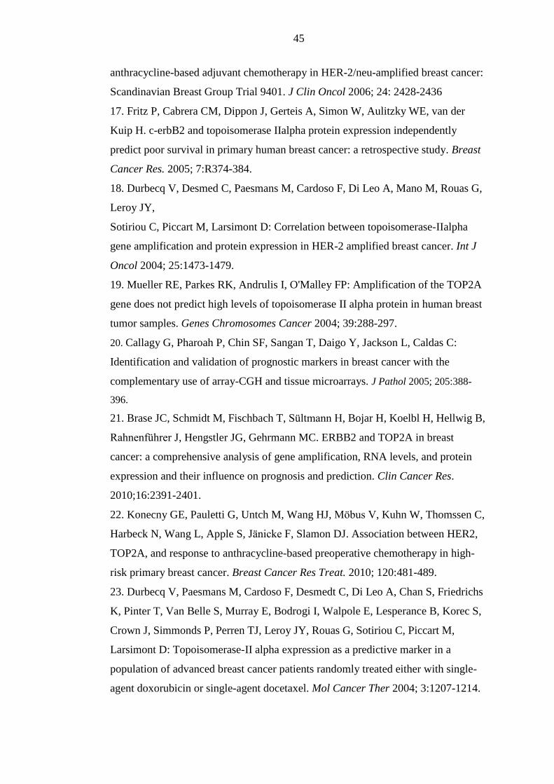
45
anthracycline-based adjuvant chemotherapy in HER-2/neu-amplified breast cancer:
Scandinavian Breast Group Trial 9401. J Clin Oncol 2006; 24: 2428-2436
17. Fritz P, Cabrera CM, Dippon J, Gerteis A, Simon W, Aulitzky WE, van der
Kuip H. c-erbB2 and topoisomerase IIalpha protein expression independently
predict poor survival in primary human breast cancer: a retrospective study. Breast
Cancer Res. 2005; 7:R374-384.
18. Durbecq V, Desmed C, Paesmans M, Cardoso F, Di Leo A, Mano M, Rouas G,
Leroy JY,
Sotiriou C, Piccart M, Larsimont D: Correlation between topoisomerase-IIalpha
gene amplification and protein expression in HER-2 amplified breast cancer. Int J
Oncol 2004; 25:1473-1479.
19. Mueller RE, Parkes RK, Andrulis I, O'Malley FP: Amplification of the TOP2A
gene does not predict high levels of topoisomerase II alpha protein in human breast
tumor samples. Genes Chromosomes Cancer 2004; 39:288-297.
20. Callagy G, Pharoah P, Chin SF, Sangan T, Daigo Y, Jackson L, Caldas C:
Identification and validation of prognostic markers in breast cancer with the
complementary use of array-CGH and tissue microarrays. J Pathol 2005; 205:388-
396.
21. Brase JC, Schmidt M, Fischbach T, Sültmann H, Bojar H, Koelbl H, Hellwig B,
Rahnenführer J, Hengstler JG, Gehrmann MC. ERBB2 and TOP2A in breast
cancer: a comprehensive analysis of gene amplification, RNA levels, and protein
expression and their influence on prognosis and prediction. Clin Cancer Res.
2010;16:2391-2401.
22. Konecny GE, Pauletti G, Untch M, Wang HJ, Möbus V, Kuhn W, Thomssen C,
Harbeck N, Wang L, Apple S, Jänicke F, Slamon DJ. Association between HER2,
TOP2A, and response to anthracycline-based preoperative chemotherapy in high-
risk primary breast cancer. Breast Cancer Res Treat. 2010; 120:481-489.
23. Durbecq V, Paesmans M, Cardoso F, Desmedt C, Di Leo A, Chan S, Friedrichs
K, Pinter T, Van Belle S, Murray E, Bodrogi I, Walpole E, Lesperance B, Korec S,
Crown J, Simmonds P, Perren TJ, Leroy JY, Rouas G, Sotiriou C, Piccart M,
Larsimont D: Topoisomerase-II alpha expression as a predictive marker in a
population of advanced breast cancer patients randomly treated either with single-
agent doxorubicin or single-agent docetaxel. Mol Cancer Ther 2004; 3:1207-1214.

46
24. Kahan Z, Uhercsak G, Hajnal-Papp R, Boda K, Thurzo L. Dose-dense
sequential adriamycin-Paclitaxel-cyclophosphamide chemotherapy is well tolerated
and safe in high-risk early breast cancer. Oncology. 2005;68:446-453.
25. Kahán Z, Spanik S, Wagnerova M, Skacel T, Planko B, Fitzthum E, Lindner E,
Soldatenkova V, Zielinski CC, Brodowicz T.: Feasibility of two dose-dense FEC
regimens with growth factor support for adjuvant therapy in patients with early
breast cancer: results from a randomised study of the Central European Cooperative
Oncology Group (CECOG). Breast Cancer Res Treat. 2008; 112:557-563.
26. Kahán Z., Csenki M., Varga Z., et al. The risk of early and late lung sequelae
after conformal radiotherapy in breast cancer patients. Int J Radiat Oncol Biol Phys
2007; 68:673-681.
27. Fein DA, McGee KP, Schultheiss TE, Fowble B, Hanks GE. Intra- and
interfractional reproducibility of tangential breast fields: A prospective on-line
portal imaging study. Int J Radiation Oncology Biol Phys 1996; 34:733-740.
28. Lirette A, Pouliot J, Aubin M, Larochelle M. The role of electronic portal
imaging in tangential breast irradiation: a prospective study. Radiother Oncol 1995;
37:241-245.
29. Hurkmans CW, Remeijer P, Lebesque JV, Mijnheer B. Set-up verification using
portal imaging; review of current clinical practice. Radiother Oncol 2001; 58:105-
120.
30. Stroom JC, Heijmen BJM. Geometrical uncertainties, radiotherapy planning
margins, and the ICRU-62 report. Radiother Oncol 2002; 64:75-83.
31. Arriola E, Rodriguez-Pinilla SM, Lambros MBK, Jones RL, James M, Savage
K, Smith IE, Dowsett M, Reis-Filho JS: Topoisomerase II alpha amplification may
predict benefit from adjuvant anthracyclines in HER2 positive early breast cancer.
Breast Cancer Res Treat 2007; 106:181-189.
32. Arpino G, Ciocca DR, Weiss H, Allred DC, Daguerre P, Vargas-Roig L, Leuzzi
M, Gago F, Elledge R, Mohsin SK. Predictive value of apoptosis, proliferation,
HER-2, and topoisomerase IIalpha for anthracycline chemotherapy in locally
advanced breast cancer. Breast Cancer Res Treat. 2005;92:69-75.
33. Nocito A, Kononen J, Kallioniemi OP, Sauter G. Tissue microarrays (TMAs)
for high-throughput molecular pathology research. Int J Cancer 2001;94:1–5.

47
34. Park K, Kim J, Lim S, Han S. Topoisomerase II-alpha (topoII) and HER2
amplification in breast cancers and response to preoperative doxorubicin
chemotherapy. Eur J Cancer. 2003;395:631-634.
35. Harris LN, Broadwater G, Abu-Khalaf M, Cowan D, Thor AD, Budman D,
Cirrincione CT, Berry DA, Winer EP, Hudis CA, Hayes DF, Friedman P, Ellis M,
Dressler L. Topoisomerase II{alpha} amplification does not predict benefit from
dose-intense cyclophosphamide, doxorubicin, and fluorouracil therapy in HER2-
amplified early breast cancer: results of CALGB 8541/150013. J Clin Oncol. 2009;
27:3430-3436.
36. Orlando L, Del Curto B, Gandini S, Ghisini R, Pietri E, Torrisi R, Balduzzi A,
Cardillo A, Dellapasqua S, Veronesi P, Viale G, Goldhirsch A, Colleoni M:
Topoisomerase IIalpha gene status and prediction of pathological complete
remission after anthracycline-based neoadjuvant chemotherapy in endocrine non-
responsive Her2/neu-positive breast cancer. Breast 2008; 17: 506-511.
37. Tubbs R, Barlow WE, Budd GT, Swain E, Porter P, Gown A, Yeh IT, Sledge
G, Shapiro C, Ingle J, Haskell C, Albain KS, Livingston R, Hayes DF. Outcome of
patients with early-stage breast cancer treated with doxorubicin-based adjuvant
chemotherapy as a function of HER2 and TOP2A status. J Clin Oncol.
2009;27:3881-3886.
38. Arpino G, Ciocca DR, Weiss H, Allred DC, Daguerre P, Vargas-Roig L, Leuzzi
M, Gago F, Elledge R, Mohsin SK. Predictive value of apoptosis, proliferation,
HER-2, and topoisomerase IIalpha for anthracycline chemotherapy in locally
advanced breast cancer. Breast Cancer Res Treat. 2005;92:69-75.
39. Bhargava R, Lal P, Chen B: HER-2/neu and topoisomerase IIa gene
amplification and protein expression in invasive breast carcinomas: chromogenic in
situ hybridization and immunohistochemical analyses. Am J Clin Pathol 2005;
123:889-895
40. Gómez HL, Pinto JA, Olivera M, Vidaurre T, Doimi FD, Vigil CE, Velarde RG,
Abugattas JE, Alarcón E, Vallejos CS. Topoisomerase II-alpha as a predictive
factor of response to therapy with anthracyclines in locally advanced breast cancer.
Breast. 2010 Aug 10. [Epub ahead of print]
41. Cardoso F, Durbecq V, Larsimont D, Paesmans M, Leroy JY, Rouas G, Sotiriou
C, Renard N, Richard V, Piccart MJ, Di Leo A. Correlation between complete
response to anthracycline-based chemotherapy and topoisomerase II-alpha gene

48
amplification and protein overexpression in locally advanced/metastatic breast
cancer. Int J Oncol 2004; 24:201-209.
42. Di Leo A, Larsimont D, Gancberg D, Jarvinen T, Beauduin M, Vindevoghel A,
Michel J, Focan CH, Ries F, Gobert PH, Closon-Dejardin MT, Dolci S, Rouas G,
Paesmans M, Lobelle JP, Isola J, Piccart MJ: HER-2 and topo-isomerase IIalpha as
predictive markers in a population of node-positive breast cancer patients randomly
treated with adjuvant CMF or epirubicin plus cyclophosphamide. Ann Oncol 2001;
12:1081-1089.
43. Gunnarsdóttir KA, Jensen MB, Zahrieh D, Gelber RD, Knoop A, Bonetti M,
Mouridsen H, Ejlertsen B. CEF is superior to CMF for tumours with TOP2A
aberrations: a Subpopulation Treatment Effect Pattern Plot (STEPP) analysis on
Danish Breast Cancer Cooperative Group Study 89D. Breast Cancer Res Treat
2010; 123:163-169.
44. Hannemann J, Kristel P, van Tinteren H, Bontenbal M, van Hoesel QG, Smit
WM, Nooij MA, Voest EE, van der Wall E, Hupperets P, de Vries EG, Rodenhuis
S, van de Vijver MJ. Molecular subtypes of breast cancer and amplification of
topoisomerase II alpha: predictive role in dose intensive adjuvant chemotherapy. Br
J Cancer 2006; 95:1334-1341.
45. Mukherjee A, Shehata M, Moseley P, Rakha E, Ellis I, Chan S. Topo2α protein
expression predicts response to anthracycline combination neo-adjuvant
chemotherapy in locally advanced primary breast cancer. Br J Cancer.
2010;103:1794-1800.
46. Press MF, Sauter G, Buyse M, Bernstein L, Guzman R, Santiago A, Villalobos
IE, Eiermann W, Pienkowski T, Martin M, Robert N, Crown J, Bee V, Taupin H,
Flom KJ, Tabah-Fisch I, Pauletti G, Lindsay MA, Riva A, Slamon DJ. Alteration of
Topoisomerase II-Alpha Gene in Human Breast Cancer: Association With
Responsiveness to Anthracycline-Based Chemotherapy. J Clin Oncol. 2010 Dec 28.
[Epub ahead of print]
47. Mueller RE, Parkes RK, Andrulis I, O'Malley FP: Amplification of the TOP2A
gene does not predict high levels of topoisomerase II alpha protein in human breast
tumor samples. Genes Chromosomes Cancer 2004; 39:288-297.
48. Järvinen TA, Kononen J, Pelto-Huikko M, Isola J. Expression of topoisomerase
IIalpha is associated with rapid cell proliferation, aneuploidy, and c-erbB2
overexpression in breast cancer. Am J Pathol. 1996;148:2073-2082.

49
49. Bartlett JM, Munro A, Cameron DA, Thomas J, Prescott R, Twelves CJ. Type 1
receptor tyrosine kinase profiles identify patients with enhanced benefit from
anthracyclines in the BR9601 adjuvant breast cancer chemotherapy trial. J Clin
Oncol. 2008; 26: 5027-5035.
50. Rouzier R, Perou CM, Symmans WF, Ibrahim N, Cristofanilli M, Anderson K,
Hess KR, Stec J, Ayers M, Wagner P, Morandi P, Fan C, Rabiul I, Ross JS,
Hortobagyi GN, Pusztai L. Breast cancer subtypes respond differently to
preoperative chemotherapy. Clin Cancer Res 2005; 11:5678-5685.
51. Rody A, Karn T, Ruckhäberle E, Müller V, Gehrmann M, Solbach C, Ahr A,
Gätje R, Holtrich U, Kaufmann M: Gene expression of topoisomerase II alpha
(TOP2A) by microarray analysis is highly prognostic in estrogen receptor (ER)
positive breast cancer. Breast Cancer Res Treat, 2009;113:4574-66
52. Griem KL, Fetherston P, Kuznetsova M, et al. Three-dimensional photon
dosimetry: A comparison of treatment of the intact breast in the supine and prone
position. Int J Radiation Oncology Biol Phys 2003; 57:891-899.
53. Kurtman C, Andrieu MN, Hicsönmez A, Celebioglu B. Three-dimensional
conformal breast irradiation in the prone position. Brazilian Journal of Medical and
Biological Research 2003; 36:1441-1446.
54. Buijsen J, Jager JJ, Bovendeerd J, et al. Prone breast irradiation for pendulous
breasts. Radiotherapy and Oncology 2007; 82:337-340.
55. Goodman KA, Hong L, Wagman R, Hunt MA, McCormick B. Dosimetric
analysis of a simplified intensity modulation technique for prone breast
radiotherapy. Int J Radiation Oncology Biol Phys 2004; 60:95-102.
56. Stegman LD, Beal KP, Hunt MA, Fornier MN, McCormick B. Long-term
clinical outcomes of whole-breast irradiation delivered in the prone position. Int J
Radiation Oncology Biol Phys 2007; 68:73-81.
57. Grann A, McCormick B, Chabner ES, et al. Prone breast radiotherapy in early-
stage breast cancer: A preliminary analysis. Int J Radiation Oncology Biol Phys
2000; 47:319-325.
58. Fein DA, McGee KP, Schultheiss TE, Fowble B, Hanks GE. Intra- and
interfractional reproducibility of tangential breast fields: A prospective on-line
portal imaging study. Int J Radiation Oncology Biol Phys 1996; 34:733-740.

50
59. Algan Ö, Fowble B, McNeeley S, Fein D. Use of the prone position in radiation
treatment for women with early stage breast cancer. Int J Radiation Oncology Biol
Phys 1998; 40:1137-1140.
60. Morrow NV, Stepaniak C, White J, Wilson JF, Li AX. Intra- and interfractional
variations for prone breast irradiation: An indication for image-guided radiotherapy.
Int J Radiation Oncology Biol Phys 2007; 69:910-917.
61. Mahe M-A, Classe J-M, Dravet F, Cussac A, Cuilliere J-C. Preliminary results
for prone-position breast irradiation. Int J Radiation Oncology Biol Phys 2002;
52:156-160.
62. Moody AM, Mayles WP, Bliss JM, et al. The influence of breast size on late
radiation effects and association with radiotherapy dose inhomogeneity. Int J
Radiation Oncology Biol Phys 1994; 33:106-112.
63. Johansen J, Overgaard J, Rose C, et al. Cosmetic outcome and breast morbidity
in breast-conserving treatment. Results from the Danish DBCG-82TM national
randomized trial in breast cancer. Acta Oncol 2002; 41:369-380.
64. International Commission on Radiation Units and Measurements. Prescribing,
recording and reporting photon beam therapy (supplement to ICRU Report 50)
ICRU Report 62. Bethesda, MD, USA: ICRU Publications, 1999.
65. Merchant TE, McCormick B. Prone position breast irradiation. Int J Radiat
Oncol Biol Phys 1994; 30:197-203.
66. Formenti SC, Gidea-Addeo D, Goldberg JD, et al. Phase I-II trial of prone
accelerated intensity modulated radiation therapy to the breast to optimally spare
normal tissue. J Clin Oncol 2007; 25:2236-2242.
67. van Herk M. Errors and margins in radiotherapy. Semin Radiat Oncol 2004;
14:52-64.
68. Saliou MG, Giraud P, Simon L, et al. Radiotherapy for breast cancer:
Respiratory and set-up uncertainties. Cancer Radiother 2005; 9:414-421.
69. Chino JP, Marks LB. Prone positioning causes the heart to be displaced
anteriorly within the thorax: Implications for breast cancer treatment. Int J
Radiation Oncology Biol Phys 2008; 70:916-920.
70. Early Breast Cancer Trialists’ Collaborative Group (EBCTCG ). Effects of
radiotherapy and of differences in the extent of surgery for early breast cancer on
local recurrence and 15-year survival: an overview of the randomised trials. Lancet
2005; 366:2087-2106.

51
71. Levi F, Randimbison L, Te VC, La Vecchia C. Cancer risk after radiotherapy
for breast cancer. Br J Cancer 2006; 95:390-392.
72. Schüller P, Bruns F, Hesselmann S, et al. Simulator verification of the accuracy
of patient repositioning after virtual simulation. Strahlenther Onkol 2002; 178:715-
721.
73. Horst E, Schuck A, Moustakis C, et al. CT simulation in nodal positive breast
cancer. Strahlenther Onkol 2001; 177:511-516.

52
APPENDIX

53
I

54
Tumor topoisomerase II alpha status and response to
anthracycline-based neoadjuvant chemotherapy in breast cancer
Alíz Nikolényi1, Farkas Sükösd
2, László Kaizer
2, Erika Csörgő
2, András Vörös
2,
Gabriella Uhercsák1, Katalin Ormándi
3, 5, György
Lázár
4, László Thurzó
1, Thomas
Brodowicz6, Zsuzsanna Kahán
1
1Department of Oncotherapy,
2 Department of Pathology,
3Department of
Radiology, 4Department of Surgery, University of Szeged,
5Euromedic Diagnostics
Szeged Ltd, Szeged, Hungary; 6Central European Cooperative Oncology Group,
Vienna, Austria
Corresponding author: Zsuzsanna Kahán, Department of Oncotherapy, University
of Szeged, Korányi fasor 12, H-6720 Szeged, Hungary; Phone: 36-62-545406; Fax:
36-62-545922; E-mail: [email protected]
Running title: Topoisomerase II alpha in breast cancer
Key-words: anthracyclines, breast cancer, chemosensitivity, primary neoadjuvant
chemotherapy, topoisomerase II alpha

55
Abstract
Objectives: The individualized chemotherapy of breast cancer improves the
outcome. Anthracyclines target the enzyme topoisomerase II alpha (TOP2A).
Methods: Forty-three patients with 45 breast cancers were treated with neoadjuvant
Taxotere-Epirubicin±Xeloda chemotherapy. The TOP2A status of the cancers,
determined retrospectively by FISH and IHC, was analyzed in relation to the
standard clinical and pathological data.
Results: Clinically and pathologically complete remission (pCR) was achieved in
15 (33.3%) and 9 (20%) cases, respectively. The TOP2A gene was amplified in 2
HER2-positive cancers (8%), and 32 (84.2%) overall exhibited TOP2A expression
in >15% of the cells. The expression of TOP2A exhibited a strong correlation with
the expression of Ki67 (R=0.743, p<0.001), and was negatively correlated with ER
(R=0.404, p=0.012) and PgR (R=0.430, p=0.007). The expression of TOP2A was
not related to the amplification of the TOP2A gene or the HER2 status of the tumor.
The proportions of Ki67- and TOP2A-positive tumor cells were significantly
reduced after chemotherapy (56.1±23.6 vs. 19.0±27.7%, p=0.004, and 41.0±27.9%
vs. 12.7±24.8%, p<0.001, respectively). The development of pCR was related to a
high grade (p=0.054), ER negativity (p=0.027) and high TOP2A expression
(p=0.037). The expression of TOP2A was an independent predictor of pCR
(OR=1.460, for every 10% increase, 95% CI: 1.016-2.096, p=0.041). After a
median follow-up time of 31.0 months, neither RFS nor OS was related to the
tumor response.

56
Conclusions: TOP2A expression is a marker of the tumor’s proliferation rate and
sensitivity to anthracycline-based chemotherapy, and does not depend on the
amplification of its gene.
Introduction
The delivery of chemotherapy preoperatively in the neoadjuvant setting is an option
in all cases that would require adjuvant chemotherapy after surgery [1]. The
response to neoadjuvant chemotherapy has been demonstrated by multiple studies
to correlate with the prognosis [1]. The achievement of pathologically complete
regression (pCR), i.e. the absence of any remaining cancerous tissue after primary
systemic therapy, indicates an excellent outcome, and is commonly applied as a
surrogate end-point in clinical studies on neoadjuvant systemic therapy [1, 2]. Since
breast cancer is a heterogeneous disease entity at all stages, the identification of
predictive factors with the aim of facilitating the choice of systemic therapy is a
relevant approach to improvement of the long-term outcome.
Topoisomerase II alpha (TOP2A) is an enzyme that plays a key role in DNA
replication and cell proliferation [3-5]. Targeted inhibition of this enzyme at a
molecular level is responsible for the cytotoxic effect of the TOP2A inhibitors,
including the anthracycline class. TOP2A is located on chromosome 17 q12-q21,
next to the HER2 gene, and its aberrations (amplification or deletion) have been
demonstrated mostly in HER2-positive breast cancers [3, 4]. Around one-third of all
HER2-positive breast tumors, and at least one-tenth of all breast cancers present
with TOP2A gene amplifications, and 4-13% with deletion of the gene [4, 6-14].
TOP2A gene abnormalities of such as amplification or deletion have been related to
chemosensitivity in many studies [6-11, 13-16]. The expression of TOP2A has been
less extensively studied than its gene abnormalities, but tumors with high TOP2A
57
expression have been found to be more responsive to anthracycline-based
chemotherapy in the adjuvant [12], neoadjuvant [16, 17] and metastatic setting [18].
Amplification [19, 20] or increased expression [21] of the TOP2A gene have been
shown to be a predictor of a poor prognosis among patients with ER-positive breast
cancer, and of a good prognosis in HER2-positive cases [11]. Despite these
achievements, at present the data are inconclusive concerning the predictive role of
the TOP2A gene or protein status due to the different methods and study designs
used, and the retrospective nature of most analyses.
We set out to perform a retrospective study of the presence of gene abnormalities
and the expression of TOP2A in a cohort of breast cancers treated with neoadjuvant
anthracycline-based chemotherapy.
Patients and Methods
Patients with operable T2≥3 cm or T3-4 and/or N1-2 and M0 breast cancer were
eligible (if clinically node-positive, T1 tumor size was permitted). Full blood count,
standard serum biochemistry and imaging examinations including chest X-ray,
abdominal ultrasonography and bone scan, were carried out to rule out distant
metastases. Through physical examination, mammography, ultrasonography and
breast MRI, the initial local/regional tumor status and that after six cycles of
chemotherapy were evaluated. Via core needle biopsy, 3 tissue cylinders were taken
in each case preoperatively with a 16 G core needle for histopathological
examinations. Before or during the chemotherapy, a clip (O-Twist-Marker, BIP
Biomed. Instrumente & Produkte GmbH, Germany) was inserted into the tumor for
visualization purposes.
58
All patients received docetaxel 75 mg/m2 and epirubicin 75 mg/m
2 on day 1 (TE
regimen), which in the case of tumor stage T1-T3 was supplemented with
capecitebine 2x1000 mg/m2 daily on days 1-14 (EDC regimen), irrespective of the
nodal status. Six cycles of neoadjuvant chemotherapy were delivered every 3 weeks
with filgrastim or pegfilgrastim supportation. The clinical response was classified
according to the WHO criteria [22]. The tumor characteristics were determined with
standardized methods. Tumor regression was graded via the semiquantitative
scoring system developed by Sinn et al., as follows: 0 = no effect, 1 = resorption
and tumor sclerosis, 2 = minimal residual invasive tumor [< 0.5 cm], 3 = residual
noninvasive tumor only, 4 = no tumor detectable [23]. A pCR was taken as the
absence of any invasive or in situ tumor in the breast or the axilla.
Relapse-free survival (RFS) and overall survival (OS) were calculated from day 1
of chemotherapy to the date of appearance of local/distant metastasis, or the date of
death for any reason (or the date of the last follow-up), respectively. Analyses were
carried out on the associations of the tumor response, the RFS and the OS with the
tumor characteristics, such as the histological type, the pathological stage, the
grade, the ER, PgR and HER2 status, the Ki67 and TOP2A protein expressions and
the amplification of the TOP2A gene, and the relation between the tumor
characteristics before and after chemotherapy.
Tissue micro array (TMA) construction
From the postsurgical specimens or biopsied tissues, an experienced pathologist
(LK) selected the most cellular region. The TMA was constructed as described
previously [24]. A tissue core 2 mm in diameter was punched for the TMA and
59
embedded in an acceptor block. Slides for TOP2A FISH and IHC examinations
were made from every block.
Fluorescent in situ hybridization (FISH)
The TMA slides were subjected to triplet color FISH assay (LSI TOP2A spectrum
Green /HER2 spectrum Orange/ CEP 17 Spectrum Aqua, Vysis, Downers Grove,
IL, USA) for simultaneous evaluation of TOP2A and HER2 genes and chromosome
17-copy number according to the manufacturer’s instructions. A ZEISS Axioimager
Z2 fluorescence microscope and the Mark and Find System (Carl Zeiss, AxioVision
4.8) were used to identify every spot, in each of which 20 cells were counted and
the number of gene copies was assessed. The numbers of the green signals of
TOP2A and the orange signals of the HER2 gene and centromere 17 (CEP17) were
recorded for each nucleus, and the ratios of the numbers of signals for the gene
probes TOP2A and HER2 divided by the number of signals for CEP17 were
calculated. TOP2A/CEP17 and HER2/CEP17 ratios >2.2 were defined as gene
amplification, and those of <0.8 as deletion. Polysomy was taken as 5 or more copy
numbers of centromeres for chromosome 17 per cell.
Immunohistochemistry (IHC)
IHC was done on paired tumor samples taken from the pretreatment biopsies and
surgical specimens. All samples were formalin-fixed and paraffin-embedded. If the
complete disappearance of the cancer was obtained, only the pre-chemotherapy
value could be determined.
ER, PgR, HER2 and Ki67 IHC was carried out with an automatic staining system
applying the peroxidase-streptavidin-biotin technique (Dako Autostainer). Slides

60
were deparaffinized, rehydrated and treated with 3% H2O2 in methanol for 10
minutes to block endogenous peroxidase activity. Sections were immersed in 10
mmol/L sodium citrate buffer (pH 6.0), subjected to heat-induced antigen retrieval
in a microwave oven for 15 minutes, and then cooled for 20 minutes. A peroxydase-
based detection system was used according to the manufacturer’s instructions. For
HER2 IHC, the HercepTest (Dako Glostrup, Denmark) was used. The Ki67
labeling index was assessed with the MIB1 monoclonal antibody.
The threshold for ER or PgR positivity was 10%. HER2 expression was scored
semiquantitavely according to the ASCO/CAP guidelines.
TOP2A IHC involved use of the primary specific monoclonal antibody
Topoisomerase II alpha Ki-s1 (Lab Vision, Fremont, CA, USA). Antigen retrieval
was achieved by autoclaving in citrate buffer, pH 6.0, for 10 min at 121°C, and an
EnVision + System (Dako) was applied as the detection system. Immunostained
sections were evaluated by two independent pathologists who had no prior
knowledge of the clinicopathologic variables. Each pathologist counted at least 50
cells within randomly selected and outlined areas on each slide, and the percentage
of immunostained cells was determined. Disagreement between the pathologists
prompted reassesment of the results and a consensus was reached by a joint re-
evaluation of the slide.
A cut-off value of 15% separated negative (≤15%) and positive cases (>15%).
For HER2 IHC, the standard method was used. HER2 expression was scored
semiquantitatively with scores 0-3+, following the accepted criteria; HER2 2+ was
regarded as indeterminate, and required HER2 FISH examination. We used Ki67
labeling indices as continuous variables, thus no threshold was used in the analyses.

61
Statistical analysis
The associations between the binary or multiple versus the continuous variables
were analyzed by the independent sample t-test or one-way ANOVA, respectively.
The relationships of the qualitative data were tested by chi-square tests. To examine
the changes in the tumor markers after chemotherapy, the paired sample t-test and
McNemar test were used for the continuous and categorical variables, respectively.
The relationship between the continuous variables was examined by correlational
analysis. The effects of the tumor markers on RFS and OS were analyzed with the
linear regression model. SPSS version 17.0 for Windows (SPSS Inc., Chicago, IL,
USA) was applied for statistical analysis.
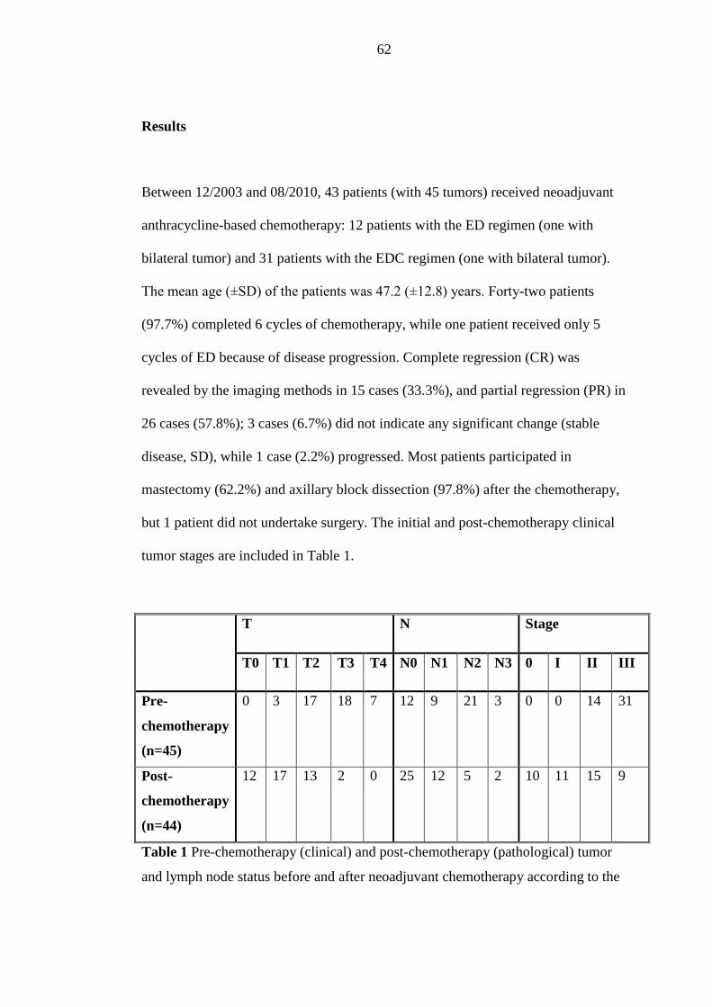
62
Results
Between 12/2003 and 08/2010, 43 patients (with 45 tumors) received neoadjuvant
anthracycline-based chemotherapy: 12 patients with the ED regimen (one with
bilateral tumor) and 31 patients with the EDC regimen (one with bilateral tumor).
The mean age (±SD) of the patients was 47.2 (±12.8) years. Forty-two patients
(97.7%) completed 6 cycles of chemotherapy, while one patient received only 5
cycles of ED because of disease progression. Complete regression (CR) was
revealed by the imaging methods in 15 cases (33.3%), and partial regression (PR) in
26 cases (57.8%); 3 cases (6.7%) did not indicate any significant change (stable
disease, SD), while 1 case (2.2%) progressed. Most patients participated in
mastectomy (62.2%) and axillary block dissection (97.8%) after the chemotherapy,
but 1 patient did not undertake surgery. The initial and post-chemotherapy clinical
tumor stages are included in Table 1.
T N Stage
T0 T1 T2 T3 T4 N0 N1 N2 N3 0 I II III
Pre-
chemotherapy
(n=45)
0 3 17 18 7 12 9 21 3 0 0 14 31
Post-
chemotherapy
(n=44)
12 17 13 2 0 25 12 5 2 10 11 15 9
Table 1 Pre-chemotherapy (clinical) and post-chemotherapy (pathological) tumor
and lymph node status before and after neoadjuvant chemotherapy according to the

63
UICC/AJCC TNM classification. Note that one patient did not undergo surgery
after neoadjuvant chemotherapy.
About half of the tumors were ER-positive, and one-third of them PgR-positive.
HER2 positivity was demonstrated by HER2 IHC and/or FISH in 18% of all
samples (Table 2).
Tumor feature N %
Histological type
IDC 38 84.4
ILC 3 6.7
other 4 8.9
Histologic grade
1 0 0
2 11 24.4
3 33 73.3
unknown 1 2.2
ER
negative 25 55.6
positive 20 44.4
PR negative 28 62.2
positive 17 37.8
HER2 negative 37 82.2
positive 8 17.8
Proportion (±SD) of Ki67-positive
cells (%)
56.1±23.6
TOP2A
Negative
(≤15%)
6 15.8
Positive
(>15%)
32 84.2
unknown 7
Proportion (±SD) of TOP2A-
positive cells (%)
41.0±27.9

64
Table 2 Pathological features of breast cancers before neoadjuvant chemotherapy
No significant change was observed in the ER, PgR or HER2 status of the tumors
after chemotherapy. The proportion of Ki67-positive tumor cells was significantly
reduced by the chemotherapy (56.1±23.6 vs. 19.0±27.7%, p=0.004).
The pathological tumor responses to chemotherapy are listed in Table 3.
Histological tumor regression
grade (TRG)
Overall n=44
(%)
EDC n=31
(%)
EC n=13
(%)
TRG0 1 (2.3) 0 (0.0) 1 (7.7)
TRG1 20 (45.5) 16 (51.6) 4 (30.8)
TRG2 10 (22.6) 6 (19.4) 4 (30.8)
TRG3 1 (2.3) 1 (3.2) 0 (0.0)
TRG4 12 (27.3) 8 (25.8) 4 (30.7)
Table 3 Pathological tumor response after neoadjuvant ED or EDC chemotherapy
(p=0.50)
Although complete disappearance of the primary tumor (TRG 4) was detected in 12
cases, the axillary lymph nodes were still involved in 3 of these cases, and 9 (20%)
cases were therefore classified as pCR. No significant difference existed between
tumor response according to the chemotherapy regimen (p=0.50): the proportions of
major tumor responses (TRG3-4) were 29% (n=9) and 30.7% (n=4) among the
patients treated with the TEX or the TE regimens, respectively (Table 3), while the
respective rates of pCR were 22.6% (n=7) and 15.4% (n=2). In an additional lymph
node-negative case, only a small DCIS focus remained. The association between the

65
clinical and pathological tumor responses proved to be statistically significant
(p<0.001).
TOP2A FISH/IHC
For technical reasons, the TOP2A FISH and TOP2A IHC results were assessable in
only 25 and 38 cases, respectively. With FISH, 23 tumors (92%) exhibited a normal
TOP2A gene copy number, while in 2 (8%), the TOP2A gene was amplified; both
were HER2-positive by means of IHC and FISH. Despite the fact that the median
proportion of IHC-stained cells was 50%, in view of the reference data in the
literature [11, 17, 18], we used >15% as a cut-off value for the definition of TOP2A
positivity (Table 2). Thirty-two (84.2%) tumors were classified as TOP2A-positive
and 6 (15.8%) as TOP2A-negative from the core biopsy. No significant correlation
was found between the TOP2A status as determined by FISH and IHC (p=0.52).
The average (±SD) proportion of TOP2A-positive cells in the evaluable samples
was 41.0±27.9% before, and 12.7±24.8% after the chemotherapy (p<0.001).
The expression of TOP2A showed a strong correlation with that of Ki67 (R=0.743,
p<0.001), and was negatively correlated with ER (R=0.404, p=0.012) and PgR
(R=0.430, p=0.007) (Figure 1), irrespective of the HER2 status (data not shown).

66
Figure 1. Correlation between the expression of TOP2A and Ki67
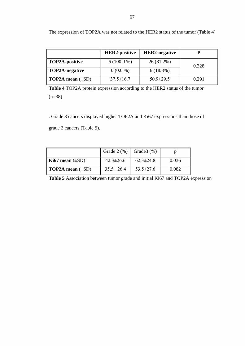
67
The expression of TOP2A was not related to the HER2 status of the tumor (Table 4)
HER2-positive HER2-negative P
TOP2A-positive 6 (100.0 %) 26 (81.2%) 0.328
TOP2A-negative 0 (0.0 %) 6 (18.8%)
TOP2A mean (±SD) 37.5±16.7 50.9±29.5 0.291
Table 4 TOP2A protein expression according to the HER2 status of the tumor
(n=38)
. Grade 3 cancers displayed higher TOP2A and Ki67 expressions than those of
grade 2 cancers (Table 5).
Grade 2 (%) Grade3 (%) p
Ki67 mean (±SD) 42.3±26.6 62.3±24.8 0.036
TOP2A mean (±SD) 35.5 ±26.4 53.5±27.6 0.082
Table 5 Association between tumor grade and initial Ki67 and TOP2A expression

68
Association between tumor response and tumor characteristics
A major tumor response was seen mostly for grade 3 and ER-negative cancers
(Table 6).
pCR
p yes no
Grade2 0 11 p=0.054
Grade3 9 24
ER-negative 8 16 p=0.023
ER-positive 1 19
PR-negative 7 20 p=0.230
PR-positive 2 15
HER2-negative 6 30 p=0.196
HER2-positive 3 5
Table 6 Tumor characteristics in relation with tumor response to neoadjuvant
anthracycline-based chemotherapy (n=44)
The development of pCR was related to high grade (grade 3) (p=0.054) and ER
negativity (p=0.027). While the mean (±SD) pre-chemotherapy TOP2A expression
was 66.9±26.3% in cases with pCR, it was 41.8±26.6% in cases without pCR
(p=0.037). Eight pCRs (21%) occurred among those cases that were assessed for
TOP2A IHC, and all the pCRs occurred in TOP2A-positive cancers. Although no
association was found with TOP2A amplification, both TOP2A-amplified tumors
gave a major response: pCR in one, and a reduction in tumor size from 70 to 15 mm
in the other. Ki67 was not predictive of the tumor response in univariate analysis
(OR=1.027, 95% CI: 0.992-1.062, p=0.167). In the logistic regression model
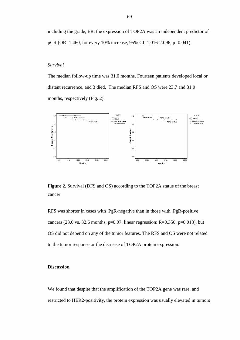
69
including the grade, ER, the expression of TOP2A was an independent predictor of
pCR (OR=1.460, for every 10% increase, 95% CI: 1.016-2.096, p=0.041).
Survival
The median follow-up time was 31.0 months. Fourteen patients developed local or
distant recurrence, and 3 died. The median RFS and OS were 23.7 and 31.0
months, respectively (Fig. 2).
Figure 2. Survival (DFS and OS) according to the TOP2A status of the breast
cancer
RFS was shorter in cases with PgR-negative than in those with PgR-positive
cancers (23.0 vs. 32.6 months, p=0.07, linear regression: R=0.350, p=0.018), but
OS did not depend on any of the tumor features. The RFS and OS were not related
to the tumor response or the decrease of TOP2A protein expression.
Discussion
We found that despite that the amplification of the TOP2A gene was rare, and
restricted to HER2-positivity, the protein expression was usually elevated in tumors

70
with high proliferation rate; anthracycline-based chemotherapy resulted in the
reduction of the expression of TOP2A. TOP2A positivity was an independent
predictor of pCR, and a 10% increase of TOP2A IHC staining resulted in a 46%
increase of the likelihood of obtaining a pCR.
The amplification of the TOP2A gene is a rare abnormality, and is restricted to
HER2 positive tumors [6, 7, 10-13, 16, 24, 25]. The deletion of the gene is less
frequent, and its role in anthracycline-sensitivity is controversial [6, 7, 9, 10, 13-16,
26]. In our cohort, 2 cases with TOP2A gene amplification exhibited HER2
amplification and high TOP2A expression (data not shown), and showed excellent
response to the therapy, however, the small number of cases limits the interpretation
of the findings.
The cut-off point for defining TOP2A-positivity by IHC varied in different studies.
The most often used threshold value was 10-15% (range 5-25%) [11, 17, 18, 27-
30], and the rate of TOP2A-positive tumors varied between 5-45% [12, 18, 30]. In a
series of 245 tumors, the median proportion of TOP2A-positive cells was 27%, and
about half of the tumors rated positive [11]. For the TOP2A-positive category, we
used the cut-off value of >15%. In our cohort, the median proportion of TOP2A-
stained cells was relatively high (50%), and the majority of tumors classified as
TOP2A-positive. The first reason for this finding is the selected nature of our study
population: only patients with rapidly proliferating tumors (based on the knowledge
of standard tumor characteristics), likely chemosensitive were chosen for
neoadjuvant chemotherapy. Second, the methods applied (the tumor regions with
the highest cellularity were used; TOP2A-positive cells were counted regardless of
the intensity of the staining) also favored high TOP2A values. Of note is that using
the same methods, the median TOP2A IHC value in another cohort of our patients

71
selected for adjuvant chemotherapy and in the whole population irrespective of
tumor characteristics was 20 (range: 0-90) and 5 (range: 0-80), respectively (data
not shown).
In our study, TOP2A protein expression was found an independent predictor of
pCR after neoadjuvant docetaxel-epirubicin chemotherapy, and the probability of
pCR increased by almost 50% with every 10% increase of the TOP2A positive
tumor cells. The predictive role of the tumor TOP2A status for anthracycline-based
chemotherapy has been investigated with different methods, in different settings.
The correlation between the efficiency of adjuvant anthracycline-based
chemotherapy and the presence of TOP2A gene amplification [13, 14, 31] or the
amplification and deletion of the gene [6-8, 10] in the breast tumor is well
demonstrated. In those randomized trials which compared an anthracycline-
containing chemotherapy with a non-anthracycline containing regimen, the benefit
of the former was limited to tumors with the presence of the amplification [13, 31],
or the amplification or the deletion of the TOP2A gene [7, 8, 10]. Some studies
demonstrated that the presence of TOP2A gene amplification is predictive for the
benefit of the dose elevation of the anthracylines [14, 32]. In contrast, a
retrospective analysis of the CALGB 8541 study did not support a difference of
benefit if doxorubicin was administered at different doses in tumors with TOP2A
gene aberrations, but in that study, standard dose was compared with suboptimal
doses [9]. In the neoadjuvant setting, the retrospective study of 350 cases showed
that the amplification of the TOP2A gene involves a 3 times higher probability of
pCR in patients treated with neoadjuvant anthracycline-based chemotherapy [16].
Likewise, the amplification of the TOP2A gene or the polysomy of chromosome 17
indicated a more than 4-times increased probability to obtain pCR in HER2-positive

72
and ER-, PgR-negative tumors [15]. Park et al. found that the chance of obtaining a
response including pCR after neoadjuvant doxorubicin was associated with TOP2A
and HER2 coamplification [25]. In a relatively small neoadjuvant study, in
consistence with our findings, serial TOP2A IHC determination showed a trend for
better response to anthracyclines in tumors with higher TOP2A expression, and a
significant decrease after therapy, in responders [17]. Mukherjee et al. found the
extent of TOP2A protein expression predictive of the pCR in 91 patients treated
with preoperative FEC chemotherapy [33]. The robust study of Press et al. provides
evidence that the coamplification of the TOP2A and HER2 genes is a clinically
useful predictive marker of an enhanced response to anthracycline-based
chemotherapy in metastatic breast cancer [34]. In a phase III study in advanced
breast cancer, the TOP2A IHC status was predictive for response to doxorubicin
monotherapy, and every 10% increase in TOP2A expression was associated with a
9% increase in the probability of response, while no such effect was demonstrated
for the treatment with docetaxel monotherapy [18].
The main outcome and novelty of our study is the demonstration that a simple tool
such as TOP2A IHC is an useful, semiquantitative predictive marker of the benefit
of neoadjuvant anthracycline-based chemotherapy in breast cancer. The design of
our study is not appropriate to answer whether TOP2A IHC staining is a specific
marker of anthracycline sensitivity or that of chemosensitivity only. Other studies,
however, support the hypothesis that high TOP2A protein expression is predictive
of anthracycline sensitivity. In an early study of Di Leo et al., it seemed that the
>10% expression of TOP2A protein favors the benefit of both the choice and the
higher dose of an adjuvant EC regimen [30]. Likewise, Durbecque et al. in a
retrospective analysis of the TAX 303 randomized study, demonstrated that

73
although docetaxel is more efficient than doxorubicin in the population of advanced
breast cancer patients overall, increasing TOP2A expression is associated with a
higher chance to obtain a response in the doxorubicin arm, but not in the docetaxel
arm [18].
Many studies examined the correlation between the TOP2A gene status and the
TOP2A protein expression [11, 12, 27, 29, 35, 37]. Although gene amplification
favored high protein expression, the presence of the enzyme was not dependent on
the gene abnormality. Jarvinen et al. found that TOP2A protein expression
correlated well with TOP2A mRNA [37]. Brase et al., analyzed TOP2A at the
levels of gene amplification, RNA expression and protein expression, and studied
their correlations. No correlation was found between gene amplification and RNA
or protein expression, but a strong correlation existed between TOP2A RNA and
protein levels [20]. The conclusion was drawn that unlike in the case of the HER2
status, TOP2A protein expression is highly regulated at the RNA level. In our
cohort, most tumors exhibited high TOP2A expression without the presence of
TOP2A gene alteration, as a function of high proliferative activity. Schindlbeck et
al. based on their study on patients treated with adjuvant anthracycline-based
chemotherapy, concluded that it is the TOP2A IHC and not the gene status that
predicts benefit of the treatment [12]. The relevance of TOP2A expression in the
prediction of anthracycline-sensitivity merits further studies, however, the
reconsideration of the optimal method is needed.
We did not find association between outcome and TOP2A expression or the fall of
TOP2A expression after neoadjuvant chemotherapy. This result is limited by the
relatively short follow-up time. Survival was neither different in other studies by the
TOP2A status among patients treated with anthracyclines [11, 26, 28]. Of note is,

74
however, that the administration of anthracyclines did improve outcome in HER2-
and TOP2A-coamplified tumors to a level that was obtained without such
chemotherapy, but with the addition of Herceptin [13].
In conclusion, our findings suggest that the IHC determination of the TOP2A
protein is a useful tool for the estimation of the sensitivity of breast cancer to
anthracycline-based chemotherapy.
75
References
1. Kaufmann M, von Minckwitz G, Bear HD, Buzdar A, McGale P, Bonnefoi H,
Colleoni M, Denkert C, Eiermann W, Jackesz R, Makris A, Miller W, Pierga JY,
Semiglazov V, Schneeweiss A, Souchon R, Stearns V, Untch M, Loibl S.
Recommendations from an international expert panel on the use of neoadjuvant
(primary) systemic treatment of operable breast cancer: new perspectives 2006. Ann
Oncol. 2007; 18:1927-1934.
2. Steger GG, Greil R, Jakesz R, Lang A, Mlineritsch B, Melbinger-Zeinitzer E,
Marth C, Samonigg H, Kubista E, Gnant M. Final results of ABCSG-24, a
randomized phase III study comparing epiribicin, docetaxel, and capecitabine
(EDC) to epirubicin and docetaxel (ED) as neoadjuvant treatment for early breast
cancer and comparing ED/EDC + trastuzumab (T) to ED/EDC as neoadjuvant
treatment for early HER-2 positive breast cancer. Cancer Res 2009; 69: S565 [abstr.
1081]
3. Di Leo A, Biganzoli L, Claudion W, Licitra S, Pestrin M, Larsimont D:
Topoisomerase II alpha as a marker predicting anthracyclines’ activity in early
breast cancer patients: ready for the primetime? Eur J Cancer, 44: 2791-2798, 2008
4. Järvinen TA, Liu ET. HER-2/neu and topoisomerase IIalpha in breast cancer.
Breast Cancer Res Treat. 2003; 78:299-311.
76
5. Isaacs RJ, Davies SL, Sandri MI, Redwood C, Wells NJ, Hickson ID.
Physiological regulation of eukaryotic topoisomerase II. Biochim Biophys Acta.
1998;1400:121-137.
6. Di Leo A, Gancberg D, Larsimont D, Tanner M, Jarvinen T, Rouas G, Dolci S,
Leroy JY, Paesmans M, Isola J, Piccart MJ. HER-2 amplification and
topoisomerase IIalpha gene aberrations as predictive markers in node-positive
breast cancer patients randomly treated either with an anthracycline-based therapy
or with cyclophosphamide, methotrexate, and 5-fluorouracil. Clin Cancer Res 2002;
8:1107-1116.
7. Knoop AS, Knudsen H, Balslev E, Rasmussen BB, Overgaard J, Nielsen KV,
Schonau A, Gunnarsdóttir K, Olsen KE, Mouridsen H, Ejlertsen B. Danish Breast
Cancer Cooperative Group. Retrospective analysis of topoisomerase IIa
amplifications and deletions as predictive markers in primary breast cancer patients
randomly assigned to cyclophosphamide, methotrexate, and fluorouracil or
cyclophosphamide, epirubicin, and fluorouracil: Danish Breast Cancer Cooperative
Group. J Clin Oncol 2005; 23:7483-7490.
8. Nielsen KV, Ejlertsen B, Moller S, Jorgensen JT, Knoop A, Knudsen H,
Mouridsen HT: The value of TOP2A gene copy number variation as a biomarker in
breast cancer: Update of DBCG trial 89D. Acta Oncol 2008; 47: 725-734.
77
9. Harris LN, Broadwater G, Abu-Khalaf M, Cowan D, Thor AD, Budman D,
Cirrincione CT, Berry DA, Winer EP, Hudis CA, Hayes DF, Friedman P, Ellis M,
Dressler L. Topoisomerase II{alpha} amplification does not predict benefit from
dose-intense cyclophosphamide, doxorubicin, and fluorouracil therapy in HER2-
amplified early breast cancer: results of CALGB 8541/150013. J Clin Oncol. 2009;
27:3430-3436.
10. O'Malley FP, Chia S, Tu D, Shepherd LE, Levine MN, Bramwell VH, Andrulis
IL, Pritchard KI. Topoisomerase II alpha and responsiveness of breast cancer to
adjuvant chemotherapy. J Natl Cancer Inst. 2009; 101:644-650.
11. Arriola E, Rodriguez-Pinilla SM, Lambros MBK, Jones RL, James M, Savage
K, Smith IE, Dowsett M, Reis-Filho JS: Topoisomerase II alpha amplification may
predict benefit from adjuvant anthracyclines in HER2 positive early breast cancer.
Breast Cancer Res Treat 2007; 106:181-189.
12. Schindlbeck C, Mayr D, Olivier C, Rack B, Engelstaedter V, Jueckstock J,
Jenderek C, Andergassen U, Jeschke U, Friese K. Topoisomerase IIalpha
expression rather than gene amplification predicts responsiveness of adjuvant
anthracycline-based chemotherapy in women with primary breast cancer. J Cancer
Res Clin Oncol. 2010;136:1029-1037.
13. Slamon D, EiermannW, Robert N et al. BCIRG 006: 2nd interim analysis phase
II randomized trial comparing doxorubicin and cyclophosphamide followed by
docetaxel positive early (AC®TH) with docetaxel, carboplatin and trastuzumab

78
(TCH) in HER2 positive early breast cancer patients: BCIRG 006 study. Breast
Cancer Res Treat 2005; 94:S5 [abstr. 1]
14. Tanner M, Isola J, Wiklund T, Erikstein B, Kellokumpu-Lehtinen P,
Malmström P, Wilking N, Nilsson J, Bergh J: Topoisomerase IIalpha gene
amplification predicts favorable treatment response to tailored and dose-escalated
anthracycline-based adjuvant chemotherapy in HER-2/neu-amplified breast cancer:
Scandinavian Breast Group Trial 9401. J Clin Oncol 2006; 24: 2428-2436.
15. Orlando L, Del Curto B, Gandini S, Ghisini R, Pietri E, Torrisi R, Balduzzi A,
Cardillo A, Dellapasqua S, Veronesi P, Viale G, Goldhirsch A, Colleoni M:
Topoisomerase IIalpha gene status and prediction of pathological complete
remission after anthracycline-based neoadjuvant chemotherapy in endocrine non-
responsive Her2/neu-positive breast cancer. Breast 2008; 17: 506-511.
16. Konecny GE, Pauletti G, Untch M, Wang HJ, Möbus V, Kuhn W, Thomssen C,
Harbeck N, Wang L, Apple S, Jänicke F, Slamon DJ. Association between HER2,
TOP2A, and response to anthracycline-based preoperative chemotherapy in high-
risk primary breast cancer. Breast Cancer Res Treat. 2010; 120:481-489.
17. Arpino G, Ciocca DR, Weiss H, Allred DC, Daguerre P, Vargas-Roig L, Leuzzi
M, Gago F, Elledge R, Mohsin SK. Predictive value of apoptosis, proliferation,
79
HER-2, and topoisomerase IIalpha for anthracycline chemotherapy in locally
advanced breast cancer. Breast Cancer Res Treat. 2005;92:69-75.
18. Durbecq V, Paesmans M, Cardoso F, Desmedt C, Di Leo A, Chan S, Friedrichs
K, Pinter T, Van Belle S, Murray E, Bodrogi I, Walpole E, Lesperance B, Korec S,
Crown J, Simmonds P, Perren TJ, Leroy JY, Rouas G, Sotiriou C, Piccart M,
Larsimont D: Topoisomerase-II alpha expression as a predictive marker in a
population of advanced breast cancer patients randomly treated either with single-
agent doxorubicin or single-agent docetaxel. Mol Cancer Ther 2004; 3:1207-1214.
19. Rody A, Karn T, Ruckhäberle E, Müller V, Gehrmann M, Solbach C, Ahr A,
Gätje R, Holtrich U, Kaufmann M: Gene expression of topoisomerase II alpha
(TOP2A) by microarray analysis is highly prognostic in estrogen receptor (ER)
positive breast cancer. Breast Cancer Res Treat, 2009;113:4574-66
20. Brase JC, Schmidt M, Fischbach T, Sültmann H, Bojar H, Koelbl H, Hellwig B,
Rahnenführer J, Hengstler JG, Gehrmann MC. ERBB2 and TOP2A in breast
cancer: a comprehensive analysis of gene amplification, RNA levels, and protein
expression and their influence on prognosis and prediction. Clin Cancer Res.
2010;16:2391-2401.

80
21. Sparano JA, Goldstein LJ, Childs BH, Shak S, Brassard D, Badve S, Baehner
FL, Bugarini R, Rowley S, Perez E, Shulman LN, Martino S, Davidson NE, Sledge
GW Jr, Gray R. Relationship between Topoisomerase 2A RNA Expression and
Recurrence after Adjuvant Chemotherapy for Breast Cancer. Clin Cancer Res.
2009;15:7693-7700.
22. Miller AB, Hoogstraten B, Staquet M, Winkler A. Reporting results of cancer
treatment. Cancer 1981; 47:207-214.
23. Sinn HP, Schmid H, Junkermann H, Huober J, Leppien G, Kaufmann M,
Bastert G, Otto HF: Histologic regression of breast cancer after primary
(neoadjuvant) chemotherapy. Geburtshilfe Frauenheilkd 1994; 54:552-558.
24. Nocito A, Kononen J, Kallioniemi OP, Sauter G. Tissue microarrays (TMAs)
for high-throughput molecular pathology research. Int J Cancer 2001;94:1–5.
25. Park K, Kim J, Lim S, Han S. Topoisomerase II-alpha (topoII) and HER2
amplification in breast cancers and response to preoperative doxorubicin
chemotherapy. Eur J Cancer. 2003;395:631-634.
26. Tubbs R, Barlow WE, Budd GT, Swain E, Porter P, Gown A, Yeh IT, Sledge G,
Shapiro C, Ingle J, Haskell C, Albain KS, Livingston R, Hayes DF. Outcome of
81
patients with early-stage breast cancer treated with doxorubicin-based adjuvant
chemotherapy as a function of HER2 and TOP2A status. J Clin Oncol.
2009;27:3881-3886.
27. Bhargava R, Lal P, Chen B: HER-2/neu and topoisomerase IIa gene
amplification and protein expression in invasive breast carcinomas: chromogenic in
situ hybridization and immunohistochemical analyses. Am J Clin Pathol 2005;
123:889-895
28. Gómez HL, Pinto JA, Olivera M, Vidaurre T, Doimi FD, Vigil CE, Velarde RG,
Abugattas JE, Alarcón E, Vallejos CS. Topoisomerase II-alpha as a predictive
factor of response to therapy with anthracyclines in locally advanced breast cancer.
Breast. 2010 Aug 10. [Epub ahead of print]
29. Cardoso F, Durbecq V, Larsimont D, Paesmans M, Leroy JY, Rouas G, Sotiriou
C, Renard N, Richard V, Piccart MJ, Di Leo A. Correlation between complete
response to anthracycline-based chemotherapy and topoisomerase II-alpha gene
amplification and protein overexpression in locally advanced/metastatic breast
cancer. Int J Oncol 2004; 24:201-209.
30. Di Leo A, Larsimont D, Gancberg D, Jarvinen T, Beauduin M, Vindevoghel A,
Michel J, Focan CH, Ries F, Gobert PH, Closon-Dejardin MT, Dolci S, Rouas G,
Paesmans M, Lobelle JP, Isola J, Piccart MJ: HER-2 and topo-isomerase IIalpha as
predictive markers in a population of node-positive breast cancer patients randomly

82
treated with adjuvant CMF or epirubicin plus cyclophosphamide. Ann Oncol 2001;
12:1081-1089.
31. Gunnarsdóttir KA, Jensen MB, Zahrieh D, Gelber RD, Knoop A, Bonetti M,
Mouridsen H, Ejlertsen B. CEF is superior to CMF for tumours with TOP2A
aberrations: a Subpopulation Treatment Effect Pattern Plot (STEPP) analysis on
Danish Breast Cancer Cooperative Group Study 89D. Breast Cancer Res Treat
2010; 123:163-169.
32. Hannemann J, Kristel P, van Tinteren H, Bontenbal M, van Hoesel QG, Smit
WM, Nooij MA, Voest EE, van der Wall E, Hupperets P, de Vries EG, Rodenhuis
S, van de Vijver MJ. Molecular subtypes of breast cancer and amplification of
topoisomerase II alpha: predictive role in dose intensive adjuvant chemotherapy. Br
J Cancer 2006; 95:1334-1341.
33. Mukherjee A, Shehata M, Moseley P, Rakha E, Ellis I, Chan S. Topo2α protein
expression predicts response to anthracycline combination neo-adjuvant
chemotherapy in locally advanced primary breast cancer. Br J Cancer.
2010;103:1794-1800.
34. Press MF, Sauter G, Buyse M, Bernstein L, Guzman R, Santiago A, Villalobos
IE, Eiermann W, Pienkowski T, Martin M, Robert N, Crown J, Bee V, Taupin H,
Flom KJ, Tabah-Fisch I, Pauletti G, Lindsay MA, Riva A, Slamon DJ. Alteration of
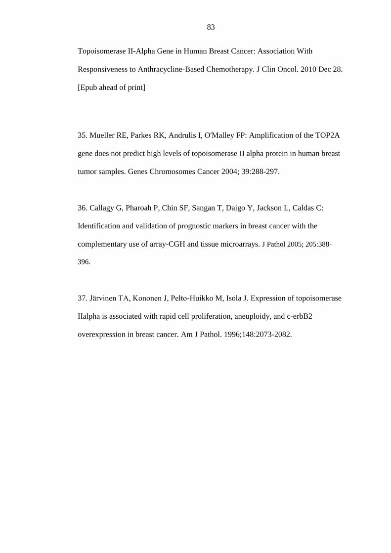
83
Topoisomerase II-Alpha Gene in Human Breast Cancer: Association With
Responsiveness to Anthracycline-Based Chemotherapy. J Clin Oncol. 2010 Dec 28.
[Epub ahead of print]
35. Mueller RE, Parkes RK, Andrulis I, O'Malley FP: Amplification of the TOP2A
gene does not predict high levels of topoisomerase II alpha protein in human breast
tumor samples. Genes Chromosomes Cancer 2004; 39:288-297.
36. Callagy G, Pharoah P, Chin SF, Sangan T, Daigo Y, Jackson L, Caldas C:
Identification and validation of prognostic markers in breast cancer with the
complementary use of array-CGH and tissue microarrays. J Pathol 2005; 205:388-
396.
37. Järvinen TA, Kononen J, Pelto-Huikko M, Isola J. Expression of topoisomerase
IIalpha is associated with rapid cell proliferation, aneuploidy, and c-erbB2
overexpression in breast cancer. Am J Pathol. 1996;148:2073-2082.

84
II

85
Running head: Topoisomerase II alpha expression and adjuvant chemotherapy in
breast cancer
Manuscript type: original paper
Tumour topoisomerase II alpha protein expression and outcome
after adjuvant dose-dense anthracycline-based chemotherapy
Alíz Nikolényi1, Gabriella Uhercsák
1, Melinda Csenki
1, Sándor Hamar
2, Erika
Csörgő2, Ervin Tánczos
3, László Thurzó
1, Thomas Brodowicz
4, Maria Wagnerova
5,
Zsuzsanna Kahán1
1Department of Oncotherapy,
2 Department of Pathology,
3Department of Medical
Informatics, 4Department of Surgery, University of Szeged, Szeged, Hungary;
4Central European Cooperative Oncology Group, Vienna, Austria;
5Oncology
Institute, Department of Radiotherapy and Oncology, Kosice, Slovakia
Corresponding author: Zsuzsanna Kahán, Department of Oncotherapy, University
of Szeged, Korányi fasor 12, H-6720 Szeged, Hungary; Phone: 36-62-545406; Fax:
36-62-545922; E-mail: [email protected]
Key-words: anthracyclines, adjuvant chemotherapy, breast cancer, dose-dense
chemotherapy, topoisomerase II alpha

86
Abbreviations
A adriamycin
ADC adriamycin (A)-docetaxel (D)-cyclophosphamide (C) chemotherapy
study
ATC adriamycin (A)-paclitaxel (T)-cyclophosphamide (C) chemotherapy
study
C cyclophosphamide
CECOG Central European Cooperative Oncology Group
D docetaxel
ER estrogen receptor
FEC fluorouracil-epirubicin-cyclophosphamide chemotherapy
FISH fluorescence in situ hybridisation
GCSF granulocyte colony stimulating factor
IHC immunohistochemistry
LVI lymphovascular invasion
OS overall survival
PR progesterone receptor
RFS recurrence-free survival
RNA ribonucleic acid
T paclitaxel
TOP2A topoisomerase II alpha

87
Abstract
There is a need for the selection of those breast cancers where benefit may be
attained from the addition of an anthracycline to the adjuvant chemotherapy. The
expression of topoisomerase II alpha (TOP2A) protein in 3 cohorts of breast
cancers treated with adjuvant dose-dense anthracycline-based chemotherapy was
determined retrospectively. The TOP2A status was analysed in relation with the
other standard tumour features and the outcome. TOP2A IHC results were
assessable in 106 patients: with a cut-off value of 15%, 48% of the tumours were
classified as TOP2A-positive. The expression of TOP2A correlated with that of
Ki67 (R=0.532, p<0.001) and a high grade (p=0.04) , but did not correlate with the
proportion of ER- or PR-positive cells in the tumour. More tumors were TOP2A-
negative among the ER- or PR-positive cancers than among the ER/PR-negative
cancers (p=0.021 and p=0.002, respectively). After a median follow-up time of 64.5
months, 31 relapses (23.5%) and 23 deaths (17.4%) had occurred in 131 patients.
The overall survival was longer in the TOP2A-positive cases than in the TOP2A-
negative cases. The recurrence-free survival and the overall survival were
significantly more favourable in the ER/PR-negative and TOP2A-positive tumours
than in other subgroups. In a Cox proportional hazards model, the grade and
TOP2A remained significant determinants in the ER/PR-negative subgroup.
TOP2A positivity and grade 3 indicated a decrease in the risk of death with
HR=0.211 (95% CI: 0.042-1.05, p=0.056) and HR=0.216 (95% CI: 0.047-0.990,
p=0.048), respectively. A higher sensitivity to anthracycline-containing regimens is
suggested in ER/PR-negative and TOP2A-positive cancers.
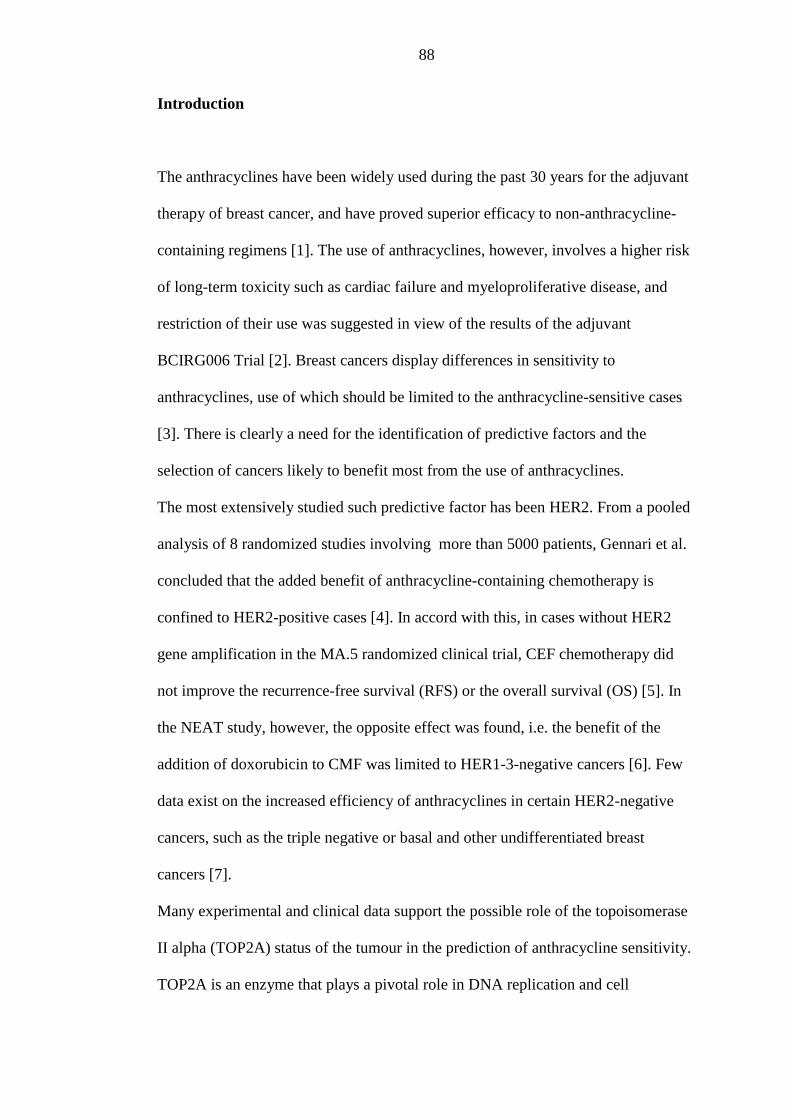
88
Introduction
The anthracyclines have been widely used during the past 30 years for the adjuvant
therapy of breast cancer, and have proved superior efficacy to non-anthracycline-
containing regimens [1]. The use of anthracyclines, however, involves a higher risk
of long-term toxicity such as cardiac failure and myeloproliferative disease, and
restriction of their use was suggested in view of the results of the adjuvant
BCIRG006 Trial [2]. Breast cancers display differences in sensitivity to
anthracyclines, use of which should be limited to the anthracycline-sensitive cases
[3]. There is clearly a need for the identification of predictive factors and the
selection of cancers likely to benefit most from the use of anthracyclines.
The most extensively studied such predictive factor has been HER2. From a pooled
analysis of 8 randomized studies involving more than 5000 patients, Gennari et al.
concluded that the added benefit of anthracycline-containing chemotherapy is
confined to HER2-positive cases [4]. In accord with this, in cases without HER2
gene amplification in the MA.5 randomized clinical trial, CEF chemotherapy did
not improve the recurrence-free survival (RFS) or the overall survival (OS) [5]. In
the NEAT study, however, the opposite effect was found, i.e. the benefit of the
addition of doxorubicin to CMF was limited to HER1-3-negative cancers [6]. Few
data exist on the increased efficiency of anthracyclines in certain HER2-negative
cancers, such as the triple negative or basal and other undifferentiated breast
cancers [7].
Many experimental and clinical data support the possible role of the topoisomerase
II alpha (TOP2A) status of the tumour in the prediction of anthracycline sensitivity.
TOP2A is an enzyme that plays a pivotal role in DNA replication and cell
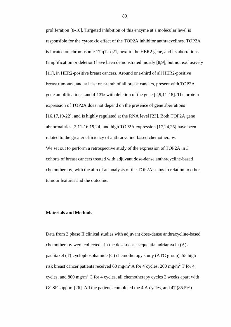
89
proliferation [8-10]. Targeted inhibition of this enzyme at a molecular level is
responsible for the cytotoxic effect of the TOP2A inhibitor anthracyclines. TOP2A
is located on chromosome 17 q12-q21, next to the HER2 gene, and its aberrations
(amplification or deletion) have been demonstrated mostly [8,9], but not exclusively
[11], in HER2-positive breast cancers. Around one-third of all HER2-positive
breast tumours, and at least one-tenth of all breast cancers, present with TOP2A
gene amplifications, and 4-13% with deletion of the gene [2,9,11-18]. The protein
expression of TOP2A does not depend on the presence of gene aberrations
[16,17,19-22], and is highly regulated at the RNA level [23]. Both TOP2A gene
abnormalities [2,11-16,19,24] and high TOP2A expression [17,24,25] have been
related to the greater efficiency of anthracycline-based chemotherapy.
We set out to perform a retrospective study of the expression of TOP2A in 3
cohorts of breast cancers treated with adjuvant dose-dense anthracycline-based
chemotherapy, with the aim of an analysis of the TOP2A status in relation to other
tumour features and the outcome.
Materials and Methods
Data from 3 phase II clinical studies with adjuvant dose-dense anthracycline-based
chemotherapy were collected. In the dose-dense sequential adriamycin (A)-
paclitaxel (T)-cyclophosphamide (C) chemotherapy study (ATC group), 55 high-
risk breast cancer patients received 60 mg/m2
A for 4 cycles, 200 mg/m2 T for 4
cycles, and 800 mg/m2 C for 4 cycles, all chemotherapy cycles 2 weeks apart with
GCSF support [26]. All the patients completed the 4 A cycles, and 47 (85.5%)

90
patients completed all 12 cycles. In the very similar dose-dense sequential
adriamycin (A)-docetaxel (D)-cyclophosphamide (C) chemotherapy study (ADC
group), 34 breast cancer patients received 60 mg/m2
A for 4 cycles, 75 mg/m2 D for
4 cycles, and 800 mg/m2 C for 4 cycles, all chemotherapy cycles 2 weeks apart,
with GCSF support. Of the 34 patients enrolled, 33 (97%) completed all 12 cycles,
whereas one was excluded after the first 7 cycles because of disease progression. In
the dose-dense FEC study (CECOG group), breast cancer patients were randomized
to 6 cycles of FEC75 or FEC90 (fluorouracil 500 mg/m2, epirubicin 75 or 90 mg/m
2,
respectively and cyclophosphamide 500 mg/m2) with pegfilgasrtim support [27].
Most of the enrolled 51 patients completed the study, but the clinical data and the
tumour samples were accessable in only 43 cases treated at the Hungarian and the
Slovakian centres. Patient- and tumour-related data, such as the pathological stage,
the grade, and the ER, PR, HER2 and Ki67 status, determined by standard methods
in the 3 study populations, are included in Table 1.
The RFS and the OS were calculated from day 1 of chemotherapy to the date of
appearance of local recurrence/distant metastasis, or the date of death for any reason
(or the date of the last follow-up), respectively. Analyses were carried out on the
associations of the RFS and the OS with the tumour characteristics.
Tissue microarray (TMA) construction
TMAs were constructed from formalin-fixed and paraffin-embedded tumour blocks
as described previously [28]. An experienced pathologist (SH) selected the most
cellular region. A tissue core 2 mm in diameter was punched for the TMA and
embedded in an acceptor block. Slides for FISH and IHC examinations were made
from every block.

91
Immunohistochemistry (IHC)
TOP2A IHC involved use of the primary specific monoclonal antibody TOP2A Ki-
s1 (Lab Vision, Fremont, CA, USA) with an automatic staining machine (Dako
Autostainer). Antigen retrieval was achieved by autoclaving in citrate buffer, pH
6.0, for 10 min at 121°C, and an EnVision + System (Dako) was applied as the
detection system. The nuclei of 50 tumour cells were counted under the microscope
by two independent examiners, and the proportion of stained cells was recorded. A
cut-off value of 15% separated negative (≤15%) and positive cases (>15%).
For HER2 IHC, the standard method was used. HER2 expression was scored
semiquantitatively with scores in the range 0-3+, following the accepted criteria;
HER2 2+ was regarded as indeterminate, and required HER2 FISH examination.
Statistical analysis
Univariate comparisons of groups was performed by one-way ANOVA and chi-
square testing in cases of continuous and categorical variables, respectively. Two-
by-two frequency tables were evaluated by means of Fisher’s exact test. The
relationship between the continuous variables was examined by correlational
analysis. The dependence of the durations of the RFS and OS on the possible risk
factors was analysed by means of the Kaplan-Meier method. To estimate the effects
of the TOP2A protein expression and the conventional prognostic factors on the
outcome, the Cox proportional hazards model was utilized. A stepwise selection
method was performed, using the likelihood-ratio statistics based on the maximum
partial likelihood estimates. SPSS version 17.0 for Windows (SPSS Inc., Chicago,
IL, USA) was applied for statistical analysis.

92
Results
The patient- and tumour-related characteristics within the 3 study cohorts and in the
overall population are included in Table 1.
ATC (n=55) ADC (n=34) CECOG
(n=43)
Overall
(n=132)
Age (mean±SE) 59.8±1.2 54.9±1.6 54.9±1.6 57.0±0.8
pT (mean±SE,
mm)
35.6±2.8 16.7±2.7 22.5±2.3 26.3±1.6
pN+ (median) 6 0.5 2 3
Histological
type (%)
IDC
ILC
Medullary
Other
43 (78.2)
6 (10.9)
1 (1.8)
5 (9.1)
30 (88.3)
1 (2.9)
0 (0)
3 (8.8)
35 (81.4)
4 (9.3)
3 (7.0)
1 (2.3)
108 (81.8)
11 (8.3)
4 (3.0)
9 (6.9)
LVI present
(%)
38 (69.1) 9 (26.5) 28 (65.1) 75 (56.8)
Grade 1
Grade 2
Grade 3
Grade
unknown
3 (5.4)
10 (18.2)
27 (49.1)
15 (27.3)
0 (0)
12 (35.3)
17 (50)
5 (14.7)
2 (4.7)
18 (41.9)
22 (51.2)
1 (2.3)
5 (3.8)
40 (30.3)
66 (50.0)
21 (15.9)
ER positive (%) 25 (45.5) 17 (50) 16 (37.2) 33 (25)
PR positive (%) 23 (41.8) 15 (44.1) 15 (34.9) 30 (22.7)
HER2
positive(%)
13 (23.6) 9 (26.5) 7 (16.3) 29 (22.0)
Ki67 (mean±SE,
%)
Ki67 (median,
%)
29.3±4.2
20
25.0±4.5
20
42.3±5.5
30
32.3±2.7
25

93
Table 1. Patient- and tumour-related characteristics within the study groups and the
overall population
The median follow-up time for the entire population was 64.5 months, and for the
ATC, ADC and CECOG cohorts was 103, 44.5 and 60 months, , respectively.
Altogether 31 relapses (23.5%) and 23 deaths (17.4%) occurred. The OS differed
significantly in the 3 cohorts: the ATC cohort exhibited the worst, and the ADC
cohort the best survival (p<0.01). Among the standard prognostic factors, the
pathological tumour size (pT) and the number of positive lymph nodes were
associated with the RFS in the overall study population (p<0.05), while the
presence of LVI was related to the RFS in the ADC cohort.
TOP2A IHC
For technical reasons, the TOP2A IHC results were assessable in only 106 cases. In
the overall population, the average and median proportions of the TOP2A-positive
cells were 21% and 10%, respectively. With a cut-off value of 15%, 48% of the
tumours were classified as TOP2A-positive (Table 2).
ATC ADC CECOG Overall
TOP2 A IHC
(n)
40 27 39 106
TOP2A
(mean±SE, %)
18.3±3.4
17.33±5.0
24.5±5.0
21.02±2.3
TOP2A
(median, %)
10 15 10 10
TOP2A+
(n)(%)
16 (40) 14 (51.9) 21 (53.8) 51 (48.1)

94
Table 2 TOP2A IHC status in the study groups and the overall population
Most of the TOP2A-positive tumours were of grade 3 (p=0.004). The expression of
TOP2A correleted significantly with that of Ki67 (R=0.532, p<0.001), but not with
ER or PR. Among the ER- and/or PR-positive cancers, more were TOP2A-negative
than among the ER- and PR-negative cancers (p=0.021 and p=0.002, respectively)
(Table 3).

95
ATC (n=40) ADC (n=27) CECOG (n=39) Overall (n=106)
TOP2
A-
TOP2A
+
TOP2
A-
TOP2A
+
TOP2
A-
TOP2A
+
TOP2
A-
TOP2A
+
ER
-
11 11 6 8 5 13 22 32
ER
+
13 5 7 6 13 8 33 19
p 0.203 0.706 0.054 0.021
PR- 10 12 7 10 6 15 23 37
PR
+
14 4 6 4 12 6 32 14
p 0.054 0.440 0.026 0.002
Table 3 TOP2A IHC status according to the ER/PR status of the tumour
All hormone receptor-negative cancers were of grade 2 or 3, and TOP2A-positive
cases were more frequently of grade 3 (p=0.066 and p=0.040 in the ER-negative
and the PR-negative groups, respectively). No association was detected between the
TOP2A status and the grade of the tumour in the hormone receptor-positive group.
The expression of TOP2A was not related to the tumour size, the number of
positive nodes or the HER2 status of the tumour. The protein expressions of
TOP2A and Ki67 increased with the grade (p=0.162 and p=0.005, respectively).
Association between outcome and tumour TOP2A status
In the overall population, more relapses and more deaths occurred among the
TOP2A-negative cases than among the TOP2A-positive cases, and the RFS and OS
were longer accordingly (Table 4, Fig. 1).

96
TOP2A IHC number of
deaths (%)
OS (mean±SE)
(months)
number of
relapses (%)
RFS
(mean±SE)
(months)
Negative 14/55 (25.5) 93.3±6.0 14/55 (25.5) 93.7±6.1
Positive 6/51 (11.8) 103.8±4.3 8/51 (15.7) 96.8±5.9
p (Mantel-Cox) 0.081 0.229
Table 4 Survival (OS and RFS) according to the TOP2A status of the tumour
Figure 1. Survival (OS and RFS) according to the TOP2A status of the tumour

97
The outcome in the hormone receptor-positive and hormone receptor-negative
subgroups was analysed separately (Table 5, Fig. 2).
ER-negative ER-positive PR-negative PR-positive
TOP2
A-
TOP2
A+
TOP2
A-
TOP2
A+
TOP2
A-
TOP2
A+
TOP2
A-
TOP2
A+
number of
deaths
6/22 2/32 8/33 4/19 7/23 2/37 7/32 4/14
OS
(mean±SE)
(months)
93.1±
9.3
109.3±
3.9
92.9±
7.6
82.7±
6.8
89.0±
9.6
110.5±
3.1
95.7±
7.4
81.3±
10.2
p (Mantel-
Cox)
0.035 0.916 0.005 0.494
number of
relapses
7/22 5/32 7/33 5/19 7/23 6/37 7/32 4/14
RFS(mean
±SE)
(months)
87.2±
10.3
97.1±
7.5
97.2±
7.3
77.6±
8.1
87.3±
10.3
97.2±
6.7
97.7±
7.1
79.5±
11.2
p (Mantel-
Cox)
0.176 0.774 0.169 0.639
Table 5 Survival (OS and RFS) according to the TOP2A and ER/PR status of the
tumor

98
Figure 2. a,c Survival (OS and RFS) according to the tumor TOP2A IHC status in
the ER and/or PR positive tumors
Figure 2 b,d Survival (OS and RFS) according to the tumor TOP2A IHC status in
the ER and/or PR negative tumors
While there was no difference in the number of events, or in the OS and the RFS in
the ER- and the PR-positive subgroups according to the TOP2A status, the OS and
RFS were significantly improved in the ER- or PR-negative and TOP2A-positive
cases as compared with the TOP2A-negative cases (Table 5, Fig. 2). Figure 1
presents the RFS and OS as functions of the TOP2A expression status in ER/PR-
negative cases.
In order to estimate the dependence of the OS and the RFS on the tumour TOP2A
and Ki67 status, the tumour grade and the nodal status in ER- and/or PR-negative
cancer, these variables were studied in a Cox proportional hazards model. In grade
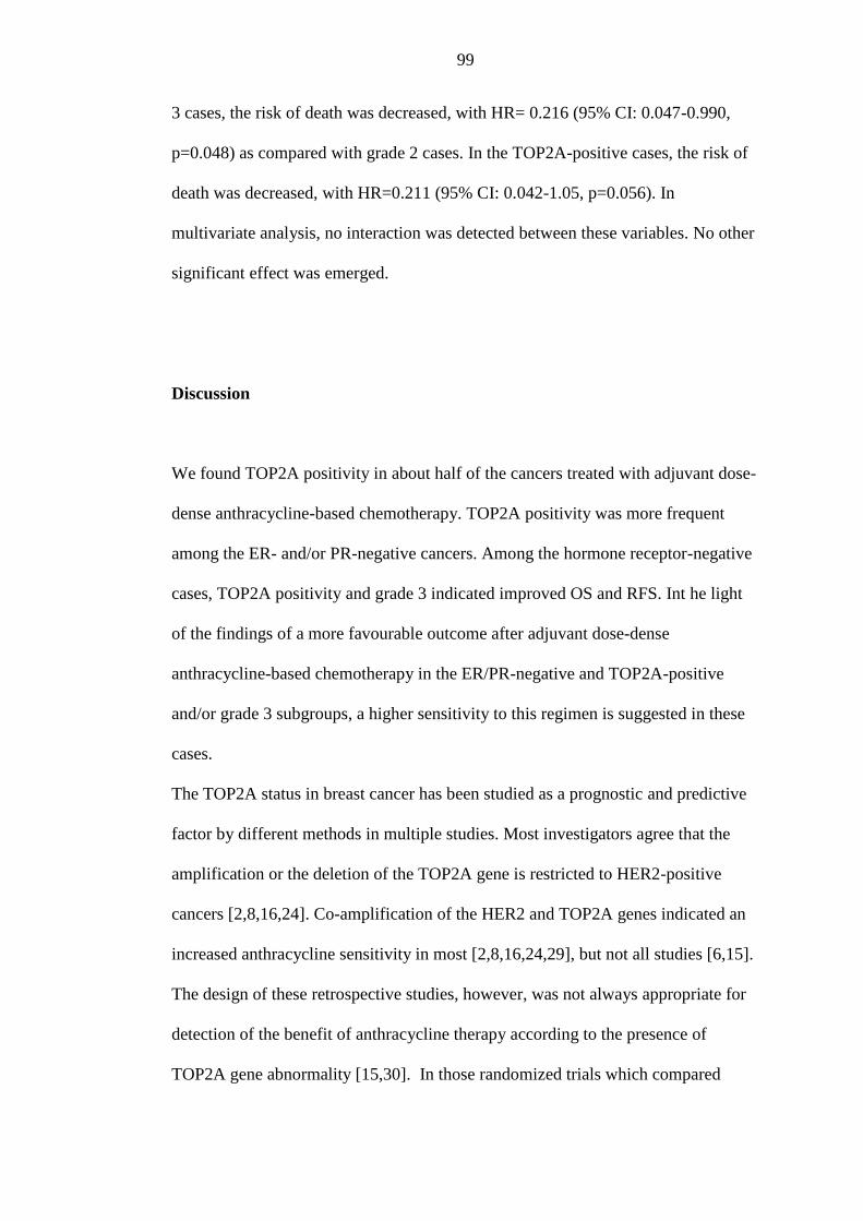
99
3 cases, the risk of death was decreased, with HR= 0.216 (95% CI: 0.047-0.990,
p=0.048) as compared with grade 2 cases. In the TOP2A-positive cases, the risk of
death was decreased, with HR=0.211 (95% CI: 0.042-1.05, p=0.056). In
multivariate analysis, no interaction was detected between these variables. No other
significant effect was emerged.
Discussion
We found TOP2A positivity in about half of the cancers treated with adjuvant dose-
dense anthracycline-based chemotherapy. TOP2A positivity was more frequent
among the ER- and/or PR-negative cancers. Among the hormone receptor-negative
cases, TOP2A positivity and grade 3 indicated improved OS and RFS. Int he light
of the findings of a more favourable outcome after adjuvant dose-dense
anthracycline-based chemotherapy in the ER/PR-negative and TOP2A-positive
and/or grade 3 subgroups, a higher sensitivity to this regimen is suggested in these
cases.
The TOP2A status in breast cancer has been studied as a prognostic and predictive
factor by different methods in multiple studies. Most investigators agree that the
amplification or the deletion of the TOP2A gene is restricted to HER2-positive
cancers [2,8,16,24]. Co-amplification of the HER2 and TOP2A genes indicated an
increased anthracycline sensitivity in most [2,8,16,24,29], but not all studies [6,15].
The design of these retrospective studies, however, was not always appropriate for
detection of the benefit of anthracycline therapy according to the presence of
TOP2A gene abnormality [15,30]. In those randomized trials which compared

100
anthracycline-containing chemotherapy with a non-anthracycline-containing
regimen, the benefit of the former was limited to tumours with an abnormal TOP2A
gene status [2,11,13,14,31]. Some studies have demonstrated that the presence of a
TOP2A gene alteration is predictive of the benefit of an elevation of the
anthracyline dose [19,32]. Deletion of the gene is less frequent, and its role in
anthracycline sensitivity seems rather controversial [2,12-14,18,24,30]. In line with
the contradictory results, it is noteworthy that, although TOP2A gene abnormalities
have been observed exclusively in HER2-positive breast cancers, high
anthracycline sensitivity is not limited to this special group [7].
Investigations of whether the expression of TOP2A is a specific marker of
anthracycline sensitivity gave more concordant results. The early study by Di Leo et
al. led to the conclusion that a finding of TOP2A positivity by means of IHC
determination favoured the benefit of both the choice and a higher dose of an
adjuvant EC regimen [33]. Likewise, in a retrospective analysis of the TAX 303
randomized study, Durbecque et al. demonstrated that, although docetaxel is more
efficient than doxorubicin in the population of advanced breast cancer patients
overall, increase of the TOP2A protein expression is associated with a higher
chance of obtaining a response in the doxorubicin arm, but not in the docetaxel arm
[25]. The greater sensitivity to anthracycline-based adjuvant chemotherapy of
ER/PR-negative breast cancers as compared with ER- or PR-positive tumours has
been well demonstrated [7,34]. Our own study suggests that one of the related key
factors is the more frequent TOP2A positivity among the ER/PR-negative tumours,
and we advocate TOP2A IHC as a tool to select those hormone receptor-negative
cases which would benefit from adjuvant anthracyclines. In a patient population
treated with adjuvant anthracycline-containing chemotherapy, Schindlbeck et al.

101
retrospectively examined the TOP2A status. About 50% of the cases proved to be
TOP2A-positive, and after a median survival time of 42 months, the survival was
significantly poorer among the TOP2A-negative cases [17]. Brase et al.
demonstrated the strong negative prognostic power of an elevated TOP2A RNA
level in 782 untreated breast cancer patients, which remained significant after
further analyses in the ER-positive and the HER2-negative and triple-negative
subgroups. In the same paper, complete tumour regression to chemotherapy with
EC was reported to be related to the high TOP2A and low ER RNA levels, results
which support our finding that anthracyclines result in a favourable outcome in ER-
negative and TOP2A-positive cancers [23]. Rody et al.followed up more than 1300
patients, and found that the TOP2A expression was the strongest indicator of a poor
prognosis among hormone receptor-positive cases, while no such effect was
detected among the ER-negative cases [35]. Although the prognostic effect of
TOP2A positivity was found to be independent of the systemic therapy, the nature
of the chemotherapy given in about half of the patients, was not reported. It may be
speculated that the similar outcome in the TOP2A-positive and -negative cases in
the ER-negative group may be due to the higher chemosensitivity of the TOP2A-
positive cases.
The expression of TOP2A seems to be regulated most strongly at the RNA level,
and its gene status is probably less determinative of its functional capacity. Jarvinen
et al. and Brase et al. found no correlation between gene amplification and protein
expression, but there was a strong correlation between the TOP2A RNA and protein
levels [23,36]. Accordingly, although gene amplification favoured a high protein
expression in those studies that examined the correlation between the TOP2A gene
status and the TOP2A protein expression, the presence of the enzyme was not

102
dependent on the gene abnormality [16,17,20-22]. Their findings led Brase et al. to
recommend determination of the RNA expression, while Schindlbeck et al.
suggested determination of the protein expression of TOP2A for patient selection,
rather than examination of the gene status [17,23].
Our own data indicate that a simple tool such as TOP2A IHC ( together with the
grade) is a useful predictive marker, at least in the hormone receptor-negative cases,
and should be implemented in routine practice for the selection of those who can be
expected to benefit from adjuvant anthracycline-based chemotherapy. The usually
poor outcome in the group of hormone receptor-negative and TOP2A-positive cases
may be reversed by the application of anthracycline-containing chemotherapy.

103
References
1. Early Breast Cancer Trialists' Collaborative Group (EBCTCG): Effects of
chemotherapy and hormonal therapy for early breast cancer on recurrence and 15-
year survival: an overview of the randomised trials. Lancet. 2005; 365:1687-1717.
2. Slamon D, EiermannW, Robert N et al. BCIRG 006: 2nd interim analysis phase
II randomized trial comparing doxorubicin and cyclophosphamide followed by
docetaxel positive early (AC®TH) with docetaxel, carboplatin and trastuzumab
(TCH) in HER2 positive early breast cancer patients: BCIRG 006 study. Breast
Cancer Res Treat 2005; 94:S5 [abstr. 1]
3. Gianni L, Norton L, Wolmark N, Suter TM, Bonadonna G, Hortobagyi GN. Role
of anthracyclines int he treatment of early breast cancer. J Clin Oncol 2009; 27:
4798-4808.
4. Gennari A, Sormani MP, Pronzato P, Puntoni M, Colozza M, Pfeffer U, Bruzzi
P. HER2 status and efficacy of adjuvant anthracyclines in early breast cancer: a
pooled analysis of randomized trials. J Natl Cancer Inst. 2008; 100:14-20.
5. Pritchard KI, Shepherd LE, O'Malley FP, Andrulis IL, Tu D, Bramwell VH,
Levine MN; National Cancer Institute of Canada Clinical Trials Group. HER2 and
responsiveness of breast cancer to adjuvant chemotherapy. N Engl J Med. 2006;
354: 2103-2111.
6. Bartlett JM, Munro A, Cameron DA, Thomas J, Prescott R, Twelves CJ. Type 1
receptor tyrosine kinase profiles identify patients with enhanced benefit from
anthracyclines in the BR9601 adjuvant breast cancer chemotherapy trial. J Clin
Oncol. 2008; 26: 5027-5035.
7. Rouzier R, Perou CM, Symmans WF, Ibrahim N, Cristofanilli M, Anderson K,
Hess KR, Stec J, Ayers M, Wagner P, Morandi P, Fan C, Rabiul I, Ross JS,

104
Hortobagyi GN, Pusztai L. Breast cancer subtypes respond differently to
preoperative chemotherapy. Clin Cancer Res 2005; 11:5678-5685.
8. Di Leo A, Biganzoli L, Claudion W, Licitra S, Pestrin M, Larsimont D:
Topoisomerase II alpha as a marker predicting anthracyclines’ activity in early
breast cancer patients: ready for the primetime? Eur J Cancer, 44: 2791-2798, 2008
9. Järvinen TA, Liu ET. HER-2/neu and topoisomerase IIalpha in breast cancer.
Breast Cancer Res Treat. 2003; 78:299-311.
10. Isaacs RJ, Davies SL, Sandri MI, Redwood C, Wells NJ, Hickson ID.
Physiological regulation of eukaryotic topoisomerase II. Biochim Biophys Acta.
1998;1400:121-137.
11. Nielsen KV, Ejlertsen B, Moller S, Jorgensen JT, Knoop A, Knudsen H,
Mouridsen HT: The value of TOP2A gene copy number variation as a biomarker in
breast cancer: Update of DBCG trial 89D. Acta Oncol 2008; 47: 725-734.
12. Di Leo A, Gancberg D, Larsimont D, Tanner M, Jarvinen T, Rouas G, Dolci S,
Leroy JY, Paesmans M, Isola J, Piccart MJ. HER-2 amplification and
topoisomerase IIalpha gene aberrations as predictive markers in node-positive
breast cancer patients randomly treated either with an anthracycline-based therapy
or with cyclophosphamide, methotrexate, and 5-fluorouracil. Clin Cancer Res 2002;
8:1107-1116.
13. Knoop AS, Knudsen H, Balslev E, Rasmussen BB, Overgaard J, Nielsen KV,
Schonau A, Gunnarsdóttir K, Olsen KE, Mouridsen H, Ejlertsen B. Danish Breast
Cancer Cooperative Group. Retrospective analysis of topoisomerase IIa

105
amplifications and deletions as predictive markers in primary breast cancer patients
randomly assigned to cyclophosphamide, methotrexate, and fluorouracil or
cyclophosphamide, epirubicin, and fluorouracil: Danish Breast Cancer Cooperative
Group. J Clin Oncol 2005; 23:7483-7490.
14. O'Malley FP, Chia S, Tu D, Shepherd LE, Levine MN, Bramwell VH, Andrulis
IL, Pritchard KI. Topoisomerase II alpha and responsiveness of breast cancer to
adjuvant chemotherapy.
J Natl Cancer Inst. 2009; 101:644-650.
15. Harris LN, Broadwater G, Abu-Khalaf M, Cowan D, Thor AD, Budman D,
Cirrincione CT, Berry DA, Winer EP, Hudis CA, Hayes DF, Friedman P, Ellis M,
Dressler L. Topoisomerase II{alpha} amplification does not predict benefit from
dose-intense cyclophosphamide, doxorubicin, and fluorouracil therapy in HER2-
amplified early breast cancer: results of CALGB 8541/150013. J Clin Oncol. 2009;
27:3430-3436.
16. Arriola E, Rodriguez-Pinilla SM, Lambros MBK, Jones RL, James M, Savage
K, Smith IE, Dowsett M, Reis-Filho JS: Topoisomerase II alpha amplification may
predict benefit from adjuvant anthracyclines in HER2 positive early breast cancer.
Breast Cancer Res Treat 2007; 106:181-189.
17. Schindlbeck C, Mayr D, Olivier C, Rack B, Engelstaedter V, Jueckstock J,
Jenderek C, Andergassen U, Jeschke U, Friese K. Topoisomerase IIalpha
expression rather than gene amplification predicts responsiveness of adjuvant
anthracycline-based chemotherapy in women with primary breast cancer. J Cancer
Res Clin Oncol. 2010;136:1029-1037.

106
18. Tanner M, Isola J, Wiklund T, Erikstein B, Kellokumpu-Lehtinen P,
Malmström P, Wilking N, Nilsson J, Bergh J: Topoisomerase IIalpha gene
amplification predicts favorable treatment response to tailored and dose-escalated
anthracycline-based adjuvant chemotherapy in HER-2/neu-amplified breast cancer:
Scandinavian Breast Group Trial 9401. J Clin Oncol 2006; 24: 2428-2436.
19. Fritz P, Cabrera CM, Dippon J, Gerteis A, Simon W, Aulitzky WE, van der
Kuip H. c-erbB2 and topoisomerase IIalpha protein expression independently
predict poor survival in primary human breast cancer: a retrospective study. Breast
Cancer Res. 2005; 7:R374-384.
20. Durbecq V, Desmed C, Paesmans M, Cardoso F, Di Leo A, Mano M, Rouas G,
Leroy JY,
Sotiriou C, Piccart M, Larsimont D: Correlation between topoisomerase-IIalpha
gene amplification and protein expression in HER-2 amplified breast cancer. Int J
Oncol 2004; 25:1473-1479.
21. . Mueller RE, Parkes RK, Andrulis I, O'Malley FP: Amplification of the TOP2A
gene does not predict high levels of topoisomerase II alpha protein in human breast
tumor samples. Genes Chromosomes Cancer 2004; 39:288-297.
22. Callagy G, Pharoah P, Chin SF, Sangan T, Daigo Y, Jackson L, Caldas C:
Identification and validation of prognostic markers in breast cancer with the
complementary use of array-CGH and tissue microarrays. J Pathol 2005; 205:388-
396.

107
23. Brase JC, Schmidt M, Fischbach T, Sültmann H, Bojar H, Koelbl H, Hellwig B,
Rahnenführer J, Hengstler JG, Gehrmann MC. ERBB2 and TOP2A in breast
cancer: a comprehensive analysis of gene amplification, RNA levels, and protein
expression and their influence on prognosis and prediction. Clin Cancer Res.
2010;16:2391-2401.
24. Konecny GE, Pauletti G, Untch M, Wang HJ, Möbus V, Kuhn W, Thomssen C,
Harbeck N, Wang L, Apple S, Jänicke F, Slamon DJ. Association between HER2,
TOP2A, and response to anthracycline-based preoperative chemotherapy in high-
risk primary breast cancer. Breast Cancer Res Treat. 2010; 120:481-489.
25. Durbecq V, Paesmans M, Cardoso F, Desmedt C, Di Leo A, Chan S, Friedrichs
K, Pinter T, Van Belle S, Murray E, Bodrogi I, Walpole E, Lesperance B, Korec S,
Crown J, Simmonds P, Perren TJ, Leroy JY, Rouas G, Sotiriou C, Piccart M,
Larsimont D: Topoisomerase-II alpha expression as a predictive marker in a
population of advanced breast cancer patients randomly treated either with single-
agent doxorubicin or single-agent docetaxel. Mol Cancer Ther 2004; 3:1207-1214.
26. Kahan Z, Uhercsak G, Hajnal-Papp R, Boda K, Thurzo L. Dose-dense
sequential adriamycin-Paclitaxel-cyclophosphamide chemotherapy is well tolerated
and safe in high-risk early breast cancer. Oncology. 2005;68:446-453.
27. Kahán Z, Spanik S, Wagnerova M, Skacel T, Planko B, Fitzthum E, Lindner E,
Soldatenkova V, Zielinski CC, Brodowicz T.: Feasibility of two dose-dense FEC

108
regimens with growth factor support for adjuvant therapy in patients with early
breast cancer: results from a randomised study of the Central European Cooperative
Oncology Group (CECOG). Breast Cancer Res Treat. 2008; 112:557-563.
28. Nocito A, Kononen J, Kallioniemi OP, Sauter G. Tissue microarrays (TMAs)
for high-throughput molecular pathology research. Int J Cancer 2001;94:1–5.
29. Park K, Kim J, Lim S, Han S. Topoisomerase II-alpha (topoII) and HER2
amplification in breast cancers and response to preoperative doxorubicin
chemotherapy. Eur J Cancer. 2003;395:631-634.
30. Tubbs R, Barlow WE, Budd GT, Swain E, Porter P, Gown A, Yeh IT, Sledge G,
Shapiro C, Ingle J, Haskell C, Albain KS, Livingston R, Hayes DF. Outcome of
patients with early-stage breast cancer treated with doxorubicin-based adjuvant
chemotherapy as a function of HER2 and TOP2A status. J Clin Oncol.
2009;27:3881-3886.
31. Gunnarsdóttir KA, Jensen MB, Zahrieh D, Gelber RD, Knoop A, Bonetti M,
Mouridsen H, Ejlertsen B. CEF is superior to CMF for tumours with TOP2A
aberrations: a Subpopulation Treatment Effect Pattern Plot (STEPP) analysis on
Danish Breast Cancer Cooperative Group Study 89D. Breast Cancer Res Treat
2010; 123:163-169.
32. Hannemann J, Kristel P, van Tinteren H, Bontenbal M, van Hoesel QG, Smit
WM, Nooij MA, Voest EE, van der Wall E, Hupperets P, de Vries EG, Rodenhuis
S, van de Vijver MJ. Molecular subtypes of breast cancer and amplification of

109
topoisomerase II alpha: predictive role in dose intensive adjuvant chemotherapy. Br
J Cancer 2006; 95:1334-1341.
33. Di Leo A, Larsimont D, Gancberg D, Jarvinen T, Beauduin M, Vindevoghel A,
Michel J, Focan CH, Ries F, Gobert PH, Closon-Dejardin MT, Dolci S, Rouas G,
Paesmans M, Lobelle JP, Isola J, Piccart MJ: HER-2 and topo-isomerase IIalpha as
predictive markers in a population of node-positive breast cancer patients randomly
treated with adjuvant CMF or epirubicin plus cyclophosphamide. Ann Oncol 2001;
12:1081-1089.34. Berry DA, Cirrincione C, Henderson IC, Citron ML, Budman
DR, Goldstein LJ, Martino S, Perez EA, Muss HB, Norton L, Hudis C, Winer EP.
Estrogen-receptor status and outcomes of modern chemotherapy for patients with
node-positive breast cancer. JAMA. 2006; 295:1658-1667.
35. Rody A, Karn T, Ruckhäberle E, Müller V, Gehrmann M, Solbach C, Ahr A,
Gätje R, Holtrich U, Kaufmann M: Gene expression of topoisomerase II alpha
(TOP2A) by microarray analysis is highly prognostic in estrogen receptor (ER)
positive breast cancer. Breast Cancer Res Treat, 2009;113:4574-66
36. Järvinen TA, Kononen J, Pelto-Huikko M, Isola J. Expression of topoisomerase
IIalpha is associated with rapid cell proliferation, aneuploidy, and c-erbB2
overexpression in breast cancer. Am J Pathol. 1996;148:2073-2082.

110
III
CLINICAL INVESTIGATION Breast
INDIVIDUAL POSITIONING: A COMPARATIVE STUDY OF ADJUVANT BREASTRADIOTHERAPY IN THE PRONE VERSUS SUPINE POSITION
ZOLTAN VARGA, KATALIN HIDEGHETY, M.D., PH.D., TAMAS MEZ}O, ALIZ NIKOLENYI, M.D.,LASZLO THURZO, M.D., PH.D., AND ZSUZSANNA KAHAN, M.D., PH.D.
Department of Oncotherapy, University of Szeged, Szeged, Hungary
Purpose: To study breast radiotherapy in the prone vs. supine positions through dosimetry and clinical implemen-tation.Methods and Materials: Conformal radiotherapy plans in 61 patients requiring only breast irradiation were de-veloped for both the prone and supine positions. After evaluation of the of the first 20 plan pairs, the patients wereirradiated in the prone or supine position in a randomized fashion. These cases were analyzed for repositioningaccuracy and skin reactions related to treatment position and patient characteristics.Results: The planning target volume covered with 47.5–53.5 Gy in the prone vs. the supine position was 85.1% ±4.2% vs. 89.2 ± 2.2%, respectively (p < 0.0001). Radiation exposure of the ipsilateral lung, expressed in terms of themean lung dose and the V20Gy, was dramatically lower in the prone vs. supine position (p < 0.0001), but the doses tothe heart did not differ. There was no difference in the need to correct positioning during radiotherapy, but theextent of displacement was significantly higher in the prone vs. supine position (p = 0.021). The repositioningaccuracy in the prone position exhibited an improvement over time and did not depend on any patient-relatedparameters. Significantly more radiodermatitis of Grade 1–2 developed following prone vs. supine irradiation(p = 0.025).Conclusions: Conformal breast radiotherapy is feasible in the prone position. Its primary advantage is the substan-tially lower radiation dose to the ipsilateral lung. The higher dose inhomogeneity and increased rate of Grade 1–2skin toxicity, however, may be of concern. � 2009 Elsevier Inc.
Breast cancer, Conformal radiotherapy, Prone treatment position, Supine treatment position, Repositioningaccuracy.
INTRODUCTION
Postoperative radiotherapy has become an integral part of the
complex treatment of breast cancer. The risk of late radio-
genic sequelae such as lung fibrosis, cardiovascular events,
or secondary cancers increases with radiation exposure of
the organs at risk (OARs) (1–4), and selective irradiation
of the target organ is therefore mandatory. The simplest
way to protect the OARs during breast radiotherapy is indi-
vidual patient positioning. It has been observed that a prone
position during breast radiotherapy results in a substantially
lower dose to OARs such as the ipsilateral lung (5–9) and the
heart (5, 8), with the additional advantage of improved dose
homogeneity (5, 6, 9). This mode of positioning has been
shown to be feasible (10, 11), even in obese patients (8),
and to provide a similar long-term outcome and toxicity as
with standard supine tangents (11, 12). Because we had
earlier found the prone position to be helpful in a few diffi-
cult cases, we set out to perform a prospective study to
compare radiotherapy in the prone position with our usual
technique in the supine position with excellent repositioning
accuracy. The study comprised two phases: the first phase
served as a setup period for the acquisition of experience
with patient positioning and radiotherapy planning in the
prone position, but the radiotherapy was in fact delivered
in the conventional supine position; in the second phase, ra-
diotherapy administered in the prone vs. the supine position
in a randomized fashion was studied. The radiotherapy plans
were analyzed for the overall study population, whereas the
implementation of breast radiotherapy in the prone position
was the subject of only the second phase of the study. We
aimed to identify those patients who benefit most from prone
positioning by means of dosimetry (dose homogeneity and
Reprint requests to: Zsuzsanna Kahan, Department of Oncother-apy, University of Szeged, Koranyi fasor 12, H-6720 Szeged,Hungary. Tel: (+36) 62-545406; Fax: (+36) 62-545922; E-mail:[email protected]
A portion of this article was presented at the 27th Annual Meetingof the European Society of Therapeutic Radiology and Oncology,Goteborg, Sweden, September 14–18, 2008.
Conflict of interest: Katalin Hideghety plays an advisory role inthe technical development at ORFIT, for which she receives regularhonoraria.Acknowledgments—The authors are grateful to Dr. Krisztina Bodafor statistical analysis counseling.
Received Sept 3, 2008, and in revised form Oct 24, 2008.Accepted for publication Oct 28, 2008.
94
Int. J. Radiation Oncology Biol. Phys., Vol. 75, No. 1, pp. 94–100, 2009Copyright � 2009 Elsevier Inc.
Printed in the USA. All rights reserved0360-3016/09/$–see front matter
doi:10.1016/j.ijrobp.2008.10.045

protection of the OARs) and feasibility (including reposi-
tioning accuracy).
METHODS AND MATERIALS
The study was approved by the Institutional Review Board of the
University of Szeged, and all enrolled patients gave their written
informed consent before being registered in the study.
Early breast cancer patients after surgery requiring only radiother-
apy of the operated breast were included in the study. No restriction
existed regarding the size of the breast or the patient.
In the first phase of the study (n = 20), although radiotherapy plan-
ning was performed in both positions, all patients received radiother-
apy in the supine position. The 41 patients enrolled in the second
phase were randomized to radiotherapy in the prone vs. the supine
position, but the position for radiotherapy randomized to the patient
was blinded to the physician who performed the contouring.
The patients were positioned on the supine thorax and the prone
breast modules of the AIO (All In One) Solution (ORFIT, Wijne-
gem, Belgium) system, which contains special cushion sets fixed
to a universal baseplate. In the supine position, the patient was
laid on a 15� thorax wedge cushion with both arms elevated, resting
on an arm support, and held on an adjustable grip pole. The head was
placed in the head support secured to a supplementary baseplate at-
tached to the thorax cushion. In the prone position, the head was
resting on a pillow, both arms were placed superolaterally, sup-
ported by the cranial part of the prone breast cushion, and the target
breast lay across the semicircular aperture of the platform. The
patient was rotated slightly to allow the ipsilateral chest wall to ex-
tend into the aperture. A thermoplastic mask (five-point fixation,
breast precut; ORFIT) was applied in the supine position, molded
around the chin, the neck, the thorax (excluding the target breast),
and the abdomen. The opposite breast was covered with the mask
and carefully positioned away from the radiation fields. Mask fixa-
tion was not used in the prone position, but a polyfoam wedge was
placed under the contralateral breast to displace it. On the basis of
experience gained during the first phase of the study, in the second
41 patients, a different polyfoam wedge was applied as a new devel-
opment of the AIO system for better protection of the opposite breast
(Fig. 1). Positioning landmarks were drawn on the skin or the mask,
using two lateral lasers and one overhead laser. All patients were
scanned on a Somatom Emotion 6 CT simulator (Siemens, Erlan-
gen, Germany) in both positions. The planning target volume
(PTV) and OARs were contoured on the CT slices throughout the
entire planning volume in the XiO (CMS, Maryland Heights,
MO) treatment planning system, according to the local protocol
(13). The PTV was defined as the entire breast delineated on the
CT data set, extending to within 4 mm of the skin surface. Treatment
plans were developed by applying conventional 6-MV tangential
photon fields set up isocentrically and a median of 2 (range, 1–3) in-
dividually weighted 6/15-MV segmental fields superimposed on the
tangential fields by using a multileaf collimator. Wedges were used
in almost all cases. A mean dose to the PTV of 50 Gy and a uniform
distribution (� 10%) of the prescribed dose to 95% of the PTV were
aimed for. Dose homogeneity within the PTV was characterized by
the volume of the breast receiving at least 47.5 Gy but less than 53.5
Gy (V95%–107%). Radiation exposure of the OARs (the volume of the
ipsilateral lung receiving more than 20 Gy [V20Gy], the mean lung
dose [MLD], the mean dose to the heart [MHD], the volume of
the heart receiving more than 25 or 30 Gy [V25Gy and V30Gy], the
volume of the contralateral breast receiving more than 5 Gy
[V5Gy], and the mean dose to the contralateral breast) was registered
in both positions. The central lung distance (CLD) and breast
separation were determined in the supine position as measures of
the patient anatomy.
The objectives in the second phase of the study were patient ad-
herence to the protocol, repositioning accuracy, and toxicity during
radiotherapy. Before the commencement of radiotherapy, the
position of the isocenter in the patient was checked under the CT
simulator. The necessary displacement in three dimensions was reg-
istered as the first datum of the repositioning accuracy. Radiotherapy
was delivered with a linear accelerator (Primus, Siemens) in five
fractions per week. The accuracy of patient repositioning during ra-
diotherapy was checked three times per week with an electronic por-
tal imaging device (Beamview version 2.2, Siemens), with the help
of radio-opaque markers placed on the skin and mask as reference
markers. (The dose delivered by portal imaging was taken into con-
sideration in the calculation of the final dose received by the patient.)
One portal image for one of the tangentional beams was recorded
and compared with the corresponding beam’s eye view digitally re-
constructed radiograph generated from the planning system. The
need to correct the position of the table in two dimensions was estab-
lished and recorded by one or two physicians (AN or ZK). Analysis
of each port image involved determination of the distances between
the radio-opaque skin markers, and measurements of the CLD, the
lung area included in the field, the central flash distance, and the in-
ferior central margin (14, 15). The action level was set at 3 mm. Sys-
tematic and random errors generated from the three-dimensional
Fig. 1. Typical supine and prone positioning during breast radio-therapy.
Breast radiotherapy in the prone position d Z. VARGA et al. 95

vector of displacement during the CT simulation and the two dimen-
sional vector of displacement during the radiotherapy were calcu-
lated according to conventional definitions (16, 17). Acute skin
reactions (graded by the Common Terminology Criteria for Adverse
Events, version 3.0) were compared in 41 patients randomized to ra-
diotherapy in the prone vs. supine position, at the end of the whole
breast irradiation. The relations between the data obtained by anal-
ysis of the radiotherapy plans and repositioning accuracy vs. the
patient characteristics were analyzed with the Student t test, the
chi-square test, regression analysis, analysis of variance, and logistic
regression. Statistical analysis was performed with SPSS 11.0 for
Windows.
RESULTS
The first phase of the study and the second feasibility phase
involved 20 and 41 patients, respectively. The mean (� SD)
age of the overall study population was 56.0 � 9.6 (range,
29.3–73.9), and that of the second phase was 56.6 � 9.9
(range, 29.3–73.6) years. Twenty-seven patients underwent
right-sided and 34 left-sided breast irradiation. The age,
weight, waist, hip size, and breast separation did not differ
significantly between patients randomized to radiotherapy
in the prone or the supine position (Table 1). Tumor bed
boost irradiation and systemic treatments did not differ
significantly between the two groups.
Radiotherapy plans for the prone vs. the supine positionThe radiotherapy plans were first analyzed in the overall
population. Mean (� SD) percentage PTV covered by
47.5–53.5 Gy (V95%–107%) in the prone vs. the supine position
was 85.1� 4.2% and 89.2� 2.2%, respectively (p < 0.0001).
Dose homogeneity did not depend on PTV or breast separa-
tion. The irradiated volume of and the dose to the ipsilateral
lung, determined in terms of MLD and V20Gy, were dramati-
cally lower in the prone position than in the supine position
(Table 2). No significant difference was detected in the
mean dose to the heart and the volumes of the heart receiving
at least 25 Gy or 30 Gy in 34 left-sided breast cancer patients
according to their position during radiotherapy (Table 2). The
first 20 pairs of treatment plans revealed significantly higher
doses to the contralateral breast in the prone position than in
the supine position. In the second phase of the study (n = 41),
as a consequence of the more complete displacement of the
opposite breast due to the use of a new polyfoam wedge, there
was no longer any significant difference (Table 3).
We hoped to identify parameters related to patient anat-
omy that indicate high lung doses if radiotherapy is given
in the supine position to select those patients who would ben-
efit most from radiotherapy in the prone position. With regard
to the volume of the target breast, breast separation, and
CLD, only CLD was significantly associated with MLD
(r = 0.843, p < 0.0001) and V20Gy (r = 0.733, p < 0.0001).
Implementation of breast radiotherapy in the proneposition
In the second phase of the study, adherence to the study
protocol, repositioning accuracy, and early skin reactions
Tab
le1
.C
har
acte
rist
ics
(mea
n�
SD
)o
fp
atie
nts
ran
do
miz
edto
rad
ioth
erap
yin
the
pro
ne
vs.
sup
ine
po
siti
on
s
Ag
e(y
ears
)W
eig
ht
(kg
)H
eig
ht
(cm
)B
MI
(kg
/cm
2)
Wai
stsi
ze(c
m)
Hip
size
(cm
)B
reas
tse
par
atio
n(c
m)
Su
pin
e(n
=2
1)
59
.1�
9.3
(42
.1–
75
.0)
71
.6�
12
.4(5
2.0
–9
6.0
)1
62
.1�
7.7
(15
0–
17
5)
27
.2�
3.9
(20
.9–
33
.2)
93
.3�
14
.4(7
8–
14
5)
10
7.4�
12
.1(9
5–
15
0)
21
.1�
2.7
(16
.4–
26
.9)
Pro
ne
(n=
20
)5
6.9�
10
.7(3
0.7
–7
2.4
)6
9.9�
12
.4(5
0.0
–1
02
.0)
16
1.0�
4.3
(15
2–
16
8)
27
.1�
5.3
(17
.7–
38
.9)
89
.3�
10
.6(6
9–
10
8)
10
4.4�
9.9
(87
–1
24
)2
0.7�
3.1
(14
.2–
26
.9)
pv
alu
e0
.49
0.6
60
.56
0.9
40
.32
0.4
00
.64
Abb
revi
atio
n:B
MI
=b
od
ym
ass
ind
ex.
96 I. J. Radiation Oncology d Biology d Physics Volume 75, Number 1, 2009

were analyzed. The protocol was tolerated well by all
patients; only one treated in the prone position required
a 1-week break because of radiodermatitis. It was necessary
to correct the location of the isocenter in the simulator or
the position of the table during radiotherapy in 20.3% (61/
301) and 20.3% (62/306) of all checks in the prone and the
supine position, respectively (p = 0.999). The mean length
of the displacement vector was 8.06 � 4.66 mm (range,
3.00–22.56 mm) and 6.60 � 3.05 mm (range, 3.00–21.19
mm) in the prone and supine positions, respectively (p =
0.021). The population random errors were 3.89 mm and
2.97 mm, whereas the population systematic errors were
0.86 mm and 0.82 mm for the prone and the supine position,
respectively. The random errors in the two groups are shown
in Table 4. A trend was detected for better overall reposition-
ing accuracy in the supine position (p = 0.061). We analyzed
whether repositioning accuracy changed from patient to pa-
tient during the study period. The individual random errors
for repositioning in the prone position decreased with time,
whereas no change was detected in the group randomized
to radiotherapy in the supine position (Fig. 2). Repositioning
accuracy in the prone position did not depend on any patient-
related parameter. In the supine position, however, it was sig-
nificantly related to lower weight (p = 0.01), body mass index
(p = 0.011), waist size (p = 0.039), volume of the ipsilateral
breast (p = 0.007), and breast separation (p = 0.001). Grade 1
radiodermatitis developed in 55% and 38.1% of patients and
Grade 2 radiodermatitis in 35% and 19.5% of the patients re-
ceiving radiotherapy in the prone or the supine position,
respectively (p = 0.025). Acute skin reactions were not re-
lated to dose homogeneity in the PTV or the random errors
for repositioning, regarded as measures of systematic and
random overdosage, respectively.
DISCUSSION
We evaluated our initial experience regarding the dosime-
try and feasibility of conformal breast radiotherapy in the
prone position and identified its place in everyday practice.
Our results indicate that its primary advantage is the signifi-
cantly reduced radiation exposure of the ipsilateral lung. The
doses to the heart and the contralateral breast are similar in
the prone and supine positions. Special practice in and atten-
tion to accurate repositioning are necessary if the prone posi-
tion is applied, and dose inhomogeneity and acute skin
reactions may increase slightly.
There have been few studies on prone breast radiotherapy.
Some focused on dose distribution (6, 7, 9, 18) and others on
clinical implementation (11, 12, 14, 19, 20); only one study
with both dosimetric aspects and feasibility (10). This study
is the first randomized clinical trial to compare breast radio-
therapy in the prone vs. the supine position.
Use of the prone position during breast radiotherapy raises
special considerations because of the altered shape, motion,
and position of the organs present in the region. The altered
shape of the target breast hanging down across the aperture
of the positioning device results in a different dose distribu-
tion relative to that in the supine position. Improved dose uni-
formity, particularly avoidance of an overdosage within the
PTV, have been associated with a better cosmetic outcome
(21, 22). A higher dose inhomogeneity is related to larger
breasts if conventional tangent beams are used (21). Buijsen
et al. (9) compared prone and supine breast irradiation in 10
patients with pendulous breasts, and concluded that the dose
homogeneity was better in the prone than the supine position.
In fact, this was based on a comparison of the PTV overdosed
(V105% and V107%) in the supine vs. prone position, but the
significantly lower mean dose and PTV coverage represent-
ing an underdosage were neglected. Similarly, in another
study (6), larger volumes receiving > 52.5 Gy within the
PTV were found in the supine than the prone position, but
no other information on dose distribution was reported. We
examined V95%–107% as a measure of dose homogeneity
within the PTV, according to International Commission on
Radiation Units and Measurements Report 62 (23), and
found that the dose distribution was significantly more
Table 2. Radiation doses to the ipsilateral lung and the heart in the overall study population (mean � SD)
Lung (n = 61) Heart (n = 34)
MLD (Gy) V20Gy (%) Mean dose (Gy) V25Gy (%) V30Gy (%)
Supine 7.45 � 2.62 14.3 � 5.4 3.51 � 2.33 4.7 � 4.6 4.1 � 4.3Prone 2.02 � 1.23 3.3 � 2.5 3.18 � 1.31 3.6 � 2.5 3.0 � 2.2p value <0.0001 <0.0001 0.413 0.171 0.152
Abbreviation: MLD = mean lung dose.
Table 3. Radiation dose to the opposite breast in the two consecutive cohorts of the study
First phase (n = 20) Second phase (n = 41) p for first vs. second phase
Mean dose (Gy) V5Gy (%) Mean dose (Gy) V5Gy (%) Mean dose V5Gy
Supine 0.85 � 0.47 2.7 � 2.0 0.61 � 0.73 1.7 � 2.8 0.096 0.073Prone 1.26 � 0.78 4.5 � 3.4 0.74 � 0.44 2.2 � 2.0 0.00092 0.001p for supine vs. prone 0.0038 0.0057 0.162 0.159
Breast radiotherapy in the prone position d Z. VARGA et al. 97

uniform in the supine position, regardless of the size or shape
of the target breast. None of the radiotherapy plans indicated
measurable volumes receiving > 53.5 Gy. Our dose prescrip-
tion strategy was different from those of Buijsen et al. (9),
and Griem et al. (6). A mean dose of 50 Gy was prescribed
to the entire PTV, provided that the dose range is between
45 and 55 Gy in at least 95% of the PTV, instead of specify-
ing a dose to a dose prescription point. We believe that our
approach reliably represents the dose homogeneity within
the PTV. Goodman et al. (18) reported a simplified inten-
sity-modulated radiation therapy (IMRT) technique that im-
proved dose homogeneity within the target breast in the
prone position compared with the unacceptably high doses
generated if conventional tangents were used. The greatest
improvement was seen in women with the most pendulous
breasts. Although in our study dose uniformity was accept-
able in both positions, in certain cases, the IMRT approach
could be followed to prevent the early and late consequences
of dose inhomogeneity. In accordance with these data, in an
investigation of 35 patients with large pendulous breasts,
Mahe et al. (10) found that when conventional tangents
were used, the dose was 105%–110% in one third of patients.
Despite the use of in-field segments, we observed hot spots at
the top and bottom of the target breast in the prone position,
which is consistent with the experience of Mahe et al. The ap-
plication of intensity-modulated beams in our study may
have played a role in the apparent lack of a relation between
dose uniformity and breast size.
Because of the different shape of the chest wall when the
patient is positioned prone, the lung volume included in the
tangent fields is considerably less. All authors agree that
lung doses are dramatically reduced if breast radiotherapy
is performed with the patient prone (5–9). The beneficial ef-
fect of prone positioning on the protection of the ipsilateral
lung is further enhanced if the almost absent intrafractional
motion of the chest wall is taken into account for the calcula-
tion of safety margins around the CTV (20, 24, 25).
When left-sided irradiation is performed, the irradiated
volume of the heart is not reduced, despite the fact that less
intrathoracic volume is exposed to radiation in the prone
than in the supine position. Reports on heart doses are not
concordant, however. Some studies suggest a reduction in
heart doses as a result of prone positioning but do not provide
direct comparisons with supine positioning (5, 8). Others are
consistent with our results in showing no significant differ-
ence in doses to the heart as a function of the treatment posi-
tion (6, 7, 9). This finding may be accepted if the change in
position of the heart by treatment position is taken into con-
sideration. In fact, the prone position causes an anterior dis-
placement of the heart within the thorax by 19 mm on
average, as demonstrated by CT and MRI measurements in
breast cancer patients receiving radiotherapy (26).
Because breast radiotherapy increases the risk of late con-
tralateral breast cancer by 18%–34%, special attention is nec-
essary to protect the opposite breast (3, 4). Although some
studies allude to the radiation dose to the opposite breast in
the prone position, detailed dose–volume histogram data
have not been provided (5, 6). No widely accepted dose con-
straints exist for the contralateral breast. We registered V5Gy
and the mean dose to the healthy breast. In the first phase of
the study, we detected higher doses to the opposite breast in
the prone than supine position, a consequence of suboptimal
positioning in the prone state. Following revision of the
positioning method, no difference was observed in the sec-
ond phase of the study. We consider careful application of
the polyfoam wedge in the prone position, and of mask fixa-
tion in the supine position, to be important in removing the
opposite breast from the radiation fields.
The largest prospective Phase I–II study on prone breast ir-
radiation is that of Formenti et al. (8). Accelerated whole
breast radiotherapy was feasible in 90 patients, with high
setup reproducibility, although numerical data were not pro-
vided. In another feasibility study (10), prolonged adequate
immobilization could not be achieved in 3 of 35 patients
with large pendulous breasts in the prone position. In one ret-
rospective study (11), 5% of the patients during prone breast
Table 4. Random errors for repositioning in the prone andsupine positions
Mean � SE (mm) Median (mm)
Supine 2.75 � 0.27 2.58Prone 3.46 � 0.37 3.48p value 0.061
Fig. 2. Random errors for repositioning among the patients whoreceived radiotherapy prone (a) and those received radiotherapysupine (b) by sequence of enrolment in the study.
98 I. J. Radiation Oncology d Biology d Physics Volume 75, Number 1, 2009

radiotherapy complained of chest wall or rib pain, and 2 of
248 patients suffered a rib fracture (11), as did 1 of 35 in
the previous study (10). All our patients considered the prone
radiotherapy convenient and completed the course of radio-
therapy. We believe that the comfortable positioning system
was essential to achieve such good adherence to the protocol.
It is our view that repositioning accuracy is a key condition
for radiotherapy, particularly if inverse or forward intensity
modulation is applied (24, 25). During simulation in 308
patients with various cancer sites, Schuller et al. (27) found
that the repositioning accuracy was improved in the entire
patient population if positioning aids or mask fixation were
used, but this was not affected by prone or supine positioning.
Breast irradiation was performed without mask fixation in the
supine position for 64 patients. Of the various tumor sites, the
breast exhibited the poorest repositioning accuracy. Dis-
placement was carried out in 27 patients (42.2%) and ex-
ceeded 1 cm in many cases. In another study of 25 breast
cancer patients irradiated in the supine position (28), the iso-
center displacement on simulation was 5.7 mm on average.
Morrow et al. (20) studied interfractional error in reposition-
ing in 15 patients and recommended image guidance during
prone breast radiotherapy because of the need for frequent
and large displacements. In agreement with our results,
they observed no relation between the breast size and the re-
positioning accuracy. Interestingly, however, we found that
the repositioning accuracy in the supine position is signifi-
cantly worse in obese patients. To the best of our knowledge,
no such data have been published previously. If confirmed,
they indicate that increased attention must be paid to the po-
sition of overweight patients during breast radiotherapy. We
believe that the relatively good repositioning accuracy in our
study, was related to the comfortable positioning device used
for both the prone and the supine position and to the mask fix-
ation used in the supine position. Repositioning accuracy in
the prone position improved over time, indicating the need
for experience and expertise. Furthermore, our study war-
rants the development of mask fixation in the prone position,
which would reduce setup uncertainty.
In other publications (10, 11), acute skin reactions after
breast radiotherapy in the prone position were reported in
similar incidences as among our patients. Mahe et al. (10)
found that acute skin reactions were most frequent at the
top and the bottom of the fields, in accordance with the
high dose regions. In our study, radiodermatitis in the prone
position was not related to the size of the breast or the dose
inhomogeneity in it.
Merchant and McCormick (5) recommend breast radio-
therapy in the prone position if the supine position is likely
to result in unacceptable dose inhomogeneity or significant
doses to normal tissues. We hoped to identify those patients
who would benefit most from the prone position during
breast radiotherapy. Because we could not detect any advan-
tage of prone radiotherapy other than the absence of radiation
exposure of the lung, we set out to identify those patient-re-
lated parameters that are associated with a higher lung dose if
the patient is irradiated in a supine position. Consideration of
breast volume, breast separation, and CLD as measures of the
PTV shape indicated that only CLD was related to the dose to
the ipsilateral lung. Thus, we recommend monitoring of the
CLD as a primary measure for the indication of prone radio-
therapy. Moreover, because the risk of early and late lung se-
quelae is strongly related to patient age (13), the presence of
lung disease, and possibly to certain systemic therapies, these
factors should be taken into account when a decision is made
concerning the position used during breast radiotherapy.
REFERENCES
1. Kwa SLS, Lebesque JV, Theuws JC, et al. Radiation pneumo-nitis as a function of mean lung dose: An analysis of pooleddata of 540 patients. Int J Radiat Oncol Biol Phys 1998;42:1–9.
2. Darby S, McGale P, Taylor CW, Peto R. Long-term mortalityfrom heart disease and lung cancer after radiotherapy for earlybreast cancer: Prospective cohort study of about 300 000women in US SEER cancer registries. Lancet Oncol 2005;6:557–565.
3. Early Breast Cancer Trialists’ Collaborative Group (EBCTCG).Effects of radiotherapy and of differences in the extent of sur-gery for early breast cancer on local recurrence and 15-year sur-vival: An overview of the randomised trials. Lancet 2005;366:2087–2106.
4. Levi F, Randimbison L, Te VC, La Vecchia C. Cancer risk afterradiotherapy for breast cancer. Br J Cancer 2006;95:390–392.
5. Merchant TE, McCormick B. Prone position breast irradiation.Int J Radiat Oncol Biol Phys 1994;30:197–203.
6. Griem KL, Fetherston P, Kuznetsova M, et al. Three-dimen-sional photon dosimetry: A comparison of treatment of the in-tact breast in the supine and prone position. Int J RadiationOncology Biol Phys 2003;57:891–899.
7. Kurtman C, Andrieu MN, Hicsonmez A, Celebioglu B. Three-dimensional conformal breast irradiation in the prone position.Brazilian J Med Biol Res 2003;36:1441–1446.
8. Formenti SC, Gidea-Addeo D, Goldberg JD, et al. Phase I–IItrial of prone accelerated intensity modulated radiation therapyto the breast to optimally spare normal tissue. J Clin Oncol2007;25:2236–2242.
9. Buijsen J, Jager JJ, Bovendeerd J, et al. Prone breast irradiationfor pendulous breasts. Radiother Oncol 2007;82:337–340.
10. Mahe M-A, Classe J-M, Dravet F, et al. Preliminary results forprone-position breast irradiation. Int J Radiation Oncology BiolPhys 2002;52:156–160.
11. Stegman LD, Beal KP, Hunt MA, et al. Long-term clinical out-comes of whole-breast irradiation delivered in the prone posi-tion. Int J Radiation Oncology Biol Phys 2007;68:73–81.
12. Grann A, McCormick B, Chabner ES, et al. Prone breast radio-therapy in early-stage breast cancer: A preliminary analysis. IntJ Radiation Oncology Biol Phys 2000;47:319–325.
13. Kahan Z, Csenki M, Varga Z, et al. The risk of early and latelung sequelae after conformal radiotherapy in breast cancer pa-tients. Int J Radiat Oncol Biol Phys 2007;68:673–681.
14. Fein DA, McGee KP, Schultheiss TE, et al. Intra- and interfrac-tional reproducibility of tangential breast fields: A prospectiveon-line portal imaging study. Int J Radiation Oncology BiolPhys 1996;34:733–740.
15. Lirette A, Pouliot J, Aubin M, Larochelle M. The role of elec-tronic portal imaging in tangential breast irradiation: A prospec-tive study. Radiother Oncol 1995;37:241–245.
Breast radiotherapy in the prone position d Z. VARGA et al. 99

16. Hurkmans CW, Remeijer P, Lebesque JV, Mijnheer B. Set-upverification using portal imaging; review of current clinicalpractice. Radiother Oncol 2001;58:105–120.
17. Stroom JC, Heijmen BJM. Geometrical uncertainties, radiother-apy planning margins, and the ICRU-62 report. Radiother On-col 2002;64:75–83.
18. Goodman KA, Hong L, Wagman R, et al. Dosimetric analysisof a simplified intensity modulation technique for prone breastradiotherapy. Int J Radiation Oncology Biol Phys 2004;60:95–102.
19. Algan O, Fowble B, McNeeley S, Fein D. Use of the proneposition in radiation treatment for women with early stage breastcancer. Int J Radiation Oncology Biol Phys 1998;40:1137–1140.
20. Morrow NV, Stepaniak C, White J, et al. Intra- and interfrac-tional variations for prone breast irradiation: An indication forimage-guided radiotherapy. Int J Radiation Oncology BiolPhys 2007;69:910–917.
21. Moody AM, Mayles WP, Bliss JM, et al. The influence of breastsize on late radiation effects and association with radiotherapydose inhomogeneity. Int J Radiation Oncology Biol Phys1994;33:106–112.
22. Johansen J, Overgaard J, Rose C, et al. Cosmetic outcome andbreast morbidity in breast-conserving treatment. Results fromthe Danish DBCG-82TM national randomized trial in breastcancer. Acta Oncol 2002;41:369–380.
23. International Commission on Radiation Units and Measure-ments. Prescribing, recording and reporting photon beam ther-apy (supplement to ICRU Report 50) (ICRU Report 62).Bethesda, MD: ICRU Publications; 1999.
24. van Herk M. Errors and margins in radiotherapy. Semin RadiatOncol 2004;14:52–64.
25. Saliou MG, Giraud P, Simon L, et al. Radiotherapy for breastcancer: Respiratory and set-up uncertainties. Cancer Radiother2005;9:414–421.
26. Chino JP, Marks LB. Prone positioning causes the heart to bedisplaced anteriorly within the thorax: Implications for breastcancer treatment. Int J Radiation Oncology Biol Phys 2008;70:916–920.
27. Schuller P, Bruns F, Hesselmann S, et al. Simulator verificationof the accuracy of patient repositioning after virtual simulation.Strahlenther Onkol 2002;178:715–721.
28. Horst E, Schuck A, Moustakis C, et al. CT simulation in nodalpositive breast cancer. Strahlenther Onkol 2001;177:511–516.
100 I. J. Radiation Oncology d Biology d Physics Volume 75, Number 1, 2009