NEW COMPOSITIONAL AND STRUCTURAL DATA VALIDATE THE …
14
NEW COMPOSITIONAL AND STRUCTURAL DATA VALIDATE THE STATUS OF JAMBORITE LUCA BINDI § Dipartimento di Scienze della Terra, Università degli Studi di Firenze, Via G. La Pira 4, I-50121 Firenze, Italy ANDREW G. CHRISTY Centre for Advanced Microscopy, and Department of Applied Mathematics, Research School of Physics and Engineering, Australian National University, Canberra, ACT 0200, Australia STUART J. MILLS Geosciences, Museum Victoria, GPO Box 666, Melbourne, Victoria 3001, Australia MARCO E. CIRIOTTI Associazione Micromineralogica Italiana, Via San Pietro 55, I-10073 Devesi-Ciriè, Torino, Italy ERICA BITTARELLO Dipartimento di Scienze della Terra, Università degli Studi di Torino, Via Tommaso Valperga Caluso, 35, I-10125 Torino, Italy ABSTRACT Jamborite was originally described with the formula (Ni 2+ ,Ni 3+ ,Fe)(OH) 2 (OH,S,H 2 O) from Ca’ de’ Ladri and Monteacuto Ragazza near Bologna, and Castelluccio di Moscheda near Modena, Italy. Re-examination of the mineral from the type local- ities and Rio Vesale, Sestola, Val Panaro (Emilia-Romagna, Italy), led to the discovery of a crystal suitable for study by single-crystal and powder X-ray diffraction, SEM-EDS, and Raman spectroscopy. Jamborite crystallizes in the space group R 3m, with the unit-cell parameters a 3.068(4) Å, c 23.298(11) Å, and Z = 3. The structure refinement (R 1 = 0.0818) showed that jamborite contains brucite-like sheets of edge-sharing octahedra (Ni 2+ ,M 3+ )(O,OH) 6 with a distinctive double layer of par- tially occupied H 2 O molecules between them. Raman data indicate that the sulfur is present as sulfate rather than sulfide. The new analytical data were recalculated on the basis of 1 (Ni+Ca+Co+Fe) to give the formula [(Ni 2+ 0.902 Ca 2+ 0.002 ) (Co 3+ 0.072 Fe 3+ 0.024 )] Σ1.000 (OH) 1.884 Cl 0.012 (H 2 O) 0.004 (SO 4 ) 0.100 ·0.900H 2 O. The sulfur occupancy was too low to be located in the refinement, but the ≈1:1 ratio of M 3+ :S from the chemical analysis implies that SO 4 2− replaces OH − in the brucite sheet rather than sitting in the interlayer space. The splitting of the H 2 O layer allows avoidance of short SO 4 2− ···H 2 O distances. Thus, jamborite is not a member of the hydrotalcite supergroup. Jamborite is redefined as M 2+ 1−x M 3+ x (OH) 2−x (SO 4 ) x ·nH 2 O, where M 2+ is dominantly Ni, M 3+ is dominantly Co, x ≤ 1 / 3 and probably ≤ 1 / 7 (x = 0.10 for the neotype sample), and n < (1−x). The low M 3+ /M 2+ ratio relative to honessite and hydrohonessite and high Co content may explain the rarity of jamborite as an early alteration product of millerite. The redefinition of jamborite and designation of the neotype specimen from Rio Vesale have been approved by the Commission on New Minerals, Nomenclature and Classification (CNMNC), voting proposal 14-E. Keywords: jamborite, brucite-like sheets, sulfate, redefinition, crystal structure, CNMNC. § Corresponding author e-mail address: [email protected] 1 The Canadian Mineralogist Vol. 0, pp. 1-14 (2015) DOI: 10.3749/canmin.1400050
Transcript of NEW COMPOSITIONAL AND STRUCTURAL DATA VALIDATE THE …

canmin.0.0.1400050 18-08-15 9:9
NEW COMPOSITIONAL AND STRUCTURAL DATA VALIDATE THE STATUS OFJAMBORITE
LUCA BINDI§
Dipartimento di Scienze della Terra, Università degli Studi di Firenze, Via G. La Pira 4, I-50121 Firenze, Italy
ANDREW G. CHRISTY
Centre for Advanced Microscopy, and Department of Applied Mathematics, Research School of Physics and Engineering,Australian National University, Canberra, ACT 0200, Australia
STUART J. MILLS
Geosciences, Museum Victoria, GPO Box 666, Melbourne, Victoria 3001, Australia
MARCO E. CIRIOTTI
Associazione Micromineralogica Italiana, Via San Pietro 55, I-10073 Devesi-Ciriè, Torino, Italy
ERICA BITTARELLO
Dipartimento di Scienze della Terra, Università degli Studi di Torino, Via Tommaso Valperga Caluso, 35,I-10125 Torino, Italy
ABSTRACT
Jamborite was originally described with the formula (Ni2+,Ni3+,Fe)(OH)2(OH,S,H2O) from Ca’ de’ Ladri and MonteacutoRagazza near Bologna, and Castelluccio di Moscheda near Modena, Italy. Re-examination of the mineral from the type local-ities and Rio Vesale, Sestola, Val Panaro (Emilia-Romagna, Italy), led to the discovery of a crystal suitable for study bysingle-crystal and powder X-ray diffraction, SEM-EDS, and Raman spectroscopy. Jamborite crystallizes in the space groupR3m, with the unit-cell parameters a 3.068(4) Å, c 23.298(11) Å, and Z = 3. The structure refinement (R1 = 0.0818) showedthat jamborite contains brucite-like sheets of edge-sharing octahedra (Ni2+,M3+)(O,OH)6 with a distinctive double layer of par-tially occupied H2O molecules between them. Raman data indicate that the sulfur is present as sulfate rather than sulfide.The new analytical data were recalculated on the basis of 1 (Ni+Ca+Co+Fe) to give the formula [(Ni2+0.902Ca
2+0.002)
(Co3+0.072Fe3+
0.024)]Σ1.000(OH)1.884Cl0.012(H2O)0.004(SO4)0.100·0.900H2O. The sulfur occupancy was too low to be located inthe refinement, but the ≈1:1 ratio of M3+:S from the chemical analysis implies that SO4
2− replaces OH− in the brucite sheetrather than sitting in the interlayer space. The splitting of the H2O layer allows avoidance of short SO4
2−···H2O distances.Thus, jamborite is not a member of the hydrotalcite supergroup. Jamborite is redefined as M2+
1−xM3+
x(OH)2−x(SO4)x·nH2O,where M2+ is dominantly Ni, M3+ is dominantly Co, x ≤ 1/3 and probably ≤ 1/7 (x = 0.10 for the neotype sample), andn < (1−x). The low M3+/M2+ ratio relative to honessite and hydrohonessite and high Co content may explain the rarity ofjamborite as an early alteration product of millerite. The redefinition of jamborite and designation of the neotype specimenfrom Rio Vesale have been approved by the Commission on New Minerals, Nomenclature and Classification (CNMNC),voting proposal 14-E.
Keywords: jamborite, brucite-like sheets, sulfate, redefinition, crystal structure, CNMNC.
§ Corresponding author e-mail address: [email protected]
1
The Canadian MineralogistVol. 0, pp. 1-14 (2015)DOI: 10.3749/canmin.1400050

canmin.0.0.1400050 18-08-15 9:9
INTRODUCTION
Jamborite was originally described by Morandi &Dalrio (1973) as (Ni2+,Ni3+,Fe)(OH)2(OH,S,H2O);they discovered it in ophiolitic rocks at Ca’ de’ Ladriand Monteacuto Ragazza near Bologna, andCastelluccio di Moscheda near Modena, Emilia-Romagna, Italy. Jamborite typically forms green fib-rous-lamellar pseudomorphs after millerite. The X-raypowder pattern indicated a hexagonal cell with thedimensions a 3.07 and c 23.3 Å. In their recent reviewof the hydrotalcite supergroup, Mills et al. (2012)noted that, on the basis of original analytical data, the“…formula might be [(Ni2+6Ni
3+2)(OH)16]S
2−·4H2O,but at the same time it must be noted that the coex-istence of oxidized Ni3+ and reduced S2− in an oxy-compound is unusual, and that the pale green colorof jamborite is not consistent with charge transferbetween Ni2+ and Ni3+.” Since the presence of Ni3+
and S2− was not confirmed, and as the true formulaof jamborite is unknown, Mills et al. (2012) desig-nated jamborite as a “questionable species”. Becauseof these findings, we embarked upon a multi-meth-odological approach to define the true compositionand structure of jamborite and report the resultsherein.
Ideally, questions relating to the identity and def-inition of a mineral species should be settled byexamination of the type material. Unfortunately, ofthe co-type specimens deposited at the University ofBologna from Ca’ de’ Ladri, Monteacuto Ragazza,and Castelluccio di Moscheda, too little materialremained for samples to be released for study. Thisimplied that a neotype specimen needed to bedefined. We examined many specimens from thethree cotype localities, but found none that yieldedmaterial suitable for single-crystal X-ray diffractionstudy. The only sample that did so was from RioVesale, Sestola, Val Panaro, very near the cotype loc-ality Castelluccio di Moscheda. The Rio Vesale sam-ple was identified as jamborite since its unit-cellparameters agreed well with the data of Morandi &Dalrio (1973), while its major element compositionwas also in broad agreement with theirs. Thus, thismaterial may serve as a neotype. Powder XRD,energy-dispersive X-ray spectrometry (EDS), andRaman spectroscopy verified that the same phase alsooccurs at the three original type localities. The neo-type sample is deposited in the mineralogical collec-tions of the Natural History Museum of theUniversity of Florence, Italy, specimen number 3141/I, and the single crystal mounted in epoxy, in the col-lections of the Museo Regionale di Scienze Naturali,Torino, number M/15850. The results and designationof the neotype specimen have been approved by theCommission on New Minerals, Nomenclature andClassification (CNMNC) of the InternationalMineralogical Association, voting proposal 14-E.
EXPERIMENTAL METHODS
Chemical analyses
The crystal from Rio Vesale studied by single-crystal X-ray diffraction (see below) was subsequentlymounted into an epoxy tablet, polished, and quantita-tively analyzed (23 points) with a scanning electronmicroscope using an X-ray energy-dispersive system(SEM-EDS). Specifically, we used a Stereoscan S360Cambridge electron microscope with an OxfordInstruments INCA analyzer equipped with aPentaFET Link SATW detector. The working condi-tions were: working distance 25 mm, acceleratingvoltage 15 kV, beam current 1.30 nA, and a live timeof 50 s. Since the sample damaged rapidly under thebeam, it was not re-analyzed using wavelength-dispersive spectrometry.
Raman spectroscopy
The Raman spectrum of jamborite, of type hones-site (NMNH #117698) and of type hydrohonessite(WAM #M77.1991b), were obtained using a micro/macro Jobin Yvon LabRam HRVIS, equipped with amotorized x-y stage and an Olympus microscope. Thebackscattered Raman signal was collected using a50× objective; the Raman spectrum pertains to anunoriented crystal. We used the 632.8 nm line of anHe-Ne laser for excitation; the laser power was con-trolled by means of a series of density filters. The lat-eral and depth resolutions were about 2 and 5 μm,respectively. We calibrated the system using the520.6 cm−1 Raman band of silicon before eachexperimental session. The spectra were collected withmultiple acquisitions (2 to 10), with single countingtimes ranging between 5 and 30 seconds. Spectralmanipulation such as baseline adjustment, smoothing,and normalization were performed using the Labspec5 software package. Band-component analysis wasundertaken using the Fityk software package (Wojdyr2010), which enabled the type of fitting function to beselected and allows specific parameters to be fixed orvaried accordingly. We recorded the spectrum using theLabSpec 5 program from 130 to 4000 cm−1; the resultsof the spectroscopic analysis are reported below.
Single crystal X-ray diffraction, structure solution,and refinement
Several crystal fragments were hand-picked from arock sample (kindly provided by Massimo Batoni)collected at Rio Vesale, Modena, Italy. In the rocksample, jamborite occurs either as transparent singlecrystal pseudomorphs after millerite, or as green coat-ings on the walls of cavities lined by calcite, dolo-mite, and quartz. Several seemingly single crystalswere selected and preliminarily examined with a
2 THE CANADIAN MINERALOGIST

canmin.0.0.1400050 18-08-15 9:9
Bruker-Enraf MACH3 single-crystal diffractometerusing graphite-monochromatized MoKα radiation.Most did not show any diffraction peaks, implyingthat they are X-ray amorphous. Finally, one crystal(30 × 35 × 130 μm in size) showed weak and broadpeaks. Although the diffraction quality was not ideal,it was selected for a full data collection, which wasdone with an Oxford Diffraction Xcalibur 3 diffracto-meter (MoKα X-ray radiation) fitted with a Sapphire2 CCD detector. Intensity integration and standardLorentz-polarization corrections were undertaken withthe CrysAlis RED (Oxford Diffraction 2006) softwarepackage. Crystal shape and dimension optimizationwere performed with X-shape (Stoe & Cie 1996),based on the Habitus program (Herrendorf 1993). Theset of reflections was corrected for absorption via aGaussian analytical method and averaged accordingto the 3m point group. The only systematic absenceswere those referring to the R lattice, so the structuresolution was initiated in the space group R3m. Theposition of the Ni atom (at the origin of the unit cell;Wyckoff position 3a) was determined from the three-dimensional Patterson synthesis (Sheldrick 2008).Three-dimensional difference Fourier synthesisyielded the position of the remaining two O atoms (atpositions [0; 0; z]: Wyckoff site 6c). The full-matrixleast-squares program SHELXL-97 (Sheldrick 2008)was used for the refinement of the structure. Oneoxygen site was found to be partially occupied(44%). Hydrogen atoms could not be located. At the
final stage, with isotropic atomic displacement para-meters for all atoms and no constraints, the residualvalue settled at R = 0.0818 for 45 independentobserved reflections [2σ(I) level] and seven para-meters. The very low (observed reflections)/(refinedparameters) ratio prevented refinement of an aniso-tropic model of the structure. Neutral scatteringcurves for Ni and O were taken from theInternational Tables for X-ray Crystallography (Ibers& Hamilton 1974). Inspection of the difference-Fourier map revealed maximum positive and negativepeaks of 0.51 e−/Å3 (0.98 Å from O1) and 0.53e−/Å3 (1.77 Å from O2), respectively.
RESULTS
Raman spectroscopy
The Raman spectrum of neotype jamborite isdominated by an intense band at 527 cm−1 with alow-intensity band at 460 cm−1 (Fig. 1, Table 1).These bands may be attributed to ν4(SO4) andν2(SO4), respectively, whereas the two bands at 973and 1061 cm−1 may be attributed to ν1(SO4) andν3(SO4), respectively. The presence of these fourbands indicates that sulfate is present in jamborite,rather than sulfide, as described by Morandi & Dalrio(1973). No bands were observed that could be attribu-ted to Si–O or Si–OH vibrations. In the low-wave-number region, jamborite displays two bands at 167
FIG. 1. Raman spectra of jamborite in the 130–4000 cm−1 region.
STATUS OF JAMBORITE 3

canmin.0.0.1400050 18-08-15 9:9
and 286 cm−1. These bands are assigned to latticevibrations of Ni/Co–O.
In the region between 2500 and 4000 cm−1
(hydroxyl-stretching region), the spectrum displays aconsiderable amount of noise; some bands show lowintensity, but it is possible to observe a broad enve-lope of overlapping bands centered upon 2900 cm−1.Band-component analysis enables modes to beresolved, with three intense bands at 2874, 2956, and2988 cm−1. Another broad and weak band isobserved at 3614 cm−1.
In order to confirm the presence of sulfate in jam-borite, we investigated the type samples of honessite([Ni1−xFe
3+x(OH)2](SO4)x/2·nH2O, where x < 0.5 and
n < 3x/2; NMNH #117698) and hydrohonessite([Ni1−xFe
3+x(OH)2](SO4)x/2·nH2O, where x < 0.5
and n > 3x/2; WAM #M77.1991b). For honessite,the sulfate bands were observed at 468, 528, 979,and 1032 cm−1, whereas for hydrohonessite, theywere observed only at 533 and 976 cm−1.
Chemical analyses
Results of the EDS analysis of the mounted jam-borite crystal are reported in Table 2. In addition tothe analytes listed, Mg was sought but not foundabove the detection limit. The crystal was also ana-lyzed for oxygen, but the result is not shown in thetable, as the calculated O content is preferred forrecalculation of the formula, given the large correc-tions and uncertainties involved in quantifying themeasured values of the oxygen content. Oxygenatomic percentages were 23.42–38.01 wt.%, mean =31.06 wt.% and standard deviation 4.68 wt.% (SiO2
standard), which may be compared with the 41.57wt.% oxygen calculated below. The data in Table 2
are broadly in accord with the less complete analysisof Morandi & Dalrio (1973), who gave (wt.%) Mg <0.2%, S 3.5%, Fe 0.9%, Co 1.9%, and Ni in the inter-val 42.0–49.4%.
The analytical results were initially recalculatedmaking the following assumptions:
1) The mean atomic ratio S:Si in the EDS ana-lyses is about 4:1. However, the Ramanspectrum showed sulfate bands but no silic-ate bands. Hence, the small amount ofSi was attributed to a silica impurity (onlyabout 1 wt.% of the total sample, and unlikelyto be detected by XRD) and excluded. The Cawas included with Ni.
2) Nickel can occur only in the 2+ oxidationstate, as Ni3+ is known only from a smallnumber of synthetic compounds formedunder very oxidizing conditions, and hasnever been proven to be present in a mineral.
3) Conversely, Co and Fe may be in the 2+ or 3+states. Iron oxidizes more readily than Co; forCo3+ to be present, all Fe is assumed to be Fe3+.
As indicated above, input from other techniques wasused to further constrain the treatment of the analy-tical data. The Raman spectrum indicates that S ispresent as SO4
2−, but shows no evidence of CO32− or
S2−. Hence, sulfur is in the 6+ oxidation state.Detailed consideration of these analytical data in
conjunction with the structure refinement and crystal-chemical constraints allow us to construct a structuremodel for jamborite, which is derived in detail below.The model implies that the maximally hydrated
TABLE 1. RAMAN BANDS (cm−1) AND ASSIGNMENTS FOR JAMBORITE, HONESSITE, ANDHYDROHONESSITE
jamborite honessite hydrohonessite assignment
167 lattice vibrations of Ni/Co–O286 302 297 lattice vibrations of Ni/Co–O460 468 ν2(SO4)527 528 533 ν4(SO4)628 Eg(T) mode Ni/Co–OH852 826 unassigned973 979 976 ν1(SO4)1061 1032 ν3(SO4)
1592 H–O–H bending mode2874 2512 Stretching mode of H2O molecules2956 2950 2612 Stretching mode of H2O molecules2988 2768 Stretching mode of H2O molecules3614 3573 3621 Stretching mode of OH groups
3626 3647 Stretching mode of OH groups
4 THE CANADIAN MINERALOGIST

canmin.0.0.1400050 18-08-15 9:9
stoichiometry is Ni2+1−xM3+
x(OH)2−x(SO4)x(1−x)H2O.For such a stoichiometry, the most sensible methodof recalculating the formula, in the absence of directdetermination of H2O content and cation oxidationstates, is as follows.
1) Use the atomic ratio S/(Ni+Co+Fe) to obtainthe parameter x in the formula above.
2) Recalculate to a convenient fixed number of(Ni+Co+Fe).
3) Assume that Fe and then Co are trivalent, upto a maximum set by the number of SO4
2−.4) Assume that Cl− substitutes for OH−.5) Initially, assume (2−x)(OH− + Cl−) per octa-
hedrally coordinated cation, but replace someof the OH− with O2− or with H2O asrequired to maintain electroneutrality.
6) Add additional H2O to give the maximumH2O content allowed by the formula above.
For the analysis in Table 2, the ratio x = S/(Ni+Co+Fe+Ca) = 0.0999, and we recalculated the numbers ofatoms to 1 (Ni+Co+Fe+Ca). Following the steps listedabove, we obtained the formula: [(Ni2+0.902Ca
2+0.002)
(Co3+0.072Fe3+
0.024)]Σ1.000(OH)1.884Cl0.012(H2O)0.004(SO4)0.100·0.900H2O. All Fe and Co are trivalent,with Co predominating (atomic Co/Fe = 3).Although the sulfate content appears to be minor inthe formula expressed this way, it is likely thatthere is strong two-dimensional ordering of SO4
groups as discussed below, as well as of divalentand trivalent octahedral cations, but such order doesnot propagate between layers to produce a super-structure in X-ray diffraction. Such low-dimensionalorder is well documented for the structurally relatedhydrotalcite supergroup of minerals (Mills et al.2012, Génin et al. 2014). By analogy with species
of the woodwardite group in the hydrotalcite super-group (Mills et al. 2012), it is reasonable to definejamborite and any related minerals that may be dis-covered in the future on the basis of the dominantdivalent and trivalent octahedrally coordinatedcations. The analytical data indicate that Ni2+ andCo3+ are the dominant octahedral species in jambor-ite, such that the ideal formula is Ni2+1−xCo
3+x
(OH)2−x(SO4)x·nH2O. In the material analyzed, x isapproximately 0.1, although in the discussion below,we indicate that x ≤ ⅓ (and probably ≤ 1/7), withn ≤ (1 − x).
Crystal-structure model for jamborite
Refinement of the single-crystal XRD data forjamborite indicates that the structure contains brucite-like MnX2n layers [with bond distances M‒X = 1.977(7) Å], where M = Ni, Co, and Fe, and X is predomi-nantly OH−, with additional species (SO4
2, H2O)located in the interlayers. Thus, jamborite bears astrong structural and compositional resemblance tothe woodwardite-group mineral honessite, Ni2+1−xFe
3+x
(OH)2(SO4)x/2·nH2O (Mills et al. 2012). Like othermembers of the hydrotalcite supergroup, the excesspositive charge due to M3+ in the hydroxide layer ofhonessite is balanced by interlayer anions such thatthere is 2M3+ for every SO4
2−. However, jamboritediffers from honessite in that the interlayer spacing isnarrower (7.46 Å as opposed to 8.90 Å), and therefined average structure shows two distinct layers ofatoms between each pair of brucite-like layers.Furthermore, the analytical data indicate that thepotential M3+ cations (Co and Fe) and SO4
2− arealmost perfectly in a 1:1 ratio in jamborite, ratherthan the 2:1 ratio typical of “layered double hydrox-ide” phases such as honessite. This in turn suggeststhat charge balance in jamborite is achieved notthrough SO4
2− occurring as isolated groups in the
TABLE 2. EDX ANALYSIS OF JAMBORITE
Element wt.% el. (mean of 23) Range SD StandardAtoms per 1
(Ni+Co+Fe+Ca)
S 2.57 1.87–3.28 0.34 FeS2 0.09995Cl 0.35 0.21–0.55 0.08 KCl 0.01231Si 0.58 0.06–1.59 0.54 SiO2 0.02574Ca 0.06 0.01–0.17 0.04 CaSiO3 0.00187Fe 1.08 0.47–1.73 0.37 Fe metal 0.02411Co 3.41 0.74–4.95 1.09 Co metal 0.07215Ni 42.46 31.85–46.87 3.35 Ni metal 0.90188Subtotal 50.51O (calc.)* 17.88 1.39331H2O (calc.)† 26.67 1.84577Total 95.06
*Oxygen required to balance charge of analysed cations. †Additional H2O required to fit max-imally hydrated formula derived from structure model.
STATUS OF JAMBORITE 5

canmin.0.0.1400050 18-08-15 9:9
interlayer space, but through a hydroxide of thebrucite-like layer being replaced by an apical oxygenatom of the sulfate tetrahedron. Sulfate tetrahedra arestrongly bound to an otherwise brucite-like layer inthis fashion in minerals such as spangolite, Cu6Al(OH)12(SO4)Cl·3H2O (Hawthorne et al. 1993).Incorporation of the sulfate group into the “brucite”layer explains the contraction of the jamborite inter-layer space relative to honessite, in which a completelyseparate sulfate tetrahedron is linked to the “brucite”layers only through hydrogen bonds. Thus, jamborite,like spangolite, lies outside the hydrotalcite supergroupas defined by Mills et al. (2012), but is nevertheless amember of a broader family of brucite-derived layeredstructures. The inferred stoichiometry of jamborite is
Ni2+1−xM3+
x(OH)2−x(SO4)x·nH2O, where the analyticaldata show that x is approximately equal to 0.1. Themaximum value of n is discussed below.
The X-ray unit cell of jamborite has a ≈ 3 Å,implying that there is only one octahedrally coordi-nated cation (Ni, Co, and Fe) per unit mesh in eachbrucite-like layer, and that there is no three-dimen-sional long-range order involving M3+ or SO4
2−. Thetwo layers of interlayer atoms located in the structurerefinement are too strongly scattering to correspondto very sparsely occupied sulfate groups, and hencemust be H2O molecules. The refinement indicates thatthese lie at similar x and y coordinates to the hydro-xide groups of the “brucite” layers immediately aboveand below them (Fig. 2).
FIG. 2. The jamborite structure, viewed down the x direction. (left) The atoms located in the crystal-structure refinement.Sheets of edge-sharing (Ni,Co)O6 octahedra (green) stack in an ABC pattern to give a three-layer rhombohedral unit cell.The interlayer spaces contain two layers of scatterers (pink); these are deduced to be H2O molecules at ~50% occupancy.(right) A more complete depiction of the structure as deduced here. The majority of anions bonded to (Ni,Co) are OH−,for which the H atoms are indicated (small spheres). However, about 5% of the OH− are replaced by the apical O2−
ligands of SO42− groups (yellow tetrahedra).
6 THE CANADIAN MINERALOGIST

canmin.0.0.1400050 18-08-15 9:9
Suppose we represent anion coordinates by A =[0; 0; z], B = [⅔; ⅓; z], and C = [⅓; ⅔; z], and octa-hedral cation coordinates by the corresponding Greekletters α, β, and γ. Then, between two successive bru-cite-type layers AβC and CαB, the H2O molecules areat the C positions. In the refinement, the distancesbetween oxygen atoms along the z direction across
the interlayer OH…H2O…H2O…OH are 2.372, 1.266,and 2.372 Å (Fig. 3). Clearly, the two H2O positionscannot be occupied simultaneously. These must bemutually exclusive sites with ≤50% occupancy, withH2O located at either the lower or upper position, butnot both. The 2.372 Å distance is also very short foran O–H…O hydrogen-bonded distance. The usual
FIG. 3. The structure of jamborite with sulfate groups omitted. The short distance between the two H2O sites implies thatonly the upper or lower site in each pair can be occupied. The O–H…H2O distance shown is also short, but becomesreasonable if the H2O molecules are actually displaced slightly from their average positions.
STATUS OF JAMBORITE 7
canmin.0.0.1400050 18-08-15 9:9
bond valences of approximately 0.8 valence units(v.u.) for O–H and 0.2 v.u. for H…O would corres-pond to a O–H…O distance of about 2.7 Å (Brown& Altermatt 1985). However, this distance can beincreased by small sideways displacements of theH2O molecule about these average positions.
Above a “brucite” layer AβC, the S of the sulfategroup in our model for jamborite must also lie at a
C position, vertically above the sulfate oxygen that isreplacing a hydroxide. Thus, the oxygen atoms of theSO4
2− group that point into the interlayer cannot beat C positions, but must be at approximate A or Bpositions; the position close to B, above the octahed-rally coordinated cation, would be electrostatically themost favorable. If we assume that the sulfate S‒Odistance is close to the grand average value of 1.459
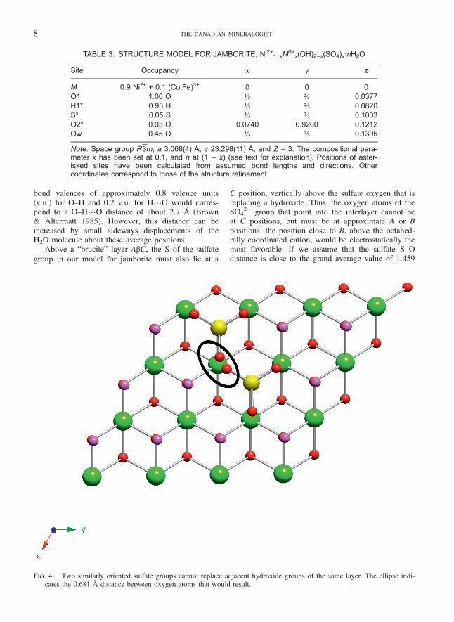
TABLE 3. STRUCTURE MODEL FOR JAMBORITE, Ni2+1−xM3+
x(OH)2−x(SO4)x·nH2O
Site Occupancy x y z
M 0.9 Ni2+ + 0.1 (Co,Fe)3+ 0 0 0O1 1.00 O ⅓ ⅔ 0.0377H1* 0.95 H ⅓ ⅔ 0.0820S* 0.05 S ⅓ ⅔ 0.1003O2* 0.05 O 0.0740 0.9260 0.1212Ow 0.45 O ⅓ ⅔ 0.1395
Note: Space group R3̄m, a 3.068(4) Å, c 23.298(11) Å, and Z = 3. The compositional para-meter x has been set at 0.1, and n at (1 − x) (see text for explanation). Positions of aster-isked sites have been calculated from assumed bond lengths and directions. Othercoordinates correspond to those of the structure refinement
FIG. 4. Two similarly oriented sulfate groups cannot replace adjacent hydroxide groups of the same layer. The ellipse indi-cates the 0.681 Å distance between oxygen atoms that would result.
8 THE CANADIAN MINERALOGIST

canmin.0.0.1400050 18-08-15 9:9
FIG. 5. If the upper sulfate group is present, then the sulfate tetrahedron immediately below it cannot be occupied owing tounrealistically short O…O distances. However, other sulfate positions in the lower layer are sufficiently far away to beoccupied.
STATUS OF JAMBORITE 9

canmin.0.0.1400050 18-08-15 9:9
Å (Hawthorne et al. 2000), we can estimate coordi-nates for the sulfur and the rest of the associated oxy-gen atoms. Table 3 shows a partially complete modelof the average structure, including some atoms thatwere not detected in the structure refinement owingto the low occupancy of their sites. A position hasbeen estimated for the hydroxide hydrogen atom,based on an O–H distance of 1.03 Å (0.8 v.u.), butnot for the hydrogen atoms of H2O.
In order to avoid impossibly small distancesbetween oxygen atoms of two different sulfate tetra-hedra, a sulfate group cannot have a second sulfatetetrahedron replacing any of the six immediatelyneighboring OH− groups of the same layer, if the sul-fate tetrahedra are oriented similarly (Fig. 4). Also,there is insufficient space for two sulfate groups toface each other across the interlayer (Fig. 5). Thesesteric factors limit the maximum value of sulfate
FIG. 6. A sulfate group attached to the upper “brucite” layer implies that immediately neighboring H2O molecules must beat the lower position, and vice versa.
10 THE CANADIAN MINERALOGIST

canmin.0.0.1400050 18-08-15 9:9
content x to ⅓. However, it will be seen below thatconsideration of sulfate–water interactions mayimpose even tighter constraints upon the maximumvalue of x.
Although the XRD results indicate that there is nolong-range order of sulfate and H2O positions in jam-borite, steric considerations imply considerable coup-ling between their local occupancy patterns. It isobvious that if S replaces H1 above a given O1 atom,then there is no room for H2O at either of the splitOw positions at the same x and y coordinates in thesame interlayer. The distances from O2 of a sulfategroup to the neighboring Ow sites are also shortenough that there is coupling between sulfate andH2O occupancies (Fig. 6). If a sulfate group isattached to the upper “brucite” layer, then H2O mole-cules at the “upper” position would be at only 2.042Å from the nearest sulfate oxygen atom, whereas thecorresponding “lower” positions are at 2.618 Å.Hence, an “upper” sulfate must be surrounded byH2O molecules at the “lower” position, and viceversa.
Thus, the double interlayer of H2O apparent fromthe structure refinement occurs because a sulfategroup attaches randomly either to the “brucite” layerbelow or to the one above, and drives the neighboringinterlayer H2O molecules to adopt either of two dif-ferent z coordinates. The maximum number of H2Oper formula unit Ni2+1−xM
3+x(OH)2−x(SO4)x·nH2O is
thus n = (1 − x), but as this number is spread over asite with four times the multiplicity of the M site, themaximum occupancy of the Ow site is only (0.5 –x/2) (Table 3). The mean composition of jamborite inthis study is x = 0.1, such that the maximum Owoccupancy in the structure model is 0.45 (Table 3).This value is in almost perfect agreement with theoccupancy of 0.44 obtained from the structure refine-ment, which both supports the correctness of themodel and implies that jamborite is indeed fullyhydrated.
It was noted above that in order to ensure anadequate distance between sulfate groups, x cannotexceed ⅓. However, for this composition, every H2Omolecule in an interlayer is adjacent to three SO4
FIG. 7. The densest possible occupancy of sulfate groups attached to one side of a given “brucite” layer, with one-third ofthe hydroxide groups replaced. This density of sulfate occupancy can only be achieved if all sulfate tetrahedra are at the‘lower’ position and all H2O (pink) are at ‘upper’ positions, or vice versa.
STATUS OF JAMBORITE 11

canmin.0.0.1400050 18-08-15 9:9
groups (Fig. 7). Because H2O must occupy the‘upper’ position if any adjacent SO4 group is at its‘lower’ position and vice versa, the only way thatsuch a high value of x can be accommodated is if allSO4 groups in a given layer are at the same height,as are all H2O molecules. Thus, there would have tobe complete two-dimensional upper-lower orderwithin a single interlayer, and equal occupancy of thetwo H2O positions in the average structure can thenonly occur through lack of correlation between suc-cessive interlayers. In order to avoid any couplingbetween the heights of neighboring sulfate groups,each H2O molecule must be adjacent to at most onesulfate group. Hence, there must be at least two H2Omolecules between each pair of sulfate groups, whichis only possible up to a maximum x = 1/7 (Fig. 8).The up or down orientations of individual sulfate tet-rahedra are now quite independent of one another,
although each sulfate group determines the positionof the sextet of H2O molecules that surround it. Theeven lower value x of 0.1 that is observed for jambor-ite suggests that sulfate heights are disordered in thisfashion, such that the sextets of H2O surroundingthem occur at both heights equally; H2O groups alsoexist that are not adjacent to any sulfate groups, andthese can occupy either height randomly.
DEFINITION OF JAMBORITE
In our crystal structure refinement, the feature thatdistinguishes jamborite from all related mineralsdescribed to date is the double layer of partially occu-pied H2O sites. We show above that the splitting ofthe H2O layer can be ascribed to avoidance of SO4
groups which bond directly to octahedral cationseither above or below, unlike the interlayer anions of
FIG. 8. “Brucite” layer of jamborite, showing the densest possible packing of sulfate tetrahedra in the overlying interlayerfor which there is no short-range coupling between up/down orientations of sulfate tetrahedra, corresponding to the com-position x = 1/7. Sulfate tetrahedra point downward and bond to the underlying “brucite” layer, or point upwards andbond to the “brucite” layer above, with equal probability. Downward-pointing tetrahedra are surrounded by 6 H2O at the‘upper’ position (saturated pink color), whereas upward-pointing tetrahedra are surrounded by 6 H2O at the ‘lower’ posi-tion (pinkish-grey color).
12 THE CANADIAN MINERALOGIST

canmin.0.0.1400050 18-08-15 9:9
the hydrotalcite supergroup. The occupancy of theSO4 groups themselves is so low in the average struc-ture that they cannot be located in the refinement, butthe S content is readily analyzed by EDS, and Ramanspectroscopy confirms its speciation. Replacement ofOH− by SO4
2− is charge-balanced by substitution ofa trivalent cation for Ni2+, and Co3+ predominates byfar as the trivalent species in the sample studied.Thus, we propose that the name “jamborite” appliesto the species with the structure type refined here andthe composition M2+
1−xM3+
x(OH)2−x(SO4)x·nH2O,where M2+ is predominantly Ni and M3+ is predomi-nantly Co. Compositions where trivalent cations otherthan Co, or anions other than sulfate, are dominantwould represent different species. In particular, it iseasy to envisage the possible occurrence of an Fe3+
-dominant analogue. However, the fact that we havenot detected such a phase, when Ni2+−Fe3+−SO4 mem-bers of the hydrotalcite supergroup are well known(honessite and hydrohonessite), suggests that the Fe3+
analogue is of limited stability at best.
OCCURRENCE OF JAMBORITE, HONESSITE, AND
HYDROHONESSITE
Jamborite has been reported from about twentylocations (cf. www.mindat.org), mostly replacing mill-erite, as described in the original article by Morandi& Dalrio (1973). We doubt that all of these occur-rences are actually jamborite; many may be of visu-ally similar green secondary minerals. Specimensfrom Hoopeston, Illinois and Halls Gap, Kentucky,USA, were analyzed at Museum Victoria and wereidentified as hydrohonessite rather than jamborite orhonessite. However, as noted in the introduction, wereconfirmed by powder XRD, EDS, and Raman spec-troscopy that the specimens from Ca’ de’ Ladri,Monteacuto Ragazza, and Castelluccio di Moschedado contain jamborite, although much material of sim-ilar appearance and composition from these localitiesas well as Rio Vesale is amorphous to X-rays.Honessite similarly appears to be rare, with specimensconverting readily to hydrohonessite, although hones-site from the type locality (Lindon, Wisconsin, USA)has remained stable and has not converted to hydro-honessite. Nickel & Wildman (1981) and Bish &Livingstone (1981) noted that honessite and hydroho-nessite interconvert readily, depending on temperatureand humidity. In nature, jamborite occurs rarely as afirst stage in the alteration of millerite to hydrotalcite-supergroup minerals. This is consistent with its lessoxidized composition [M3+/(M2++M3+) = 0.10 for thejamborite of this study, but 0.25 for honessite orhydrohonessite]. Our data, and the lack of observa-tion of an Fe3+ analogue to date, suggest that whetherit forms or not depends on the availability of cobalt.As alteration proceeds, the progressively more
hydrated phases honessite and then hydrohonessitereplace it.
CONCLUSIONS
1. The ideal formula for jamborite is now Ni2+1−xCo3+x(OH)2−x(SO4)x·nH2O rather than (Ni2+,Ni3+,Fe)(OH)2(OH,S,H2O).
2. Structural investigation shows that jamboritelies outside the hydrotalcite supergroup asdefined by Mills et al. (2012).
3. Jamborite is no longer a “questionable species”.
ACKNOWLEDGEMENTS
We thank Paul Pohwat at the SmithsonianInstitution and Peter Downes at the WesternAustralian Museum for loaning portions of the typespecimens of honessite and hydrohonessite, respect-ively. The manuscript took advantage of reviews byUwe Kolitsch, Robert Martin, and an anonymousreviewer. The research was supported by “Progettod’Ateneo 2012, University of Firenze” to LB and byproject MIUR 101742-2013, co-financed by AMI“Characterization of new and/or rare mineral species”to MEC and EB.
REFERENCES
BISH, D.L. & LIVINGSTONE, A. (1981) The crystal chemistryand paragenesis of honessite and hydrohonessite: thesulphate analogues of reevesite. Mineralogical Magazine44, 339–343.
BROWN, I.D. & ALTERMATT, D. (1985) Bond-valence para-meters obtained from a systematic analysis of the InorganicCrystal Structure Database. Acta Crystallographica B41,244–247.
GÉNIN, J.-M.R., MILLS, S.J., CHRISTY, A.G., GUÉRIN, O.,HERBILLON, A.J., KUZMANN, E., ONA-NGUEMA, G., RUBY,C., & UPADHYAY, C. (2014) Mössbauerite, Fe3+6O4(OH)8[CO3]·3H2O, the fully oxidized ‘green rust’ mineralfrom Mont Saint-Michel Bay, France. MineralogicalMagazine 78, 447–465.
HAWTHORNE, F.C., KIMATA, M., & EBY, R.K. (1993) Thecrystal structure of spangolite, a complex copper sulfatesheet mineral. American Mineralogist 78, 649–652.
HAWTHORNE, F.C., KRIVOVICHEV, S.V., & BURNS, P.C. (2000)The crystal chemical of sulfate minerals. In Sulfate Minerals— Crystallography, Geochemistry, and EnvironmentalSignificance (C.N. Alpers, J.L. Jambor, & D.K. Nordstrom,eds.). Reviews in Mineralogy 40, 1–112.
HERRENDORF, W. (1993) Habitus. Ph.D. dissertation,University of Karlsruhe, Germany.
STATUS OF JAMBORITE 13

canmin.0.0.1400050 18-08-15 9:9
IBERS, J.A. & HAMILTON, W.C. (EDS.) (1974) InternationalTables for X-ray Crystallography, Volume IV. Kynock,Dordrecht, Netherlands, 366p.
MILLS, S.J., CHRISTY, A.G., GÉNIN, J.-M.R., KAMEDA, T., &COLOMBO, F. (2012) Nomenclature of the hydrotalcitesupergroup: natural layered double hydroxides.Mineralogical Magazine 76, 1289–1336.
MORANDI, N. & DALRIO, G. (1973) Jamborite: a new nickelhydroxide mineral from the northern Apennines, Italy.American Mineralogist 58, 835–839.
NICKEL, E.H. & WILDMAN, J.E. (1981) Hydrohonessite – anew hydrated Ni-Fe hydroxysulphate mineral; its relation-ship to honessite, carrboydite and minerals of the pyr-oaurite group. Mineralogical Magazine 44, 333–337.
OXFORD DIFFRACTION (2006) CrysAlis RED (Version1.171.31.2) and ABSPACK in CrysAlis RED. OxfordDiffraction Ltd., Abingdon, Oxfordshire, England.
SHELDRICK, G.M. (2008) A short history of SHELX. ActaCrystallographica A64, 112–122.
STOE & CIE (1996) X-shape (version 1.02). Stoe & Cie,Darmstadt, Germany.
WOJDYR, M. (2010) Fityk: a general-purpose peak fittingprogram. Journal of Applied Crystallography 43,1126–1128.
Received June 13, 2014, Revised manuscript acceptedNovember 23, 2014.
14 THE CANADIAN MINERALOGIST


