ORIGINAL ARTICLE Role of acid sphingomyelinase and IL-6 as ...
1521-0103/368/3/338–352$35.00 https://doi.org/10.1124/jpet.118.253955THE JOURNAL OF PHARMACOLOGY AND EXPERIMENTAL THERAPEUTICS J Pharmacol Exp Ther 368:338–352, March 2019Copyright ª 2019 by The American Society for Pharmacology and Experimental Therapeutics
Neutral Sphingomyelinase Inhibition Alleviates LPS-InducedMicroglia Activation and Neuroinflammation after ExperimentalTraumatic Brain Injury
Asit Kumar,1 Rebecca J. Henry, Bogdan A. Stoica, David J. Loane, Gelareh Abulwerdi,Shahnawaz A. Bhat, and Alan I. FadenDepartment of Anesthesiology and Shock, Trauma and Anesthesiology Research (STAR) Center, University of Maryland Schoolof Medicine, Baltimore, Maryland
Received September 26, 2018; accepted December 14, 2018
ABSTRACTNeuroinflammation is one of the key secondary injury mech-anisms triggered by traumatic brain injury (TBI). Microglialactivation, a hallmark of brain neuroinflammation, plays acritical role in regulating immune responses after TBI andcontributes to progressive neurodegeneration and neurologicdeficits following brain trauma. Here we evaluated the role ofneutral sphingomyelinase (nSMase) in microglial activation byexamining the effects of the nSMase inhibitors altenusin andGW4869 in vitro (using BV2 microglia cells and primarymicroglia), as well as in a controlled cortical injury (CCI) modelin adult male C57BL/6 mice. Pretreatment of altenusin orGW4869 prior to lipopolysaccharide (LPS) stimulation for 4 or24 hours, significantly downregulated gene expression of the
pro-inflammatory mediators TNF-a, IL-1b, IL-6, iNOS, andCCL2 in microglia and reduced the release of nitric oxide andTNF-a. These nSMase inhibitors also attenuated the release ofmicroparticles and phosphorylation of p38 MAPK and ERK1/2.In addition, altenusin pretreatment also reduced the geneexpression of multiple inflammatory markers associated withmicroglial activation after experimental TBI, including TNF-a,IL-1b, IL-6, iNOS, CCL2, CD68, NOX2, and p22phox. Overall,our data demonstrate that nSMase inhibitors attenuate mul-tiple inflammatory pathways associated with microglial acti-vation in vitro and after experimental TBI. Thus, nSMaseinhibitors may represent promising therapeutics agents tar-geting neuroinflammation.
IntroductionRecent clinical and experimental studies show that trau-
matic brain injury (TBI) initiates a sustained neuroinflamma-tory response with microglial activation that contributes toposttraumatic neurodegeneration and chronic neurologic deficits(Loane and Byrnes, 2010; Loane et al., 2014a). Such neurotoxicmicroglial activation is associated with both phenotypic andgenotypic changes (Loane and Kumar, 2016). The latter includeactivation of inflammatory cytokines, chemokines, and micro-RNAs (miRs) (Perry et al., 2010; Prinz and Priller, 2014).Activated microglia also release extracellular vesicles (EVs),
including microparticles (MP; also called microvesicles) andexosomes containing pro-inflammatory molecules, which canfurther enhance and potentially propagate the inflammatoryresponse after TBI (Hazelton et al., 2018; Paolicelli et al.,2018). Several in vitro and in vivo models, including lipopoly-saccharide (LPS)-treated primary microglia, cell lines (e.g.,BV2 microglia), and LPS-challenged mice have been used tomimic such neurotoxic microglial activation (Loane et al.,2009; Mao et al., 2012; Fenn et al., 2014; Yuan et al., 2015;Hung et al., 2017). The LPS model stimulates microglialactivation pro-inflammatory phenotypic changes, and it hasalso been used in EVs studies whereby MP released fromactivated microglia are enriched in pro-inflammatory media-tors such as interleukin-1b (IL-1b) (Kumar et al., 2017; Yanget al., 2018). Experimental neuroprotection strategies forTBI have included modulation of upstream activation mecha-nisms using NADPH-oxidase (NOX2) inhibitors or mGluR5agonists (Loane et al., 2013; Kumar et al., 2016a,b), as well asstrategies serving to inhibit the release of specific inflammatory
This work was supported by the National Institutes of Health [GrantNumber R01NS037313 (A.I.F.), R01NS082308 (D.J.L.), and R01NS096002(B.A.S.)].
1Current affiliation: Department of Neurology, Richard T Johnson Divisionof Neuroimmunology and Neurological Infections, Johns Hopkins UniversitySchool of Medicine, Baltimore, Maryland.
https://doi.org/10.1124/jpet.118.253955.
ABBREVIATIONS: AD, Alzheimer’s disease; Arg-1, arginase 1; CCI, controlled cortical injury; CCL2, C-C motif chemokine ligand 2; D68, cluster ofdifferentiation 68; ERK, extracellular signal-regulated kinase; EV, extracellular vesicles; IkBa, nuclear factor of kappa light polypeptide geneenhancer in B-cells inhibitor, alpha; IL-1b, interleukin-1b; IL-6, interleukin 6; IL-4ra, IL-4 receptor subunit alpha; JNK, c-Jun N-terminal kinase; LPS,lipopolysaccharide; MAPK, mitogen-activated protein kinase; miR, microRNA; MP, microparticles; Mrc1/CD206, mannose receptor C-type 1/clusterof differentiation 206; MTT, tetrazolium salt 3-(4,5-dimethylthiazol-2-yl)-2,5-diphenyltetrazolium bromide; NF-kB, nuclear factor kappa-light-chain-enhancer of activated B cells; Nos2/iNOS, nitric oxide synthase 2/inducible nitric oxide synthase; NOX2, NADPH oxidase 2; nSMase, neutralsphingomyelinase; TBI, traumatic brain injury; TBS-T, Tris-buffered saline containing 0.1% Tween 20; TNF-a, tumor necrosis factor-alpha;Ym1/Chil3, Ym1/chitinase-like 3.
338
at ASPE
T Journals on June 26, 2020
jpet.aspetjournals.orgD
ownloaded from
factors such as pro-inflammatory cytokines (Kumar et al.,2016b). We recently showed in a murine experimental TBImodel that neutralization of EVs using a novel surfactantpolyethylene glycol telomere B can serve to reduce the releaseof pro-inflammatory factors (Kumar et al., 2017); this treat-ment was also found to be neuroprotective in a porcine modelof TBI (Bohman et al., 2016).A number of studies have also begun to examine the effects
of nSMase inhibitors, such as altenusin andGW4869, on brainimmune cell function and as potential anti-inflammatoryagents (Iguchi et al., 2016; Chua et al., 2017; Dickens et al.,2017; Huang et al., 2018; Sobue et al., 2018). The nSMases area family of sphingomyelin phosphodiesterase enzymes thatcatalyze hydrolysis of sphingomyelin, a major component ofeukaryotic cell membrane, to produce phosphocholine andceramide (Wu et al., 2010; Figuera-Losada et al., 2015).Regulation of innate immune responses mediated throughnSMase inhibition might provide beneficial effects for severalneurodegenerative diseases (Guo et al., 2015; Dinkins et al.,2016; Bilousova et al., 2018). Prior studies demonstrate thatnSMase inhibitors can limit neuroinflammation and neuronalcell death (Brann et al., 2002). Thus, such drugs can protectneurons, astrocytes, and oligodendrocytes against ceramide-induced cell death produced by pro-inflammatory molecules(tumor necrosis factor-a; TNF-a), b-amyloid, and cerebralischemia (Luberto et al., 2002; Lee et al., 2004; Zeng et al.,2005; Martinez et al., 2012; Gu et al., 2013). Recent stud-ies indicate that myeloid cells can release EVs/MP thatspread inflammatory signals both in vitro and in vivo to alterneuronal functions (Harrison-Brown et al., 2016; Nigro et al.,2016; Beneventano et al., 2017; Kumar et al., 2017). Impor-tantly, EV levels are significantly increased following TBI inhumans and animal models, as well as in chronic neurode-generative and autoimmune disorders such as Alzheimer’sdisease (AD) and multiple sclerosis, supporting the conceptthat spreading inflammation via EVs release may play a rolein these neuropathologies (Taylor and Gercel-Taylor, 2014;Xiao et al., 2017; Pieragostino et al., 2018). Thus, nSMaseinhibitors have emerged as new pharmacological agents forpreventing release of EVs and thus can modulate inflamma-tion and oxidative stress.In the present study, we investigated the therapeutic
potential of the nSMase inhibitors altenusin and GW4869in BV2 murine microglial cell line and primary microgliain vitro, as well as in the mice subjected to controlled corticalimpact (CCI), a well-characterized experimental TBI model.More specifically, we examined their ability to modulateLPS-induced microglial activation, as well as release of pro-inflammatory molecules and EVs in vitro, and on markersof secondary neuroinflammation following TBI in mice. Inaddition, we evaluated the potential underlying signaltransduction mechanisms involved.
Materials and MethodsAnimals. Studies were performed using 10- to 12-week-old
C57BL/6 male mice (22–26 g wt; Taconic Biosciences, Germantown,NY).Micewere housed in theAnimalCare Facility at theUniversity ofMaryland School of Medicine under standard laboratory conditions,maintained on 12-hour light/dark cyclewith temperature of 24°C,withad libitum access to food and water. Mice were acclimatized in theanimal surgery room half an hour before the surgery. All surgical
procedures were performed in accordance with protocols approved bythe Institutional Animal Care and Use Committee (IACUC) at theUniversity of Maryland School of Medicine.
Controlled Cortical Impact. Brain injury in mice was inducedusing our custom-designed CCI injury device, consisting of amicroprocessor-controlled pneumatic impactor with a 3.5 mm di-ameter tip as described previously (Kabadi et al., 2012). Briefly,mice were anesthetized in gas chamber containing evaporatedmixture of gases 70%N2Oand 30%O2 alongwith isoflurane (inductionat 4% and maintenance at 2%) and directed through a nose mask.Depth of anesthesia was assessed, before initiation of the surgicalprocedure, by monitoring respiration rate and pedal withdrawalreflexes. Animals were placed on a heated pad to maintain a bodytemperature of 37°C under the anesthesia. The headwasmounted in astereotaxic frame and a 10-mm midline incision was made over theskull. After reflecting the skin and fascia, a 5-mm craniotomy wasmade on the central aspect of the left parietal bone. The impounder tipof the injury device was positioned to the surface of the exposed duraafter extending to its full stroke distance (44 mm) and reset to impactthe cortical surface. Moderate injury was induced using an impactorvelocity of 6 m/s and deformation depth of 2 mm. After performing theCCI injury, the incision was closed with interrupted 6-0 silk sutures.After anesthesiawithdrawal, the animalwas placed into a heated cagefor at least 45 min postinjury to maintain the normal body temper-ature. All animals were monitored carefully for at least 4 hours aftersurgery. Sham animals underwent the same procedure as injuredmice except for craniotomy and cortical impact.
In Vivo Drug Treatments. At 30 minutes postinjury injured,mice received a single intraperitoneal injection of altenusin (MilliporeSigma, St. Louis, MO) at 2 or 10 mg/kg in saline1 dimethyl sulfoxide(DMSO;Millipore Sigma) or equal volume of vehicle (saline1DMSO).Drug dosages were partly based upon prior in vivo studies foraltenusin in mice (Chua et al., 2017). At 24 hour postinjury, micewere humanely euthanized and brain tissues were rapidly dissectedand stored at 280°C following transcardial perfusion with ice-coldsterile phosphate-buffered saline (PBS).
BV2 Cell Culture. BV2 murine microglial cells were cultured inDulbecco’s modified Eagle’s medium (DMEM; Life Technologies,Carlsbad, CA) supplemented with 10% fetal calf serum (Life Technol-ogies) and 1% penicillin and streptomycin (40 U/ml and 40 mg/ml,respectively; Millipore Sigma) at 37°Cwith 5%CO2. Cells were split 1:5 until their confluency using 0.05% Trypsin-EDTA (Life Technolo-gies) solution in phosphate buffered saline (PBS). For activationof BV-2 microglia, lipopolysaccharide (LPS from Escherichia coli;20 ng/ml; Millipore Sigma) was used. BV2 microglia were seeded at adensity of either 3 � 105 cells/well (6-well plate), 1.5 � 105 cells/well(12-well plate), 0.75� 105 cells/well (24-well plate), or 20,000 cells/well(96-well plate). Cells were incubated with or without various concen-tration of altenusin (10–100 mM) or GW4869 (5–10 mM; MilliporeSigma) followed by LPS stimulation (20 ng/ml) for either 4 or 24 hours.
Primary Microglia Cultures. Primary microglia cultures wereprepared from cerebral cortices of postnatal day P1–3 Sprague-Dawley rats as previously described (Olajide et al., 2014; Singhet al., 2014). In brief, brainswere carefully taken, and cerebral corticeswere collected and freed from meninges in Hanks’ balanced saltsolution (without calcium, magnesium, and phenol red; Life Technol-ogies). Forebrains were minced and gently dissociated by repeatedpipetting in DMEM/F-12 medium (Life Technologies) and filtered bypassing through 70-mm cell strainer (Millipore Sigma). Cells werecollected by centrifugation (1000 g, 10 minutes at 4°C) and resus-pended in DMEM/F-12 containing 10% fetal calf serum (Life Technol-ogies) and 1% penicillin and streptomycin (40 U/ml and 40 mg/ml,respectively) and cultured on Poly-D-lysine coated 75 cm2 cell cultureflask (Millipore Sigma) in 5%CO2 at 37°C. After 12–14 days in culture,floating microglia were harvested from mixed glia (astrocyte-microglia) cultures and reseeded into cell culture plates at the densityof either 0.75 � 105 cells/well (24-well plate) or 1.5 � 105 cells/well(12-well plate). On the next day, nonadherent cells were removed by
Neutral Sphingomyelinase Inhibition Alleviates Neuroinflammation 339
at ASPE
T Journals on June 26, 2020
jpet.aspetjournals.orgD
ownloaded from
changing the media, and after 1 hour cells were used for experiments.Cells were incubated with altenusin (10–50 mM) followed by LPSstimulation (20 ng/ml) for either 4 or 24 hours.
Cell Viability Assay. Cell viability was determined using atetrazolium salt 3-(4,5-dimethylthiazol-2-yl)-2,5-diphenyltetrazoliumbromide (MTT; Millipore Sigma) colorimetric assay (Mosmann, 1983).BV2 microglia cells were incubated in 96-well plates in DMEMcontaining 10% fetal calf serum and 1% penicillin and streptomycin(40 U/ml and 40 mg/ml, respectively) containing various concentra-tions of altenusin (1–100 mM) and GW4869 (1–20 mM) for 24 hours.Ten microliters of MTT at a final concentration of 0.5 mg/ml wereadded to each well. After 3-hour incubation in 5% CO2 at 37°C, mediawas discarded and formazan crystals were dissolved by adding100 ml of DMSO to each well. The absorbance was measured at540 nm using absorbance microplate reader (Biotek, Winooski, VT)and is directly proportional to cell viability. Cell viability wasexpressed as a percentage of surviving cells compared with thenonstimulated control cells.
mRNA Real-Time PCR. RNA was isolated using Direct-zolRNA mini prep kit (ZYMO Research, Irvine, CA). For cDNAsynthesis, total RNA was reverse transcribed using High-CapacitycDNA Reverse Transcription Kit (ThermoFisher Scientific,Waltham, MA) as per manufacturer’s instructions. In brief, each20 ml of reverse transcription (RT) reaction consisted of 2 ml of 10�RT buffer, 0.8 ml of 25� dNTP mix (100 mM), 2.0 ml of 10� RTrandom primers, 1.0 ml of MultiScribe reverse transcriptase, 4.2 mlof nuclease-free water, and 10 ml of RNA sample. Thermal cyclingconditions used for reverse transcription are 10 minutes at 25°C,120 minutes at 37°C, and finally 5 minutes at 85°C followed bystop at 4°C. The synthesized cDNA was the template for the real-time PCR amplification that was carried out either by ABI7900HT or Quant Studio 5 real-time PCR System (ThermoFisherScientific), using TaqMan Universal Master Mix II, no UNG(ThermoFisher Scientific) as per the company’s protocol. Reactionconditions were 10 minutes at 95°C, followed by 40 cycles of15 seconds at 95°C, 60 seconds at 60°C, followed by the final plateread. The following primers were used in this study: Mm00443258_m1TNF-a (mouse), Rn01525859_g1 TNF-a (rat), Mm00446190_m1 IL-6(mouse), Rn01410330_m1 IL-6 (rat), Mm01336189_m1 IL-1b (mouse),Rn00580432_m1 IL-1b (rat), Mm00440502_m1Nos2/iNOS (mouse),Rn00561646_m1 Nos2/iNOS (rat), Mm00441242_m1 CCL2(mouse), Rn00580555_m1 CCL2 (rat), Mm01288386_m1 IL-10(mouse), Mm99999915_g1, GAPDH (mouse), Rn01775763_g1GAPDH (rat), Mm03047343_m1 CD68 (mouse), Mm01287743_m1CYBB/NOX2 (mouse), Mm00514478_m1 CYBA/p22phox (mouse),Mm01275139_m1 IL4ra (mouse), Mm01329362_m1 Mrc1/CD206(mouse), Mm00475988_m1 Arg-1 (mouse), Mm00657889_mHYm1/Chil3 (mouse). GAPDH served as an internal control for samplenormalization and the comparative cycle threshold method (22DDCt)was used for data quantification as described previously (Livak andSchmittgen, 2001).
microRNA Real-Time PCR. Following RNA isolation usingDirect-zol RNA mini prep kit (ZYMO Research), miRNAs werereverse-transcribed by using TaqMan microRNA reverse transcrip-tion kit (ThermoFisher Scientific) according to manufacturer’s protocol.In brief, themastermix of 7ml contained 1.5ml of reverse transcriptionbuffer (10�), 0.15 ml of dNTPs (100 mM; with deoxythymidinetriphosphate), 1 ml of MultiScribe Reverse Transcriptase (50 U/ml),0.19 ml of RNase inhibitor (20 U/ml), and 4.16 ml nuclease-free water.Each 15 ml of RT reaction consisted of 7 ml of master mix, 3 ml ofRT primer (5�), and 5 ml of RNA sample (10 ng). Thermal cyclingconditions used for reverse transcription are 30 minutes at 16°C,30 minutes at 42°C, and finally 5 minutes at 85°C followed by stop at4°C. For real-time PCR amplification of mature miRNA, TaqManUniversal PCR master mix II (2�) with no UNG and TaqMan SmallRNA assays (20�) were used according to manufacturer’s protocol(mmu480953_mir miR-155, mmu478399_mir miR-146a, 001973 U6snRNA). In brief, the reaction setup for real-time PCR of 20 ml
contained 10 ml of TaqMan Universal PCR master mix II (2�), 1 ml ofTaqMan Small RNA assay (20�), 1.33 ml of template cDNA, and 7.67of ml nuclease-free water. The cycling condition for real-time PCRinvolved 40 cycles started with PCR initial enzyme activation step for10 minutes at 95°C and then 15 seconds at 95°C and 60 seconds at60°C. The real-time qPCR results were normalized to RNU6 (LifeTechnologies) and quantified using comparative Ct method 22DDCt
(Livak and Schmittgen, 2001). PCR reactions were carried out either byABI 7900HT or Quant Studio 5 real-time PCR System (ThermoFisherScientific).
Microparticle Analysis by Flow Cytometry. Microparticles(MP), a special class of extracellular vesicles released into BV2microglial media were characterized using a MACSQuant flowcytometer (Miltenyi Biotec, Auburn, CA) as described previously(Kumar et al., 2017). For MP analysis, fetal calf serum (LifeTechnologies) was subjected to 18 hours centrifugation at 100,000 g.DMEMmedia was filtered using 0.22-mm filter (Millipore). Cells werepretreated with altenusin and GW4869 for 30 minutes followed byLPS stimulation for 24 hours. To induce the MP shedding, cells wereconditioned with 100 mM of BzATP for 30 minutes. At the end of theexperiment, cell supernatants were used to collect total MP that werepurified by using ultracentrifugation at the speed of 300 g for10 minutes, 2100 g for 10 minutes followed by 100,000 g for 1 hourat 4°C as previously described (Poncelet et al., 2015; Tian et al., 2015;Kumar et al., 2017) with minor modification. To characterize the sizeof MP, MACSQuant was first calibrated with calibration beads(Miltenyi Biotec), and forward and side scatters were set at logarith-mic gain. Photomultiplier tube voltage and triggers were optimized todetect submicron-sized particles.Microbead standards of various sizes300 nm (Sigma, St. Louis, MO; LB3), 1090 (BCP-10-5; Spherotech),and 3000 nm (BP-30-5; Spherotech, Lake Forest, IL) were used to setthe initial parameters in the flow cytometer. MP were distinguishedfrom larger (apoptotic body; .1000 nm) and smaller (exosomes;,100 nm) vesicles based on size (SSC), and their phenotype wasconfirmed using the APC-conjugated Annexin V (Catalog No. 550474;BD Bioscience, San Jose, CA). All reagents and solutions used for MPanalysis were sterile and filtered (0.1 or 0.22 mm filter; Millipore)before use.
Western Blotting. To generate whole cell extracts, cells werewashed with cold PBS and lysed in the radioimmunoprecipitationassay buffer (RIPA buffer; Teknova, Hollister, CA), with 3% phospha-tase (Phosphatase Inhibitor Cocktail 2 and 3; Millipore Sigma) andprotease inhibitor cocktail (Millipore Sigma). Protein concentration ofthe samples was measured using the bicinchoninic acid protein assaykit (ThermoFisher Scientific) according to the manufacturer’s instruc-tions. For Western blotting, 15 mg of total protein from each samplewas subjected to 5%–20% gradient gels for sodium dodecyl sulfate-polyacrylamide gel electrophoresis. Proteins were transferred ontonitrocellulose membranes and blocked in 5% BSA in Tris-bufferedsaline containing 0.1% Tween 20 (TBS-T). After blocking, membraneswere incubated with primary antibodies, including phospho-p38MAPK (1:1000; Cell Signaling Technology, Danvers, MA), phospho-ERK1/2 (1:1000; Cell SignalingTechnology), phospho-JNK (1:1000; CellSignaling Technology), anti-IkB-a (1:1000; Cell Signaling Technology),and rabbit anti-actin (1:5000; Cell Signaling Technology). Primaryantibodies were diluted in TBS-T and 5% BSA. Membranes wereincubated with the primary antibody overnight at 4°C followed byincubation inappropriate horseradish peroxidase-conjugated secondaryantibodies (Jackson Immuno Research Laboratories, West Grove, PA)for 1 hour at room temperature. After extensive washing (3 times for15minutes each in TBS-T), proteinswere visualized using Super SignalWest Dura Extended Duration Substrate (ThermoFisher Scientific).Chemiluminescence was captured using ChemiDoc TM XRS1 System(Bio-Rad, Hercules, CA). Densitometry analyses of the proteins wereperformed using ImageJ software (NIH, Bethesda, MD), and b-actinwasused to confirmequal sample loading andnormalization of thedata.
Nitrite Release. Nitrite levels in cell culture supernatant weremeasured using a Griess reagent kit (ThermoFisher Scientific)
340 Kumar et al.
at ASPE
T Journals on June 26, 2020
jpet.aspetjournals.orgD
ownloaded from

according to manufacturer’s instruction. Absorbance wasmeasured at548 nm using the Synergy HT multi-mode microplate reader (Biotek)and normalized with the reference sample. Amount of nitrite(expressed in micromoles) released by microglia cells in each samplewas calculated by plotting standard curve.
Determination of TNF-a Release from LPS-Activated Micro-glia. Levels of TNF-a released in BV2 and primary rat microglia cellculture supernatant weremeasured usingmouse and rat ELISA kit fromR&DSystems,Minneapolis,MN; andBDBiosciences, respectively, as permanufacturer’s instructions. Briefly, standards or samples were added toantibody-coated 96-well plates and incubated for 2 hours at RT. Afterwashing the plates with wash buffer, samples were incubated withdetection antibody for another 2 hours at RT. Plates were washed andincubated in horseradish peroxidase-conjugated streptavidin for 20 min-utes at RT in dark. Substrate solution was added and plate was left forincubation 30minutes in the dark. After the incubation, stop solutionwasadded and absorbance was measured at 450 nm using a Synergy HTMulti-Mode Microplate Reader (Biotek). A standard curve was used tocalculate the levels of TNF-a release and expressed as picograms ofcytokine/milliliters.
Statistical Analysis. Statistical analyses were performed usingGraphPad Prism software (GraphPad Software Inc., San Diego,CA). Values of all experiments are represented as mean 6 S.E.M.of at least three independent experiments. Values were comparedusing one-way analysis of variance with Tukey’s post hoc correc-tion (multiple comparisons). The level of significance was set at*P , 0.05, **P , 0.01, ***P , 0.001.
ResultsEffects of Altenusin and GW4869 on the Microglial
Cell Viability. To determine the cytotoxic potential of alte-nusin (Fig. 1A) and GW4869 (Fig. 1B), the viability of BV2microglia was evaluated using an MTT assay. Treatment withaltenusin (ranging from 1 to 100 mM) prior to LPS stimulation(20 ng/ml) did not adversely affect BV2microglia viabilitywhencompared with control or unstimulated cells (Fig. 1C). Simi-larly, pretreatment of GW4869 (ranging from 1 to 10 mM) did
not affect microglial cell viability (Fig. 1D); however, 20 mMGW4869 with (P , 0.05) and without LPS (P , 0.01)significantly decreased cell viability, indicating GW4869cytotoxicity .10 mM.Altenusin and GW4869 Attenuate LPS-Induced Acti-
vation of BV2 Microglia. To investigate whether altenusinand GW4869 exerts anti-inflammatory effects, BV2 microgliawere preincubated with altenusin and GW4869 for 30 minutesand then stimulated with or without LPS. We evaluated geneexpression levels of key pro-inflammatory cytokines (TNF-a,IL-1b, and IL-6), inducible form of nitric oxide synthase (iNOS),chemokine (CCL2), and the anti-inflammatory cytokine (IL-10).Based on time-course analysis (datanot shown),we identified thetimepoint formRNAanalysis to be 4hours post-LPS stimulationwhen pro-inflammatory mRNAs were robustly induced. LPSstimulation of BV2 microglia significantly increased the expres-sion of TNF-a (LPSvs. control,P, 0.001), IL-1b (LPSvs. control,P , 0.001), IL-6 (LPS vs. control, P , 0.001), iNOS (LPS vs.control,P, 0.001), andCCL2 (LPS vs. control,P, 0.001) (Fig. 2,A–E). Pretreatment of altenusin significantly reduced expressionof TNF-a (Fig. 2A) and IL-6 (Fig. 2B) at 75 (TNFa, P , 0.001;IL-6,P, 0.001) and 100 mM (TNF-a,P, 0.001; IL-6,P, 0.001)comparedwith LPS alone. Altenusin also significantly decreasedexpression of IL-1b (Fig. 2C), iNOS (Fig. 2D), andCCL2 (Fig. 2E),starting at 50 mM (LPS vs. LPS 1 altenusin; IL-1b, P , 0.001;iNOS, P , 0.001; CCL2, P , 0.01). Similarly, pretreatment ofGW4869 for 30 minutes significantly reduced gene expressionof TNF-a (5–10 mM, P , 0.001; Fig. 2G), IL-6 (5–10 mM,P, 0.001; Fig. 2H), IL-1b (5–10 mM, P, 0.001; Fig. 2I), iNOS(10mM, P, 0.05, Fig. 2J), CCL2 (5–10mM,P, 0.001; Fig. 2K)following LPS stimulation. Altenusin and GW4869 did notsignificantly increase IL-10 gene expression following LPSstimulation (Fig. 2, F and L).With regard to protein markers, peak expression of TNF-a
and nitric oxide production in BV2 and primary microglia isat 24 hours following LPS stimulation (Loane et al., 2014;
Fig. 1. Effects of altenusin (A) and GW4869 (B) on the viability of BV2microglia cells. (C and D) Cells were pretreated with altenusin (1–100 mM; C) andGW4869 (1–20 mM; D) for 30 minutes, afterward cells were stimulated with or without LPS (20 ng/ml) for the next 24 hours. At the end of incubationperiod, MTT assays were performed on the cells to assess the cell viability. All values are expressed as mean 6 S.E.M. for at least three independentexperiments. Data were analyzed using one-way analysis of variance (ANOVA) for multiple comparison with post hoc Tukey’s test. *P, 0.05, **P, 0.01compared with control cells.
Neutral Sphingomyelinase Inhibition Alleviates Neuroinflammation 341
at ASPE
T Journals on June 26, 2020
jpet.aspetjournals.orgD
ownloaded from

Stoica et al., 2014; Kumar et al., 2017). As predicted, weobserved a significant increase in the nitrite levels and TNF-aprotein in the supernatant of BV2 stimulated with LPS at24 hours compared with control treated cells (Fig. 3). Altenusinpretreatment significantly decreased nitrite production in a dose-dependent manner, starting at 25 mM (LPS vs. LPS1 altenusin;P , 0.001; Fig. 3A). Similarly, GW4869 pretreatment signifi-cantly reduced nitrite release following LPS stimulation, start-ing at 5 mM (LPS vs. LPS 1 GW4869; P , 0.001; Fig. 3B).Altenusin and GW4869 treatment did not significantlychange the nitrite levels when added to BV2 microglia in
the absence of LPS. Pretreatment of altenusin significantly de-creasedTNF-a protein levels followingLPSstimulation, startingat75 mM (LPS vs. LPS 1 altenusin; P , 0.001; Fig. 3C), whereasTNF-a levels were significantly decreased following GW4869treatment at 10 mM (LPS vs. LPS1GW4869; P, 0.05; Fig. 3D).Altenusin Attenuate LPS-Induced Activation of Pri-
mary Microglia. To confirm the data obtained from BV2microglial cells, we also tested the anti-inflammatory proper-ties of altenusin in primary rat microglia. In response to LPSstimulation, we observed a robust increase in themRNA levelsof TNF-a (LPS vs. control, P , 0.001), IL-6 (LPS vs. control,
Fig. 2. Effects of altenusin and GW4869 on the expression of pro-inflammatory mediators in BV2 microglia (A–L). Cells were pretreated with altenusin(10–100 mM) for 30 minutes, afterward cells were stimulated with or without LPS (20 ng/ml) for 4 hours for studying gene expression of TNF-a (A), IL-6(B), IL-1b (C), iNOS (D), CCL2 (E), IL-10 (F). Similarly, for analyzing effect of GW4869 on gene expression of inflammatory mediators, cells werepretreated with GW4869 (5–10 mM) for 30 minute followed by stimulation with or without LPS (20 ng/ml) for the next 4 hour for analyzing geneexpression of TNF-a (G), IL-6 (H), IL-1b (I), iNOS (J), CCL2 (K), IL-10 (L). Data are presented as percentage control of LPS. Statistical analyses werecarried out by using one-way ANOVA with post hoc Tukey’s test (multiple comparisons). Results are expressed as mean 6 S.E.M. of at least threeindependent experiments. *P , 0.05, **P , 0.01, ***P , 0.001 in comparison with LPS; ###P , 0.001 in comparison with control.
342 Kumar et al.
at ASPE
T Journals on June 26, 2020
jpet.aspetjournals.orgD
ownloaded from

P , 0.001), IL-1b (LPS vs. control, P , 0.01), iNOS (LPS vs.control,P, 0.001), and CCL2 (LPS vs. control, P, 0.01; Fig. 4).Pretreatment with altenusin resulted in a significant down-regulation of TNF-a (Fig. 4A), IL-6 (Fig. 4B), and iNOS (Fig. 4D)starting at 10 mM (TNFa, P , 0.001; IL-6, P , 0.001; iNOS,P , 0.001), whereas IL-1b (Fig. 4C) and CCL2 (Fig. 4E)expression were significantly decreased starting at 25 (IL-1b,P , 0.05; CCL2, P , 0.01) and 50 mM (IL-1b, P , 0.01; CCL2,P , 0.01) of altenusin. Next, we investigated the LPS-inducedrelease of nitrite and TNF-a in primary rat microglia (Fig. 5, Aand B). LPS stimulation resulted in a significant increase in therelease of nitrite (Fig. 5A) and TNF-a protein (Fig. 5B). Pre-treatment with altenusin significantly reduced nitrite levelsstarting at 10mM (LPS vs. LPS1 altenusin;P, 0.001; Fig. 5A),whereas levels of TNF-a protein was significantly reducedstarting at 25mM (LPS vs. LPS1 altenusin;P, 0.001; Fig. 5B).Altenusin and GW4869 Attenuate LPS-Induced Ele-
vation of Pro-inflammatory miR-155. We assessed changesin pro-inflammatory miR-155 and miR-146 levels in BV2microglia pretreated with altenusin and GW4869 and stim-ulated with LPS (Fig. 5, C and D). Altenusin or GW4869treatment prior to LPS stimulation significantly decreasedmiR-155 expression at 100 mM altenusin (P , 0.01 vs. LPS)and at 10 mM GW4869 (P , 0.01 vs. LPS) (Fig. 5C),respectively, but these inhibitors had no significant effectson LPS-induced miR-146 expression (Fig. 5D).Effects of Altenusin and GW4869 on the Microparti-
cle Release Induced by Combined Treatment with LPSand BzATP. Next, we investigated if altenusin and GW4869had any effect on theMP release in activatedmicroglia (Fig. 6).P2X7 triggers MP shedding in the microglia, which can beactivated by agonists such as ATP or BzATP (synthetic analog
of ATP) (Bianco et al., 2005, 2009). Therefore, BV2 microgliawere pretreated with altenusin and GW4869 for 30 minutesand then stimulated with or without LPS for 24 hours followedby an additional BzATP stimulation for 30 minutes to induceMP shedding (Kumar et al., 2017). MP were quantified byflow cytometry using annexin V, which binds the externalizedphosphatidylserine present on the MP surface. Apoptoticbodies (.1000 nm) and exosomes (,100 nm) were excludedby gating analysis, and MP ranging in size between 300 and1000 nm were analyzed (Fig. 6A). LPS/BzATP significantlyinduced the shedding of annexin V positiveMP compared withMP levels in control BV2 microglia (P , 0.001; Fig. 6B).Interestingly, we found that altenusin (50 and 100 mM) andGW4869 (5 and 10 mM) significantly decreased the micropar-ticle count (P , 0.001) in the activated BV2 microglia.Altenusin Inhibits the Pro-inflammatory Pathways
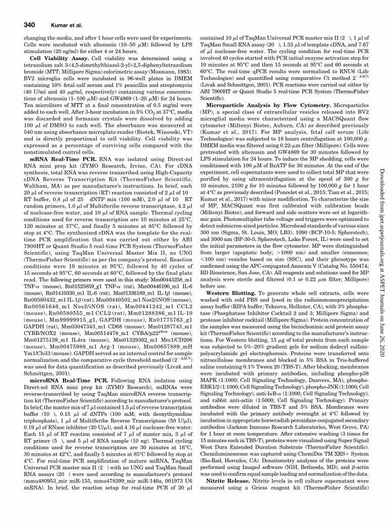
p38 MAPK and ERK1/2. Mitogen-activated protein kinase(MAPK) pathways are implicated in several cellular process-es, including neuroinflammation (Kaminska, 2005; Kaminskaet al., 2009). To further investigate the mechanism by whichaltenusin modulates cytokines production, we assessed alte-nusin effects on the activation of various components ofMAPKin LPS-activated BV2 microglia (Fig. 7A). LPS treatment ofmicroglia for 30 minutes led to the phosphorylation, andthus activation of p38 MAPK (P , 0.001), ERK1/2 (P , 0.05),and JNK (P , 0.001) (Fig. 7, B–D). Notably, pretreatment ofaltenusin at 100 mM significantly reduces phosphorylation ofp38 (P , 0.05 vs. LPS; Fig. 7B). Similarly, there was asignificant reduction in the phosphorylation of ERK1/2 byaltenusin (P , 0.05 vs. LPS; Fig. 7C). In contrast, altenusinfailed to reduce LPS-stimulated phosphorylation of JNK (Fig. 7D)or degradation of IkBa (Fig. 7E).
Fig. 3. Effects of altenusin and GW4869 on the release of nitrite (A and B) and TNF-a (C and D) in BV2microglia cells. For nitrite estimation, cells werepretreated with altenusin [10–100 mM (A)] and GW4869 [5–10 mM (B)]. For TNF-a ELISA, cells were pretreated with altenusin [50–100 mM (C)] andGW4869 [5–10 mM (D)]. Cells were pretreated for 30 minutes followed by stimulation with or without LPS (20 ng/ml) for the next 24 hours. At the end ofincubation, cell supernatants were collected, centrifuged, and levels of nitrite, and TNF-a was quantified by using Griess reagent assay andmouse specific ELISA assay, respectively. Statistical analyses were carried out by using one-way ANOVA with post hoc Tukey’s test (multiplecomparisons). Results are expressed as mean 6 S.E.M. of at least three independent experiments. *P , 0.05, **P , 0.01, ***P , 0.001 incomparison with LPS; ###P , 0.001 in comparison with control.
Neutral Sphingomyelinase Inhibition Alleviates Neuroinflammation 343
at ASPE
T Journals on June 26, 2020
jpet.aspetjournals.orgD
ownloaded from

Systemic Treatment of Altenusin Attenuates AcuteNeuroinflammation following Experimental TraumaticBrain Injury in Mice. We then investigated whether alte-nusin has similar anti-inflammatory properties in vivo. Weused a well-established model of CCI in adult male C57BL/6mice to induce a cortical focal contusion injury and examinedwhether the increases in gene expression of proinflammatorycytokines and chemokines were ameliorated by altenusintreatment. Two and tenmilligrams per kilogram of altenusinwas administered systemically (intraperitoneally) tomoderate-level CCI mice starting at 30 minutes postinjury (Fig. 8A).Sham-treated and CCI mice were euthanized 24 hours later,and cortical tissue was collected for mRNA analysis of TNFa,IL-6, IL-1b, iNOS, CCL2, CD68, NOX2, and p22phox. Follow-ing CCI, there was a robust increase in mRNA for pro-inflammatorymediators TNFa, IL-6, IL-1b, iNOS, and CCL2(P , 0.001 vs. sham; Fig. 8, B–F). Altenusin (10 mg/kg)treatment significantly reduced the expression levels ofTNF-a (P , 0.01; Fig. 8B), IL-6 (P , 0.05; Fig. 8C), IL-1b
(P , 0.05; Fig. 8D), iNOS (P , 0.05; Fig. 8E), and CCL2(P , 0.01; Fig. 8F) compared with vehicle-treated CCIgroup. We also evaluated the expression of CD68, a markerof activated and phagocytic microglia (Song et al., 2011; Fuet al., 2014; Walker and Lue, 2015). CD68 expression wassignificantly increased in the vehicle-treated CCI group(P , 0.05 vs. sham; Fig. 8G), and its expression wassignificantly reduced with altenusin (10 mg/kg) treatment(P , 0.05 vs. vehicle-treated CCI). NOX2 has been impli-cated in microglial-mediated neurotoxicity in TBI (Loaneet al., 2014; Kumar et al., 2016), so we analyzed the geneexpression of NOX2 and p22phox in the injured cortex. Therewas a significant increase in the expression of NOX2 andp22phoxmRNA expression with CCI (P, 0.001 vs. sham; Fig. 8,H and I), and altenusin treatment (10 mg/kg) significantlyreduced both subunit expression (P , 0.05 vs. vehicle-treatedCCI).Finally, to investigate whether altenusin has modulatory
effects on anti-inflammatory and proresolution immune
Fig. 4. Effects of altenusin on the expression of pro-inflammatory mediators in primary rat microglia (A–E). Cells were pretreated with altenusin (10–50mM) for 30 minutes; afterward cells were stimulated with LPS (20 ng/ml) for 4 hours for studying gene expression of TNF-a (A), IL-6 (B), IL-1b (C), iNOS(D), CCL2 (E). Data are presented as percentage control of LPS. Statistical analyses were carried out by using one-way ANOVA with post hoc Tukey’stest (multiple comparisons). Results are expressed as mean 6 S.E.M. of at least three independent experiments. *P, 0.05; **P , 0.01; ***P, 0.001 incomparison with LPS; ##P , 0.01; ###P , 0.001 in comparison with control.
344 Kumar et al.
at ASPE
T Journals on June 26, 2020
jpet.aspetjournals.orgD
ownloaded from

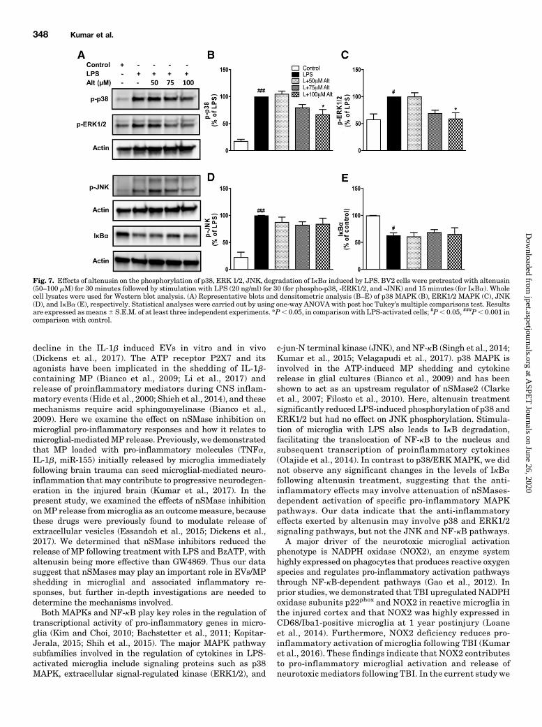
responses, we assessed anti-inflammatory and alternativeactivationmicroglialmarkers (IL-10, IL-4ra,Arg-1,Mrc1/CD206,and Ym1/Chil3). CCI induced significant increase in IL-10,IL-4ra, Arg-1 mRNA expression in the injured cortex (P ,0.05–0.001 vs. sham; Fig. 9, A, B, and D), as well as increasedMrc1/CD206 and Ym1/Chil3 expression that failed to reachstatistical significance (Fig. 9, C and E). Post-injury treatmentof altenusin at both doses (2 and 10 mg/kg) did not increaseexpression of these anti-inflammatory and proresolvinggenes in the injured cortex beyond vehicle-treated CCIlevels (Fig. 9).
DiscussionIn the present study, we demonstrate the anti-inflammatory
effects of nSMase inhibitors in activatedmicroglia in vitro andin an experimental TBImodel inmice. Altenusin andGW4869significantly suppressed neuroinflammation, reducing thegene expression of proinflammatory cytokines (TNFa, IL-1b,IL-6), chemokines (CCL2), and iNOS, as well as microgliaactivation markers (CD68) and components of NADPH oxi-dase (NOX2, p22phox). They also reduced the extracellularrelease of TNF-a and nitric oxide in activated BV2 cells andprimarymicroglia. These inflammatorymolecules are known tobe important mediators of microglial activation and neuro-inflammation associated with several neurologic conditions,
including TBI (Hanisch, 2002; Woodcock and Morganti-Kossmann, 2013; Thelin et al., 2017).GW4869 is a selective, noncompetitive nSMase2 inhibitor
that shows no inhibitory activity on acid sphingomyelinase(aSMase) (Luberto et al., 2002), whereas altenusin is a non-steroidal fungal metabolite with broader nSMase inhibitoractivity, known to target multiple isoforms of nSMase(Uchida et al., 1999; Dickens et al., 2017). In the currentstudy, we demonstrated that both nSMase inhibitors atten-uated LPS-induced microglial activation and the release ofpro-inflammatory mediators in vitro, although altenusinhad greater potency and less cytotoxicity than GW4869.Some prior studies have demonstrated therapeutic effectsof GW4869 at 20 mM in breast cancer cells, macrophages,and mesenchymal stem cells (Wu et al., 2005; Essandohet al., 2015; Xiao et al., 2018); however, in our studies 20 mMGW4869 significantly induced cytotoxicity, thereby nega-tively impacting microglial cell viability beginning atconcentration of .10 mM. The differences we identified inthe GW4869 cytotoxicity profile compared with the above-mentioned studies may be due to the different cell typesexamined (microglia vs. non-brain-derived cells). Collec-tively, our data suggest that a more general inhibition ofnSMase activity may improve effectiveness. A limitation ofpharmaceutical intervention approaches used to addresspotential mechanisms is that they may have off-target
Fig. 5. Effects of altenusin on the release of nitrite and TNF-a in primary rat microglia (A and B). For estimation of nitrite and TNF-a release, cells werepretreated with altenusin (10–50 mM) for 30 minutes followed by stimulation with LPS (20 ng/ml) for the next 24 hours. At the end of incubation, cellsupernatants were collected and centrifuged and levels of nitrite (A), and TNF-a (B) was quantified by using Griess reagent assay and rat specific ELISAassay, respectively. For miRNA expression of miR-155 (C) and miR-146 (D), cells were pretreated with altenusin (50–100 mM) and GW4869 (5–10 mM)for 30 minutes, followed by LPS stimulation for 4 hours. Altenusin (100 mM) and GW4869 (10 mM) significantly decreased the miR-155 (C), but not miR-146 (D), expression in LPS-stimulated BV2 cells. Statistical analyses were carried out by using one-way ANOVA with post hoc Tukey’s test (multiplecomparisons). Results are expressed as means 6 S.E. of at least three independent experiments. **P , 0.01; ***P , 0.001 in comparison with LPS;###P , 0.001 in comparison with control.
Neutral Sphingomyelinase Inhibition Alleviates Neuroinflammation 345
at ASPE
T Journals on June 26, 2020
jpet.aspetjournals.orgD
ownloaded from

Fig. 6. Microglia-derived MP are decreased in the BV2 cell culture supernatant following treatment of altenusin and GW4869. BV2 microglial cellswere pretreated with altenusin (50–100 mM) and GW4869 (5–10 mM) for 30 minutes followed by LPS (20 ng/ml) stimulation for 24 hours. Afterwardcells were incubated with BzATP (100 mM) for 30 minutes. (A) At the end of incubation, cell supernatants were collected, centrifuged, and release ofMPs were quantified by flow cytometry. Flow cytometry analysis of enriched MP isolated from the supernatant using sequential centrifugation at300 g for 10 minutes, 2100 g for 10 minutes followed by 100,000 g for 1 hour. Representation of gating strategy used to characterize MP/MV using
346 Kumar et al.
at ASPE
T Journals on June 26, 2020
jpet.aspetjournals.orgD
ownloaded from

activity. For example, altenusin has been shown to haveinhibitory effects on a number of kinases, including Srckinase (Aly et al., 2008), and it is an agonist of the FarnesoidX receptor, a member of the nuclear receptor subfamily(Zheng et al., 2017).Recent preclinical studies have implicated nSMases in neuro-
degenerative disorders and as regulators of neuroinflammation.A recent study in ananimalmodel of AD suggested that nSMase2may contribute to the progression of the disease and thatnSMase2 loss of function improves neurocognitive function(Dinkins et al., 2016) and limits pathologic changes. Pharmaco-logical inhibition of nSMases with altenusin also inhibits aggre-gation of tau protein in vitro (Chua et al., 2017). Gene expressionof mitochondrial-associated sphingolipid-metabolizing enzymeswas shown to increase significantly in the brain of TBI mice(Novgorodov et al., 2014). Another study reported the activa-tion of the nSMase2/ceramide pathway in astrocytes, but notneurons, in the hippocampus after cerebral ischemia in rats(Gu et al., 2013). nSMases appear to regulate leukocytemigration to the brain following IL-1b-induced brain injury,and their pharmacological inhibition by intrastriatal admin-istration of altenusin significantly reduced glial cell activationand cytokine expression (Dickens et al., 2017). Furthermore,GW4869 significantly downregulates the gene expression ofpro-inflammatory cytokines (TNFa, IL-1b, IL-6) in rat hippo-campus after cerebral ischemia (Gu et al., 2013). Notably,GW4869 suppressed the production of pro-inflammatorycytokines in LPS-stimulated RAW264.7 macrophages andin a sepsis-induced inflammatory model (Essandoh et al.,2015). Furthermore, mature dendritic cells, upon LPS stim-ulation, can produce pro-inflammatory cytokines, includingTNF-a contained exosomes that are attenuated by GW4869(Gao et al., 2016).In previously published studies, we demonstrated that
secondary neurodegeneration and chronic neurologic deficitsfollowing experimental TBI are driven by a pronounced pro-inflammatory and neurotoxic microglial activation that per-sists in injured cortex for days, weeks, or potentially months toyears following the initial traumatic event (Byrnes et al., 2012;Loane et al., 2014a,b; Kumar et al., 2016a,b). We also showedthat LPS stimulation of BV2 microglia and primary microgliarepresent excellent in vitro model that mimic the pro-inflammatory neuroinflammatory events initiated acutelyin our TBI model (Loane et al., 2009, 2013). By using an LPSmodel of microglial activation in vitro, we demonstrate thatboth altenusin and GW4869 treatment attenuate the geneexpression of TNFa, IL-1b, IL-6, iNOS, and CCL2. A previousstudy has shown that nSMase2 is activated by TNF-a (Clarkeet al., 2011). Thus, our data are consistent with othersindicating that nSMase2 activation is an important step forthe amplification (positive feedback) of the neuroinflamma-tory signaling program.We investigated the effects of nSMaseinhibition on the gene expression of anti-inflammatory cyto-kine IL-10 in activated microglia. We found that althoughtranscriptional levels of IL-10 increased somewhat followingaltenusin and GW4869 pretreatment, such changes did not
reach statistical significance. These preliminary results indicatethat nSMase inhibition produces robust anti-inflammatory re-sponse in microglia without upregulating inflammation-resolving programs driven by IL-10. Further investigationinto mechanisms by which nSMase inhibition regulates pro-and anti-inflammatory microglial activation phenotypes willbe required to identify signal transduction pathways leadingto beneficial inflammation-resolving responses in microglia.Accumulating evidence supports the involvement of
miRNAs as key regulators in the pathogenesis of variousneurologic conditions, including neuroinflammation (Guedeset al., 2013), neurodegeneration (Sabirzhanov et al., 2018),autoimmune diseases (Ponomarev et al., 2011), and centralnervous system injury (Sabirzhanov et al., 2014). miR-155 andmiR-146 are two well-characterized pro-inflammatory miRsthat are associated with microglial activation and regulatecytokine release, chemokine production, and oxidative stress(Cardoso et al., 2012; Saba et al., 2012, 2014; Guedes et al.,2014; Butovsky et al., 2015). Prior work has shown thatmiR-155 and miR-146 may be involved in modulation ofmicroglial-mediated neuroinflammation by inducing proin-flammatory cytokines in microglia (Jayadev et al., 2013; Yinet al., 2017) and neuronal cell death in various brain injurymodels (Caballero-Garrido et al., 2015; Harrison et al., 2016,2017; Pena-Philippides et al., 2016). It has been demonstratedthat miRNA-155 and -146a mediate cell-to-cell communica-tion through exosomes/microvesicles (MV) and participate intarget gene modulation in bone marrow-derived dendriticcells and cardiovascular disease (Montecalvo et al., 2012;Hulsmans and Holvoet, 2013). We recently showed that miR-155 is significantly upregulated in cortex after experimentalTBI and that central administration of miR-155 inhibitors(antagomirs) can reduce neuroinflammation and improveoutcomes (Henry et al., 2018). In the present study, upregu-lation of miR-155 and miR-146 expression were observed inLPS-activated BV2 microglial cells. Following treatment withaltenusin or GW4869, expression of miR-155 was substan-tially decreased, whereas no changes were observed inmiR-146 expression, providing evidence that nSMase2 actsupstream of the key pro-inflammatory miR-155.We and others previously showed that microglia can release
EVs that further activate the neighboring microglia andpropagate the neuroinflammatory responses in the injuredbrain (Brites and Fernandes, 2015; Kumar et al., 2017;Paolicelli et al., 2018; Yang et al., 2018). The nSMase pathwayis known to be involved in the biogenesis of EVs (Menck et al.,2017). Studies have shown that LPS-induced systemic in-flammation causes the choroid plexus to secrete exosome andpropagate a pro‐inflammatory response in the brain and thatthese processes can be attenuated by GW4869 (Balusu et al.,2016). LPS also enhances exosome release from pulmonaryartery smooth muscle cells, promoting cell proliferation andapoptosis resistance and contributing to the pathogenesis ofpulmonary hypertension; GW4869 may attenuate pulmonaryhypertension by inhibiting the exosome release (Zhao et al.,2017). Inhibition of nSMase with altenusin resulted in sharp
SSC-H and standard microbeads (300–1000 nm diameter). Standard microbeads were used as an internal control to determine the size of MP in thesupernatant, and annexin V staining confirmed MP characteristics. (B) Flow cytometric quantification. Bars represent mean 6 S.E.M. Datarepresent results of at least three independent experiments. ***P, 0.001 in comparison with LPS and BzATP combined; ###P, 0.001 in comparisonwith control.
Neutral Sphingomyelinase Inhibition Alleviates Neuroinflammation 347
at ASPE
T Journals on June 26, 2020
jpet.aspetjournals.orgD
ownloaded from
decline in the IL-1b induced EVs in vitro and in vivo(Dickens et al., 2017). The ATP receptor P2X7 and itsagonists have been implicated in the shedding of IL-1b-containing MP (Bianco et al., 2009; Li et al., 2017) andrelease of proinflammatory mediators during CNS inflam-matory events (Hide et al., 2000; Shieh et al., 2014), and thesemechanisms require acid sphingomyelinase (Bianco et al.,2009). Here we examine the effect on nSMase inhibition onmicroglial pro-inflammatory responses and how it relates tomicroglial-mediatedMP release. Previously, we demonstratedthat MP loaded with pro-inflammatory molecules (TNFa,IL-1b, miR-155) initially released by microglia immediatelyfollowing brain trauma can seed microglial-mediated neuro-inflammation that may contribute to progressive neurodegen-eration in the injured brain (Kumar et al., 2017). In thepresent study, we examined the effects of nSMase inhibitiononMP release frommicroglia as an outcomemeasure, becausethese drugs were previously found to modulate release ofextracellular vesicles (Essandoh et al., 2015; Dickens et al.,2017). We determined that nSMase inhibitors reduced therelease of MP following treatment with LPS and BzATP, withaltenusin being more effective than GW4869. Thus our datasuggest that nSMases may play an important role in EVs/MPshedding in microglial and associated inflammatory re-sponses, but further in-depth investigations are needed todetermine the mechanisms involved.Both MAPKs and NF-kB play key roles in the regulation of
transcriptional activity of pro-inflammatory genes in micro-glia (Kim and Choi, 2010; Bachstetter et al., 2011; Kopitar-Jerala, 2015; Shih et al., 2015). The major MAPK pathwaysubfamilies involved in the regulation of cytokines in LPS-activated microglia include signaling proteins such as p38MAPK, extracellular signal-regulated kinase (ERK1/2), and
c-jun-N terminal kinase (JNK), and NF-kB (Singh et al., 2014;Kumar et al., 2015; Velagapudi et al., 2017). p38 MAPK isinvolved in the ATP-induced MP shedding and cytokinerelease in glial cultures (Bianco et al., 2009) and has beenshown to act as an upstream regulator of nSMase2 (Clarkeet al., 2007; Filosto et al., 2010). Here, altenusin treatmentsignificantly reduced LPS-induced phosphorylation of p38 andERK1/2 but had no effect on JNK phosphorylation. Stimula-tion of microglia with LPS also leads to IkB degradation,facilitating the translocation of NF-kB to the nucleus andsubsequent transcription of proinflammatory cytokines(Olajide et al., 2014). In contrast to p38/ERK MAPK, we didnot observe any significant changes in the levels of IkBafollowing altenusin treatment, suggesting that the anti-inflammatory effects may involve attenuation of nSMases-dependent activation of specific pro-inflammatory MAPKpathways. Our data indicate that the anti-inflammatoryeffects exerted by altenusin may involve p38 and ERK1/2signaling pathways, but not the JNK and NF-kB pathways.A major driver of the neurotoxic microglial activation
phenotype is NADPH oxidase (NOX2), an enzyme systemhighly expressed on phagocytes that produces reactive oxygenspecies and regulates pro-inflammatory activation pathwaysthrough NF-kB-dependent pathways (Gao et al., 2012). Inprior studies, we demonstrated that TBI upregulated NADPHoxidase subunits p22phox and NOX2 in reactive microglia inthe injured cortex and that NOX2 was highly expressed inCD68/Iba1-positive microglia at 1 year postinjury (Loaneet al., 2014). Furthermore, NOX2 deficiency reduces pro-inflammatory activation of microglia following TBI (Kumaret al., 2016). These findings indicate that NOX2 contributesto pro-inflammatory microglial activation and release ofneurotoxic mediators following TBI. In the current study we
Fig. 7. Effects of altenusin on the phosphorylation of p38, ERK 1/2, JNK, degradation of IkBa induced by LPS. BV2 cells were pretreated with altenusin(50–100 mM) for 30 minutes followed by stimulation with LPS (20 ng/ml) for 30 (for phospho-p38, -ERK1/2, and -JNK) and 15 minutes (for IkBa). Wholecell lysates were used for Western blot analysis. (A) Representative blots and densitometric analysis (B–E) of p38 MAPK (B), ERK1/2 MAPK (C), JNK(D), and IkBa (E), respectively. Statistical analyses were carried out by using one-way ANOVA with post hoc Tukey’s multiple comparisons test. Resultsare expressed asmeans6 S.E.M. of at least three independent experiments. *P, 0.05, in comparison with LPS-activated cells; #P, 0.05, ###P, 0.001 incomparison with control.
348 Kumar et al.
at ASPE
T Journals on June 26, 2020
jpet.aspetjournals.orgD
ownloaded from

demonstrate the effect of nSMase inhibition on the expres-sion of NADPH oxidase subunit gene expression (NOX2,p22phox) and pro-inflammatory gene expression followingTBI, indicating that this important neurotoxic microglialactivation pathway is inhibited by nSMase inhibitors.Notably, systemic administration of altenusin immediatelyfollowing TBI significantly decreased the expression of pro-inflammatory and reactive microglial markers TNFa, IL-6,IL-1b, iNOS, CCL2, CD68, NOX2, and p22phox in the injuredcortex of mice.
Microglia are not only pro-inflammatory or neurotoxic butalso produce anti-inflammatory cytokines, alternative activa-tion markers, and neurotrophic factors that can have positiveand beneficial functions in resolving inflammation (Loane andKumar, 2016). To address the effect of effects of altenusin onthese programs, we examined the gene expression of anti-inflammatory cytokines and alternative activation markers(IL-10, IL-4ra, Arg-1, Mrc1, and Ym1) in the injured cortexat 24 hours postinjury. Our results indicate that altenusinreduces pro-inflammatory responses in the injured brain
Fig. 8. Systemic administration of altenusin (2 and 10 mg/kg) at 30 minutes postinjury. Mice were euthanized and perfused after 24 hours, and corticaltissue was processed for mRNA analysis (A). Gene expression of proinflammatory molecules were analyzed in the cortex of sham, vehicle, and altenusinadministered TBI mice (vehicle-treated CCI significantly induced expression of proinflammatory mediators, TNF-a (B), IL-6 (C), IL-1b (D), iNOS (E)CCL2 (F), and microglia activation marker CD68 (G), reactive oxygen species (ROS) inducer NOX2 (H) and p22-phox (I), compared with sham-treatedanimals. Altenusin treatment (10 mg/kg) significantly reduced expression of all above mentioned genes. Analysis done by one-way ANOVA, followedby post hoc adjustments using Tukey’s multiple comparison test. Results are expressed as means 6 S.E.M. (N = 5–7 animals each group). *P , 0.05,**P , 0.01 in comparison with TBI (vehicle); ##P , 0.01, ###P , 0.001 in comparison with sham.
Neutral Sphingomyelinase Inhibition Alleviates Neuroinflammation 349
at ASPE
T Journals on June 26, 2020
jpet.aspetjournals.orgD
ownloaded from
without upregulating anti-inflammatory and alternative ac-tivation pathways. Given altenusin’s ability to attenuate earlyinflammatory events, it may be effective in improving long-term neurobehavioral and neuropathological outcomes afterTBI. A challenge for the therapeutic application of nSMaseinhibitors is that inhibition of nSMase2 may cause memoryimpairment (Tabatadze et al., 2010) and motor deficits (Tanet al., 2018). However, when examined in the context ofAD-associated pathology, genetic nSMase2 deficiency im-proved cognition (Dinkins et al., 2016), suggesting that inconditions where nSMases play a key role for the develop-ment of the pathologic process, their inhibition may, onbalance, have significant therapeutic value.Together, our studies underscore the anti-inflammatory
effects of selective nSMase inhibition following experimentalTBI and their potential as therapeutic target after headinjury. Further investigation is warranted to elucidate theeffects of nSMase inhibitors, particularly altenusin, on be-havioral outcomes following brain trauma.
Acknowledgments
We thank Ming Yang, Victoria Meadows, and Niaz Khan for theirexcellent technical assistance. We are grateful to Stephen R. Thom(University of Maryland Baltimore) for providing us the access to useflow cytometer instrument.
Authorship Contributions
Participated in research design: Kumar, Stoica, Loane.Conducted experiments: Kumar, Henry, Abulwerdi, Bhat.Performed data analysis: Kumar.Wrote or contributed to the writing of the manuscript: Kumar,
Stoica, Loane, Faden.
References
Aly AH, Edrada-Ebel R, Indriani ID, Wray V, Müller WEG, Totzke F, Zirrgiebel U,Schächtele C, Kubbutat MHG, Lin WH, et al. (2008) Cytotoxic metabolites from thefungal endophyte Alternaria sp. and their subsequent detection in its host plantPolygonum senegalense. J Nat Prod 71:972–980.
Bachstetter AD, Xing B, de Almeida L, Dimayuga ER, Watterson DM, and Van EldikLJ (2011) Microglial p38a MAPK is a key regulator of proinflammatory cytokineup-regulation induced by toll-like receptor (TLR) ligands or beta-amyloid (Ab).J Neuroinflammation 8:79.
Balusu S, Van Wonterghem E, De Rycke R, Raemdonck K, Stremersch S,Gevaert K, Brkic M, Demeestere D, Vanhooren V, Hendrix A, et al. (2016)Identification of a novel mechanism of blood-brain communication during pe-ripheral inflammation via choroid plexus-derived extracellular vesicles.EMBO Mol Med 8:1162–1183.
Beneventano M, Spampinato SF, Merlo S, Chisari M, Platania P, Ragusa M, PurrelloM, Nicoletti F, and Sortino MA (2017) Shedding of microvesicles from microgliacontributes to the effects induced by metabotropic glutamate receptor 5 activationon neuronal death. Front Pharmacol 8:812.
Bianco F, Perrotta C, Novellino L, Francolini M, Riganti L, Menna E, Saglietti L,Schuchman EH, Furlan R, Clementi E, et al. (2009) Acid sphingomyelinase activitytriggers microparticle release from glial cells. EMBO J 28:1043–1054.
Bianco F, Pravettoni E, Colombo A, Schenk U, Möller T, Matteoli M, and Verderio C(2005) Astrocyte-derived ATP induces vesicle shedding and IL-1 beta release frommicroglia. J Immunol 174:7268–7277.
Bilousova T, Elias C, Miyoshi E, Alam MP, Zhu C, Campagna J, Vadivel K, Jagod-zinska B, Gylys KH, and John V (2018) Suppression of tau propagation using aninhibitor that targets the DK-switch of nSMase2. Biochem Biophys Res Commun499:751–757.
Bohman L-E, Riley J, Milovanova TN, Sanborn MR, Thom SR, and Armstead WM(2016) Microparticles impair hypotensive cerebrovasodilation and cause hippo-campal neuronal cell injury after traumatic brain injury. J Neurotrauma 33:168–174.
Brann AB, Tcherpakov M, Williams IM, Futerman AH, and Fainzilber M (2002)Nerve growth factor-induced p75-mediated death of cultured hippocampal neuronsis age-dependent and transduced through ceramide generated by neutral sphin-gomyelinase. J Biol Chem 277:9812–9818.
Brites D and Fernandes A (2015) Neuroinflammation and depression: microglia ac-tivation, extracellular microvesicles and microRNA dysregulation. Front CellNeurosci 9:476.
Butovsky O, Jedrychowski MP, Cialic R, Krasemann S, Murugaiyan G, Fanek Z,Greco DJ, Wu PM, Doykan CE, Kiner O, et al. (2015) Targeting miR-155 re-stores abnormal microglia and attenuates disease in SOD1 mice. Ann Neurol77:75–99.
Byrnes KR, Loane DJ, Stoica BA, Zhang J, and Faden AI (2012) Delayed mGluR5activation limits neuroinflammation and neurodegeneration after traumatic braininjury. J Neuroinflammation 9:43.
Fig. 9. Systemic administration of altenusin (2 and 10 mg/kg) at 30 minutes postinjury. Mice were euthanized and perfused after 24 hours, and corticaltissue was processed for mRNA analyses. Gene expression of anti-inflammatory and alternative activation markers IL-10 (A), IL-4ra (B),Mrc1/CD206 (C), Arg-1 (D), and Ym1/Chil3 (E) were analyzed in the cortex of sham, vehicle, and altenusin administered TBI mice. Analysisdone by one-way ANOVA, followed by post hoc adjustments using Tukey’s multiple comparison test. Results are expressed as means 6 S.E.M.(N = 4–7 animals each group). #P , 0.05, ##P , 0.01, ###P , 0.001 in comparison with sham treatment.
350 Kumar et al.
at ASPE
T Journals on June 26, 2020
jpet.aspetjournals.orgD
ownloaded from

Caballero-Garrido E, Pena-Philippides JC, Lordkipanidze T, Bragin D, Yang Y,Erhardt EB, and Roitbak T (2015) In vivo inhibition of mir-155 promotes recoveryafter experimental mouse stroke. J Neurosci 35:12446–12464.
Cardoso AL, Guedes JR, Pereira de Almeida L, and Pedroso de Lima MC (2012) miR-155 modulates microglia-mediated immune response by down-regulating SOCS-1and promoting cytokine and nitric oxide production. Immunology 135:73–88.
Chua SW, Cornejo A, van Eersel J, Stevens CH, Vaca I, Cueto M, Kassiou M,Gladbach A, Macmillan A, Lewis L, et al. (2017) The polyphenol altenusin inhibitsin vitro fibrillization of tau and reduces induced tau pathology in primary neurons.ACS Chem Neurosci 8:743–751.
Clarke CJ, Cloessner EA, Roddy PL, and Hannun YA (2011) Neutral sphingomyelinase2 (nSMase2) is the primary neutral sphingomyelinase isoform activated by tumournecrosis factor-a in MCF-7 cells. Biochem J 435:381–390.
Clarke CJ, Truong T-G, and Hannun YA (2007) Role for neutral sphingomyelinase-2in tumor necrosis factor a-stimulated expression of vascular cell adhesionmolecule-1 (VCAM) and intercellular adhesion molecule-1 (ICAM) in lung epithe-lial cells: p38 MAPK is an upstream regulator of nSMase2. J Biol Chem 282:1384–1396.
Dickens AM, Tovar-Y-Romo LB, Yoo SW, Trout AL, Bae M, Kanmogne M, Megra B,Williams DW, Witwer KW, Gacias M, et al. (2017) Astrocyte-shed extracellularvesicles regulate the peripheral leukocyte response to inflammatory brain lesions.Sci Signal 10.
Dinkins MB, Enasko J, Hernandez C, Wang G, Kong J, Helwa I, Liu Y, Terry AV Jr,and Bieberich E (2016) Neutral sphingomyelinase-2 deficiency amelioratesAlzheimer’s disease pathology and improves cognition in the 5XFAD mouse.J Neurosci 36:8653–8667.
Essandoh K, Yang L, Wang X, Huang W, Qin D, Hao J, Wang Y, Zingarelli B, Peng T,and Fan G-C (2015) Blockade of exosome generation with GW4869 dampens thesepsis-induced inflammation and cardiac dysfunction. Biochim Biophys Acta 1852:2362–2371.
Fenn AM, Gensel JC, Huang Y, Popovich PG, Lifshitz J, and Godbout JP (2014)Immune activation promotes depression 1 month after diffuse brain injury: a rolefor primed microglia. Biol Psychiatry 76:575–584.
Figuera-Losada M, Stathis M, Dorskind JM, Thomas AG, Bandaru VVR, Yoo S-W,Westwood NJ, Rogers GW, McArthur JC, Haughey NJ, et al. (2015) Cambinol, anovel inhibitor of neutral sphingomyelinase 2 shows neuroprotective properties.PLoS One 10:e0124481.
Filosto S, Fry W, Knowlton AA, and Goldkorn T (2010) Neutral sphingomyelinase2 (nSMase2) is a phosphoprotein regulated by calcineurin (PP2B). J Biol Chem285:10213–10222.
Fu R, Shen Q, Xu P, Luo JJ, and Tang Y (2014) Phagocytosis of microglia in thecentral nervous system diseases. Mol Neurobiol 49:1422–1434.
Gao H-M, Zhou H, and Hong J-S (2012) NADPH oxidases: novel therapeutic targetsfor neurodegenerative diseases. Trends Pharmacol Sci 33:295–303.
Gao W, Liu H, Yuan J, Wu C, Huang D, Ma Y, Zhu J, Ma L, Guo J, Shi H, et al. (2016)Exosomes derived from mature dendritic cells increase endothelial inflammationand atherosclerosis via membrane TNF-a mediated NF-kB pathway. J Cell MolMed 20:2318–2327.
Gu L, Huang B, Shen W, Gao L, Ding Z, Wu H, and Guo J (2013) Early activation ofnSMase2/ceramide pathway in astrocytes is involved in ischemia-associated neu-ronal damage via inflammation in rat hippocampi. J Neuroinflammation 10:109.
Guedes J, Cardoso ALC, and Pedroso de Lima MC (2013) Involvement of microRNAin microglia-mediated immune response. Clin Dev Immunol 2013:186872.
Guedes JR, Custódia CM, Silva RJ, de Almeida LP, Pedroso de Lima MC,and Cardoso AL (2014) Early miR-155 upregulation contributes to neuro-inflammation in Alzheimer’s disease triple transgenic mouse model. Hum MolGenet 23:6286–6301.
Guo BB, Bellingham SA, and Hill AF (2015) The neutral sphingomyelinase pathwayregulates packaging of the prion protein into exosomes. J Biol Chem 290:3455–3467.
Hanisch U-K (2002) Microglia as a source and target of cytokines. Glia 40:140–155.Harrison EB, Emanuel K, Lamberty BG, Morsey BM, Li M, Kelso ML, YelamanchiliSV, and Fox HS (2017) Induction of mir-155 after brain injury promotes type1 interferon and has a neuroprotective effect. Front Mol Neurosci 10:228.
Harrison EB, Hochfelder CG, Lamberty BG, Meays BM, Morsey BM, Kelso ML, FoxHS, and Yelamanchili SV (2016) Traumatic brain injury increases levels of miR-21in extracellular vesicles: implications for neuroinflammation. FEBS Open Bio 6:835–846.
Harrison-Brown M, Liu G-J, and Banati R (2016) Checkpoints to the brain: directingmyeloid cell migration to the central nervous system. Int J Mol Sci 17.
Hazelton I, Yates A, Dale A, Roodselaar J, Akbar N, Ruitenberg MJ, Anthony DC,and Couch Y (2018) Exacerbation of acute traumatic brain injury by circulatingextracellular vesicles. J Neurotrauma 35:639–651.
Henry RJ, Doran SJ, Barrett JP, Meadows VE, Sabirzhanov B, Stoica BA, Loane DJ,and Faden AI (2018) Inhibition of mir-155 limits neuroinflammation and improvesfunctional recovery after experimental traumatic brain injury in mice. NeurotherapeuticsDOI: 10.1007/s13311-018-0665-9 [published ahead of print].
Hide I, Tanaka M, Inoue A, Nakajima K, Kohsaka S, Inoue K, and Nakata Y (2000)Extracellular ATP triggers tumor necrosis factor-alpha release from rat microglia.J Neurochem 75:965–972.
Huang Y, Li Y, Zhang H, Zhao R, Jing R, Xu Y, He M, Peer J, Kim YC, Luo J,et al. (2018) Zika virus propagation and release in human fetal astrocytes canbe suppressed by neutral sphingomyelinase-2 inhibitor GW4869. Cell Discov 4:19.
Hulsmans M and Holvoet P (2013) MicroRNA-containing microvesicles regulatinginflammation in association with atherosclerotic disease. Cardiovasc Res 100:7–18.
Hung T-H, Shyue S-K, Wu C-H, Chen C-C, Lin C-C, Chang C-F, and Chen S-F (2017)Deletion or inhibition of soluble epoxide hydrolase protects against brain damageand reduces microglia-mediated neuroinflammation in traumatic brain injury.Oncotarget 8:103236–103260.
Iguchi Y, Eid L, Parent M, Soucy G, Bareil C, Riku Y, Kawai K, Takagi S, Yoshida M,Katsuno M, et al. (2016) Exosome secretion is a key pathway for clearance ofpathological TDP-43. Brain 139:3187–3201.
Jayadev S, Case A, Alajajian B, Eastman AJ, Möller T, and Garden GA (2013)Presenilin 2 influences miR146 level and activity in microglia. J Neurochem 127:592–599.
Kabadi SV, Stoica BA, Hanscom M, Loane DJ, Kharebava G, Murray Ii MG, CabatbatRM, and Faden AI (2012) CR8, a selective and potent CDK inhibitor, providesneuroprotection in experimental traumatic brain injury. Neurotherapeutics 9:405–421.
Kaminska B (2005) MAPK signalling pathways as molecular targets for anti-inflammatory therapy--from molecular mechanisms to therapeutic benefits. BiochimBiophys Acta 1754:253–262.
Kaminska B, Gozdz A, Zawadzka M, Ellert-Miklaszewska A, and Lipko M (2009)MAPK signal transduction underlying brain inflammation and gliosis as thera-peutic target. Anat Rec (Hoboken) 292:1902–1913.
Kim EK and Choi E-J (2010) Pathological roles of MAPK signaling pathways inhuman diseases. Biochim et Biophys Acta 1802:396–405.
Kopitar-Jerala N (2015) Innate immune response in brain, NF-kappa B signaling andcystatins. Front Mol Neurosci 8:73.
Kumar A, Alvarez-Croda D-M, Stoica BA, Faden AI, and Loane DJ (2016a)Microglial/Macrophage polarization dynamics following traumatic brain injury.J Neurotrauma 33:1732–1750.
Kumar A, Barrett JP, Alvarez-Croda D-M, Stoica BA, Faden AI, and Loane DJ(2016b) NOX2 drives M1-like microglial/macrophage activation and neuro-degeneration following experimental traumatic brain injury. Brain Behav Immun58:291–309.
Kumar A, Bhatia HS, de Oliveira ACP, and Fiebich BL (2015) microRNA-26a mod-ulates inflammatory response induced by toll-like receptor 4 stimulation inmicroglia. J Neurochem 135:1189–1202.
Kumar A, Stoica BA, Loane DJ, Yang M, Abulwerdi G, Khan N, Kumar A, Thom SR,and Faden AI (2017) Microglial-derived microparticles mediate neuroinflammationafter traumatic brain injury. J Neuroinflammation 14:47.
Lee J-T, Xu J, Lee J-M, Ku G, Han X, Yang D-I, Chen S, and Hsu CY (2004) Amyloid-beta peptide induces oligodendrocyte death by activating the neutralsphingomyelinase-ceramide pathway. J Cell Biol 164:123–131.
Li J, Li X, Jiang X, Yang M, Yang R, Burnstock G, Xiang Z, and Yuan H (2017)Microvesicles shed from microglia activated by the P2X7-p38 pathway are involvedin neuropathic pain induced by spinal nerve ligation in rats. Purinergic Signal 13:13–26.
Livak KJ and Schmittgen TD (2001) Analysis of relative gene expression data usingreal-time quantitative PCR and the 2(-D D C(T)) Method. Methods 25:402–408.
Loane DJ and Byrnes KR (2010) Role of microglia in neurotrauma.Neurotherapeutics7:366–377.
Loane DJ and Kumar A (2016) Microglia in the TBI brain: the good, the bad, and thedysregulated. Exp Neurol 275:316–327.
Loane DJ, Kumar A, Stoica BA, Cabatbat R, and Faden AI (2014a) Progressiveneurodegeneration after experimental brain trauma: association with chronicmicroglial activation. J Neuropathol Exp Neurol 73:14–29.
Loane DJ, Stoica BA, Byrnes KR, Jeong W, and Faden AI (2013) Activation ofmGluR5 and inhibition of NADPH oxidase improves functional recovery aftertraumatic brain injury. J Neurotrauma 30:403–412.
Loane DJ, Stoica BA, Pajoohesh-Ganji A, Byrnes KR, and Faden AI (2009) Activationof metabotropic glutamate receptor 5 modulates microglial reactivity and neuro-toxicity by inhibiting NADPH oxidase. J Biol Chem 284:15629–15639.
Loane DJ, Stoica BA, Tchantchou F, Kumar A, Barrett JP, Akintola T, Xue F, ConnPJ, and Faden AI (2014b) Novel mGluR5 positive allosteric modulator improvesfunctional recovery, attenuates neurodegeneration, and alters microglial polari-zation after experimental traumatic brain injury. Neurotherapeutics 11:857–869.
Luberto C, Hassler DF, Signorelli P, Okamoto Y, Sawai H, Boros E, Hazen-MartinDJ, Obeid LM, Hannun YA, and Smith GK (2002) Inhibition of tumor necrosisfactor-induced cell death in MCF7 by a novel inhibitor of neutral sphingomyelinase.J Biol Chem 277:41128–41139.
Mao S-S, Hua R, Zhao X-P, Qin X, Sun Z-Q, Zhang Y, Wu Y-Q, Jia M-X, Cao J-L,and Zhang Y-M (2012) Exogenous administration of PACAP alleviates traumaticbrain injury in rats through a mechanism involving the TLR4/MyD88/NF-kBpathway. J Neurotrauma 29:1941–1959.
Martinez TN, Chen X, Bandyopadhyay S, Merrill AH, and Tansey MG (2012)Ceramide sphingolipid signaling mediates Tumor Necrosis Factor (TNF)-dependenttoxicity via caspase signaling in dopaminergic neurons. Mol Neurodegener 7:45.
Menck K, Sönmezer C, Worst TS, Schulz M, Dihazi GH, Streit F, Erdmann G, KlingS, Boutros M, Binder C, et al. (2017) Neutral sphingomyelinases control extra-cellular vesicles budding from the plasma membrane. J Extracell Vesicles 6:1378056.
Montecalvo A, Larregina AT, Shufesky WJ, Stolz DB, Sullivan MLG, Karlsson JM,Baty CJ, Gibson GA, Erdos G, Wang Z, et al. (2012) Mechanism of transfer offunctional microRNAs between mouse dendritic cells via exosomes. Blood 119:756–766.
Mosmann T (1983) Rapid colorimetric assay for cellular growth and survival: appli-cation to proliferation and cytotoxicity assays. J Immunol Methods 65:55–63.
Nigro A, Colombo F, Casella G, Finardi A, Verderio C, and Furlan R (2016)Myeloid extracellular vesicles: messengers from the demented brain. FrontImmunol 7:17.
Novgorodov SA, Riley CL, Yu J, Borg KT, Hannun YA, Proia RL, Kindy MS,and Gudz TI (2014) Essential roles of neutral ceramidase and sphingosine in mi-tochondrial dysfunction due to traumatic brain injury. J Biol Chem 289:13142–13154.
Olajide OA, Kumar A, Velagapudi R, Okorji UP, and Fiebich BL (2014) Punicalagininhibits neuroinflammation in LPS-activated rat primary microglia.Mol Nutr FoodRes 58:1843–1851.
Neutral Sphingomyelinase Inhibition Alleviates Neuroinflammation 351
at ASPE
T Journals on June 26, 2020
jpet.aspetjournals.orgD
ownloaded from
Paolicelli RC, Bergamini G, and Rajendran L (2018) Cell-to-cell communication byextracellular vesicles: focus on microglia. Neuroscience DOI: 10.1016/j.neurosci-ence.2018.04.003 [published ahead of print].
Pena-Philippides JC, Caballero-Garrido E, Lordkipanidze T, and Roitbak T (2016) Invivo inhibition of miR-155 significantly alters post-stroke inflammatory response.J Neuroinflammation 13:287.
Perry VH, Nicoll JAR, and Holmes C (2010) Microglia in neurodegenerative disease.Nat Rev Neurol 6:193–201.
Pieragostino D, Cicalini I, Lanuti P, Ercolino E, di Ioia M, Zucchelli M, Zappacosta R,Miscia S, Marchisio M, Sacchetta P, et al. (2018) Enhanced release of acidsphingomyelinase-enriched exosomes generates a lipidomics signature in CSF ofMultiple Sclerosis patients. Sci Rep 8:3071.
Poncelet P, Robert S, Bailly N, Garnache-Ottou F, Bouriche T, Devalet B, SegatchianJH, Saas P, and Mullier F (2015) Tips and tricks for flow cytometry-based analysisand counting of microparticles. Transfus Apher Sci 53:110–126.
Ponomarev ED, Veremeyko T, Barteneva N, Krichevsky AM, and Weiner HL (2011)MicroRNA-124 promotes microglia quiescence and suppresses EAE by deactivatingmacrophages via the C/EBP-a-PU.1 pathway. Nat Med 17:64–70.
Prinz M and Priller J (2014) Microglia and brain macrophages in the molecular age:from origin to neuropsychiatric disease. Nat Rev Neurosci 15:300–312.
Saba R, Gushue S, Huzarewich RLCH, Manguiat K, Medina S, Robertson C,and Booth SA (2012) MicroRNA 146a (miR-146a) is over-expressed during priondisease and modulates the innate immune response and the microglial activationstate. PLoS One 7:e30832.
Saba R, Sorensen DL, and Booth SA (2014) MicroRNA-146a: a dominant, negativeregulator of the innate immune response. Front Immunol 5:578.
Sabirzhanov B, Faden AI, Aubrecht T, Henry R, Glaser E, and Stoica BA (2018)MicroRNA-711-Induced downregulation of angiopoietin-1 mediates neuronal celldeath. J Neurotrauma 35:2462–2481.
Sabirzhanov B, Zhao Z, Stoica BA, Loane DJ, Wu J, Borroto C, Dorsey SG, and FadenAI (2014) Downregulation of miR-23a and miR-27a following experimental trau-matic brain injury induces neuronal cell death through activation of proapoptoticBcl-2 proteins. J Neurosci 34:10055–10071.
Shieh C-H, Heinrich A, Serchov T, van Calker D, and Biber K (2014) P2X7-dependent, but differentially regulated release of IL-6, CCL2, and TNF-a in cul-tured mouse microglia. Glia 62:592–607.
Shih R-H, Wang C-Y, and Yang C-M (2015) NF-kappaB signaling pathways inneurological inflammation: a mini review. Front Mol Neurosci 8:77.
Singh V, Bhatia HS, Kumar A, de Oliveira ACP, and Fiebich BL (2014) Histone deace-tylase inhibitors valproic acid and sodium butyrate enhance prostaglandins release inlipopolysaccharide-activated primary microglia. Neuroscience 265:147–157.
Sobue A, Ito N, Nagai T, Shan W, Hada K, Nakajima A, Murakami Y, Mouri A,Yamamoto Y, Nabeshima T, et al. (2018) Astroglial major histocompatibilitycomplex class I following immune activation leads to behavioral and neuropatho-logical changes. Glia 66:1034–1052.
Song L, Lee C, and Schindler C (2011) Deletion of the murine scavenger receptorCD68. J Lipid Res 52:1542–1550.
Stoica BA, Loane DJ, Zhao Z, Kabadi SV, Hanscom M, Byrnes KR, and Faden AI(2014) PARP-1 inhibition attenuates neuronal loss, microglia activation and neu-rological deficits after traumatic brain injury. J Neurotrauma 31:758–772.
Tabatadze N, Savonenko A, Song H, Bandaru VVR, Chu M, and Haughey NJ (2010)Inhibition of neutral sphingomyelinase-2 perturbs brain sphingolipid balance andspatial memory in mice. J Neurosci Res 88:2940–2951.
Tan LH-R, Tan AJ-R, Ng Y-Y, Chua JJ-E, Chew W-S, Muralidharan S, Torta F, DuttaB, Sze SK, Herr DR, et al. (2018) Enriched expression of neutral sphingomyelinase2 in the striatum is essential for regulation of lipid raft content and motor coordi-nation. Mol Neurobiol 55:5741–5756.
Taylor DD and Gercel-Taylor C (2014) Exosome platform for diagnosis and moni-toring of traumatic brain injury. Philos Trans R Soc Lond B Biol Sci 369.
Thelin EP, Tajsic T, Zeiler FA, Menon DK, Hutchinson PJA, Carpenter KLH, Morganti-Kossmann MC, and Helmy A (2017) Monitoring the neuroinflammatory responsefollowing acute brain injury. Front Neurol 8:351.
Tian Y, Salsbery B, Wang M, Yuan H, Yang J, Zhao Z, Wu X, Zhang Y, Konkle BA,Thiagarajan P, et al. (2015) Brain-derived microparticles induce systemic co-agulation in a murine model of traumatic brain injury. Blood 125:2151–2159.
Uchida R, Tomoda H, Dong Y, and Omura S (1999) Alutenusin, a specific neutralsphingomyelinase inhibitor, produced by Penicillium sp. FO-7436. J Antibiot(Tokyo) 52:572–574.
Velagapudi R, Kumar A, Bhatia HS, El-Bakoush A, Lepiarz I, Fiebich BL,and Olajide OA (2017) Inhibition of neuroinflammation by thymoquinone requiresactivation of Nrf2/ARE signalling. Int Immunopharmacol 48:17–29.
Walker DG and Lue L-F (2015) Immune phenotypes of microglia in human neuro-degenerative disease: challenges to detecting microglial polarization in humanbrains. Alzheimers Res Ther 7:56.
Woodcock T and Morganti-Kossmann MC (2013) The role of markers of inflammationin traumatic brain injury. Front Neurol 4:18.
Wu BX, Rajagopalan V, Roddy PL, Clarke CJ, and Hannun YA (2010) Identificationand characterization of murine mitochondria-associated neutral sphingomyelinase(MA-nSMase), the mammalian sphingomyelin phosphodiesterase 5. J Biol Chem285:17993–18002.
Wu M, Harvey KA, Ruzmetov N, Welch ZR, Sech L, Jackson K, Stillwell W, ZalogaGP, and Siddiqui RA (2005) Omega-3 polyunsaturated fatty acids attenuate breastcancer growth through activation of a neutral sphingomyelinase-mediated path-way. Int J Cancer 117:340–348.
Xiao C, Wang K, Xu Y, Hu H, Zhang N, Wang Y, Zhong Z, Zhao J, Li Q, Zhu D, et al.(2018) Transplanted mesenchymal stem cells reduce autophagic flux in infarctedhearts via the exosomal transfer of mir-125b. Circ Res 123:564–578.
Xiao T, Zhang W, Jiao B, Pan C-Z, Liu X, and Shen L (2017) The role of exosomes inthe pathogenesis of Alzheimer’ disease. Transl Neurodegener 6:3.
Yang Y, Boza-Serrano A, Dunning CJR, Clausen BH, Lambertsen KL, and DeierborgT (2018) Inflammation leads to distinct populations of extracellular vesicles frommicroglia. J Neuroinflammation 15:168.
Yin H, Song S, and Pan X (2017) Knockdown of miR-155 protects microglia againstLPS-induced inflammatory injury via targeting RACK1: a novel research for in-tracranial infection. J Inflamm (Lond) 14:17.
Yuan Y, Zhu F, Pu Y, Wang D, Huang A, Hu X, Qin S, Sun X, Su Z, and He C (2015)Neuroprotective effects of nitidine against traumatic CNS injury via inhibitingmicroglia activation. Brain Behav Immun 48:287–300.
Zeng C, Lee JT, Chen H, Chen S, Hsu CY, and Xu J (2005) Amyloid-beta peptideenhances tumor necrosis factor-alpha-induced iNOS through neutral sphingo-myelinase/ceramide pathway in oligodendrocytes. J Neurochem 94:703–712.
Zhao L, Luo H, Li X, Li T, He J, Qi Q, Liu Y, and Yu Z (2017) Exosomes derived fromhuman pulmonary artery endothelial cells shift the balance between proliferationand apoptosis of smooth muscle cells. Cardiology 137:43–53.
Zheng Z, Zhao Z, Li S, Lu X, Jiang M, Lin J, An Y, Xie Y, Xu M, Shen W, et al.(2017) Altenusin, a nonsteroidal microbial metabolite, attenuates nonalcoholicfatty liver disease by activating the farnesoid X receptor. Mol Pharmacol 92:425–436.
Address correspondence to: Dr. Alan I. Faden, Departments of Anesthe-siology, Anatomy and Neurobiology, Neurosurgery, and Neurology, and Centerfor Shock, Trauma and Anesthesiology Research (STAR), University ofMaryland School of Medicine, Baltimore, MD 21201. E-mail: [email protected]; or Dr. Asit Kumar, Department of Anesthesiology and Shock,Trauma and Anesthesiology Research (STAR) Center, University of MarylandSchool of Medicine, Baltimore, MD 21201. E-mail: [email protected]
352 Kumar et al.
at ASPE
T Journals on June 26, 2020
jpet.aspetjournals.orgD
ownloaded from