Neurotransmitters, receptors and synaptic...
96
Ladislav Vyklický Institute of Physiology Academy of Sciences CR, Prague http://www.biomed.cas.cz/d331/index.html Neurotransmitters, receptors and synaptic transmission 2 nd grade – Medical faculty LF UK 1
Transcript of Neurotransmitters, receptors and synaptic...

Ladislav Vyklický
Institute of Physiology Academy of Sciences CR, Prague http://www.biomed.cas.cz/d331/index.html
Neurotransmitters, receptors and synaptic transmission
2nd grade – Medical faculty LF UK
1

2
Brain
Colonies of yeast
…communicate using the released ammonia – this is a sign of „… this is my place, go away"

Jan E. Purkyně 1787-1869
3
Ramon y Cajal 1852-1934
Human brain consists of ~ 50, 000, 000, 000 neurons

1921 Otto Loewi (1873-1961) (in 936 awarded by NP)
1st heart 2nd heart
Vagus stimulation
Extracellular fluid
↓ Reduction in the heart rate
Reduction in the heart rate
Acetylcholine
Chemical synapse
4
The designation "synapse" comes from Charles Scott Sherrington (1857-1952; in 1932, he was awarded the Nobel Prize)
The neurons of adult humans form 1 01 4 to 5 × 1 01 4 (1 00- 500 trillion) of the synaptic contacts

Chemical signalisation
For multicellular organisms there is a need to coordinate the activity of individual cells and tissues- this happens by means of chemical signaling.
Signals e.g. external (odorants), internal (hormonas, growth factors, ionts, neurotransmitters); serve as a chemical signal molecules linking adjacent or remote cells
Even external signals, which are not chemical, e.g. light or heat are in converted to chemical signal
5
Receptor- ion channels
G protein coupled receptors
Receptors heaving direct enzymatic activity
Intracellular receptors

The substance could be considered a neurotransmitter if it meets the following criteria (Paton 1958) :
1. Presynaptic neuron must contain expected substance and be able to release it
2. The substance must be released after stimulation of the
presynaptic axon 3. The application of the substance on the postsynaptic neuron gives
rise to the same effect as the natural neurotransmitter 4. The effect of substances on the postsynaptic neuron must be
pharmacologically affected similarly to the the action natural neurotransmitter
5. In the vicinity of synapses must be present "enzyme", which uptakes or decomposes the neurotransmitter
Definition of the neurotransmitter
6 6

Ionotropic receptors Metabotropic receptors
Receptors for neurotransmitters
7
Enzyme / ion channel

Ionotropic receptor Metabotropic receptor
8
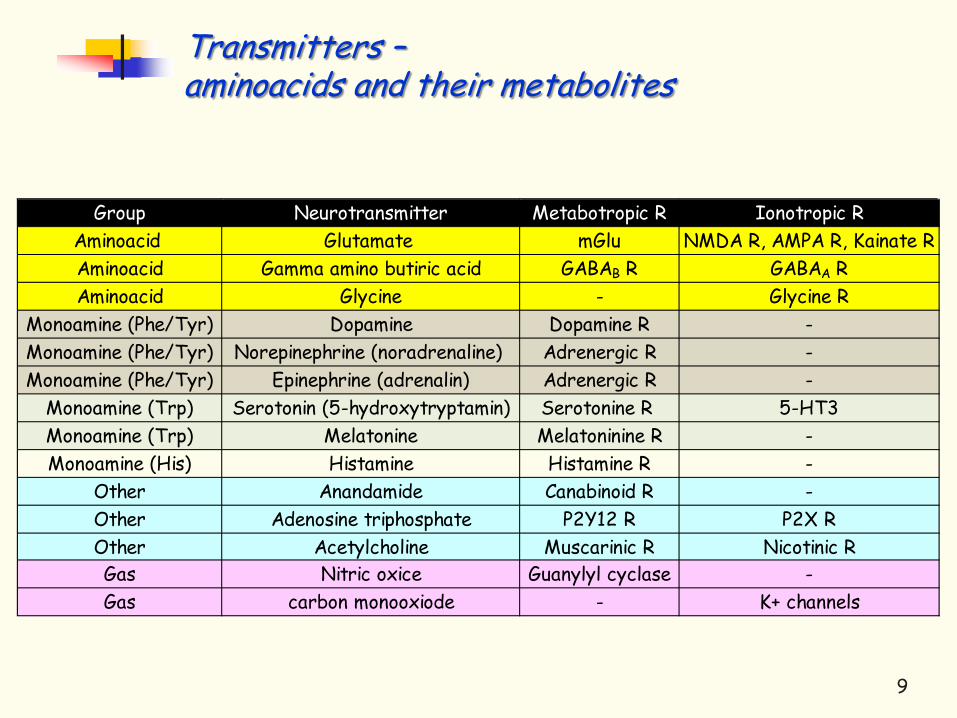
Transmitters – aminoacids and their metabolites
Group Neurotransmitter Metabotropic R Ionotropic RAminoacid Glutamate mGlu NMDA R, AMPA R, Kainate RAminoacid Gamma amino butiric acid GABAB R GABAA RAminoacid Glycine - Glycine R
Monoamine (Phe/Tyr) Dopamine Dopamine R -Monoamine (Phe/Tyr) Norepinephrine (noradrenaline) Adrenergic R -Monoamine (Phe/Tyr) Epinephrine (adrenalin) Adrenergic R -
Monoamine (Trp) Serotonin (5-hydroxytryptamin) Serotonine R 5-HT3Monoamine (Trp) Melatonine Melatoninine R -Monoamine (His) Histamine Histamine R -
Other Anandamide Canabinoid R -Other Adenosine triphosphate P2Y12 R P2X ROther Acetylcholine Muscarinic R Nicotinic RGas Nitric oxice Guanylyl cyclase -Gas carbon monooxiode - K+ channels
9
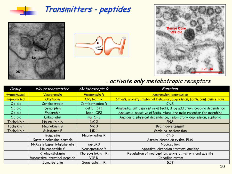
Transmitters - peptides
10
…activate only metabotropic receptors Group Neurotransmitter Metabotropic R Function
Hypophyseal Vasopressin Vasopresin R Aggression, depressionHypophyseal Oxytocin Oxytocin R Stress, anxiety, maternal behavior, aggression, faith, confidence, love
Opioid Corticotropin Corticotropine R CNSOpioid Dynorphin delta OP1 Analgesia, antidepressive effects, drug addiction, cocaine dependenceOpioid Endorphin kapa OP2 Analgesia, sedative effects, miosa, the main receptor for morphineOpioid Enkephalin mu OP3 Analgesia, physical dependence, respiratory depression, euphoria
Tachykinin Neurokinin A NK 2 PNSTachykinin Neurokinin B NK 3 Brain developmentTachykinin Substance P NK 1 Vomiting, nociception
Bombesin Neuromedine R CNSGastrin releasing peptide - Stress, circadian rythm, PNS
N-Acetylaspartylglutamate mGluR3 NociceptionNeuropeptide Y Neuropeptide Y Appetite, circadian rhythms, anxietyCholecystokinin Cholecystokinin R Regulation of nociception, anxiety, memory and apetite
Vasoactive intestinal peptide VIP R Circadian rythmSomatostatin Somatostatin R GIT
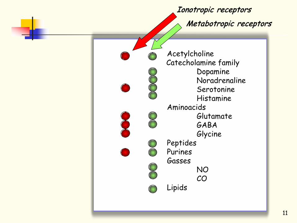
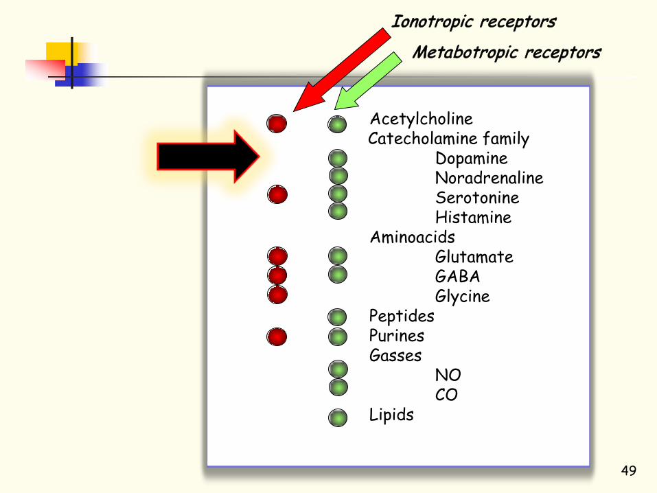
Acetylcholine Catecholamine family Dopamine Noradrenaline Serotonine Histamine Aminoacids
Glutamate GABA Glycine
Peptides Purines Gasses NO
CO Lipids
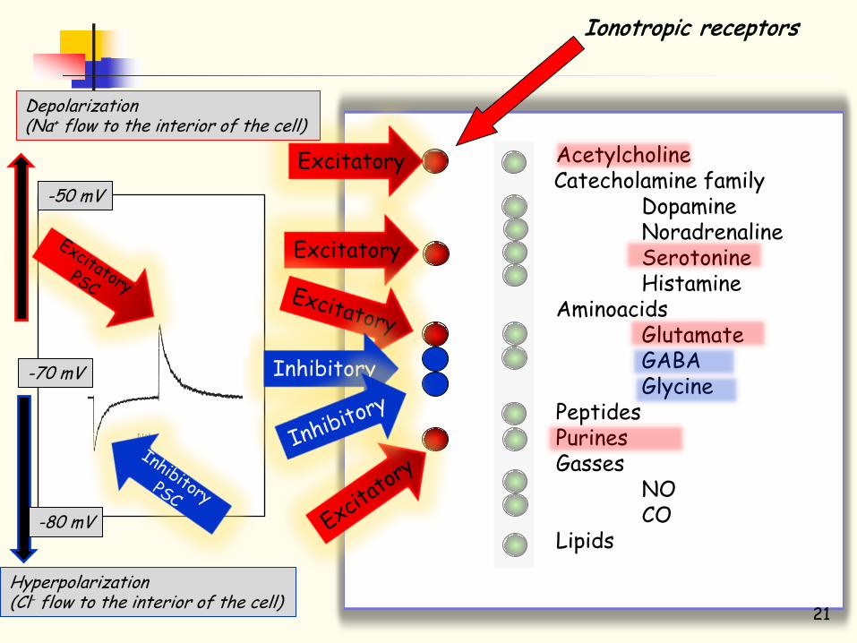
Ionotropic receptors
Metabotropic receptors
11

Chemical synapse
12
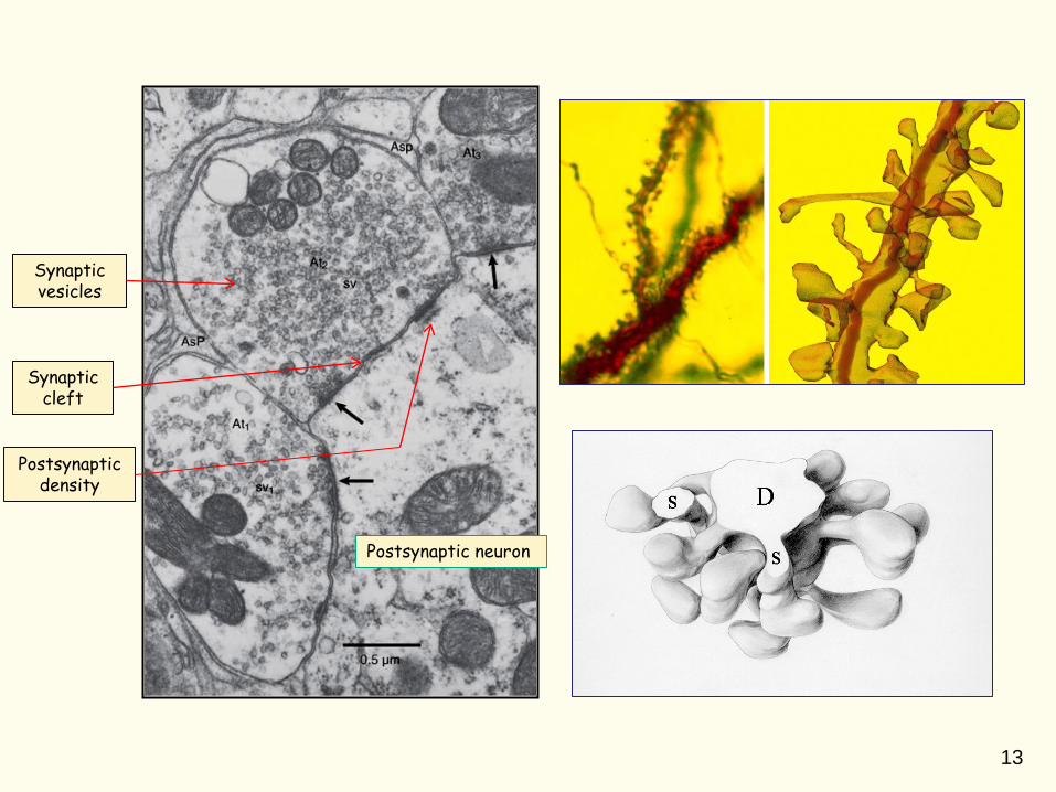
Postsynaptic density
Synaptic vesicles
Synaptic cleft
Postsynaptic neuron
13

3-D reconstruction of a molecule of ACh receptor
Ion channels are trensmembrane proteins forming a pore, that alows ion flux acros the cytoplasmatic membrane
Selective filter
Sensor
Gate
Structure of ion channels
Membrane
14

15
Neurotransmitter activated ion channels are usually oligomeric complexes composed of several subunits
Trimer Tetramer Hexamer Pentamer
Conexonone channels
Glutamate receptors
Cis – loop receptors ATP – IK GABA Glycine Acetylcholine Serotonine
Basic principles of neurotransmitter activation of ion channels
16

17 17
Presynaptic site Postsynaptic
site
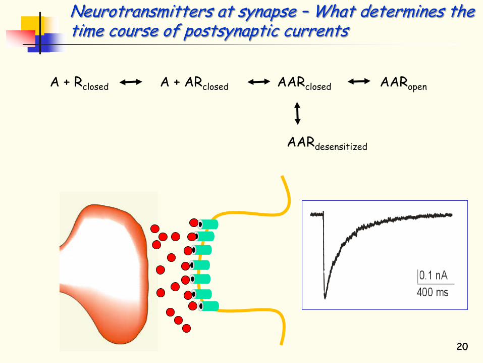
A + Rclosed A + ARclosed AARclosed AARopen
AARdesensitized
Neurotransmitters at synapse – What determines the time course of postsynaptic currents
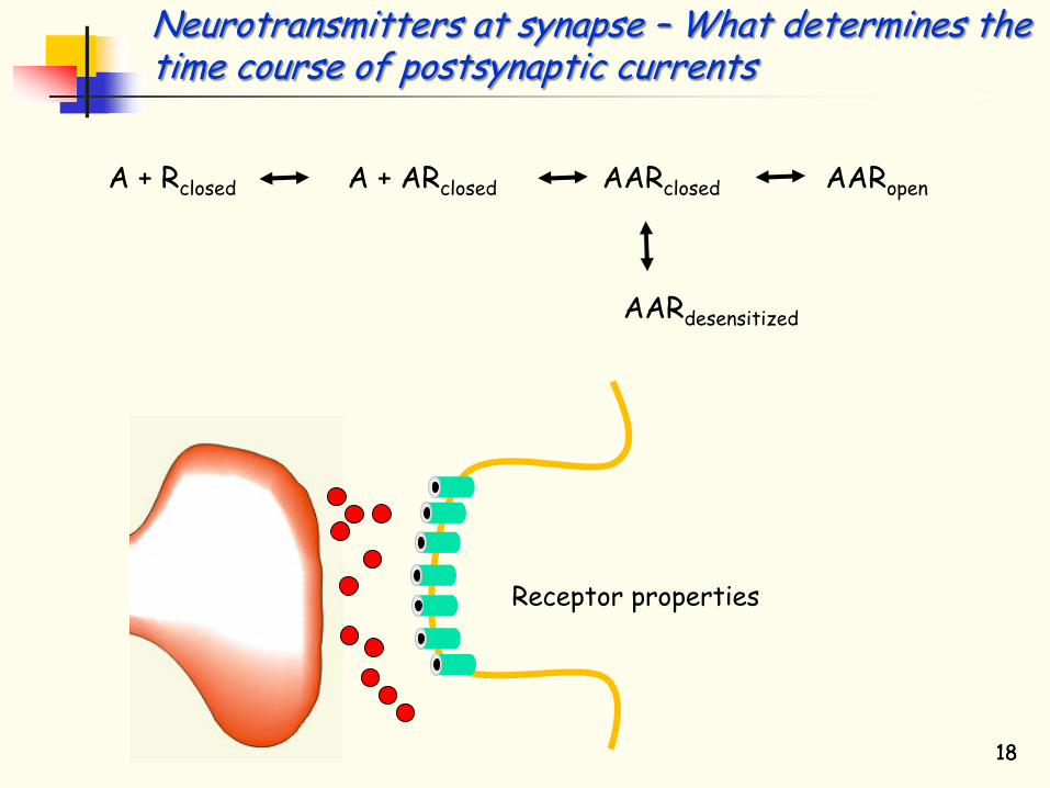
Receptor properties
18 18
A + Rclosed A + ARclosed AARclosed AARopen
AARdesensitized
Neurotransmitters at synapse – What determines the time course of postsynaptic currents

19 19
Bound transmitter Unbound transmitter
A + Rclosed A + ARclosed AARclosed AARopen
AARdesensitized
Neurotransmitters at synapse – What determines the time course of postsynaptic currents
20 20
A + Rclosed A + ARclosed AARclosed AARopen
AARdesensitized
Neurotransmitters at synapse – What determines the time course of postsynaptic currents
Acetylcholine Catecholamine family Dopamine Noradrenaline Serotonine Histamine Aminoacids
Glutamate GABA Glycine
Peptides Purines Gasses NO
CO Lipids
Ionotropic receptors
21
Excitatory
Excitatory
Inhibitory -70 mV
-50 mV
Hyperpolarization (Cl- flow to the interior of the cell)
Depolarization (Na+ flow to the interior of the cell)
-80 mV
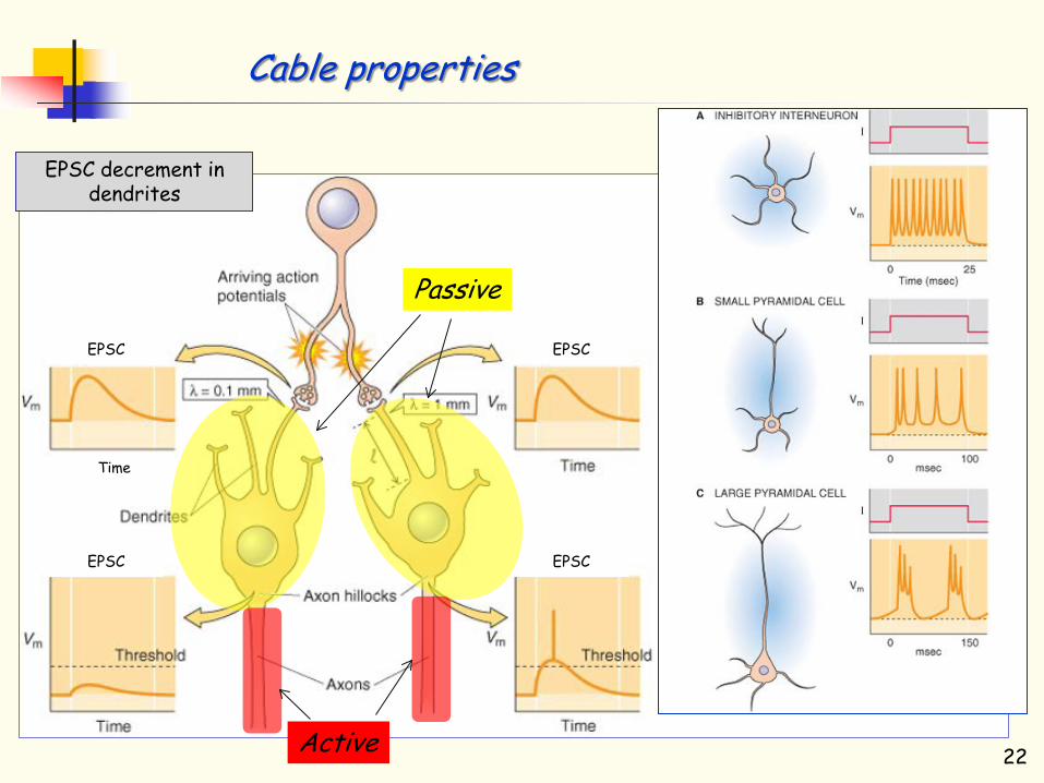
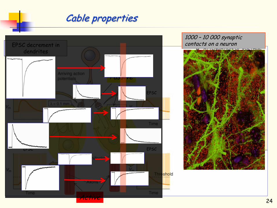
EPSC decrement in dendrites
Time
EPSC EPSC
EPSC EPSC
Relativní vzdálenost
Cable properties
22
1000 – 10 000 synaptic contacts on a neuron
Passive
Active
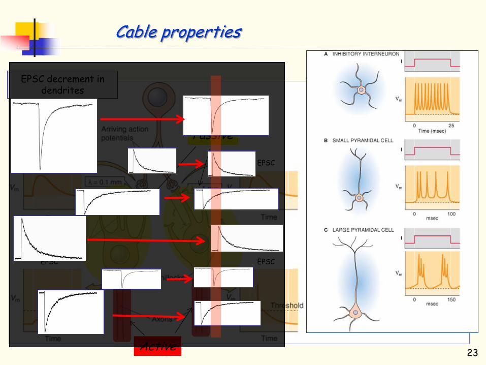
EPSC decrement in dendrites
Time
EPSC EPSC
EPSC EPSC
Relativní vzdálenost
Cable properties
23
1000 – 10 000 synaptic contacts on a neuron
Passive
Active
EPSC decrement in dendrites
Time
EPSC EPSC
EPSC EPSC
Relativní vzdálenost
Cable properties
24
1000 – 10 000 synaptic contacts on a neuron
Passive
Active

Summation of postsynaptic currents
25

Receptor (R) consists of seven membrane spanning regions
Extracellulr space
Cytosol G protein
Ligan binding and receptor activation
Receptor interacts with the G protein to promote conformational change and the change of GDP for GTP
G protein dissociates from the receptor α-GTP and βγ
subunits dissociate
Both α-GTP and βγ can now interact with their appropriate effectors
Members of the RGS family of G-protein regulators stimulate GTP hydrolysis
α-catalyzed hydrolysis of GTP to GDP inactivates α and promotes reassembly of the trimer
Metebotropic receptors
1
6 5
4 3
2
26
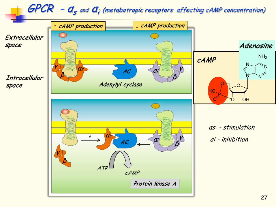
Extracellular space
Intracellular space Adenylyl cyclase
αs
αs
γ β β
β
β γ
αi γ
γ αi
ATP cAMP
Protein kinase A
↑ cAMP production ↓ cAMP production
AC
AC +
-
27
GPCR - αs and αi (metabotropic receptors affecting cAMP concentration)
cAMP
Adenosine
αs - stimulation
αi - inhibition

cAMP and phosphorylation/dephosphorylation
28
Stimulation of metabotropic receptors leads to 5-fold increase in the intracellular concentration of cAMP for ~5 seconds. This increase is only temporary – cAMP is enzymatically broken down into AMP by cAMP phosphodiesterase.
cAMP- dependent protein kinase A (PKA)
protein … serine … protein protein … threonine … protein
P
intracellular proteins – ion channels, receptors, enzymes
PKA Kinase A ancoring protein
(A kinase anchoring protein - AKAP)
serine/threonine phsphoprotein phosphatase (PP) typ 1, 2a, 2b a 2c.
cAMP
Adenosine

29
Transmitters and hormons acting through activation of Gαs (cAMP increase) adenosine, epinephrine, norepinephrine, dopamine, histamine, prostaglandins, serotonine, ACTH, antidiuretic hormone = vasopresin, calcitonin, CRH, FSH, glucagone, oxytocine, secretine, VIP, Thyreotropin–releasing hormone (TRH),TSH Transmitters and hormons acting through activation of Gαi (cAMP decrease) Acetylcholine, adenosine, epinephrine, norepinephrine, dopamine, GABA, glutamate, neuropeptide Y, opioids, serotonine, Angiotensine, melatonine, Growth hormone inhibitory hormone (somatostatine)
GPCR - αs and αi (metabotropic receptors affecting concentration of cAMP)
αi
αs

GPCR - αq (metabotropic receptors … activation PKC)
Intracellullar space
Fosfolipase C
αq γ
β
ER
Komplex of the metabotropic receptor and G- proteins
IP3 stimulates release of Ca2+ from ER
αq PLC
PIP2
PLC
DAG
PLC
IP3
IP3 receptor
Protein kinase C
PKC
PKC
Inositol trisphosphate (IP3)
Ca2+ Calmodulin dependent protein kinase (CAMP)
PLC
30
Inositol trisfosphate (IP3)
Diacylglycerol (DAG)

GPCR - αq (metabotropic receptors … activation PKC)
Intracellullar space
Fosfolipase C
αq γ
β
ER
Komplex of the metabotropic receptor and G- proteins
IP3 stimulates release of Ca2+ from ER
αq PLC
PIP2
PLC
DAG
PLC
IP3
IP3 receptor
Protein kinase C
PKC
PKC
Inositol trisphosphate (IP3)
Ca2+ Calmodulin dependent protein kinase (CAMP)
Fosfatidylinositol-4,5-bisfosfát
PLC
31
Inositol trisfosphate (IP3)
Diacylglycerol (DAG)

GPCR - αq (metabotropic receptors … activation PKC)
Intracellullar space
Fosfolipase C
αq γ
β
ER
Komplex of the metabotropic receptor and G- proteins
IP3 stimulates release of Ca2+ from ER
αq PLC
PIP2
PLC
DAG
PLC
IP3
IP3 receptor
Protein kinase C
PKC
PKC
Inositol trisphosphate (IP3)
Ca2+ Calmodulin dependent protein kinase (CAMP)
Fosfatidylinositol-4,5-bisfosfát
PLC
32
Inositol trisfosphate (IP3)
Calmodulin
• Ca2+ - Calmodulin dependent protein kinase
• Phosphatase (Calcineurin)
• Protease (Calpain)
• Endonuclease
Ca2+
Diacylglycerol (DAG)

33
Transmitters and hormons that activate IP3 a DAG acetylcholine, epinephrine, norepinephrine, bradykinine, endotheline, glutamate, histamine, leukotriens, prostaglandins, serotonine, tachykinine, thromboxane A2. antidiuretic hormone = vasopresin, cholecystokinin, gastrine, neurotensine, oxytocine, Thyreotropin–releasing hormone (TRH), TSH
GPCR - αq (metabotropic receptors … activation PKC)
αq

Acetylcholine Catecholamine family Dopamine Noradrenaline Serotonine Histamine Aminoacids
Glutamate GABA Glycine
Peptides Purines Gasses NO
CO Lipids
Ionotropic receptors
Metabotropic receptors
34

Cholin
CH3 - COOH Acetate
Cholin acetyltransferase
Acetylcholine (ACh) was discovered by Henry H. Dale (in 1914) … its role of neurotransmitter was described by Otto Loewi … in 1936 both were awarded by NP
Acetylcholinesterase
Acetyl-CoA
Acetylcholine „Vagusstoff“ 35

8 nm
2,5 nm
Nicotinic ACh receptor
36

8 nm
2,5 nm
17 nACh subunits
(α1)2β1δε (α1)2β1δγ
(α3)2(β4)3 Ganglia
(α4)2(β2)3 CNS (α3)2(β4)3 (α7)5
Nicotinic ACh receptor
37

1.5-4.0 x 10⁷ Ach receptors
60 vesicles is released during EPSC – Each contain 104 molecules of ACh
38

Botulinum toxin is a protein produced by the bacterium Clostridium botulinum, and is considered the most powerful neurotoxin ever discovered - 100 g would be sufficient to kill all people. BT – prevents fusion of vesicles with the presynaptic membrane
39
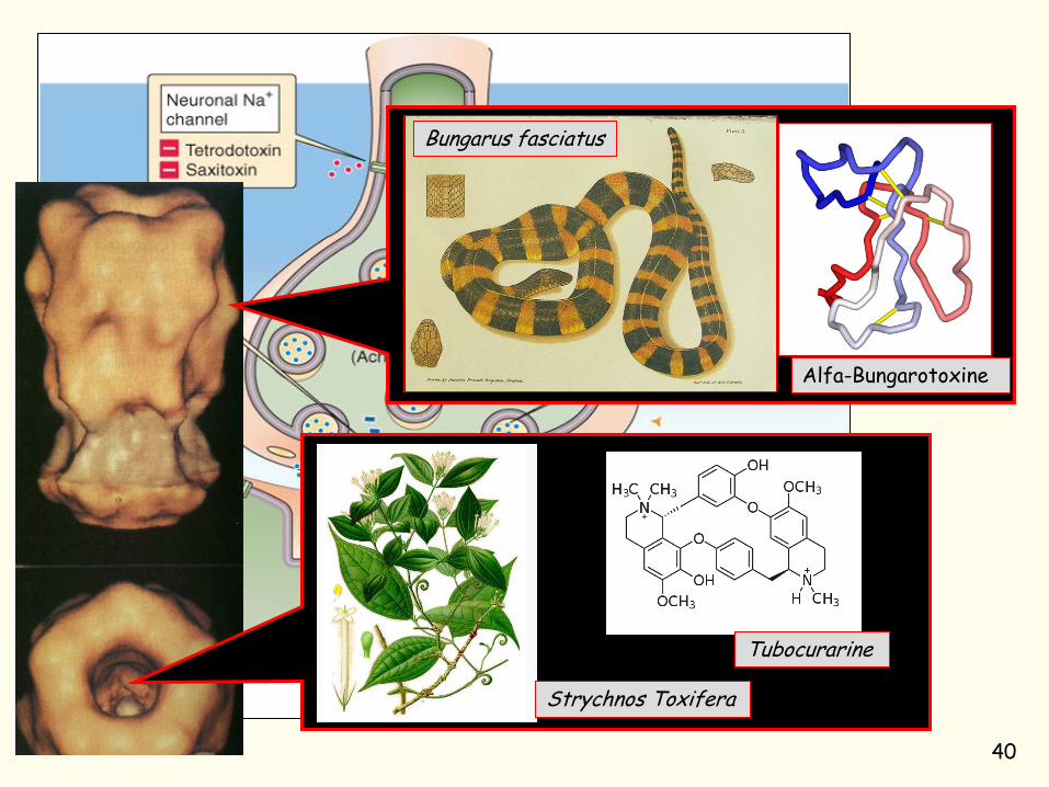
Bungarus fasciatus
Alfa-Bungarotoxine
Tubocurarine
Strychnos Toxifera
40
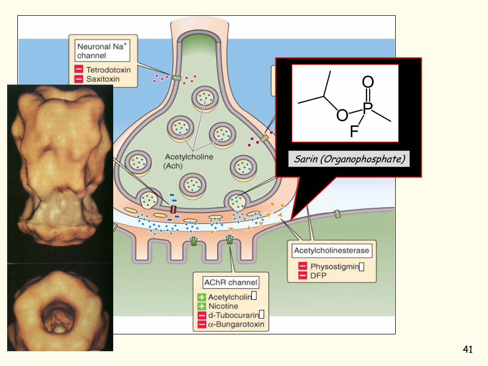
Sarin (Organophosphate)
41

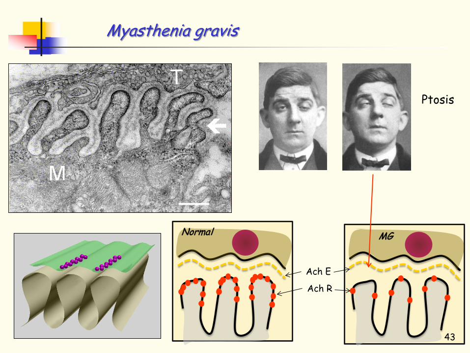
Myasthenia gravis
Ptosis
Normal
Ach E
MG
Ach R
Physostigmine
Physostigma venenosum
42
Myasthenia gravis
Ptosis
Normal
Ach E
MG
Ach R
43
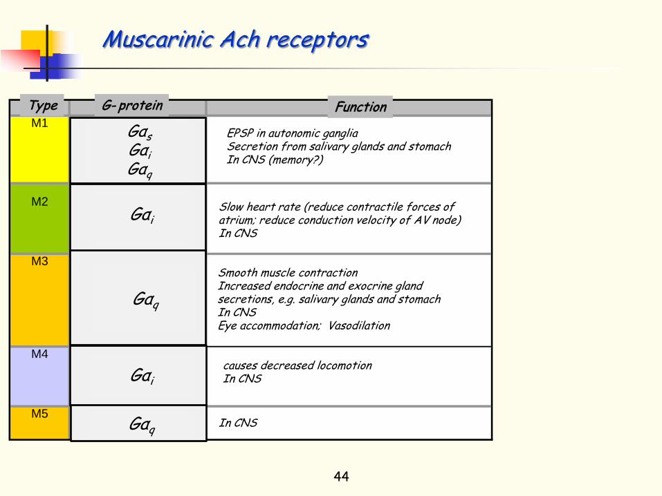
Typ G-protein FunkceM1 Gq EPSP v autonomních ganglích
(Gi) sekrece sliných žláz a žaludku(Gs): v CNS (paměť?)Slow EPSP.
M2 Gi zpomaluje srdeční činnost↑ K+ vodivost snižuje kontraktilní sílu srdce
↓ Ca2+ vodivost v CNSM3 Gq kontrakce hladkého svalstva
zvyšuje sekreci žláz - slinných a žaludku v CNS akomodace oka vasodilatace zvracení
M4 Gi zvýšená lokomoce↑ K+ vodivost v CNS↓ Ca2+ vodivost
M5 Gq v CNS
44
EPSP in autonomic ganglia Secretion from salivary glands and stomach In CNS (memory?)
Slow heart rate (reduce contractile forces of atrium; reduce conduction velocity of AV node) In CNS
conductance
Smooth muscle contraction Increased endocrine and exocrine gland secretions, e.g. salivary glands and stomach In CNS Eye accommodation; Vasodilation
causes decreased locomotion In CNS
In CNS
conductance
conductance conductance
Function Type G- protein
Muscarinic Ach receptors
Gαs Gαi Gαq
Gαi
Gαq
Gαi
Gαq

Typ G-protein FunkceM1 Gq EPSP v autonomních ganglích
(Gi) sekrece sliných žláz a žaludku(Gs): v CNS (paměť?)Slow EPSP.
M2 Gi zpomaluje srdeční činnost↑ K+ vodivost snižuje kontraktilní sílu srdce
↓ Ca2+ vodivost v CNSM3 Gq kontrakce hladkého svalstva
zvyšuje sekreci žláz - slinných a žaludku v CNS akomodace oka vasodilatace zvracení
M4 Gi zvýšená lokomoce↑ K+ vodivost v CNS↓ Ca2+ vodivost
M5 Gq v CNS
45
EPSP in autonomic ganglia Secretion from salivary glands and stomach In CNS (memory?)
Slow heart rate (reduce contractile forces of atrium; reduce conduction velocity of AV node) In CNS
conductance
Smooth muscle contraction Increased endocrine and exocrine gland secretions, e.g. salivary glands and stomach In CNS Eye accommodation; Vasodilation
causes decreased locomotion In CNS
In CNS
conductance
conductance conductance
Function Type G- protein
Atropa belladonna (Rulík zlomocný)
Atropin (antagonist)
Muscarinic Ach receptors
Gαs Gαi Gαq
Gαi
Gαq
Gαi
Gαq
Amanita muscaria
Muscarine (agonist)

CNS
Autonomic nervous system
Nervs and proximal ganglia Organs (muscle, heart, glands)
Preganglion fibre
Ganglion
ACh
N2 nicotinic ACh R
ACh
Muscarinic ACh R
Ganglion
ACh
N2 nicotinic ACh R
NA
α or β adrenergic receptors
Postganglion fibre
Synapse „en passant“
46
Parasympathetic nervous system
Sympathetic nervous system
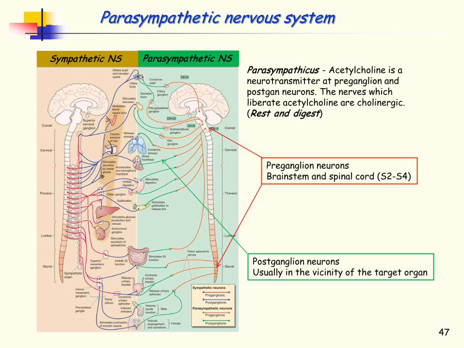
Parasympathicus - Acetylcholine is a neurotransmitter at preganglion and postgan neurons. The nerves which liberate acetylcholine are cholinergic. (Rest and digest)
Parasympathetic NS Sympathetic NS
Preganglion neurons Brainstem and spinal cord (S2-S4)
Postganglion neurons Usually in the vicinity of the target organ
47
Parasympathetic nervous system
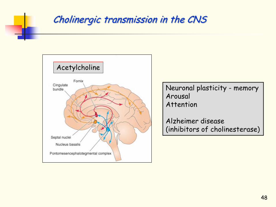
Acetylcholine
Neuronal plasticity - memory Arousal Attention Alzheimer disease (inhibitors of cholinesterase)
Cholinergic transmission in the CNS
48
Acetylcholine Catecholamine family Dopamine Noradrenaline Serotonine Histamine Aminoacids
Glutamate GABA Glycine
Peptides Purines Gasses NO
CO Lipids
Ionotropic receptors
Metabotropic receptors
49

Phenylalanine
Phenylalanine hydroxylase (livr)
Adrenaline
CNS
Sympathetic NS
50 Adrenal gland

Phenylalanine
Phenylalanine hydroxylase (livr)
Adrenaline
CNS
Sympathetic NS
Phenylpyruvate oligophernia (Phenylketonuria) Is an autosomal recessive metabolic genetic disorder characterized by a mutation in the gene for the hepatic enzyme phenylalanine hydroxylase • Mental retardation • Brain damage • Oligophrenia • Seizures
51 Adrenal gland

Dopamine transporter DAT1
Reuptake
Dopaminergic neuron
Brain, striatum, bazal ganglia
MAO
Norepinephrinephrine transporter NET
Reuptake
Dopaminergic neuron COMT
Prefrontal cortex
D1 Gαs
D2 Gαi
D3 Gαi
D4 Gαi
D5 Gαs
Postsynaptic site Postsynaptic site
Dopaminergic system - CNS
52

Dopamine degradation
53

Parkinson disease – Schizophrenia – Drug ubusus
Reuptake
Postsynaptic site
Dopaminergic neuron
Treatment of schizophrenia D4 is 4-fold increased
Haloperidol (inhibitor of D receptors
Dihydroxyphenylalanine DOPA
Treatment of Parkinson disease
Cocaine (inhibitor of dopamine and
norepinephrine transporter)
Erythroxylum coca 54

Parkinson disease – Schizophrenia – Drug ubusus
Reuptake
Postsynaptic site
Dopaminergic neuron
Treatment of schizophrenia D4 is 4-fold increased
Haloperidol (inhibitor of D receptors
Dihydroxyphenylalanine DOPA
Treatment of Parkinson disease
Cocaine (inhibitor of dopamine and
norepinephrine transporter)
Erythroxylum coca
PET
Substancia nigra
55

Dopamine receptors • Control of movement • Memory, attention, motivation • Sleep • Control of vomiting and nausea • A system of pleasure, aggression, addiction • The behavior of "the search for reward„ • Anhedonia – the inability to feel pleasure • Schizophrenia • Bipolar disorder • Alcohol dependency • Control of food intake • Sexual behavior • Social phobia • Pain • ADHD Attention-deficit hyperactivity disorder • Parkinson disease
56

Acetylcholine Catecholamine family Dopamine Noradrenaline Serotonine Histamine Aminoacids
Glutamate GABA Glycine
Peptides Purines Gasses NO
CO Lipids
Ionotropic receptors
Metabotropic receptors
57

CNS
Autonomic nervous system
Nerve and proximal ganglia
Organs (muscle and glands)
Preganglion fibre
Ganglion
ACh
N2 nicotinic ACh R
ACh
Muscarinic ACh R
Ganglion
ACh
N2 nicotinic ACh R
NA
α or β adrenergic receptors
Postganglion fibre
Synapse „en passant“
58
Parasympathetic nervous system
Sympathetic nervous system

Sympathetic nervous system Acetylcholine is the neurotransmitter on the preganglionic neurons. On the postganglionic neurons it is the norepinephrine (noradrenaline). The nerves which liberate noradrenaline are called adrenergic. (Hunting and defence)
Parasymphatetic NS Sympathetic NS
Preganglion neurons Intermediolateral part of the spinal cord (T1-L3)
Prevertebral and paravertebral ganglia
PNS – Sympathetic nervous system - Norepinephrine
59

Uptake
Norepinephrine neuron
MAO
Postsynaptic site
COMT
60
PNS – Sympathetic nervous system - Norepinephrine
Receptor GPCR Effect α1 Gq ↑ Vasoconstriction; ↓ intestine mobility; α2 Gi ↓ Insulin release; ↓ Transmitter release;
Sfincter contraction β1 Gs ↑ Heart rate;
↑ Lipolysis β2 Gs Relaxation of smooth muscle β3 Gs ↑ Lipolysis

Serotonine and norepinephrine
transporter
Uptake
CNS MAO
Postsynaptic site
Norepinephrine neuron
Depression, mood disorders, anxiety, Attention Deficit Hyperactivity Disorder (ADHD)
Noradrenaline - CNS
Antidepressants (Serotonin–norepinephrine reuptake inhibitors - SNRIs) Inhibit reuptake of norepinephrine (NE activate postsynaptic receptor for longer)
Noradrenaline
61

Acetylcholine Catecholamine family Dopamine Noradrenaline Serotonine Histamine Aminoacids
Glutamate GABA Glycine
Peptides Purines Gasses NO
CO Lipids
Ionotropic receptors
Metabotropic receptors
62

Biosynthesis and metabolism of serotonin
MAO
63

Biosynthesis and metabolism of serotonin
MAO Ecstasy (MDMA - 3,4-methylendioxymetamfetamin) It is the most typical representative of the so called “recreation drug“ (originally developed as a to lose weight on the basis of the suppression of appetite)
… it releases serotonin from the synaptic vesicles
Serotonin transporter (SERT) - polymorphism of this gene may play a role in: sudden death of newborns, aggressive, degenerative diseases (AD), posttraumatic stress, sensitivity to the depression.
Selective serotonin reuptake inhibitors (SSRI 's) are used for psychiatric illness and in particular for obsessive compulse disorder.
64

Typ Iono/Metabotropní Mechanismus Účinek
5-HT1 Gi/Go Snižuje hladinu cAMP Inhibitory
5-HT2 Gq/G11 Zvyšuje IP3 and DAG. Excitační
5-HT3 Ligand-gated Na+ and K+ kanál. Depolarizace Excitační
5-HT4 Gs Zvyšuje hladinu cAMP Excitační
5-HT5 Gi/Go Snižuje hladinu cAMP Inhibiční
5-HT6 Gs Zvyšuje hladinu cAMP Excitační
5-HT7 Gs Zvyšuje hladinu cAMP Excitační
Type Inono/metabotropis Action 5-HT receptors
Serotoninové receptory modulují uvolňování řady neuropřenašeču glutamátu, GABA, dopaninu, noradrenalinu, acetylcholinu….
Serotonine (5HT) receptors
65
Inhibitory Excitatory Excitatory Excitatory Inhibitory Excitatory Excitatory
In Ligand-gated Na+ channel
5-HT3 are ionotropic receptors – are excitatory CNS – anxiety PNS – nociception
Direct
Indirect
Indirect

Subtypes of serotonine receptors
66

Subtypes of serotonine receptors
67
Ergot (fungi)
Agonist of 5-HT1A, 1B, 1D, rec. Migrene, to stop haemorrhage
Ergotamine
Antagonists of 5HT6 5HT7 Antipsychotics (schizophrenia) and antidepresants (depressioin).

Matabotropní serotoninové receptory
Aphrodisiac Yohimbine is found naturally in an African tree Yohimbine – (Pausinystalia yohimbe). The main effect is to increase blood flow in the region of pelvis – promot blood circulation in penis.
Yohimbine – antagonist of 5-HT1B, 5-HT1D, 5-HT2A, 5-HT2B and agonist of 5-HT1A.
68

Matabotropic serotonine receptors
69
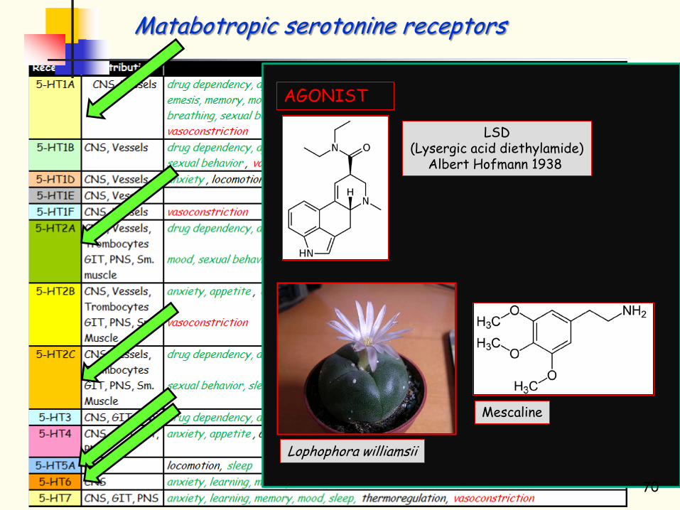
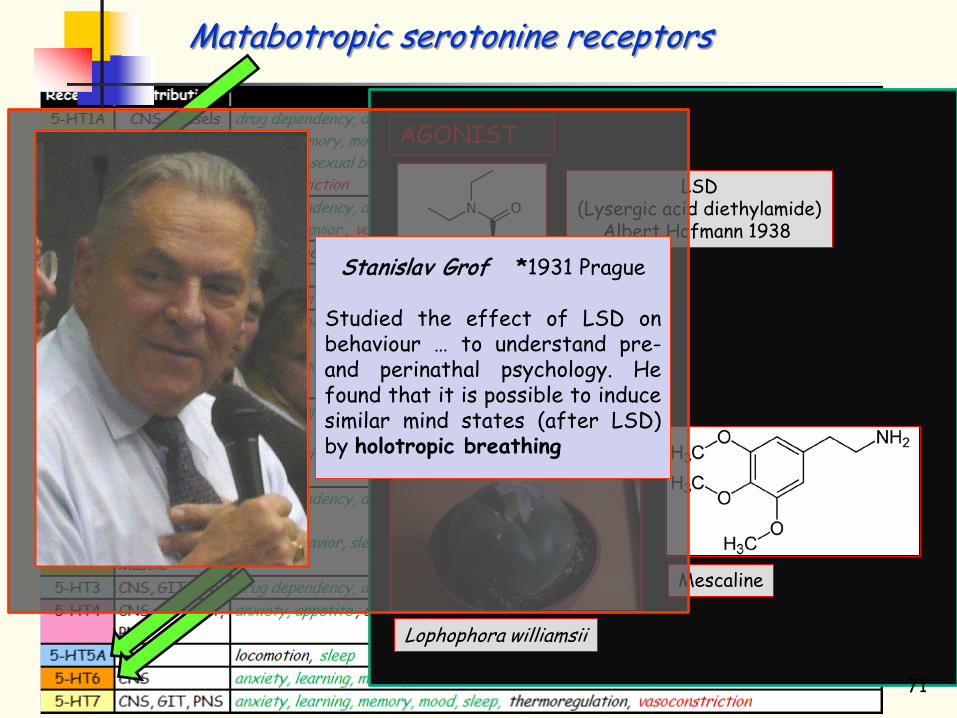
Matabotropic serotonine receptors
LSD (Lysergic acid diethylamide)
Albert Hofmann 1938
Mescaline
Lophophora williamsii
AGONIST
70
Matabotropic serotonine receptors
LSD (Lysergic acid diethylamide)
Albert Hofmann 1938
Mescaline
Lophophora williamsii
AGONIST
Stanislav Grof *1931 Prague
Studied the effect of LSD on behaviour … to understand pre- and perinathal psychology. He found that it is possible to induce similar mind states (after LSD) by holotropic breathing
71

Matabotropic serotonine receptors
LSD (Lysergic acid diethylamide)
Albert Hofmann 1938
Mescaline
Lophophora williamsii
AGONIST
Stanislav Grof *1931 Prague
Studied the effect of LSD on behaviour … to understand pre- and perinathal psychology. He found that it is possible to induce similar mind states (after LSD) by holotropic breathing
Salvador Dalí
72

Acetylcholine Catecholamine family Dopamine Noradrenaline Serotonine Histamine Aminoacids
Glutamate GABA Glycine
Peptides Purines Gasses NO
CO Lipids
Ionotropic receptors
Metabotropic receptors
73

Mast cells are capable to release heparin and histamine - importance for allergic reactions.
Histidine Histamine
74
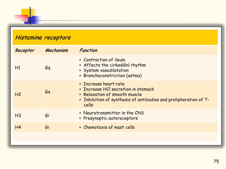
Histamine receptors
Receptor Mechanism Function
H1 Gq
• Contraction of ileum • Affects the cirkadální rhythm • System vasodilatation • Bronchoconstriction (astma)
H2 Gs
• Increase heart rate • Increase HCl secretion in stomach • Relaxation of smooth muscle • Inhibition of synthesis of antibodies and prolipheration of T-
cells
H3 Gi • Neurotransmitter in the CNS • Presynaptic autoreceptors
H4 Gi • Chemotaxis of mast cells
75

76
Acetylcholine Catecholamine family Dopamine Noradrenaline Serotonine Histamine Aminoacids
Glutamate GABA Glycine
Peptides Purines Gasses NO
CO Lipids
Ionotropic receptors
Metabotropic receptors

GABAergic Glycinergic
Purinergic (ATP) Cholinergic Serotonergic
Glutamatergic
Relative representation of synapses in the CNS
77

Glutamát vázající domena
78

Glutamic acid
AMPA (α-amino-3-hydroxyl-5-methyl-4-isoxazole-propionate)
NMDA N-methyl-D-aspartate
Kainate
Ionotropic glutamate receptors
79
Receptor SubunitAMPA GluR1
GluR2GluR3GluR4
Kainate GluR5GluR6GluR7KA-1KA-2
NMDA NR1NR2ANR2BNR2CNR2DNR3ANR3B
Glutamate receptors are thought to be responsible for the reception and transduction of umami taste stimuli.
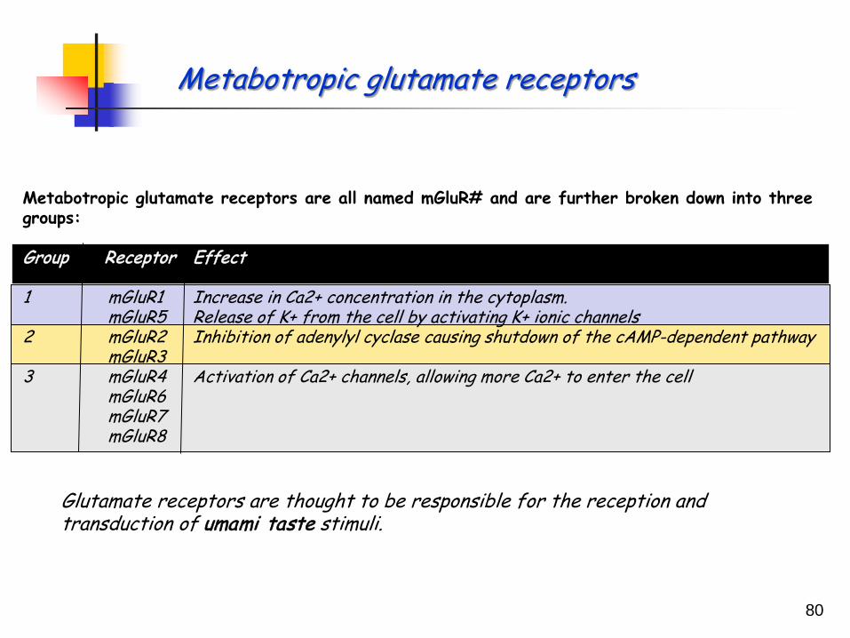
Metabotropic glutamate receptors
80
Metabotropic glutamate receptors are all named mGluR# and are further broken down into three groups: Group Receptor Effect 1 mGluR1 Increase in Ca2+ concentration in the cytoplasm. mGluR5 Release of K+ from the cell by activating K+ ionic channels 2 mGluR2 Inhibition of adenylyl cyclase causing shutdown of the cAMP-dependent pathway mGluR3 3 mGluR4 Activation of Ca2+ channels, allowing more Ca2+ to enter the cell mGluR6 mGluR7 mGluR8

↑ [Ca2+]i
Depolarisation
Glu Gly Glu Gly
Activation of NMDA R
81
10 ms
1 pA (10-12 A)
Closed
Open

Long term potentiation LTP
82

Before glutamate 30 min after glutamate
A B
Excitotoxicity
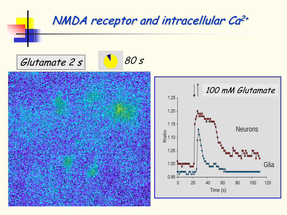
80 s Glutamate 2 s
Time (s)
Ra
tio
100 M glutamateµ
Neurons
Glia
100 mM Glutamate
NMDA receptor and intracellular Ca2+

Ca2+
Cell death
Increase in the intracellular Ca2+ concentration – an important step in excitotoxicity induction
Endonuclease
DNA Fragmentation
Phospholipase
Arachidonic acid
Protease (Calpain)
Ca-binding prot. (Calmodulin)
Mitochondria damage
Free radicals
Cytoskeleton disruption
Nitric oxide synthase
↓ ATP
↓ pH
Glutamate NMDA receptor

Alzheimer dementia
Functional changes of glutamatergic neurons
Neurodegeneration In: hippocampus, nucleus basalis Meynerti, amygdala, cortex
Glutamate and Alzheimer dementia
? Pathology
Tonic increase in the extracellular glutamate concentration
Excesive activation of NMDA receptors
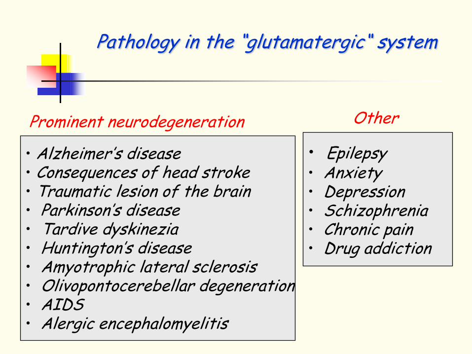
• Alzheimer’s disease • Consequences of head stroke • Traumatic lesion of the brain • Parkinson’s disease • Tardive dyskinezia • Huntington’s disease • Amyotrophic lateral sclerosis • Olivopontocerebellar degeneration • AIDS • Alergic encephalomyelitis
• Epilepsy • Anxiety • Depression • Schizophrenia • Chronic pain • Drug addiction
Prominent neurodegeneration Other
Pathology in the “glutamatergic“ system

Acetylcholine Catecholamine family Dopamine Noradrenaline Serotonine Histamine Aminoacids
Glutamate GABA Glycine
Peptides Purines Gasses NO
CO Lipids
Ionotropic receptors
Metabotropic receptors
88

GABAA receptors – ionotropic receptors GABAB receptors – metabotropic receptors • GABAB1 • GABAB2
Reduce activity of adenylyl cyclase (reduce Ca2+ conductance and increase K+)
GABA receptors
89
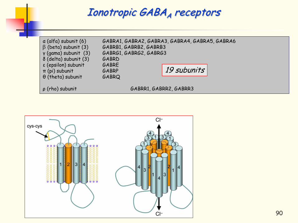
α (alfa) subunit (6) GABRA1, GABRA2, GABRA3, GABRA4, GABRA5, GABRA6 β (beta) subunit (3) GABRB1, GABRB2, GABRB3 γ (gama) subunit (3) GABRG1, GABRG2, GABRG3 δ (delta) subunit (3) GABRD ε (epsilon) subunit GABRE π (pi) subunit GABRP θ (theta) subunit GABRQ ρ (rho) subunit GABRR1, GABRR2, GABRR3
Ionotropic GABAA receptors
90
19 subunits
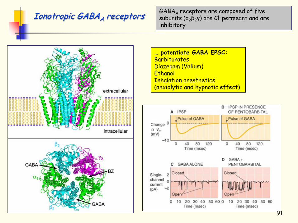
… potentiate GABA EPSC: Barbiturates Diazepam (Valium) Ethanol Inhalation anesthetics (anxiolytic and hypnotic effect)
GABAA receptors are composed of five subunits (α2β2γ) are Cl- permeant and are inhibitory
Ionotropic GABAA receptors
91

92
Acetylcholine Catecholamine family Dopamine Noradrenaline Serotonine Histamine Aminoacids
Glutamate GABA Glycine
Peptides Purines Gasses NO
CO Lipids
Ionotropic receptors
Metabotropic receptors

Glycine activated ion channel contains five subunits α-subunit (α1-4) GLRA1, GLRA2, GLRA3, GLRA4 - (binds glycine) β-subunit (GLRB) Ion channels are Cl- permeant – therefore inhibitory
Is expresed only in the spinal cord There are pure glycinergic synapses as well as mixed where glycine is released together with GABA
Ionotropic glycine receptor
93

Strychnos nux-vomica L Strychnine
Pharmacology of glycine receptor
94

-
+
- Renshaw cell
Motoneurone
Motoneurone
ACh
GLY
GLY
Mechanism of strychnin induced convulsions
95
96 96
Thank you for your attention