Neurotransmitter vijay
33
NEUROTRANSMITTERS -VIJAY P J
-
Upload
vijaypj17 -
Category
Technology
-
view
1.032 -
download
3
Transcript of Neurotransmitter vijay
NEUROTRANSMITTERS
-VIJAY P J
Introduction
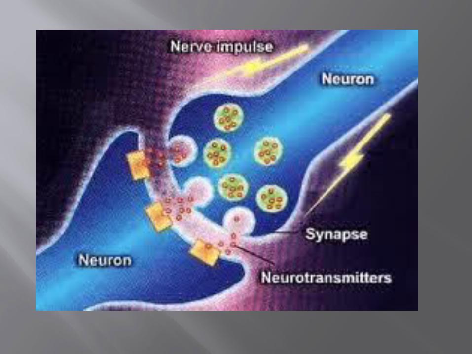
Neurotransmitters are the brain chemicals that communicate information throughout our brain and body.
They relay signals between nerve cells, called “neurons.”
The brain uses neurotransmitters to tell heart to beat, lungs to breathe, and stomach to digest.
They can also affect mood, sleep, concentration, weight, and can cause adverse symptoms when they are out of balance.
Definition
Neurotransmitters are chemicals found and produced in the brain to allow the transmission of impulses from one nerve cell to the next across synapses. They aid in the conduction of information throughout the body. These chemicals fit into specific receptor cells embedded in the membrane of the dendrite that either fuel up or excite action in the cells (excitatory) or stop or inhibit an action (inhibitory).
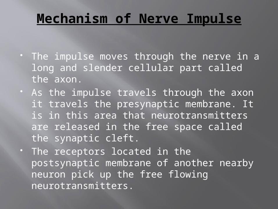
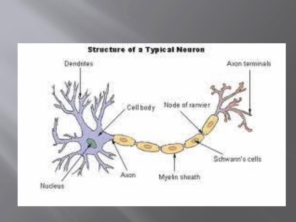
Mechanism of Nerve Impulse
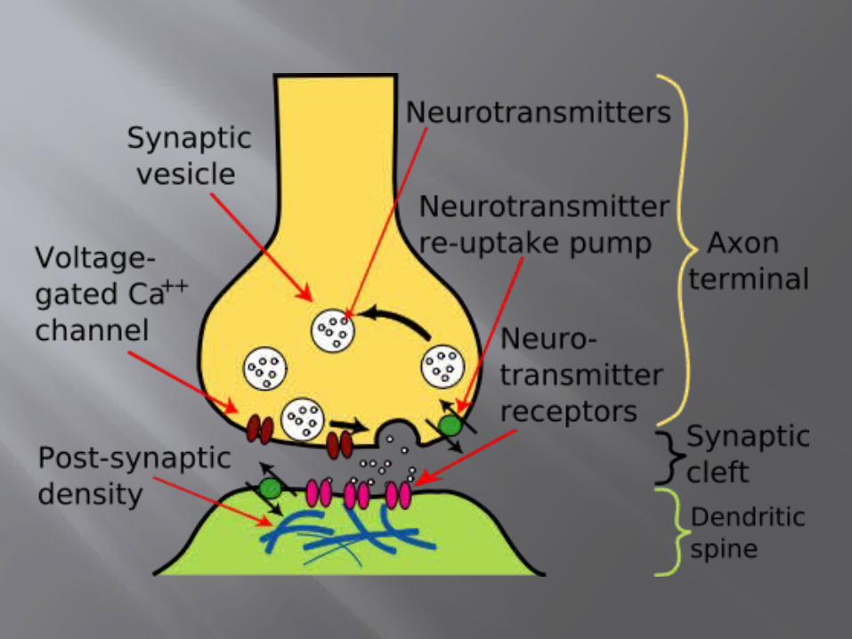
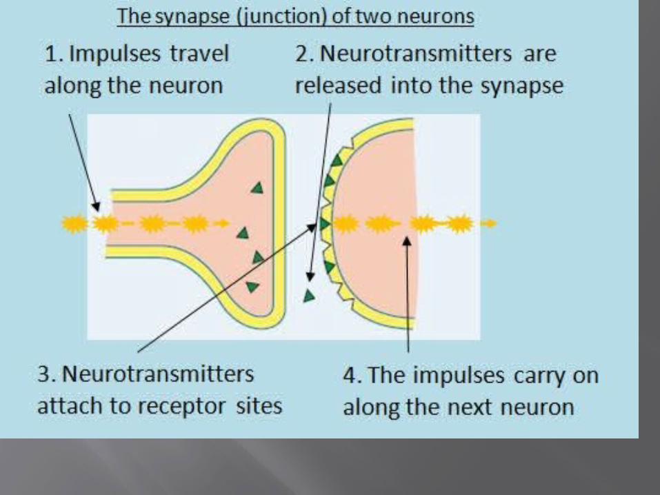
The impulse moves through the nerve in a long and slender cellular part called the axon.
As the impulse travels through the axon it travels the presynaptic membrane. It is in this area that neurotransmitters are released in the free space called the synaptic cleft.
The receptors located in the postsynaptic membrane of another nearby neuron pick up the free flowing neurotransmitters.
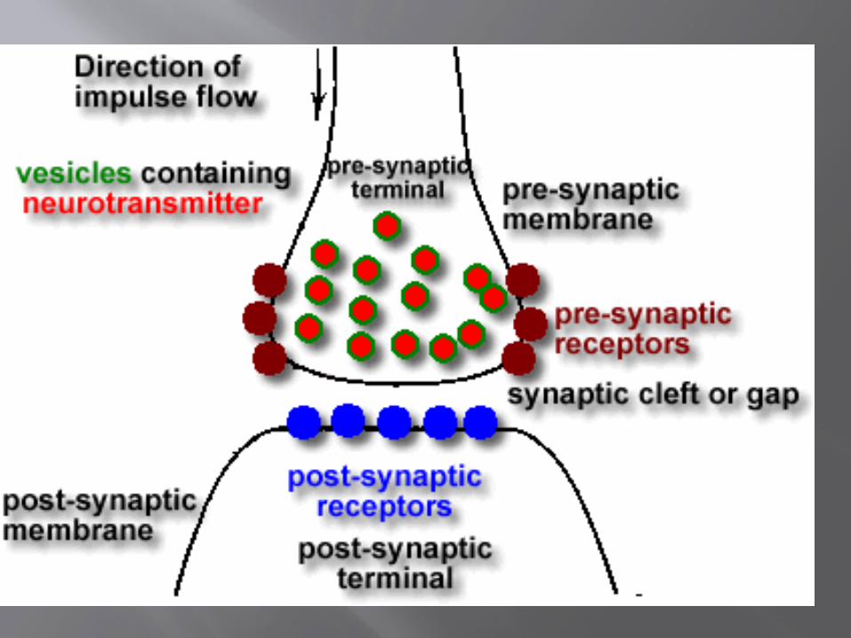
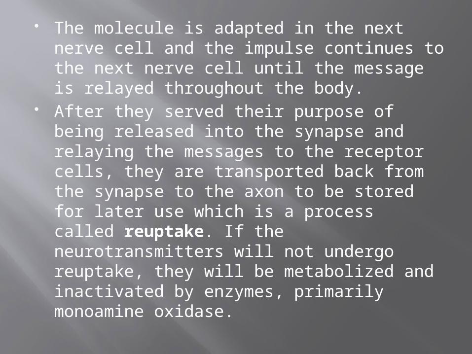
The molecule is adapted in the next nerve cell and the impulse continues to the next nerve cell until the message is relayed throughout the body.
After they served their purpose of being released into the synapse and relaying the messages to the receptor cells, they are transported back from the synapse to the axon to be stored for later use which is a process called reuptake. If the neurotransmitters will not undergo reuptake, they will be metabolized and inactivated by enzymes, primarily monoamine oxidase.
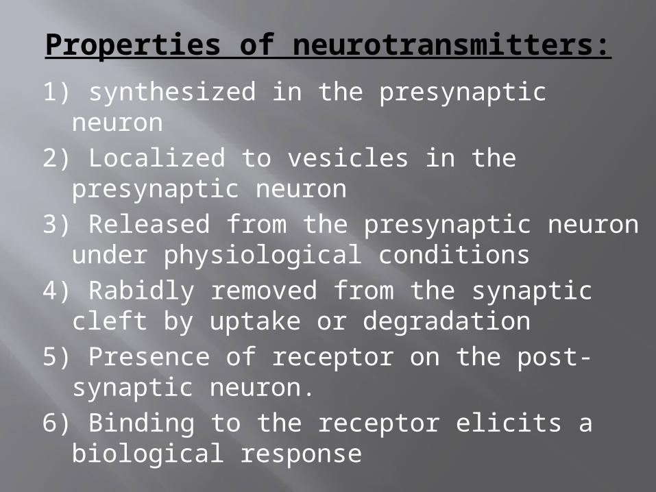
Properties of neurotransmitters:
1) synthesized in the presynaptic neuron2) Localized to vesicles in the presynaptic
neuron3) Released from the presynaptic neuron
under physiological conditions4) Rabidly removed from the synaptic cleft by
uptake or degradation5) Presence of receptor on the post-synaptic
neuron.6) Binding to the receptor elicits a biological
response
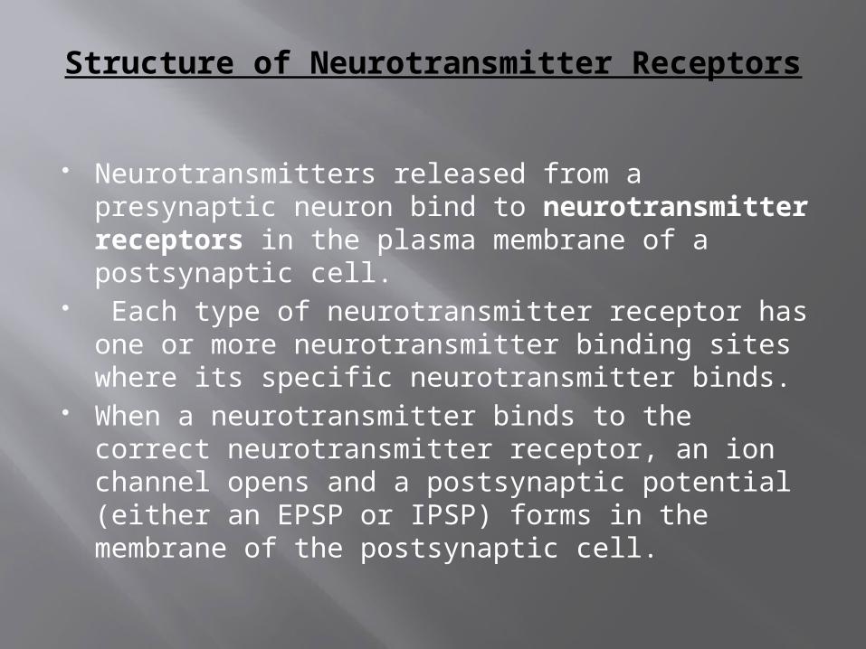
Structure of Neurotransmitter Receptors
Neurotransmitters released from a presynaptic neuron bind to neurotransmitter receptors in the plasma membrane of a postsynaptic cell.
Each type of neurotransmitter receptor has one or more neurotransmitter binding sites where its specific neurotransmitter binds.
When a neurotransmitter binds to the correct neurotransmitter receptor, an ion channel opens and a postsynaptic potential (either an EPSP or IPSP) forms in the membrane of the postsynaptic cell.
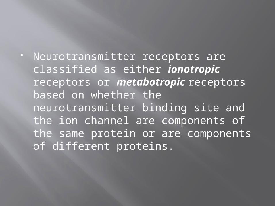
Neurotransmitter receptors are classified as either ionotropic receptors or metabotropic receptors based on whether the neurotransmitter binding site and the ion channel are components of the same protein or are components of different proteins.
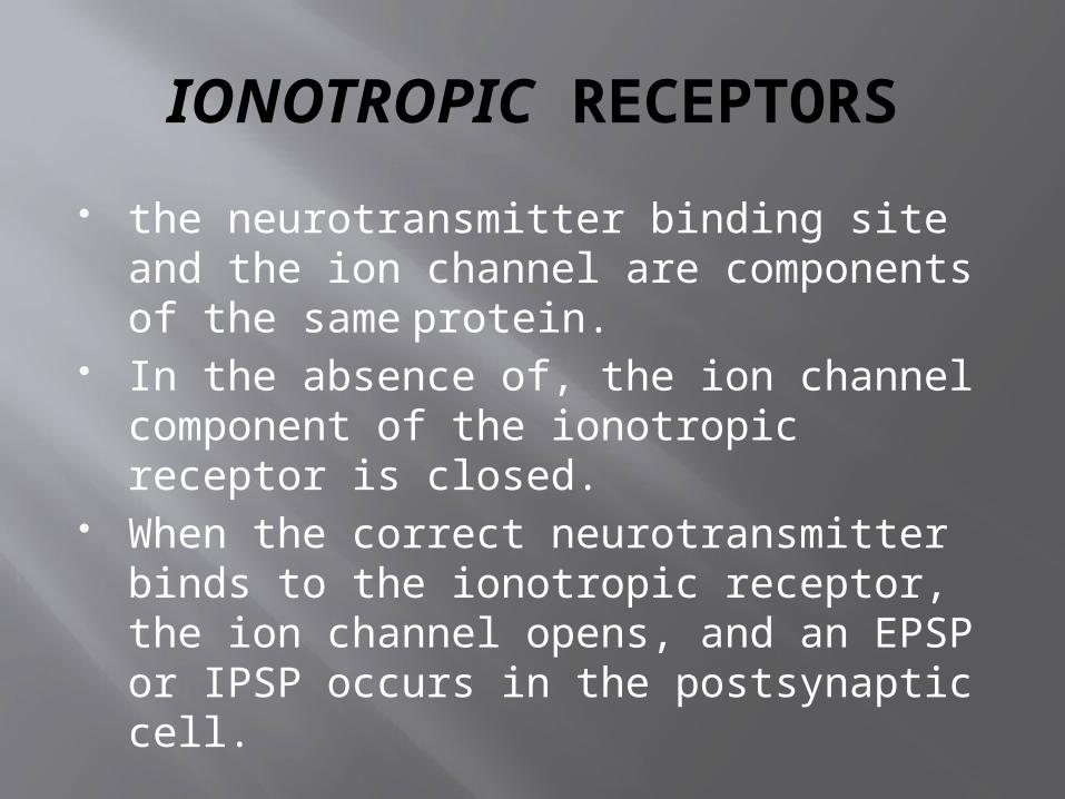
IONOTROPIC RECEPTORS
the neurotransmitter binding site and the ion channel are components of the same protein.
In the absence of, the ion channel component of the ionotropic receptor is closed.
When the correct neurotransmitter binds to the ionotropic receptor, the ion channel opens, and an EPSP or IPSP occurs in the postsynaptic cell.
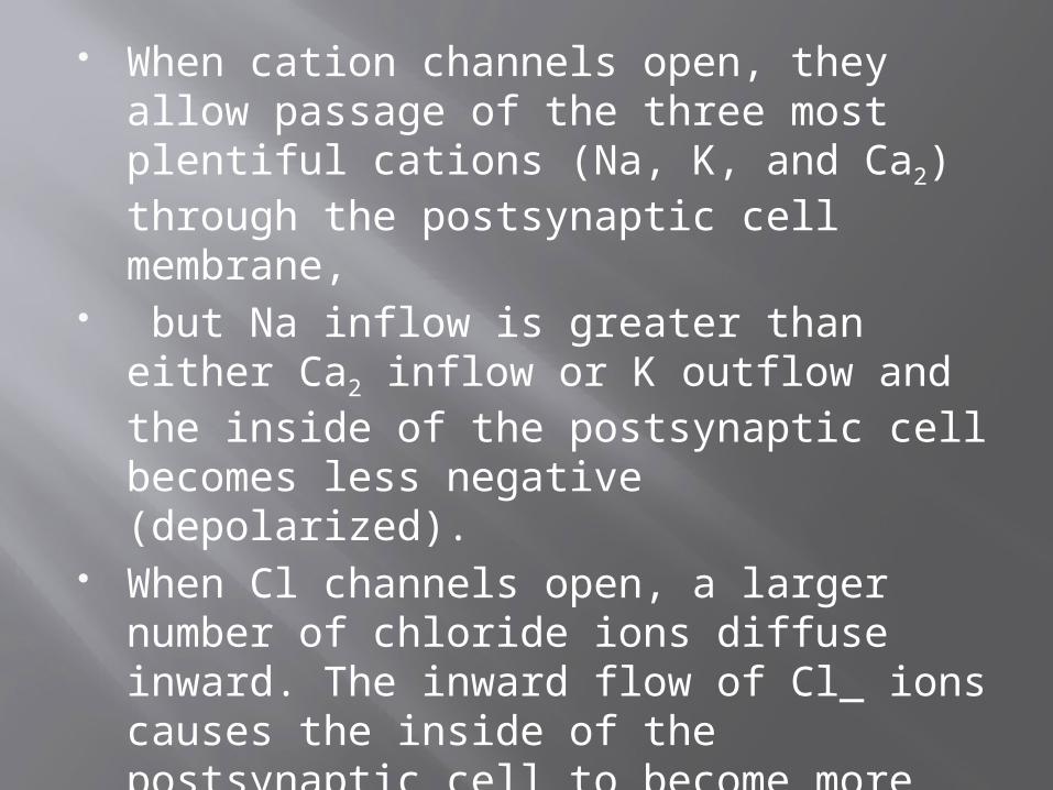
When cation channels open, they allow passage of the three most plentiful cations (Na, K, and Ca2) through the postsynaptic cell membrane,
but Na inflow is greater than either Ca2 inflow or K outflow and the inside of the postsynaptic cell becomes less negative (depolarized).
When Cl channels open, a larger number of chloride ions diffuse inward. The inward flow of Cl_ ions causes the inside of the postsynaptic cell to become more negative (hyperpolarized).
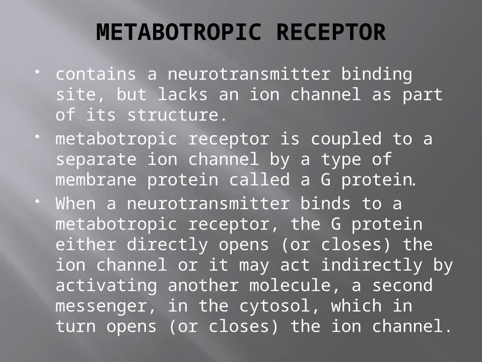
METABOTROPIC RECEPTOR
contains a neurotransmitter binding site, but lacks an ion channel as part of its structure.
metabotropic receptor is coupled to a separate ion channel by a type of membrane protein called a G protein.
When a neurotransmitter binds to a metabotropic receptor, the G protein either directly opens (or closes) the ion channel or it may act indirectly by activating another molecule, a second messenger, in the cytosol, which in turn opens (or closes) the ion channel.
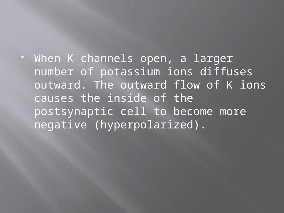
When K channels open, a larger number of potassium ions diffuses outward. The outward flow of K ions causes the inside of the postsynaptic cell to become more negative (hyperpolarized).
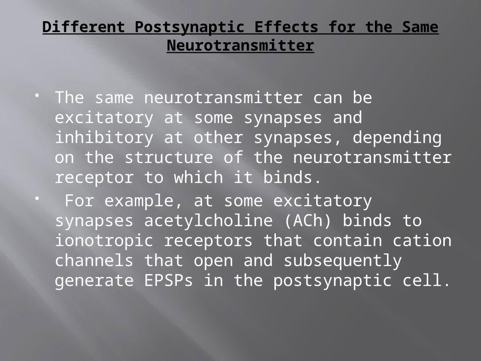
Different Postsynaptic Effects for the Same Neurotransmitter
The same neurotransmitter can be excitatory at some synapses and inhibitory at other synapses, depending on the structure of the neurotransmitter receptor to which it binds.
For example, at some excitatory synapses acetylcholine (ACh) binds to ionotropic receptors that contain cation channels that open and subsequently generate EPSPs in the postsynaptic cell.
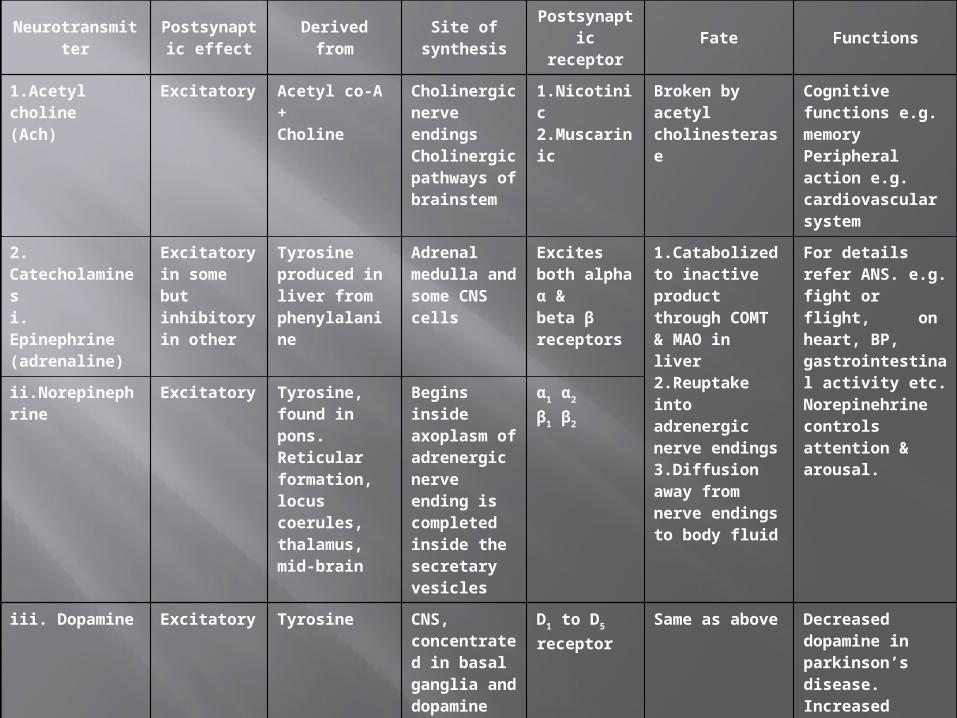
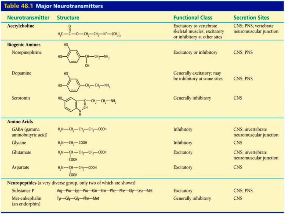
NeurotransmitterPostsynaptic
effectDerived from
Site of synthesis
Postsynaptic receptor
Fate Functions
1.Acetyl choline(Ach)
Excitatory Acetyl co-A +Choline
Cholinergic nerve endingsCholinergic pathways of brainstem
1.Nicotinic2.Muscarinic
Broken by acetyl cholinesterase
Cognitive functions e.g. memoryPeripheral action e.g. cardiovascular system
2. Catecholaminesi. Epinephrine (adrenaline)
Excitatory in some but inhibitory in other
Tyrosine produced in liver from phenylalanine
Adrenal medulla and some CNS cells
Excites both alpha α &beta β receptors
1.Catabolized to inactive product through COMT & MAO in liver2.Reuptake into adrenergic nerve endings3.Diffusion away from nerve endings to body fluid
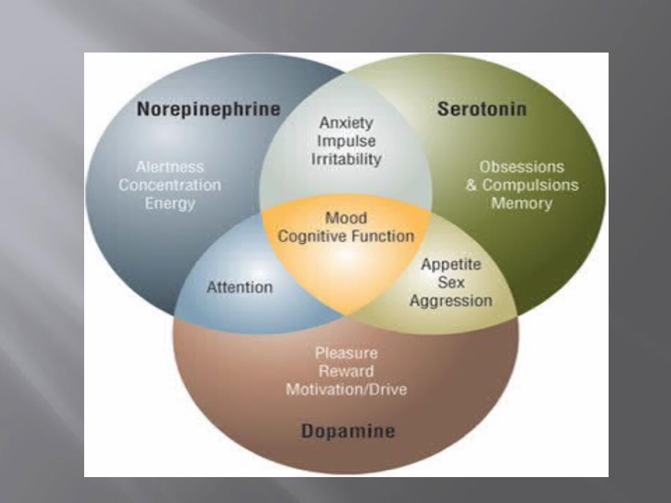
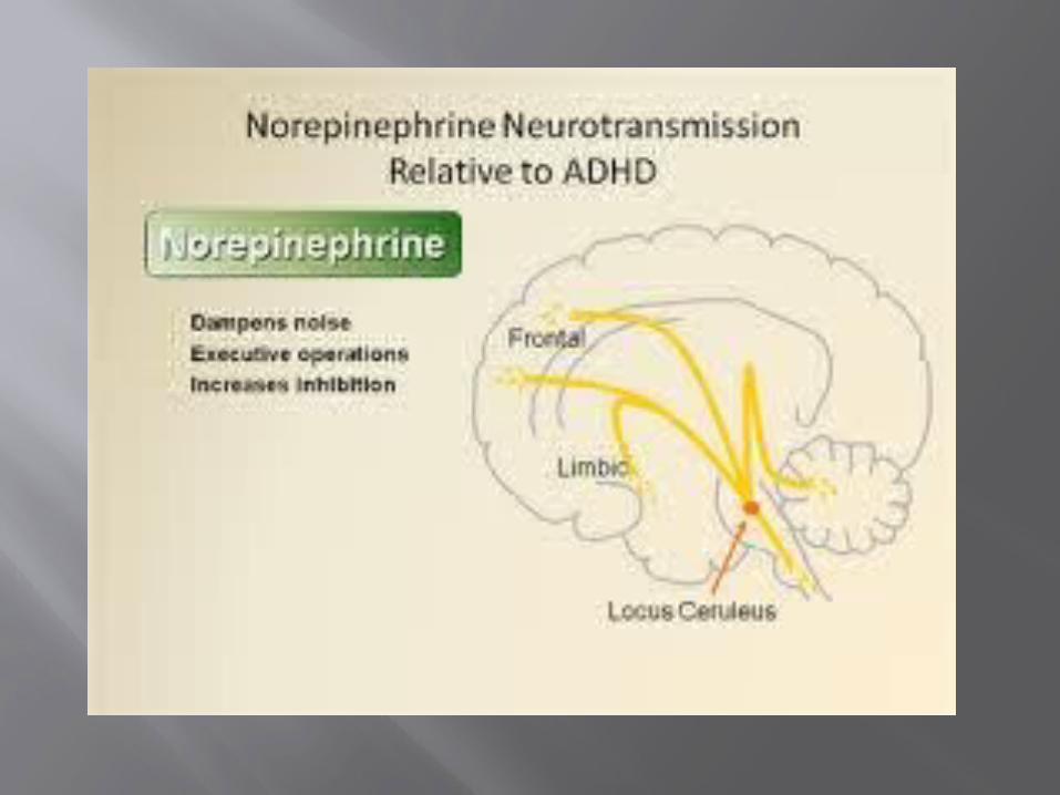
For details refer ANS. e.g. fight or flight, on heart, BP, gastrointestinal activity etc. Norepinehrine controls attention & arousal.
ii.Norepinephrine Excitatory Tyrosine, found in pons. Reticular formation, locus coerules, thalamus, mid-brain
Begins inside axoplasm of adrenergic nerve ending is completed inside the secretary vesicles
α1 α2
β1 β2
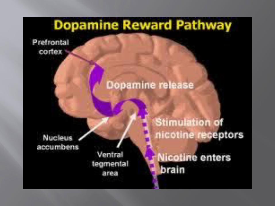
iii. Dopamine Excitatory Tyrosine CNS, concentrated in basal ganglia and dopamine pathways e.g. nigrostriatal, mesocorticolimbic and tubero-hypophyseal pathway
D1 to D5 receptor
Same as above Decreased dopamine in parkinson’s disease.Increased dopamine concentration causes schizophrenia
NeurotransmitterPostsynaptic
effect Derived fromSite of
synthesisPostsynaptic
receptorFate Functions
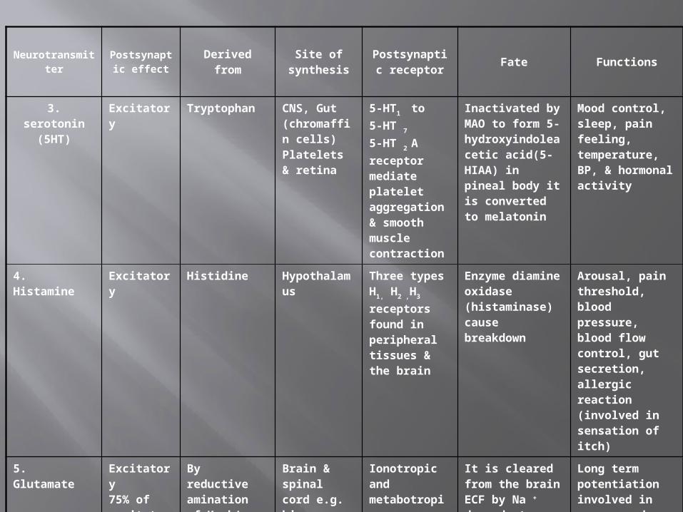
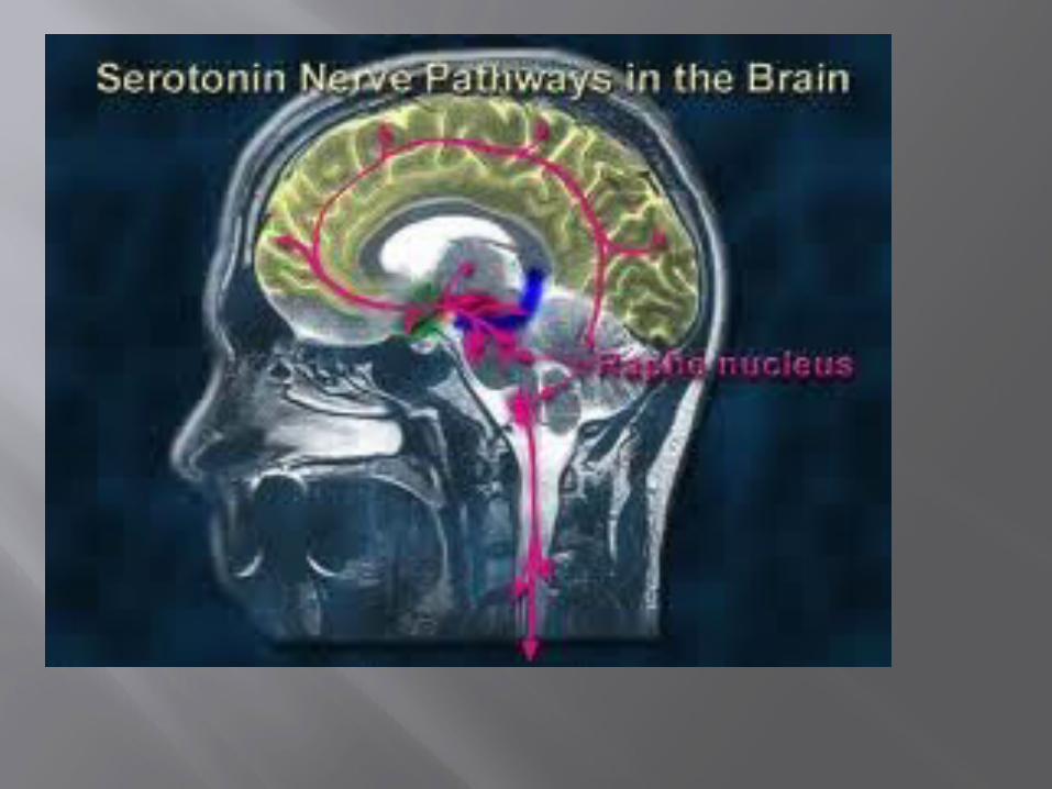
3. serotonin(5HT)
Excitatory Tryptophan CNS, Gut (chromaffin cells) Platelets & retina
5-HT1 to 5-HT
7
5-HT 2 A receptor mediate platelet aggregation & smooth muscle contraction
Inactivated by MAO to form 5-hydroxyindoleacetic acid(5-HIAA) in pineal body it is converted to melatonin
Mood control, sleep, pain feeling, temperature, BP, & hormonal activity
4. Histamine Excitatory Histidine Hypothalamus Three types H1, H2 ,H3 receptors found in peripheral tissues & the brain
Enzyme diamine oxidase (histaminase) cause breakdown
Arousal, pain threshold, blood pressure, blood flow control, gut secretion, allergic reaction (involved in sensation of itch)
5. Glutamate Excitatory75% of excitatory transmission in the brain
By reductive amination of Kreb’s cycle intermediate α –ketoglutarate.
Brain & spinal cord e.g. hippocampus
Ionotropic and metabotropic receptors.Three types of ionotropic receptors e.g. NMDA, AMPA and kainate receptors.
It is cleared from the brain ECF by Na + dependent uptake system in neurons and neuroglia.
Long term potentiation involved in memory and learning by causing Ca++ influx.
NeurotransmitterPostsynaptic
effect Derived fromSite of
synthesisPostsynaptic
receptorFate Functions
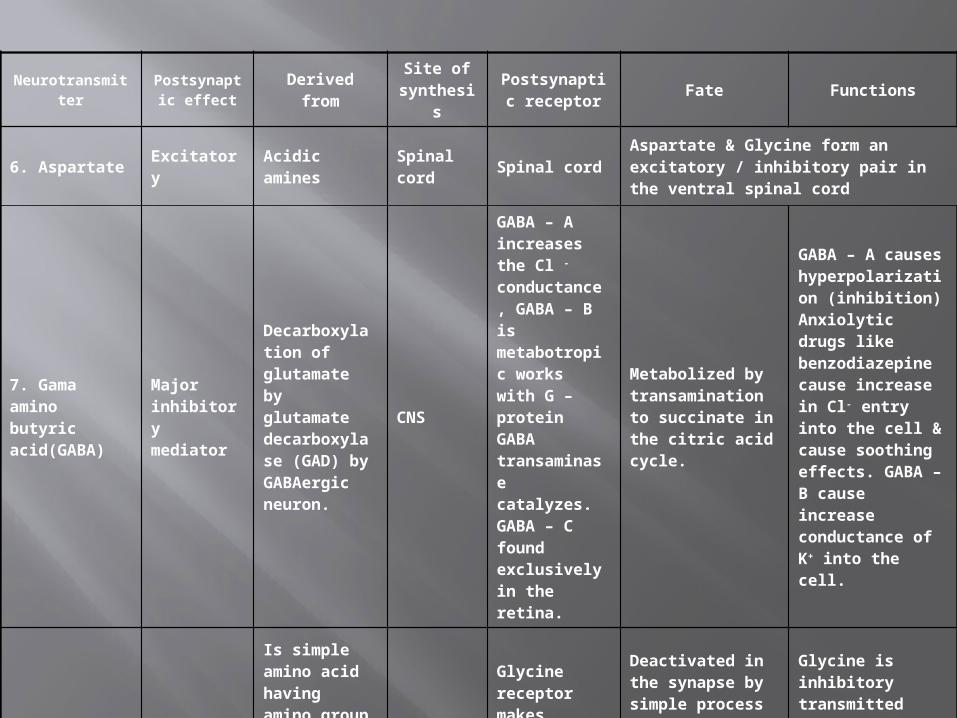
6. Aspartate Excitatory Acidic amines Spinal cord Spinal cordAspartate & Glycine form an excitatory / inhibitory pair in the ventral spinal cord
7. Gama amino butyric acid(GABA)
Major inhibitory mediator
Decarboxylation of glutamate by glutamate decarboxylase (GAD) by GABAergic neuron.
CNS
GABA – A increases the Cl - conductance, GABA – B is metabotropic works with G – protein GABA transaminase catalyzes. GABA – C found exclusively in the retina.
Metabolized by transamination to succinate in the citric acid cycle.
GABA – A causes hyperpolarization (inhibition) Anxiolytic drugs like benzodiazepine cause increase in Cl- entry into the cell & cause soothing effects. GABA – B cause increase conductance of K+ into the cell.
8. Glycine Inhibitory
Is simple amino acid having amino group and a carboxyl group attached to a carbon atom
Spinal cord
Glycine receptor makes postsynaptic membrane more permeable to Cl- ion.
Deactivated in the synapse by simple process of reabsorbtion by active transport back into the presynaptic membrane
Glycine is inhibitory transmitted found in the ventral spinal cord. It is inhibitory transmitter to Renshaw cells.
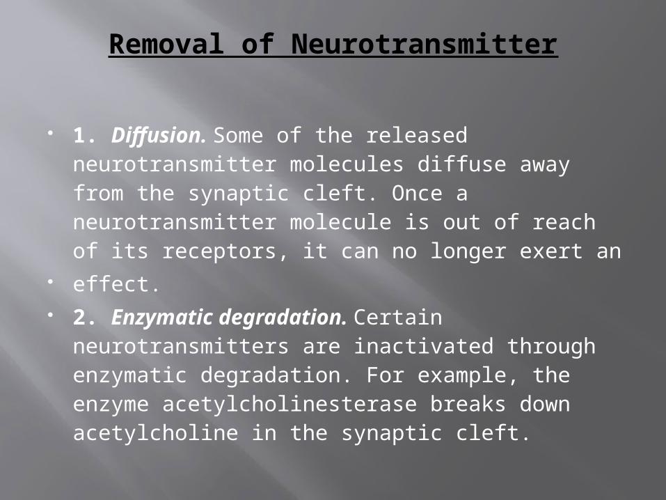
Removal of Neurotransmitter
1. Diffusion. Some of the released neurotransmitter molecules diffuse away from the synaptic cleft. Once a neurotransmitter molecule is out of reach of its receptors, it can no longer exert an
effect. 2. Enzymatic degradation. Certain
neurotransmitters are inactivated through enzymatic degradation. For example, the enzyme acetylcholinesterase breaks down acetylcholine in the synaptic cleft.
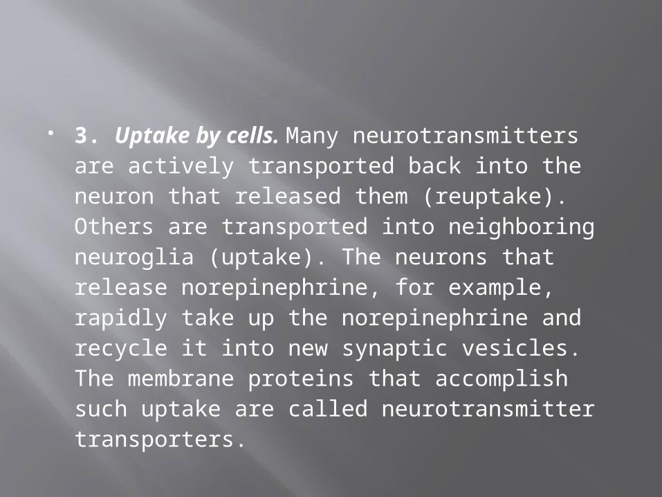
3. Uptake by cells. Many neurotransmitters are actively transported back into the neuron that released them (reuptake). Others are transported into neighboring neuroglia (uptake). The neurons that release norepinephrine, for example, rapidly take up the norepinephrine and recycle it into new synaptic vesicles. The membrane proteins that accomplish such uptake are called neurotransmitter transporters.
NEUROPEPTIDES
Substance P Found in sensory neurons, spinal
cord pathways, and parts of brain associated with pain; enhances perception of pain.
Enkephalins
Inhibit pain impulses by suppressing release of substance P; may have a role in memory and learning, control of body temperature, sexual activity, and mental illness.
Endorphins Inhibit pain by blocking release of
substance P; may have a role in memory and learning, sexual activity, control of body temperature, and mental illness.
Dynorphins May be related to controlling pain and
registering emotions.
Hypothalamic releasing Produced by the hypothalamus;
regulate and inhibiting hormones the release of hormones by the anterior pituitary.
Angiotensin II
Stimulates thirst; may regulate blood pressure in the brain. As a hormone causes vasoconstriction and promotes release of aldosterone, which increases the rate of salt and water reabsorption by the kidneys.
Cholecystokinin (CCK)
Found in the brain and small intestine; may
regulate feeding as a “stop eating” signal. As a hormone, regulates pancreatic enzyme secretion during digestion, and contraction of smooth muscle in the gastrointestinal tract.

THANK YOU