
Neurotransmitter Detection Using Corona Phase Molecular...
12
Neurotransmitter Detection Using Corona Phase Molecular Recognition on Fluorescent Single-Walled Carbon Nanotube Sensors Sebastian Kruss, † Markita P. Landry, † Emma Vander Ende, Barbara M.A. Lima, Nigel F. Reuel, Jingqing Zhang, Justin Nelson, Bin Mu, Andrew Hilmer, and Michael Strano* Department of Chemical Engineering, Massachusetts Institute of Technology, Cambridge, Massachusetts 02139, United States * S Supporting Information ABSTRACT: Temporal and spatial changes in neurotransmitter concentrations are central to information processing in neural networks. Therefore, biosensors for neurotransmitters are essential tools for neuroscience. In this work, we applied a new technique, corona phase molecular recognition (CoPhMoRe), to identify adsorbed polymer phases on fluorescent single-walled carbon nanotubes (SWCNTs) that allow for the selective detection of specific neurotransmitters, including dopamine. We functionalized and suspended SWCNTs with a library of different polymers (n = 30) containing phospholipids, nucleic acids, and amphiphilic polymers to study how neurotransmitters modulate the resulting band gap, near-infrared (nIR) fluorescence of the SWCNT. We identified several corona phases that enable the selective detection of neurotransmitters. Catecholamines such as dopamine increased the fluorescence of specific single-stranded DNA- and RNA- wrapped SWCNTs by 58−80% upon addition of 100 μM dopamine depending on the SWCNT chirality (n,m). In solution, the limit of detection was 11 nM [K d = 433 nM for (GT) 15 DNA-wrapped SWCNTs]. Mechanistic studies revealed that this turn-on response is due to an increase in fluorescence quantum yield and not covalent modification of the SWCNT or scavenging of reactive oxygen species. When immobilized on a surface, the fluorescence intensity of a single DNA- or RNA-wrapped SWCNT is enhanced by a factor of up to 5.39 ± 1.44, whereby fluorescence signals are reversible. Our findings indicate that certain DNA/ RNA coronae act as conformational switches on SWCNTs, which reversibly modulate the SWCNT fluorescence. These findings suggest that our polymer−SWCNT constructs can act as fluorescent neurotransmitter sensors in the tissue-compatible nIR optical window, which may find applications in neuroscience. ■ INTRODUCTION Neurotransmitter release is the basis of neurotransmission in chemical synapses and a central part of data processing in the brain. Therefore, the spatiotemporal concentration profile of neurotransmitters is of intrinsic interest for neuroscience and necessary to fully understand neural networks. Many diseases are directly related to altered patterns of neurotransmission, including Parkinson’s disease, which is directly linked to depletion of dopaminergic neurons and low levels of the neurotransmitter dopamine. 1 However, few analytical methods exist that can measure neurotransmitter gradients with high spatial and temporal resolution. In this work, we employed corona phase molecular recognition (CoPhMoRe) to develop novel fluorescent sensors for neurotransmitters. This technique involves screening and selection of a library of nanoparticle- adsorbed organic phases, or coronae, for their ability to distinguish specific molecules. 2 Semiconducting single-walled carbon nanotubes (SWCNTs) conjugated with polymers are a new class of biosensors. 3 SWCNTs consist only of a carbon monolayer. Therefore, small perturbations in the nanotube environment, which we call the nanotube corona, can be used to detect single-molecule analytes. 4 SWCNTs fluoresce in the near-infrared (nIR), which is optimal for biomedical applications because it falls into the optical tissue transparency window. 5 SWCNTs can be dispersed in various polymers, and this non-covalent function- alization renders the sensor biocompatible, water-soluble, and specific for certain analytes. Recently, sensors for reactive oxygen species (ROS), nitric oxide (NO), glucose, and DNA have been developed. 4,6−9 The detection of neurotransmitters is very challenging for various reasons: Interfering substances such as chemical or structural neurotransmitter analogues prevent sensitive detec- tion, whereas the short time scales, small length scales, and low neurotransmitter concentrations make it difficult to observe neurotransmitter release in real time. 10−12 Nevertheless, sensors are necessary tools to study neurotransmitter release on the single-cell/synapse level and also in small networks of neurons or in vivo. Electrochemical methods such as amperometry or cyclic voltammetry have provided valuable information on redox- active neurotransmitters such as dopamine, epinephrine, and serotonin. Many biological studies were only possible because these methods provided quantitative information about neuro- transmitter concentrations around cells, in brain slices, and in Received: October 10, 2013 Published: December 19, 2013 Article pubs.acs.org/JACS © 2013 American Chemical Society 713 dx.doi.org/10.1021/ja410433b | J. Am. Chem. Soc. 2014, 136, 713−724
Transcript of Neurotransmitter Detection Using Corona Phase Molecular...

Neurotransmitter Detection Using Corona Phase MolecularRecognition on Fluorescent Single-Walled Carbon Nanotube SensorsSebastian Kruss,† Markita P. Landry,† Emma Vander Ende, Barbara M.A. Lima, Nigel F. Reuel,Jingqing Zhang, Justin Nelson, Bin Mu, Andrew Hilmer, and Michael Strano*
Department of Chemical Engineering, Massachusetts Institute of Technology, Cambridge, Massachusetts 02139, United States
*S Supporting Information
ABSTRACT: Temporal and spatial changes in neurotransmitterconcentrations are central to information processing in neuralnetworks. Therefore, biosensors for neurotransmitters are essentialtools for neuroscience. In this work, we applied a new technique,corona phase molecular recognition (CoPhMoRe), to identify adsorbedpolymer phases on fluorescent single-walled carbon nanotubes(SWCNTs) that allow for the selective detection of specificneurotransmitters, including dopamine. We functionalized andsuspended SWCNTs with a library of different polymers (n = 30)containing phospholipids, nucleic acids, and amphiphilic polymers to study how neurotransmitters modulate the resulting bandgap, near-infrared (nIR) fluorescence of the SWCNT. We identified several corona phases that enable the selective detection ofneurotransmitters. Catecholamines such as dopamine increased the fluorescence of specific single-stranded DNA- and RNA-wrapped SWCNTs by 58−80% upon addition of 100 μM dopamine depending on the SWCNT chirality (n,m). In solution, thelimit of detection was 11 nM [Kd = 433 nM for (GT)15 DNA-wrapped SWCNTs]. Mechanistic studies revealed that this turn-onresponse is due to an increase in fluorescence quantum yield and not covalent modification of the SWCNT or scavenging ofreactive oxygen species. When immobilized on a surface, the fluorescence intensity of a single DNA- or RNA-wrapped SWCNTis enhanced by a factor of up to 5.39 ± 1.44, whereby fluorescence signals are reversible. Our findings indicate that certain DNA/RNA coronae act as conformational switches on SWCNTs, which reversibly modulate the SWCNT fluorescence. These findingssuggest that our polymer−SWCNT constructs can act as fluorescent neurotransmitter sensors in the tissue-compatible nIRoptical window, which may find applications in neuroscience.
■ INTRODUCTION
Neurotransmitter release is the basis of neurotransmission inchemical synapses and a central part of data processing in thebrain. Therefore, the spatiotemporal concentration profile ofneurotransmitters is of intrinsic interest for neuroscience andnecessary to fully understand neural networks. Many diseasesare directly related to altered patterns of neurotransmission,including Parkinson’s disease, which is directly linked todepletion of dopaminergic neurons and low levels of theneurotransmitter dopamine.1 However, few analytical methodsexist that can measure neurotransmitter gradients with highspatial and temporal resolution. In this work, we employedcorona phase molecular recognition (CoPhMoRe) to developnovel fluorescent sensors for neurotransmitters. This techniqueinvolves screening and selection of a library of nanoparticle-adsorbed organic phases, or coronae, for their ability todistinguish specific molecules.2
Semiconducting single-walled carbon nanotubes (SWCNTs)conjugated with polymers are a new class of biosensors.3
SWCNTs consist only of a carbon monolayer. Therefore, smallperturbations in the nanotube environment, which we call thenanotube corona, can be used to detect single-moleculeanalytes.4 SWCNTs fluoresce in the near-infrared (nIR),which is optimal for biomedical applications because it falls
into the optical tissue transparency window.5 SWCNTs can bedispersed in various polymers, and this non-covalent function-alization renders the sensor biocompatible, water-soluble, andspecific for certain analytes. Recently, sensors for reactiveoxygen species (ROS), nitric oxide (NO), glucose, and DNAhave been developed.4,6−9
The detection of neurotransmitters is very challenging forvarious reasons: Interfering substances such as chemical orstructural neurotransmitter analogues prevent sensitive detec-tion, whereas the short time scales, small length scales, and lowneurotransmitter concentrations make it difficult to observeneurotransmitter release in real time.10−12 Nevertheless, sensorsare necessary tools to study neurotransmitter release on thesingle-cell/synapse level and also in small networks of neuronsor in vivo.Electrochemical methods such as amperometry or cyclic
voltammetry have provided valuable information on redox-active neurotransmitters such as dopamine, epinephrine, andserotonin. Many biological studies were only possible becausethese methods provided quantitative information about neuro-transmitter concentrations around cells, in brain slices, and in
Received: October 10, 2013Published: December 19, 2013
Article
pubs.acs.org/JACS
© 2013 American Chemical Society 713 dx.doi.org/10.1021/ja410433b | J. Am. Chem. Soc. 2014, 136, 713−724

vivo.10,13 However, electrochemical methods are limited tomolecules that can be oxidized or reduced at the electrode.Moreover, electrodes are usually large compared to the site ofneurotransmitter release, so parallel/spatial detection ofneurotransmitter release has not been achieved at the scale ofthe synapse. Other approaches for neurotransmitter detectionare based on fluorescent labeling. Neurotransmitter-containingvesicles can be labeled with dyes, and the fusion of thesevesicles with the synaptic cell membrane can be observed byfluorescence microscopy. Vesicles that contain neurotransmit-ters have different pH values compared with the extracellularspace. As such, pH-dependent fluorescent proteins/dyes havebeen used to visualize exocytosis.11 However, this approachlacks direct optical neurotransmitter identification and rathervisualizes structural changes of vesicles and membranes insteadof directly visualizing the neurotransmitter itself. In contrast,fluorescent false neurotransmitters (FFNs) are fluorescent dyeshaving chemical structures that are similar to the neuro-transmitter of interest and are loaded into synaptic vesicles byneurotransmitter transporters. This method was used tovisualize fluorescence depletion upon stimulation in singlesynapses of dopaminergic neurons14,15 but does not directlydetect natural neurotransmitters. Another approach is based onmodifying biological recognition units of neurotransmitters andconjugating them with fluorescent dyes. This method was usedto engineer γ-aminobenzoic acid (GABA) sensors andglutamate sensors.16,17 Recently, glutamate was measured byusing green fluorescent protein (GFP)-conjugated glutamatereceptors that can be also transfected into cells.18 While theseefforts have provided us with many tools to study neuronalpathways, there is still a great need for a direct high-resolutionspatiotemporal detection tool for neurotransmitters, which willenable completely new insights into how neurons communicatewith each other.Here we present a new fluorescent sensor platform for the
optical detection of neurotransmitters that is based on the nIRfluorescence of SWCNTs. Fluorescent sensors with distinctsensitivity and specificity for different neurotransmitters weresynthesized by wrapping carbon nanotubes with differentpolymers. The specificity of these sensors does not arise fromusing known biological recognition elements. Rather, it is basedon the unique conformational changes that happen when apolymer is pinned at a nanoparticle surface, creating a definedcorona. Only the combination of a polymer and its confinementon the nanoparticle cause recognition and signal transductionin the presence of the analyte according to this scheme, whichwe have introduced previously as corona phase molecularrecognition (CoPhMoRe).2
We show that catecholamines such as dopamine andepinephrine increase the fluorescence of DNA- or RNA-wrapped carbon nanotubes by enhancing their fluorescencequantum yield. This fluorescence increase is an importantfinding with respect to carbon nanotube photophysics, as itpresents the first example of CoPhMoRe that leads to afluorescence turn-on signal. We further show how theseunderlying mechanisms are the founding principle for afluorescent turn-on sensor for dopamine.
■ EXPERIMENTAL SECTIONMaterials. Chemicals, including the neurotransmitters, were
purchased from Sigma-Aldrich (USA), unless stated otherwise.Single-stranded DNA (ssDNA) and RNA (ssRNA) sequences (N1−N13) were purchased from IDT (USA). HiPCO SWCNTs were
purchased from Unidym and processed according to the suggestionsby the manufacturer (extraction of non-SWCNT material by phaseseparation in water/hexane). Phospholipids were purchased fromAvanti Polar Lipids (USA). Compounds P1−P4 were synthesized aspreviously reported.2,19 Compound P5 was purchased from Poly-scitech (USA).
Preparation and Characterization of Wrapped SWCNTs. Weused direct probe-tip sonication (Cole Parmer) to encapsulate HiPCOSWCNTs (Unidym) with the several nucleic acid derivatives used inthis work (10 min, 40% amplitude). For this purpose, the nucleic acidwas dissolved in a 0.1 M NaCl solution (100 mg/mL). Next, 20 μL ofthe DNA solution was added to 980 μL of 0.1 M NaCl solution and 1mg HiPCO SWCNTs, and the resulting suspension was tip-sonicatedfor 10 min (3 mm tip diameter, 40% amplitude) in an ice bath. Aftersonication, we centrifuged the samples two times for 90 min at 16100gand collected the supernatant. For all other polymers we used dialysis:First, sodium cholate (SC)-wrapped SWCNTs were prepared by tipsonication of 2 wt % SC in water (6 mm tip diameter, 40% amplitude,1 h). These samples were centrifuged at 150000g (Beckmann Coulterultracentrifuge) for 4 h. Second, the polymer (1 wt %) was dissolved ina solution of SC-wrapped SWCNTs and dialyzed for 5 days againstwater in a 3.5 kDa molecular-weight cutoff dialysis bag (Spectra/Pordialysis membrane). All of the samples were analyzed by UV−vis−nIRspectroscopy (Shimadzu UV-3101PC).
CoPhMoRe Screening and Solution-Based Experiments.Polymer-wrapped SWCNTs were diluted in phosphate-buffered saline(PBS) (pH 7.4, 10 mM) to a SWCNT concentration corresponding toan absorbance of 0.036 at 632 nm. To 96 well-plates (Microtest 96tissue culture plate, BD) were added 198 μL aliquots of this solution.The nIR fluorescence was collected on a Zeiss AxioVision invertedmicroscope coupled to a PI Acton SP2500 spectrometer and aPrinceton Instruments InGaAs OMA V array detector. The samplewas excited with a 785 nm photodiode laser (450 mW). Exposuretimes were 10 s. The neurotransmitter was dissolved in PBS (10 mMstock solution), and 2 μL of the solution was added into the wells at afinal concentration of 100 μM or smaller. In a typical experiment, thefluorescence responses of nine different neurotransmitters for onepolymer−SWCNT combination were measured in triplicate (n = 3).The dopamine solution was freshly prepared and used within 10 min.Subsequent measurement of the samples in the well plate took around10 min. The experiments with ROS scavengers were done similarly:Spectra were collected before addition of the scavengers, after additionof the scavengers, and after addition of dopamine (100 μM). All of theexperiments were performed in PBS. We added neurotransmitters andother compounds in small volumes (1 vol %), which changed theSWCNT concentration only slightly (∼1%).
Microscopy Setup. Visible-Wavelength TIRF Microscopy. Thetotal internal reflection fluorescence (TIRF) microscope uses asupercontinuum excitation source (NKT, SuperK Extreme EXR15),which is relayed through several conditioning optics, including a 10×beam expansion telescope to expand the diameter of the excitationbeam to approximately 1.5 cm. This allows overfill into the backaperture of the microscope objective and enables maximal excitationarea of the sample. The expanded beam is directed into a plano-convexfocusing lens (TIR lens), which is mounted on a three-axistranslational stage [Figure S3a in the Supporting Information (SI)].The angle of beam incidence (Θ) is changed by moving the TIR lensin the plane perpendicular to beam propagation. When Θ = Θc (thecritical angle), all of the incident light is reflected from the surface ofthe glass−water interface and generates an evanescent field that excitesfluorophores in the vicinity of the glass−water interface.
Before light enters the microscope objective, a 531 nm narrow-bandpass filter (FF01-531/40-25, Semrock) tunes the excitationwavelength relayed to the sample through the objective. A 562 nmdichroic mirror (FF562-Di03-25 × 36, Semrock) reflects thisexcitation wavelength while passing the Cy3 characteristic emissionby passing wavelengths above ∼562 nm. The emission path of theprism-TIRF microscope begins with the collection of the emissionfrom the sample by the objective. This emission passes through a 593nm bandpass filter (FF01-593/40-25, Semrock) that reflects the
Journal of the American Chemical Society Article
dx.doi.org/10.1021/ja410433b | J. Am. Chem. Soc. 2014, 136, 713−724714

excitation laser light (532 nm) but transmits the fluorescence emissionwavelength of the Cy3 dye (570 nm). A pair of doublet lenses expandthe 75 μm × 75 μm image to fill the CCD sensor (8.2 mm × 8.2 mm;iXon3 EMCCD, Andor Technology).nIR Epifluoresence Microscopy. Single-SWCNT data were
collected on an inverted microscope (Zeiss Axiovert 200) equippedwith a 100× objective (Zeiss, α-Plan-APOCHROMAT 100×/1.46 OilDIC (UV) VIS-IR) attached to a 2D InGaAs CCD array (OMA-V 2D,Princeton Instruments) (SI Figure S3b). Fluorescence was excited by a658 nm 200 mW diode-pumped solid-state laser (CrystaLaser, RCL-100-660).Microfluidic Chamber Preparation. Microfluidic chambers for
visible TIRF experiments were surface-passivated with bovine serumalbumin (BSA) to avoid nonspecific adsorption of the fluorescentlytagged biomolecules to the glass surface, as previously described.20
The formation of microfluidic flow channels was achieved by cutting
several channels out of double-sided tape and sealing the tape betweenthe quartz slide and the coverslip.
For flow experiments, a micropipet tip was introduced at the flowinlet of a single microfluidic channel, and a syringe was coupled intothe flow outlet to change the buffer conditions of the experimentalchamber. To passivate these chambers, BSA coating of the glass slidesurface and a glass coverslip was performed by flowing in a 100 nMsolution of BSA tagged with biotin (BSA-Bt).21 Subsequently, PBSbuffer was flowed through the microfluidic channel to rinsenonadhered BSA-Bt. Next, a 33 nM solution of neutravidin proteinwas flowed in and allowed to incubate for 10 min for binding to thebiotin moiety on the surface-bound BSA-Bt. Neutravidin proteinbound nonspecifically to the SWCNT surface, allowing surfaceimmobilization of the DNA-wrapped SWCNT samples within themicrofluidic chambers.
Figure 1. Screening of SWCNT−polymer conjugates for fluorescence modulation by neurotransmitters. (a) Fluorescence responses of polymer−SWCNT conjugates (x axis) to neurotransmitters (y axis). The normalized fluorescence changes (I − I0)/I0 of different polymer−SWCNTconjugates upon addition of different neurotransmitters (100 μM) are shown in a color-coded heat map. Polymer structures are shown in Figure 2.N1−N13 are nucleic acids, PL1−PL12 are phospholipids, and P1−P5 are amphiphilic polymers. Red indicates a fluorescence increase and blue afluorescence decrease. (b) nIR fluorescence spectrum (785 nm excitation) of (GT)15−SWCNT before and after addition of 100 μM dopamine inPBS. The fluorescence intensity increased by up to 80% depending on the chirality of the SWCNTs. The distinct peaks correspond to differentSWCNT chiralities in the sample. (c) Schematic of the fluorescent turn-on sensor for dopamine.
Journal of the American Chemical Society Article
dx.doi.org/10.1021/ja410433b | J. Am. Chem. Soc. 2014, 136, 713−724715

For reversibility experiments, ibidi flow chambers were used (ibidi,sticky-Slide VI 0.4) and attached to a pump (Ismatec). Glass substrateswere functionalized with (3-aminopropyl)triethoxysilane (APTES) inethanol (1% APTES, 1% water) and then mounted under the flowchamber. Next, 1 mg/L (GT)15- or (GU)15-wrapped SWCNT solutionin PBS was incubated in the channels to adsorb SWCNTs to thesurface. For the reversibility and single-SWCNT experiments,dopamine (100 μM) and PBS were periodically washed over thesurface (flow rate 50 μL/min).
■ RESULTS
CoPhMoRe Screening for Polymer−SWCNT Conju-gates That Change Fluorescence upon Neurotransmit-ter Addition. We suspended HiPCO SWCNTs in variouspolymers (see the Experimental Section) to solubilize them inaqueous solution, render them sensitive for neurotransmitters,and measure the influence of neurotransmitters on theirfluorescence spectra (Figure 1). We elected to use unseparatedSWCNTs in order to detect band gap- or diameter-dependentanalyte responses. For this purpose, we used either directultrasonication or dialysis of SC-wrapped carbon nanotubes.The structures are shown in Figure 2. The polymers areabbreviated as follows: N1−N12 are ssDNA sequences, N13 isthe ssRNA sequence (GU)15, and PL1−PL12 are phospholipidswith different head and tail groups. P1−P5 are differentamphiphilic polymers with a hydrophobic group that binds tothe carbon surface and a hydrophilic group that renders the
conjugate water-soluble. All of the SWCNT−polymer hybridcombinations were characterized by absorption spectroscopy.Samples were then diluted to a final concentration of 1 mg/L inPBS at pH 7.4. SWCNT−polymer conjugates were excited by a785 nm photodiode laser, and their emission spectra between950 and 1250 nm were recorded. Fluorescence spectra werecollected before and after addition of the neurotransmitters(final concentration 100 μM). The library of neurotransmittersincluded acetylcholine, serotonin, dopamine, histamine, GABA,glutamic acid, glycine, aspartic acid, and (−)-epinephrine.Fluorescence changes were calculated by comparing the
SWCNT intensities at the fluorescence emission peak of the(9,4) chirality SWCNTs (around 1132 nm for nucleic acid-wrapped SWCNTs) because in most cases the (9,4) peakprovided the largest fluorescence count in our setup. Thesensor response was defined as (I − I0)/I0, that is, thedifference between the final fluorescence intensity I and thestarting intensity I0, (I − I0), normalized by the startingintensity I0. For all of the polymers (Figure 2), fluorescenceintensity modulations but no wavelength peak shifts wereobserved. The optical responses for the different SWCNT−polymer hybrids in the presence of various neurotransmittersare shown in Figure 1. The fluorescence changes are color-coded (ΔI/I0 > 0, red; ΔI/I0 < 0, blue), and the intensity of thecolor corresponds to the absolute intensity of the response. Wefound several SWCNT−polymer conjugates with clear
Figure 2. Polymers that were used to suspend/functionalize SWCNTs and generate a sensor library. N1−N12 are ssDNA sequences, N13 is anssRNA sequence, PL1−PL12 are phospholipids with different head and tail groups, and P1−P5 are amphiphilic polymers with hydrophobic andhydrophilic groups.
Journal of the American Chemical Society Article
dx.doi.org/10.1021/ja410433b | J. Am. Chem. Soc. 2014, 136, 713−724716

modulations in nIR fluorescence upon neurotransmitteraddition.All of the DNA- or RNA-wrapped SWCNTs (N1−N13)
showed an increase in fluorescence from 11% to 80% uponaddition of dopamine and 3% to 62% for (−)-epinephrine.Most nucleic acid-wrapped SWCNTs showed a fluorescencedecrease upon addition of 100 μM serotonin. In general,aromatic (and redox-active) neurotransmitters such as dop-amine showed stronger responses than non-redox-activeneurotransmitters such as glycine. The SWCNT−polymerconjugate with the strongest fluorescence increase, N1,corresponds to (GT)15 DNA-wrapped SWCNTs. We usedthis specific DNA sequence as an exemplary model to study thefluorescence enhancement of nucleic acid−SWCNT conjugatesto dopamine and to investigate the mechanism of thismolecular recognition. In particular, we focused on dopaminebecause it is one of the central neurotransmitters in the brainand is linked to reward processing and learning.22
In Figure 1b, the fluorescence spectrum of (GT)15−SWCNTin PBS before and after the addition of 100 μM dopamine isshown. Upon addition of dopamine, the fluorescence increasedby a factor of 58−81%. Different peaks correspond to differentchiralities of SWCNTs that are present in a typical HiPCOsample, where the fluorescence response depended on thechirality (n,m) of the SWCNTs [(6,5), 991 nm, 0.80 ± 0.12;(7,5), 1044 nm, 0.63 ± 0.088; (10,2), 1077 nm, 0.66 ± 0.08;(9,4), 1132 nm, 0.73 ± 0.11; (8,6), 1203 nm, 0.58 ± 0.09].Dopamine changed the SWCNT fluorescence intensity but didnot shift the spectrum. Figure 1c shows the basic principle ofthis sensor platform: a polymer is pinned to a carbon nanotubesurface and mediates the interaction of the analyte with theSWCNT corona.To determine the source of increased SWCNT fluorescence
upon dopamine addition, we took absorption spectra of(GT)15−SWCNT. The SWCNT absorption spectra did notchange upon addition of 100 μM dopamine (SI Figure S1a).Both the intensities and positions of the E11 and E22 opticaltransitions remained identical before and after dopamineaddition. Therefore, the change in SWCNT fluorescence(Figure 1b) could not be attributed to changes in the
absorption cross section upon dopamine addition and mustbe due to an increase in quantum yield of (GT)15−SWCNTfluorescence.Another potential source of SWCNT fluorescence change
could be covalent modification of the SWCNT surface bydopamine. Dopamine is a reactive molecule that is known topolymerize readily in solution and could covalently modify thecarbon surface and thus induce a change in SWCNTfluorescence.23,24 The Raman spectrum of a 1 mg/L (GT)15−SWCNT sample in PBS before and 10 min after the addition ofdopamine showed no difference in Raman intensity (SI FigureS1b). The G peak at 1591 cm−1 and the D peak at 1307 cm−1
did not change upon dopamine addition. This invariance of theG/D ratio indicates that no sp3 defects were introduced intothe carbon lattice by dopamine.
Sensitivity and Kinetics of the Fluorescent SWCNT-Based Dopamine Sensors. We used the peak of the (9,4)SWCNTs to calculate the sensor response (I − I0)/I0 anddetermine a calibration curve (Figure 3a). Even at dopamineconcentrations of 100 pM, a fluorescence increase of 5% wasobserved. The approximate linear regime of the sensor wasbetween 10 nM and 10 μM, on the basis of the sensor responseat 10 μM, 1 μM, 100 nM, and 10 nM (linear fit on alogarithmic scale, R2 > 0.98). At higher concentrations (>10μM), the sensor response saturated. The calibration curve canbe described by a kinetic adsorption model in which A is theanalyte (dopamine) and θ is an available recognition site on theSWCNT sensor. The relationship between the analyte andavailable docking sites for dopamine can be described asfollows:
θ θ+ ⇄A A (1)
As we show in later single-molecule results, this turn-onresponse is instantaneous. Therefore, the equilibrium for thisreaction can be modeled as an instantaneous reaction and canbe described by the following equilibrium constant:
θθ
=K[A ]
[A][ ]A(2)
Figure 3. Calibration curve and dopamine response kinetics of the (GT)15−SWCNT sensor. (a) Calibration curve of the dopamine sensor insolution (PBS), n = 3, three replicates each. (b) Time-dependent response after addition of 100 μM dopamine (at t = 0 min, the starting intensitywas measured). The decrease of fluorescence is most probably related to dopamine polymerization. Errors are standard deviations.
Journal of the American Chemical Society Article
dx.doi.org/10.1021/ja410433b | J. Am. Chem. Soc. 2014, 136, 713−724717

The total concentration of available recognition sites, [θ]tot, isthe sum of the concentrations of the free and occupied sites:
θ θ θ= +[ ] [A ] [ ]tot (3)
We can use eqs 2 and 3 to derive the following expression for[θ]tot in terms of the dopamine concentration [A]:
θ θ θ
θ θ
θ
θ
= +
= +
= +
=+
⎛⎝⎜
⎞⎠⎟
⎛⎝⎜
⎞⎠⎟
K
K
KK
[ ] [A ] [ ]
[A ][A ]
[A]
[A ] 11
[A]
[A ][A] 1
[A]
tot
A
A
A
A (4)
If we assume that the sensor response (i.e., the normalizedintensity change) is proportional to the Aθ/θtot ratio, we findthat
α θθ
α−
= + =+
+I I
IB
KK
B[A ][ ]
([A] )([A] ) 1
n
n0
0 tot
A
A (5)
where the parameter B accounts for background and n forcooperativity. Fitting the curve in Figure 3a results in a
proportionality factor α = 0.55 with B = 0.032, Kd = 1/KA = 433nM, and n = 0.66, indicating negative cooperativity (R2 > 0.99).The limit of detection was 11 nM; this value was calculated byadding the sensor response for the addition of only buffer(PBS) to 3 times the standard deviation (noise) and then usingthe fit to eq 5 to calculate a concentration (limit of detection)from this value.We observed time-dependent variations in the absolute
magnitude of the sensor response, which we attributed to theknown phenomenon of dopamine polymerization in solu-tion.23,25,26 The time dependence of the sensor response can beexplained in terms of a polymerization product C that quenchesthe SWCNT fluorescence. Dopamine (A) is oxidized todopamine quinone (B), which then polymerizes to give C:
→ →A B Ck k1 2
Because of the time frame of the experiment, we can assumethat [A] ≫ [B] ≫ [C] and neglect back-reactions. The firstreaction does depend on oxidative species such as O2 and istherefore not dependent on [A]. Therefore, we find that
= k t[B] 1 (6)
=t
kd[C]
d[B]2 (7)
Figure 4. Dopamine-induced conformational shift of DNA. (GT)15 DNA was 5′ terminally tagged with a Cy3 fluorophore, conjugated to a SWCNT,and immobilized on a surface prior to flowing in dopamine. (a) Upon addition of 100 μM dopamine, dequenching of the Cy3 fluorophore due tofluorophore destacking was observed. (b) A positive control showed similar fluorophore dequenching when the short unlabeled complementaryoligonucleotide (AC)6 (50 nM) was added to the system and allowed to hybridize to the Cy3-labeled end of the DNA on the SWCNT. (c) Anegative control with unlabeled DNA showed no signal before or after dopamine addition. (d) A negative control with Cy3-labeled DNA on asurface without the SWCNT also showed no change in Cy3 fluorescence upon addition of dopamine. The histograms show the fluorophore countsbefore (blue) and after (red) addition of dopamine/(AC)6.
Journal of the American Chemical Society Article
dx.doi.org/10.1021/ja410433b | J. Am. Chem. Soc. 2014, 136, 713−724718

If we assume that the sensor response can be described by alinear combination of effects due to quenching (from C) anddequenching (from A), we find that
β−
=−
+=
⎛⎝⎜
⎞⎠⎟
⎛⎝⎜
⎞⎠⎟
I II
I II
tt t
0
0
0
0 0
2
(8)
The parameter β contains the rate constants and a quenchingfactor (β < 0). The sensor response over the time course of 90min is shown in Figure 3b [fit parameters: ((I − I0)/I0)t=0 =0.81 and β = −2.73 × 10⁻⁵ min−2). The initial fluorescenceincrease was always instantaneous. It was only limited bydiffusion of dopamine to the sensor (see the single-SWCNTdata below). However, the fluorescence response changed from80% fluorescence increase to approximately 60% increasewithin 90 min of solubilizing dopamine in PBS buffer, which weattribute to the gradual polymerization of dopamine.23,25,26
Again, the absorption spectra of the SWCNTs were notaffected by the dopamine polymerization within the first 90min, suggesting that precipitation of the sensor or lightabsorption by polymerized dopamine did not play a role (SIFigure S2). Therefore, the age of the dopamine solution andthe time needed to perform the measurements have aninfluence on the fluorescence change. Of note, a 10 mMdopamine stock solution in PBS showed signs of polymer-ization, as evidenced by darkening of the solution within 1 h.After 1 day, a black precipitate had formed. Therefore, wealways prepared fresh dopamine solutions for all assayspresented in this work and discarded dopamine solutions thatwere more than 10 min old (see the Experimental Section).Variations of the initial absolute response in independentexperiments were most likely due to slight variations in the ageof the dopamine solution and the time needed to measure allsamples.Conformational Changes of the Nucleic Acid Back-
bone during Dopamine Recognition. In the absence ofcovalent modification of the SWCNT by dopamine, and
without ROS generation in the sensor system, we turned tosingle-molecule imaging of the sample to visualize anyconformational changes that may accompany dopaminedetection. We encapsulated the SWCNT sample with 5′terminally labeled (GT)15 DNA, where we used the dye Cy3 asa visible tag that reports on the structure of the 5′ end of theDNA. Quenching of organic fluorophores occurs as a functionof proximity to the surface of the SWCNT, thereby creating afluorescent ruler to quantify the degree of DNA desorption.27 Alack of Cy3 fluorescence is expected for a DNA polymer thatremains adsorbed to the SWCNT surface (i.e., within a distanceof 1 nm from the SWCNT surface). Conversely, bright Cy3fluorescence is expected for DNA polymers that desorb fromthe SWCNT surface. We immobilized Cy3-labeled (GT)15−SWCNT complexes [(GT)15−Cy3−SWCNT] to the surface ofa microfluidic channel as described in the Experimental Sectionand employed proper passivation of the slide surfaces to avoidnonspecific interactions of molecules with the surface. Tovisualize the fluorescence signal from surface-adsorbedSWCNT sensors, we used a custom-built TIRF microscope.This excitation method drastically reduced the fluorescentbackground and enabled high-resolution observation ofdynamic single-biomolecule behavior.Immobilized (GT)15−Cy3−SWCNT showed a highly
quenched Cy3 signal, suggesting that the Cy3 dye wasphysically stacked on the SWCNT surface and thereforephotophysically quenched. Upon addition of 100 μm dopaminein PBS buffer, the Cy3 signal instantly brightened, and weobserved a marked increase in the number of Cy3 moleculesthat fluoresced [from 432.9 ± 11.4 pre-dopamine to 1601.8 ±30.9 post-dopamine (mean ± SE); Figure 4a]. The time scalefor the Cy3 brightening and SWCNT nIR brightening shown inFigure 4 was instantaneous within the 0.5 s frame rate,suggesting that both processes [dopamine detection viaSWCNT brightening and desorption of the Cy3 dye on the(GT)15 terminus] occur simultaneously. These results suggest
Figure 5. Reversible dopamine response of immobilized single (GT)15−SWCNT sensors. (a) nIR fluorescence images of sensors that wereimmobilized to a surface in a flow chamber. The red arrows indicate three different carbon nanotubes, the intensities of which are shown in (b−d).The images are from the same spot at different time points, as indicated in (b−d). The surface was periodically exposed to 100 μM dopamine andwashed with PBS. (b−d) Intensity traces of 2 pixel × 2 pixel (585 nm × 585 nm) regions of the SWCNTs indicated by the red arrows in (a). Thesensor response was reversible with a small hysteresis. The sensor response is convoluted by the concentration profile of dopamine (mixing).
Journal of the American Chemical Society Article
dx.doi.org/10.1021/ja410433b | J. Am. Chem. Soc. 2014, 136, 713−724719

that dopamine interacts with the terminal end of the (GT)15−Cy3 DNA in the process of dopamine sensing. To confirm thatdequenching of Cy3 occurs when the fluorophore destacksfrom the SWCNT surface, we performed a positive control toconfirm simultaneous dequenching and destacking for (GT)15−Cy3−SWCNT. To the same surface-immobilized (GT)15−SWCNT we introduced 100 nM unlabeled (AC)6 oligonucleo-tide, which is complementary to the ends of (GT)15−Cy3DNA. Upon introduction of the (AC)6 oligonucleotide to themicrofluidic channel, we observed the same dequenching of theterminal Cy3 dye from the SWCNT surface [from 10.4 ± 0.8pre-(AC)6 to 60.7 ± 1.6 post-(AC)6 (mean ± SE); Figure 4b],albeit at a lower rate due to the slower hybridization dynamicsof (AC)6 to the terminal end of (GT)15−Cy3 DNA.Additional negative controls were performed to ensure that
no Cy3 signal was observed in the absence of the Cy3fluorophore or the SWCNT. First, surface-immobilized(GT)15−SWCNT lacking the Cy3 dye upon the introductionof 100 μM dopamine showed no signal, as expected (Figure4c). Second, we confirmed that no change in Cy3 signal wasobserved when dopamine was introduced to surface-immobi-lized Cy3−(GT)15 DNA without a SWCNT (Figure 4d).Differences in surface coverage due to differences in SWCNTversus DNA surface immobilization were observed (see theExperimental Section), and therefore, our analysis focused onthe relative rather than absolute changes in signal. We alsoestablished that neither dopamine nor (AC)6 alone producedan appreciable signal in the Cy3 emission channel at itscharacteristic emission wavelength of 570 nm, and weconfirmed that addition of a noncomplementary DNA strand,cDNA (GATGCGTGTCTAAGAT), did not increase the Cy3fluorescence [from 15.8 ± 1.5 pre-cDNA to 13.1 ± 1.2 post-cDNA (mean ± SE)].Reversibility of SWCNT-Based Dopamine Sensors. We
tested the reversibility of (GT)15−SWCNT and (GU)15−SWCNT sensors by exposing them periodically to dopamine ina flow chamber. For this purpose, (GT)15−SWCNT and(GU)15−SWCNT were adsorbed to APTES-functionalizedglass slides. The glass slides were mounted into a flow chamberand connected to a pump. The sensors were periodicallyexposed to 100 μM dopamine in PBS and washed with PBS.The results are shown in Figure 5 for (GT)15−SWCNT (alsosee the movie in the Supporting Information).We analyzed the fluorescence intensity traces of 2 pixel × 2
pixel (585 nm × 585 nm) regions, which were most likelysingle SWCNTs. Representative traces for three sensors areshown in Figure 5b−d. Whenever the sensors were exposed todopamine, their fluorescence intensity increased by a factor of5.39 ± 1.44 (mean ± SD) for (GT)15−SWCNT (analysis offirst response peak for n = 20 sensors). The fluorescence of(GU)15−SWCNT increased by a factor of 3.67 ± 1.18. Whenthe surface was washed with PBS, the fluorescence returnedapproximately to the starting intensity. However, a slightincrease in the base intensity level was observed over the timecourse of seven dopamine additions. When the washing timewas extended to >30 min, the initial intensity level wasrecovered. The time constants for recovery of the half-maximum intensity after washing with PBS were 49 ± 34 sfor (GT)15−SWCNT and 34 ± 19 s for (GU)15−SWCNT. Itshould be noted that the return to the base level is convolutedby the concentration profile of dopamine that forms in thetubing/flow chamber, so the real recovery times are likelyshorter than noted above.
Selectivity of Fluorescent SWCNT-Based DopamineSensors. The response of the (GT)15−SWCNT sensor tomolecules with structural or functional homology to dopaminecan help reveal the signal transduction mechanism bycomparison of the structures and basic chemical properties ofvarious analytes. We studied the influence of structuralhomologues and substances such as uric acid that are knownto interfere with existing electrochemical dopamine sensors.28
For this purpose, we first added possible interfering chemicals(100 μM) to a 1 mg/L solution of (GT)15−SWCNT in PBS(Figure 6a). (−)-Epinephrine and (−)-norepinephrine showed
a similar response as dopamine. L-3,4-Dihydroxyphenylalanine(L-DOPA) produced a medium response, and 3,4-dihydrox-yphenylacetic acid (DOPAC) produced a small responserelative to dopamine. On the other side, uric acid, L-tryptophan,L-tyrosine, homovanillic acid, and L-phenylalanine showed nosignificant response.To the sensors pre-exposed to these dopamine homologues,
100 μM dopamine was added to gauge whether a response to
Figure 6. Interference of (GT)15−SWCNT by compounds of similarstructure. (a) Sensor response (I − I0)/I0 after addition of possibleinterfering molecules (100 μM). (b) Sensor response (I − I0)/I0 afteraddition of 100 μM dopamine to solutions already containing theinterfering substances. The sensor response is additive with amaximum possible fluorescence increase. Errors are standarddeviations.
Journal of the American Chemical Society Article
dx.doi.org/10.1021/ja410433b | J. Am. Chem. Soc. 2014, 136, 713−724720
dopamine could occur in their presence (Figure 6b). Theresults showed that the sensor response is additive with respectto interfering molecules such that if an interfering substanceincreased the (GT)15−SWCNT fluorescence, the absolutedopamine response was smaller. This finding can be explainedby occupation of dopamine binding sites θ by interferingsubstances.Another possible interferent is ascorbic acid, which is found
abundantly in the brain. Ascorbic acid increased thefluorescence of (GT)15−SWCNT but had a Kd of 18.3 μM,which is 2 orders of magnitude larger than that for dopamine(SI Figure S3).Role of Reactive Oxygen Species. Dopamine is an
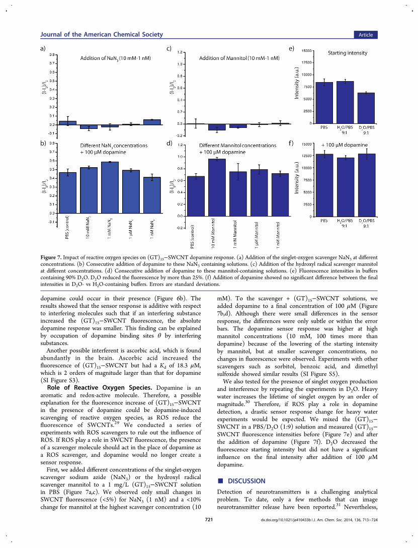
aromatic and redox-active molecule. Therefore, a possibleexplanation for the fluorescence increase of (GT)15−SWCNTin the presence of dopamine could be dopamine-inducedscavenging of reactive oxygen species, as ROS reduce thefluorescence of SWCNTs.29 We conducted a series ofexperiments with ROS scavengers to rule out the influence ofROS. If ROS play a role in SWCNT fluorescence, the presenceof a scavenger molecule should act in the place of dopamine asa ROS scavenger, and dopamine would no longer create asensor response.First, we added different concentrations of the singlet-oxygen
scavenger sodium azide (NaN3) or the hydroxyl radicalscavenger mannitol to a 1 mg/L (GT)15−SWCNT solutionin PBS (Figure 7a,c). We observed only small changes inSWCNT fluorescence (<5%) for NaN3 (1 nM) and a <10%change for mannitol at the highest scavenger concentration (10
mM). To the scavenger + (GT)15−SWCNT solutions, weadded dopamine to a final concentration of 100 μM (Figure7b,d). Although there were small differences in the sensorresponse, the differences were only subtle or within the errorbars. The dopamine sensor response was higher at highmannitol concentrations (10 mM, 100 times more thandopamine) because of the lowering of the starting intensityby mannitol, but at smaller scavenger concentrations, nochanges in fluorescence were observed. Experiments with otherscavengers such as sorbitol, benzoic acid, and dimethylsulfoxide showed similar results (SI Figure S5).We also tested for the presence of singlet oxygen production
and interference by repeating the experiments in D2O. Heavywater increases the lifetime of singlet oxygen by an order ofmagnitude.30 Therefore, if ROS play a role in dopaminedetection, a drastic sensor response change for heavy waterexperiments would be expected. We mixed the (GT)15−SWCNT in a PBS/D2O (1:9) solution and measured (GT)15−SWCNT fluorescence intensities before (Figure 7e) and afterthe addition of dopamine (Figure 7f). D2O decreased thefluorescence starting intensity but did not have a significantinfluence on the final intensity after addition of 100 μMdopamine.
■ DISCUSSION
Detection of neurotransmitters is a challenging analyticalproblem. To date, only a few methods that can imageneurotransmitter release have been reported.31 Nevertheless,
Figure 7. Impact of reactive oxygen species on (GT)15−SWCNT dopamine response. (a) Addition of the singlet-oxygen scavenger NaN3 at differentconcentrations. (b) Consecutive addition of dopamine to these NaN3 containing solutions. (c) Addition of the hydroxyl radical scavenger mannitolat different concentrations. (d) Consecutive addition of dopamine to these mannitol-containing solutions. (e) Fluorescence intensities in bufferscontaining 90% D2O. D2O reduced the fluorescence by more than 25%. (f) Addition of dopamine showed no significant difference between the finalintensities in D2O- vs H2O-containing buffers. Errors are standard deviations.
Journal of the American Chemical Society Article
dx.doi.org/10.1021/ja410433b | J. Am. Chem. Soc. 2014, 136, 713−724721

neurotransmitters are a very important class of biomolecules,and nanosensors can facilitate new insights in neurobiology.We used a new approach for the detection of neuro-
transmitters that is based on corona phase molecularrecognition (CoPhMoRe) of fluorescent single-walled carbonnanotubes (SWCNTs).2 For this purpose, SWCNTs werefunctionalized with different polymers. Through a screening, weinvestigated how nine different neurotransmitters affect the nIRfluorescence signature of 30 SWCNT−polymer hybridstructures. Interestingly, we found several polymer−SWCNThybrids whose nIR fluorescence intensities changed in thepresence of neurotransmitters (Figure 1a). In theory, everypolymer−SWCNT hybrid structure whose nIR fluorescencechanges in the presence of a neurotransmitter is a possiblesensor, yet a certain degree of specificity for the analyte is alsorequired. We attribute the selectivity of these sensors to theCoPhMoRe provided by the organic phase around the carbonnanotube. In this scheme, it is the organic corona phase thatcontrols how the analyte influences the fluorescence of theSWCNTs. For example, the presence of a particular analytemay impart conformational changes in the organic coronaphase around the SWCNT that lead to quenching ordequenching of the fluorescence, thereby imparting the sensorwith selectivity and sensitivity for that particular analyte.A central finding of our screening approach is that nucleic
acid-wrapped carbon nanotubes brighten in the nIR in thepresence of catecholamines such as dopamine or (−)-epi-nephrine. In contrast, most SWCNTs coated with phospholipidderivatives show either no change or a decrease in nIRfluorescence. Therefore, nucleic acid-wrapped SWCNTs have ahigh potential for use as dopamine sensors with high specificityand sensitivity. It is well-known that dopamine is a reactivemolecule and undergoes oxidation to dopamine quinone andother products readily.23,25,26,32 We attribute the time-depend-ent response (Figure 3b) to these reactions. Interestingly,polymerization of dopamine might have been a primarycontributor to results observed in previous work from ourlab, where dopamine was shown to quench the SWCNTfluorescence of DNA-wrapped SWCNTs.2,4 These experimentswere performed at higher dopamine concentrations (500 μM)and after longer incubation times, conditions under whichdopamine is known to polymerize readily. It is likely that inprevious publications, a complex mixture of polymerizeddopamine instead of pure dopamine was measured and thatSWCNTs aggregated/precipitated and reduced the fluores-cence count. When our samples were measured several hoursor 1 day after the addition of dopamine, we also observed astrong decrease in fluorescence. In contrast, herein we showresults based on more biologically pertinent dopamineconcentrations and at time scales that are more relevant forneuronal studies: the present experiments were conducted atmuch lower dopamine concentrations (<100 μM) with freshdopamine solutions at shorter incubation times (seconds orminutes) and revealed that dopamine and catecholamines ingeneral increase the fluorescence of nucleic acid-wrappedSWCNTs, with the magnitude of the fluorescence increasedepending on the nucleic acid sequence (Figure 1a). Thecalibration curve of the (GT)15−SWCNT dopamine sensor(Figure 3a) shows that the lower bound of dopamineconcentrations that can be detected is in the nanomolarrange. While we always prepared fresh dopamine solutions foreach experiment, the slight variations in sensor responses are
likely due to small variations in the time to prepare the solution,prepare the wells, and measure the sample.The polymer−SWCNT hybrids with the highest relative
fluorescence increases were those with the DNA sequence(GT)15 and the RNA sequence (GU)15. In solution, 100 μMdopamine produced an up to 80% increase in the fluorescence.This increase in fluorescence intensity is the basis for a turn-ondopamine sensor. In contrast, previously reported SWCNT-based sensors for small molecules are based on SWCNTfluorescence quenching.4,6
A feasible mechanism for dopamine sensing could potentiallyinvolve covalent modification of the SWCNT. First andforemost, we ruled out this possibility by verifying that theRaman spectra of the SWCNTs before and after the addition ofdopamine remained invariant, as did the D/G Raman peak ratio(SI Figure S1). Thus, a mechanism based on covalentmodification of the SWCNT can be ruled out. In addition,the reversibility of the sensing response indicates that neitherthe SWCNT nor the nucleic acid wrapping are covalentlymodified by dopamine (Figure 5). We have also verified thatthe SWCNT absorption cross section is invariant withdopamine addition, confirming that the quantum yield of(GT)15−SWCNT increases in the presence of dopamine (SIFigures S1 and S2). Additionally, scavenging of reactive oxygenspecies by dopamine was ruled out as the main mechanism byexperiments with competing reactive oxygen scavengers (Figure7 and SI Figure S5).Probing the local structure of the (GT)15 polymer via
fluorophore tagging of the DNA polymer 5′ end stronglysuggested that a small local perturbation of the polymer endsoccurs when dopamine enters the organic corona of theSWCNT (Figure 4). In this manner, the interaction ofdopamine with the nucleic acid or the SWCNT surfaceperturbs the nucleic acid conformation. This conformationalchange decreases SWCNT exciton decay routes, leading to anincrease in SWCNT fluorescence (quantum yield). It is likelythat the nucleic acid conformational changes lead to removal ofquenching species from the solvent or quenching groups of thenucleic acid backbone from the surface of the SWCNT. Forinstance, protons are known to quench SWCNT fluorescence,and their removal could explain the increase in fluorescence.33
In this picture, the selectivity of the sensor response isdetermined by the local polymer structure within the SWCNTcorona phase. Our results show that even small differences inthe nucleic acid sequence cause strong changes in the sensorresponse (Figure 1a) and that dopamine homologues exhibitdifferent responses compared with dopamine. For instance,both (GT)15−SWCNT and its RNA analogue (GU)15−SWCNT show a strong dopamine response (79% vs 44%fluorescence increase) but a completely different response tothe catecholamine (−)-epinephrine (62% vs ∼0% fluorescenceincrease) (Figure 1a). All these data support the CoPhMoReconcept and suggest that further engineering and screening ofthe organic phase of the SWCNT will provide even moreselective and sensitive sensors.The interaction between the nucleic acid−SWCNT and
dopamine could involve either direct physical contact or aredox reaction. A redox reaction of the nucleic acid polymer islikely to change the polymer structure within the SWCNTcorona phase, thus changing the quantum yield of SWCNTfluorescence. To understand the details of this mechanism, it isnoteworthy that dopamine is easily oxidized to dopaminequinone, which can undergo further downstream reactions.26
Journal of the American Chemical Society Article
dx.doi.org/10.1021/ja410433b | J. Am. Chem. Soc. 2014, 136, 713−724722

Additional evidence for redox-chemistry-based CoPhMoRe isprovided by the redox potentials of compounds similar to thedopamine analyte that produce similar SWCNT fluorescenceresponses (SI Figure S6). For instance, ascorbic acid has asimilar redox potential but a different structure compared withdopamine and also produces a response (SI Figure S4). L-Dopaand DOPAC produce sensor responses similar to that ofdopamine but have similar structures and redox potentials. Incontrast, uric acid has a different structure but a redox potentialthat is only slightly larger than that of dopamine, but itproduces no response when added to (GT)15−SWCNT.Plotting sensor responses against the redox peak currentpotentials (from cyclic voltammetry data) shows that the redoxpotential of the analyte can play a role in SWCNT-basedsensing (SI Figure S6).This hypothesis is also in agreement with our finding that
(GT)15−SWCNT showed a higher response than (AT)15−SWCNT [0.79 for (GT)15 vs 0.15 (AT)15; Figure 1a], since theguanine nucleotide is known to act as a oxidation sink innucleic acids, and therefore, higher guanine content should leadto higher sensor responses.34
Recently it was reported that DNA-functionalized SWCNTsshow a fluorescence increase in the presence of reducing agentssuch as trolox or mercaptoethanol.35 These findings along withours indicate that substances that are readily oxidized cangenerally increase the fluorescence of polynucleotide-function-alized SWCNTs, although exact mechanisms might be differentfor different substances.A redox-based mechanism presupposes that the nucleic acid
is already oxidized before dopamine is added and is thenreduced by dopamine. However, the reversibility of the sensorresponse (Figure 5) implies that there is an oxidizedequilibrium state of (GT)15 DNA on the SWCNT and thatthe nucleotide radical cations do not irreversibly react in thisstate, which is rather unlikely. Dopamine also shares anaromatic structure with L-Dopa, DOPAC, (−)-epinephrine, and(−)-norepinephrine. Therefore, another plausible mechanismof the molecular recognition could be based on π−π stacking ofthe aromatic ring of dopamine with the SWCNT. In thispicture, the DNA sequence serves as a diffusion barrier, thusproviding selectivity. When dopamine binds to the SWCNTsurface it displaces parts of the DNA backbone, which increasesthe fluorescence quantum yield.Although the exact nature of the dopamine−sensor
interaction remains under investigation, both mechanisms arecompatible with the CoPhMoRe concept because it does notpresuppose the type of the interaction between the organiccorona and the analyte.In this work we focused on the development and
characterization of a dopamine sensor, but the screening inFigure 1a clearly shows many other promising polymer−SWCNT sensors for other neurotransmitters. Future work willfocus on increasing the sensitivity and selectivity of the sensorsfor dopamine and other neurotransmitters using various parallelapproaches. First, we envision the development of ratiometricsensors using different SWCNT chiralities to expand the libraryof neurotransmitters that we can detect. In this work we usedHiPCO SWCNTs, which comprise a mixture of all SWCNTchiralities, not all of which can be excited simultaneously. Theuse of single-chirality samples should increase the sensitivity offuture dopamine sensors by enabling excitation of all SWCNTswithin a sample. Combinations of different SWCNT chiralitiescould also be used to synthesize ratiometric sensors by using a
combination of two or more different polymer−SWCNTsamples to gain higher selectivity and sensitivity throughsimultaneous multiplexed detection.Tools in neuroscience for the optical detection of dopamine
are lacking, particularly at the nanoscale. For instance, there areno tools currently available for direct measurements ofdopamine concentrations in the synaptic cleft. Fast-scan cyclicvoltammetry measurements in combination with geometricalassumptions suggest that extracellular dopamine levels are 250nM in the nucleus accumbens of the rat, 1.6 mM (transient) inthe synaptic cleft, and 25 mM in dopamine-containingvesicles.36 Our optical dopamine sensor could be used tomeasure static dopamine concentrations and also dynamicchanges in dopamine concentrations with high spatial andtemporal precision. Additionally, our sensor could be used totrack the secretion of dopamine from dopaminergic neurons,providing a fundamental understanding of how neuronal cellscommunicate with each other.In addition to its use as a sensor, the brightening of nucleic
acid-wrapped SWCNTs in the presence of dopamine/catechol-amines can be helpful for SWCNT imaging in general. Figure 5shows that single (GT)15−SWCNTs get brighter by a factor of>5. Therefore, adding fresh dopamine to nucleic acid-wrappedSWCNTs, particularly (GT)15−SWCNTs, might be useful toachieve higher fluorescence signals for applications requiringbright SWCNTs, such as single particle imaging.Lastly, it is important to note that this work presents a novel
approach to SWCNT-based sensor development. Whileprevious SWCNT sensor developments have relied on knownbiological recognition elements such as antibodies, the workpresented here is based on CoPhMoRe provided by polymersthat have no pre-existing affinity for dopamine. We envisionthat future generations of SWCNT-based sensors will not belimited to using pre-existing molecular recognition elementsand instead will exploit molecular recognition provided by thestructure of the corona phase of a SWCNT−polymerconjugate. Through further screening rounds of additionalpolymer−SWCNT hybrids, the selectivity and specificity ofthese sensors can be further improved, and novel sensors canbe discovered for which there currently exist no naturallyoccurring biological recognition elements.
■ CONCLUSIONThis work presents a new method for the optical detection ofneurotransmitters by using fluorescent single-walled carbonnanotubes (SWCNTs). Our approach is based on chemicalengineering of the organic phase (corona) around the carbonnanotube, resulting in corona phase molecular recognition(CoPhMoRe) of neurotransmitter analytes. By using differentpolymers wrapped around the carbon nanotube, we cangenerate polymer−SWCNT hybrids that are able to detectvarious neurotransmitters. One specific finding of our screeningshows that (GT)15 DNA- and (GU)15 RNA-wrapped SWCNTscan serve as highly selective and sensitive fluorescence turn-onsensors for catecholamine neurotransmitters such as dopamine.Our approach incorporates three very important figures ofmerit for sensor development: the sensors provide a strongoptical turn-on response in the nIR, are highly sensitive andreversible, and are fully functional at the nanoscale. Mechanisticstudies have shown that the nucleic acid backbone most likelyacts as a reversible dopamine-dependent switch that canmodulate exciton decay routes and thereby the SWCNTfluorescence. In the future, these sensors could be used for
Journal of the American Chemical Society Article
dx.doi.org/10.1021/ja410433b | J. Am. Chem. Soc. 2014, 136, 713−724723

spatiotemporal neurotransmitter detection near synapses or inneural networks.
■ ASSOCIATED CONTENT
*S Supporting InformationRaman spectra, absorption spectra, and absorption kinetics(Figures S1 and S2); schematic of the optical setup (FigureS3); ascorbic acid calibration curves (Figure S4); additionalROS scavenger experiments (Figure S5); and plot of sensorresponse versus redox potential (Figure S6). This material isavailable free of charge via the Internet at http://pubs.acs.org.
■ AUTHOR INFORMATION
Corresponding [email protected]
Author Contributions†S.K. and M.P.L. contributed equally.
NotesThe authors declare no competing financial interest.
■ ACKNOWLEDGMENTS
This work was supported primarily by the National ScienceFoundation under Award 1213622. S.K. was supported by apostdoctoral fellowship from the Deutsche Forschungsgemein-schaft (DFG). M.P.L. acknowledges an NSF postdoctoralresearch fellowship under Award 1306229.
■ REFERENCES(1) Davie, C. A. Br. Med. Bull. 2008, 86, 109.(2) Zhang, J.; Landry, M. P.; Barone, P. W.; Kim, J. H.; Lin, S.; Ulissi,Z. W.; Lin, D.; Mu, B.; Boghossian, A. A.; Hilmer, A. J.; Rwei, A.;Hinckley, A. C.; Kruss, S.; Shandell, M. A.; Nair, N.; Blake, S.; Sen, F.;Sen, S.; Croy, R. G.; Li, D.; Yum, K.; Ahn, J. H.; Jin, H.; Heller, D. A.;Essigmann, J. M.; Blankschtein, D.; Strano, M. S. Nat. Nanotechnol.2013, 8, 959.(3) Kruss, S.; Hilmer, A. J.; Zhang, J.; Reuel, N. F.; Mu, B.; Strano, M.S. Adv. Drug Delivery Rev. 2013, 65, 1933.(4) Zhang, J.; Boghossian, A. A.; Barone, P. W.; Rwei, A.; Kim, J. H.;Lin, D.; Heller, D. A.; Hilmer, A. J.; Nair, N.; Reuel, N. F.; Strano, M.S. J. Am. Chem. Soc. 2011, 133, 567.(5) O’Connell, M. J.; Bachilo, S. M.; Huffman, C. B.; Moore, V. C.;Strano, M. S.; Haroz, E. H.; Rialon, K. L.; Boul, P. J.; Noon, W. H.;Kittrell, C.; Ma, J.; Hauge, R. H.; Weisman, R. B.; Smalley, R. E. Science2002, 297, 593.(6) Jin, H.; Heller, D. A.; Kalbacova, M.; Kim, J. H.; Zhang, J.;Boghossian, A. A.; Maheshri, N.; Strano, M. S. Nat. Nanotechnol. 2010,5, 302.(7) Barone, P. W.; Parker, R. S.; Strano, M. S. Anal. Chem. 2005, 77,7556.(8) Heller, D. A.; Baik, S.; Eurell, T. E.; Strano, M. S. Adv. Mater.2005, 17, 2793.(9) Heller, D. A.; Jin, H.; Martinez, B. M.; Patel, D.; Miller, B. M.;Yeung, T. K.; Jena, P. V.; Hobartner, C.; Ha, T.; Silverman, S. K.;Strano, M. S. Nat. Nanotechnol. 2009, 4, 114.(10) Adams, K. L.; Puchades, M.; Ewing, A. G. Annu. Rev. Anal.Chem. 2008, 1, 329.(11) Kim, D.; Koseoglu, S.; Manning, B. M.; Meyer, A. F.; Haynes, C.L. Anal. Chem. 2011, 83, 7242.(12) Savtchenko, L. P.; Rusakov, D. A. Proc. Natl. Acad. Sci. U.S.A.2007, 104, 1823.(13) Wightman, R. M.; Jankowski, J. A.; Kennedy, R. T.; Kawagoe, K.T.; Schroeder, T. J.; Leszczyszyn, D. J.; Near, J. A.; Diliberto, E. J., Jr.;Viveros, O. H. Proc. Natl. Acad. Sci. U.S.A. 1991, 88, 10754.
(14) Rodriguez, P. C.; Pereira, D. B.; Borgkvist, A.; Wong, M. Y.;Barnard, C.; Sonders, M. S.; Zhang, H.; Sames, D.; Sulzer, D. Proc.Natl. Acad. Sci. U.S.A. 2013, 110, 870.(15) Gubernator, N. G.; Zhang, H.; Staal, R. G.; Mosharov, E. V.;Pereira, D. B.; Yue, M.; Balsanek, V.; Vadola, P. A.; Mukherjee, B.;Edwards, R. H.; Sulzer, D.; Sames, D. Science 2009, 324, 1441.(16) Masharina, A.; Reymond, L.; Maurel, D.; Umezawa, K.;Johnsson, K. J. Am. Chem. Soc. 2012, 134, 19026.(17) Okubo, Y.; Sekiya, H.; Namiki, S.; Sakamoto, H.; Iinuma, S.;Yamasaki, M.; Watanabe, M.; Hirose, K.; Iino, M. Proc. Natl. Acad. Sci.U.S.A. 2010, 107, 6526.(18) Marvin, J. S.; Borghuis, B. G.; Tian, L.; Cichon, J.; Harnett, M.T.; Akerboom, J.; Gordus, A.; Renninger, S. L.; Chen, T. W.;Bargmann, C. I.; Orger, M. B.; Schreiter, E. R.; Demb, J. B.; Gan, W.B.; Hires, S. A.; Looger, L. L. Nat. Methods 2013, 10, 162.(19) Mu, B.; McNicholas, T. P.; Zhang, J.; Hilmer, A. J.; Jin, Z.;Reuel, N. F.; Kim, J. H.; Yum, K.; Strano, M. S. J. Am. Chem. Soc. 2012,134, 17620.(20) Lamichhane, R.; Solem, A.; Black, W.; Rueda, D. Methods 2010,52, 192.(21) Roy, R.; Hohng, S.; Ha, T. Nat. Methods 2008, 5, 507.(22) Steinberg, E. E.; Keiflin, R.; Boivin, J. R.; Witten, I. B.;Deisseroth, K.; Janak, P. H. Nat. Neurosci. 2013, 16, 966.(23) Lee, H.; Dellatore, S. M.; Miller, W. M.; Messersmith, P. B.Science 2007, 318, 426.(24) Dreyer, D. R.; Miller, D. J.; Freeman, B. D.; Paul, D. R.;Bielawski, C. W. Langmuir 2012, 28, 6428.(25) Liebscher, J.; Mrowczyn ski, R.; Scheidt, H. A.; Filip, C.; Hadade,N. D.; Turcu, R.; Bende, A.; Beck, S. Langmuir 2013, 29, 10539.(26) Lynge, M. E.; van der Westen, R.; Postma, A.; Stadler, B.Nanoscale 2011, 3, 4916.(27) Yang, R.; Jin, J.; Chen, Y.; Shao, N.; Kang, H.; Xiao, Z.; Tang,Z.; Wu, Y.; Zhu, Z.; Tan, W. J. Am. Chem. Soc. 2008, 130, 8351.(28) Wightman, R. M.; May, L. J.; Michael, A. C. Anal. Chem. 1988,60, 769A.(29) Sen, F.; Boghossian, A. A.; Sen, S.; Ulissi, Z. W.; Zhang, J.;Strano, M. S. ACS Nano 2012, 6, 10632.(30) Merkel, P. B.; Kearns, D. R.; Nilsson, R. J. Am. Chem. Soc. 1972,94, 1030.(31) Sames, D.; Dunn, M.; Karpowicz, R. J.; Sulzer, D. ACS Chem.Neurosci. 2013, 4, 648.(32) Herlinger, E.; Jameson, R. F.; Linert, W. J. Chem. Soc., PerkinTrans. 2 1995, 259.(33) Strano, M. S.; Huffman, C. B.; Moore, V. C.; O’Connell, M. J.;Haroz, E. H.; Hubbard, J.; Miller, M.; Rialon, K.; Kittrell, C.; Ramesh,S.; Hauge, R. H.; Smalley, R. E. J. Phys. Chem. B 2003, 107, 6979.(34) Steenken, S.; Jovanovic, S. V. J. Am. Chem. Soc. 1997, 119, 617.(35) Lee, A. J.; Wang, X.; Carlson, L. J.; Smyder, J. A.; Loesch, B.; Tu,X.; Zheng, M.; Krauss, T. D. Nano Lett 2011, 11, 1636.(36) Garris, P. A.; Ciolkowski, E. L.; Pastore, P.; Wightman, R. M. J.Neurosci. 1994, 14, 6084.
Journal of the American Chemical Society Article
dx.doi.org/10.1021/ja410433b | J. Am. Chem. Soc. 2014, 136, 713−724724