Neuroprotective and Therapeutic Role of Omega-3...
11
Clinical Neurology and Neuroscience 2018; 2(1): 12-22 http://www.sciencepublishinggroup.com/j/cnn doi: 10.11648/j.cnn.20180201.13 Neuroprotective and Therapeutic Role of Omega-3 Against Oxidative Stress and Neurotransmitter Disturbances in Rotenone-Induced Mice Model of Parkinson's Disease Nagi Ali Ibrahim 1, * , Yasser Ashry Khadrawy 2 , Soliman Sayed Ibrahim 1 , Noura El-Sayed Ezzat 1 1 Zoology Department, Faculty of Science, Zagazig University, Zagazig, Egypt 2 Department of Medical Physiology, National Research Center, Dokki, Giza, Egypt Email address: * Corresponding author To cite this article: Nagi Ali Ibrahim, Yasser Ashry Khadrawy, Soliman Sayed Ibrahim, Noura El-Sayed Ezzat. Neuroprotective and Therapeutic Efficacy of Omega-3 Against Oxidative Stress and Neurotransmitter Disturbances in Brain of Rotenone-Induced Mice Model of Parkinson's Disease. Clinical Neurology and Neuroscience. Vol. 2, No. 1, 2018, pp. 12-22. doi: 10.11648/j.cnn.20180201.13 Received: November 16, 2017; Accepted: December 4, 2017; Published: February 7, 2018 Abstract: The present study aimed at evaluating the protective and therapeutic efficacy of omega-3 against motor impairment and brain biochemical disturbances in rotenone-induced mice model of Parkinson's disease (PD). Sixty animals were divided into six groups (10 each): mice of the 1 st group were used as controls, they were injected subcutaneously (sc) with the vehicle (50 µl dimethylsulfoxide (DEMSO) + 950 µl sunflower oil /kg body weight) every other day for 30 days; the 2 nd group, mice model of Parkinson’s disease (PD), were injected (sc) with rotenone (3 mg/kg dissolved in vehicle every other day for 30 days). the 3 rd group, mice were given rotenone for 30 days followed by a stopping (recovery) period of other 30 days to validate the persistency of the PD model; the 4 th group (protection group), mice received orally Omega-3 oil (300 mg/kg) daily an hour before every rotenone injection for 30 days; the 5 th and 6 th groups (therapeutic groups), mice were treated orally with Omega-3 oil daily for 7 and 15 days respectively after the induction of PD mice model. Data obtained revealed an impairment of the motor activity in mice of PD model as indicated from the decreased time of the forelimb hanging test. This was associated with a state of oxidative stress in the brain of PD model as indicated from the increase in lipid peroxidation (increased malondialdehyed, MDA, level) and nitric oxide (NO), and the decrease in reduced glutathione (GSH). A significant decrease in the levels of dopamine, norepinephrine, serotonin, AChE activity and a significant increase in TNF-α level was recorded in the PD model. The present findings show that both the protection by or oral treatment with omega-3 for 15 days could ameliorate the rotenone- induced oxidative stress and inflammation in brain of PD mice model. In addition, omega-3 either as protection or treatment daily for 15 days was effective in restoring the decrease in dopamine and norepinephrine induced in the brain of PD mice model. In conclusion, the present study demonstrates that omega-3 supplementation potentially reverses the motor, and neurochemical alternations induced by rotenone in mice model of PD. Keywords: Parkinson's Disease, Omega-3, Oxidative Stress, Neurotransmitters 1. Introduction Parkinson's disease (PD) is the second most common neurodegenerative disorder after Alzheimer's disease, with a male –to –female ratio of about 3:2 in most studies [1, 2]. Parkinson's disease is characterized by a loss of dopaminergic neurons in the substantianigra in the midbrain and also the presence of Lewy bodies, which are intracellular inclusions enriched in the protein α-synuclein [3]. The most common motor symptoms of PD include bradykinesia, rest tremor, rigidity, and postural and gait impairment [4]. Various risk factors have been found for sporadic PD, including exposure to pesticides and other toxins, positive family history, and oophorectomy, but age remains the most important one documented so far [1, 5, 6]. Parkinson’s disease is a multifactorial disease characterized
Transcript of Neuroprotective and Therapeutic Role of Omega-3...

Clinical Neurology and Neuroscience 2018; 2(1): 12-22
http://www.sciencepublishinggroup.com/j/cnn
doi: 10.11648/j.cnn.20180201.13
Neuroprotective and Therapeutic Role of Omega-3 Against Oxidative Stress and Neurotransmitter Disturbances in Rotenone-Induced Mice Model of Parkinson's Disease
Nagi Ali Ibrahim1, *
, Yasser Ashry Khadrawy2, Soliman Sayed Ibrahim
1, Noura El-Sayed Ezzat
1
1Zoology Department, Faculty of Science, Zagazig University, Zagazig, Egypt 2Department of Medical Physiology, National Research Center, Dokki, Giza, Egypt
Email address:
*Corresponding author
To cite this article: Nagi Ali Ibrahim, Yasser Ashry Khadrawy, Soliman Sayed Ibrahim, Noura El-Sayed Ezzat. Neuroprotective and Therapeutic Efficacy of
Omega-3 Against Oxidative Stress and Neurotransmitter Disturbances in Brain of Rotenone-Induced Mice Model of Parkinson's Disease.
Clinical Neurology and Neuroscience. Vol. 2, No. 1, 2018, pp. 12-22. doi: 10.11648/j.cnn.20180201.13
Received: November 16, 2017; Accepted: December 4, 2017; Published: February 7, 2018
Abstract: The present study aimed at evaluating the protective and therapeutic efficacy of omega-3 against motor
impairment and brain biochemical disturbances in rotenone-induced mice model of Parkinson's disease (PD). Sixty animals
were divided into six groups (10 each): mice of the 1st group were used as controls, they were injected subcutaneously (sc)
with the vehicle (50 µl dimethylsulfoxide (DEMSO) + 950 µl sunflower oil /kg body weight) every other day for 30 days; the
2nd
group, mice model of Parkinson’s disease (PD), were injected (sc) with rotenone (3 mg/kg dissolved in vehicle every other
day for 30 days). the 3rd
group, mice were given rotenone for 30 days followed by a stopping (recovery) period of other 30
days to validate the persistency of the PD model; the 4th
group (protection group), mice received orally Omega-3 oil (300
mg/kg) daily an hour before every rotenone injection for 30 days; the 5th
and 6th
groups (therapeutic groups), mice were treated
orally with Omega-3 oil daily for 7 and 15 days respectively after the induction of PD mice model. Data obtained revealed an
impairment of the motor activity in mice of PD model as indicated from the decreased time of the forelimb hanging test. This
was associated with a state of oxidative stress in the brain of PD model as indicated from the increase in lipid peroxidation
(increased malondialdehyed, MDA, level) and nitric oxide (NO), and the decrease in reduced glutathione (GSH). A significant
decrease in the levels of dopamine, norepinephrine, serotonin, AChE activity and a significant increase in TNF-α level was
recorded in the PD model. The present findings show that both the protection by or oral treatment with omega-3 for 15 days
could ameliorate the rotenone- induced oxidative stress and inflammation in brain of PD mice model. In addition, omega-3
either as protection or treatment daily for 15 days was effective in restoring the decrease in dopamine and norepinephrine
induced in the brain of PD mice model. In conclusion, the present study demonstrates that omega-3 supplementation
potentially reverses the motor, and neurochemical alternations induced by rotenone in mice model of PD.
Keywords: Parkinson's Disease, Omega-3, Oxidative Stress, Neurotransmitters
1. Introduction
Parkinson's disease (PD) is the second most common
neurodegenerative disorder after Alzheimer's disease, with a
male –to –female ratio of about 3:2 in most studies [1, 2].
Parkinson's disease is characterized by a loss of
dopaminergic neurons in the substantianigra in the midbrain
and also the presence of Lewy bodies, which are intracellular
inclusions enriched in the protein α-synuclein [3]. The most
common motor symptoms of PD include bradykinesia, rest
tremor, rigidity, and postural and gait impairment [4]. Various
risk factors have been found for sporadic PD, including
exposure to pesticides and other toxins, positive family
history, and oophorectomy, but age remains the most
important one documented so far [1, 5, 6].
Parkinson’s disease is a multifactorial disease characterized

Clinical Neurology and Neuroscience 2018; 2(1): 12-22 13
by self-perpetuating cascades involving a myriad of deleterious
events at various stages including mitochondrial dysfunction,
short-term and long-term oxidative and nitrosative stress,
energy crisis, excitotoxicity, neuroinflammation and protein
aggregation [7]. These events work together to promote cell
death. Oxidative stress and mitochondrial dysfunction are
responsible for the pathological features of PD, such as
neuronal death and apoptosis [8, 9].
On the other hand, PD is now considered a multisystem
degenerative process, affecting not only the nigrostriatal
dopaminergic system, responsible for the majority of the motor
symptoms, but also other neurotransmitter systems, like
cholinergic, serotonergic, and noradrenergic systems associated
with several non motor symptoms such as sleep disturbances,
fatigue, anxiety, depression, cognitive impairment, dementia,
olfactory dysfunction, pain, sweating and constipation [10].
It is well known that the symptoms of PD worsen with
time. The medications of PD provide the patients with a
relief period from symptoms. These medications include
levodopa [11], dopamine agonists [12], and catechol-O-
methyl transferase (COMT) inhibitors that are added to
levodopa treatment to overcome motor complications and to
prolong the bioavailability of levodopa [13]. The
anticholinergic drugs used in the treatment of PD are
potentially helpful in delaying the need for levodopa
treatment. In addition, they may allow for a reduction in the
dose of levodopa required in more advanced cases, thus
further extending the use of levodopa [14]. Although these
medications could relief the symptoms of PD, many side
effects have been reported including oxidative stress and the
degeneration of residual dopamine neurons in patients with
PD [15, 16, 17], confusion, hallucinations, depression,
psychosis, dyskinesia, and mental changes [18]
It is clear that there is no exact cure for PD and many side
effects have been reported due to the use of these
medications. Therefore, there is a persistent need for new,
safer and effective agents for the treatment of PD. Omega-3
polyunsaturated fatty acids (Omega-3 PUFAs), including
eicosapentaenoic acid (EPA) and docosahexaenoic acid
(DHA) are dietary fats with an array of health benefits [19].
DHA is a key component of all cell membranes and is found
in abundance in the brain and retina [20]. EPA and DHA are
the precursors of several metabolites that are potent lipid
mediators, considered by many investigators to be beneficial
in the prevention or treatment of several diseases [21]. These
fatty acids are predominantly found in fish and fish oils [22].
Omega-3 PUFAs have a crucial role for optimal brain
function by enabling fluidity in neuronal membranes and
regulation of neurotransmitters [23]. DHA supplementation
has been found to ameliorate memory and cognitive
impairment in healthy older adults with age related cognitive
decline without side effects [24]. It has been found that
omega-3 supplementation may enhance resistance to free
radical attack and reduce lipid peroxidation and may be an
effective dietary supplement in the management of various
diseases in which oxidant / antioxidant balance is disturbed
as in aged brain tissue [25].
Bousquet et al. (2008) [26] found that omega-3 PUFAs had
a partially protective effect against 1-methyl-4-phenyl-1, 2, 3,
6-tetrahydropyridine (MPTP)-induced reduction in dopamine
production, storage, and/or release. It has been found that
when rats are fed a diet deficient in omega-3 PUFAs, they
displayed inadequate storage of newly synthesized dopamine
[27]. Daily omega-3 PUFAs supplementation reversed the 5-
HT stress –induced effects but without impact on the
physical state or behavior of the animals. This suggests that
omega-3 PUFAs can improve resistance to stress [28].
Li et al. (2014)[29] found that marine derived omega-3
PUFAs supplementation had a significant lowering effect on
the levels of C-reactive protein (CRP), interleukin 6 (IL-6)
and tumor necrosis factor-alpha (TNF-α) particularly in non
obese subjects.
Although several approved drugs may alleviate PD
symptoms, long term use of these drugs is often associated
with aggravating side effects, and none of these drugs can slow
down, prevent or even reverse the progress of PD. Therefore,
the aim of the present study is to evaluate the protective and
therapeutic effect of omega-3 against mice model of PD
induced by rotenone. This is carried out by investigating the
effect of omega-3 on the motor and neurochemical changes
induced in the brain of mice PD model.
2. Materials and Methods
2.1. Animals
Forty eight Swiss male albino mice (weighing 30-40 g.)
were used in the present study. The animals were obtained
from Animal House Colony of National Research Centre,
Giza, Egypt. On arrival, animals were housed ten per cage in
stainless steel cages with ad libitum access to standard
laboratory diet and tap water in a temperature-controlled (20-
25°C) and artificially illuminated (12 hrs. dark / light cycle)
room free of any chemical contamination. All animals
received human care in compliance with the guidelines of the
Animals Care and Use Committee of the National Research
Centre, Egypt, on July 2009 with registration number 09/093.
2.2. Drugs and Chemicals
Rotenone was purchased from Sigma Chemical Co. (St.
Louis MO, USA). It was dissolved first in dimethylsulfoxide
(1.5 mg/50µl) and then the volume was completed with
sunflower oil to have a final concentration of 1.5 mg/ml.
Omega-3 oil was purchased from Arab Co. for Gelatin and
Pharmaceutical Products, Egypt.
2.3. Experimental Design
Animals were divided into 6 groups, mice of the 1st group
were used as control, they received orally the vehicle (50 µl
dimethylsulfoxide (DEMSO) + 950 µl sunflower oil/kg body
weight) every other day for 30 days; the 2nd
group, mice model
of Parkinson’s disease (PD), were treated with subcutaneous (sc)
injection of rotenone (3 mg/kg body weight. dissolved in
dimethylsulfoxide + sunflower oil every other day for 30 days);

14 Nagi Ali Ibrahim et al.: Neuroprotective and Therapeutic Efficacy of Omega-3 Against Oxidative Stress and Neurotransmitter
Disturbances in Brain of Rotenone-Induced Mice Model of Parkinson's Disease
the 3rd
group, mice were injected s c with rotenone for 30 days
followed by a recovery period for other 30 days to investigate
whether the mice model of PD could recover to normal or not
after stopping rotenone injection; the 4th group (protection
group) mice received orally Omega-3 oil daily (300 mg/kg) an
hour before every rotenone injection for 30 days; the 5th and 6
th
groups (therapeutic groups), mice were treated orally with
Omega-3 oil daily for 7 and 15 days respectively after the
induction of mice model of PD.
2.4. Motor Activity (Hanging Wire Test)
This test was used as a measure of muscular strength and
motor neuron integrity. Mice used their forelimbs to suspend
their body weight on a wire stretched between two posts 60
cm and 20 cm above a foam pillow. The time (in seconds)
until the rat fell was recorded [30].
2.5. Preparation of Brain Samples
Following motor testing, animals were sacrificed by sudden
decapitation after being fasted for 12 hours. The brain of each
animal was quickly disected out and rapidly transferred to an
ice-cold Petri dish. Each brain was divided into two right and
left halves. The right half was used to measure oxidative stress
parameters [Malondialdehyde (MDA), Nitric oxide (NO),
reduced glutathione (GSH)], acetylcholineesterase (AchE)
activity and tumor necrosis factor-alpha (TNF-α) level. The
left half was used for the analysis of other neurotransmitter
including dopamin (DA), norepinephrine (NE) and serotonin
(5-hydroxytryptamine; 5-HT). Each brain area was weighed
and frozen at -80°C until analyzed.
The right half of each mouse brain was homogenized in
phosphate buffer (pH 7.4). This homogenate was centrifuged
at 5000 rpm and 4°C for 10 minutes; the supernatant was
stored at -80°C until analysis. This supernatant was used for
the determination of oxidative stress parameters, AchE
activity and TNF-α level.
2.6. Neurochemical Analysis
2.6.1. Determination of Lipid Peroxidation
MDA, a measure of lipid peroxidation, was estimated
according to the method of Ruiz-Larrea et al. (1994) [31].
Malondialdehyde was determined by measuring
thiobarbituric acid reactive species (TBARS). One molecule
of MDA reacts with two molecules of thiobarbituric acid in
acidic medium at a temperature of 95°C for 20 minutes to
form TBARS. The absorbance of the resultant pink product
was measured at 532 nm.
2.6.2. Determination of NO
NO was determined colorimetrically in the brain tissue
according to the method described by Montgomery and
Dymock (1961) [32]. This is based on the measurement of
endogenous nitrite concentration as an indicator of NO
production. It depends on the addition of Griess reagent
which converts nitrite into a deep purple azo compound
whose absorbance is read at 540 nm.
2.6.3. Determination of GSH
Ellman's method [33] was used to measure GSH level. The
procedure is based on the reduction of Ellman´s reagent by –SH
groups of GSH to form 2-nitro-s-mercaptobenzoic acid whose
intense yellow color is measured spectrophotometrically at 412
nm.
2.6.4. Determination of TNF-α
Estimation of brain TNF-α level expressed in pg/g tissue
was carried out using rat TNF-α ELISA Kit supplied by
Koma Biotech INC, Seoul (Korea).
2.6.5. Neurotransmitter Analysis
The procedure used for the determination of AchE activity
in the midbrain and striatum was a modification of the method
of Ellman et al. (1961) [34] as described by Gorun et al. (1978)
[35]. The principle of the method depends on the hydrolysis of
acetylthiocholine iodide by acetycholinesterase to produce
thiocholine. Thiocholine is allowed to react with the -SH
reagent 5,5’-dithiobis-(2-nitrobenzoic acid) (DTNB), which is
reduced to thionitrobenzoic acid, a yellow colored anion
whose absorption is read spectrophotometrically at 412 nm.
The left half of each mouse was homogenized in an ice-
cold solution of acidified n-butanol. The homogenates were
centrifuged at 2000 r. p. m. for 5 minutes. These supernatants
were used for the estimation of DA, NE and 5-HT according
to the fluorometric method described by Ciarlone (1978)
[36]. The fluorescence was measured using
spectrofluorometer (model Jasco-FP-6500, Japan) with a
source of xenon arc lamp 150W (excitation slit band width of
excitation monochromator: 5 nm., emission slit band width of
emission monochromator: 5 nm.).
2.7. Statistical Analysis
The data presented in the study were statistically evaluated
as a mean value for each group and its corresponding
standard error. Statistical significance between the groups
under investigation was tested by one-way analysis of
variance (ANOVA) using Statistical Package for Social
Sciences (SPSS) program, version 14 followed by post hoc
test using Duncan to compare significance between groups.
Difference was considered significant at P-value <0.05.
3. Results
3.1. Motor Activity
Data demonstrated in figure (1) revealed that rotenone
injection (3 mg/kg every other day) for 30 days resulted in a
significant decrease in the hanging time of mice below the
control value. The daily oral protection with omega-3 (300
mg/kg) against rotenone induce model of PD restored the
hanging time of mice to control –like value. Moreover, when
the mice model of PD were treated daily with omega-3 for 7
and 15 days, the decrease in forelimb hanging time induced
by rotenone returned to non-significant change as compared
to control value.
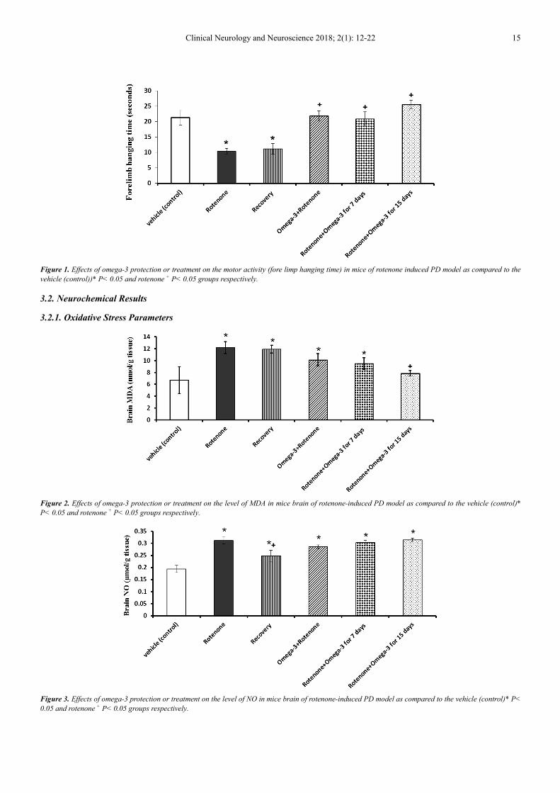
Clinical Neurology and Neuroscience 2018; 2(1): 12-22 15
Figure 1. Effects of omega-3 protection or treatment on the motor activity (fore limp hanging time) in mice of rotenone induced PD model as compared to the
vehicle (control))* P< 0.05 and rotenone + P< 0.05 groups respectively.
3.2. Neurochemical Results
3.2.1. Oxidative Stress Parameters
Figure 2. Effects of omega-3 protection or treatment on the level of MDA in mice brain of rotenone-induced PD model as compared to the vehicle (control)*
P< 0.05 and rotenone + P< 0.05 groups respectively.
Figure 3. Effects of omega-3 protection or treatment on the level of NO in mice brain of rotenone-induced PD model as compared to the vehicle (control)* P<
0.05 and rotenone + P< 0.05 groups respectively.

16 Nagi Ali Ibrahim et al.: Neuroprotective and Therapeutic Efficacy of Omega-3 Against Oxidative Stress and Neurotransmitter
Disturbances in Brain of Rotenone-Induced Mice Model of Parkinson's Disease
Figure 4. Effects of omega-3 protection or treatment on the level of GSH in mice brain of rotenone induced PD model as compared to the vehicle (control) *
P< 0.05 and rotenone + P< 0.05 groups respectively.
Data illustrated in figures (2 and 3) show that rotenone-
induced mice model of PD was accompanied by significant
increases in brain levels of lipid peroxidation (MDA) by
82.29% and NO by 60.82% respectively and a significant
decrease in the level of GSH by 35.30% Figure (4) as all
compared to their levels in control mice. The protection and
daily treatment with Omega-3 for 7 days failed to restore the
increased levels of lipid peroxidation and nitric oxide
induced in the brain of PD mice model, however, GSH value
showed non significant change. Daily treatment of PD mice
model with omega-3, for 15 days restored brain levels of
both MDA and GSH toward the control values, whereas, NO
showed elevated level (figures 2, 3 and 4) respectively.
Data demonstrated in figure (5) showed a significant
increase in TNF-α by 33.6% in the brain of mice model of
PD as compared to the control value. This increase was
reduced to control like value when the mice model of PD
were protected or treated daily for 15 days with omega-3.
Figure 5. Effects of omega-3 protection or treatment on the level of TNF-α in mice brain of rotenone induced PD model as compared to the vehicle (control) *
P< 0.05 and rotenone + P< 0.05 groups respectively.
3.2.2. Neurotransmitters
Figure (6) shows a significant decrease jn brain AchE
activity by -21.28% below the control value. Both the
protection and treatment with omega-3 for 7 days reversed
the decrease in the AchE activity to a significant increase.
However, treatment of mice model of PD with omega-3 was
extended to 15 days AchE activity was restored to control
like value.
In mice model of PD significant decreases in the brain
levels of DA (figure 7), NE (figure 8) and 5-HT (Figure 9)
were observed recording -14.68%, -15.49% and -58.71%,
respectively, as compared to control values. The protection
with omega-3 prevented the decrease in dopamine and
norepinephrine induced in mice model of PD but failed to
restore the decrease in serotonin. Similarly the daily
treatment of mice model of PD with omega-3 for 7 and 15
days restored the brain levels of DA and NE to non
significantly changed as compared to control animals.
However, omega-3 treatment was unable to prevent the
decrease in 5-HT induced in mice model of PD.

Clinical Neurology and Neuroscience 2018; 2(1): 12-22 17
Figure 6. Effects of omega-3 protection or treatment on the level of AchE activity in mice brain of rotenone induced PD model as compared to the vehicle
(control) * P< 0.05 and rotenone + P< 0.05 groups respectively.
Figure 7. Effects of omega-3 protection or treatment on the level of DA in mice brain of rotenone induced PD model as compared to the vehicle (control). *
P< 0.05 and rotenone + P< 0.05 groups respectively.
Figure 8. Effects of omega-3 protection or treatment on the level of NE in mice brain of rotenone-induced PD model as compared to the vehicle (control) * P<
0.05 and rotenone + P< 0.05 groups respectively.

18 Nagi Ali Ibrahim et al.: Neuroprotective and Therapeutic Efficacy of Omega-3 Against Oxidative Stress and Neurotransmitter
Disturbances in Brain of Rotenone-Induced Mice Model of Parkinson's Disease
Figure 9. Effects of omega-3 protection or treatment on the level of 5-HT in mice brain of rotenone induced PD model as compared to the vehicle (control)*
P< 0.05 and rotenone + P< 0.05 groups respectively.
4. Discussion
In the present study, a significant increase in the levels of
MDA (a marker of lipid peroxidation) and NO together with
the significant decrease in reduced glutathione GSH after the
daily s. c. injection of rotenone (3 mg/kg every other day)
indicate the development of oxidative stress in the brain
tissue of rotenone-treated mice.
Rotenone is a potent dopaminergic neurotoxicant owing to
its highly lipophilicity, its easy crossing of the blood-brain
barrier and its accumulation in subcellular organelles
including the mitochondria [37]. It has the ability
tobindspecifically to complex I, disrupting mitochondrial
respiration and increasing reactive oxygen species (ROS)
production [38]. ROS in turn could attack lipid rich
membranes of the brain causing an increase in lipid
peroxidation [39]. Thus, the present significant increase in
the level of lipid peroxidation in the brain tissue could be
attributed to the attack of cell membranes of the brain by the
free radicals evolved by rotenone.
In addition, the significant increase in the level of NO in
the brain tissue recorded in the present study could be
mediated by the activation of neuronal nitric oxide synthase
(nNOS). Supporting our findings is the study of He et al.
(2003) [40] who found that chronic rotenone administration
leads to increased activity of NOS in the cerebral tissues.
Nitric oxide may inhibit key enzymes of energy metabolism,
damage DNA, deplete intracellular glutathione and react with
ROS, especially superoxide (O˙) to form peroxynitrite
(ONOO-), a potent oxidant and nitrating agent which has a
potent damaging activity [41]. Moreover, the potent
oxidant ONOO– may directly oxidize DA [42]
causingdopaminergic damage [43]. This could explain the
reduced level of dopamine induced by rotenone.
The present recorded reduction of GSH could be attributed
to its exhaustion in scavenging the rapidly generating free
radicals. Reduced glutathione (GSH) has crucial metabolic
importance and its decreased levels might play an important
role in inducing oxidative stress in brain [44].
TNF-α a potent pro-inflammatory cytokine has a
promoting role in neuroinflammation-mediated progressive
degeneration of dopaminergic neurons in PD [45, 46]. Thus,
the increase in brain TNF-α induced by rotenone in the
present study could mediate the neuronal inflammation and
consequently the dopaminergic damage. Zaitone et al. (2012)
[47] observed that rotenone activates the release of TNF-α
from microglia.
Accordingly, it could be concluded that the oxidative and
nitrosative stress together with the neuroinflammation
induced by rotenone in the brain tissue could underlie
dopaminergic neurons neurodegeneration and consequently
the reduced level of DA.
The present findings indicate that rotenone induced a
significant decrease in cerebral AchE activity. According to
the DA-Ach balance hypothesis, DA could inhibit
acetylcholine releasein the striatum [48]. Therefore, the
depletion of dopamine in the striatum could result in
dopaminergic/cholinergic imbalance [49] leading
toanincrease the level of Ach. This continuous stimulation
without inhibition could underlie the characteristic symptoms
of tremor, rigidity and muscle fatigue that develop leading to
postural instability [50]. AchE is the enzyme responsible for
the degradation of acetylcholine. Accordingly, the present
decreased activity of AchE could arise from its consumption
in the degradation of the abnormal increased Ach content
generated during the development of PD.
In agreement with the present findings, Swathi and
Rajendra (2014) [51] found that the i. p. injection of rotenone
induced a significant decrease in brain DA, NE and 5-HT
levels.
The removal of monoamines from the synaptic cleft takes
place by a specific vesicular monoamine transporter
(VMAT2) [52] which is responsible for repacking
monoamine neurotransmitters including DA, NE and 5-HT
into their vesicles [53]. This protects them from degradation
by monoamine oxidase enzyme (MAO). Watabe and Nakaki
(2008) [54] found that rotenone inhibited the activity of
VMAT2. In addition, it has been reported that chronic
rotenone administration significantly increased the activity of

Clinical Neurology and Neuroscience 2018; 2(1): 12-22 19
MAO enzyme [51]. Therefore the present decrease in the
cerebral level of DA, NE and 5-HT may arise from the
inhibition of VMAT2 and the increased MAO activity
induced by rotenone.
In the present study, the forelimb hanging test was used to
assess the disturbance of locomotor activity that represents
the main symptom in PD. The present data indicate that the s.
c. injection of rotenone (model group) caused a significant
decrease in the time of the forelimb hanging test. This
finding ensure the establishment of the rat model of PD and
reflect a serious model disability. The motor effects are
closely linked to the degree of neuronal dysfunction [55].
Therefore, the decrease in DA level may explain the deficit
in motor activity induced by rotenone.
The present data revealed that the use of omega-3 either as
a protection or a therapy against the mouse model of PD
prevented the decrease in GSH level induced by rotenone in
the brain tissue, however, it failed to prevent the increased
NO level. In addition, only the daily treatment of mouse
model of PD with omega-3 for 15 days restored the lipid
peroxidation to control-like value.
It has been suggested that DHA, one of omega-3
components, is required for the organization and function of
membrane proteins [56]. This may be particularly important
in mitochondria, where electron transfer is tightly coupled
between the complexes of the embedded electron transport
chain. Therefore, accumulation of omega-3 PUFAs within
mitochondrial membranes may affect mitochondrial
bioenergetics, a suggestion supported by the recent
observation that the unsaturation index of mitochondrial
membranes in rodents is positively associated with rates of
palmitate oxidation [57]. Therefore, omega-3 improves the
efficiency of mitochondria either through the greater content
of the electron transport complexes, or through the enhanced
kinetics of existing proteins [58]. Consequently, omega-3
could prevent the reported inhibition of complex-I induced
by rotenone in the mitochondria. This effect may prevent the
leakage of single oxygen to the cytoplasm and the formation
of free radicals.
In addition, DHA has been found to increase the activities
of the antioxidant enzymes glutathione peroxidase and
glutathione reductase [59]. Thus, the recorded amelioration
in brain GSH levels, in the present study, could be attributed
to the stimulatory effect of DHA on the activity of
glutathione reductase. This may explain the restoration of
GSH level during the protection and therapeutic application.
Production of NO via constitutive nitric oxide synthase has
been linked to homeostasis, for instance, the regulation of
arterial blood pressure, whereas NO produced after inducible
nitric oxide synthase induction appears to be involved in
pathophysiological phenomena [60].
The present results recorded a significant increase in NO
levels when the mice model of PD was protected or treated
with omega-3. In the brain, NO is mainly synthesized in
synaptic terminals by a neuronal NOS isoform, acting as a
neuromodulator [61, 62]. In the rat medial preoptic area, NO
increased the release of both dopamine and serotonin [63].
Therefore, the present increase in the brain nitric oxide level
as a result of omega-3 protection or treatment could play a
role in the recovery of dopamine level.
Several studies reported that EPA or fish oil inhibited
endotoxin-induced TNF-α production [64, 65]. In addition, it
has been reported that omega-3 PUFAs induce their anti-
inflammatory effects via reduction of the transcription factor
nuclear factor-κB activation which is a potent inducer of
proinflammatory cytokine-like TNF-α [66]. Therefore, the
ability of omega-3 to restore the elevated level of TNF-α
induced by rotenone could be mediated by inhibiting its
production.
Our results showed that both protection and daily
treatment (for 7 days) of mouse model of PD with omega-3
reversed the decrease in AchE activity into an increase,
however, the enzyme activity returned to control like value
after 15 days of daily treatment with omega-3. This may
represent the mechanism by which omega-3 acts to attenuate
the increase in the cholinergic activity. The control-like value
of AchEactivity that has been recorded after 15 days
indicates the restoration of cholinergic activity to its normal
value. This effect was associated with the recovery in
dopamine level and therefore, it may attenuate tremors and
dyskinesia associated with PD [67].
Omega-3 PUFAs influence dopaminergic, noradrenergic,
serotonergic and GABAergic neurotransmission in particular
areas of the brain [68]. Dietary supplementation with omega-
3 fatty acids increases dopamine levels and D2 receptor
binding, and lowers monoamine oxidase B (MAO-B) activity
in prefrontal cortex and D2 receptor binding in striatum [69].
Thus, the recovery in dopamine level that was observed after
the protection or daily treatment of mice model of PD with
omega-3 could be attributed to its inhibitory effect on MAO.
This effect could also explain the restoration of NE level.
It is clear from the present results that daily omega-3
therapy for 15 days was effective in preventing the depletion
of brain NE level induced in the brain of mouse model of PD,
as it restored the changes in NE to control-like value.
Unfortunately, the reduced level in cerebral 5HT was still
evident when the mice model of PD was protected or treated
with omega-3. However, the percentage of decrease was
reduced from -58.7% in the parkinsonian mice to -30.2% and
-34.8% when the mice model was protected or treated with
omega-3 for 7 days and 15 days, respectively. These results
indicate a slight improvement that may need more time to be
effective.
The present results show that the changes induced by
omega-3 in the mice model of PD were associated with
noticeable improvement in the motor activity as indicated
from the recovered hanging time. This could be attributed to
the recovery of DA levels and consequently DA/Ach
balance.
5. Conclusion
In light of data obtained in the current study. It could be
concluded that oral supplementation of omega-3
20 Nagi Ali Ibrahim et al.: Neuroprotective and Therapeutic Efficacy of Omega-3 Against Oxidative Stress and Neurotransmitter
Disturbances in Brain of Rotenone-Induced Mice Model of Parkinson's Disease
polyunsaturated fatty acids (omega-3 PUFAs) may constitute
neuroprotective and therapeutic strategies for Parkinson's
disease (PD). These beneficial effects of omega-3 could be
attributed partially to its antioxidant and anti- inflammatory
potential.
References
[1] de Lau, L. M. L., Breteler, M. M. B. (2006). Epidemiology of Parkinson’s disease. Lancet Neurol, 5, 525-535.
[2] Alves, G., Forsaa, E. B., Pedersen, K. F., Gjerstad, M. D., Larsen, J. P. (2008). Epidemiology of Parkinson's disease. J Neurol, 255, 18-32.
[3] de Rijk, M. C., Rocca, W. A., Anderson, D. W., Melcon, M. O., Breteler, M. M., Maraganore, D. M. (1997). A population perspective on diagnostic criteria for Parkinson's disease. Neurology, 48, 1277-1281.
[4] Massano, J., Bhatia, K. P. (2012). Clinical approach to Parkinson's disease: features, diagnosis, and principles of management. Cold Spring Harb Perspect Med, 2, a008870.
[5] Elbaz, A., Moisan, F. (2008). Update in the epidemiology of Parkinson’s disease. Curr Opin Neurol, 21, 454–460.
[6] Bronstein, J., Carvey, P., Chen, H., Cory-Slechta, D., DiMonte, D., et al (2009). Meeting report: Consensus statement—Parkinson’s disease and the environment: Collaborative on health and the environment and Parkinson’s Action Network (CHE PAN) conference, 26–28 June 2007. Environ Health Perspect, 117, 117–121.
[7] Moore, D. J., West, A. B., Dawson, V. L., Dawson, T. M. (2005). Molecular pathophysiology of Parkinson's disease. Annu. Rev. Neurosci., 28, 57- 87.
[8] Smith, M. P., Cass, W. A. (2007). Oxidative stress and dopamine depletion in an intrastriatal 6-hydroxydopamine model of Parkinson’s disease. Neuroscience, 144, 1057–1066.
[9] Exner, N., Lutz, A. K., Haass, C., Winklhofer, K. F. (2012). Mitochondrial dysfunction in Parkinson’s disease: molecular mechanisms and pathophysiological consequences. EMBO J., 31, 3038–3062.
[10] Giza, E., Gotzamani-Psarrakou, A., Bostantjopoulou, S. (2012). Imaging beyond the striatonigral dopaminergic system in Parkinson's disease. Hell. J. Nucl. Med., 15, 224-232.
[11] Schapira, A. H., Emre, M., Jenner, P., Poewe, W. (2009). Levodopa in the treatment of Parkinson’s disease. Eur J Neurol, 16, 982–986.
[12] Pahwa, R., Koller, W. C. (1995). Dopamine agonists in the treatment of Parkinson's disease. Cleve Clin J Med, 62, 212-217.
[13] Rivest, J., Barclay, C. L., Suchowersky, O. (1999). COMT inhibitors in Parkinson’s disease. Can. J. Neurol. Sci., 26, 34–38.
[14] Bassi, S., Albizzati, M. G., Calloni, E., Sbacchi, M., Frattola, L. (1986). Treatment of Parkinson’s disease with or phenadrine alone and in combination with L-dopa. Br J ClinPract, 40, 273-275.
[15] Fahn, S. (1996). Is levodopa toxic? Neurology, 47, 184-195.
[16] Idem. (1997). Levodopa-induced neurotoxicity: does it represent a problem for the treatment of Parkinson’s disease? CNS Drugs, 8, 376-393.
[17] Barzilai, A., Melamed, E., Shirvan, A. (2001). Is there a rationale for neuroprotection against dopamine toxicity in Parkinson’s disease? Cell MolNeurobiol, 21, 215-235.
[18] Hoehn, M. M. M., Elton, R. L. (1985). Low dosages of bromocriptine added to levodopa in Parkinson's disease. Neurology, 35, 199-206.
[19] Su Kp, Huang SY, Chiu TH, Huang KC, Huang CL, Chang HC, Pariante CM. Omega-3 fatty acids for major depressive disorder during pregnancy: results from a randomized, double-blind, placebo-controlled trial. J Clin Psychiatry 2008; 69, 644–651.
[20] Krauss-Etschmann, S., Shadid, R., Campoy, C., Hoster, E., Demmelmair, H., Jimenez, M., Gil, A., Rivero, M., Veszpremi, B., Decsi, T., Koletzko, B. V., Nutrition and Health lifestyle (NUHEAL) Study Group. (2007). Effects of fish oil and folate supplementation of pregnant women on maternal and fetal plasma concentrations of docosahexaenoic acid and eicosapentaenoic acid: a European randomized multicenter trial. Am J ClinNutr., 85, 1392–400.
[21] Serhan, C. N., Chiang, N., Van Dyke, T. E. (2008). Resolving inflammation: dual anti-inflammatory and pro-resolution lipid mediators. Nat Rev Immunol, 8, 349–361.
[22] Wang, Y., Li, L., Jiang, W., Yang, Z., Zhang, Z. (2006). Synthesis and preliminary antitumor activity evaluation of a DHA and doxorubicin conjugate. Bioorg Med ChemLett., 16, 2974–2977.
[23] Yehuda, S., Rabinovitz, S., Mostofsky, D. I. (1999). Essentialfatty acids are mediators of brain biochemistry and cognitive functions. J. Neurosci. Res., 56, 565–570.
[24] Yurko-Mauro, K., McCarthy, D., Rom, D., Nelson, E. B., Ryan, A. S., Blackwell, A., Salem, N., Jr., Stedman, M., MIDAS Investigators. (2010). Beneficial effects of docosahexaenoic acid on cognition in age-related cognitive decline. Alzheimers Dement., 6, 456–464.
[25] Avramovic, N., Dragutinovic, V., Krstic, D., Colovic, M. B., Trbovic, A., de Luka, S., Milovanovic, I., Popovic, T. (2012). The effects of omega 3 fatty acid supplementation on brain tissue oxidative status in aged wistar rats. Hippokratia., 16, 241–245.
[26] Bousquet, M., Saint-Pierre, M., Julien, C., Salem, N. Jr., Cicchetti, F. (2008). Beneficial effects of dietary omega-3 polyunsaturated fatty acid on toxin-induced neuronal degeneration in an animal model of Parkinson's disease. FASEB J, 22, 1213-1225.
[27] Zimmer, L., Durand, G., Guilloteau, D., Chalon, S. (1999). n-3 polyunsaturated fatty acid deficiency and dopamine metabolism in the rat frontal cortex. Lipids, 34, 251.
[28] Vancassel, S., Leman, S., Hanonick, L., Denis, S., Roger, J., Nollet, M., Bodard, S., Kousignian, I., Belzung, C., Chalon, S. (2008). n-3 Polyunsaturated fatty acid supplementation reverses stress-induced modifications on brain monoamine levels in mice. J Lipid Res, 49, 340-348.
[29] Li, K., Huang, T., Zheng, J., Wu, K., Li, D., (2014). Effect of Marine-Derived n-3 Polyunsaturated Fatty Acids on C-Reactive Protein, Interleukin 6 and Tumor Necrosis Factor α: A Meta-Analysis. PLoS One, 9, e88103.

Clinical Neurology and Neuroscience 2018; 2(1): 12-22 21
[30] Zhang, L., Haraguchi, S., Koda, T., Hashimoto, K., Nakagawara, A. (2010). Muscle atrophy and motor neuron degeneration in human NEDL1 transgenic mice. J. Biomed. Biotechnol., 2011, 1-7.
[31] Ruiz-Larrea, M. B., Leal, A. M., Liza, M., Lacort, M., de Groot, H (1994). Antioxidant effects of estradiol and 2-hydroxyestradiol on iron-induced lipid peroxidation of rat liver microsomes. Steroids, 59, 383-388.
[32] Montgomery, H. A. C., Dymock, J. F. (1961). The determination of nitrite in water. Analyst, 86, 414- 416.
[33] Ellman, G. L. (1959). Tissue sulfhydryl groups, Arch Biochem., 82, 70-77.
[34] Ellman, G. L., Courtney, K. D., Andres, V., Featherstone, R. M. (1961). A new and rapid colorimetric determination of acetylcholinesterase activity. Biochem. Pharmacol., 7, 88-95.
[35] Gorun, V., Proinov, I., Baltescu V., Balaban, G., Barzu, O. (1978). Modified Ellman procedure for assay of cholinesterase in crude-enzymatic preparations. Anal. Biochem., 86, 324-326.
[36] Ciarlone, A. E. (1978). Further modification of a fluoromertric method for analyzing brain amines. Microchem. J., 23, 9-12.
[37] Talpade, D. J., Greene, J. G. Higgins, D. S. Jr., Greenamyre, J. T. (2000). In vivo labeling of mitochondrial complex I (NADH: ubiquinone oxidoreductase) in rat brain using [(3) H] dihydrorotenone.
[38] Betarbet, R., Sherer, T. B., MacKenzie, G., Garcia-Osuna, M., Panov, A. V., Greenmyre, J. T. (2000). Chronic systemic pesticide exposure reproduces features of Parkinson’s disease. Nat. Neurobiol., 3, 1301-1306.
[39] Kale, M., Rathore, N., John, S., Bhatnagar, D. (1999). Lipid peroxidative damage on pyrethroid exposure and alterations in antioxidant status in rat erythrocytes: a possible involvement of reactive oxygen species. Toxicol. Lett., 105, 197-205.
[40] He, Y., Imam, S. Z., Dong, Z., Jankovic, J., Ali, S. F., Appel, S. H., Le, W. (2003). Role of nitric oxide in rotenone-induced nigro-striatal injury. J. Neurochem., 86, 1338-1345.
[41] Korhonen, R., Lahti, A., Kankaanranta, H., Moilanen, E. (2005). Nitric oxide production and signaling in inflammation. Curr. Drug Targets Inflamm. Allergy., 4, 471- 479.
[42] LaVoie, M. J., Hastings, T. G. (1999). Peroxynitrite- and nitrite-induced oxidation of dopamine: implications for nitric oxide in dopaminergic cell loss. J. Neurochem., 73, 2546- 2554.
[43] Imam, S. Z., Newport, G. D., Itzhak, Y., Cadet, J. L., Islam, F., Slikker, W. Jr., Ali, S. F. (2001). Peroxynitrite plays a role in methamphetamine- induced dopaminergic neurotoxicity: evidence from mice lacking neuronal nitric oxide synthase gene or overexpressing copper-zinc superoxide dismutase. J. Neurochem., 76, 745-749.
[44] Dickinson, D. A., Forman, H. J. (2002). Cellular glutathione and thiols metabolism. Biochem. Pharm., 64, 1019–1026.
[45] Frankola, K. A., Greig, N. H., Luo, W., Tweedie, D. (2011). Targeting TNF-alpha to elucidate and ameliorate neuroinflammation in neurodegenerative diseases. CNS and Neurological Disorders— DrugTargets, 10, 391–403.
[46] Montgomery, S. L., Bowers, W. J. (2012). Tumor necrosis
factor alpha and the roles it plays in homeostatic and degenerative processes within the central nervous system. Jounal of Neuroimmune Pharmacology, 7, 42–59.
[47] Zaitone, S. A., Abo-Elmatty, D. M., Elshazly, S. M. (2012). Piracetam and vinpocetine ameliorater otenone-induced Parkinsonism in rats. Indian J. Pharmacol., 44, 774-779.
[48] Stoof, J. C., Drukarch, B., de Boer, P., Westerink, B. H., Groenewegen, H. J. (1992). Regulation of the activity of striatal cholinergic neurons by dopamine. Neuroscience, 47, 755-770.
[49] Aosaki, T., Miura, M., Suzuki, T., Nishimura, K., Masuda, M. (2010). Acetylcholine-dopamine balance hypothesis in the striatum. GeriatrGerontolInt, 10, 148-157.
[50] Swathi, G., Bhuvaneswar, C., Rajendra, W. (2013). Alterations of cholinergic neurotransmission in rotenone induced parkinson’s disease: protective role of bacopamonnieri. Int. J. Pharm. Biol. Sci., 3, 286-292.
[51] Swathi, G., Rajendra, W. (2014). Protective role of bacopamonnierion induced Parkinson’s disease with particular reference to catecholamine system. Int. J. Pharm. Pharm. Sci., 6, 379-382.
[52] Njus, D., Kelley, P. M., Hardabek, G. J. (1986). Bioenergetics of secretory vesicles. Biochim. Biophys. Acta., 853, 237-265.
[53] Nirenberg, M. J., Chan, J., Liu, Y., Edwards, R. H., Pickel, V. M. (1996). Ultrastructural localization of the vesicular monoamine transporter-2 in midbrain dopaminergic neurons: potential sites for somatodendritic storage and release of dopamine. J. Neurosci., 16, 4135- 4145.
[54] Watabe, M., Nakaki, T. (2008). Mitochondrial complex I inhibitor rotenone inhibits and redistributes vesicular monoamine transporter 2 via nitration in human dopaminergic SH-SY5Y cells. Mol. Pharmacol., 74, 933-940.
[55] Schwarting, R. K., Bonatz, A. E., Carey, R. J., Huston, J. P. (1991). Relationships between indices of behavioral asymmetries and neurochemical changes following mesencephalic 6-hydroxydopamine injections. Brain Res., 554, 46-55.
[56] Infante, J. P., Huszagh, V. A. (2000). Secondary carnitine deficiency and impaired docosahexaenoic (22: 6n-3) acid synthesis: a common denominator in the pathophysiology of diseases of oxidative phosphorylation and β-oxidation. FEBS Lett, 468, 1–5.
[57] Holloway, G. P., Fajardo, V. A., McMeekin, L., LeBlanc, P. J. (2012). Unsaturation of mitochondrial membrane lipids is related to palmitate oxidation in subsarcolemmal and intermyofibrillar mitochondria. J Membr Biol., 245, 165–176.
[58] Peoples, G. E., McLennan, P. L. (2010). Dietary fish oil reduces skeletal muscle oxygen consumption, provides fatigue resistance and improves contractile recovery in the rat in vivo hindlimb. Br J Nutr, 104, 1771–1779.
[59] Wang, J. Y., Sekine, S., Saito, M. (2003). Effect of docosahexaenoic acid and ascorbate on peroxidation of retinal membranes of ODS rats. Free Radic. Res., 37, 419–424.
[60] Moncada, S., Palmer, R. M. J., Higgs, E. A. (1991). Nitric oxide: physiology, pathophysiology, and pharmacology. J. Pharm. Exp. Ther., 43, 109–141.

22 Nagi Ali Ibrahim et al.: Neuroprotective and Therapeutic Efficacy of Omega-3 Against Oxidative Stress and Neurotransmitter
Disturbances in Brain of Rotenone-Induced Mice Model of Parkinson's Disease
[61] Garthwaite, J. (2008). Concepts of neural nitric oxide-mediated transmission. European Journal of Neuroscience, 27, 2783–2802.
[62] Steinert, J. R., Chernova, T., Forsythe, I. D. (2010). Nitric oxide signaling in brain function, dysfunction, and dementia. Neuroscientist, 16, 435–452.
[63] Lorrain, D. S., Hull, E. M. (1993). Nitric oxide increases dopamine and serotonin release in the medial preoptic area. Neuroreport, 5, 87-89.
[64] Lo, C. J., Chiu, K. C., Fu, M., Lo, R., Helton, S. (1999). Fish oil decreases macrophage tumor necrosis factor gene transcription by altering the NF kappa B activity. J. Surg. Res., 82, 216–221.
[65] Babcock, T. A., Novak, T., Ong, E., Jho, D. H., Helton, W. S., Espat, N. J. (2002). Modulation of lipopolysaccharide-stimulated macrophage tumor necrosis factor-α production by ω-3 fatty acid is associated withdifferential cyclooxygenase-2
protein expression and is independent of interleukin-10. J. Surg. Res., 107, 135–139.
[66] Siriwardhana, N., Kalupahana, N. S., Moustaid-Moussa, N. (2012). Health benefits of n-3 polyunsaturated fatty acids: eicosapentaenoic acid and docosahexaenoic acid. Adv Food Nutr Res., 65, 211–222.
[67] Won, L., Ding, Y., Singh, P., Kang, U. J. (2014). Striatal cholinergic cell ablation attenuates L-DOPAinduceddyskinesia in Parkinsonian mice. J. Neurosci., 34, 3090-3094.
[68] Das, U. N., Fams. (2003). Long-chain polyunsaturated fatty acids in the growth and development of the brain and memory. Nutrition, 19, 62-65.
[69] Chalon, S., Delion-Vancassel, S., Belzung, C., Guilloteau, D., Leguisquet, A., Besnard, J. C., Durand, G. (1998). Dietary fish oil affects mono aminergicneuro transmission and behavior in rats. J Nutr., 128, 2512–2519.