Neuron Report - Neuroscienceneuroscience.jhu.edu/files2/Dolen_et__al_Neuron.pdfNeuron Report...
16
Neuron Report Correction of Fragile X Syndrome in Mice Gu ¨ l Do ¨ len, 1,2 Emily Osterweil, 1 B.S. Shankaranarayana Rao, 3 Gordon B. Smith, 1 Benjamin D. Auerbach, 1 Sumantra Chattarji, 4 and Mark F. Bear 1, * 1 Howard Hughes Medical Institute, The Picower Institute for Learning and Memory, Department of Brain and Cognitive Sciences, Massachusetts Institute of Technology, Cambridge, MA 02139, USA 2 Department of Neuroscience, Brown Medical School and the Division of Biology and Medicine, Providence, RI 02912, USA 3 Department of Neurophysiology, National Institute of Mental Health and Neuroscience, Bangalore 560 002, India 4 National Center for Biological Sciences, Tata Institute of Fundamental Research, Bangalore 560 002, India *Correspondence: [email protected] DOI 10.1016/j.neuron.2007.12.001 SUMMARY Fragile X syndrome (FXS) is the most common form of heritable mental retardation and the leading identified cause of autism. FXS is caused by transcriptional silencing of the FMR1 gene that encodes the fragile X mental retardation protein (FMRP), but the pathogenesis of the dis- ease is unknown. According to one proposal, many psychiatric and neurological symptoms of FXS result from unchecked activation of mGluR5, a metabotropic glutamate receptor. To test this idea we generated Fmr1 mutant mice with a 50% reduction in mGluR5 expres- sion and studied a range of phenotypes with rel- evance to the human disorder. Our results dem- onstrate that mGluR5 contributes significantly to the pathogenesis of the disease, a finding that has significant therapeutic implications for fragile X and related developmental disorders. INTRODUCTION Despite progress understanding the etiology of fragile X, it is still unknown how disruption of brain function by the FMR1 mutation leads to a devastating syndrome that includes altered neural development, cognitive impair- ment, childhood epilepsy, and autism (Bernardet and Cru- sio, 2006). There is no treatment for fragile X syndrome (FXS), and the prospects for therapy by gene replacement are not promising (Peier et al., 2000). Future therapeutic approaches must therefore be based on a more complete understanding of the basic pathogenesis of the disease. FMRP is enriched postsynaptically in the brain, particu- larly at synapses that use the major excitatory neurotrans- mitter glutamate, so much attention has been focused on synaptic dysfunction in FXS. Recently a ‘‘metabotropic glutamate receptor (mGluR) theory’’ of fragile X pathogen- esis was proposed (Bear et al., 2004), based on the follow- ing four observations: (1) FMRP can function as a repressor of mRNA translation at synapses (Brown et al., 2001; Qin et al., 2005); (2) synaptic protein synthesis is stimulated potently by activation of group 1 (Gp1) mGluRs, compris- ing mGluR1 and mGluR5 (Weiler and Greenough, 1993); (3) many of the lasting consequences of activating Gp1 mGluRs depend on synaptic mRNA translation (Huber et al., 2000; Karachot et al., 2001; Merlin et al., 1998; Ray- mond et al., 2000; Vanderklish and Edelman, 2002; Zho et al., 2002); and (4) in the absence of FMRP, several pro- tein synthesis-dependent consequences of activating mGluRs are exaggerated (Chuang et al., 2005; Hou et al., 2006; Huber et al., 2002; Koekkoek et al., 2005). Together, these findings have led to the idea that FMRP and Gp1 mGluRs normally work in functional opposition, and that in the absence of FMRP, unchecked mGluR-dependent protein synthesis leads to the pathogenesis of FXS (Fig- ure S1 available online). The appeal of the mGluR theory stems from its simplic- ity and the potentially profound therapeutic implication— that downregulating Gp1 mGluR signaling could correct multiple symptoms of FXS. However, the theory remains controversial. To date, the strongest evidence in favor of the mGluR theory (McBride et al., 2005; Tucker et al., 2006) has been indirect, relying on drug treatments in non- mammalian species with mGluR orthologs coupled to dif- ferent signaling cascades than mammalian Gp1 mGluRs (Bjarnadottir et al., 2005). It has been shown in fragile X knockout mice that acute administration of MPEP [2-methyl-6-(phenylethynyl)-pyridine], an mGluR5 antago- nist, can reversibly suppress seizure phenotypes (Chuang et al., 2005; Yan et al., 2005). However, in addition to off- target activity of MPEP (Heidbreder et al., 2003; Lea and Faden, 2006), interpretation of this finding is complicated by the fact that the drug is anticonvulsant in wild-type mice as well. Thus, it remains to be established if chronic down- regulation of Gp1 mGluR signaling can correct altered development in fragile X, as predicted by the mGluR theory. In the current study, we used a genetic strategy to definitively address this critical question. RESULTS Rescue Strategy and Rationale Because both the human FMR1 and GRM5 genes have functional homologs in the mouse (Fmr1 and Grm5), we were able to generate Fmr1 knockout mice with reduced Neuron 56, 955–962, December 20, 2007 ª2007 Elsevier Inc. 955
Transcript of Neuron Report - Neuroscienceneuroscience.jhu.edu/files2/Dolen_et__al_Neuron.pdfNeuron Report...
Neuron
Report
Correction of Fragile X Syndrome in MiceGul Dolen,1,2 Emily Osterweil,1 B.S. Shankaranarayana Rao,3 Gordon B. Smith,1 Benjamin D. Auerbach,1
Sumantra Chattarji,4 and Mark F. Bear1,*1Howard Hughes Medical Institute, The Picower Institute for Learning and Memory, Department of Brain and Cognitive Sciences,
Massachusetts Institute of Technology, Cambridge, MA 02139, USA2Department of Neuroscience, Brown Medical School and the Division of Biology and Medicine, Providence, RI 02912, USA3Department of Neurophysiology, National Institute of Mental Health and Neuroscience, Bangalore 560 002, India4National Center for Biological Sciences, Tata Institute of Fundamental Research, Bangalore 560 002, India
*Correspondence: [email protected] 10.1016/j.neuron.2007.12.001
SUMMARY
Fragile X syndrome (FXS) is the most commonform of heritable mental retardation and theleading identified cause of autism. FXS is causedby transcriptional silencing of the FMR1 genethat encodes the fragile X mental retardationprotein (FMRP), but the pathogenesis of the dis-ease is unknown. According to one proposal,many psychiatric and neurological symptomsof FXS result from unchecked activation ofmGluR5, a metabotropic glutamate receptor.To test this idea we generated Fmr1 mutantmice with a 50% reduction in mGluR5 expres-sion and studied a range of phenotypes with rel-evance to the human disorder. Our results dem-onstrate that mGluR5 contributes significantlyto the pathogenesis of the disease, a findingthat has significant therapeutic implications forfragile X and related developmental disorders.
INTRODUCTION
Despite progress understanding the etiology of fragile X, it
is still unknown how disruption of brain function by the
FMR1 mutation leads to a devastating syndrome that
includes altered neural development, cognitive impair-
ment, childhood epilepsy, and autism (Bernardet and Cru-
sio, 2006). There is no treatment for fragile X syndrome
(FXS), and the prospects for therapy by gene replacement
are not promising (Peier et al., 2000). Future therapeutic
approaches must therefore be based on a more complete
understanding of the basic pathogenesis of the disease.
FMRP is enriched postsynaptically in the brain, particu-
larly at synapses that use the major excitatory neurotrans-
mitter glutamate, so much attention has been focused on
synaptic dysfunction in FXS. Recently a ‘‘metabotropic
glutamate receptor (mGluR) theory’’ of fragile X pathogen-
esis was proposed (Bear et al., 2004), based on the follow-
ing four observations: (1) FMRP can function as a repressor
of mRNA translation at synapses (Brown et al., 2001; Qin
et al., 2005); (2) synaptic protein synthesis is stimulated
Neu
potently by activation of group 1 (Gp1) mGluRs, compris-
ing mGluR1 and mGluR5 (Weiler and Greenough, 1993);
(3) many of the lasting consequences of activating Gp1
mGluRs depend on synaptic mRNA translation (Huber
et al., 2000; Karachot et al., 2001; Merlin et al., 1998; Ray-
mond et al., 2000; Vanderklish and Edelman, 2002; Zho
et al., 2002); and (4) in the absence of FMRP, several pro-
tein synthesis-dependent consequences of activating
mGluRs are exaggerated (Chuang et al., 2005; Hou et al.,
2006; Huber et al., 2002; Koekkoek et al., 2005). Together,
these findings have led to the idea that FMRP and Gp1
mGluRs normally work in functional opposition, and that
in the absence of FMRP, unchecked mGluR-dependent
protein synthesis leads to the pathogenesis of FXS (Fig-
ure S1 available online).
The appeal of the mGluR theory stems from its simplic-
ity and the potentially profound therapeutic implication—
that downregulating Gp1 mGluR signaling could correct
multiple symptoms of FXS. However, the theory remains
controversial. To date, the strongest evidence in favor of
the mGluR theory (McBride et al., 2005; Tucker et al.,
2006) has been indirect, relying on drug treatments in non-
mammalian species with mGluR orthologs coupled to dif-
ferent signaling cascades than mammalian Gp1 mGluRs
(Bjarnadottir et al., 2005). It has been shown in fragile X
knockout mice that acute administration of MPEP
[2-methyl-6-(phenylethynyl)-pyridine], an mGluR5 antago-
nist, can reversibly suppress seizure phenotypes (Chuang
et al., 2005; Yan et al., 2005). However, in addition to off-
target activity of MPEP (Heidbreder et al., 2003; Lea and
Faden, 2006), interpretation of this finding is complicated
by the fact that the drug is anticonvulsant in wild-type mice
as well. Thus, it remains to be established if chronic down-
regulation of Gp1 mGluR signaling can correct altered
development in fragile X, as predicted by the mGluR
theory. In the current study, we used a genetic strategy
to definitively address this critical question.
RESULTS
Rescue Strategy and RationaleBecause both the human FMR1 and GRM5 genes have
functional homologs in the mouse (Fmr1 and Grm5), we
were able to generate Fmr1 knockout mice with reduced
ron 56, 955–962, December 20, 2007 ª2007 Elsevier Inc. 955

Neuron
Correction of Fragile X Syndrome in Mice
expression of mGluR5, the major Gp1 mGluR in the fore-
brain. By crossing two mutant lines, the functional relation-
ship between two protein products can be examined;
genetic ‘‘rescue’’ occurs when single mutant phenotypes
are attenuated in the double mutant. The power of this
approach in the murine model is two-fold: (1) it is a precise
and selective method to reduce mGluR5 function, and (2) it
permits analysis of diverse phenotypes across many de-
velopmental time points, using a variety of experimental
methods both in vitro and in vivo. In addition, unlike simpler
genetically modifiable organisms, endophenotypes identi-
fied in this mammalian model not only can serve to estab-
lish genetic interaction, but also may bear direct relation to
the phenotype in humans with the disease.
Fmr1 mutant mice (The Dutch-Belgian Fragile X Consor-
tium, 1994) were crossed with Grm5 mutant mice (Lu et al.,
1997) to produce Fmr1 knockout animals with a selective
reduction in mGluR5 expression (Figure S2). To increase
the therapeutic relevance, we concentrated on animals
with a 50% reduction in mGluR5 rather than a complete
knockout (which impairs brain function [Jia et al., 1998;
Lu et al., 1997]). Littermates with four different genotypes
were created in our cross: wild-type [Fmr1 (+/Y) Grm5
(+/+)], Fmr1 knockout [Fmr1 (�/Y) Grm5 (+/+)], Grm5 het-
erozygote [Fmr1 (+/Y) Grm5 (+/�)], and the knockout/het-
erozygote cross [Fmr1 (�/Y) Grm5 (+/�)]; these animals
are termed WT, KO, HT, and CR, respectively. In all cross-
ings, animals were on the C57Bl/6J clonal background.
The key question that we address in this study is if a
reduction of mGluR5 expression will correct diverse fragile
X mutant phenotypes, as predicted by the mGluR theory
(Figure S1). Our genetic rescue strategy rests on the as-
sumption that the FMRP-regulated ‘‘readout’’ of mGluR5
activation is modulated by Grm5 gene dosage. One
FMRP-regulated consequence of mGluR5 activation is
hippocampal long-term synaptic depression (LTD), which
is approximately doubled in the KO (Huber et al., 2002). It
had already been established that there is a significant
effect of mGluR5 expression level on LTD in the C57Bl/
6J WT background (Huber et al., 2001), and we confirmed
in the present study that a 50% reduction in mGluR5 pro-
tein expression also significantly reduces LTD in the Fmr1
KO background (Figure S2). We therefore went on to
examine diverse phenotypes with relevance to the human
disorder, including experience-dependent cortical devel-
opment, hippocampus-dependent memory, altered
body growth, seizure, and postpubertal macroorchidism.
All analyses of these mice were performed ‘‘blind,’’ with-
out experimenter knowledge of the genotype. Note that,
in each experiment, three outcomes were possible: the re-
duced Grm5 gene dosage could ameliorate, exacerbate,
or have no effect on Fmr1 mutant phenotypes.
Altered Ocular Dominance Plasticity in Fmr1 KOMice Is Rescued by Reducing mGluR5 ExpressionOcular dominance (OD) plasticity in visual cortex, elicited by
temporary monocular deprivation (MD), is the classic exam-
ple of how experience modifies the brain during critical pe-
956 Neuron 56, 955–962, December 20, 2007 ª2007 Elsevier In
riods of development. Here, we use this paradigm to study
the interaction of genesandenvironment ina diseasemodel.
Visually evoked potentials (VEPs) were recorded in the
visual cortex of awake mice (Figure 1A), as described pre-
viously (Frenkel and Bear, 2004). We initially assessed
absolute levels of visual responsiveness across genotypes
on postnatal day (P) 28 and found no difference (Figure 1B).
Additional mice were studied before and after MD begun
on P28. Previous studies using the chronic VEP method
have shown how visual responses evolve during the
course of MD (Figure S3). Closure of the contralateral eye-
lid initially causes depression of responses to the deprived
(contralateral)-eye (apparent at 3 days MD), followed by
potentiation of nondeprived (ipsilateral)-eye responses
(apparent by 7 days MD) (Frenkel and Bear, 2004). Be-
cause they are recorded chronically, changes in VEPs for
each animal can be conveniently described by two values:
the fractional change from baseline in contralateral-eye
response, and the fractional change from baseline in the
ipsilateral-eye response. For reference, average effects
(±SEM) of 3 and 7 days of MD in WT mice from a previous
study (Frenkel and Bear, 2004) appear in Figure S3.
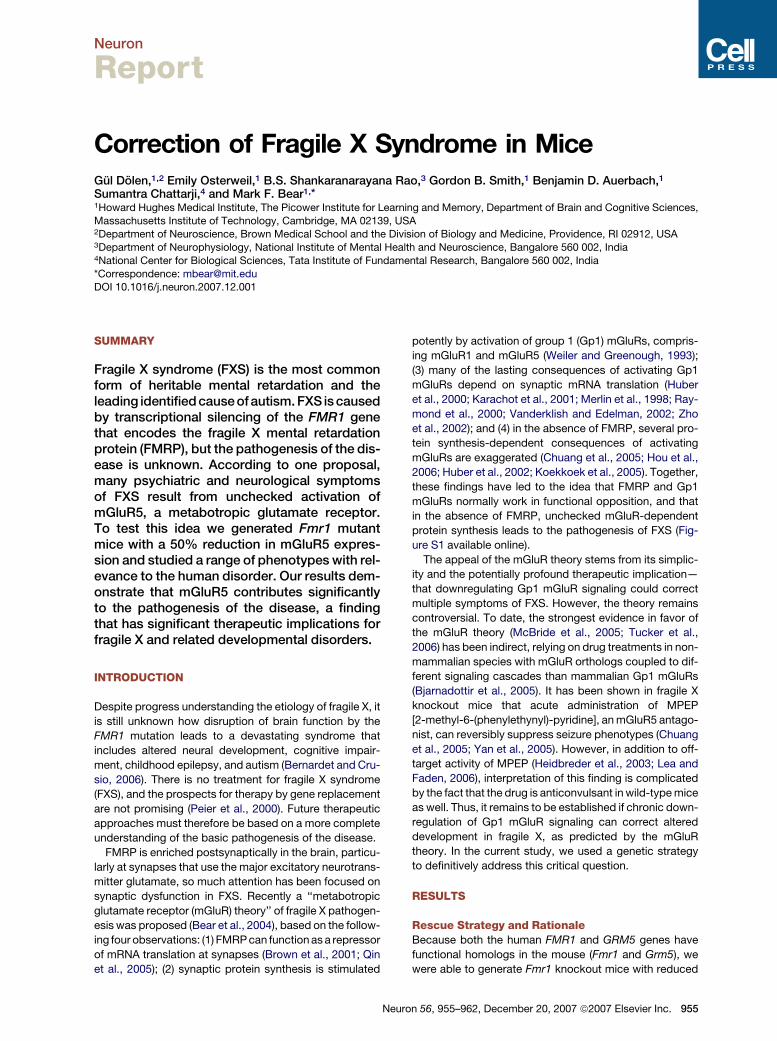
In the current study we also found that the response to
3 days MD in WT mice was dominated by deprived-eye
depression, as expected. In KO littermates, however, the
response to brief MD was characterized by substantial
open-eye potentiation, reminiscent of what happens in
WT mice after longer periods of MD. On the other hand,
the HT mice showed a ‘‘hypoplastic’’ response to MD,
as they lacked significant deprived-eye depression. How-
ever, crossing the two mutant mice resulted in a pheno-
type very similar to WT that was again dominated by
deprived-eye depression (Figure 1C).
Plots of the average (±SEM) fractional changes after
3 days MD in the four genotypes are shown in Figure 1D.
The KO mice displayed increased plasticity compared to
the WT (MANOVA WT:KO, p = 0.011); HT mice displayed
diminished plasticity compared to WT (MANOVA WT:HT,
p = 0.013); CR mice showed a rescue of the KO phenotype
and were not significantly different from WT (MANOVA
WT:CR, p = 0.8268, KO:CR p = 0.037, HT:CRS p = 0.161).
Since the KO and HT mutations affected OD plasticity in
opposite directions, one could question whether the CR
phenotype reflects rescue or the simple addition of two
independent effects. However, a compound phenotype
would be the absence of deprived-eye depression (the ef-
fect of reducing mGluR5) and an exaggeration of open-
eye potentiation (the effect of reducing FMRP). Instead,
we observe a phenotype in the CR mice that is signifi-
cantly different from KO mice, and not significantly differ-
ent from WT. Thus, reducing mGluR5 by 50% corrects the
defect in plasticity caused by the absence of FMRP.
Density of Dendritic Spines on Cortical PyramidalNeurons Is Increased in Fmr1 KO and Rescued byReducing mGluR5 ExpressionAbnormalities in dendritic spines, the major targets of ex-
citatory synapses in the brain, have long been associated
c.
Neuron
Correction of Fragile X Syndrome in Mice
with various forms of human mental retardation, including
FXS. The increased spine density phenotype observed in
humans has been recapitulated in the Fmr1 KO mouse
Figure 1. Genetic Rescue of OD Plasticity Phenotype in FXS
(A) Schematic of the mouse visual pathway and position of the record-
ing electrode in primary visual cortex.
(B) Absolute VEP amplitudes recorded during binocular viewing across
contrasts (0–100%, square reversing at 1 Hz, 0.05 cycles/degree).
No significant differences across genotypes (n = 46 WT, n = 33 KO,
n = 8 HT, n = 20 CR hemispheres, MANOVA p = 0.0868).
(C) Effect of 3 day MD on VEP amplitude (data expressed as
mean ± SEM, normalized to day 0 ipsilateral eye value. (C1) WT mice
(n = 19). Note significant deprived eye depression. (C2) KO mice
(n = 18). Note significant open eye potentiation. (C3) HT mice (n = 16).
Note absence of deprived eye depression. (C4) CR mice (n = 13).
Note rescue of KO phenotype. Post hoc Student’s t tests: *Significantly
different from baseline (day 0).
(D) Plots (mean ± SEM) of the fractional change in open and deprived eye
responses after 3 day MD show rescue of the KO phenotype in CR mice.
Neu
(reviewed by Grossman et al. [2006]). Because one protein
synthesis-dependent consequence of activating Gp1
mGluRs on cortical neurons in vitro is an increase in the
density of long, thin spines (Vanderklish and Edelman,
2002), we hypothesized that FMRP and mGluR5 antago-
nistically regulate dendritic spine density in vivo.
We chose to examine this question in layer 3 pyramidal
neurons of binocular visual cortex at P30, since we had
established that OD plasticity at this age was altered in
the Fmr1 KO mice. Dendritic spine density was analyzed
separately in apical and basal branches across the four
genotypes, using the Golgi-Cox silver staining method
(Figure 2A). We observed a highly significant increase in
total dendritic spine density in the KO, readily apparent
as a rightward shift in the cumulative probability histogram
(Figure 2B). Reducing mGluR5 expression had no effect
on spine density in the HT mice, but the fragile X pheno-
type was completely rescued in the CR mice (apical
Kruskal-Wallis test, p < 0.0001; Kolmogorov-Smirnov
test, WT:KO p < 0.0001, WT:HT p = 0.3920, CR:WT p =
0.4407, CR:KO p < 0.0001; basal Kruskal-Wallis test, p <
0.0001; Kolmogorov-Smirnov test WT:KO p < 0.0001,
WT:HT p > 0.9999, CR:WT p > 0.9999, CR:KO p < 0.0001).
We also performed a segmental analysis of spine den-
sity across the four genotypes. Consistent with previous
observations, we observed an inverted U-shaped distribu-
tion of synapses in both apical and basal branches across
all genotypes. However, as shown in Figure 2C, the den-
sity of spines was uniformly increased in the Fmr1 KO
and rescued in the CR (repeated-measures ANOVA:
apical distance p < 0.0001, apical distance 3 genotype
p < 0.0001, apical genotype p < 0.0001, basal distance
p < 0.0001, basal distance 3 genotype p = 0.0181, basal
genotype p < 0.0001; ANOVA genotype: apical, basal, 10–
100, in 10 mm segments p < 0.0001; unpaired Student’s t
tests apical, basal, 10–100, in 10 mm segments WT:KO p <
0.05, WT:HT p > 0.05, WT:CR p > 0.05, KO:CR p < 0.05).
These results suggest that neither the Fmr1 KO phenotype
nor the rescue by selective reduction in gene dosage in
the CR reflects a redistribution of synapses within the
segment.
Increased Basal Protein Synthesisin Hippocampus of Fmr1 KO Mice IsRescued by Reducing mGluR5 ExpressionA previous study reported an elevated basal rate of in vivo
protein synthesis in the hippocampus of Fmr1 KO mice
(Qin et al., 2005). We asked if this difference could also
be observed in hippocampal slices in vitro by examining
the incorporation of 35S-methionine/cysteine into new
protein. We observed a significant effect of genotype on
protein synthesis (Figure 3A). The increased protein syn-
thesis seen in KO hippocampus was prevented by selec-
tive reduction in mGluR5 gene dosage.
Electrophoretic separation of radiolabeled translation
products (Figure 3B) suggests that increased protein syn-
thesis in the KO is not limited to one or few predominant
protein species but rather extends across a broad range
ron 56, 955–962, December 20, 2007 ª2007 Elsevier Inc. 957
Neuron
Correction of Fragile X Syndrome in Mice
of resolved molecular weights. Because the rate of protein
synthesis was unaffected in the HT mice relative to WT,
the rescue in the CR mice is unambiguous and does not
simply reflect an offsetting decrease in synthesis of a
separate pool of proteins.
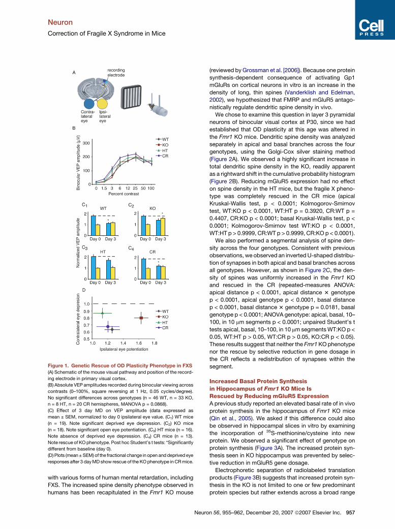
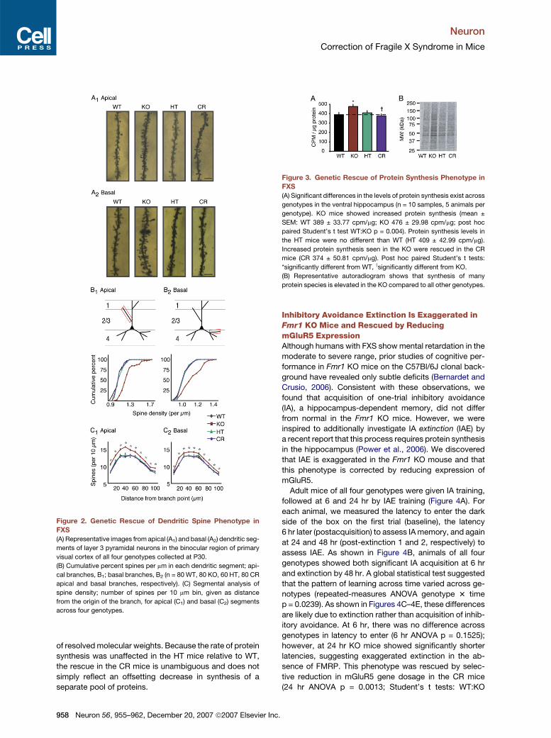
Figure 2. Genetic Rescue of Dendritic Spine Phenotype in
FXS
(A) Representative images from apical (A1) and basal (A2) dendritic seg-
ments of layer 3 pyramidal neurons in the binocular region of primary
visual cortex of all four genotypes collected at P30.
(B) Cumulative percent spines per mm in each dendritic segment; api-
cal branches, B1; basal branches, B2 (n = 80 WT, 80 KO, 60 HT, 80 CR
apical and basal branches, respectively). (C) Segmental analysis of
spine density; number of spines per 10 mm bin, given as distance
from the origin of the branch, for apical (C1) and basal (C2) segments
across four genotypes.
958 Neuron 56, 955–962, December 20, 2007 ª2007 Elsevier In
Inhibitory Avoidance Extinction Is Exaggerated inFmr1 KO Mice and Rescued by ReducingmGluR5 ExpressionAlthough humans with FXS show mental retardation in the
moderate to severe range, prior studies of cognitive per-
formance in Fmr1 KO mice on the C57Bl/6J clonal back-
ground have revealed only subtle deficits (Bernardet and
Crusio, 2006). Consistent with these observations, we
found that acquisition of one-trial inhibitory avoidance
(IA), a hippocampus-dependent memory, did not differ
from normal in the Fmr1 KO mice. However, we were
inspired to additionally investigate IA extinction (IAE) by
a recent report that this process requires protein synthesis
in the hippocampus (Power et al., 2006). We discovered
that IAE is exaggerated in the Fmr1 KO mouse and that
this phenotype is corrected by reducing expression of
mGluR5.
Adult mice of all four genotypes were given IA training,
followed at 6 and 24 hr by IAE training (Figure 4A). For
each animal, we measured the latency to enter the dark
side of the box on the first trial (baseline), the latency
6 hr later (postacquisition) to assess IA memory, and again
at 24 and 48 hr (post-extinction 1 and 2, respectively) to
assess IAE. As shown in Figure 4B, animals of all four
genotypes showed both significant IA acquisition at 6 hr
and extinction by 48 hr. A global statistical test suggested
that the pattern of learning across time varied across ge-
notypes (repeated-measures ANOVA genotype 3 time
p = 0.0239). As shown in Figures 4C–4E, these differences
are likely due to extinction rather than acquisition of inhib-
itory avoidance. At 6 hr, there was no difference across
genotypes in latency to enter (6 hr ANOVA p = 0.1525);
however, at 24 hr KO mice showed significantly shorter
latencies, suggesting exaggerated extinction in the ab-
sence of FMRP. This phenotype was rescued by selec-
tive reduction in mGluR5 gene dosage in the CR mice
(24 hr ANOVA p = 0.0013; Student’s t tests: WT:KO
Figure 3. Genetic Rescue of Protein Synthesis Phenotype in
FXS
(A) Significant differences in the levels of protein synthesis exist across
genotypes in the ventral hippocampus (n = 10 samples, 5 animals per
genotype). KO mice showed increased protein synthesis (mean ±
SEM: WT 389 ± 33.77 cpm/mg; KO 476 ± 29.98 cpm/mg; post hoc
paired Student’s t test WT:KO p = 0.004). Protein synthesis levels in
the HT mice were no different than WT (HT 409 ± 42.99 cpm/mg).
Increased protein synthesis seen in the KO were rescued in the CR
mice (CR 374 ± 50.81 cpm/mg). Post hoc paired Student’s t tests:
*significantly different from WT, ysignificantly different from KO.
(B) Representative autoradiogram shows that synthesis of many
protein species is elevated in the KO compared to all other genotypes.
c.
Neuron
Correction of Fragile X Syndrome in Mice
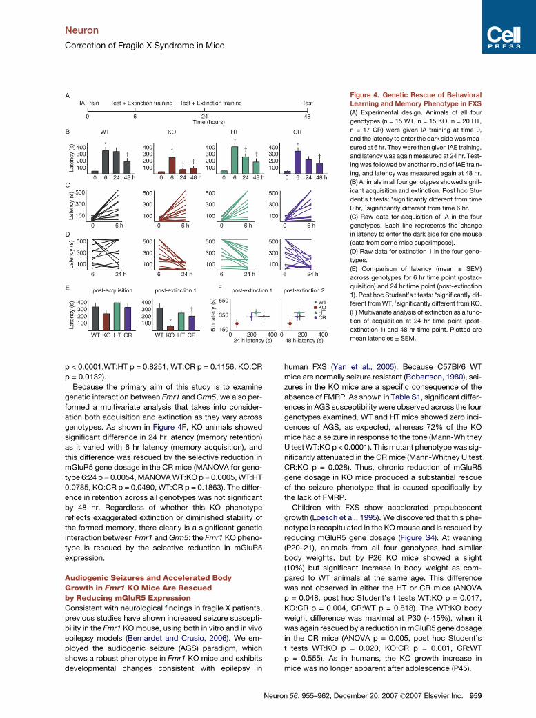
Figure 4. Genetic Rescue of Behavioral
Learning and Memory Phenotype in FXS
(A) Experimental design. Animals of all four
genotypes (n = 15 WT, n = 15 KO, n = 20 HT,
n = 17 CR) were given IA training at time 0,
and the latency to enter the dark side was mea-
sured at 6 hr. They were then given IAE training,
and latency was again measured at 24 hr. Test-
ing was followed by another round of IAE train-
ing, and latency was measured again at 48 hr.
(B) Animals in all four genotypes showed signif-
icant acquisition and extinction. Post hoc Stu-
dent’s t tests: *significantly different from time
0 hr, ysignificantly different from time 6 hr.
(C) Raw data for acquisition of IA in the four
genotypes. Each line represents the change
in latency to enter the dark side for one mouse
(data from some mice superimpose).
(D) Raw data for extinction 1 in the four geno-
types.
(E) Comparison of latency (mean ± SEM)
across genotypes for 6 hr time point (postac-
quisition) and 24 hr time point (post-extinction
1). Post hoc Student’s t tests: *significantly dif-
ferent from WT, ysignificantly different from KO.
(F) Multivariate analysis of extinction as a func-
tion of acquisition at 24 hr time point (post-
extinction 1) and 48 hr time point. Plotted are
mean latencies ± SEM.
p < 0.0001,WT:HT p = 0.8251, WT:CR p = 0.1156, KO:CR
p = 0.0132).
Because the primary aim of this study is to examine
genetic interaction between Fmr1 and Grm5, we also per-
formed a multivariate analysis that takes into consider-
ation both acquisition and extinction as they vary across
genotypes. As shown in Figure 4F, KO animals showed
significant difference in 24 hr latency (memory retention)
as it varied with 6 hr latency (memory acquisition), and
this difference was rescued by the selective reduction in
mGluR5 gene dosage in the CR mice (MANOVA for geno-
type 6:24 p = 0.0054, MANOVA WT:KO p = 0.0005, WT:HT
0.0785, KO:CR p = 0.0490, WT:CR p = 0.1863). The differ-
ence in retention across all genotypes was not significant
by 48 hr. Regardless of whether this KO phenotype
reflects exaggerated extinction or diminished stability of
the formed memory, there clearly is a significant genetic
interaction between Fmr1 and Grm5: the Fmr1 KO pheno-
type is rescued by the selective reduction in mGluR5
expression.
Audiogenic Seizures and Accelerated BodyGrowth in Fmr1 KO Mice Are Rescuedby Reducing mGluR5 ExpressionConsistent with neurological findings in fragile X patients,
previous studies have shown increased seizure suscepti-
bility in the Fmr1 KO mouse, using both in vitro and in vivo
epilepsy models (Bernardet and Crusio, 2006). We em-
ployed the audiogenic seizure (AGS) paradigm, which
shows a robust phenotype in Fmr1 KO mice and exhibits
developmental changes consistent with epilepsy in
Neu
human FXS (Yan et al., 2005). Because C57Bl/6 WT
mice are normally seizure resistant (Robertson, 1980), sei-
zures in the KO mice are a specific consequence of the
absence of FMRP. As shown in Table S1, significant differ-
ences in AGS susceptibility were observed across the four
genotypes examined. WT and HT mice showed zero inci-
dences of AGS, as expected, whereas 72% of the KO
mice had a seizure in response to the tone (Mann-Whitney
U test WT:KO p < 0.0001). This mutant phenotype was sig-
nificantly attenuated in the CR mice (Mann-Whitney U test
CR:KO p = 0.028). Thus, chronic reduction of mGluR5
gene dosage in KO mice produced a substantial rescue
of the seizure phenotype that is caused specifically by
the lack of FMRP.
Children with FXS show accelerated prepubescent
growth (Loesch et al., 1995). We discovered that this phe-
notype is recapitulated in the KO mouse and is rescued by
reducing mGluR5 gene dosage (Figure S4). At weaning
(P20–21), animals from all four genotypes had similar
body weights, but by P26 KO mice showed a slight
(10%) but significant increase in body weight as com-
pared to WT animals at the same age. This difference
was not observed in either the HT or CR mice (ANOVA
p = 0.048, post hoc Student’s t tests WT:KO p = 0.017,
KO:CR p = 0.004, CR:WT p = 0.818). The WT:KO body
weight difference was maximal at P30 (�15%), when it
was again rescued by a reduction in mGluR5 gene dosage
in the CR mice (ANOVA p = 0.005, post hoc Student’s
t tests WT:KO p = 0.020, KO:CR p = 0.001, CR:WT
p = 0.555). As in humans, the KO growth increase in
mice was no longer apparent after adolescence (P45).
ron 56, 955–962, December 20, 2007 ª2007 Elsevier Inc. 959
Neuron
Correction of Fragile X Syndrome in Mice
Macroorchidism in Fmr1 KO Mice Is Not Rescuedby Reducing mGluR5 ExpressionChildren with FXS (and KO mice) have dysmorphic fea-
tures, including postadolescent macroorchidism. Testes
express Gp1 mGluRs (Storto et al., 2001), so we won-
dered if this phenotype might also be rescued in our CR
mice. Postadolescent testicular weight was increased by
24% in KO mice compared to WT (p < 0.0004; Student’s
t test); however, there was no rescue of this phenotype
in the CR mice (Figure S5). To investigate if the absence
of rescue was a matter of gene dosage, we generated
KO mice that had a complete absence of mGluR5 [Fmr1
(�/Y) Grm5 (�/�), dKO]. Again, however, there was no
rescue of the testicular phenotype.
DISCUSSION
The goal of this study was to test a key prediction of the
mGluR theory—that aspects of FXS can be corrected by
downregulating signaling through group 1 mGluRs (Bear
et al., 2004). Each analysis was designed to examine a dif-
ferent dimension of the disorder in mice with relevance to
the human syndrome, ranging from the cognitive to the
somatic. The experiments assayed dysfunction in very
different neural circuits; and for each, three outcomes
were possible: amelioration, exacerbation, or persistence
of Fmr1 mutant phenotypes in mice with reduced expres-
sion of mGluR5. Thus, it is remarkable that by reducing
mGluR5 gene dosage by 50%, we were able to bring mul-
tiple, widely varied fragile X phenotypes significantly
closer to normal.
A novel aspect of the current study was the use of OD
plasticity as an in vivo assay of how experience-dependent
synaptic modification is altered by the loss of FMRP. MD
sets in motion a sequence of synaptic changes in visual
cortex, characterized by a rapid and persistent loss of re-
sponsiveness to the deprived eye and a slower compensa-
tory increase in responsiveness to the nondeprived eye
(Frenkel and Bear, 2004). Because MD triggers mecha-
nisms of synaptic depression and potentiation, as well as
homeostatic adaptations to an altered environment, OD
plasticity is a particularly rich paradigm for understanding
the interactions of genes and experience. The intersecting
trends of using mice to study OD plasticity mechanisms
and to model human diseases provided the opportunity
to use this paradigm to get a more precise understanding
of how development goes awry in a genetic disorder.
Previous work suggested that Gp1 mGluR signaling is
highest in visual cortex during the period of maximal syn-
aptic plasticity (Dudek and Bear, 1989), and the current
findings strongly suggest an important role for mGluR5
in OD plasticity. Although more experiments will be re-
quired to pinpoint this role, an obvious clue comes from
the genetic interaction with Fmr1. FMRP can act as
a translational suppressor (Brown et al., 2001; Qin et al.,
2005), and OD plasticity, like many forms of persistent
synaptic modification, requires protein synthesis (Taha
and Stryker, 2002). Thus, our findings suggest the intrigu-
960 Neuron 56, 955–962, December 20, 2007 ª2007 Elsevier
ing hypothesis that the rate of plasticity in visual cortex is
determined by the level of activity-dependent protein syn-
thesis, which is stimulated by mGluR5 and inhibited by
FMRP. Consistent with this model, the phenotype in
Fmr1 KO mice appears to reflect ‘‘hyperplasticity,’’ since
3 days of MD yielded effects on VEPs that normally require
7 days. This exaggerated plasticity was corrected by re-
ducing mGluR5 expression by 50%.
Although we observed an increased spine density in the
visual cortex of KO mice, there was no apparent difference
in the amplitude of VEPs at P30. This discrepancy may be
because VEPs were recorded in layer 4, whereas the spine
measurements were made on layer 3 neurons. In any
case, the clear mutant spine phenotype in layer 3 gave
us the opportunity to examine if this structural defect
could also be corrected by decreasing mGluR5. We ob-
served a remarkable rescue of the fragile X spine pheno-
type in the CR mice, despite the fact that reducing
mGluR5 expression in the HT mice had no effect on spine
density. Thus, although the reduction in Grm5 gene dos-
age is not sufficient to alter spine density by itself, it com-
pletely corrects the defect in fragile X mice.
Strain-specific variation has confounded previous at-
tempts to identify a behavioral learning and memory
phenotype in the Fmr1 KO (Bernardet and Crusio, 2006).
Consistent with earlier findings, we were unable to detect
a significant IA deficit in the Fmr1 KO mice on the C57BL/6
background. On the other hand, we were able to detect
a difference in the rate of IA extinction that could be cor-
rected by reducing mGluR5 expression. Because IA in-
duces LTP of Schaffer collateral synapses in area CA1 of
the hippocampus (Whitlock et al., 2006), it is tempting to
speculate that IA extinction is exaggerated in Fmr1 KO
mice due, at least in part, to excessive mGluR-dependent
synaptic weakening (Huber et al., 2002; Zho et al., 2002).
Unbalanced LTD could account for the cognitive impair-
ment that is the hallmark of fragile X.
Fragile X is a syndromic disorder. In addition to mental
retardation, associated features of the disease in humans
include childhood epilepsy, altered body growth, and
postpubertal macroorchidism. In the case of epilepsy
and macroorchidism, these phenotypes have been reca-
pitulated in the mouse model (Bernardet and Crusio,
2006); however, it was previously unknown that Fmr1
KO mice show a similar disruption in body growth. Both
the body growth and AGS phenotypes were ameliorated
in the CR mice; however, there was no evidence of an in-
teraction between FMRP and mGluR5 in the control of tes-
ticle size. These results argue against a role for mGluR5 in
the pathogenesis of the macroorchidism phenotype in
FXS, but we cannot rule out a contribution of the other
Gp1 mGluR (mGluR1).
ConclusionAlthough we studied a range of phenotypes, a simple way
to conceptualize the constellation of findings is that fragile
X is a disorder of excess—excessive sensitivity to environ-
mental change, synaptic connectivity, protein synthesis,
Inc.
Neuron
Correction of Fragile X Syndrome in Mice
memory extinction, body growth, and excitability—and
these excesses can be corrected by reducing mGluR5.
Although the precise molecular basis of the interaction
remains to be determined, the data show unambiguously
that mGluR5 and FMRP act as an opponent pair in several
functional contexts and support the theory that many CNS
symptoms in fragile X are accounted for by unbalanced
activation of Gp1 mGluRs. These findings have major ther-
apeutic implications for FXS and autism (see Bear et al.,
2008).
EXPERIMENTAL PROCEDURES
Animals
Fmr1 mutant mice (Jackson Labs) were crossed with Grm5 mutants
(Jackson Labs) to produce mice of four genotypes. In all crossings,
animals were on the C57Bl/6J clonal background. In an effort to reduce
variability due to rearing conditions, all experimental animals were
bred from Fmr1 heterozygote mothers, group housed (animals weaned
to solitary housing were excluded), and maintained in a 12:12 hr light:
dark cycle. Paternal genotype varied between crossings and included
WT, Grm5 HT, or Grm5 KO.
Genotyping
See Figure S2 and the Supplemental Data.
Electrophysiology and Spine Measurements
Transverse hippocampal slices were prepared from P25–30 mice and
mGluR-LTD was studied as described by Huber et al. (2001). VEP
recordings and monocular deprivation were performed as previously
described (Frenkel and Bear, 2004). Spines were analyzed using the
Golgi-Cox method as described by Hayashi et al. (2004). Animal n =
8 WT, 8 KO, 6 HT, 8 CR; dendritic segment n = 80 WT, 80 KO,
60 HT, 80 CR apical and basal branches, respectively. All protrusions,
irrespective of their morphological characteristics, were counted as
spines if they were in direct continuity with the dendritic shaft. In total,
68,032 spines were counted across all four genotypes.
Inhibitory Avoidance Extinction, Metabolic Labeling, and
Audiogenic Seizure
IAE experiments were performed as previously described (Power et al.,
2006). Metabolic labeling experiments were similar to those described
in Raymond et al. (2000). AGS experiments were performed as
described by Yan et al. (2005). See Supplemental Data.
Statistical Analysis
In all cases, post hoc comparisons between genotypes were made
only if global analysis indicated a statistically significant (p < 0.05)
effect of genotype. Outliers (R2 SD from the mean) were excluded.
For AGS experiments, nonparametric statistics (Kruskall-Wallis,
Mann-Whitney U) were used since incidence scores were bimodal
(yes/no). For all other analysis, parametric tests (ANOVA, MANOVA,
two-tailed paired and unpaired Student’s t tests, assuming equal var-
iance) were used. For the metabolic labeling experiments, the post hoc
paired Student’s t test was used to eliminate the variability due to
strength of radioactive label on different experimental days and was
justified by the experimental design (samples were collected with
yoked, rather than randomized, controls).
Supplemental Data
The Supplemental Data for this article can be found online at http://
www.neuron.org/cgi/content/full/56/6/955/DC1/.
Neu
ACKNOWLEDGMENTS
We are grateful to M. Shuler for helpful discussion of appropriate
statistical analysis; K. Oram, C. Dudley, A. Topolszki, T. Udaka, and
C. Poo for genotyping and animal care; E. Sklar for technical assis-
tance and construction of AGS stimulus; M. Frenkel for use of previ-
ously published data; S. Meagher for administrative support; A. Kraev
and J. Roder for Grm5 genotyping protocols; K. Huber, K. Wiig, A. Go-
vindarajan, R. Paylor, W. Chen, B. Yoon, Q. Yan, R. Bauchwitz, M.
Tranfaglia, E. Klann, E. Berry-Kravis, and A. Heynen for helpful discus-
sions. Supported by the NIMH, NICHD, National Fragile X Foundation,
FRAXA, Simons Foundation. M.F.B. discloses a financial interest in
Seaside Therapeutics.
Received: March 29, 2007
Revised: October 8, 2007
Accepted: December 3, 2007
Published: December 19, 2007
REFERENCES
Bear, M.F., Huber, K.M., and Warren, S.T. (2004). The mGluR theory of
fragile X mental retardation. Trends Neurosci. 27, 370–377.
Bear, M.F., Dolen, G., Osterweil, E., and Nagarajan, N. (2008). Fragile
X: Translation in action. Neuropsychopharmacology 33, 84–87.
Bernardet, M., and Crusio, W.E. (2006). Fmr1 KO mice as a possible
model of autistic features. ScientificWorldJournal 6, 1164–1176.
Bjarnadottir, T.K., Schioth, H.B., and Fredriksson, R. (2005). The
phylogenetic relationship of the glutamate and pheromone G-pro-
tein-coupled receptors in different vertebrate species. Ann. N Y
Acad. Sci. 1040, 230–233.
Brown, V., Jin, P., Ceman, S., Darnell, J.C., O’Donnell, W.T.,
Tenenbaum, S.A., Jin, X., Feng, Y., Wilkinson, K.D., Keene, J.D.,
et al. (2001). Microarray identification of FMRP-associated brain
mRNAs and altered mRNA translational profiles in fragile X syndrome.
Cell 107, 477–487.
Chuang, S.C., Zhao, W., Bauchwitz, R., Yan, Q., Bianchi, R., and
Wong, R.K. (2005). Prolonged epileptiform discharges induced by al-
tered group I metabotropic glutamate receptor-mediated synaptic re-
sponses in hippocampal slices of a fragile X mouse model. J. Neurosci.
25, 8048–8055.
Dudek, S.M., and Bear, M.F. (1989). A biochemical correlate of the
critical period for synaptic modification in the visual cortex. Science
246, 673–675.
The Dutch-Belgian Fragile X Consortium. (1994). Fmr1 knockout mice:
A model to study fragile X mental retardation. Cell 78, 23–33.
Frenkel, M.Y., and Bear, M.F. (2004). How monocular deprivation shifts
ocular dominance in visual cortex of young mice. Neuron 44, 917–923.
Grossman, A.W., Aldridge, G.M., Weiler, I.J., and Greenough, W.T.
(2006). Local protein synthesis and spine morphogenesis: Fragile X
syndrome and beyond. J. Neurosci. 26, 7151–7155.
Hayashi, M.L., Choi, S.Y., Rao, B.S., Jung, H.Y., Lee, H.K., Zhang, D.,
Chattarji, S., Kirkwood, A., and Tonegawa, S. (2004). Altered cortical
synaptic morphology and impaired memory consolidation in
forebrain-specific dominant-negative PAK transgenic mice. Neuron
42, 773–787.
Heidbreder, C.A., Bianchi, M., Lacroix, L.P., Faedo, S., Perdona, E.,
Remelli, R., Cavanni, P., and Crespi, F. (2003). Evidence that the
metabotropic glutamate receptor 5 antagonist MPEP may act as an in-
hibitor of the norepinephrine transporter in vitro and in vivo. Synapse
50, 269–276.
Hou, L., Antion, M.D., Hu, D., Spencer, C.M., Paylor, R., and Klann, E.
(2006). Dynamic translational and proteasomal regulation of fragile X
ron 56, 955–962, December 20, 2007 ª2007 Elsevier Inc. 961
Neuron
Correction of Fragile X Syndrome in Mice
mental retardation protein controls mGluR-dependent long-term
depression. Neuron 51, 441–454.
Huber, K.M., Kayser, M.S., and Bear, M.F. (2000). Role for rapid
dendritic protein synthesis in hippocampal mGluR-dependent long-
term depression. Science 288, 1254–1257.
Huber, K.M., Roder, J.C., and Bear, M.F. (2001). Chemical induction of
mGluR5- and protein synthesis-dependent long-term depression in
hippocampal area CA1. J. Neurophysiol. 86, 321–325.
Huber, K.M., Gallagher, S.M., Warren, S.T., and Bear, M.F. (2002).
Altered synaptic plasticity in a mouse model of fragile X mental retar-
dation. Proc. Natl. Acad. Sci. USA 99, 7746–7750.
Jia, Z., Lu, Y., Henderson, J., Taverna, F., Romano, C., Abramow-
Newerly, W., Wojtowicz, J.M., and Roder, J. (1998). Selective abolition
of the NMDA component of long-term potentiation in mice lacking
mGluR5. Learn. Mem. 5, 331–343.
Karachot, L., Shirai, Y., Vigot, R., Yamamori, T., and Ito, M. (2001).
Induction of long-term depression in cerebellar Purkinje cells requires
a rapidly turned over protein. J. Neurophysiol. 86, 280–289.
Koekkoek, S.K., Yamaguchi, K., Milojkovic, B.A., Dortland, B.R.,
Ruigrok, T.J., Maex, R., De Graaf, W., Smit, A.E., VanderWerf, F.,
Bakker, C.E., et al. (2005). Deletion of FMR1 in Purkinje cells enhances
parallel fiber LTD, enlarges spines, and attenuates cerebellar eyelid
conditioning in Fragile X syndrome. Neuron 47, 339–352.
Lea, P.M., IV, and Faden, A.I. (2006). Metabotropic glutamate receptor
subtype 5 antagonists MPEP and MTEP. CNS Drug Rev. 12, 149–166.
Loesch, D.Z., Huggins, R.M., and Hoang, N.H. (1995). Growth in stat-
ure in fragile X families: A mixed longitudinal study. Am. J. Med. Genet.
58, 249–256.
Lu, Y.M., Jia, Z., Janus, C., Henderson, J.T., Gerlai, R., Wojtowicz,
J.M., and Roder, J.C. (1997). Mice lacking metabotropic glutamate
receptor 5 show impaired learning and reduced CA1 long-term poten-
tiation (LTP) but normal CA3 LTP. J. Neurosci. 17, 5196–5205.
McBride, S.M., Choi, C.H., Wang, Y., Liebelt, D., Braunstein, E.,
Ferreiro, D., Sehgal, A., Siwicki, K.K., Dockendorff, T.C., Nguyen,
H.T., et al. (2005). Pharmacological rescue of synaptic plasticity, court-
ship behavior, and mushroom body defects in a Drosophila model of
fragile X syndrome. Neuron 45, 753–764.
Merlin, L.R., Bergold, P.J., and Wong, R.K. (1998). Requirement of pro-
tein synthesis for group I mGluR-mediated induction of epileptiform
discharges. J. Neurophysiol. 80, 989–993.
Peier, A.M., McIlwain, K.L., Kenneson, A., Warren, S.T., Paylor, R., and
Nelson, D.L. (2000). (Over)correction of FMR1 deficiency with YAC
transgenics: Behavioral and physical features. Hum. Mol. Genet. 9,
1145–1159.
962 Neuron 56, 955–962, December 20, 2007 ª2007 Elsevier In
Power, A.E., Berlau, D.J., McGaugh, J.L., and Steward, O. (2006). Ani-
somycin infused into the hippocampus fails to block ‘‘reconsolidation’’
but impairs extinction: The role of re-exposure duration. Learn. Mem.
13, 27–34.
Qin, M., Kang, J., Burlin, T.V., Jiang, C., and Smith, C.B. (2005). Post-
adolescent changes in regional cerebral protein synthesis: An in vivo
study in the FMR1 null mouse. J. Neurosci. 25, 5087–5095.
Raymond, C.R., Thompson, V.L., Tate, W.P., and Abraham, W.C.
(2000). Metabotropic glutamate receptors trigger homosynaptic
protein synthesis to prolong long-term potentiation. J. Neurosci. 20,
969–976.
Robertson, H.A. (1980). Audiogenic seizures: Increased benzodiazepin
receptor binding in a susceptible strain of mice. Eur. J. Pharmacol. 66,
249–252.
Storto, M., Sallese, M., Salvatore, L., Poulet, R., Condorelli, D.F.,
Dell’Albani, P., Marcello, M.F., Romeo, R., Piomboni, P., Barone, N.,
et al. (2001). Expression of metabotropic glutamate receptors in the
rat and human testis. J. Endocrinol. 170, 71–78.
Taha, S., and Stryker, M.P. (2002). Rapid ocular dominance plasticity
requires cortical but not geniculate protein synthesis. Neuron 34,
425–436.
Tucker, B., Richards, R.I., and Lardelli, M. (2006). Contribution of
mGluR and Fmr1 functional pathways to neurite morphogenesis, cra-
niofacial development and fragile X syndrome. Hum. Mol. Genet. 15,
3446–3458.
Vanderklish, P.W., and Edelman, G.M. (2002). Dendritic spines elon-
gate after stimulation of group 1 metabotropic glutamate receptors
in cultured hippocampal neurons. Proc. Natl. Acad. Sci. USA 99,
1639–1644.
Weiler, I.J., and Greenough, W.T. (1993). Metabotropic glutamate re-
ceptors trigger postsynaptic protein synthesis. Proc. Natl. Acad. Sci.
USA 90, 7168–7171.
Whitlock, J.R., Heynen, A.J., Shuler, M.G., and Bear, M.F. (2006).
Learning induces long-term potentiation in the hippocampus. Science
313, 1093–1097.
Yan, Q.J., Rammal, M., Tranfaglia, M., and Bauchwitz, R.P. (2005).
Suppression of two major Fragile X Syndrome mouse model pheno-
types by the mGluR5 antagonist MPEP. Neuropharmacology 49,
1053–1066.
Zho, W.M., You, J.L., Huang, C.C., and Hsu, K.S. (2002). The group I
metabotropic glutamate receptor agonist (S)-3,5-dihydroxyphenylgly-
cine induces a novel form of depotentiation in the CA1 region of the
hippocampus. J. Neurosci. 22, 8838–8849.
c.
1
Neuron, Volume 56
Supplemental Data
Correction of Fragile X Syndrome in Mice Gül Dölen, Emily Osterweil, B.S. Shankaranarayana Rao, Gordon B. Smith, Benjamin D. Auerbach, Sumantra Chattarji, and Mark F. Bear Supplemental Experimental Procedures Genotyping Screening for the presence or absence of the wild-type allele on the Fmr1 locus was performed using primer S1(5’ GTG GTT AGC TAA AGT GAG GAT GAT 3’) and S2 (5’ CAG GTT TGT TGG GAT TAA CAG ATC 3’) and the knockout allele using primer M2 (5’ ATC TAG TCA TGC TAT GGA TAT CAG C 3’) and N2 (5’ GTG GGC TCT ATG GCT TCT GAG G 3’). For the Fmr1 locus the following PCR program was used: 95 °C for 5 min; 34 PCR cycles were performed composed of 30 sec at 95 °C, 30 sec at 61 °C, and 1min at 72°C. KO and WT PCR reactions were run separately; the reaction products were then combined and electrophoresed on a 1.5% agarose gel [WT: 465 BP (S1/S2); KO: 800 BP (M2/N2)]. Screening for the presence or absence of the knockout allele on the Grm5 locus was performed using primer P(5’ AGG GGA GGA GTA GAA GGT GGC GCG A 3’) and F(5’GCT CAC ATG CCA GGT GAC ATT ATT ATT GGA 3’) and the wild-type allele using F (see above) and R (5’ CCA TGC TAG TTG CAG AGT AAG CAA TCT GAG GT 3’). For the Grm5 locus the following PCR program was used: 95 °C for 5 min; 34 PCR cycles were performed composed of 30 sec at 95 °C, 55 sec at 61 °C, and 1min at 72 °C. KO and WT PCR reactions were run separately; the reaction products were then combined and electrophoresed on a 1.5% agarose gel [WT: 445 BP (F/R); KO: 550 BP (F/P)]. All primers were generated by Invitrogen. Inhibitory Avoidance Extinction On the day before training adult mice of all four genotypes (P60-P110) were moved into the behavioral testing room. On the day of testing, animals were placed into the dark compartment of an IA training box (a two-chambered Perspex box consisting of a lighted safe side and a dark shock side separated by a trap door) for 30 seconds followed by 90 seconds in the light compartment for habituation. Following the habituation period, the door separating the two compartments was opened and animals were allowed to enter the dark compartment. Latency to enter following door opening was recorded (“baseline”, time 0, 8a.m.-12p.m.); animals with baseline entrance latencies of greater than120 seconds were excluded. After each animal stepped completely into the dark compartment with all four paws, the sliding door was closed and the animal received a single scrambled foot-shock (0.5mA, 2.0 sec) via electrified steel rods in the floor of the box. This intensity and duration of shock consistently caused animals to vocalize and jump. Animals remained in the dark compartment for 15 sec following the shock and were then returned to their home cages. Six to seven hours following IA training, mice received a retention test (“post-acquisition”, time 6 hours, 2p.m.-6p.m.). During post-acquisition retention testing each animal was placed in the lit compartment as in training; after a 90 second delay, the
2
door opened, and the latency to enter the dark compartment was recorded (cutoff time 540 sec). For inhibitory avoidance extinction (IAE) training, animals were allowed to explore the dark compartment of the box for 200 seconds in the absence of foot-shock (animals remaining in the lit compartment after the cutoff were gently guided, using an index card, into the dark compartment); following IAE training animals were returned to their home cages. Twenty-four hours following initial IA training, mice received a second retention test (“post-extinction 1”, time 24 hours, 8a.m-12p.m.). Animals were tested in the same way as at the six hour time point, followed by a second 200 second extinction trial in the dark side of the box; following training animals were again returned to their home cages. Forty-eight hours following avoidance training, mice received a third and final retention test (“post-extinction 2”, time 48 hours, 8a.m.-12p.m.). Metabolic Labeling Pilot studies indicated that the difference between WT and KO protein synthesis was most pronounced in the ventral hippocampus so all subsequent experiments were restricted to slices from this region. Briefly, 500 µm sections were prepared from one hippocampus per animal (average age P84) using a tissue slicer (Stoelting Co.), and transferred to a custom-made incubation manifold with multiple separate inserts (15 mm diameter netwells, Electron Microscopy Sciences) suspended in carbogenated, 32.5°C ACSF (124 mM NaCl; 5 mM KCl; 1.25 mM NaH2PO4; 26 mM NaHCO3; 0.8 mM MgCl2; 1.8 mM CaCl2; and 10 mM dextrose saturated with 95% O2, 5% CO2). Following a 4 hour recovery, netwells containing slices were transferred to a second manifold chamber containing 100 ml carbogenated ACSF with 11 µCi/�l 35S-Met/Cys (EasyTag Express protein labeling mix, Perkin Elmer) and incubated for 1 hour. Netwells were then transferred to 12-well dish containing ice-cold dissection buffer to stop protein synthesis and remove excess 35S-Met/Cys, and slices were subsequently removed and homogenized in ice-cold homogenization buffer. Samples were then incubated in Trichloroacetic acid (TCA; 10% final) for 10 minutes on ice, spun at 21,000xg, 10 minutes, 4°C, and the pellet washed with ice-cold ddH2O. Pellets were then resolubilized in 37°C 1N NaOH, and pH adjusted with 1N HCl. For each sample, triplicate aliquots were then read with a scintillation counter (Beckman Instruments) using HiSafe 2 scintillation cocktail (Perkin Elmer), and triplicate aliquots were subjected to a protein concentration assay (Bio-Rad). These triplicate readings were averaged, and the counts per minute (CPM) obtained were divided by the CPM obtained (in triplicate) from the 35S-Met/Cys ASCF used for incubation. Final data were thus expressed as incorporated CPM per µg protein. Equal amounts of protein from each sample were subjected to SDS-PAGE and transferred to nitrocellulose membranes. Autoradiography was performed on the dried membranes. Staining with MemCode blot stain (Pierce) confirmed equal loading. One dimensional line scans were performed using the gel analyzer tool in Image J to visualize the breadth of radiolabeled species across genotypes. Audiogenic Seizure Animals at P19-21 (immediately following weaning) were habituated to the behavioral chamber (28x17.5x12 cm transparent plastic box) for 2 minutes prior to stimulus onset. AGS stimulus was a 125 dbSPL at 0.25 m siren (modified personal alarm, Radioshack model 49-1010, powered from a DC converter). Seizures were scored for incidence during a 5 minute stimulus presentation or until animal reached AGS endpoint (status epilepticus/ respiratory arrest/ death).
3
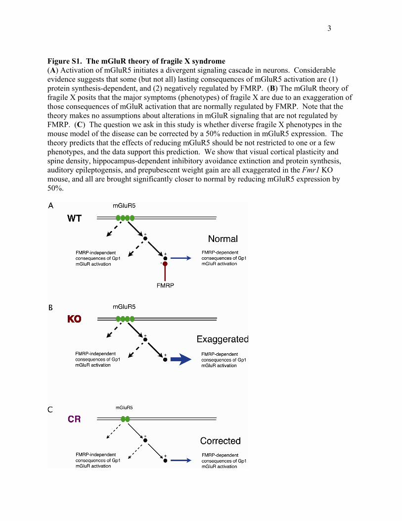
Figure S1. The mGluR theory of fragile X syndrome (A) Activation of mGluR5 initiates a divergent signaling cascade in neurons. Considerable evidence suggests that some (but not all) lasting consequences of mGluR5 activation are (1) protein synthesis-dependent, and (2) negatively regulated by FMRP. (B) The mGluR theory of fragile X posits that the major symptoms (phenotypes) of fragile X are due to an exaggeration of those consequences of mGluR activation that are normally regulated by FMRP. Note that the theory makes no assumptions about alterations in mGluR signaling that are not regulated by FMRP. (C) The question we ask in this study is whether diverse fragile X phenotypes in the mouse model of the disease can be corrected by a 50% reduction in mGluR5 expression. The theory predicts that the effects of reducing mGluR5 should be not restricted to one or a few phenotypes, and the data support this prediction. We show that visual cortical plasticity and spine density, hippocampus-dependent inhibitory avoidance extinction and protein synthesis, auditory epileptogensis, and prepubescent weight gain are all exaggerated in the Fmr1 KO mouse, and all are brought significantly closer to normal by reducing mGluR5 expression by 50%.
4
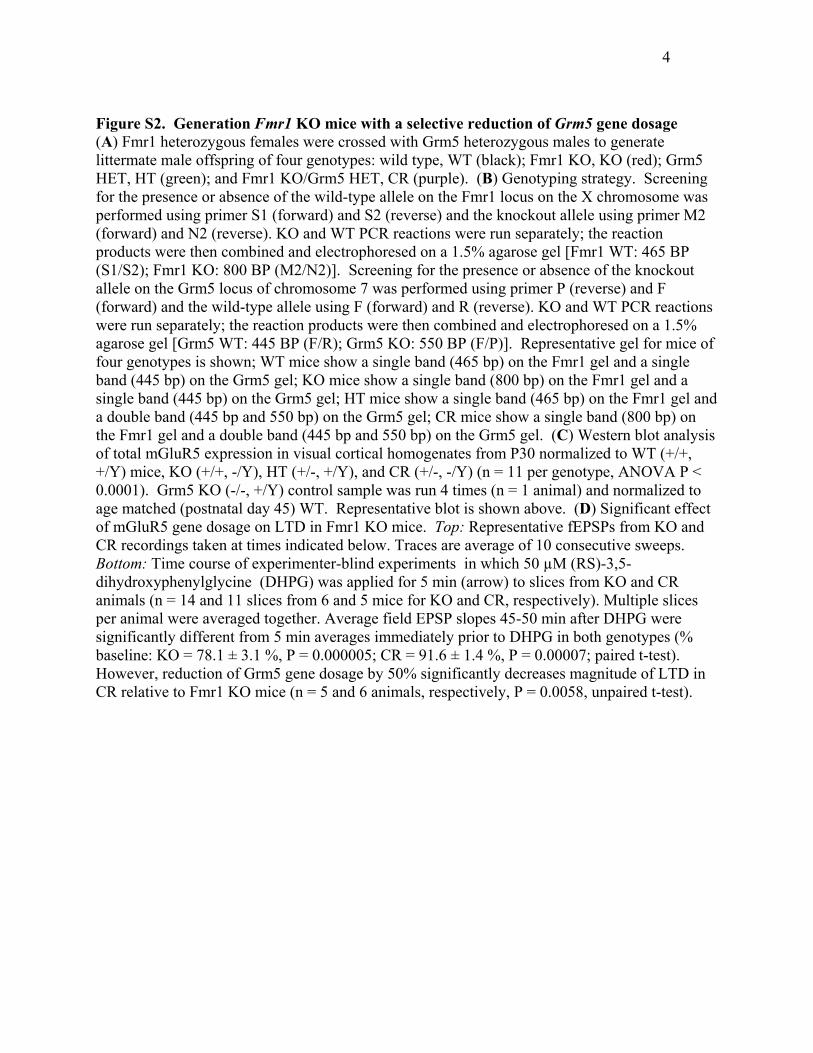
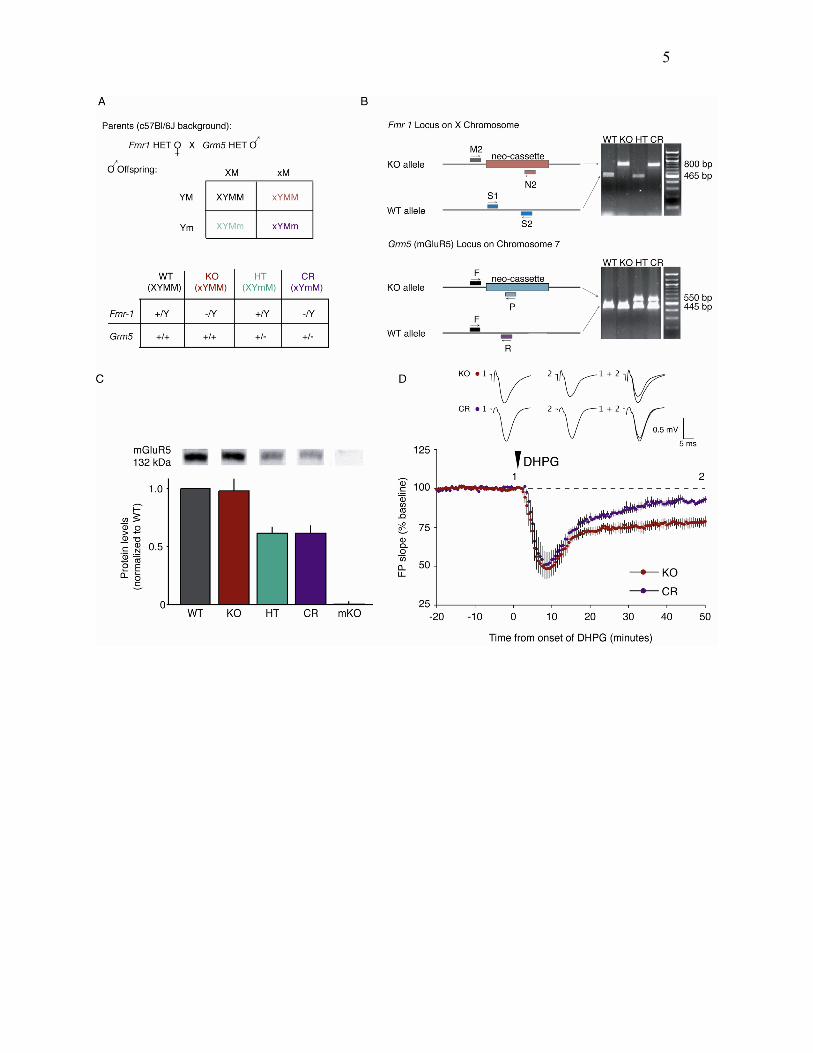
Figure S2. Generation Fmr1 KO mice with a selective reduction of Grm5 gene dosage (A) Fmr1 heterozygous females were crossed with Grm5 heterozygous males to generate littermate male offspring of four genotypes: wild type, WT (black); Fmr1 KO, KO (red); Grm5 HET, HT (green); and Fmr1 KO/Grm5 HET, CR (purple). (B) Genotyping strategy. Screening for the presence or absence of the wild-type allele on the Fmr1 locus on the X chromosome was performed using primer S1 (forward) and S2 (reverse) and the knockout allele using primer M2 (forward) and N2 (reverse). KO and WT PCR reactions were run separately; the reaction products were then combined and electrophoresed on a 1.5% agarose gel [Fmr1 WT: 465 BP (S1/S2); Fmr1 KO: 800 BP (M2/N2)]. Screening for the presence or absence of the knockout allele on the Grm5 locus of chromosome 7 was performed using primer P (reverse) and F (forward) and the wild-type allele using F (forward) and R (reverse). KO and WT PCR reactions were run separately; the reaction products were then combined and electrophoresed on a 1.5% agarose gel [Grm5 WT: 445 BP (F/R); Grm5 KO: 550 BP (F/P)]. Representative gel for mice of four genotypes is shown; WT mice show a single band (465 bp) on the Fmr1 gel and a single band (445 bp) on the Grm5 gel; KO mice show a single band (800 bp) on the Fmr1 gel and a single band (445 bp) on the Grm5 gel; HT mice show a single band (465 bp) on the Fmr1 gel and a double band (445 bp and 550 bp) on the Grm5 gel; CR mice show a single band (800 bp) on the Fmr1 gel and a double band (445 bp and 550 bp) on the Grm5 gel. (C) Western blot analysis of total mGluR5 expression in visual cortical homogenates from P30 normalized to WT (+/+, +/Y) mice, KO (+/+, -/Y), HT (+/-, +/Y), and CR (+/-, -/Y) (n = 11 per genotype, ANOVA P < 0.0001). Grm5 KO (-/-, +/Y) control sample was run 4 times (n = 1 animal) and normalized to age matched (postnatal day 45) WT. Representative blot is shown above. (D) Significant effect of mGluR5 gene dosage on LTD in Fmr1 KO mice. Top: Representative fEPSPs from KO and CR recordings taken at times indicated below. Traces are average of 10 consecutive sweeps. Bottom: Time course of experimenter-blind experiments in which 50 µM (RS)-3,5-dihydroxyphenylglycine (DHPG) was applied for 5 min (arrow) to slices from KO and CR animals (n = 14 and 11 slices from 6 and 5 mice for KO and CR, respectively). Multiple slices per animal were averaged together. Average field EPSP slopes 45-50 min after DHPG were significantly different from 5 min averages immediately prior to DHPG in both genotypes (% baseline: KO = 78.1 ± 3.1 %, P = 0.000005; CR = 91.6 ± 1.4 %, P = 0.00007; paired t-test). However, reduction of Grm5 gene dosage by 50% significantly decreases magnitude of LTD in CR relative to Fmr1 KO mice (n = 5 and 6 animals, respectively, P = 0.0058, unpaired t-test).
5
6
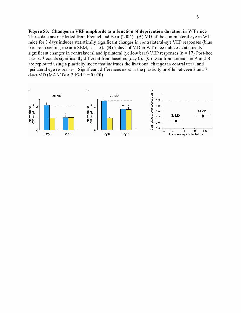
Figure S3. Changes in VEP amplitude as a function of deprivation duration in WT mice These data are re-plotted from Frenkel and Bear (2004). (A) MD of the contralateral eye in WT mice for 3 days induces statistically significant changes in contralateral-eye VEP responses (blue bars representing mean ± SEM, n = 15). (B) 7 days of MD in WT mice induces statistically significant changes in contralateral and ipsilateral (yellow bars) VEP responses (n = 17) Post-hoc t-tests: * equals significantly different from baseline (day 0). (C) Data from animals in A and B are replotted using a plasticity index that indicates the fractional changes in contralateral and ipsilateral eye responses. Significant differences exist in the plasticity profile between 3 and 7 days MD (MANOVA 3d:7d P = 0.020).
7
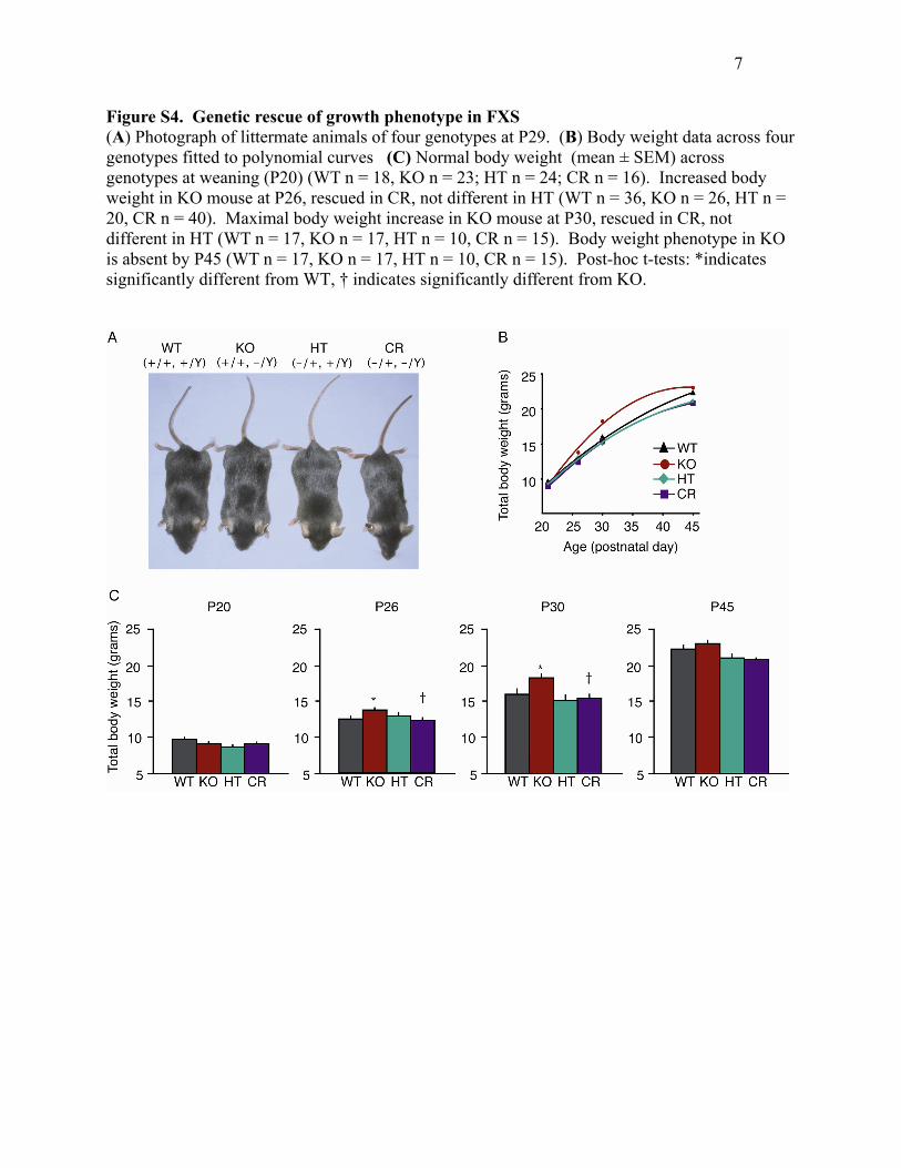
Figure S4. Genetic rescue of growth phenotype in FXS (A) Photograph of littermate animals of four genotypes at P29. (B) Body weight data across four genotypes fitted to polynomial curves (C) Normal body weight (mean ± SEM) across genotypes at weaning (P20) (WT n = 18, KO n = 23; HT n = 24; CR n = 16). Increased body weight in KO mouse at P26, rescued in CR, not different in HT (WT n = 36, KO n = 26, HT n = 20, CR n = 40). Maximal body weight increase in KO mouse at P30, rescued in CR, not different in HT (WT n = 17, KO n = 17, HT n = 10, CR n = 15). Body weight phenotype in KO is absent by P45 (WT n = 17, KO n = 17, HT n = 10, CR n = 15). Post-hoc t-tests: *indicates significantly different from WT, † indicates significantly different from KO.
8
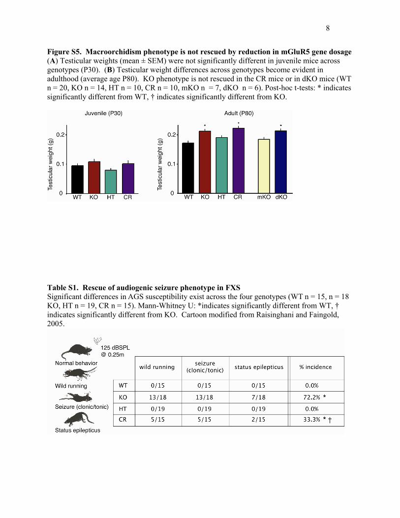
Figure S5. Macroorchidism phenotype is not rescued by reduction in mGluR5 gene dosage (A) Testicular weights (mean ± SEM) were not significantly different in juvenile mice across genotypes (P30). (B) Testicular weight differences across genotypes become evident in adulthood (average age P80). KO phenotype is not rescued in the CR mice or in dKO mice (WT n = 20, KO n = 14, HT n = 10, CR n = 10, mKO n = 7, dKO n = 6). Post-hoc t-tests: * indicates significantly different from WT, † indicates significantly different from KO.
Table S1. Rescue of audiogenic seizure phenotype in FXS Significant differences in AGS susceptibility exist across the four genotypes (WT n = 15, n = 18 KO, HT n = 19, CR n = 15). Mann-Whitney U: *indicates significantly different from WT, † indicates significantly different from KO. Cartoon modified from Raisinghani and Faingold, 2005.