
Neuromuscular Junctions in Health and Disease …€¦ · Neuromuscular Junctions in Health and...
53
Biomedical Sciences (Neuroscience) Honours Elective Neuromuscular Junctions in Health and Disease (NEBM10033) 2013-14 a lifetime, a part; neuro-muscular junctions: mind meeting matter.
-
Upload
nguyenngoc -
Category
Documents
-
view
218 -
download
0
Transcript of Neuromuscular Junctions in Health and Disease …€¦ · Neuromuscular Junctions in Health and...

Biomedical Sciences (Neuroscience) Honours Elective
Neuromuscular Junctions in Health and Disease (NEBM10033)
2013-14
a lifetime, a part; neuro-muscular junctions:
mind meeting matter.
Contents Teaching Staff
3
Course Aims and Objectives Required Background Knowledge
4 5
Learning Outcomes
6
Timetable
7
Recommended Reading
8
Expectations
10
Seminars
10
Late Submission of Assessed Work
12
Common Marking Scheme
13
Special Circumstances APPENDICES
13 14

3
Teaching Staff Course Organiser: Professor Richard R Ribchester Euan MacDonald Centre for MND Research Centre for Integrative Physiology University of Edinburgh Hugh Robson Building George Square
Edinburgh EH8 9XD
Tel: 0131 6503256/3257 email: [email protected] Deputy Organiser: Professor Tom Gillingwater email: [email protected] Course Administrator: Caroline Morris BMTO, Medical School Teviot Place Edinburgh, EH8 9AG [email protected], 0131 651 3255 Please contact Caroline in the first instance if you have any queries.
PLEASE NOTE:
There is a comprehensive website for the NMJiHaD course, containing all the information required to carry out the prescribed work for the course:
http://www.dns.ed.ac.uk/rrrweb/nmjhdhons/NMJhonsIndex.htm

4
Course Aims and Objectives The Aims of the "NMJiHaD" course are to:
• Enhance your knowledge and understanding of the anatomy, physiology and cell biology of neuromuscular junctions; and facilitate your understanding of the importance of synaptic strength and synaptic homeostasis in the healthy nervous/neuromuscular system and after injury or in disease;
• Develop your evidence-based reasoning and critical skills in appraisal and integration of findings reported in original research literature, in the context of knowledge, understanding and research on neuromuscular junctions;
• Provide generic skills training in problem solving, team working, presentation of research material, orally and in writing.
These aims will be met in the following ways:
The course this year will comprise four "mini-symposia", in which we shall explore different aspects of the neuromuscular junction in health, injury and in disease. Seminars, discussions, and student presentations are the main teaching methods. There will also be a workshop on quantal analysis of synaptic transmission; and training and opportunities to practice computer-based measurement and analysis of the structure and function of neuromuscular junctions will be provided. Examination is by written exam paper only, taken at the December diet: there is no in-course assessment. However, we provide feedback by way of "formative assessment", to give you a clear understanding of the assessment criteria that will be applied to your answers on the December exam paper. The mini-symposia will take place in Weeks 2,5,7, and 11 of the course. The sessions in Weeks 1,3,6, and10 will comprise background lectures and group discussions of the Abstracts of key research papers, in preparation for the subsequent mini-symposia. A set of "Big Burning Questions" will be identified by the class at the end of these preparatory discussions. Each subsequent mini-symposium will then comprise student presentations of the papers that have been discussed in the preceding week.
Weeks 4 and 9 are for self-directed learning, when you will be given computer-based exercises in data acquisition and analysis based on data generated either by computer models or from real experiments.
Week 8 will include a discussion of typical exam questions, what examiners expect of the answers, and how they are marked. We will use examples from last year's exam paper. You will then have an opportunity to practice writing an essay with these marking criteria in mind.
Week 12 will comprise a revision session, in which we will also go over quantal analysis problems. There will also be an optional demonstration of neuromuscular junction physiology and morphology in Week 12.

5
Required Background Knowledge
The following list constitutes an overview of what you should have familiarity with as background to this course. Some, but not all, of these points will be revised, consolidated and extended during the course:
• Basic structure and organization of mammalian spinal cord circuitry including monosynaptic and polysynaptic peripheral reflexes; motor unit organization in the ventral horn and lateral motor columns; pyramidal and extra-pyramidal descending pathways from the brain. Basic principles of motor control including initiation, execution and feedback. Principle brain areas involved in the control of movement, including motor cortex, basal ganglia and cerebellum. Size principle of motor unit activation. Regulation of muscle force by recruitment of motor units, motor unit size, and firing frequency.
• Techniques for studying NMJ: isometric tension, intracellular recording, voltage clamp, Ca-imaging, transmitted light microscopy (including phase and interference contrast), confocal fluorescence microscopy, and, immunocytochemistry, transgenic expression of marker genes (eg GFP variants); scanning and transmission electron microscopy.
• Motor unit structure in adult muscle: motor neurone cell body and dendrites in spinal cord; myelinated axon entering peripheral nerve via ventral roots; intramuscular nerve branching and mononeuronal innervation of muscle fibres at motor endplates by motor nerve terminals. Basic structure of muscle spindles and their sensory and motor innervation, distinct from innervation of extrafusal muscle fibres.
• Cellular composition of the vertebrate/mammalian NMJ: presynaptic axon terminal, terminal Schwann cell, kranocyte, motor endplate.
• Cytoarchitecture of NMJ: synaptic vesicles, mitochondria, active zones, extracellular matrix, postsynaptic densities of ACh receptors on junctional folds
• Proteins located at active zones (including SNARE proteins); postsynaptic densities (including ACh receptors and associated cytoskeletal proteins; and synaptic basal lamina (including acetylcholinesterase).
• Basic physiology of neuromuscular transmission: resting potential, action potential, membrane resistance, membrane capacitance, membrane time constant, length constant, ‘input resistance’; EPP’s/EPC’s; mEPP’s/mEPC’s; quantal content, quantal size, safety factor for neuromuscular transmission; facilitation, depression; muscle twitch, muscle tetanus.
• Effects of specific toxins, drugs and ions (especially Ca2+ and Mg2+) on synthesis, storage, release, action and inactivation of acetylcholine at NMJ

6
• Development of NMJ: prenatal formation of motor neurons and naturally cell death by apoptosis; specificity of motor innervation; postnatal synapse elimination; transition from gamma-subunit to epsilon-subunit forms of the ACh Receptor.
• Diseases and pathology of the NMJ, causes and characteristics; specifically: Myasthenia gravis, Lambert-Eaton Myasthenic Syndrome, Congential Myasthenic Syndrome, Botulism, Nerve agent poisoning, Amyotrophic Lateral Sclerosis, Spinal Muscular Atrophy.
• Responses of NMJ to injury: axotomy-induced degeneration; repair by collateral nerve sprouting and axon regeneration
• Effects of denervation or paralysis on properties of muscle relevant to synaptic transmission, including changes in resting membrane potential, excitability, ACh supersensitivity, resistance of action potentials to TTX
Learning Outcomes
In addition to familiarity with the required background knowledge, by the end of the course you should be able to provide informed, evidence-based answers to the following questions, which therefore form the basis for the Learning Objectives/Outcomes of the course. Your understanding of these learning outcomes will be tested in the Course Examination, whose questions will be based on the topics covered in the mini-symposia and the papers presented in them. You will be expected to support answers with experimental or clinical evidence derived from the topics you have researched and discussed in classes, the assignments and your other reading.
• Anatomy and Physiology What cell types are present at neuromuscular junctions and what are their functions? What molecules are located at neuromuscular synapses? What techniques enable the structure of neuromuscular junctions to be visualised? How is the physiological function of the neuromuscular junction analysed? What is the 'quantal content' of an end-plate potential (or current) and how is it measured? What determines the amplitude and frequency of MEPPs? What determines the amplitude and quantal content of the EPP? Why do end-plate potentials sum non-linearly but end-plate currents sum linearly? What is the role of Ca2+ ions at the neuromuscular junction and how may the effects of Ca2+ concentration be measured? What is the 'safety factor' for neuromuscular transmission and how has it been measured? What is the function of the junctional folds? How are synaptic vesicles and neurotransmitter recycled in motor nerve terminals? What is the relationship between the form (morphology) and function (physiology) of the neuromuscular junction in different species?

7
Contd..
• Development, maintenance and plasticity How and when are neuromuscular junctions formed in early development? How are neuromuscular connections remodelled postnatally? How do junctional acetylcholine receptor sub-types change in expression and distribution during development or after muscle denervation? What properties of the NMJ appear to be homeostatically regulated? How is the size of a neuromuscular junction related to its function? What causes neuromuscular synapses to degenerate after nerve injury? What are the causes and consequences of motor axons sprouting? How are neuromuscular connections remodelled during/after nerve injury and repair?
• Disease How has fundamental biology of the neuromuscular junction informed our understanding of neuromuscular disease? How have animal models been used to study the involvement of NMJ's in neuromuscular disease? What causes Myasthenia Gravis and how is it currently treated? What causes Lambert-Eaton Myasthenic Syndrome and how is it currently treated? What causes Congenital Myasthenic Syndrome and how is it currently treated? What causes the symptoms and signs of Botulism and how is it treated? What are the symptoms and signs of nerve agent (anticholinesterase) toxicity and how are they treated? How are NMJ's affected in Motor Neurone Disease (ALS) and Spinal Muscular Atrophy (SMA)? What are the future prospects for treatment of ALS and/or SMA?
Timetable
The course takes place in Semester 1, Mondays, 9:30-11:30 am
Locations:
v Greenfield Suite Small Lab, George Square (Weeks 1,3,9) v Biomedical Seminar Room 3, Anatomy Building, Teviot Place.
Note: The location of the first session on Monday September 16 will be the Greenfield Suite Small Lab
See the course website for a detailed breakdown of topics: http://www.dns.ed.ac.uk/rrrweb/NMJHDhons/NMJhonsIndex.htm

8
Reading
Science students: Review lecture notes from Neuroscience with Pharmacology 2; Brain & Behaviour 3 and/or Mechanisms of Brain Development 3
Intercalated Medical students: Review ‘Molecules to Society’ courses ‘ia’ (Modules 1A,1B), 2a (Neuroscience, Statistics), 2b (Clinical Genetics)
Intercalated Veterinary students: Review neurological components of Y3 course in Veterinary Pathology and relevant cell/neurophysiology components of Y1/2 courses on The Animal Body 1 & 3
Textbooks:
Byrne, JH & Roberts, JL (2009) From Molecules to Networks 2nd edn. Sinauer. Chapters 2, 5, 8,11,13,16, 20
And/or
Nicholls, JG et al (2012) From Neuron to Brain. 5th edn. Sinauer. Chapters 11,13,15, 24, 25, 27
Reviews:
Schwarz,T. (2005) Transmitter Release at the Neuromuscular Junction. Int Rev Neurobiol 75,105-144. (See website for PDF) Slater, CR Reliability of neuromuscular transmission and how it is maintained.Handbook of Neurology. 2008: 91,27-101. (See website for PDF) Sanes JR, Lichtman JW. Development of the vertebrate neuromuscular junction. Annu Rev Neurosci. 1999;22:389-442. PMID: 10202544 Katz B. Neural transmitter release: from quantal secretion to exocytosis and beyond. J Neurocytol. 2003 Jun-Sep;32(5-8):437-46. PMID: 15034246 Hughes BW, Kusner LL, Kaminski HJ. Molecular architecture of the neuromuscular junction. Muscle Nerve. 2006 Apr;33(4):445-61. PMID: 16228970 Hirsch NP. Neuromuscular junction in health and disease. Br J Anaesth. 2007 Jul;99(1):132-8. PMID: 17573397 Davis GW. Homeostatic control of neural activity: from phenomenology to molecular design. Annu Rev Neurosci. 2006;29:307-23. PMID: 16776588 Ribchester RR. Mammalian neuromuscular junctions: modern tools to monitor synaptic form and function. Curr Opin Pharmacol. 2009 Jun;9(3):297-305. PMID: 19394273

9
Original papers:
RRR’s Top Ten 1: Fatt P, Katz B. Spontaneous subthreshold activity at motor nerve endings. J Physiol. 1952 May;117(1):109-28.PMID: 14946732 2: Boyd IA, Martin AR. The end-plate potential in mammalian muscle. J Physiol. 1956 Apr 27;132(1):74-91. PMID: 13320373 3: Dodge FA Jr, Rahamimoff R.Co-operative action a calcium ions in transmitter release at the neuromuscular junction. J Physiol. 1967 Nov;193(2):419-32. PMID: 6065887 4: Brown MC, Jansen JK, Van Essen D. Polyneuronal innervation of skeletal muscle in new-born rats and its elimination during maturation. J Physiol. 1976 Oct;261(2):387-422. PMID: 978579 5: McLachlan EM, Martin AR. Non-linear summation of end-plate potentials in the frog and mouse. J Physiol. 1981 Feb;311:307-24. PMID: 6267255 6: Mishina M, Takai T, Imoto K, Noda M, Takahashi T, Numa S, Methfessel C, Sakmann B. Molecular distinction between fetal and adult forms of muscle acetylcholine receptor. Nature. 1986 May 22-28;321(6068):406-11. PMID: 2423878 7: Betz WJ, Bewick GS. Optical analysis of synaptic vesicle recycling at the frog neuromuscular junction. Science. 1992 Jan 10;255(5041):200-3. PMID: 1553547. 8: Wood SJ, Slater CR. The contribution of postsynaptic folds to the safety factor for neuromuscular transmission in rat fast- and slow-twitch muscles. J Physiol. 1997 Apr 1;500 ( Pt 1):165-76. PMID: 9097941 9: Harlow ML, Ress D, Stoschek A, Marshall RM, McMahan UJ. The architecture of active zone material at the frog's neuromuscular junction. Nature. 2001 Jan 25;409(6819):479-84. PMID: 11206537 10: Walsh MK, Lichtman JW. In vivo time-lapse imaging of synaptic takeover associated with naturally occurring synapse elimination. Neuron. 2003 Jan 9;37(1):67-73. PMID: 12526773
Additional reading is indicated in the APPENDIX and on the course website:
http://www.dns.ed.ac.uk/rrrweb/nmjhdhons/NMJreading.htm

10
Expectations The course represents a co-operative learning, mutual education forum in which all students play a part and therefore depend upon and benefit greatly from one another’s presence and participation. You are therefore required and expected to attend all timetabled classes and to participate actively in small group and class discussions, for the benefit of yourselves and your classmates. You will work in small groups for discussion and presentation of research papers in “mini-symposia”. You will each be required to give an oral presentation of one of the papers prescribed for the course. You are expected to prepare your presentation carefully and thoughtfully, and in a structured, logical way that your classmates can follow and understand. If you are unavoidably absent from one of the classes, you should inform the course organiser by email, giving reasons, ideally in advance with several days notice. Absence from three or more classes will be noted to your Personal Tutor.
Seminars Details of relevant research seminars will be posted on the course website or sent via email. The Edinburgh Neuroscience website (http://www.edinburghneuroscience.ed.ac.uk/) lists seminars of potential relevance and interest.
Assessment The course will be assessed by a two-hour written examination in December. Students will be expected to answer two questions from a choice of five or six questions, one of which may be a quantitative data analysis problem. Marking will be in accordance with the University common marking scheme. Feedback, justifying the marks, will be made available early in Semester 2, after the exam board has met to consider the marks. Questions will be based on the topics covered in the mini-symposia and the papers presented in them. You will be expected to support answers with experimental or clinical evidence derived from the topics you have researched and the papers discussed in classes, the assignments given and other background reading, including from sources indicated on the course website. Each answer will be anonymously marked and moderated, respectively, by two members of academic staff. Examiners of essays for this course will be looking for evidence that you have assimilated and understood the main findings of the core papers we have discussed in each minisymposium, supplemented by the background reading and other learning resources indicated for each of these. They will also take account of the Learning Objectives/Outcomes for the course. Extra credit will be given for evidence of additional reading around the subject, provided it shows knowledge and understanding relevant to the question. (ALWAYS ANSWER THE QUESTION POSED, not the question you would like to have been posed!) Formative assessment will be provided during the course, on practice essays, problem solving, and oral presentations. You will be provided with detailed feedback. Past exam papers are available through the library website and through the course website.

11
Formative Assessment
In the NMJiHaD course we provide opportunities for formative assessment and feedback in four ways: first, an optional essay to help develop your scientific writing skills; second, a compulsory exam essay based on an initial appraisal and consideration of the marking criteria used by assessors on an answer to a question from last year's exam paper, followed by an opportunity for you to write an essay that fits the criteria. This will help you to understand what is required in the summative assessment for the course, which is the exam you will take in December. Third, we give you practice in solving quantal analysis problems, using questions from past exam papers. Finally, we provide advice and appraisal of your oral presentations given during the course. This will help you to develop your scientific communication skills, important for presentation of your Dissertations at the end of the year.
What is expected of a good answer to a question on the December exam paper? The examiners' marking criteria are indicated on a marksheet, that is also designed to provide you with feedback on the best aspects of your answer and the ways it could have been improved. The examiners look for an answer to the question that is put (and not the question that you have preferred it to be!). We expect the answer to be well structured, written in good English, and to apply evidence-based knowledge and understanding with reference to - and providing good coverage of - background reading, lectures and research papers you have studied on the course. Your essays should be critical but balanced. Evidence of additional relevant reading may be given extra credit and attract a higher mark, as will insightful summary diagrams and/or creative suggestions/hypotheses for further research. As an exercise, we will review the text of two answers given to a question on last year's exam paper. We will see how the mark for this answer was arrived at. We will then examine the text of a different answer to the same question: only this time, you - the students - will 'mark' the answer, applying the examiners' criteria. We will then compare the consensus mark with the one actually awarded by the examiners. Finally, you may attempt the same question (or a different one from last year’s paper), under self-imposed exam conditions if you wish. Submit your answer for marking. You will receive feedback using the same criteria and categories indicated on the marksheet.
Optional Assignments with Formative Assessment and Feedback
Note: Hand in your practice essay(s) and/or quantal-analysis problems any time after Week 5 and before the end of Week 8 if you wish to receive feedback before the examination at the end of the semester. The work may not be marked and feedback cannot be guaranteed if you submit your assignments after that date.
1. Write an essay (<2500 words) on either: "The molecular physiology of synaptic transmission at the neuromuscular junction". This essay could include - for example- a review of the ultrastructure of the active zone, the molecular composition of synaptic vesicles, and the mechanisms of synaptic vesicle recycling; or alternatively, focus on the organization and function of acetylcholine receptors, sodium ion channels and cytoskeletal proteins; or the functions of molecules in the basal lamina, including acetylcholinesterase.
12
Or:
"Molecular dissection of neuromuscular synaptic development, structure and function in Drosophila". This essay should critically evaluate the application of molecular genetic techniques to determine how the size and strength of synapses is regulated during development.
In either case, discuss the relevance of the findings to the understanding of neuromuscular disease. If you wish to write both essays, both will be marked and feedback provided.
2. Choose a question from last year's Exam Paper and write an essay on that. To help you prepare for this, in Week 8 we will discuss examiners guidance notes, marking criteria and a couple of examples of answers that were given to one of the questions on last year's exam paper (see above).
3. Measure the amplitudes of the EPPs/MEPP's and calculate the mean quantal content of the EPPs using the Direct Method, the Variance Method, and the Method of Failures in the downloadable recordings you will find on the Course website
Late Submission of Assessed Work There is no In-Course Assessment for this course. However, all work for formative assessment must be submitted between Week 5 and Week 8, otherwise the work may not be marked.
13
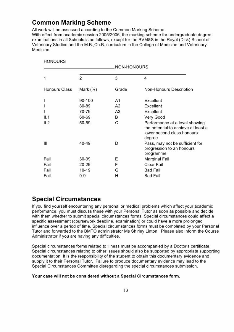
Common Marking Scheme All work will be assessed according to the Common Marking Scheme With effect from academic session 2005/2006, the marking scheme for undergraduate degree examinations in all Schools is as follows, except for the BVM&S in the Royal (Dick) School of Veterinary Studies and the M.B.,Ch.B. curriculum in the College of Medicine and Veterinary Medicine.
HONOURS NON-HONOURS
1 2 3 4 Honours Class Mark (%) Grade Non-Honours Description I 90-100 A1 Excellent I 80-89 A2 Excellent I 70-79 A3 Excellent II.1 60-69 B Very Good II.2 50-59 C Performance at a level showing
the potential to achieve at least a lower second class honours degree
III 40-49 D Pass, may not be sufficient for progression to an honours programme
Fail 30-39 E Marginal Fail Fail 20-29 F Clear Fail Fail 10-19 G Bad Fail Fail 0-9 H Bad Fail
Special Circumstances If you find yourself encountering any personal or medical problems which affect your academic performance, you must discuss these with your Personal Tutor as soon as possible and decide with them whether to submit special circumstances forms. Special circumstances could affect a specific assessment (coursework deadline, examination) or could have a more prolonged influence over a period of time. Special circumstances forms must be completed by your Personal Tutor and forwarded to the BMTO administrator Ms Shirley Linton. Please also inform the Course Administrator if you are having any difficulties. Special circumstances forms related to illness must be accompanied by a Doctor’s certificate. Special circumstances relating to other issues should also be supported by appropriate supporting documentation. It is the responsibility of the student to obtain this documentary evidence and supply it to their Personal Tutor. Failure to produce documentary evidence may lead to the Special Circumstances Committee disregarding the special circumstances submission. Your case will not be considered without a Special Circumstances form.

14
APPENDIX I : About the course/ Course structure We can’t live without our neuromuscular junctions (NMJ’s). Every breath we take depends on them and cessation or failure of neuromuscular transmission in our respiratory muscles is, arguably, the ultimate cause of death for all of us. So it is important to understand how neuromuscular junctions work: and in order to do so we must understand their structure, molecular composition, physiology, development, and plasticity; how they respond to injury; and their involvement in disease. Historically, studies of neuromuscular junctions provided deep insights into fundamental mechanisms of synaptic transmission. In the words of the late doyen of NMJ biology, Nobel Laureate Professor Sir Bernard Katz: “the neuromuscular junction is an experimentally favourable object whose study could throw considerable light on synaptic mechanisms elsewhere.” Thus, our understanding of all the principles of chemical synaptic transmission in the brain is based on analysis of synaptic structure and function that was initially carried out by Katz and his colleagues, in their pioneering studies of vertebrate neuromuscular junctions. In a nutshell, thousands of motor neurones, organized into motor units, each supply an axon branch that divides intramuscularly into hundreds of collaterals, each ending in a motor nerve terminal on the motor endplate on a single skeletal muscle fibre. Activation of motor units is combined in a myriad of ways, enabling an extraordinary range of delicate-to-intense voluntary movements, thereby linking cognition and intention to behaviour. Nerve terminals are capped by terminal Schwann cells and fibroblast-like cells we call kranocytes. Each motor terminal arbor forms synapses on the surface of a single muscle fibre at a single patch, about 400 µm2 in area; and each of these motor endplates is endowed with tens of millions (at about 105/µm2) of ligand-gated acetylcholine receptors (AChR) and voltage-gated sodium ion channels, organized around post-synaptic densities in junctional folds. High-fidelity synaptic transmission - that is, a high quantal content of synaptic transmission, produces large endplate currents (EPC), leading to depolarising endplate potentials (EPP) in muscle fibres, evoked by nerve excitation. This response is mediated by calcium-dependent exocytosis of synaptic vesicles from motor nerve terminals. Spontaneous exocytosis of these 30nm spheres, each containing about 5000 molecules of the neurotransmitter acetylcholine (ACh) also occurs at low frequency, about 1/s, producing miniature endplate currents and potentials (MEPCs/MEPPs), with a quantal size of about 1 mV. Thus, small electrical signals in axons are amplified at NMJ’s and translated, ultimately, into powerful muscle contractions. Homeostatic synaptic strength-regulation empowers NMJ’s, enabling them to activate skeletal muscles with an extraordinary degree of reliability. This regulation maintains the safety-factor for neuromuscular transmission at about three: that is, about three times as much neurotransmitter is released over the amount required to trigger a muscle action potential and a twitch. The action of ACh is terminated by the enzyme acetylcholinesterase (AChE), located in the synaptic basal lamina between the presynaptic terminal and the postsynaptic muscle fibre. Choline is taken up into nerve terminals and utilized by the cytoplasmic enzyme cholineacetyltransferase (ChAT) in the resynthesis of ACh. Synaptic vesicles are also recycled, following their clathrin-dynamin mediated endocytosis. Mature patterns of motor unit organization and muscle fibre innervation come about during development and following refinement of an initial pattern that is highly distributed and overlapping, producing polyneuronal innervation of muscle fibres. The adult pattern emerges during a late developmental process called synapse elimination, which reduces the number of muscle fibres contacted by each motor neurone until each muscle fibre is contacted by one and only one motor nerve terminal. Synapse elimination is still poorly understood but evidently involves an exquisite interplay of different molecular and cellular components of neuromuscular synapses. The overall process seems somewhat akin to the co-operativity of an intimate love-affair versus the competitive struggle of all-out war! The form and function of mature neuromuscular junctions are normally maintained and preserved throughout adult life and only

15
start to break down during senescence, partly in response to wear-and-tear and wasting or degeneration of muscle fibres. When such highly-tuned synaptic connections fail - as they may do following injury, poisoning or disease – affected individuals suffer symptoms and show signs of severe motor disturbances, ranging from painful seizures or cramps to weakness or complete paralysis. In fact, respiratory paralysis - due to failure of neuromuscular transmission - is a critical feature in infections or illnesses, such as botulism or myasthenia; and degeneration of NMJ’s in respiratory muscle is a harbinger of death in incurable motor neurone diseases such as spinal muscular atrophy (SMA) or amyotrophic lateral sclerosis (ALS). Injuries to peripheral nerves can also be highly debilitating, triggering “Wallerian” degeneration of axons and motor nerve terminals disconnected from their cell bodies, which leads to partial or complete denervation and paralysis of muscle fibres. Fortunately, injured peripheral nerve axons and motor nerve terminals show activity-dependent synaptic plasticity: motor axons and terminals react to paralysis and some forms of injury by compensatory sprouting and regeneration, unlike most axons in the Central Nervous System (CNS). Some of these processes are strongly influenced by activity, implying the expression of ‘Hebbian’ strengthening/weakening mechanisms (‘cells that fire together, wire together’; ‘use-it-or-lose-it’). The differences in repair and regeneration in the PNS and CNS add further to the value of studying mechanisms of peripheral nerve repair, since a deeper understanding of regenerative mechanisms could have an impact on the vital quest to find ways of more effectively repairing damaged neurones, axons and synapses in the brain and spinal cord.

16
APPENDIX II : Organisation and Structure of the NMJiHaD course This course is divided into “Mini-symposia”, prepared and delivered by student members of the class. Each symposium focuses on a different aspect of the structure, development, function, homeostatic maintenance, or plasticity of neuromuscular junctions. Consideration is given in each mini-symposium to the translational research relevance of these properties, for understanding and potential treatment of disease or injury affecting motor neurones and/or their neuromuscular connections. However, the course is not comprehensive and several areas of research importance are not covered, or are touched upon in only a limited way. For example, we do not consider much of the biochemistry or pharmacology of neuromuscular junctions. However, the topics we do cover will include discussion of cutting-edge research. The class will be divided into groups (“Motor Units”), each with four or five members. Each group takes responsibility for delivering one of the mini-symposia. For groups with five members, one of the group (the “Axon”) will Chair the mini-symposium and manage the discussion. The other four members (“Motor Nerve Terminals”) will deliver 15-20 minute presentations of the research papers that illustrate the topic (one paper per student). As well as steering questions from the audience, the Chair should also prompt each speaker with either spontaneous or pre-prepared questions. This is a valuable skill for anyone chairing a meeting: it is quite often necessary for the chair to get the ball rolling, or to maintain the momentum of discussion when audience members or other attendees appear reticent. The Chair shall also be a rapporteur, responsible for summarizing the mini-symposium and writing a brief (roughly 2-page) overview of all the papers presented, for circulation to the class and posting on the course website. The mini-symposia will normally be held in alternate weeks. In the interleaving weeks, the sessions will usually begin with an introductory lecture/seminar on the topic of the next mini-symposium, given by the course organiser (RRR: the “Soma”), followed by a discussion of the Abstracts of the papers to be presented by one of the groups the following week. The format of these group discussions will be structured as follows:
1. Each abstract is read aloud by a member of the group
2. The Group identifies, defines and clarifies any difficult terms or terminology
3. The Group freely discusses the issues raised by each paper
4. Each group decides on up to four “burning questions” (BQ’s) from the issues discussed (one for each paper)
5. In plenary discussion, the class narrows down the number of BQ’s to four “Big Burning
Questions” (BBQ’s) that the presenting group should endeavour to address in the following week’s mini-symposium.

17
Mini-symposium topics :
I. Structure and function of motor units and neuromuscular junctions II. Neuromuscular transmission III. Development, degeneration and regeneration of neuromuscular synapses IV. Neuromuscular synapses in larval Drosophila: synaptic
homeostasis/plasticity Introductory talks (RRR unless otherwise indicated)
1. Overview of course structure; revision of NMJ; review of anatomy and physiology of the NMJ; practical exercise: EMG recording
2. Quantal analysis and ‘safety-factor’ for neuromuscular transmission
3. Neuromuscular synapse formation, remodeling and regeneration
4. Structure, function and homeostasis of NMJ in Drosophila

18
Week 2 MINI-SYMPOSIUM I Structure and Function of Motor Units and Neuromuscular Junctions The first session will be held in the Greenfield Suite Small Computing Lab. We will begin with an exercise then discussion of EMG recordings you may make from yourselves using Backyard Brains Spiker Boxes. Download the instruction sheet from the course website. We will then adjourn to Biomedical Sciences Room 3 for an introductory background lecture and discussion of the Abstracts for the first mini-symposium. Background reading: Nicholls, JG et al (2012) From Neuron to Brain. 5th edn. Sinauer. Chapters 11,13,15, 24, 25, 27 Byrne, JH & Roberts, JL (2009) From Molecules to Networks 2nd edn. Sinauer. Chapters 2, 5, 8,11,13,16, 20 Desaki J, Uehara Y. The overall morphology of neuromuscular junctions as revealed by scanning electron microscopy. J Neurocytol. 1981 Feb;10(1):101-10. PMID: 6118394 Fischer LR, Culver DG, Tennant P, Davis AA, Wang M, Castellano-Sanchez A, Khan J, Polak MA, Glass JD. Amyotrophic lateral sclerosis is a distal axonopathy: evidence in mice and man. Exp Neurol. 2004 Feb;185(2):232-40. PMID: 14736504 Massoulié J, Millard CB. Cholinesterases and the basal lamina at vertebrate neuromuscular junctions. Curr Opin Pharmacol. 2009 Jun;9(2):316-25. PMID: 19423392. For Presentation: 1. Lu J, Tapia JC, White OL, Lichtman JW. The interscutularis muscle connectome. PLoS Biol. 2009 Feb 10;7(2):e32. PMID: 19209956 2. Thomson SR, Nahon JE, Mutsaers CA, Thomson D, Hamilton G, Parson SH, Gillingwater TH. Morphological characteristics of motor neurons do not determine their relative susceptibility to degeneration in a mouse model of severe spinal muscular atrophy. PLoS One. 2012;7(12):e52605. doi: 10.1371/journal.pone.0052605. PMID: 23285108 3. Court FA, Gillingwater TH, Melrose S, Sherman DL, Greenshields KN, Morton AJ, Harris JB, Willison HJ, Ribchester RR. Identity, developmental restriction and reactivity of extralaminar cells capping mammalian neuromuscular junctions. J Cell Sci. 2008 Dec 1;121(Pt 23):3901-11 PMID: 19001504 4. David G, Nguyen K, Barrett EF. Early vulnerability to ischemia/reperfusion injury in motor terminals innervating fast muscles of SOD1-G93A mice. Exp Neurol. 2007 Mar;204(1):411-20. Epub 2007 Jan 4. PMID: 17292357.

19
Week 5 MINI-SYMPOSIUM II Neuromuscular Transmission This block will comprise, in the first week, an introductory lecture and discussion of the Abstracts. The second week will be a workshop on neuromuscular synaptic physiology and pharmacology, including practical, computer-based analysis of data from computer models. This will be held in the Greenfield Suite Small Lab. The mini-symposium will take place in the third week. Background Reading: Schwarz,T. (2005) Transmitter Release at the Neuromuscular Junction. Int Rev Neurobiol 75,105-144. Slater, CR Reliability of neuromuscular transmission and how it is maintained.Handbook of Neurology. 2008: 91,27-101. Hirsch NP. Neuromuscular junction in health and disease. Br J Anaesth. 2007 Jul;99(1):132-8. PMID: 17573397 Spillane J, Beeson DJ, Kullmann DM. Myasthenia and related disorders of the neuromuscular junction. J Neurol Neurosurg Psychiatry. 2010 Aug;81(8):850-7. PMID: 20547629 Harlow ML, Ress D, Stoschek A, Marshall RM, McMahan UJ.The architecture of active zone material at the frog's neuromuscular junction.Nature. 2001 Jan 25;409(6819):479-84. PMID 11206537 Ribchester, R.R. (2009) Mammalian neuromuscular junctions: modern tools to monitor synaptic form and function. Curr Opin Pharmacol. 9,297-305.PMID: 19394273 Gaffield MA, Tabares L, Betz WJ. Preferred sites of exocytosis and endocytosis colocalize during high- but not lower-frequency stimulation in mouse motor nerve terminals. J Neurosci. 2009 Dec 2;29(48):15308-16. PMID: 19955383 Tsujimoto T, Umemiya M, Kuno M. Terminal sprouting is not responsible for enhanced transmitter release at disused neuromuscular junctions of the rat. J Neurosci. 1990 Jul;10(7):2059-65. PMID: 1973945 McLachlan EM, Martin AR. Non-linear summation of end-plate potentials in the frog and mouse. J Physiol. 1981 Feb;311:307-24. PMID: 6267255 For Presentation: 1. Nagwaney S, Harlow ML, Jung JH, Szule JA, Ress D, Xu J, Marshall RM, McMahan UJ. Macromolecular connections of active zone material to docked synaptic vesicles and presynaptic membrane at neuromuscular junctions of mouse. J Comp Neurol. 2009 Apr 10;513(5):457-68. PMID: 19226520 2. Ruiz R, Cano R, Casañas JJ, Gaffield MA, Betz WJ, Tabares L. Active zones and the readily releasable pool of synaptic vesicles at the neuromuscular junction of the mouse. J Neurosci. 2011 Feb 9;31(6):2000-8. PMID: 21307238. 3. Wood SJ, Slater CR.The contribution of postsynaptic folds to the safety factor for neuromuscular transmission in rat fast- and slow-twitch muscles. J Physiol. 1997 Apr 1;500 ( Pt 1):165-76. PMID: 9097941 4. Slater CR, Fawcett PR, Walls TJ, Lyons PR, Bailey SJ, Beeson D, Young C, Gardner-Medwin D. Pre- and post-synaptic abnormalities associated with impaired neuromuscular transmission in a group of patients with 'limb-girdle myasthenia'. Brain. 2006 Aug;129(Pt 8):2061-76. PMID: 16870884

20
Week 7 MINI-SYMPOSIUM III Development, degeneration and regeneration of neuromuscular synapses
Background Reading: Gillingwater TH, Ribchester RR. The relationship of neuromuscular synapse elimination to synaptic degeneration and pathology: insights from WldS and other mutant mice. J Neurocytol. 2003 Jun-Sep;32(5-8):863-81. PMID: 15034273. Brown MC, Jansen JK, Van Essen D. Polyneuronal innervation of skeletal muscle in new-born rats and its elimination during maturation.J Physiol. 1976 Oct;261(2):387-422. PMID: 978579 Coleman MP, Freeman MR. Wallerian degeneration,Wld(s), and Nmnat. Annu Rev Neurosci. 2010;33:245-67. PMID: 20345246. Pun S, Sigrist M, Santos AF, Ruegg MA, Sanes JR, Jessell TM, Arber S, Caroni P.An intrinsic distinction in neuromuscular junction assembly and maintenance in different skeletal muscles. Neuron. 2002 Apr 25;34(3):357-70. PMID: 11988168 Son YJ, Trachtenberg JT, Thompson WJ. Schwann cells induce and guide sprouting and reinnervation of neuromuscular junctions. Trends Neurosci. 1996 Jul;19(7):280-5 PMID: 879997 Walsh MK, Lichtman JW. In vivo time-lapse imaging of synaptic takeover associated with naturally occurring synapse elimination. Neuron. 2003 Jan 9;37(1):67-73.PMID: 12526773 Barry JA, Ribchester RR. Persistent polyneuronal innervation in partially denervated rat muscle after reinnervation and recovery from prolonged nerve conduction block. J Neurosci. 1995 Oct;15(10):6327-39 PMID: 7472398 Costanzo EM, Barry JA, Ribchester RR. Competition at silent synapses in reinnervated skeletal muscle. Nat Neurosci. 2000 Jul;3(7):694-700. PMID: 10862702 Wong F, Fan L, Wells S, Hartley R, Mackenzie FE, Oyebode O, Brown R, Thomson D, Coleman MP, Blanco G, Ribchester RR. Axonal and neuromuscular synaptic phenotypes in Wld(S), SOD1(G93A) and ostes mutant mice identified by fiber-optic confocal microendoscopy. Mol Cell Neurosci. 2009 Dec;42(4):296-307. PMID: 19683573 Brill MS, Lichtman JW, Thompson W, Zuo Y, Misgeld T. Spatial constraints dictate glial territories at murine neuromuscular junctions. J Cell Biol. 2011 Oct 17;195(2):293-305. PMID: 22006952.
For Presentation: 1.Turney SG, Lichtman JW. Reversing the outcome of synapse elimination at developing neuromuscular junctions in vivo: evidence for synaptic competition and its mechanism. PLoS Biol. 2012 Jun;10(6):e1001352. Epub 2012 Jun 26. PMID: 22745601.
2. Favero M, Busetto G, Cangiano A. Spike timing plays a key role in synapse elimination at the neuromuscular junction. Proc Natl Acad Sci U S A. 2012 Jun 19;109(25):E1667-1675. PMID: 22619332
3. Gillingwater TH, Thomson D, Mack TG, Soffin EM, Mattison RJ, Coleman MP, Ribchester RR. Age-dependent synapse withdrawal at axotomised neuromuscular junctions in Wld(s) mutant and Ube4b/Nmnat transgenic mice. J Physiol. 2002 Sep 15;543(Pt 3):739-55. PMID: 12231635
4. Schaefer AM, Sanes JR, Lichtman JW. A compensatory subpopulation of motor neurons in a mouse model of amyotrophic lateral sclerosis. J Comp Neurol. 2005 Sep 26;490(3):209-19. PMID: 16082680

21
Week 11 MINI-SYMPOSIUM IV Neuromuscular synapses in larval Drosophila : a model of synaptic homeostasis and disease
Background Reading: Schwarz,T. (2005) Transmitter Release at the Neuromuscular Junction. Int Rev Neurobiol 75,105-144. Collins CA, DiAntiono A. 2007. Synaptic development; insights from Drosophila. Current Opinion in Neurobiology, 17: 35-42 PMID:17229568 Costanzo EM, Barry JA, Ribchester RR. Co-regulation of synaptic efficacy at stable polyneuronally innervated neuromuscular junctions in reinnervated rat muscle. J Physiol. 1999 Dec 1;521 Pt 2:365-74. PMID: 10581308 Davis GW. Homeostatic control of neural activity: from phenomenology to molecular design. Annu Rev Neurosci. 2006;29:307-23. PMID: 16776588 Harris JB, Ribchester RR. The relationship between end-plate size and transmitter release in normal and dystrophic muscles of the mouse. J Physiol. 1979 Nov;296:245-65. PMID: 231101 Hoang B, Chiba A. Single-cell analysis of Drosophila larval neuromuscular synapses. Dev Biol. 2001 Jan 1;229(1):55-70. PMID: 11133154. Veldink JH, Bär PR, Joosten EA, Otten M, Wokke JH, van den Berg LH. Sexual differences in onset of disease and response to exercise in a transgenic model of ALS. Neuromuscul Disord. 2003 Nov;13(9):737-43. PMID: 14561497. Frank CA. Homeostatic plasticity at the Drosophila neuromuscular junction. Neuropharmacology. 2013 Jun 24. doi:pii: S0028-3908(13)00283-9. 10.1016/j.neuropharm.2013.06.015. PMID: 23806804 Frank CA, Kennedy MJ, Goold CP, Marek KW, Davis GW. Mechanisms underlying the rapid induction and sustained expression of synaptic homeostasis. Neuron. 2006 Nov 22;52(4):663-77. PMID: 17114050 Plomp JJ, van Kempen GT, Molenaar PC. The upregulation of acetylcholine release at endplates of alpha-bungarotoxin-treated rats: its dependency on calcium. J Physiol. 1994 Jul 1;478 ( Pt 1):125-36. PMID: 7965828. Dickman DK, Tong A, Davis GW. Snapin is critical for presynaptic homeostatic plasticity. J Neurosci. 2012 Jun 20;32(25):8716-24. PMID: 22723711
For presentation: 1. Lnenicka GA, Theriault K, Monroe R. Sexual differentiation of identified motor terminals in Drosophila larvae. J Neurobiol. 2006 Apr;66(5):488-98. PMID: 16470738
2. Davis GW, Goodman CS. Synapse-specific control of synaptic efficacy at the terminals of a single neuron. Nature. 1998 Mar 5;392(6671):82-6. PMID: 9510251.
3. Kauwe G, Isacoff EY. Rapid feedback regulation of synaptic efficacy during high-frequency activity at the Drosophila larval neuromuscular junction. Proc Natl Acad Sci U S A. 2013 May 28;110(22):9142-7. doi: 10.1073/pnas.1221314110.. PMID: 23674684
4. Chai A, Withers J, Koh YH, Parry K, Bao H, Zhang B, Budnik V, Pennetta G. hVAPB, the causative gene of a heterogeneous group of motor neuron diseases in humans, is functionally interchangeable with its Drosophila homologue DVAP-33A at the neuromuscular junction. Hum Mol Genet. 2008 Jan 15;17(2):266-80. PMID: 17947296

22
APPENDIX III : Knowledge Review MCQ : Knowledge Review/Revision of the Neuromuscular System 1. The following are part of the descending motor pathway EXCEPT:
a. Muscle spindles b. The motor cortex c. Upper motor neurones d. Motor neurone pools e. Lower motor neurones
2. Alpha motor neurones may receive synaptic inputs from the following EXCEPT:
a. Afferent fibres from muscle spindles b. Spinal interneurons c. Gamma motor neurons d. Upper motor neurons e. Group Ia afferent fibres
3. In muscle innervation by lower motor neurones the following is true EXCEPT:
a. Efferent axons exit the spinal cord via the ventral root b. Alpha motor neurones are responsible for the generation of muscle force c. Axons of lower motor neurones are unmyelinated d. Gamma motor neurones innervate muscle spindles e. The cell body of the alpha motor neurone is located in the ventral horn of the spinal cord
4. The compound action potential in a whole nerve:
a. is activated in an “all-or-none” manner b. is 1-2 s in duration c. is composed of small and large diameter axons with identical conduction velocities d. is mediated by ligand gated ion channels e. exhibits an absolute refractory period
5. Which of the following statements concerning the conduction of action potentials in axons is FALSE
a. Group Aα fibres may conduct at a velocity of 60 ms-1 b. Conduction in Group Aβ fibres is saltatory c. Conduction in Group Aδ fibres is faster than in Group C fibres d. Group C fibres conduct at velocities from 1ms-1 to 10 ms-1 e. Conduction in Group C axons is continuous because they are unmyelinated
6. Sodium ionic channels in motor axons are normally blocked by which of the following drugs:
a. tetrodotoxin b. µ-conotoxin c. tubocurarine d. 4-aminopyridine e. ω-agotoxin

23
7. The following events occur during chemical synaptic transmission EXCEPT:
a. The contents of a vesicle are released from the presynaptic terminal b. Calcium ions enter the presynaptic terminal c. A neurotransmitter binds to a neurotransmitter receptor d. Neurotransmitter molecules diffuse across a synaptic cleft e. Magnesium ions in the extracellular fluid enhance transmitter release
8. Indicate which of the following is FALSE. Calcium ions:
a. Are pumped out of the synaptic terminal via voltage gated ion channels following neurotransmitter release
b. Enter the synaptic terminal as a result of depolarisation c. Are at very low concentrations within the cytoplasm of resting neurones d. Can shape the neuronal action potential e. Enter the synaptic terminal via voltage gated ion channels located at active zones
9. Indicate which of the following is FALSE. Synaptic vesicles:
a. Are primed for exocytosis following docking with the presynaptic membrane b. Undergo fusion as a result of increased intracellular calcium c. Undergo fusion following inhibition of synaptotagmin d. Dock with the presynaptic membrane using synaptobrevin e. Are targeted to the active zone
10. With regard to the process that take place during exocytosis of neurotransmitter at synapses, which of the following statements is FALSE:
a. v-SNARE’s interact with t-SNARE’s to bring about vesicular fusion with synaptic terminal membranes in response when intracellular Ca ion concentration increases
b. the rate of vesicular fusion is transiently increased by application of α-latrotoxin c. acetylcholine diffuses through a fusion pore formed by a synaptic vesicle with the
presynaptic membrane d. docked vesicles may be replenished by vesicles from a reserve pool in the synaptic
terminal e. a molecular ‘cage’ of clathrin molecules forms around docked vesicles immediately prior to
exocytosis 11. Indicate which of the following is FALSE. Postsynaptic potentials:
a. Make communication between neurones possible b. Occur around 1 ms after the presynaptic action potential c. Propagate from sensory receptors d. Result from neurotransmitter molecules binding to postsynaptic receptors e. Can be either excitatory or inhibitory

24
12. A recording from a neuromuscular junction revealed spontaneous MEPPs of mean amplitude 0.5 mV; and EPPs in response to nerve stimulation (in a low Ca2+ solution), of mean amplitude 4 mV. What was the ‘quantal content’ (number of vesicles released by nerve stimulation) at this junction?
a. 0.5 mV b. 8 mV c. 2 quanta d. 4 quanta e. 8 quanta
13. If the magnesium ion concentration in a solution bathing a nerve-muscle preparation is increased to about 5 mM and Ca ionic concentration is reduced to about 0.5 mM, nerve stimulation fails to evoke transmitter release on a significant number of occasions. The average number of synaptic vesicles (quantal content, m) undergoing exocytosis can be calculated under these conditions using the formula : m=Ln (trials/failures); where Ln is the Natural Logarithm (Ln x = 2.303 log10x). In a run of 100 test stimuli during such an experiment, there was no endplate-potential response to 10 of the stimuli. This suggests the average quantal content was:
a. about 23.0 b. about 10.0 c. about 2.3 d. about 1.0 e. about 0.1
14. The rat diaphragm twitch:
a. results from release of acetylcholine at the parasympathetic neuroeffector junction b. is blocked by atropine c. is blocked by hexamethonium d. is unaffected by tubocurarine and tetrodotoxin e. is inhibited by suxamethonium
15. Application of the following drugs leads to block of synaptic transmission evoked by nerve stimulation at neuromuscular junctions of isolated nerve-muscle preparations. For which of the following drugs is the above statement FALSE :
a. botulinum toxin b. α-bungarotoxin c. atracurium d. 4-aminopyridine e. suxamethonium
16. Atracurium (AtC) is used as a muscle relaxant during surgery. Its effect and mechanism of action are similar to those of tubocurarine, If AtC were applied during a recording from a neuromuscular junction, what would be observed?
a. a decrease in the amplitude of MEPPs b. an increase in the amplitude of EPPs c. a decrease in EPP quantal content d. an increase in MEPP quantal size e. repetitive firing due to the inhibitory effect of AtC on acetylcholinesterase
25
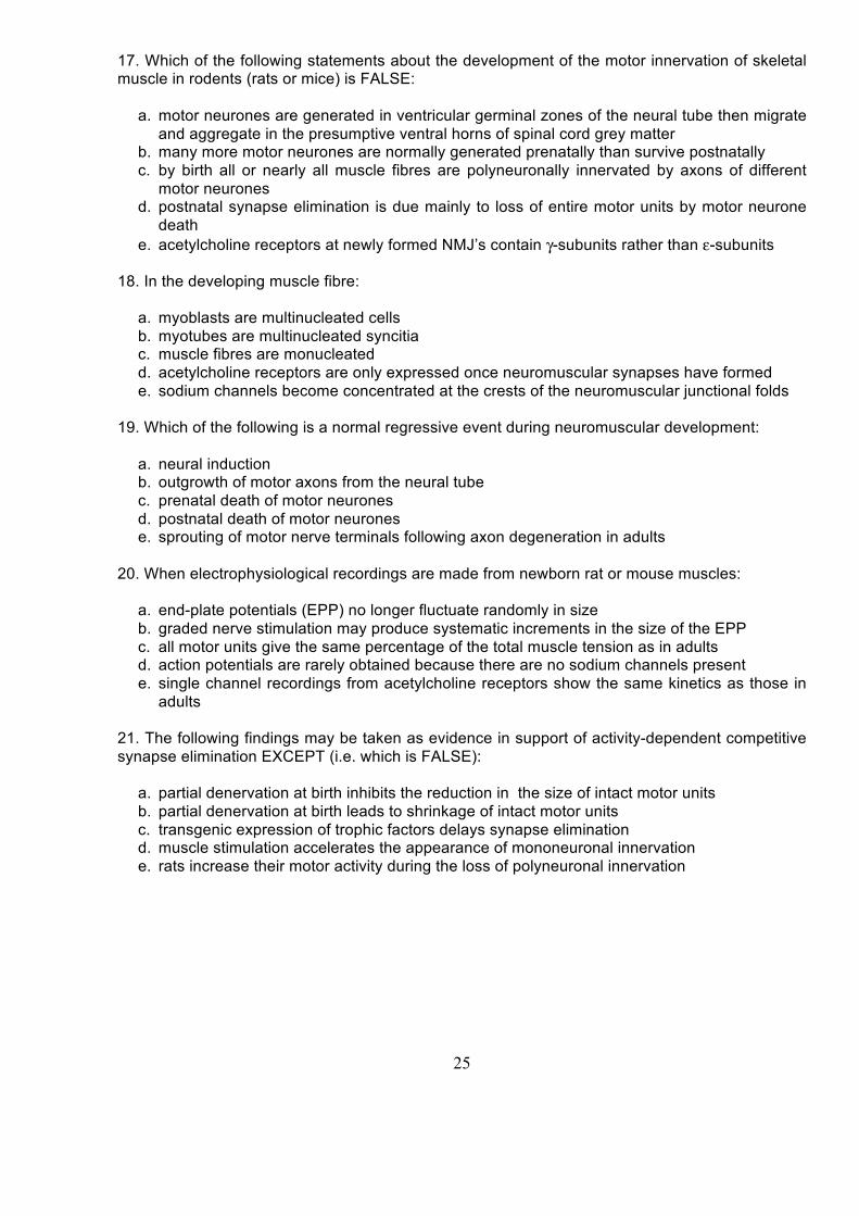
17. Which of the following statements about the development of the motor innervation of skeletal muscle in rodents (rats or mice) is FALSE:
a. motor neurones are generated in ventricular germinal zones of the neural tube then migrate and aggregate in the presumptive ventral horns of spinal cord grey matter
b. many more motor neurones are normally generated prenatally than survive postnatally c. by birth all or nearly all muscle fibres are polyneuronally innervated by axons of different
motor neurones d. postnatal synapse elimination is due mainly to loss of entire motor units by motor neurone
death e. acetylcholine receptors at newly formed NMJ’s contain γ-subunits rather than ε-subunits
18. In the developing muscle fibre:
a. myoblasts are multinucleated cells b. myotubes are multinucleated syncitia c. muscle fibres are monucleated d. acetylcholine receptors are only expressed once neuromuscular synapses have formed e. sodium channels become concentrated at the crests of the neuromuscular junctional folds
19. Which of the following is a normal regressive event during neuromuscular development:
a. neural induction b. outgrowth of motor axons from the neural tube c. prenatal death of motor neurones d. postnatal death of motor neurones e. sprouting of motor nerve terminals following axon degeneration in adults
20. When electrophysiological recordings are made from newborn rat or mouse muscles:
a. end-plate potentials (EPP) no longer fluctuate randomly in size b. graded nerve stimulation may produce systematic increments in the size of the EPP c. all motor units give the same percentage of the total muscle tension as in adults d. action potentials are rarely obtained because there are no sodium channels present e. single channel recordings from acetylcholine receptors show the same kinetics as those in
adults
21. The following findings may be taken as evidence in support of activity-dependent competitive synapse elimination EXCEPT (i.e. which is FALSE):
a. partial denervation at birth inhibits the reduction in the size of intact motor units b. partial denervation at birth leads to shrinkage of intact motor units c. transgenic expression of trophic factors delays synapse elimination d. muscle stimulation accelerates the appearance of mononeuronal innervation e. rats increase their motor activity during the loss of polyneuronal innervation

26
22. In the disease myasthenia gravis, patients have antibodies in their blood against their own acetylcholine receptors, producing symptoms and signs of muscle weakness. At a cellular level, neuromuscular junctions would be expected to show which of the following characteristics:
a. abnormally large end-plate potentials in response to nerve stimulation b. abnormally small spontaneous miniature end-plate potentials c. insensitivity to neostigmine d. insensitivity to tubocurarine e. long-lasting facilitation of end-plate potentials in response to repetitive nerve stimulation at
30 Hz 23. In the motor neurone disease Amyotrophic Lateral Sclerosis (ALS):
a. All forms of the disease are caused by mutations in the SOD1 gene b. Motor neurones supplying the legs are nearly always the first to degenerate c. Surviving motor units may be enlarged due to compensatory axonal sprouting d. There is no impairment of glutamate transport by glial cells in the spinal cord e. Riluzole, an antagonist of glutamate release, completely cures some patients
27
APPENDIX IV : QUANTAL ANALYSIS AT THE NEUROMUSCULAR JUNCTION Our present understanding of the fundamental physiological mechanism of transmitter release at synapses is mainly due to the work of B.Katz and his colleagues, based on their studies of transmission at the frog neuromuscular junction made during the 1950’s-1970’s at University College London. (Katz was awarded a Nobel Prize for his work in 1970). They showed that neurotransmitter is released in multi-molecular packets (‘quanta’). Electron microscopy established that these corresponded with synaptic vesicles in the motor nerve terminals. The evidence for quantized release of transmitter was based on Katz’s statistical analysis of electrophysiological recordings made from neuromuscular junctions during stimulation and at rest. Refinements of this ‘quantal analysis’ are still widely used in cellular electrophysiology to establish the amount of transmitter released at synapses during activity, in a variety of tissues: including the neuromuscular junction, autonomic ganglia, the hippocampus, and the cerebral cortex. At the resting neuromuscular junction, “miniature end-plate potentials” (MEPPs) are generated spontaneously at the endplate. An evoked end-plate potential (EPP) is produced in the muscle fibre following nerve stimulation. Normally this response triggers a muscle fibre action potential and contraction. When transmission is weakened either by blocking receptors with a nicotinic antagonist (e.g. tubocurarine) or by suppressing transmitter release with solutions containing reduced Ca2+ ions, the EPP becomes too small to trigger an active response: the EPP is said to be subthreshold. The essence of Katz’s quantal hypothesis of synaptic transmission was threefold: 1. The quantum of transmitter underlying the smallest nerve-evoked EPP and the spontaneous MEPP are one and the same; 2. The release of each quantum of neurotransmitter is independent of the release of other quanta and occurs with a very low statistical probability (i.e. random); 3. The evoked EPP is caused by the synchronous release of several quanta, due to a transient and large increase in the probability of release of individual quanta. Evidence supporting the hypothesis was obtained by recording intracellular EPPs and MEPPs and ascertaining the relationship between their amplitudes. In particular it was noted that EPPs are variable in amplitude from stimulus to stimulus, whereas the MEPPs are roughly constant in amplitude. The variability could be accounted for on the basis of point (2) above, by showing that the distribution of EPP amplitudes conformed to a binomial distribution, which simplified under conditions of low release probability to a poisson distribution. Binomial model of synaptic transmission Consider a nerve terminal containing a number (n) of quanta /synaptic vesicles. Suppose each has a small chance (p) of fusing with the plasma membrane and releasing transmitter across the synapse. If the synapse is stimulated repetitively, say 100 times, then the mean number (m) of quanta released will be :
m = n.p (1) By analogy, imagine tossing a coin 100 times. The probability of each toss coming up heads is 0.5. The average number of times the coin will come up heads is therefore 100 times 0.5: i.e., 50.
- 28 -
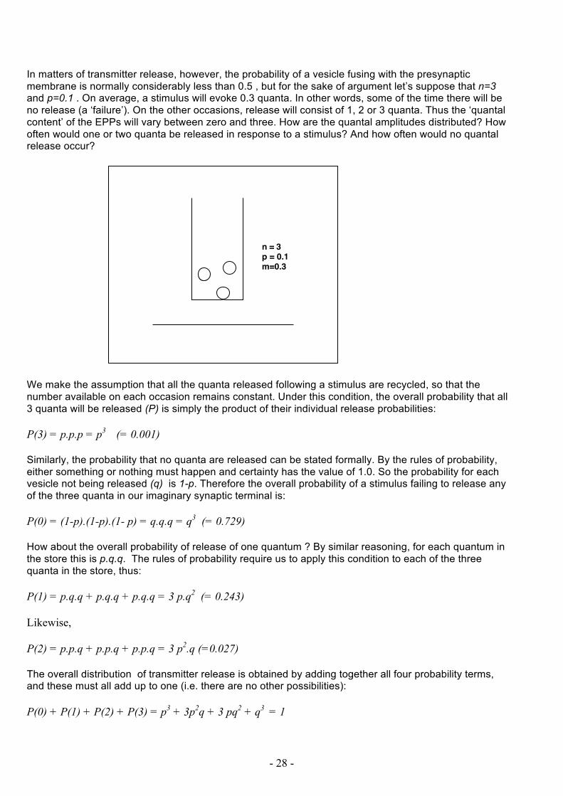
In matters of transmitter release, however, the probability of a vesicle fusing with the presynaptic membrane is normally considerably less than 0.5 , but for the sake of argument let’s suppose that n=3 and p=0.1 . On average, a stimulus will evoke 0.3 quanta. In other words, some of the time there will be no release (a ‘failure’). On the other occasions, release will consist of 1, 2 or 3 quanta. Thus the ‘quantal content’ of the EPPs will vary between zero and three. How are the quantal amplitudes distributed? How often would one or two quanta be released in response to a stimulus? And how often would no quantal release occur?
We make the assumption that all the quanta released following a stimulus are recycled, so that the number available on each occasion remains constant. Under this condition, the overall probability that all 3 quanta will be released (P) is simply the product of their individual release probabilities: P(3) = p.p.p = p3 (= 0.001) Similarly, the probability that no quanta are released can be stated formally. By the rules of probability, either something or nothing must happen and certainty has the value of 1.0. So the probability for each vesicle not being released (q) is 1-p. Therefore the overall probability of a stimulus failing to release any of the three quanta in our imaginary synaptic terminal is: P(0) = (1-p).(1-p).(1- p) = q.q.q = q3 (= 0.729) How about the overall probability of release of one quantum ? By similar reasoning, for each quantum in the store this is p.q.q. The rules of probability require us to apply this condition to each of the three quanta in the store, thus: P(1) = p.q.q + p.q.q + p.q.q = 3 p.q2 (= 0.243) Likewise, P(2) = p.p.q + p.p.q + p.p.q = 3 p2.q (=0.027) The overall distribution of transmitter release is obtained by adding together all four probability terms, and these must all add up to one (i.e. there are no other possibilities): P(0) + P(1) + P(2) + P(3) = p3 + 3p2q + 3 pq2 + q3 = 1
n = 3p = 0.1m=0.3

29
This simplifies to : (p+q)3 = 1 The above is a binomial expression : it contains two terms, p and q. Mathematical theory shows that in general we can predict from such an expression that for any number of quanta n with release probability p, that a particular nerve stimulus will release x quanta (x≤n) from the formula: P(x) = n! . px . q(n-x) The Binomial Distribution (2) (n-x)!x! Try this out on the example we have used above with n=3 and p=0.1 ( m=0.3): P(0) = P(1) = P(2) = P(3) = The Poisson model The problem with applying binomial analysis to real synapses is that there is rarely any independent way of estimating n or p . We can only estimate the mean quantal content, m, by dividing the mean EPP amplitude by the mean MEPP amplitude. It turns out that we can still nonetheless predict the distribution of amplitudes if we assume that n is very large (n>>p) and p is very small (p<<1). Under these conditions x<<n. Based on these assumptions we can make a number of simplifications to the binomial distribution (equation 2, above). For example, we may write: n! ≈ nx (e.g. try this with n=10,000 and x=3) (n-x)! and q(n-x) ≈ qn recalling that q = (1-p), we can now substitute these terms in the binomial distribution (equation 2): P(x) = nx . px. (1-p)n x!

30
since m = n.p (equation 1) this immediately simplifies to : P(x) = mx. (1-p)n (3) x! A little mathematical tinkering further simplifies the expression (1-p)n . First we apply natural logarithms in the identity: Ln (1-p)n = n. Ln (1-p) A mathematical formulation called McLaurin’s theorem can be used to express Ln (1-p): Ln (1-p) = -p - p2 - p3 - p4 - .... - p∞ 2! 3! 4! ∞ But since p<<1 by our assumption in the present analysis, then all the terms after the first one in the McLaurin series must be very small and we can ignore them. Thus: Ln(1-p) ≈ -p And, therefore: n.Ln (1-p) ≈ -n.p Taking the antilogarithm of both sides: (1-p)n = exp (-n.p) = exp (-m) [Note: Theory of logarithms - if y=Ln(x), then x=exp(y)] Substituting back in equation (3) we obtain: P(x) = mx . exp(-m) The Poisson Distribution (4) x! Once again, calculate what the distribution of probability of occurrence of EPPs containing 0,1,2,3 quanta are when the mean quantal content is 0.3 P(0) = P(1) = P(2) = P(3) =

31
Application of the Poisson Distribution to estimating quantal contents Experimentally, what does P(x) mean? It is simply the fraction of occasions on which the evoked postsynaptic potential (EPP, or in the case of CNS synapses, the EPSP) has a quantal content of x. To evaluate the quantal hypothesis and to use the Poisson distribution, we must compare the predicted variability in the amplitudes of EPPs with the actual variability observed experimentally. One of the most elegant demonstrations of the coincidence of the model and the data was obtained in a study by Boyd & Martin in a study of synaptic transmission in cat muscle: The data in the figure below were obtained from intracellular microelectrode recordings at a single neuromuscular junction in an isolated preparation in which neuromuscular transmission was depressed using a low Ca ion-high Mg ion bathing medium. The upper right of the figure shows the histogram of MEPP amplitudes as a bar chart and the superimposed graph is a fit of a normal (gaussian) distribution to the amplitudes. The lower graph shows the distribution of EPP amplitudes as a bar chart (including ‘failures’) and a fit of the Poisson distribution, taking account of the gaussian variation in MEPP amplitudes. Note that the number of failures is accurately predicted, as well as the distribution of the peaks. The mean and variance of each peak is a unit multiple of the first, which has the same mean and variance as the MEPP amplitude distribution. Data such as these provide confirmation of the quantal hypothesis.

32
ESTIMATING QUANTAL CONTENT There are three principal methods. Other methods are based on complex analysis of EPP amplitudes. 1. Direct method: Under favourable conditions, both MEPPs and EPPs can be recorded in sufficient numbers to allow cross checking of the quantal content of EPPs, comparing Poisson statistics with direct estimation of the mean quantal content m = (mean EPP amplitude) / (mean MEPP amplitude) This methos is not always possible for various technical reasons, e.g the MEPPs might be very infrequent; or they may be too small, buried in the noise of the recording system; or the mean quantal content may be large, resulting in non-linear summation of EPPs (see below). Applying the Poisson equation alone is sometimes sufficient in such cases. There are two methods of estimating quantal content based on the Poisson distribution: the Method of Failures and the Variance Method. 2. Method of Failures If the mean quantal content is low enough (as in the examples above), a significant fraction of stimuli will fail to evoke a response. This represents the P(0) expression in the Poisson distribution: P(0) = exp (-m) . m0/0! since, by mathematical definintion, both m0 and 0! are equal to 1 : P(0) = exp (-m) Taking natural logartithms : Ln (P0) = -m Substituting for P(0)=(Number of Failures)/(Number of Stimuli) and rearranging: m = Ln (Stimuli/Failures) 3. Variance method Another property of the Poisson distribution is that its variance equals its mean. From this it can be derived that: m = (mean EPP amplitude)2 (variance of EPP amplitudes) This is often expressed in terms of the coefficient of variation (standard deviation/mean = σ/µ) : m = C.V.-2

33
Non-linear summation of synaptic potentials As the amount of transmitter released onto the postsynaptic membrane increases - e.g. as the quantal content increases - the effectiveness of each quantal packet declines. This is because transmitter molecules at excitatory synapses like the neuromuscular junction act on receptors coupled to ion channels. When the channels open, positive ionic current flows into the postsynaptic cell. The electromotive driving force is determined by the ion gradient and the membrane potential. For example, at neuromuscular junctions the receptor/ion channel is the nicotinic ACh receptor which gates permeability to Na and K ions about equally. The ‘reversal potential’ is about -5 mV. This means that as the membrane potential approaches -5 mV, the ionic current flowing through the open channels becomes vanishingly small; and if membrane potential becomes more negative than -5 mV, an outward ionic current is produced instead and the EPP reverses in sign. Each quantal component of the EPP depolarises the membrane potential towards the reversal potential by a small amount, but as the amount of overall depolarisation becomes greater each successive quantal component has a weaker and weaker effect on further depolarisation. For instance, if the mean MEPP amplitude is 1 mV, then an EPP comprising 5 quanta may well produce a depolarisation of 5 mV. But an EPP comprising 20 quanta may only produce about 15 mV of depolarisation. This is called ‘non-linear summation’ of synaptic potentials. It means that under normal conditions of synaptic transmission when quantal contents can be quite high, the direct method of quantal analysis will underestimate mean quantal content and the variance method will overestimate mean quantal content. (Note: The failures method cannot usually be applied when mean quantal content is greater than about 5 because there are so few failures; P(0)=exp(-5) = 0.007; i.e. less than 1 in 100 stimuli would be expected to result in failure of transmission). The relationship between membrane potential, synaptic current and synaptic potential were investigated by McLachlan & Martin (1981), by alternately voltage- and current-clamping of the endplate. They showed that the relationship between the observed amplitude of the EPP, and the amplitude which would be obtained if transmitter quanta produced linear summation is: V’ = V / (1-f.V/E) where V’ is the predicted amplitude, V the observed amplitude; E is the difference between the resting membrane potential and the reversal potential and f - critically - is a factor which varies from muscle fibre to muscle fibre depending on its length, diameter and specific membrane and cytoplasmic electrical resistance. Normally it is not possible to measure this ‘fudge factor’ directly for every muscle fibre (it requires alternate voltage and current clamping of the endplate to do this). But there are rules of thumb: long muscle fibres mostly have an f-factor of 0.8; short muscle fibres have factors of about 0.3. Applying the correction for non linear summation to each EPP before calculating mean quantal content results in more accurate estimates by either the direct or variance methods. References Katz,B.(1969) The release of neural transmitter substances. Liverpool University Press. McLachlan EM. Martin AR. (1081) Non-linear summation of end-plate potentials in the frog and mouse. Journal of Physiology. 311:307-24. Katz,B.(1996) Neural transmitter release: from quantal secretion to exocytosis and beyond. The Fenn Lecture. Journal of Neurocytology. 25,677-86. JH Byrne & JL Roberts (2009) From molecules to networks. 2nd edn. Sinauer (Chapter 8)

34
MEPP Interval analysis Evoked release of neurotransmitter, produced by nerve excitation, can be described statistically as a binomial process, which reduces to description with the Poisson equation under appropriate conditions (see handout on Quantal Analysis). The conditions underpinning this simplification are that the quantal components of the EPP (the contents of vesicles released by exocytosis) are a small fraction of the total available (x<<n), and that each component is released independently and with a low statistical probability (p<<1). The same criteria and reasoning may be applied to spontaneous transmitter release and the occurrence of miniature endplate potentials (MEPPs). One way to demonstrate (or test) this is to count the number of MEPPs occurring in successive, identical intervals of time: for example, the number of ‘sweeps’ across an oscilloscope or a computer screen during capture of data (Gage & Hubbard, 1965). If the occurrence of MEPPs is random, then the probability of seeing a sweep containing any number of MEPPs, x, will be:
P(x) = e−mmx
x!
where P(x) = Number of sweeps containing x MEPPs Total number of sweeps and m = mean number of MEPPs per sweep Unlike the case of distribution of quantal contents during evoked release the histograms of number of MEPPs per sweep will not be complicated by any Gaussian variation : either a MEPP occurs or it does not; you can’t see half an occurrence. In other words, although the amplitude of the individual MEPPs may vary, this is irrelevant for the analysis of when they occur. A more sophisticated test of the randomness of spontaneous resease is to measure the distribution of intervals between occurrences of MEPPs. This kind of analysis was originally done by Fatt & Katz, (1952). The procedure is just as relevant today, because it is widely used in a related context , to characterize spontaneous or agonist-induced openoing and closing of ion channels seen in patch clamp recordings, for example. It is therefore important for the general neuroscientist or physiologist to grasp the principles of interval analyses and this can most simply be illustrated by considering how it was originally done for MEPPs. (In fact, interval analysis of single channel recordings is considerably more complicated , owing to variability in the open times as well as the closed times of ion channels, which may also be subject to clustering, bursting, flickery-blocks etc. See Aidley, DJ Physiology of Excitable Cells, 3rd edn for references).

35
Analysis from first principles shows that the intervals between MEPPs should also be distributed exponentially if release is random (ie events are independent and occur with a low statistical probability):
n(t + Δt) = N ΔtTe− tT
where t = elapsed time
n(t+ Δt) = Number of MEPPs within the interval of time t+ Δt N = total number of MEPPs observed T = mean interval between MEPPs
Here is one way of arriving at this relationship: Suppose we observe a number (N) of MEPPS in a period of time t. The mean interval between the MEPPs we observe will be:
T = t/N Now consider what happens when we subdivide the time t into n smaller intervals, Dt:
t = n.Δt or Δt=t/n
T = n. Δt /N Now consider the situation when N=1 , that is when there is just one MEPP in the overall period of elapsed time t. What is the probability we will see this MEPP within any defined interval Dt. If Dt =t, we would certainly see it (P=1). So, the probability of seeing the MEPP will depend on how long Dt is in relation to t.
P α Δt Thus,
P = k. Δt So it now follows that if we see the MEPP in one of the specific time intervals, Dt, then in the other (n-1) intervals of equal time we will definitely not see it. Call this probability Q:
Q = 1-P And
Q = 1-k. Δt
Q = 1-k.t/n

36
Now we ask, how long must we wait before we actually see the MEPP. We multiply the probabilities of not seeing it in each successive interval of time Dt (including the possibility that we don’t see it at all in the total time, n. Dt. Since there are n identical periods of time Dt in the total time t, it follows that:
Q(t) = (1− k tn)n
Next we apply a mathematical tool called McLaurin’s Series. This shows that:
(1− xn)n ≈ e− x
Making x=kt, we may therefore write:
Q(t) = e−kt A property of the exponential is that is rate constant, k, is the inverse of the time constant and this in turn is equal to the mean interval T. Therefore,
Q(t) = e−tT
So our chances of not seeing the MEPP, within a period of time t , decreases exponentially the longer we wait. (Like waiting at a bus stop for the next one when you know it’s going to arrive SOME time in the next hour….the longer you wait, the less likely you’ll have to wait as long….). This is provided that the occurrence of the MEPP occurs with a low probability and that the occurrence of the MEPP is not influenced by the occurrence of previous MEPPs. (Unlike buses: when one arrives, another might be right behind, for example because of the effect of queuing, the first interfering with the free transit of the other.) In practice, we normally choose our duration of time t to be long enough to observe more than one MEPP. Each interval between the MEPPs is measured and the measurements are used to construct a histogram of intervals. Each bin in the histogram represents a finite interval of time Dt. An interval measured experimentally will be placed into a particular bin, according to whether it falls within the time elapsed so far (t) and the next interval (t+ Dt). The probability of not seeing a MEPP up to time t is given by the equation above. The probability of not seeing it up to the interval of time (t+ Dt) is:
Q(t + Δt) = e−(t+Δt )T
So the probability of not seeing it in time Dt itself is:
Q(Δt) = e−(t+Δt )T − e
−tT
Conversely, the probability of seeing exactly one MEPP in time Dt is P(Dt)=1-Q(Dt) :
P(Δt) = e−tT − e
−(t+Δt )T

37
that is:
P(Δt) = e−tT (1− e
−ΔtT )
Using another spanner from the Mathematician’s Toolkit: namely, Taylor’s Series:
ex = 1+ x + x2
2!+x3
3!... x
n
n!
If x<<n then
ex ≈ 1+ x or
1− ex = −x So, making x= -Dt /T, if Dt is very small then:
(1− e−ΔtT ) = Δt
T
and
P(Δt) = ΔtTe−tT
If we define P(Dt) as the fraction of the total number of intervals in the histogram bin (t+ Dt) then:
n(t + Δt)
N=ΔtTe−tT
And
n(t + Δt) = N ΔtTe−tT
….Which is what we set out to prove: q.e.d. ! PS. By analogy with radioactive decay, the decay time constant of this exponential (that is, -1/T) can also be obtained from the ‘half life’ , t1/2 , of the curve: that is the frequency of intervals at (t+ Dt) equal to half that of the initial frequency:
t1/2 = T .ln(2)

38
N 100 T 1 dt 0.2 t+dt n 0.2 16.37 0.4 13.41 0.6 10.98 0.8 8.99 1.0 7.36 1.2 6.02 1.4 4.93 1.6 4.04 1.8 3.31 2.0 2.71 2.2 2.22 2.4 1.81 2.6 1.49 2.8 1.22 3.0 1.00 3.2 0.82 3.4 0.67 3.6 0.55 3.8 0.45 4.0 0.37 4.2 0.30
Example of a recording of MEPPs, total t = approx1 minute

39
Application to single channel recording The intervals between currents due to the opening of single ion channels (‘closed times’), observed in recordings from membranes made using the single-channel patch-clamp technique, also shows characteristics consistent with underlying events occurring with low probability and independently of one another. That is, the same set of conditions that apply for a Poisson Distribution and analysis of MEPPs.
Single-channel recording from an acetylcholine (ACh) receptor molecule in a membrane patch, in the presence of a low concentration of ACh. The histogram of closed times (intervals) shows an exponential decay with a time constant (t/T) in this example of about 20 µs. From Colquhoun, D. & Sakmann, B. (1985) J Physiol 369, 501-557.

40
References Fatt, P & Katz, B (1952) J. Physiol. 117, 109-128 Gage, P & Hubbard, J.I.(1965). Nature 208,395-397 Colquhoun,D. (1987) In Microelectrode Techniques: The Plymouth Workshop Handbook. Standen, N., Gray, P. & Whittaker, M.(eds) Company of Biologists Ltd. Pp 105-136. http://en.wikipedia.org/wiki/Half-life Neuromuscular Transmission/Quantal Analysis Problems 1. In an experiment on a partially curarised frog neuromuscular junction, acetylcholine (ACh) was applied to the endplate by iontophoresis, using 1 nA, 1 ms current pulses at a frequency of 2 Hz. A train of five endplate potentials (EPPs) was then evoked by stimulating the muscle nerve at 50Hz. The iontophoretic pulses were resumed within 20 ms of the end of the stimulus train. The following data were obtained: Mean ACh response before EPP train = 1.53 ± 0.12 mV (mean ± S.D.; n=10) Mean ACh response after EPP train = 1.51 ± 0.10 mV (mean ± S.D.; n=7) EPP number 1 2 3 4 5 Amplitude (mV) 2.2 2.7 2.1 1.3 0.9 a. calculate the amount of charge delivered by each of the iontophoretic current pulses; b. sketch the characteristic responses to ACh and nerve stimulation indicating the time course of the responses; c. how might the iontophoretic responses to ACh change, if a low concentration of ACh (1 µM) were also continuously present in the medium? d. is the hypothesis that short-term synaptic depression is caused by desensitisation of ACh receptors supported or refuted by these data? Give your reasoning. 2. Intracellular recordings were made from a mouse neuromuscular junction. The nerve supply was stimulated 150 times at 1Hz. The mean size of the EPP evoked was 1.00 mV. Five of the stimuli evoked no response (i.e. there were 5 'failures'). a What was the mean quantal content at this neuromuscular junction? b What do you predict for the quantal size, the amplitude of the uniquantal event (MEPP)? c How many of the EPPs would you predict to have quantal contents of 1,2,3 and 4 quanta? d What do you predict would be the standard deviation of the EPP amplitudes? e If the baseline ‘noise’ level peak-to-peak was 500 µV, how would this affect the accuracy of
your estimates?

41
3. In an experiment on an isolated flexor digitorum brevis nerve-muscle preparation dissected from a mouse, intracellular microelectrode recordings were made of spontaneous miniature endplate potentials (MEPP). Endplate potentials (EPP) were then evoked by nerve stimulation at a frequency of 1 Hz. In total, 97 of the stimuli applied to the nerve evoked an EPP but 3 stimuli failed to evoke any EPP. The following mean data with their standard deviations were obtained:
Mean MEPP amplitude (± SD) : 1.20 ± 0.72 mV Mean EPP amplitude (± SD) : 4.25 ± 2.42 mV
a. Speculate on the ratio of Ca2+ to Mg2+ ions in the medium bathing this preparation. b. Calculate the mean quantal content of the EPP using the Direct, Variance and Failures Methods. c. What does the standard deviation of the MEPP amplitude (quantal size) indicate and how might
this affect the estimation of mean quantal content? d. Give one other possible reason for a low quantal content, in the contexts of health and disease.
4. In a study by Wood & Slater (1997) it was found that the action potential firing threshold in rat soleus muscle fibres is reached by an endplate potential (EPP) with a mean quantal content of about 15. Quantal size did not vary substantially between muscle fibres in either soleus or extensor digitorum longus muscles in this study. Answer the following (weighting of percentage marks in brackets): a. Explain what is meant by the terms ‘quantal content’ and ‘quantal size’ b. Explain briefly how recordings of endplate currents could be made from this fibre, why endplate currents sum linearly with increasing quantal content but EPP’s sum non-linearly, and how EPP-amplitude measurements may be corrected for non-linear summation. c. Predict the number of ‘failures’ in a train of 100 EPC’s if the mean quantal content became reduced to 2 quanta. d. Use the Failures Method to calculate the probability of ‘failures’ in a train of EPC’s evoked by repetitive stimulation when the mean quantal content is 15. Calculate the predicted variance and standard deviation of quantal contents in a train of EPPs of the same mean quantal content (ie 15). e. Discuss briefly what experimental or clinical conditions lead to a reduction of quantal content and the mechanisms that can enhance the ‘safety factor’ for neuromuscular transmission.
5. Intracellular recordings of endplate potentials (EPPs) and miniature endplate potentials (MEPPs) were obtained from a muscle fibre in an isolated mouse skeletal muscle bathed in normal physiological saline but in which the Ca2+ concentration was reduced from 2mM to 1mM and the Mg2+ concentration was increased from 1mM to 4 mM. The preparation was then stimulated with 100 trains-of-four nerve stimuli, delivered at 50Hz with intervals of 10s between each stimulus train. Each stimulus train evoked a corresponding train of four EPPs (EPP1-EPP4). The acetylcholinesterase inhibitor neostigmine (final concentration 3 µM) was then added to the physiological saline. After 20 minutes, recordings were resumed from the same muscle fibre and a further 100 trains-of-four supramaximal nerve stimuli were applied to the nerve. Other constituents of the bathing solution were unaltered from that of normal physiological saline. The resting membrane potential of the muscle fibre remained steady throughout at -72 mV. The following data were obtained Before and After adding neostigmine.

42
Before adding neostigmine Mean MEPP amplitude: 1.2 mV Stimulus trials: 100
EPP1 EPP2 EPP3 EPP4 Number of EPP Failures 3 2 2 1 Mean EPP Amplitude (mV) 4.3 4.6 4.9 5.5 Rise time (ms) 1.5 1.6 1.4 1.7 Half -Decay time (ms) 2.7 2.6 2.5 2.7 After adding neostigmine Mean MEPP amplitude: 1.5 mV Stimulus trials: 100
EPP1 EPP2 EPP3 EPP4 Number of EPP Failures 4 3 2 1 Mean EPP Amplitude (mV) 4.6 5.2 5.5 5.9 Rise time (ms) 2.6 2.5 2.6. 2.7 Half-Decay time (ms) 5.4 6.7. 7.7 8.9 a. Describe and explain the responses to nerve stimulation and the effects of neostigmine on
amplitude and time course of the EPPs. Sketch their appearance in the trains-of-four before and after the change in bathing solution (25 % of marks)
b. What would you conclude from a quantal analysis of these data and should correction for non-linear summation of EPPs be considered? (25%)
c. Anticholinesterases may have additional direct effects on properties of presynaptic or post-synaptic acetylcholine receptors. Formulate hypotheses to explain the effects of neostigmine on the amplitude and time course of the synaptic potentials, with justification of your reasoning (25%).
d. Suggest how the hypotheses you have proposed might be tested experimentally and what alternative outcomes of these experiments would imply. (25%)
6. Imagine you had performed an experiment on a muscle preparation from a Drosophila larva. In this experiment, intracellular recordings were made from an identified muscle fibre, in response to stimulation of the nerve innervating the segment containing the muscle fibre. Two sets of measurements were obtained after successful microelectrode penetration of the muscle fibre in the vicinity of the neuromuscular junction. First, the nerve supply to the muscle fibre was stimulated at a frequency of 1 Hz, while the stimulus intensity was systematically increased. The mean amplitude and standard deviation of the evoked excitatory junctional potential (EJP) was measured. Second, spontaneous miniature EJP's (mEJP) were recorded from the fibre in 60 successive time intervals ('sweeps') of 1 second duration and the numbers of sweeps containing between zero and eight mEJP’s per sweep were recorded. The data obtained were as follows:
Table 1 Stimulating Voltage (V)
EJP amplitude (mV ± S.D.)
0.1 0 0.2 2.2 ± 1.7 0.3 2.1 ± 1.6 0.4 2.3 ± 1.5 0.5 10.8 ± 1.2 0.6 8.9 ± 1.6 0.7 9.5 ± 1.9 0.8 30.5 ± 2.4 0.9 29.8 ± 3.9

43
Table 2 Respond to all the following (each part carries an equal number of marks): a. With reference to Table 1, provide an interpretation of the stepwise increase in the mean amplitude of the evoked EJP as stimulus strength was increased. b. Also with reference to Table 1, use the Variance Method based on the Poisson equation to estimate the mean quantal content of the responses evoked by a stimulating voltage of 0.4V and predict from this the mean quantal size (mEJP amplitude). c. With reference to Table 2, show how the 'Method of Failures', based on the Poisson equation and the number of sweeps containing no occurrences of mEJP’s, could be used to estimate the mean frequency of mEJP’s in this recording. d. Show quantitatively that the distribution of numbers of mEJP's per 1s sweep, given in Table 2, is consistent with predictions from a Poisson model of neuromuscular transmission. e. Suppose extracellular potassium ionic concentration were increased, with the effect of doubling the mean frequency of mEJP's. Predict the number of one second sweeps out of 60 containing exactly one mEJP. f. By what mechanisms might raising extracellular potassium ions increase the frequency but decrease the amplitude of spontaneous mEJP’s? Suggest experiments that would test experimentally your proposed explanation.
Note: The Poisson equation is P(x)=mx.exp(-m)/x! where P(x) is the probability of occurrence of an EPP/EJP with a quantal content of x, given a mean quantal content equal to m. The same equation may be used to describe distributions of MEPP/MEJP frequency, with m equal the mean number of events in intervals of equal time (eg 1 second-long traces)
X (mEJP/sweep)
Number of sweeps with X mEJP
0 17 1 21 2 13 3 6 4 2 5 0 6 1 7 0 8 0

44
7. Intracellular recordings were made from neuromuscular junctions in isolated preparations of the flexor digitorum brevis (FDB) muscles of four mutant mice. The data shown in Table 1 (mean ± SEM) were obtained from recordings made 5 days after unilateral section (axotomy) of the tibial nerve supplying the FDB muscle (30 muscle fibres sampled in each muscle), and compared with recordings and data from the unoperated contralateral control muscle. The preparations were bathed in normal physiological saline throughout the experiments. P values show significance from unpaired t-tests. The data in Table 2 (mean ± SD) were obtained from recordings of single FDB muscle fibres in one of the axotomised muscles and one fibre from the contralateral control muscle. Morphological analysis was then performed, from which the endplate area and fractional occupancy of the endplate by the motor nerve terminal was derived.
Table 1: all recordings (4 muscles, 30 fibres recorded per muscle) Axotomy Control P
Resting membrane potential (mV) -54 ± 5.2 -68 ± 3.9 <0.05
Mean Frequency of MEPPs (s-1): 3.4 ± 1.1 0.3 ± 0.1 <0.05 Mean Amplitude of MEPPs (mV): 3.2 ± 1.5 0.8 ± 0.4 <0.05 Mean Amplitude of EPPs (mV): 7.9 ± 5.4 30.2 ± 4.7 <0.05 Mean Rise time of EPPs (ms): 2.6 ± 0.3 1.3 ± 0.2 <0.05 Mean 50% decay time of EPPs (ms): 5.4 ± 2.2 2.3 ± 0.6 <0.05
Table 2: single muscle fibres Axotomy Control Resting membrane potential (mV): -56 -72 Mean Frequency of MEPPs (s-1): 3.1 0.5 Mean Amplitude of MEPPs (mV): 2.2 ± 1.3 1.2 ± 0.5 Mean Amplitude of EPPs (mV): 7.9 ± 4.2 30.2 ± 2.9
Number of EPP 'failures'/Stimuli: 2/100 0/100 Endplate area (µm2): 354 381 Fractional occupancy of endplate: 0.65 0.98
Note: a.The Poisson equation is : P(x)=exp(-m).(mx)/x! For spontaneous release, P(x) is the probability of occurrence of a number of events, x, with mean frequency, m For evoked release P(x) is the probability of an EPP with a quantal content equal to x, when the mean quantal content is equal to m. b. The equation for non-linear summation is: V’=V/(1-fV/Vd) where V’ is the corrected EPP amplitude, V the observed amplitude, Vd is the ‘driving force’ equal to (Er-Em): the difference between the resting membrane potential (Em) and the ‘reversal potential’ for action of ACh on its receptors (Er = -10 mV). ‘f’ is an arbitrary factor, specific to the type of muscle fibre from which the EPP recordings are made.

45
a. From Table 1, what are plausible explanations for the differences in amplitude and time course of the evoked EPPs and spontaneous MEPPs in axotomised compared with control muscles?
b. From Table 2, predict the number of periods of one second duration, out of 100 successive test periods, when no spontaneous MEPPs would be expected to occur. What are the two main assumptions about spontaneous transmitter release underpinning your prediction?
c. From Table 2, calculate the absolute quantal content of EPPs in the axotomised and control muscle fibres using appropriate methods; then estimate the specific quantal content (quanta per µm2 of presynaptic terminal) in both cases. What do you conclude from these estimates of absolute and specific quantal content?
d. If the true quantal content for the control fibre in Table 2 were 35 quanta (determined from measurements of synaptic currents under voltage clamp). Show that assigning a value of f=1 in the equation for non-linear summation leads, in the case of the Control fibre, to overcorrection of the EPP amplitude and hence overestimation of quantal content by the Direct Method. Deduce what would be a more appropriate value of “f” for correcting for non-linear summation of quanta/EPPs in the case of the control fibre. Show that this correction makes little difference to the calculation of quantal content in the case of the axotomised fibre.
e. From your knowledge of synaptic degeneration and its control, what kind of mutation may have produced the phenotype represented by the above data; and how might the morphological analysis and measurement of fractional occupancy of the endplate in Table 2 have been obtained?
Note: The Poisson equation is P(x)=mx.exp(-m)/x! where P(x) is the probability of occurrence of an EPP with a quantal content of x, given a mean quantal content equal to m or the mean number of mEJP's per 1 second interval.

46
APPENDIX V: Tips for written and oral presentations Essays
The word “essay” is synonymous with “attempt”: thus, in an essay you are attempting to explain something. At Honours level, essays are not simply regurgitated or rearranged facts that you have stumbled upon or read about. We are looking for originality, creativity, rigour and application of critical skills. Like all science, neuroscience is evidence-based.
So, you will need to focus your essay on the evidence underpinning your attempted explanations for neuromuscular synaptic phenomena, processes, or mechanisms. You may be seeking to resolve a debatable issue. In that case you should marshal the evidence for and against. In scientific writing, evidence is usually cited in terms of who did what and when. For example, “Endplate potential recordings combined with mathematical modelling, based on Poisson statistics, established that the the release of neurotransmitter from motor nerve terminals is "quantal" (Del Castillo & Katz, 1952)”. And be sure to provide the reference for the work that you cite as evidence for such statements in a bibliography or referenc list at the end of the essay, e.g. Del Castillo, J. & Katz, B (1954) Quantal components of the end-plate potential. J Physiol. 124:560-573.
Examiners of essays for this course will be looking for evidence that you have assimilated and understood the main findings of the core papers we have discussed in each minisymposium, supplemented by the background reading and other learning resources indicated for each of these. They will also take account of the Learning Objectives for the course. Extra credit will be given for evidence of additional reading around the subject, provided it shows knowledge and understanding relevant to the question. (ALWAYS ANSWER THE QUESTION POSED, not the question you would have liked to have been posed!)
General features of good essays include:
• Good form and structure (Introduction/exposition/inference/summary)
• Good use of English (style, grammar and spelling)
• Understanding of context (the ‘big picture’)
• Sound factual background knowledge
• Critical skills including data appraisal and logical or statistical analysis
• Evidence-based reasoning
• Coherence and rigour of the arguments, including alternative explanations
• Clear statement of conclusions
• Specific suggestions for further work
• Conclusions/Summary
• Bibliography/references (not expected under closed-book/unseen exam conditions)

47
Reading research papers Critical reading of research papers:
v What is the aim of the study? Is there a hypothesis?
v How has the study been designed to address the aim/hypothesis?
v What methods/techniques have been used? Are they appropriate for the design/question?
v Figures contain the most important data: what kinds of data were acquired?
v How have the data been quantitatively analysed? Is the data analysis adequate? Could it be improved and if so how?
v Are the authors conclusions justified by the quality and quantity of the data?
v Are there alternative interpretations?
v What are the strengths and weaknesses of the study?
v What should be done next?
See also: http://www.informatics.buu.ac.th/~krisana/presentation/HowToRead/How%20to%20Read%20a%20Scientific%20Research%20Paper--.htm Presenting papers: Talks (15-20 mins) should focus on the important information in the figures but have a
v Beginning (Introduction, Aims of the study, summary of Methods used);
v Middle (presentation of Results; include original figures)
v End (strengths and weaknesses, summary of Conclusions, Suggestions for further work,). Bear in mind the mantra of good lecturing: "Tell'em what you're gonna tell 'em; Tell 'em; Tell 'em what you've told 'em". The following, light-hearted article is highly instructive on the issue of illustrative slides: Leng G. Editorial: Blue on blue doesn't work. J Neuroendocrinol. 2002 Dec;14(12):925-6. PubMed PMID: 12472871.

48
Essay Feedback Form
Course: Roll No
Overall Mark(%) Marker: Q:
Tick box which best describes the performance in each area. Note: the weighting of marks is different between each category
Description Unsatisfactory
(<40%)
Satisfactory
(40-49%)
Good
(50-59%)
Very good (60-69%)
Excellent
(>70%) Introduction/aims /hypothesis Did the introduction clearly and concisely explain the main issues to be addressed? Were the aims or objectives and hypothesis clearly stated
Coverage (ie answer question) Were the main points of question addressed ?
Knowledge/ Understanding Was the information to support the main points in the question clearly and concisely explained and supported by evidence?
Critical Evaluation Was there evidence of critical thinking? Was a viewpoint (if appropriate) expressed and supported by evidence? Was there evidence of extra reading or the use of additional sources of material?
Conclusions /future directions Were conclusions clearly defined and relevant and was there an indication of further work that could be done?
Structure/ presentation Was the essay well organised, well laid out and written in an easy to read style? Was the work of others acknowledged and referenced appropriately?
Supporting figures/tables Was appropriate use made of figures & tables? Were they neatly constructed, properly labelled and well-integrated into the text?
Comments:

49
APPENDIX VI ABSTRACTS FOR MINI-SYMPOSIUM I Architecture of Motor Units of Neuromuscular junctions PDF’s of the full articles for the first minisymposium can be downloaded from the course website: http://www.dns.ed.ac.uk/rrrweb/NMJHDhons/NMJcontents.htm or via the PubMed PMID number (if accessing from a University computer): http://www.ncbi.nlm.nih.gov/pubmed/ For all other minisymposia, you are expected to download the papers via PubMed or the University Library’s electronic journal subscriptions, for yourselves. 1. Lu J, Tapia JC, White OL, Lichtman JW. The interscutularis muscle connectome. PLoS Biol. 2009 Feb 10;7(2):e32. PMID: 19209956 2. Thomson SR, Nahon JE, Mutsaers CA, Thomson D, Hamilton G, Parson SH, Gillingwater TH. Morphological characteristics of motor neurons do not determine their relative susceptibility to degeneration in a mouse model of severe spinal muscular atrophy. PLoS One. 2012;7(12):e52605. doi: 10.1371/journal.pone.0052605. PubMed PMID: 23285108 3. Court FA, Gillingwater TH, Melrose S, Sherman DL, Greenshields KN, Morton AJ, Harris JB, Willison HJ, Ribchester RR. Identity, developmental restriction and reactivity of extralaminar cells capping mammalian neuromuscular junctions. J Cell Sci. 2008 Dec 1;121(Pt 23):3901-11 4. David G, Nguyen K, Barrett EF. Early vulnerability to ischemia/reperfusion injury in motor terminals innervating fast muscles of SOD1-G93A mice. Exp Neurol. 2007 Mar;204(1):411-20. Epub 2007 Jan 4. PubMed PMID: 17292357.

50
PLoS Biol. 2009 Feb 10;7(2):e32.
The interscutularis muscle connectome.
Lu J, Tapia JC, White OL, Lichtman JW.
Department of Molecular and Cellular Biology, Harvard University, Cambridge, Massachusetts, USA.
Erratum in
• PLoS Biol. 2009 Apr;7(4):e1000108.
Abstract
The complete connectional map (connectome) of a neural circuit is essential for understanding its structure and function. Such maps have only been obtained in Caenorhabditis elegans. As an attempt at solving mammalian circuits, we reconstructed the connectomes of six interscutularis muscles from adult transgenic mice expressing fluorescent proteins in all motor axons. The reconstruction revealed several organizational principles of the neuromuscular circuit. First, the connectomes demonstrate the anatomical basis of the graded tensions in the size principle. Second, they reveal a robust quantitative relationship between axonal caliber, length, and synapse number. Third, they permit a direct comparison of the same neuron on the left and right sides of the same vertebrate animal, and reveal significant structural variations among such neurons, which contrast with the stereotypy of identified neurons in invertebrates. Finally, the wiring length of axons is often longer than necessary, contrary to the widely held view that neural wiring length should be minimized. These results show that mammalian muscle function is implemented with a variety of wiring diagrams that share certain global features but differ substantially in anatomical form. This variability may arise from the dominant role of synaptic competition in establishing the final circuit.
PMID: 19209956

51
PLoS One. 2012;7(12):e52605.
Morphological characteristics of motor neurons do not determine their relative susceptibility to degeneration in a mouse model of severe spinal muscular atrophy. Thomson SR, Nahon JE, Mutsaers CA, Thomson D, Hamilton G, Parson SH, Gillingwater TH.
Euan MacDonald Centre for Motor Neurone Disease Research, University of Edinburgh, Edinburgh, United Kingdom.
Abstract
Spinal muscular atrophy (SMA) is a leading genetic cause of infant mortality, resulting primarily from the degeneration and loss of lower motor neurons. Studies using mouse models of SMA have revealed widespread heterogeneity in the susceptibility of individual motor neurons to neurodegeneration, but the underlying reasons remain unclear. Data from related motor neuron diseases, such as amyotrophic lateral sclerosis (ALS), suggest that morphological properties of motor neurons may regulate susceptibility: in ALS larger motor units innervating fast-twitch muscles degenerate first. We therefore set out to determine whether intrinsic morphological characteristics of motor neurons influenced their relative vulnerability to SMA. Motor neuron vulnerability was mapped across 10 muscle groups in SMA mice. Neither the position of the muscle in the body, nor the fibre type of the muscle innervated, influenced susceptibility. Morphological properties of vulnerable and disease-resistant motor neurons were then determined from single motor units reconstructed in Thy.1-YFP-H mice. None of the parameters we investigated in healthy young adult mice - including motor unit size, motor unit arbor length, branching patterns, motor endplate size, developmental pruning and numbers of terminal Schwann cells at neuromuscular junctions - correlated with vulnerability. We conclude that morphological characteristics of motor neurons are not a major determinant of disease-susceptibility in SMA, in stark contrast to related forms of motor neuron disease such as ALS. This suggests that subtle molecular differences between motor neurons, or extrinsic factors arising from other cell types, are more likely to determine relative susceptibility in SMA.

52
J Cell Sci. 2008 Dec 1;121(Pt 23):3901-11. Epub 2008 Nov 11.
Identity, developmental restriction and reactivity of extralaminar cells capping mammalian neuromuscular junctions.
Court FA, Gillingwater TH, Melrose S, Sherman DL, Greenshields KN, Morton AJ, Harris JB, Willison HJ, Ribchester RR.
Euan MacDonald Centre for Motor Neurone Disease Research, The University of Edinburgh, 1 George Square, Edinburgh EH8 9JZ, UK.
Abstract
Neuromuscular junctions (NMJs) are normally thought to comprise three major cell types: skeletal muscle fibres, motor neuron terminals and perisynaptic terminal Schwann cells. Here we studied a fourth population of junctional cells in mice and rats, revealed using a novel cytoskeletal antibody (2166). These cells lie outside the synaptic basal lamina but form caps over NMJs during postnatal development. NMJ-capping cells also bound rPH, HM-24, CD34 antibodies and cholera toxin B subunit. Bromodeoxyuridine incorporation indicated activation, proliferation and spread of NMJ-capping cells following denervation in adults, in advance of terminal Schwann cell sprouting. The NMJ-capping cell reaction coincided with expression of tenascin-C but was independent of this molecule because capping cells also dispersed after denervation in tenascin-C-null mutant mice. NMJ-capping cells also dispersed after local paralysis with botulinum toxin and in atrophic muscles of transgenic R6/2 mice. We conclude that NMJ-capping cells (proposed name 'kranocytes') represent a neglected, canonical cellular constituent of neuromuscular junctions where they could play a permissive role in synaptic regeneration.
PMID: 19001504
53
Exp Neurol. 2007 Mar;204(1):411-20. Epub 2007 Jan 4.
Early vulnerability to ischemia/reperfusion injury in motor terminals innervating fast muscles of SOD1-G93A mice.
David G, Nguyen K, Barrett EF.
Department of Physiology and Biophysics, University of Miami Miller School of Medicine, USA. [email protected]
Abstract
In mouse models of familial amyotrophic lateral sclerosis (fALS), motor neurons are especially vulnerable to oxidative stresses in vitro. To determine whether this increased vulnerability also extends to motor nerve terminals in vivo, we assayed the effect of tourniquet-induced ischemia/reperfusion (I/R) injury on motor terminals innervating fast and slow hindlimb muscles in male G93A-SOD1 mice and their wild-type littermates. These mice also expressed yellow fluorescent protein (YFP) in motor neurons. We report that in SOD1-G93A/YFP mice the motor terminals innervating two predominantly fast muscles, extensor digitorum longus (EDL) and plantaris, were more vulnerable to I/R injury than motor terminals innervating the predominantly slow soleus muscle. The mean duration of EDL ischemia required to produce a 50% reduction in endplate innervation in SOD1-G93A/YFP mice was 26 min, compared to 45 min in YFP-only mice. The post-I/R destruction of EDL terminals in SOD1-G93A mice was rapid (<2 h) and was not duplicated by cutting the sciatic nerve at the tourniquet site. The increased sensitivity to I/R injury was evident in EDL muscles of SOD1-G93A/YFP mice as young as 31 days, well before the onset of motor neuron death at approximately 90 days. This early vulnerability to I/R injury may correlate with the finding (confirmed here) that in fALS mice motor nerve terminals innervating fast hindlimb muscles degenerate before those innervating slow muscles, at ages that precede motor neuron death. Early vulnerability of fast motor terminals to I/R injury thus may signal, and possibly contribute to, early events involved in motor neuron death.