
NEUROLOGICAL EXAMINATIONS: HOW TO DETERMINE CONDITION
15
Vet Times The website for the veterinary profession https://www.vettimes.co.uk NEUROLOGICAL EXAMINATIONS: HOW TO DETERMINE CONDITION Author : MARK LOWRIE Categories : Vets Date : May 19, 2014 MARK LOWRIE MA, VetMB, MVM, DipECVN, MRCVS provides practical guidance on how to determine whether a patient has a neurological condition using traditional methods in the first of a three-part series MANY veterinary practitioners consider neurology one of the hardest disciplines. This apprehension is heightened when they are faced with an uncooperative patient, whether it is a fractious feline or a catapulting canine. These circumstances frequently result in the clinician neglecting the neurological examination in favour of making assumptions on the most likely cause for the symptoms – with no regard as to the aspect of the nervous system affected or even if a neurological problem is present. This enticement may be heightened if the veterinarian has easy access to CT, MRI or myelography. It is important to emphasise these tools are not a replacement for the traditional approach to a neurological patient and their absence should not preclude the practitioner from performing a targeted neurological examination to gain a better understanding of a patient’s problem. In these days of austerity, training, experience, clinical skills and a rational approach to a specific complaint assume even more importance, and “shotgun” diagnostics become even less justifiable. Old-fashioned way is always best The traditional or conventional method relies primarily on establishing if and where a lesion is in the 1 / 15
Transcript of NEUROLOGICAL EXAMINATIONS: HOW TO DETERMINE CONDITION

Vet TimesThe website for the veterinary professionhttps://www.vettimes.co.uk
NEUROLOGICAL EXAMINATIONS: HOW TO DETERMINECONDITION
Author : MARK LOWRIE
Categories : Vets
Date : May 19, 2014
MARK LOWRIE MA, VetMB, MVM, DipECVN, MRCVS provides practical guidance on how todetermine whether a patient has a neurological condition using traditional methods in the first of athree-part series
MANY veterinary practitioners consider neurology one of the hardest disciplines.
This apprehension is heightened when they are faced with an uncooperative patient, whether it is afractious feline or a catapulting canine. These circumstances frequently result in the clinicianneglecting the neurological examination in favour of making assumptions on the most likely causefor the symptoms – with no regard as to the aspect of the nervous system affected or even if aneurological problem is present. This enticement may be heightened if the veterinarian has easyaccess to CT, MRI or myelography.
It is important to emphasise these tools are not a replacement for the traditional approach to aneurological patient and their absence should not preclude the practitioner from performing atargeted neurological examination to gain a better understanding of a patient’s problem. In thesedays of austerity, training, experience, clinical skills and a rational approach to a specific complaintassume even more importance, and “shotgun” diagnostics become even less justifiable.
Old-fashioned way is always best
The traditional or conventional method relies primarily on establishing if and where a lesion is in the
1 / 15

nervous system and then drawing up a list of possible causes for that neurological lesion.
This list of differential diagnoses is based on the history, signalment and neuroanatomicaldiagnosis. The clinician can then carefully select the correct diagnostic procedures to investigatethis short list of diseases. The interpretation of the test results relies on a clear understanding of theneuroanatomical diagnosis and the expected disease processes involved. For example, a normalMRI scan of the T3 to L3 spinal segments in a patient with thoracolumbar disease can bediagnostic if certainty exists the test has been performed appropriately. It is the response of aclinician to these negative findings that defines his or her ability to correctly diagnose neurologicalpatients.
The aim of this series of articles is to provide the veterinarian with the necessary clinical tools totackle the neurological spinal patient, with particular emphasis on ataxia and paresis. Theemphasis will be very much on the clinical skills and approach all can learn and develop without theneed for expensive diagnostics.
This article will focus on determining whether a patient has a neurological condition or some othercause for the clinical signs.
Aims of a neurological examination
Before beginning an examination, it is important to be familiar with the aim of a neurologicalevaluation, which is to answer the following questions:
• Is the problem definitely neurological?
• What is the location of this lesion in the nervous system?
• What are the main types of disease process that can explain the clinical signs?
• How severe is the disease?
Is it neurological?
Spinal cases can present with a wide spectrum of severity – ranging from relatively vaguesymptoms that may progress gradually with time, to more acute, severe symptoms indicative ofspinal cord damage that potentially require emergency management. The majority of patients willhave an excellent outcome with the appropriate management.
Although advanced imaging is helpful in confirming a diagnosis, a lot can be ascertained from theclinical and neurological examination. This includes information about the likely diseases causingthe clinical signs and the prognosis; that is, is it worth pursuing further investigation? Before
2 / 15

undertaking a neurological examination, it is important to ask “is the patient neurological?”
This can be a very straightforward or challenging question, depending on the individual case.Alterations in ability to ambulate, weakness, altered mental status, apparent pain and paroxysmalevents are common presenting signs in animals with neurological disease. However, these signsare not exclusive to neurological conditions.
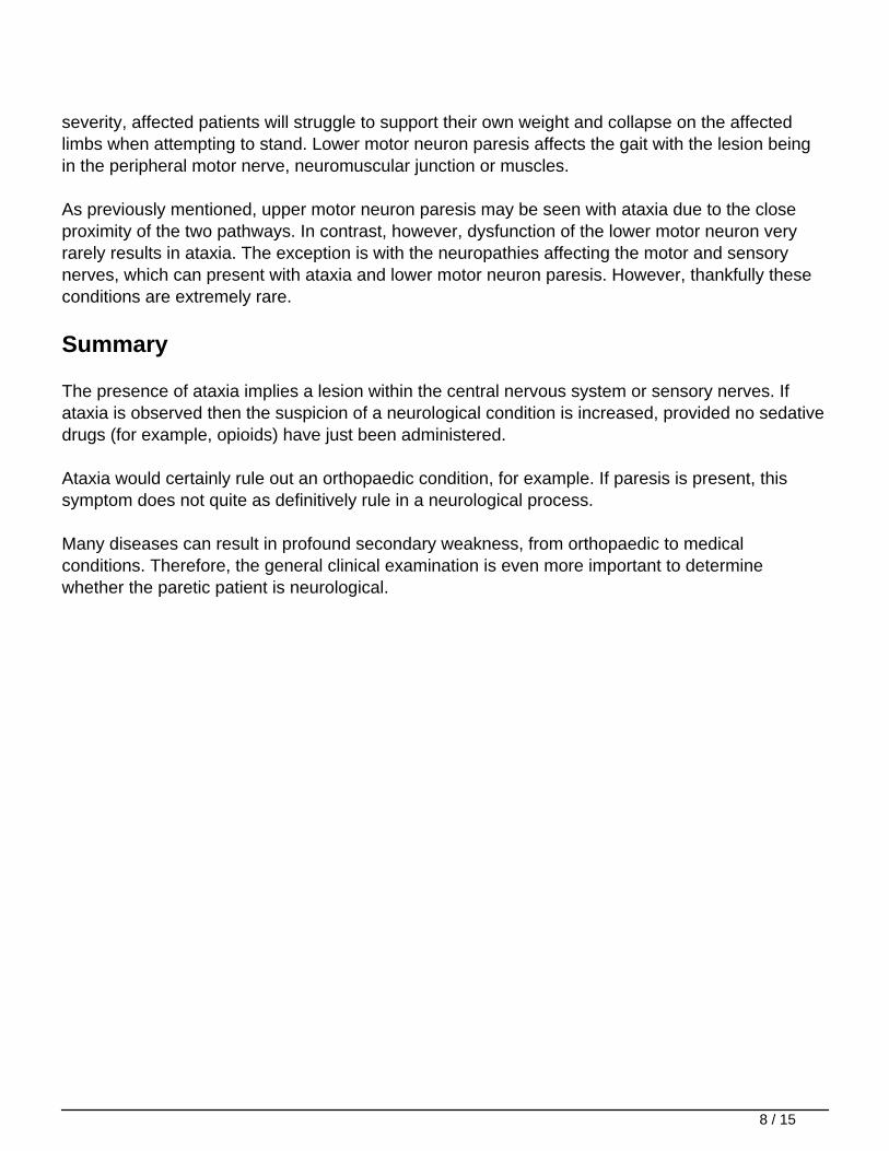
Recognition of neurological versus non-neurological disease is essential for appropriate diagnosticplanning. Inappropriate diagnostics, such as cerebrospinal fluid collection – which require generalanaesthesia and entail potential risk – should be avoided when a neurological localisation has notbeen achieved. Similarly, a neurological localisation is important if expensive procedures such asMRI are to be correctly performed. For example, a dog with referred neck and back pain due to abrain tumour may have normal spinal imaging, but an abnormal brain on MRI (Figure 1). Correctlocalisation is essential.
Pain manifestations
Apparent pain is often a difficult clinical sign to localise. Abdominal, pelvic, soft tissue andorthopaedic (particularly joint) pain may often manifest with signs such as cervical rigidity, anarched back (kyphosis) and abnormal gait – signs frequently and mistakenly assumed to beassociated with neurological disease. Examples of this include bilateral cruciate disease, aorticthromboembolism and polyarthritis, so it highlights why a general clinical examination is soimportant before a full neurological examination is embarked on.
Potential pitfalls
When determining whether a patient is neurological, we should usually look for some sort ofalteration in movement. It is important to remember an animal unwilling to move may look similar toan animal unable to move. Therefore, when a patient is presented with an inability to move, someform of encouragement for it to walk should be given.
Performing a neurological examination on a patient with non-neurological disease can cause veryconfusing results. A dog with severe orthopaedic pain will be reluctant to replace its paw whentesting conscious proprioception. A very weak patient with a severe medical condition would alsobe unable to stand and walk, and may also give the illusion of having delayed paw positioningresponses. Therefore, it is imperative a complete general clinical assessment is performed in apatient before consideration is given to a neurological examination, so as to prevent misleadingresults.
Non-neurological conditions mimicking neurological disease
3 / 15
A classic example of neurological disease being mimicked by non-neurological conditions is dogsand cats with aortic thromboembolism.
These patients will present with some degree of paresis or paralysis to one or both back legs.
If the problem is acute and severe, a femoral pulse deficit is usually obvious. However, some ofthese patients, particularly dogs, may present with an insidious and chronic onset of a pelvic limbgait abnormality or exercise intolerance. A femoral pulse deficit is expected, although this may onlybe reduced as opposed to absent. If this is not detected on general clinical examination, theneurological examination can be misleading and difficult to interpret – leading to an erroneouslocalisation.
Observe the gait
Not all dogs that are “off their legs” have neurological disease. Gait evaluation is one of the bestways to determine whether a problem is neurological, and the presence of ataxia (incoordination)would usually imply a neurological condition is present. In a clinic it is very common for animals thatare “off their legs” to be presented in the back of a car or nestled tightly in an owner’s arms, asthe problem has occurred acutely and owners are reluctant to move pets and simply want to offerthem comfort. As a consequence, this commonly leads to the pitfall of examining the patientwithout first observing the gait.
If a patient is carefully supported and encouraged to walk, it is often surprising how well it canambulate. A sling under the back legs and a harness around the chest will provide an acceptablemeans to evaluate which limbs are affected and whether movement is present in these affectedlimbs.
Basic neuroanatomy
Unfortunately, neuroanatomy plays a small role in understanding whether a patient is neurological.Spinal cord tracts can be divided into afferent (ascending or sensory) and efferent (descending ormotor) pathways.
Sensory (ascending/ afferent) function
Lesions affecting sensory function (that is, lesions affecting the peripheral sensory nerve, theascending proprioceptive pathways in the dorsal spinal cord and the sensory areas of the brain) willresult in ataxia and can be assessed by:
• observation of gait to allow detection of ataxia; and
• paw position response – testing conscious proprioception.
4 / 15
Motor (descending/ efferent) function
Lesions affecting motor function (that is, lesions affecting the motor centres of the brain, thedescending motor pathways in the ventral spinal cord, and the peripheral motor nerve andmuscles) will result in weakness, paresis or paralysis (depending on the severity) and can beassessed by:
• Observation of gait to allow detection of weakness and determine if movement is present in theaffected limbs.
• Hopping – my preferred method of assessing motor function, as it can be done in any animalregardless of size. It allows each individual limb to be assessed for weakness and allows for aneasy comparison between limbs (Figure 2).
Physiology of gait generation
The nervous system obtains sensory information from receptors, allowing identification of changesin an environment (processing), resulting in transmission of information to effectors to make anappropriate response. Therefore, in terms of proprioception, stretch receptors in the muscles, jointsand tendons convey sensory information regarding limb position in peripheral sensory nerves to thespinal cord, where it ascends (in the spinocerebellar, spinothalamic and dorsal tracts) to the brain.
The vestibular system provides similar information, allowing balance and posture to be maintainedregarding the head and trunk, which is then organised in the cerebellum to moderate activity in thedescending motor neurons and correct changes in body orientation and posture. Information alsoreaches the forebrain for integration into the consciousness.
A motor response is generated by connections from these processing centres to motor nuclei in thebrainstem. Descending motor tracts then deliver this coordinated information via the spinal cord tothe effector organs (that is, skeletal muscles) via lower motor neurons. Two types of gaitabnormality are possible with spinal cord disease that may occur alone or in combination – ataxiaand paresis.
Ataxia
Ataxia is a vague and non-specific term for an abnormality in the gait that originates from theancient Greek words “a” meaning “without” and “taxis” meaning, “order”. Ataxia is due to adefect in proprioception, that is, the ascending pathways collecting and processing informationregarding movement perception and spatial orientation. In simple anatomical terms, lesions of thesensory nerve, ascending pathways of the spinal cord and processing centres within the braincause ataxia. It is not due to defects in descending motor tracts (paresis). Disturbances in theseascending pathways cause the nervous system to fail to initiate conscious and reflex motor activity,
5 / 15

resulting in an ataxic gait (that is, one where the positional placement of the paws is inappropriatewith each stride, resulting in a different placement of the limb; Figure 3).
Ataxia can be divided into three types – cerebellar, proprioceptive and vestibular. The three formsof ataxia arise from lesions at different points in the ascending pathway.
• Vestibular ataxia describes defects in the vestibular system and associated tracts. Clinical signsmost commonly manifest as a head tilt with abnormalities in eye position (for example, strabismusand nystagmus) and difficulty in maintaining balance (falling and leaning to either side).
• Cerebellar ataxia is characterised by an inability to regulate the rate, range and force of amovement. No direct pathways run from the cerebellum to the spinal cord, meaning cerebellarlesions do not result in paresis. Instead, the loss of inhibitory control of the descending motor tractsfrom cerebellar disease results in uncontrolled and exaggerated spastic movements, for example,dysmetria and hypermetria.
Postural reaction testing (for example, hopping and placing) may yield a mildly delayed, followed byan exaggerated, response. Further evaluation of a dog with cerebellar ataxia may also reveal adecreased menace response and an intention tremor of the head and body.
• Proprioceptive ataxia is caused by lesions in the ascending sensory pathway alone and is not dueto disturbances in the descending motor tracts; hence true “proprioceptive” ataxia is notaccompanied by spasticity, paresis or involuntary movements. However, due to the anatomicalproximity of the descending motor tracts to the ascending sensory tracts in the spinal cord, it is rareto have a spinal cord lesion resulting in ataxia without some degree of paresis. Therefore, the termparaparesis is preferred terminology in the context of pelvic limb ataxia.
Proprioceptive fibres are larger in diameter and are situated in the periphery of the spinal cord,while the motor fibres are smaller and located deeper in the cord.
As a result, proprioceptive ataxia is often the first neurological sign to be observed in patientssuffering spinal cord compression followed by motor deficits, that is, paresis. In practice, thisdistinction is rarely evident.
Paresis
Paresis is defined as weakness or an inability to generate a gait. Patients with paresis appearweak, with no power to the affected limb(s), although the placement of the paws is in the correctposition with no incoordination (Figure 4).
The term paresis implies some voluntary movement is present as compared to paralysis (orplegia), in which complete loss of voluntary movement is observed. Depending on which limbs are
6 / 15
involved, paresis/paralysis can be subdivided into four groups.
• Tetraparesis/plegia is paresis or paralysis of all four limbs, and results from a lesion locatedcranial to the T2 spinal cord segment or from a generalised lower motor neuron disorder.
• Paraparesis/plegia is paresis/paralysis of the pelvic limbs caused by a lesion caudal to T2.
• Monoparesis/plegia refers to paresis/lysis of one limb caused by a lesion of the lower motorneuron innervating the affected limb.
• Hemiparesis/plegia is paresis/lysis of the limbs on one side of the body due to a lesion locatedcranial to T2 is known as hemiparesis or plegia. It is ipsilateral to a lesion located between T2 andthe mid-brain, but contralateral to a lesion located in the rostral mid-brain or cerebrum.
Paresis can affect the upper or lower motor neuron. This results in two types of paresis – uppermotor neuron and lower motor neuron paresis.
The upper motor neuron system is confined to the central nervous system (within the brain andspinal cord) and is responsible for the initiation and maintenance of movement and themaintenance of tone in extensor muscles.
Upper motor neuron paresis results from a lesion in these descending motor pathways in the brainor spinal cord, causing a spastic paresis (Table 1) with a delay in the onset of protraction (swingphase of the gait), with the resultant stride being longer than normal and with a stiff quality ofmovement.
Lesions of the upper motor neuron typically result in a release of inhibition on lower motor neuronslocated caudal to the level of the injury. This effect is most profound on the lower motor neurons tothe extensor muscles, resulting in a spastic paresis or paralysis. Lesions at many different levels ofthe central nervous system will produce the same upper motor neuron clinical signs and may alsoresult in some proprioceptive ataxia in view of the close relationship between the ascendingproprioceptive and descending motor pathways in the spinal cord and brainstem.
The lower motor neuron system connects the central nervous system with the muscle to beinnervated.
It consists of a motor neuron with the cell body located in the grey matter of the spinal cord or in thecranial nerve nucleus of the brainstem. The axons leave the central nervous system coursing to theeffector muscle. The lower motor neuron is the final part of the pathway involved in producingmuscular contraction, supporting weight and generating gait.
Lesions of the lower motor neuron produce a flaccid paresis/paralysis (Table 1). Depending on the
7 / 15
severity, affected patients will struggle to support their own weight and collapse on the affectedlimbs when attempting to stand. Lower motor neuron paresis affects the gait with the lesion beingin the peripheral motor nerve, neuromuscular junction or muscles.
As previously mentioned, upper motor neuron paresis may be seen with ataxia due to the closeproximity of the two pathways. In contrast, however, dysfunction of the lower motor neuron veryrarely results in ataxia. The exception is with the neuropathies affecting the motor and sensorynerves, which can present with ataxia and lower motor neuron paresis. However, thankfully theseconditions are extremely rare.
Summary
The presence of ataxia implies a lesion within the central nervous system or sensory nerves. Ifataxia is observed then the suspicion of a neurological condition is increased, provided no sedativedrugs (for example, opioids) have just been administered.
Ataxia would certainly rule out an orthopaedic condition, for example. If paresis is present, thissymptom does not quite as definitively rule in a neurological process.
Many diseases can result in profound secondary weakness, from orthopaedic to medicalconditions. Therefore, the general clinical examination is even more important to determinewhether the paretic patient is neurological.
8 / 15
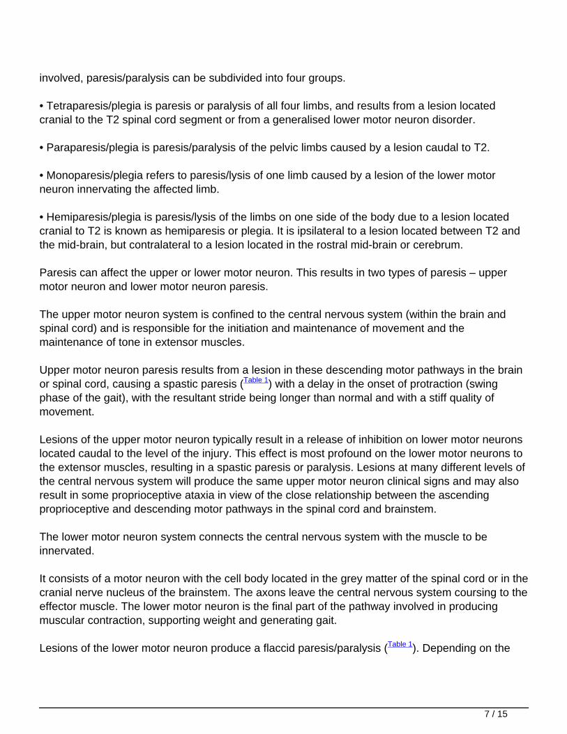
Figure 1a. A six-year-old beagle presented with the primary complaint of yelping and severe neckpain. On initial presentation, the dog had a low head carriage and obvious signs of neck pain basedon the arched back and low head carriage. However, subtle neurological deficits were present on afull examination that suggested a forebrain lesion was present.
9 / 15
Figure 1b. A sagittal (A) and transverse (B) T2-weighted MRI scan of the dog in Figure 1a. Thedotted line in A shows the level at which the transverse image B is taken. This confirms thepresence of a large forebrain mass compatible with a forebrain neoplasm.
10 / 15
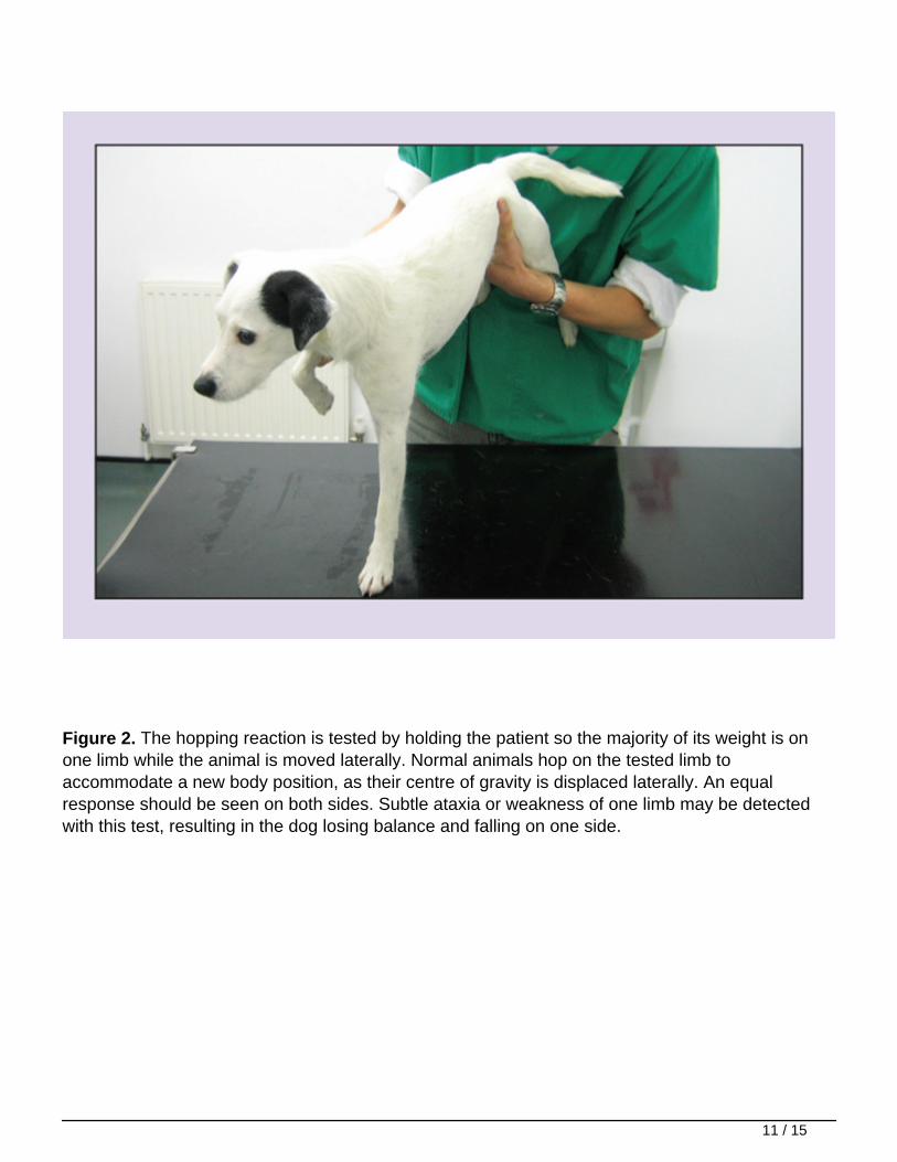
Figure 2. The hopping reaction is tested by holding the patient so the majority of its weight is onone limb while the animal is moved laterally. Normal animals hop on the tested limb toaccommodate a new body position, as their centre of gravity is displaced laterally. An equalresponse should be seen on both sides. Subtle ataxia or weakness of one limb may be detectedwith this test, resulting in the dog losing balance and falling on one side.
11 / 15

Figure 3. Screenshots (A-D) of a four-year-old Jack Russell terrier with an acute onset ofambulatory proprioceptive ataxia in the pelvic limbs (paraparesis). Notice the crossing of the backlegs and severe swaying of the trunk. The presence of weakness in the left pelvic limb (E)manifests by a delayed paw positioning response. This patient had a T3 to L3 localisation and wasdiagnosed with an acute disc herniation (Hansen Type I).
12 / 15

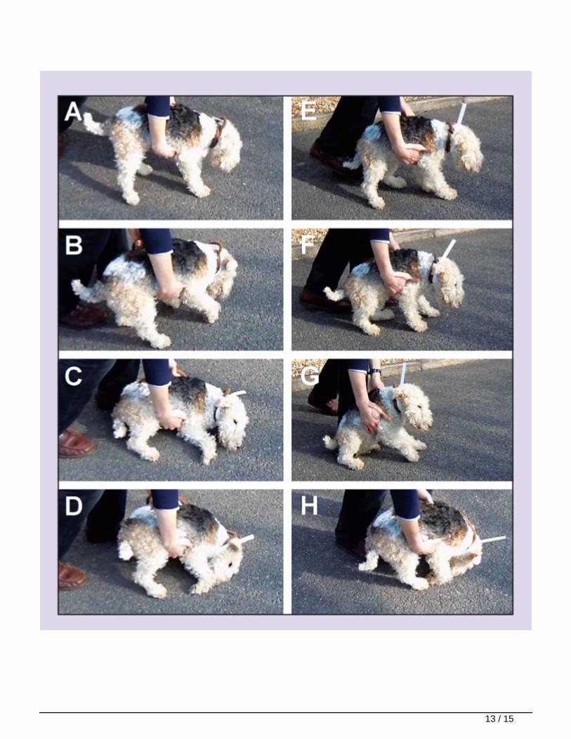
Figure 4. Screenshots (A-H) of a five-year-old fox terrier examined because of an acutelyprogressive history of non-ambulatory paresis affecting all four legs (tetraparesis). Notice theprofound weakness to all four limbs and the requirement for support. However, placement of theselimbs is appropriate with no incoordination. The patient had a neuromuscular localisation and wasdiagnosed with idiopathic polyradiculoneuritis, making a complete recovery with nursing care andrehabilitation alone.
Table 1. Clinical signs that may be seen with upper and lower motor neuron signs
14 / 15


![MAURITIUS EXAMINATIONS SYNDICATE HISTORY & …mes.intnet.mu/English/Documents/Examinations/...(b) Give one condition that can give rise to the formation of a tropical cyclone. [1]](https://static.fdocuments.net/doc/165x107/5edfc5a3ad6a402d666b1588/mauritius-examinations-syndicate-history-mes-b-give-one-condition-that.jpg)


