Neurohormonal regulation of myocardial cell apoptosis during the development of heart failure
8
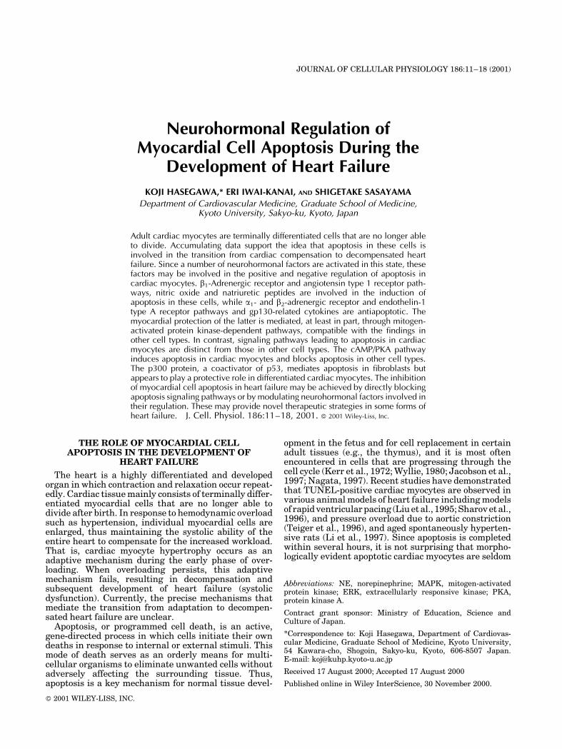
JOURNAL OF CELLULAR PHYSIOLOGY 186:11–18 (2001) Neurohormonal Regulation of Myocardial Cell Apoptosis During the Development of Heart Failure KOJI HASEGAWA,* ERI IWAI-KANAI, AND SHIGETAKE SASAYAMA Department of Cardiovascular Medicine, Graduate School of Medicine, Kyoto University, Sakyo-ku, Kyoto, Japan Adult cardiac myocytes are terminally differentiated cells that are no longer able to divide. Accumulating data support the idea that apoptosis in these cells is involved in the transition from cardiac compensation to decompensated heart failure. Since a number of neurohormonal factors are activated in this state, these factors may be involved in the positive and negative regulation of apoptosis in cardiac myocytes. b 1 -Adrenergic receptor and angiotensin type 1 receptor path- ways, nitric oxide and natriuretic peptides are involved in the induction of apoptosis in these cells, while a 1 - and b 2 -adrenergic receptor and endothelin-1 type A receptor pathways and gp130-related cytokines are antiapoptotic. The myocardial protection of the latter is mediated, at least in part, through mitogen- activated protein kinase-dependent pathways, compatible with the findings in other cell types. In contrast, signaling pathways leading to apoptosis in cardiac myocytes are distinct from those in other cell types. The cAMP/PKA pathway induces apoptosis in cardiac myocytes and blocks apoptosis in other cell types. The p300 protein, a coactivator of p53, mediates apoptosis in fibroblasts but appears to play a protective role in differentiated cardiac myocytes. The inhibition of myocardial cell apoptosis in heart failure may be achieved by directly blocking apoptosis signaling pathways or by modulating neurohormonal factors involved in their regulation. These may provide novel therapeutic strategies in some forms of heart failure. J. Cell. Physiol. 186:11–18, 2001. ß 2001 Wiley-Liss, Inc. THE ROLE OF MYOCARDIAL CELL APOPTOSIS IN THE DEVELOPMENT OF HEART FAILURE The heart is a highly differentiated and developed organ in which contraction and relaxation occur repeat- edly. Cardiac tissue mainly consists of terminally differ- entiated myocardial cells that are no longer able to divide after birth. In response to hemodynamic overload such as hypertension, individual myocardial cells are enlarged, thus maintaining the systolic ability of the entire heart to compensate for the increased workload. That is, cardiac myocyte hypertrophy occurs as an adaptive mechanism during the early phase of over- loading. When overloading persists, this adaptive mechanism fails, resulting in decompensation and subsequent development of heart failure (systolic dysfunction). Currently, the precise mechanisms that mediate the transition from adaptation to decompen- sated heart failure are unclear. Apoptosis, or programmed cell death, is an active, gene-directed process in which cells initiate their own deaths in response to internal or external stimuli. This mode of death serves as an orderly means for multi- cellular organisms to eliminate unwanted cells without adversely affecting the surrounding tissue. Thus, apoptosis is a key mechanism for normal tissue devel- opment in the fetus and for cell replacement in certain adult tissues (e.g., the thymus), and it is most often encountered in cells that are progressing through the cell cycle (Kerr et al., 1972; Wyllie, 1980; Jacobson et al., 1997; Nagata, 1997). Recent studies have demonstrated that TUNEL-positive cardiac myocytes are observed in various animal models of heart failure including models of rapid ventricular pacing (Liu et al., 1995; Sharov et al., 1996), and pressure overload due to aortic constriction (Teiger et al., 1996), and aged spontaneously hyperten- sive rats (Li et al., 1997). Since apoptosis is completed within several hours, it is not surprising that morpho- logically evident apoptotic cardiac myocytes are seldom Abbreviations: NE, norepinephrine; MAPK, mitogen-activated protein kinase; ERK, extracellularly responsive kinase; PKA, protein kinase A. Contract grant sponsor: Ministry of Education, Science and Culture of Japan. *Correspondence to: Koji Hasegawa, Department of Cardiovas- cular Medicine, Graduate School of Medicine, Kyoto University, 54 Kawara-cho, Shogoin, Sakyo-ku, Kyoto, 606-8507 Japan. E-mail: [email protected] Received 17 August 2000; Accepted 17 August 2000 Published online in Wiley InterScience, 30 November 2000. ß 2001 WILEY-LISS, INC.
-
Upload
koji-hasegawa -
Category
Documents
-
view
212 -
download
0
Transcript of Neurohormonal regulation of myocardial cell apoptosis during the development of heart failure
JOURNAL OF CELLULAR PHYSIOLOGY 186:11±18 (2001)
Neurohormonal Regulation ofMyocardial Cell Apoptosis During the
Development of Heart Failure
KOJI HASEGAWA,* ERI IWAI-KANAI, AND SHIGETAKE SASAYAMA
Department of Cardiovascular Medicine, Graduate School of Medicine,Kyoto University, Sakyo-ku, Kyoto, Japan
Adult cardiac myocytes are terminally differentiated cells that are no longer ableto divide. Accumulating data support the idea that apoptosis in these cells isinvolved in the transition from cardiac compensation to decompensated heartfailure. Since a number of neurohormonal factors are activated in this state, thesefactors may be involved in the positive and negative regulation of apoptosis incardiac myocytes. b1-Adrenergic receptor and angiotensin type 1 receptor path-ways, nitric oxide and natriuretic peptides are involved in the induction ofapoptosis in these cells, while a1- and b2-adrenergic receptor and endothelin-1type A receptor pathways and gp130-related cytokines are antiapoptotic. Themyocardial protection of the latter is mediated, at least in part, through mitogen-activated protein kinase-dependent pathways, compatible with the ®ndings inother cell types. In contrast, signaling pathways leading to apoptosis in cardiacmyocytes are distinct from those in other cell types. The cAMP/PKA pathwayinduces apoptosis in cardiac myocytes and blocks apoptosis in other cell types.The p300 protein, a coactivator of p53, mediates apoptosis in ®broblasts butappears to play a protective role in differentiated cardiac myocytes. The inhibitionof myocardial cell apoptosis in heart failure may be achieved by directly blockingapoptosis signaling pathways or by modulating neurohormonal factors involved intheir regulation. These may provide novel therapeutic strategies in some forms ofheart failure. J. Cell. Physiol. 186:11±18, 2001. ß 2001 Wiley-Liss, Inc.
THE ROLE OF MYOCARDIAL CELLAPOPTOSIS IN THE DEVELOPMENT OF
HEART FAILURE
The heart is a highly differentiated and developedorgan in which contraction and relaxation occur repeat-edly. Cardiac tissue mainly consists of terminally differ-entiated myocardial cells that are no longer able todivide after birth. In response to hemodynamic overloadsuch as hypertension, individual myocardial cells areenlarged, thus maintaining the systolic ability of theentire heart to compensate for the increased workload.That is, cardiac myocyte hypertrophy occurs as anadaptive mechanism during the early phase of over-loading. When overloading persists, this adaptivemechanism fails, resulting in decompensation andsubsequent development of heart failure (systolicdysfunction). Currently, the precise mechanisms thatmediate the transition from adaptation to decompen-sated heart failure are unclear.
Apoptosis, or programmed cell death, is an active,gene-directed process in which cells initiate their owndeaths in response to internal or external stimuli. Thismode of death serves as an orderly means for multi-cellular organisms to eliminate unwanted cells withoutadversely affecting the surrounding tissue. Thus,apoptosis is a key mechanism for normal tissue devel-
opment in the fetus and for cell replacement in certainadult tissues (e.g., the thymus), and it is most oftenencountered in cells that are progressing through thecell cycle (Kerr et al., 1972; Wyllie, 1980; Jacobson et al.,1997; Nagata, 1997). Recent studies have demonstratedthat TUNEL-positive cardiac myocytes are observed invarious animal models of heart failure including modelsof rapid ventricular pacing (Liu et al., 1995; Sharov etal.,1996), and pressure overload due to aortic constriction(Teiger et al., 1996), and aged spontaneously hyperten-sive rats (Li et al., 1997). Since apoptosis is completedwithin several hours, it is not surprising that morpho-logically evident apoptotic cardiac myocytes are seldom
Abbreviations: NE, norepinephrine; MAPK, mitogen-activatedprotein kinase; ERK, extracellularly responsive kinase; PKA,protein kinase A.
Contract grant sponsor: Ministry of Education, Science andCulture of Japan.
*Correspondence to: Koji Hasegawa, Department of Cardiovas-cular Medicine, Graduate School of Medicine, Kyoto University,54 Kawara-cho, Shogoin, Sakyo-ku, Kyoto, 606-8507 Japan.E-mail: [email protected]
Received 17 August 2000; Accepted 17 August 2000
Published online in Wiley InterScience, 30 November 2000.
ß 2001 WILEY-LISS, INC.

seen. However, key signal transducers of apoptosis areactivated in failing adult cardiomyocytes (Misao et al.,1996; Narula et al., 1999); these include upregulation ofBax, release of cytochrome C from mitochondria intocytoplasm, upregulation of caspase 3, and cleavage ofcaspase 3 to the active form. Apoptosis is primarily adefense mechanism against excessive cell proliferation.However, if apoptosis occurs in myocardial cells lackingproliferative and regenerative abilities, the number ofmyocardial cells is decreased, thus the stress on theremaining cells is increased, eventually leading to heartfailure. These ®ndings suggest that myocardial cellapoptosis is involved in the transition from compensatedcardiac hypertrophy to heart failure. In addition, it hasbeen reported that in mice lacking membrane proteingp130, considerable myocardial cell apoptosis is inducedby hypertension, which results in the rapid progressionof cardiac dilatation and heart failure (Hirota et al.,1999). This ®nding provides evidence that apoptosissignaling pathways are activated in response to hemo-dynamic overload and that myocardial cell apoptosis issuf®cient to induce decompensated heart failure inadult mice in vivo.
Since a number of neurohormonal factors are acti-vated in heart failure, these factors may positively andnegatively regulate myocardial cell apoptosis. Activa-tion of the sympathetic nervous system, renin-angio-tensin system, and endothelin-1 are closely related toincreased morbidity in heart failure (Packer, 1992;Francis et al., 1993). These factors all participate inthe regulation of myocardial cell apoptosis as well as theincreases in both preload and afterload. On the otherhand, nitric oxide and natriuretic peptides exhibitvasodilative and diuretic effects and have been shownto induce myocardial cell apoptosis. Myocardial cellapoptosis is also regulated by cytokines that act viamembrane protein gp130 such as cardiotrophin-1 (Fujioet al., 1997; Sheng et al., 1997), as well as in¯ammatorycytokines such as tumor necrosis factor-a and inter-leukin-1b. In addition, pathways that activate apoptosisin cardiac myocytes are distinct from those in other celltypes. Norepinephrine (NE) is a representative factor,the elevation of which in plasma closely correlates withthe severity and poor prognosis of heart failure. We andothers have shown that NE induces myocardial cellapoptosis via a b-adrenergic receptor-dependent path-way (Communal et al., 1998; Iwai-Kanai et al., 1999).This might provide further insight into the mechanismsthat explain the preferential effects of b-blockade onchronic congestive heart failure. This article focuses onthe regulation of myocardial cell apoptosis by adrenergicreceptor pathways, angiotensin II, endothelin-1, andgp130-related cytokines. We will also refer to thedifferential regulation of apoptosis by a p300 protein incardiac myocytes and other cell types.
DIFFERENTIAL REGULATION OF MYOCARDIALCELL APOPTOSIS BY a- AND b-ADRENERGIC
RECEPTOR PATHWAYS
The intracellular pathways for adrenergic signalingin cardiac myocytes begin with binding of NE toG-protein-coupled adrenergic receptors on cell mem-branes. At least nine subtypes of adrenergic receptorshave been identi®ed to date. Of these, the receptors
mainly expressed in cardiac myocytes are b1, b2, and a1.Both b1- and b2-adrenergic receptors are coupled with aGas protein that stimulates adenylyl cyclase activity,leading to the production of cAMP, followed by activa-tion of protein kinase A (PKA). The b2-adrenergic rece-ptor is also coupled with Gi, which inhibits adenylylcyclase activity. Thus, the b2-adrenergic receptor path-way negatively regulates the Gs-dependent pathway,which is activated by b1- and b2-adrenergic receptors.On the other hand, the a1-adrenergic receptor is coupledpredominantly with Gaq. Stimulation of Gaq results inan increase in phospholipase C that activates bothprotein kinase C and inositol 1,4,5-triphosphate kinase.These are followed by activation of the mitogen-acti-vated protein kinase (MAPK) cascade.
NE, an activator ofa- andb-adrenergic receptors, is aninducer of apoptosis in cardiac myocytes. Using a systemof cultured rat cardiac myocytes, we and others haveshown that NE-induced apoptosis is mediated throughthe b1-adrenergic receptor pathway. The cAMP/PKApathway is necessary and suf®cient to mediate b1-adre-nergic agonist-induced myocardial cell apoptosis. Inter-estingly, the cAMP/PKA pathway is anti-apoptotic inother cell types such as PC 12 cells, indicating that thispathway acts as an apoptotic messenger in a cell-typespeci®c manner (Iwai-Kanai et al., 1999). The myocar-dial cell apoptosis induced by b1-adrenergic agonists hasalso been shown in adult rat cardiac myocytes in vivo(Shizukuda et al., 1998). Two kinds of transgenic miceprovided further evidence that the induction of myocar-dial cell apoptosis by b1-adrenergic stimulation resultsin the development of heart failure. One is transgenicmice that overexpress b1-adrenergic receptors in theheart, which results in left ventricular dilation and con-tractile dysfunction with age (Liggett et al., 2000). Theother is mice overexpressing Gsa in myocardium (Genget al., 1999). These mice exhibit myocardial cell apo-ptosis and the development of cardiomyopathy. All ofthese studies indicated thatb1-adrenergic stimulation issuf®cient to induce myocardial cell apoptosis, whichresults in the development of heart failure. In contrast,transgenic mice that overexpress b2-adrenergic recep-tors in the heart do not develop heart failure. Singhand co-workers demonstrated that activation of theb2-adrenergic receptor pathway is involved in theinhibition of myocardial cell apoptosis through Gi-protein (Communal et al., 1999).
Thea1-adrenergic receptor is coupled with Gaq, whichleads to the activation of MAPK cascade. This pathwayplays an important role in the stimulation of a hyper-trophic response. In contrast to the induction of myo-cardial cell apoptosis by the b1-adrenergic receptorpathway, we found that stimulation of a1-adrenergicreceptors antagonized b-adrenergic agonist-inducedapoptosis (Iwai-Kanai et al., 1999). The protective ef-fects of a1-adrenergic agonists on cardiac myocytesagainst apoptosis are mediated, at least in part, throughthe activation of extracellular signal-regulated kinases(ERKs). Similarly, the activation of Gaq through otherreceptors such as endothelin-1 type A receptor inhibitsapoptosis in addition to inducing hypertrophy (Arakiet al., 2000). Supporting the role of Gaq in myocardialcell growth, transgenic mice with four- to ®ve-foldincreases in Gq protein levels in the myocardium exhibit
12 HASEGAWA ET AL.

cardiac hypertrophy associated with a normal lifespan(Adams et al., 1998). Interestingly, those with aneightfold-increase in myocardial Gq levels died fromheart failure at an average age of 11 weeks with amarked induction of myocardial cell apoptosis. Thisobservation led to the notion that excessive activationof Gaq-dependent pathways in cardiac myocytesresults in apoptosis, while adequate activation of thispathway mediates both hypertrophic and anti-apoptoticresponses.
ENDOTHELIN-1 IS A SURVIVAL FACTORAGAINST MYOCARDIAL CELL APOPTOSIS
Endothelin-1 is a 21-residue peptide originally islo-lated from supernatants of vascular endothelial cells(Yanagisawa et al., 1988). While endothelin-1 is mainlyproduced by endothelial cells in the basal state, anumber of cell types can synthesize endothelin-1 inresponse to various stimuli (Shichiri et al., 1991; Itoet al., 1993; Kaddoura et al., 1996; Yamazaki et al.,1996). Endothelin-1 expression in cardiac myocytes isinduced by myocardial stretch, angiotensin II andnorepinephrine (Ito et al., 1993; Kaddoura et al., 1996;Yamazaki et al., 1996). The levels of endothelin-1 inplasma and in ventricular myocardium are markedlyincreased in human and animal models of heart failure(Wei et al., 1994; Sakai et al., 1996; Iwanaga et al., 1998).Endothelin-1 exerts diverse physiological effects in-cluding vasoconstriction and growth-promotion. Endo-thelin-1 is suf®cient to induce the myocardial cellhypertrophy associated with reactivation of the fetalgene program (Shubeita et al., 1990; Ito et al., 1991).These various effects are mediated by two distinctsubtypes of G-protein-coupled heptahelical receptors,endothelin-1 type A and type B receptors, expressed in awide variety of tissues (Arai et al., 1990; Sakurai et al.,1990). Endothelin-1 is a survival factor in activelyproliferating cells such as smooth muscle cells(Wu-Wong et al., 1997) and ®broblasts (Shichiri et al.,1998). This survival effect is also seen in post-mitoticcardiac myocytes (Araki et al., 2000). This effect ismediated mainly through the endothelin-1 type Areceptor-dependent pathway, compatible with cardiacmyocytes that predominantly expressed the type Areceptor. Type A receptor is coupled with both Gq and Gi.The antiapoptotic effects of endothelin-1 are mediated,in part, through activation of Gq and MAPK pathways.Because endothelin-1 accumulates within cardiacmyocytes in failing hearts as shown by immunohisto-chemistry (Sakai et al., 1996; Iwanaga et al., 1998),endogenous ET-1 may function in ``self-protection'' by anautocrine mechanism.
Despite the evidence that endothelin-1 is a protectivefactor against myocardial cell apoptosis, upregulatedexpression of ET-1 in cardiac myocytes apparently playsa role in the deterioration of systolic function duringthe development of heart failure in vivo. Endothelin-1receptor antagonists, bosentan or BQ123, preventremodeling of the heart and have been shown to improvesurvival following myocardial infarction and pressureoverload (Sakai et al., 1996; Iwanaga et al., 1998). These®ndings suggest that the cardiac endothelin-1 pathwayis involved in the development of heart failure indepen-dent of myocardial cell apoptosis. At present, the precise
mechanisms that mediate the induction of heart failureby this pathway are unclear. One possible mechanismmight be endothelin-1-mediated decrease in intracellu-lar cAMP levels by coupling with Gi protein (James et al.,1994). Therefore, chronic elevation of endothelin-1 incardiac myocytes could decrease the systolic function ofthe entire heart.
ANGIOTENSIN II-MEDIATED REGULATION OFAPOPTOSIS BY TYPE 1 AND TYPE 2 RECEPTORS
Angiotensin II is synthesized not only by the systemicrenin±angiotensin system but also by the local renin±angiotensin system in the heart. The effects of angio-tensin II are mediated through two types of receptors,namely type 1 and type 2 receptors (Horiuchi et al.,1997). Type 1 receptor is expressed in the heart, smoothmuscle, kidney, brain, adrenal cortex, and liver. Incontrast, type 2 receptor is expressed widely in varioustissues during the fetal stage and downregulated afterbirth. However, the expression of type 2 receptor isre-induced in vascular injury, myocardial infarction,myocardial hypertrophy, and wounded skin. While bothreceptors belong to the seven-transmembrane receptorsuperfamily, recent evidence has shown that thefunctions of type 1 and type 2 receptors are mutuallyantagonistic. Type 1 receptor is coupled with Gq andpromotes vasoconstriction and cell growth. On the otherhand, type 2 receptor mediates vasodilatation, growthinhibition, and cell differentiation, although preciseintracellular signaling pathways for type 2 receptor areunknown at present. Similarly, apoptosis is differen-tially regulated by these two types of receptors. In PC 12cells, in which angiotensin II type 2 receptor is abun-dantly expressed, growth factor-mediated inhibition ofapoptosis is antagonized by angiotensin II (Yamadaet al., 1996). This effect of apoptosis promotion byangiotensin II is blocked by a type 2 receptor antagonistbut not by a type 1 receptor antagonist. The type 2receptor-dependent pathway downregulates the activa-tion of MAPK by the growth factor resulting in theinactivation of bcl-2 (Horiuchi et al., 1997). In contrast,the type 1 receptor-dependent pathway activates MAPKand inhibits apoptosis in vascular smooth muscle cells(Yamada et al., 1998).
In the heart, angiotensin II is stored in cardiacmyocytes and released from these cells in response tomyocardial stretch (Sadoshima et al., 1993). This auto-crine release of angiotensin II is involved in the stretch-induced myocardial cell hypertrophy. In addition,angiotensin II stimulates proliferation of ®broblasts.These effects are mediated via the type 1 receptor. Bothlocal and systemic renin±angiotensin systems areactivated in heart failure in humans as well as variousanimal models of heart falilure (Wollert and Drexler,1999). Pharmacological blockade of the renin±angio-tensin system is bene®cial in patients with heart failure.Despite the strong evidence that angiotensin II type 1and type 2 receptors play different roles in the regula-tion of apoptosis, and that the type 1 receptor inhibitsapoptosis, the regulation of myocardial cell apoptosis byangiotensin II appears to be unique. In contrast to the®ndings in other cell types, the angiotensin II type 1receptor pathways are involved in the promotion ofapoptosis in cardiac myocytes. Stretch-mediated release
NEUROHORMONES IN MYOCARDIAL APOPTOSIS 13

of angiotensin II induces myocyte apoptosis, which isabolished by losartan, a type 1 receptor antagonist(Kajstura et al., 1997; Leri et al., 1998). Angiotensin IIinduces the expression of p53, whose binding to thepromoter of angiotensinogen, type 1 receptor, p53, andBax was also increased. Therefore, it appears that theactivation of p53 is responsible for stretch-mediated andangiotensin II-dependent apoptosis in cardiac myocytes.These ®ndings suggest that activation of the renin±angiotensin system in heart failure is involved inpromoting myocardial cell apoptosis during the devel-opment of heart failure. This hypothesis is supported byangiotensin-converting enzyme inhibitors reducingmyocardial cell apoptosis as well as blocking theremodeling of the heart in animal models of heartfailure (Li et al., 1997; Goussev et al., 1998). Thus,bene®cial effects of these agents in patients with heartfailure might be attributable, in part, to the inhibition ofmyocardial cell apoptosis.
PROTECTION OF CARDIAC MYOCYTES AGAINSTAPOPTOSIS BY gp130-RELATED CYTOKINES
Recent work has demonstrated that members of afamily of structurally related cytokines, including leu-kemia inhibitory factor and cardiotrophin-1 protectagainst apoptosis as well as induce hypertrophy incardiomyocyte culture (Pennica et al., 1995; Wollertet al., 1996). The receptors in this cytokine family aremultimeric and share the class-speci®c transmembranesignal transducing component gp130 (Hibi et al., 1990;Taga et al., 1992; Ip et al., 1992; Yin et al., 1993; Taga,1996). Signaling is triggered through the homodimer-ization of gp130 (Murakami et al., 1993) or the hetero-dimerization of gp130 with a related transmembranesignal transducer, the leukemia inhibitory factor recep-tor subunit b (Gearing et al., 1991; Davis et al., 1993).Overexpression of both interleukin-6 and its receptorresults in constitutive tyrosine phosphorylation ofgp130 (i.e., activation) in the myocardium and leftventricular hypertrophy in vivo (Hirota et al., 1995).Members of the interleukin-6 cytokine family, includingleukemia inhibitory factor have been shown to activatethe JAK/STAT pathway and phosphorylate STAT3(Akira et al., 1994; Kisimoto et al., 1994; Narazakiet al., 1994; Taga, 1996). It is also clear that this family ofcytokines can activate Ras and MAP kinase cascades(Kisimoto et al., 1994; Heim, 1996; Taga, 1996). Domi-nant negative forms of MEK-1 and a MEK-1-speci®cinhibitor PD098059 block the activation of ERK as wellas cardiotrophin-1-mediated protection against myocar-dial cell apoptosis (Sheng et al., 1997). These ®ndingsindicate that MEK-1/ERK pathways are required for theantiapoptotic effects of gp-130-related cytokines. Cardi-otrophin-1-mediated blockade of myocardial cell apop-tosis is associated with up-regulated expression of anantiapoptotic molecule bcl-xl. STAT1 binds to its speci®cbinding site within the bcl-xl promoter and activatesthis promoter in a sequence-speci®c manner (Fujio et al.,1997). Thus, JAK/STAT as well as MEK-1/ERK path-ways are involved in the protective effects of thesecytokines. Protective effects of gp130-dependent signalson cardiac myocytes are demonstrated in the adult heartin vivo. The mice with cardiac-speci®c gp130 de®ciencyexhibit progressive dilated cardiomyopathy with mas-
sive cardiac myocyte apoptosis when subjected topressure overload (Hirota et al., 1999). This observationclearly demonstrates that pressure overload activatesapoptotic signals in the heart and that gp130-dependentpathways are required for antagonizing these signals. Inaddition, this ®nding also provides evidence thatmyocardial cell apoptosis is suf®cient to induce heartfailure in vivo
ROLE OF p300 IN THE REGULATION OFAPOPTOSIS IN CARDIAC MYOCYTES
AND OTHER CELL TYPES
Cardiac myoctes are highly differentiated and lost theability to undergo mitosis soon after birth. Because ofthese unique characteristics, signaling pathways thatactivate apoptosis in these cells are distinct from thosein other cell types. An interesting feature is the opposingeffects of cAMP on apoptosis in two different cell types.In contrast to the ®ndings in primary cardiac myocytesin culture, the administration of 8-Br-cAMP, a cell-permeable cAMP analogue, completely inhibits serumdeprivation-induced apoptosis in PC12 pheochromo-cytoma cells (Iwai-Kanai et al., 1999). Thus, cAMP-mediated apoptosis in cardiac myocytes might involvesome cell type-speci®c signal transduction mechanisms.One of the candidate molecules involved in the regula-tion of apoptosis in terminally differentiated cells is theadenovirus E1A-associated cellular protein p300. Thep300 protein is classi®ed as a transcriptional coactivatorthat communicates between the enhancer DNA and thebasal transcriptional complex formed on the promoternear the transcription initiation site (Eckner et al.,1994). This communication is required for transcrip-tional activation by the transactivators. Consistent withthis view, the p300 family of transcriptional coactivatorshas been noted to modulate many examples of enhancer-mediated transcription (Chrivia et al., 1993; Arias et al.,1994; Eckner et al., 1994, 1996; Kwok et al., 1994; Aranyet al., 1995; Lee et al., 1995; Lundblad et al., 1995;Missero et al., 1995; Bannister and Kouzarides, 1996;Chakravarti et al., 1996; Dai et al., 1996; Janknecht andNordheim, 1996; Kamei et al., 1996; Oelgeschlager et al.,1996; Trouche and Kouzarides, 1996; Yuan et al., 1996;Puri et al., 1997; Sartorelli et al., 1997). The p300 proteininteracts directly with components of the basal tran-scriptional apparatus (e.g., TFIIB and TBP (Abrahamet al., 1993; Kwok et al., 1994; Yuan et al., 1996) anddiverse enhancer binding proteins. In skeletal muscle,p300/CBP are transcriptional adaptors for MyoD andMEF-2, and these interactions are required for myogen-esis (Eckner et al., 1996; Yuan et al., 1996; Puri et al.,1997; Sartorelli et al., 1997). The p300 protein isinvolved in cardiac-speci®c transcription as a coactiva-tor of zinc-®nger proteins GATA-4/5 (Kakita et al.,1999). In addition to their possible ``bridging function,''p300 proteins possess intrinsic histone acetyl-transfer-ase activity (Bannister and Kouzarides, 1996; Ogryzkoet al., 1996), which is hypothesized to promote a locally``open'' and transcriptionally active chromatin con®g-uration. The p300 protein is a homologue of CBP, aprotein that is associated with, and coactivates thetranscription factor CREB, mediating the induction ofcAMP-responsive promoters (Arany et al., 1995; Lund-blad et al., 1995).
14 HASEGAWA ET AL.

The role of p300 in the regulation of apoptosis appearsto be distinct in cardiac myocytes from other cell types.Fibroblasts prepared from p300 knock-out mice aremore resistant to radiation-induced apoptosis thanthose from wild-type mice (Yao et al., 1998; Yuan et al.,1999). The p300 protein forms a speci®c complex withp53. This suggests a mechanism by which p300 activatesthe expression of Bax and mediates apoptosis as well asmodulates the checkpoint function in the G1 phase of thecell cycle (Avantaggiati et al., 1997; Shikama et al.,1999). In contrast to the ®ndings in ®broblasts, p300appears to play a role in the survival of cardiac myocytes.This is suggested by the study on the effects of theadenovirus E1A oncoprotein on these cells. When E1A isexpressed in differentiated cardiac muscle cells, it re-presses muscle-speci®c gene expression (Kirshenbaumand Schneider, 1995; Liu and Kitsis, 1996; Hasegawaet al., 1997) and induces apoptosis. Studies with E1Amutants indicate that interference with the p300 path-
way is suf®cient to repress muscle-speci®c gene exp-ression and induce apoptosis in cardiac myocytes(Kirshenbaum et al., 1995; Liu et al., 1996; Hasegawaet al., 1997). These experiments suggest that p300proteins are involved both in maintaining cardiac-speci®c gene expression and in myocardial cell survival.Our preliminary ®ndings suggest that a forced expres-sion of p300 inhibits the myocardial cell apoptosisinduced by 8-Br-cAMP. The possible involvement ofthe p300/CBP family in cAMP-mediated apoptosis incardiac myocytes requires further investigation.
CAN BLOCKADE OF MYOCARDIAL CELLAPOPTOSIS PREVENT THE DEVELOPMENT
OF HEART FAILURE?
It is apparent that the occurrence of myocardial cellapoptosis leads to heart failure, or at least worsens thestate of heart failure. However, it is unclear at presentwhether the blockade of apoptosis could prevent the
Fig. 1. Regulation of myocardial cell apoptosis by neurohormonal factors activated in heart failure.
Fig. 2. Differential regulation of myocardial cell growth and apoptosis by a1- and b1-adrenergicpathways.
NEUROHORMONES IN MYOCARDIAL APOPTOSIS 15

development of heart failure. In transgenic mice over-expressing an activated form of calcineurin in the heart,myocyte loss following ischemia-reperfusion is less thanin wild-type mice (De-Windt et al., 2000). Calcineurinactivation plays a role in protection against myocardialcell apoptosis. Nevertheless, the transgenic mice heartswith activated calcineurin exhibit marked dilatationand heart failure. These ®ndings suggest that heartfailure can develop independent of myocardial cellapoptosis. In contrast, doxyrubicin is a potent inducerof myocardial cell apoptosis both in culture and in adultrats in vivo. Thus, myocardial cell apoptosis maysigni®cantly contribute to the development of doxy-rubicin-induced cardiomyopathy. In most cases of end-stage heart failure, the number of cardiac myocytes isdecreased compared with a normal heart. One example,in which a loss of cardiac myocytes is prominent, is thedilated phase of hypertrophic cardiomyopathy. In thiscondition, serum levels of cardiac troponin T arecontinuously high (Sato et al., 2000), suggesting thatcardiac myocyte loss occurs during the transition to thedilated phase. The dilated phase of hypertrophic cardio-myopathy is associated with massive ®brosis and amarked decrease in myocyte number. While the con-tribution of myocardial cell apoptosis varies in differenttypes of heart failure, it is possible that blockade ofapoptosis may prevent the development of some types ofheart failure.
Prevention of apoptosis in heart failure may beachieved by directly blocking the apoptotic signals orby modulating neurohormonal factors that regulateapoptosis. The b-adrenergic pathway but not the a1-adrenergic pathway induces cell type-speci®c apoptosisin cardiac myocytes (Iwai-Kanai et al., 1999). Severalbodies of evidence suggest that activation of thesympathetic nervous system exerts a direct deleteriouseffect on the heart that is independent of the hemody-namic actions of these endogenous mechanisms. Thera-peutic intervention with b-adrenergic receptor blockersfavorably alters the natural history of heart failure, andsuch bene®ts cannot be explained solely by the effect ofthese agents on cardiac contractility and ejection frac-tion. These ®ndings might indicate that apoptosis inhi-bition is one mechanism of the bene®cial effects ofb-adrenergic receptor blockers in patients with heartfailure. Further elucidation of precise signaling path-
ways leading to myocardial cell protection and apoptosismay contribute to the establishment of novel strategiesfor heart failure therapy in humans.
ACKNOWLEDGMENTS
This work was supported in part by grants to K.H.from the Ministry of Education, Science and Culture ofJapan.
LITERATURE CITED
Abraham SE, Lobo S, Yaciuk P, Wang HG, Moran E. 1993. p300, andp300-associated proteins, are components of TATA-binding protein(TBP) complexes. Oncogene 8:1639±1647.
Adams JW, Sakata Y, Davis MG, Sah VP, Wang Y, Liggett SB, ChienKR, Brown JH, Dorn GW II. 1998. Enhanced Gaq signaling: acommon pathway mediates cardiac hypertrophy and apoptotic heartfailure. Proc Natl Acad Sci USA 95:10140±10145.
Akira S, Nisio Y, Inoue M, Wang X, Wei S, Matsusaka T, Yoshida K,SudoT, Naruto M, Kishimoto T. 1994. Molecular cloning of APRF, anovel IFN-stimulated gene factor 3 p91-related transcription factorinvolved in the gp130-mediated signaling pathway. Cell 77:63±71.
Arai H, Hori S, Aramori I, Okubo H, Nakanisi S. 1990. Cloning andexpression of a cDNA encoding an endothelin receptor. Nature348:730±932.
Araki M, Hasegawa K, Iwai-Kanai E, Fujita M, Sawamura T, KakitaT, Wada H, Morimoto T, Sasayama S. 2000. Endothelin-1 as anprotective factor against b-adrenergic agonist-induced apoptosis incardiac myocytes. J Am Coll Cardiol (in press).
Arany Z, Newsome D, Oldread E, Livingston DM, Eckner R. 1995. Afamily of transcriptional adaptor proteins targeted by the E1Aoncoprotein. Nature 374:81±84.
Arias J, Alberts AS, Brindle P, Claret FX, Smeal T, Karin M,Feramisco J, Montiminy M. 1994. Activation of cAMP and mitogenresponsive genes relies on a common nuclear factor. Nature370:226±229.
Avantaggiati ML, Ogryzko V, Gardner K, Giordano A, Levine AS,Kelly K. 1997. Recruitment of p300/CBP in p53-dependent signalpathways. Cell 89:1175±1184.
Bannister AJ, Kouzarides T. 1995. CBP-induced stimulation of c-Fosactivity is abrogated by E1A. EMBO J 14:4758±4762.
Bannister AJ, Kouzarides T. 1996. The CBP co-activator is a histoneacetyltransferase. Nature 384:641±643.
Chakravarti D, LaMorte VJ, Nelson MC, Nakajima T, Schulman IG,Juguilon H, Montminy MR, Evans RM. 1996. Role of CBP/p300 innuclear receptor signalling. Nature 383:99±103.
Chrivia JC, Kwok RP, Lamb N, Hagiwara M, Montiminy MR,Goodman RH. 1993. Phosphorylated CREB binds speci®cally tothe nuclear protein CBP. Nature 365:855±859.
Communal C, Singh K, Pimentel DR, Colucci WS. 1998. Norepinephr-ine stimulates apoptosis in adult rat ventricular myocytes byactivation of the b-adrenergic pathway. Circulation 98:1329±1334.
Communal C, Singh K, Sawyer DB, Colucci WS. 1999. Opposingeffects of beta(1) and beta(2)-adrenergic receptors on cardiac myo-cyte apoptosis: role of a pertussis toxin-sensitive G protein. Cir-culation 100:2210±2212.
Fig. 3. Roles of p300 and cAMP/PKA pathways in cardiac myocytes and in other cell types.
16 HASEGAWA ET AL.

Dai P, Akimaru H, Tanaka Y, Hou DX, Yasukawa T, Kanei-Ishii C,Takahashi T, Ishi S. 1996. CBP as a transcriptional coactivator ofc-Myb. Genes Dev 10:528±540.
Davis S, Aldrich TH, Stahl N, Pan L, Taga T, Kisimoto T, Ip NY,Yancopoulos GD. 1993. LIFR beta and gp130 as heterodimerizingsignal transducers of the tripartite CNTF receptor. Science260:1805±1808.
De-Windt LJ, Lim HW, Taigen T, Wencker D, Condorelli G, DornGW II, Kitsis RN, Molkentin JD. 2000. Calcineurin-mediatedhypertrophy protects cardiomyocytes from apoptosis in vitro andin vivo: an apoptosis-independent model of dilated heart failure.Circ Res 86:255±263.
Eckner R, Ewen ME, Newsome D, Gerdes M, DeCarpio JA, LawrenceJB, Livingston DM. 1994. Molecular cloning and functional analysisof the adenovirus E1A-associated 300-kD protein (p300) reveals aprotein with proterties of a transcriptional adaptor. Genes Dev8:869±884.
Eckner R, Yao TP, Oldread E, Livingston DM. 1996. Interaction andfunctional collaboration of p300/CBP and bHLH proteins in muscleand B-cell differentiation. Genes Dev 10:2478±2490.
Francis GS, Chon JN, Johnson G, Rector TS, Goldman S, Simon A.1993. Plasma norepinephrine, plasma renin activity, and congestiveheart failure. Relations to survival and the effects of therapy inV-HeFT II. The V-HeFT VA Cooperative Studies Group. Circulation87(Suppl 6):VI40±VI48.
Fujio Y, Kunisada K, Hirota H, Yamauchi-Takihara K, Kishimoto T.1997. Signals through gp130 up-regulates bcl-x gene expression viaSTAT1-binding cis-element in cardiac myocytes. J Clin Invest99:2898±2905.
Gearing DP, Thut CJ, VandenBos T, Gimpel SD, Delaney PB, King J,Price V, Cosman D, Beckmann MP. 1991. Leukemia inhibitoryfactor receptor is structurally related to the IL-6 signal transducer,gp130. EMBO J 10:2839±2848.
Geng YJ, Ishikawa Y, Vatner DE, Wanger TE, Bishop SP, Vatner SF,Homcy CJ. 1999. Apoptosis of cardiac myocytes in Gs a transgenicmice. Circ Res 84:34±42.
Goussev A, Sharov VG, Shimoyama H, Tanimura M, Lesch M,Goldstein S, Sabbah HN. 1998. Effects of ACE inhibition oncardiomyocyte apoptosis in dogs with heart failure. Am J Physiol275:H626±H631.
Hasegawa K, Meyers MB, Kitsis RN. 1997. Transcriptional coacti-vator p300 stimulates cell type-speci®c gene expression in cardiacmyocytes. J Biol Chem 272:20049±20054.
Heim MH. 1996. The Lak-STAT pathway: speci®c signal transductionfrom the cell membrane to the nucleus. Eur J Clin Invest 26:1±12.
Hibi M, Murakami M, Saito M, Hirno T, Taga T, Kishimoto T. 1990.Molecular cloning and expression of an IL-6 signal transducer,gp130. Cell 63:1149±1157.
Hirota H, Chen J, Betz UA, Rajewsky K, Gu Y, Ross J Jr, Muller W,Chien KR. 1999. Loss of a gp130 cardiac muscle cell survivalpathway is a critical event in the onset of heart failure duringbiomechanical stress. Cell 97:189±198.
Hirota H, Yosida K, Kisimoto T, Taga T. 1995. Continuous activationof gp130, a signal-transducing receptor component for interleukin6-related cytokines, causes myocardial hypertrophy in mice. ProcNatl Acad Sci USA 92:4862±4866.
Horiuchi M, Hayashida W, Kambe T, Yamada T, Dzau VJ. 1997.Angiotensin type 2 receptor dephosphorylates Bcl-2 by activatingmitogen-activated protein kinase phosphatase-1 and induces apop-tosis. J Biol Chem 272:19022±19026.
Ip NY, Nye SH, Boulton TG, Davis S, Taga T, Li Y, Birren SJ,Yasukawa K, Kishimoto T, Anderson DJ, Stahl N, Yancopoulos GD.1992. CNTF and LIF act on neuronal cells via shared signalingpathways that involve the IL-6 signal transducing receptorcomponent gp130. Cell 69:1121±1132.
Ito H, Hirata Y, Adachi S, Tanaka M, Tsujino M, Koike A, Nogami A,Murumo F, Hiroe M. 1993. Endothelin-1 is an autocrine/paracrinefactor in the mechanism of angiotensin II-induced hypertrophy incultured rat cardiomyocytes. J Clin Invest 92:398±403.
Ito H, Hirata Y, Hiroe M, Tsujino M, Adachi S, Takamoto T, Nitta M,Taniguchi K, Marumo F. 1991. Endothelin-1 induces hypertrophywith enhanced expression of muscle-speci®c genes in culturedneonatal rat cardiomyocytes. Circ Res 69:209±215.
Iwai-Kanai E, Hasegawa K, Araki M, Kakita T, Morimoto T,Sasayama S. 1999. a- and b-Adrenergic pathways differentiallyregulate cell type-speci®c apoptosis in rat cardiac myocytes. Circu-lation 100:305±311.
Iwanaga Y, Kihara Y, Hasegawa K, Inagaki K, Kaburagi S, Araki M,Sasayama S. 1998. Cardiac endothelin-1 plays a critical role in the
functional deterioration of left ventricles during the transition fromcompensatory hypertrophy to congestive heart failure in salt-sensitive hypertensive rats. Circulation 98:2065±2073.
Jacobson MD, Weil M, Raff MC. 1997. Programmed cell death inanimal development. Cell 88:347±354.
James AF, Xie LH, Fujitani Y, Hayashi S, Horie M. 1994. Inhibition ofthe cardiac protein kinase A-dependent chloride conductance byendothelin-1. Nature 370:297±300.
Janknecht R, Nordheim A. 1996. Regulation of the c-fos promoter bythe ternary complex factor Sap-1a and its coactivator CBP.Oncogene 12:1961±1969.
Kaddoura S, Firth JD, Boheler KR, Sugden PH, Pool-Wilson PA. 1996.Endothelin-1 is involved in norepinephrine-induced ventricularhypertrophy in vivo. Acute effects of bosentan, an orally active,mixed endothelin ETA and ETB receptor antagonist. Circulation93:2068±2079.
Kajstura J, Cigola E, Malhotra A, Li P, Cheng W, Meggs LG, AnversaP. 1997. Angiotensin Ii induces apoptosis of adult ventricularmyocytes in vitro. J Mol Cell Cardiol 29:859±870.
Kakita T, Hasegawa K, Morimoto T, Kaburagi S, Wada H, SasayamaS. 1999. p300 protein as a coactivator of GATA-5 in the transcriptionof cardiac-restricted atrial natriuretic factor gene. J Biol Chem274:34096±34102.
Kamei Y, Xu L, Heinzel T, Torchia J, Kurokawa R, Gloss B, Lin SC,Heyman RA, Rose DW, Glass CK, Rosenfeld MG. 1996. A CBPintegrator complex mediates transcriptional activation and AP-1inhibition by nuclear receptors. Cell 85:403±414.
Kerr JF, Wyllie AH, Currrie AR. 1972. Apoptosis, a basic biologicalphenomenon with wide-ranging implications in tissue kinetics. Br JCancer 26:239±257.
Kirshenbaum LA, Schneider MD. 1995. Adenovirus E1A repressescardiac gene transcription and reactivates DNA synthesis inventricular myocytes, via alternative pocket protein- and p300-binding domains. J Biol Chem 270:7791±7794.
Kisimoto T, Taga T, Akira S. 1994. Cytokine signal transduction. Cell76:253±262.
Kwok RPS, Lundblad JR, Chrivia JC, Richards JP, Bachinger HP,Brennan RG, Roberts SGE, Green MR, Goodman RH. 1994. Nuclearprotein CBP is a coactivator for the transcription factor CREB.Nature 370:223±226.
Lee JS, Galvin KM, See RH, Eckner R, Livingston D, Moran E, Shi Y.1995. Relief of YY1 transcriptional repression by adenovirus E1A ismediated by E1A-associated protein p300. Genes Dev 9:1188±1198.
Leri A, Claudio PP, Li Q, Wang X, Reiss K, Wang S, Malhotra A,Kajstura J, Anversa P. 1998. Stretch-mediate release of angiotensinII induces myocyte apoptosis by activating p53 that enhances thelocal renin±angiotensis system and decreases the Bcl-2-to-Baxprotein ratio in the cell. J Clin Invest 101:1326±1342.
Li Z, Bing OH, Long X, Robinson KG, Lakatta EG. 1997. Increasedcardiomyocyte apoptosis during the transition to heart failure in thespontaneously hypertensive rat. Am J Physiol 272:H2313±H2319.
Liggett SB, Tepe NM, Lorenz JN, Canning AM, Jantz TD, Mitarai S,Yatani A, Dorn GW II. 2000. Early and delayed consequences ofbeta(2)-adrenergic receptor overexpression in mouse hearts: criticalrole for expression level. Circulation 101:1701±1714.
Liu Y, Cigola E, Cheng W, Kajstura J, Olivetti G, Hintze TH, AnversaP. 1995. Myocyte nuclear mitotic division and programmed myocytecell death characterize the cardiac myopathy induced by rapidventricular pacing in dogs. Lab Invest 73:771±787.
Liu Y, Kitsis RN. 1996. Induction of DNA synthesis and apoptosis incardiac myocytes by E1A oncoprotein. J Cell Biol 133:325±334.
Lundblad JR, Kwok RPS, Laurance ME, Harter ML, Goodman RH.1995. Adenoviral E1A-associated protein p300 as a functional homo-logue of the transcriptional co-activator CBP. Nature 374:85±88.
Misao J, Hayakawa Y, Ohno M, Kato S, Fujiwara H. 1996. Expressionof bcl-2 protein, an inhibitor of apoptosis, and Bax, an accelerator ofapoptosis, in ventricular myocytes of human hearts with myocardialinfarction. Circulation 94:1506±1512.
Missero C, Calautti E, Eckner R, Chin J, Tsai LH, Livingston DM,Dotto GP. 1995. Involvement of the cell-cycle inhibitor Cip1/WAF1and the E1A-associated p300 protein in terminal differentiation.Proc Natl Acad Sci USA 92:5451±5455.
Murakami M, Hibi M, Nakagawa N, Nakagawa T, Yasukawa K,Yamanisi K, Taga T, Kisimoto T. 1993. IL-6-induced homodimeriza-tion of gp130 and associated activation of a tyrosine kinase. Science260:1808±1810.
Nagata S. 1997. Apoptosis by death factor. Cell 88:355±365.Narazaki M, Witthuhn BA, Yoshida K, Silvennoinen O, Yasukawa K,
Ihle JN, Kishimoto T, Taga T. 1994. Activation of JAK2 kinase
NEUROHORMONES IN MYOCARDIAL APOPTOSIS 17

mediated by the interleukin 6 signal transducer gp130. Proc NatlAcad Sci USA 91:2285±2289.
Narula J, Pandey P, Arbustini E, Haider N, Narula N, Kolodgie FD,Dal-Bello B, Semigran MJ, Bielsa-Masdeu A, Dec GW, Israels S,Ballester M, Virmani R, Saxena S, Kharbanda S. 1999. Apoptosis inheart failure: release of cytochrome c from mitochondria andactivation of caspase-3 in human cardiomyopathy. Proc Natl AcadSci USA 96:8144±8149.
Oelgeschlager M, Janknecht R, Krieg J, Schreek S, Luscher B. 1996.Interaction of the co-activator CBP with Myb proteins: effects onMyb-speci®c transactivation and on the cooperativity with NF-M.EMBO J 15:2771±2780.
Ogryzko VV, Schiltz RL, Russanova V, Howard BH, Nakatani Y. 1996.The transcriptional coactivators p300 and CBP are histoneacetyltransferases. Cell 87:953±959.
Packer M. 1992. The neurohormonal hypothesis: a theory to explainthe mechanism of disease progression in heart failure [editorial]. JAm Coll Cardiol 20:248±254.
Pennica D, King KL, Shaw KJ, Luis E, Rullams J, Louh S, DarbonneWC, Knutzon DS, Yen R, Chien KR, Baker JB, Wood WI. 1995.Expression cloning of cardiotrophin 1, a cytokine that induces car-diac myocyte hypertrophy. Proc Natl Acad Sci USA 92:1142±1146.
Puri PL, Avantaggiati ML, Balsano C, Sang N, Graessmann A,Giordano A. 1997. p300 is required for MyoD-dependent cell cyclearrest and muscle-speci®c gene transcription. EMBO J 16:369±383.
Sadoshima J, Xu Y, Slayter HS, Izumo S. 1993. Autocrine release ofangiotensin II mediates stretch-induced hypertrophy of cardiacmyocytes in vitro. Cell 75:977±984.
Sakai S, Miyauchi T, Kobayashi M, Yamaguchi I, Goto K, Sugishita K.1996. Inhibition of myocardial endothelin pathway improves long-term survival in heart failure. Nature 384:353±355.
Sakurai T, Yanagisawa M, Takuwa Y, Miyazaki H, Kimura S, Goto K,Masaki T. 1990. Cloning of a cDNA encoding a non-isopeptide-selective subtype of the endothelin receptor. Nature 348:732±735.
Sartorelli V, Huang J, Hamamori Y, Kedes L. 1997. Molecularmechanisms of myogenic coactivation by p300: direct interactionwith the activation domain of MyoD and with the MADS box ofMEF2C. Mol Cell Biol 17:1010±1026.
Sato Y, Kataoka K, Matsumori A, Sasayama S. 1997. Measuringserum aminoterminal type III procollagen peptide, 7S domain oftype IV collagen, and cardiac troponin T in patients with idiopathicdilated cardiomyopathy and secondary cardiomyopathy. Heart78:505±508.
Sharov VG, Sabbah HN, Shimoyama H, Goussev AV, Lesch M,Goldstein S. 1996. Evidence of cardiocyte apoptosis in myocardiumof dogs with chronic heart failure. Am J Pathol 148:141±149.
Sheng Z, Knowlton K, Chen J, Hoshijima M, Brown JH, Chien KR.1997. Cardiotrophin 1 (CT-1) inhibition of cardiac myocyte apoptosisvia a mitogen-activated protein kinase-dependent pathway. J BiolChem 272:5783±5791.
Shichiri M, Hirata Y, Nakajima T, Ando K, Imai T, Yanagisawa M,Masaki T, Marumo F. 1991. Endothelin-1 is an autocrine/paracrinegrowth factor for human cancer cell lines. J Clin Invest 87:1867±1871.
Shichiri M, Sedivy JM, Marumo F, Hirata Y. 1998. Endothelin-1 is apotent survival factor for c-Myc-dependent apoptosis. Mol Endocri-nol 12:172±180.
Shikama N, Lee CW, France S, Delavaine L, Lyon J, Demonacos MK,La Thangue NB. 1999. A novel cofactor for p300 that regulates thep53 response. Mol Cell 4:365±376.
Shizukuda Y, Buttrick PM, Geenen DL, Borczuk AC, Kitsis RN,Sonnenblick EH. 1998. Beta-adrenergic stimulation causes cardio-cyte apoptosis: in¯uence of tachycardia and hypertrophy. Am JPhysiol 275:H961±H968.
Shubeita HE, McDonough PM, Harris AN, Knowlton KU, GlembotskiCC, Brouwn JH, Chien KR. 1990. Endothelin induction of inositol
phospholipid hydrolysis, sarcomere assembly, and cardiac geneexpression in ventricular myocytes: a paracrine mechanism formyocardial cell hypertrophy. J Biol Chem 265:20555±20562.
Taga T. 1996. Gp130, a shared signal transducing receptor componentfor hematopoietic and neuropoietic cytokines. J Neurochem 67:1±10.
Taga T, Narazaki M, Yasukawa K, Saito T, Miki D, Hamaguchi M,Davis S, Shoyab M, Yancopoulos GD, Kishimoto T. 1992. Functionalinhibition of hematopoietic and neurotrophic cytokines by blockingthe interleukin 6 signal transducer gp130. Proc Natl Acad Sci USA89:10998±11001.
Teiger E, Than VD, Richard L, Wisnewsky C, Tea BS, Gadoury L,Tremblay J, Schwartz K, Hamet P. 1996. Apoptosis in pressureoverload-induced heart hypertrophy in the rat. J Clin Invest97:2891±2897.
Trouche D, Kouzarides T. 1996. E2F1 and E1A(12S) have a homo-logous activation domain regulated by RB and CBP. Proc Natl AcadSci USA 93:1439±1442.
Wei CM, Lerman A, Rodeheffer RJ, McGregor CGA, Brandt RR,Wright S, Heublein DM, Kao PC, Edwards WD, Burnet JC Jr. 1994.Endothelin in human congestive heart failure. Circulation 89:1580±1586.
Wollert KC, Drexler H. 1999. The renin±angiotensin system andexperimental heart failure. Cardiovasc Res 43:838±849.
Wollert KC, Taga T, Saito M, Narazaki M, Kishimoto T, GlembotskiCC, Vernallis AB, Heath JK, Pennica D, Wood WI, Chien KR. 1996.Cardiotrophin-1 activates a distinct form of cardiac muscle cellhypertrophy. Assembly of sarcomeric units in series VIA gp130/leukemia inhibitory factor receptor-dependent pathways. J BiolChem 271:9535±9545.
Wu-Wong JR, Chiou WJ, Dickinson R, Opgenorth TJ. 1997. Endothe-lin attenuates apoptosis in human smooth muscle cells. Biochem J328:733±737.
Wyllie AH. 1980. Glucocorticoid-induced thymocyte apoptosis is asso-ciated with endogenous endonuclease activation. Nature 284:555±556.
Yamada T, Akishita M, Pollman MJ, Gibbons GH, Dzau VJ, HoriuchiM. 1998. Angiotensin II type 2 receptor mediates vascularsmooth muscle cell apoptosis and antagonizes angiotensin II type 1receptor action: an in vitro gene transfer study. Life Sci 63:PL289±PL295.
Yamada T, Horiuchi M, Dzau VJ. 1996. Angiotensin II type 2 receptormediates programmed cell death. Proc Natl Acad Sci USA 93:156±160.
Yamazaki T, Komuro I, Kudoh S, Zou Y, Shiojima I, Hiroi Y, Mizuno T,Maemura K, Kurihara H, Aikawa R, Takano T, Yazaki T. 1996.Endothelin-1 is involved in mechanical stress-induced cardiomyo-cyte hypertotophy. J Biol Chem 271:3221±3228.
Yanagisawa M, Kurihara H, Kimura S, Tomobe Y, Kobayashi M,Mitsui Y, Yazaki Y, Goto K, Masaki T. 1988. A novel potent vaso-constrictor peptide produced by vascular endothelial cells. Nature332:411±415.
Yao TP, Oh SP, Fuchs M, Zhou ND, Ch'ng LE, Newsome D, BronsonRT, Li E, Livingston DM, Eckner R. 1998. Gene dosage-dependentembryonic development and proliferation defects in mice lacking thetranscriptional integrator p300. Cell 93:361±372.
Yin T, Taga T, Tsang ML, Yasukawa K, Kishimoto T, Yang Y. 1993.Involvement of IL-6 signal transducer gp130 in IL-11-mediatedsignal transduction. J Immunol 151:2555±2561.
Yuan W, Condorelli G, Caruso M, Felsani A, Giordano A. 1996.Human p300 protein is a coactivator for the transcription factorMyoD. J Biol Chem 271:9009±9013.
Yuan ZM, Huang Y, Ishiko T, Nakada S, Utsugisawa T, Shioya H,Utsugisawa Y, Shi Y, Weichselbaum R, Kufe D. 1999. Function forp300 and not CBP in the apoptotic response to DNA damage.Oncogene 18:5714±5717.
18 HASEGAWA ET AL.