
neurodynamics - University of California, San Diego · Neurodynamics Clicker Question What is a...
10
Neurodynamics Clicker Question What is a cognitive architecture? A. It is the shape of the skull which forces the brain to take on a particular organization B. It specifies the basic parts of the brain and how they are spatially organized with respect to each other C. It species the primitive operations the mind/brain is thought to perform D. It is a computer program that operates using the same symbols as the brain does 2 Clicker Question Which of the following does not illustrate the reactive paradigm? A. Recording from single cells in V1 while edges are moved in a given direction B. Recording of cells spontaneously emitting action potentials C. fMRI recording from MT as a monkey is observing moving stimuli D. ERP/EEG recordings in response to an anomaly in a sentence 3
Transcript of neurodynamics - University of California, San Diego · Neurodynamics Clicker Question What is a...
Neurodynamics
Clicker QuestionWhat is a cognitive architecture?
A. It is the shape of the skull which forces the brain to take on a particular organization
B. It specifies the basic parts of the brain and how they are spatially organized with respect to each other
C. It species the primitive operations the mind/brain is thought to perform
D. It is a computer program that operates using the same symbols as the brain does
2
Clicker QuestionWhich of the following does not illustrate the reactive paradigm?
A. Recording from single cells in V1 while edges are moved in a given direction
B. Recording of cells spontaneously emitting action potentials
C. fMRI recording from MT as a monkey is observing moving stimuli
D. ERP/EEG recordings in response to an anomaly in a sentence
3
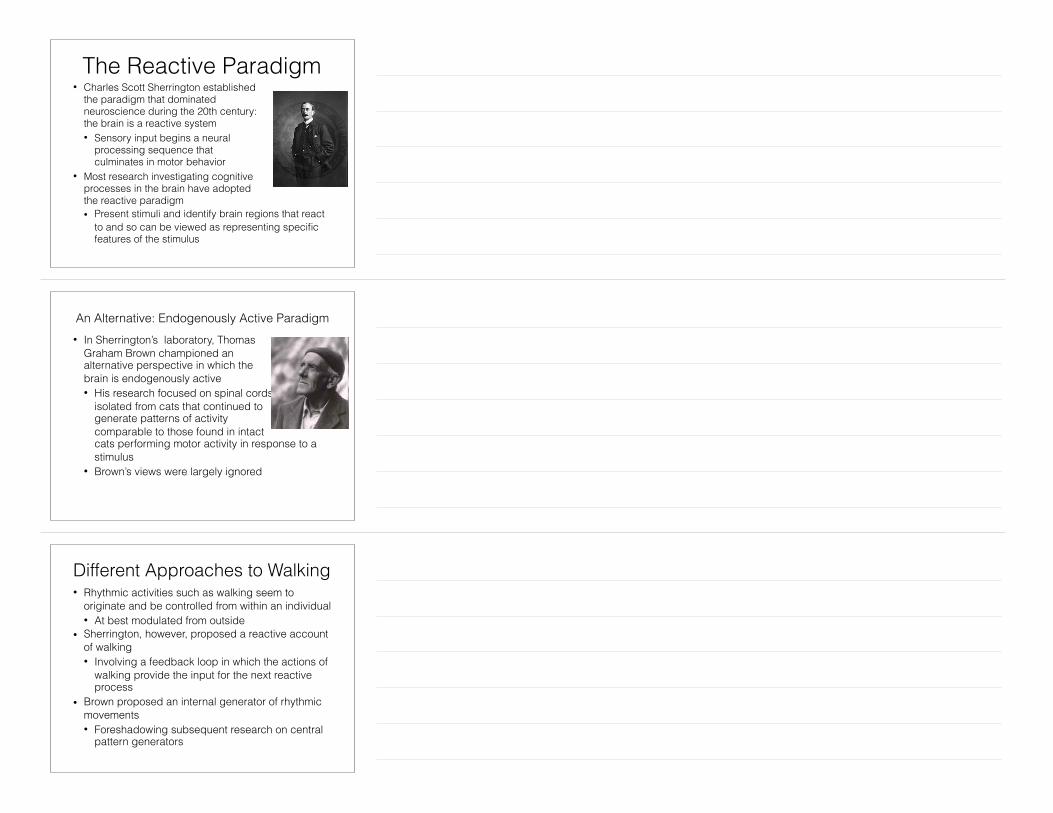
The Reactive Paradigm• Charles Scott Sherrington established
the paradigm that dominated neuroscience during the 20th century:the brain is a reactive system • Sensory input begins a neural
processing sequence that culminates in motor behavior
• Most research investigating cognitive processes in the brain have adopted the reactive paradigm • Present stimuli and identify brain regions that react
to and so can be viewed as representing specific features of the stimulus
An Alternative: Endogenously Active Paradigm• In Sherrington’s laboratory, Thomas
Graham Brown championed an alternative perspective in which the brain is endogenously active • His research focused on spinal cords
isolated from cats that continued to generate patterns of activity comparable to those found in intact cats performing motor activity in response to a stimulus
• Brown’s views were largely ignored
Different Approaches to Walking• Rhythmic activities such as walking seem to
originate and be controlled from within an individual • At best modulated from outside
• Sherrington, however, proposed a reactive account of walking • Involving a feedback loop in which the actions of
walking provide the input for the next reactive process
• Brown proposed an internal generator of rhythmic movements • Foreshadowing subsequent research on central
pattern generators
Organisms as Endogenously Active• Organisms (from single-celled organisms to us) are
highly organized systems • As such they are far from equilibrium
• Subject to continual evolution towards equilibrium
• Unless they have a constant supply of energy and direct that energy to maintain their organization
• As a result, all organisms (except seeds and spores) are continuously carrying out basic metabolic processes needed to maintain themselves • These are initiated from within the organism,
rendering the organism endogenously active
Cyclic (Feedback) Processes and Oscillations• Feedback systems can approach a steady state,
but in many conditions they generate sustained oscillations (approximating a cyclic attractor) • Systems that sustain oscillations are
endogenously active • provided they have a continued supply of
energy • Neurons are endogenously active oscillators
• A complex array of feedback processes are involved in maintaining electrical potentials across neural membranes and in many neurons support spontaneous spiking
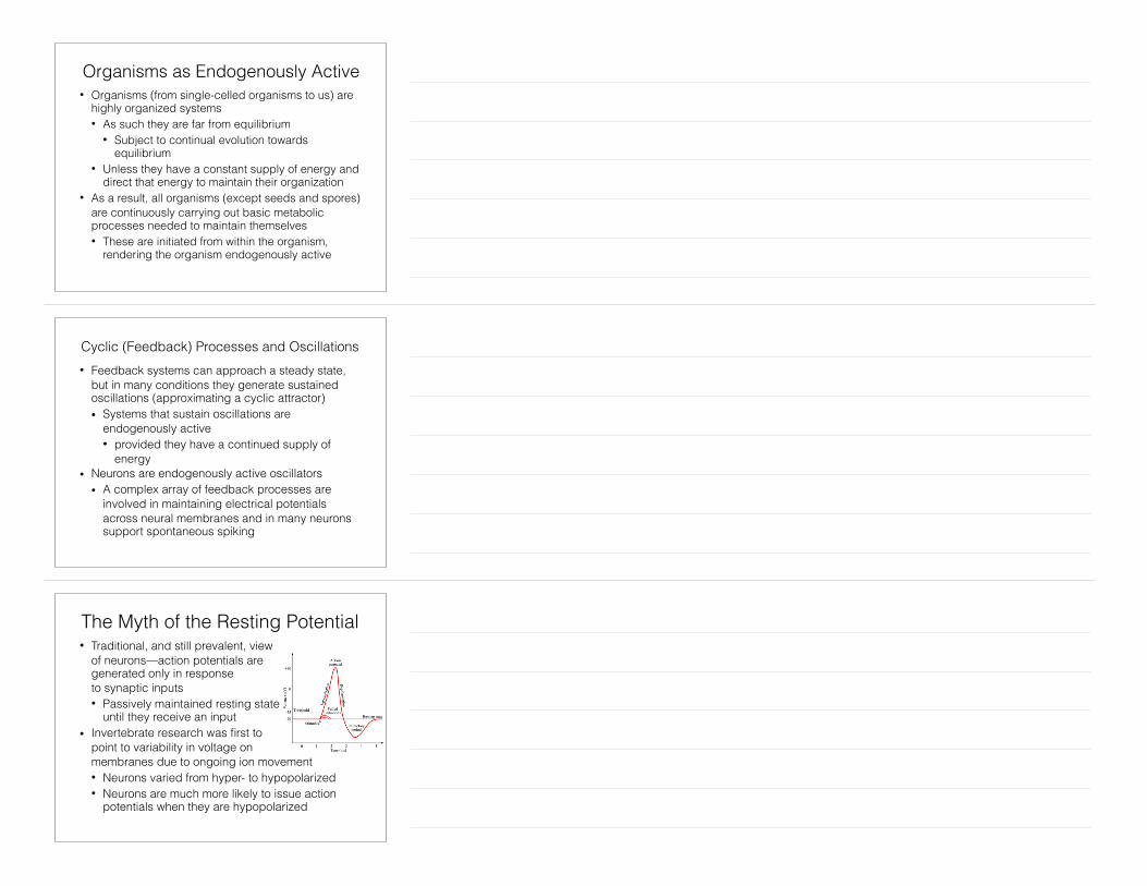
The Myth of the Resting Potential• Traditional, and still prevalent, view
of neurons—action potentials are generated only in response to synaptic inputs • Passively maintained resting state
until they receive an input • Invertebrate research was first to
point to variability in voltage on membranes due to ongoing ion movement • Neurons varied from hyper- to hypopolarized • Neurons are much more likely to issue action
potentials when they are hypopolarized
Spontaneous Firing of Neurons• If the oscillating currents along membranes cross
the threshold, they will spontaneously emit action potentials
• Rodolfo Llinás, recording from the inferior olive, identified low-threshold Ca2+ conductances • Allowed neurons to function as
single-cell oscillators “capable of self-sustained rhythmic firing independent of synaptic input” (1988)
• These intrinsic oscillations, together with synaptic processes, generate synchronous thalamic oscillations that regulate cortical activity
Synchronization of Oscillators• In his investigation of pendulums, Huygens
discovered that if a signal could be transmitted from one pendulum to another, they would tend to synchronize their oscillations
• The sub-threshold oscillations in electrical current generated in individual cells can be communicated to nearby cells • Resulting in synchronization of sub-threshold
electrical activity so that the population of cells together transition from hypo- to hyper-polarized
• The various oscillations detected in EEG corresponded to populations of cells oscillating in phase with each other
Representations and Background Rhythms• Traditional neuroscience approach to identifying
representations • Identify which stimulus drives maximal
neural firing • Place cells fire in response to specific
locations in an enclosure, for example • Firing in relation to background theta rhythms provides
information that goes beyond mere firing rates • When the rat first enters the place field, a place cell fires
a burst around the trough of the theta cycle • As the rat moves through the place field, firing
advances with respect to the theta cycle (here, 6 firing cycles to 5 theta cycles)
Representations and Background Rhythms• Firing with respect to background theta rhythm
provides a much more precise representation of location and of the sequence of locations traversed • Specifies how far through the place field the rat
has moved • Activity across a population of place cells
specifies the route the rat has taken • This also enables strengthening relevant
connections thereby enabling the construction of maps
Synchronization of Oscillations Regulates Communication
• Neurons are much more likely to produce action potentials when they are hypopolarized (closer to threshold) than when they are hyperpolarized • When their membrane potentials are
synchronized, two connected neurons will be hypopolarized together
• Then the firing of one is more likely to elicit the firing of the other
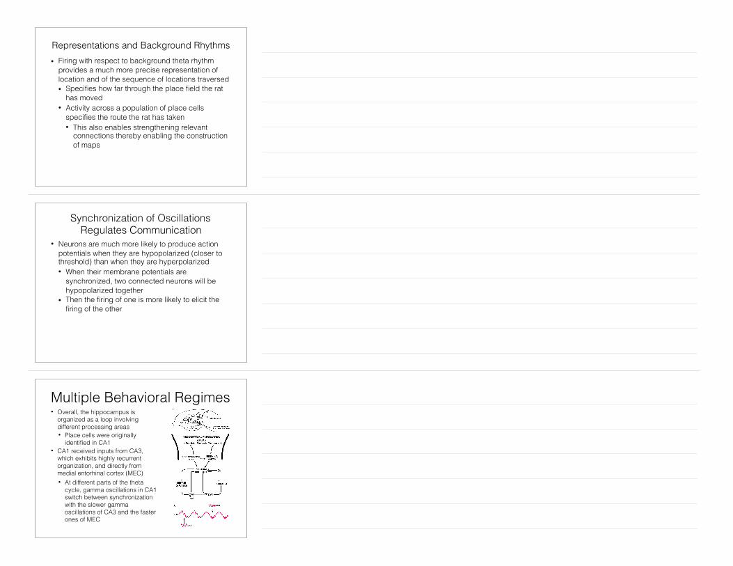
Multiple Behavioral Regimes• Overall, the hippocampus is
organized as a loop involving different processing areas • Place cells were originally
identified in CA1 • CA1 received inputs from CA3,
which exhibits highly recurrent organization, and directly from medial entorhinal cortex (MEC) • At different parts of the theta
cycle, gamma oscillations in CA1 switch between synchronization with the slower gamma oscillations of CA3 and the faster ones of MEC

Consequences for Learning via LTP• Input from CA3 signals
retrieval of a memory—learning would eradicate this memory • Appropriately, there is
no LTP when receiving input from CA3
• Input directly from MEC occurs when the stimulus is not recognized • Appropriately, LTP is
allowed when receiving input from MEC
Imaging During Resting State• Attracted renewed interest in mid-1990s, after reports that
certain brain areas consistently showed less blood flow (lower fMRI BOLD signal) in task than non-task conditions • Andreasen et al. (1995) found these areas to be less
active while performing a semantic memory task than while at rest
Semantic memory – REST:
Areas with negative peaks (pink) are more active during REST then in
task conditions
Brain Areas “Deactivated” During Task Situations• Shulman et al.’s (1997)
meta-analysis of PET studies identified severalareas with decreased activity during task conditions (versus passive viewing)
• Areas included: posterior cingulate cortex and precuneus (1), inferior parietal cortex (2, 3, 4), left dorsal lateral prefrontal cortex (5), and a medial frontal strip that continued through the inferior anterior cingulate cortex, left inferior frontal cortex and left inferior frontal gyrus to the right amygdala (6-14)
Clicker QuestionWhat is meant by the default mode network?
A. A network of brain areas that are always active to the same degree
B. A network of brain areas that responds especially strongly to a stimulus
C. A network of brain areas that is equally active in any task condition
D. A network of brain areas that exhibits more activity during the resting state than in task conditions
19
Proposal of “Default Mode Network”• “This consistency with which certain areas of the brain
participate in these decreases made us wonder whether there might be an organized mode of brain function that is present as a baseline or default state and is suspended during specific goal-directed behaviors” (Raichle et al., 2001) • Inferred that decreases during tasks “represented
the presence of functionality that was ongoing (i.e., sustained as contrasted to transiently activated) in the resting state and attenuated only when resources were temporarily reallocated during goal-directed behaviors; hence our original designation of them as default functions” (Raichle and Snyder, 2007)
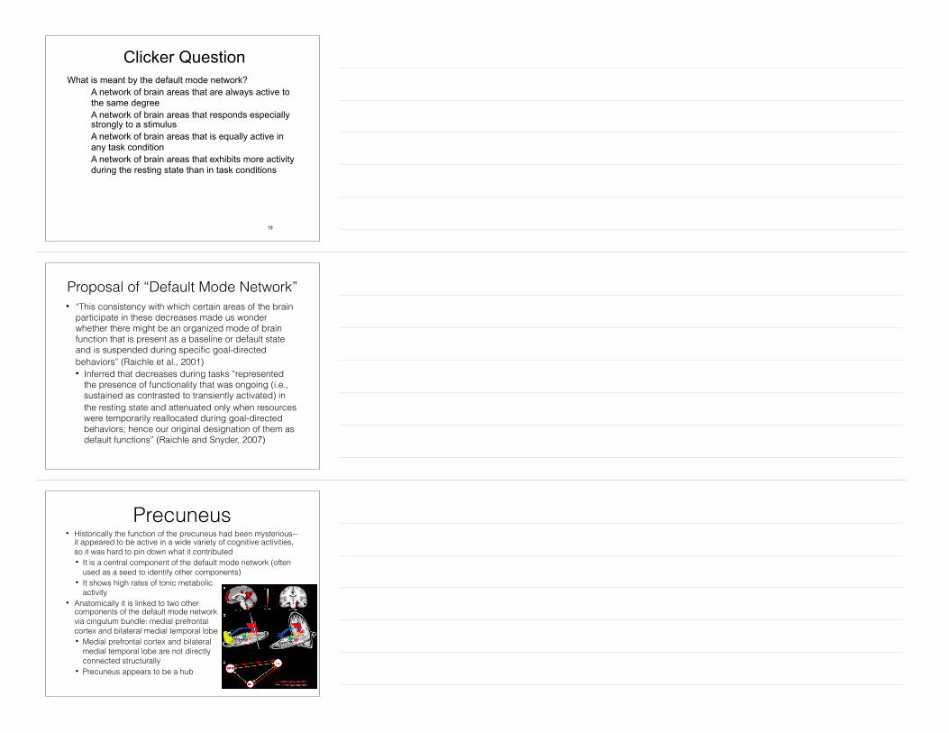
Precuneus• Historically the function of the precuneus had been mysterious--
it appeared to be active in a wide variety of cognitive activities, so it was hard to pin down what it contributed • It is a central component of the default mode network (often
used as a seed to identify other components) • It shows high rates of tonic metabolic
activity • Anatomically it is linked to two other
components of the default mode network via cingulum bundle: medial prefrontal cortex and bilateral medial temporal lobe • Medial prefrontal cortex and bilateral
medial temporal lobe are not directly connected structurally
• Precuneus appears to be a hub
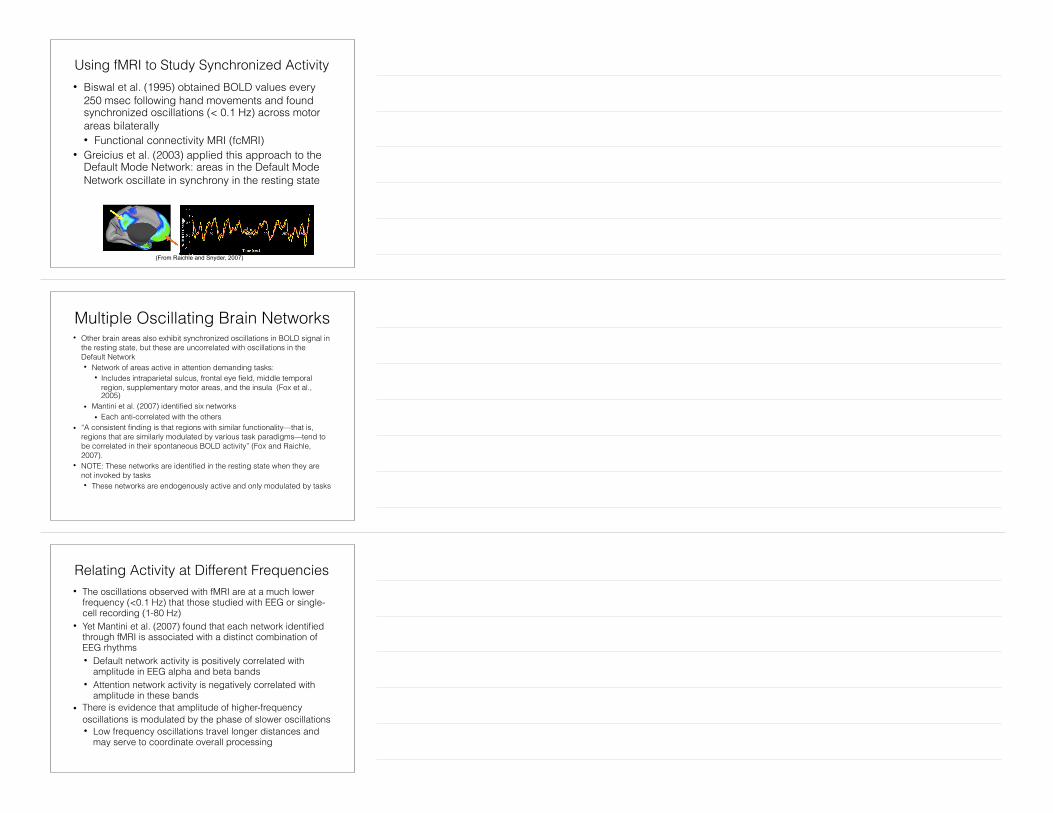
Using fMRI to Study Synchronized Activity• Biswal et al. (1995) obtained BOLD values every
250 msec following hand movements and found synchronized oscillations (< 0.1 Hz) across motor areas bilaterally • Functional connectivity MRI (fcMRI)
• Greicius et al. (2003) applied this approach to the Default Mode Network: areas in the Default Mode Network oscillate in synchrony in the resting state
(From Raichle and Snyder, 2007)
Multiple Oscillating Brain Networks• Other brain areas also exhibit synchronized oscillations in BOLD signal in
the resting state, but these are uncorrelated with oscillations in the Default Network • Network of areas active in attention demanding tasks:
• Includes intraparietal sulcus, frontal eye field, middle temporal region, supplementary motor areas, and the insula (Fox et al., 2005)
• Mantini et al. (2007) identified six networks • Each anti-correlated with the others
• “A consistent finding is that regions with similar functionality—that is, regions that are similarly modulated by various task paradigms—tend to be correlated in their spontaneous BOLD activity” (Fox and Raichle, 2007).
• NOTE: These networks are identified in the resting state when they are not invoked by tasks • These networks are endogenously active and only modulated by tasks
Relating Activity at Different Frequencies• The oscillations observed with fMRI are at a much lower
frequency (<0.1 Hz) that those studied with EEG or single-cell recording (1-80 Hz)
• Yet Mantini et al. (2007) found that each network identified through fMRI is associated with a distinct combination of EEG rhythms • Default network activity is positively correlated with
amplitude in EEG alpha and beta bands • Attention network activity is negatively correlated with
amplitude in these bands • There is evidence that amplitude of higher-frequency
oscillations is modulated by the phase of slower oscillations • Low frequency oscillations travel longer distances and
may serve to coordinate overall processing
Network Organization and Dynamics• Brain activity is neither stable nor
random--it exhibits metastability • Activity begins to settle into an
attractor, but then spontaneously breaks out of the attractor and moves toward another
• Within functionally organized networks in the brain, not all regions are synchronized at a given time • Constantly changing set of regions
that are synchronized • Growing evidence that small-world
organization with hubs and modules may support complex dynamical behavior
Rethinking Neural Processing• Traditional view in research on biological
mechanisms, including the brain: the mechanism was in the same default state until it was activated by a stimulus
• All one needed to know was how it responded to a stimulus
• Emerging view: the mechanism is constantly engaged in dynamical behavior that is directed by processes internal to it
• Stimuli perturb the ongoing dynamicalbehavior
• But the ongoing dynamics affects how the mechanism will respond to stimuli
• Identifying the structural and functional organization of neural networks is an important part of providing a dynamical mechanistic explanation of cognitive function
Fontanini and Katz (2008)
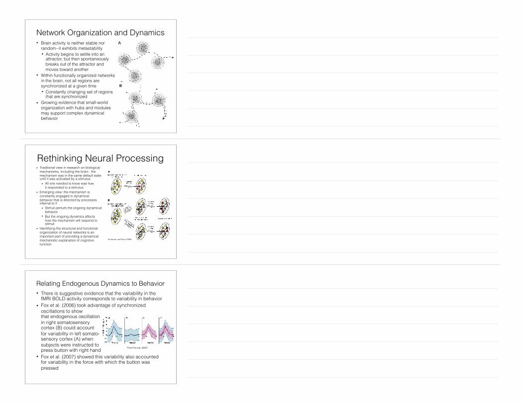
Relating Endogenous Dynamics to Behavior• There is suggestive evidence that the variability in the
fMRI BOLD activity corresponds to variability in behavior • Fox et al. (2006) took advantage of synchronized
oscillations to show that endogenous oscillation in right somatosensory cortex (B) could account for variability in left somato-sensory cortex (A) when subjects were instructed to press button with right hand
• Fox et al. (2007) showed this variability also accounted for variability in the force with which the button was pressed
From Fox et al. (2007)

Discussion QuestionWhat are the implications of endogenous dynamical activity for understanding the cognitive architecture of the mind/brain?
28


![An Effective Routing Algorithm with Chaotic Neurodynamics ...otic neurodynamics [14-20]. Chaotic neurodynamics ex- hibits a high ability to solve the various combinatorial optimization](https://static.fdocuments.net/doc/165x107/5f7460be25a1e07dee1d0a22/an-effective-routing-algorithm-with-chaotic-neurodynamics-otic-neurodynamics.jpg)
