Neurobiology of emotion
35
NEUROBIOLOGY OF EMOTIONS By Dr Sunil Suthar Under guidance of Prof. Dr. Pradeep Sharma
-
Upload
drsunil-suthar -
Category
Health & Medicine
-
view
150 -
download
8
Transcript of Neurobiology of emotion
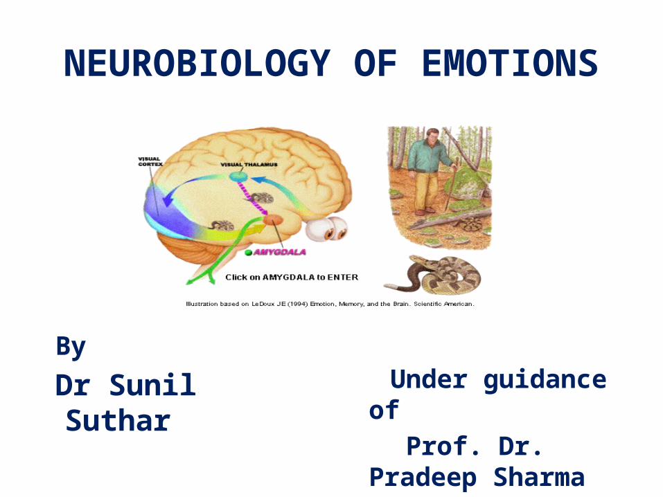
NEUROBIOLOGY OF EMOTIONS
By
Dr Sunil Suthar Under guidance of Prof. Dr. Pradeep
Sharma
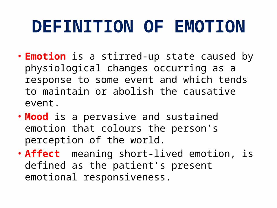
DEFINITION OF EMOTION
• Emotion is a stirred-up state caused by physiological changes occurring as a response to some event and which tends to maintain or abolish the causative event.
• Mood is a pervasive and sustained emotion that colours the person’s perception of the world.
• Affect meaning short-lived emotion, is defined as the patient’s present emotional responsiveness.
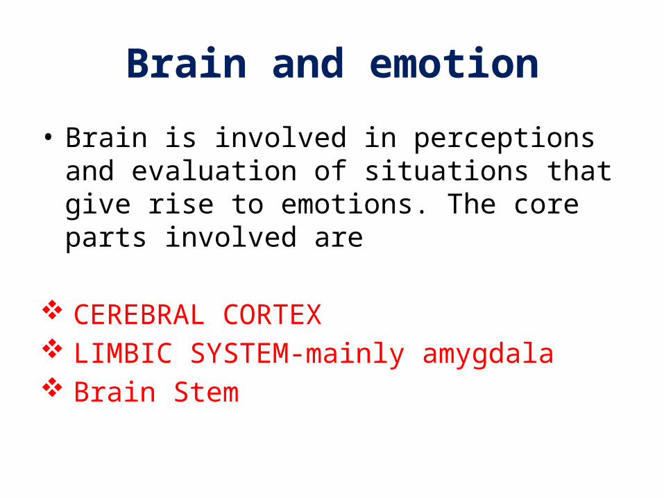
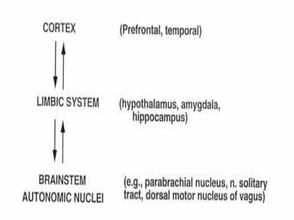
Brain and emotion
• Brain is involved in perceptions and evaluation of situations that give rise to emotions. The core parts involved are
CEREBRAL CORTEX LIMBIC SYSTEM-mainly amygdala Brain Stem
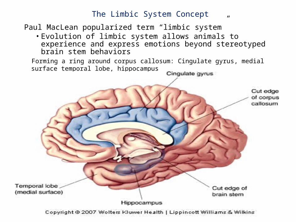
The Limbic System Concept
Paul MacLean popularized term “limbic system”• Evolution of limbic system allows animals to experience and
express emotions beyond stereotyped brain stem behaviors Forming a ring around corpus callosum: Cingulate gyrus, medial surface temporal
lobe, hippocampus
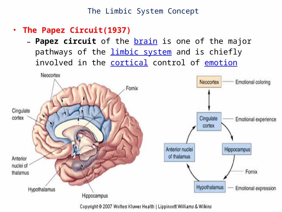
The Limbic System Concept
• The Papez Circuit(1937)– Papez circuit of the brain is one of the major pathways of the
limbic system and is chiefly involved in the cortical control of emotion
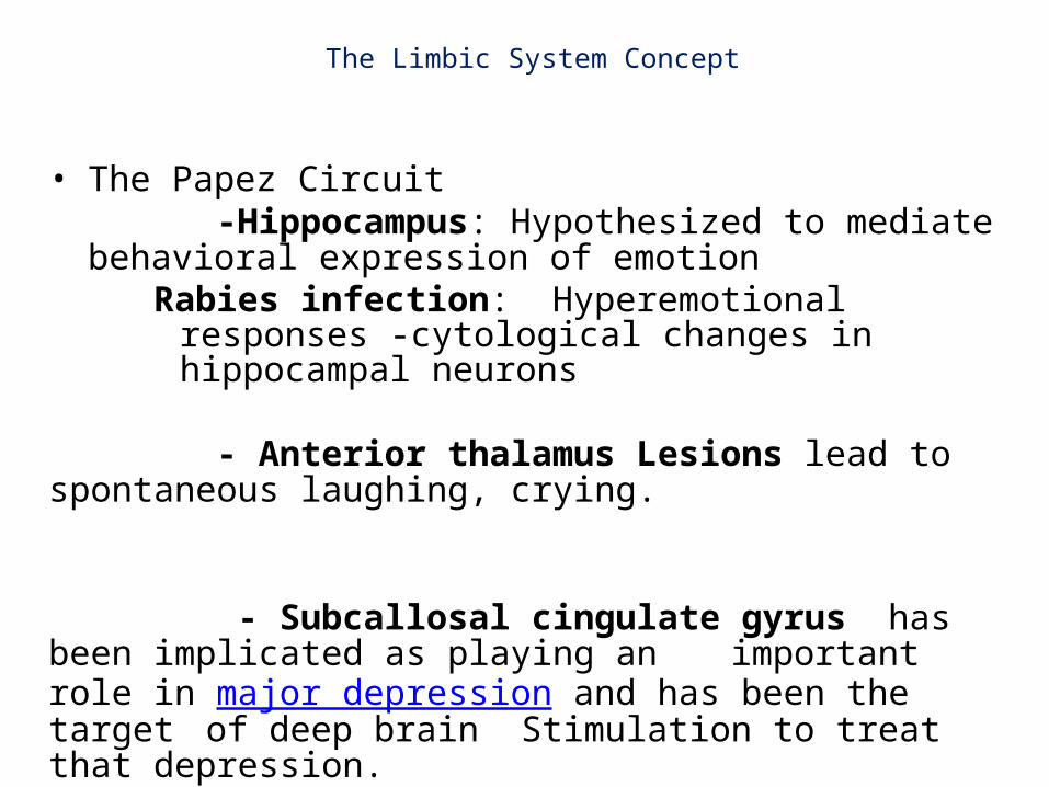
The Limbic System Concept
• The Papez Circuit -Hippocampus: Hypothesized to mediate behavioral expression
of emotionRabies infection: Hyperemotional responses -cytological
changes in hippocampal neurons
- Anterior thalamus Lesions lead to spontaneous laughing, crying.
- Subcallosal cingulate gyrus has been implicated as playing an important role in major depression and has been the target of deep brain Stimulation to treat that depression.
The Limbic System Concept
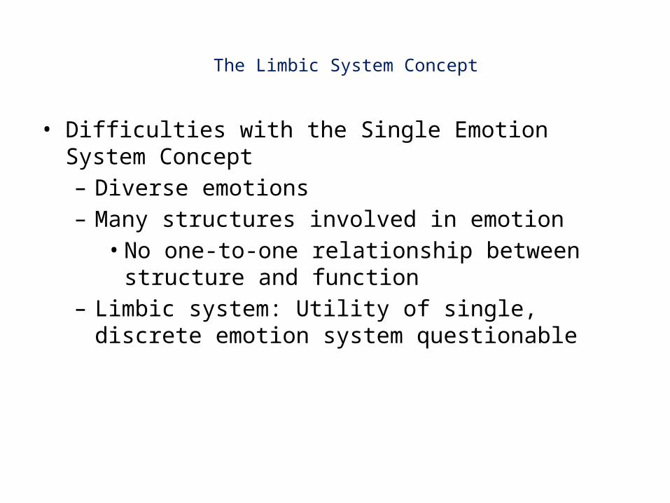
• Difficulties with the Single Emotion System Concept – Diverse emotions– Many structures involved in emotion• No one-to-one relationship between structure and
function– Limbic system: Utility of single, discrete emotion system
questionable
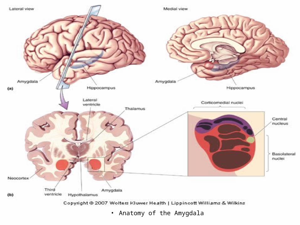
The Amygdala and AssociatedBrain Circuits
• Anatomy of the Amygdala
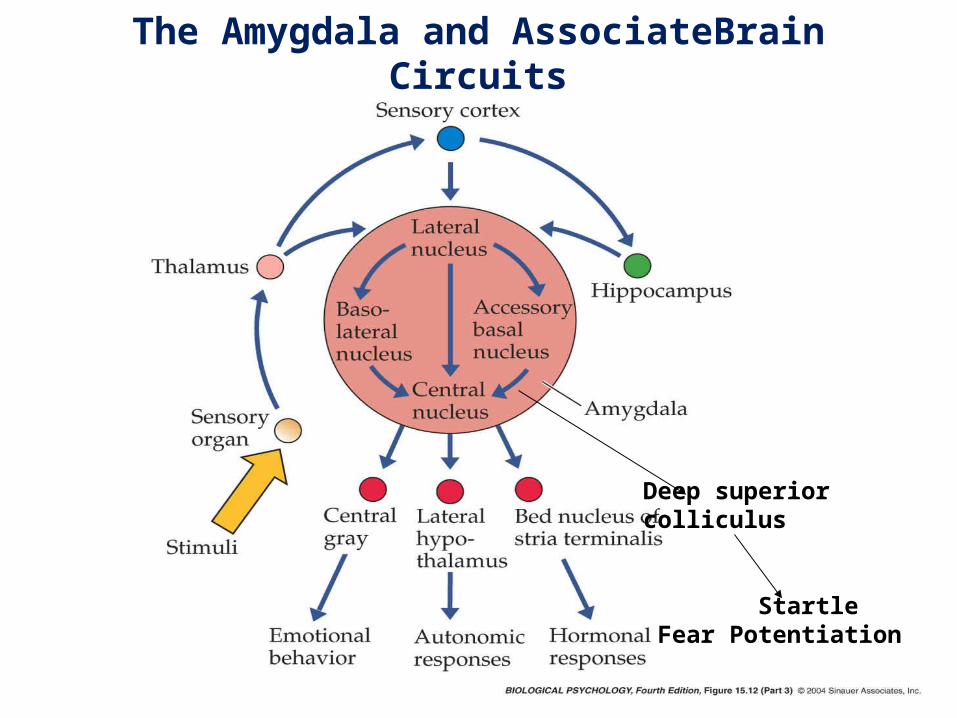
Deep superior colliculus
Startle Fear Potentiation
The Amygdala and AssociateBrain Circuits
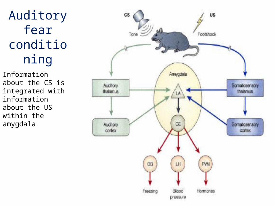
Auditory fear conditioning
Information about the CS is integrated with information about the US within the amygdala.
The Amygdala and AssociatedBrain Circuits
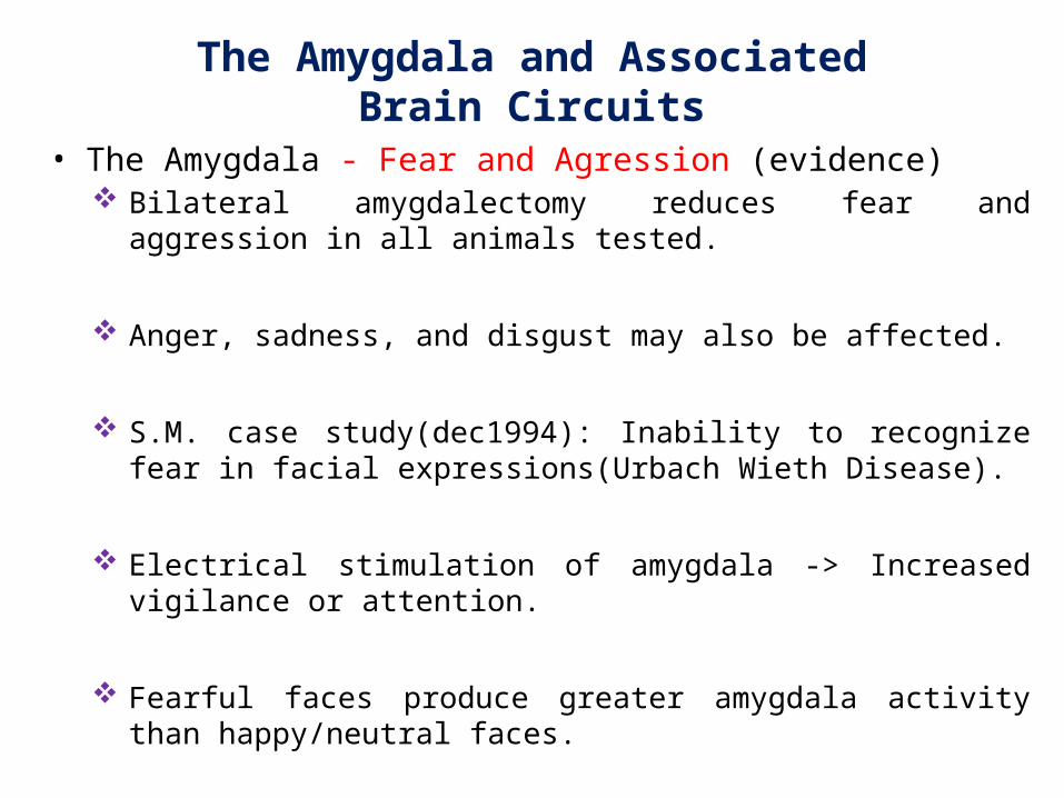
• The Amygdala - Fear and Agression (evidence)Bilateral amygdalectomy reduces fear and aggression in all animals
tested.
Anger, sadness, and disgust may also be affected.
S.M. case study(dec1994): Inability to recognize fear in facial expressions(Urbach Wieth Disease).
Electrical stimulation of amygdala -> Increased vigilance or attention.
Fearful faces produce greater amygdala activity than happy/neutral faces.
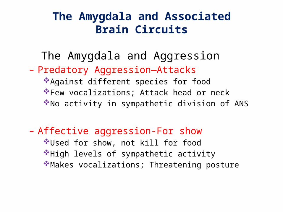
The Amygdala and Aggression– Predatory Aggression—Attacks
Against different species for foodFew vocalizations; Attack head or neckNo activity in sympathetic division of ANS
– Affective aggression-For showUsed for show, not kill for foodHigh levels of sympathetic activityMakes vocalizations; Threatening posture
The Amygdala and AssociatedBrain Circuits
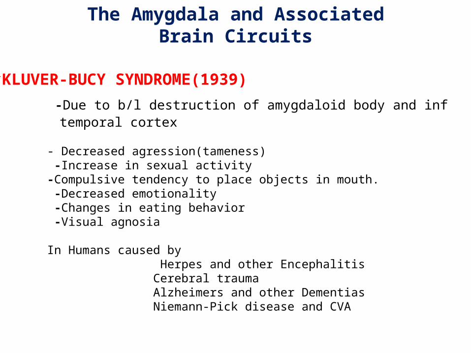
KLUVER-BUCY SYNDROME(1939)
-Due to b/l destruction of amygdaloid body and inf temporal cortex
- Decreased agression(tameness) -Increase in sexual activity -Compulsive tendency to place objects in mouth. -Decreased emotionality -Changes in eating behavior -Visual agnosia
In Humans caused by Herpes and other Encephalitis Cerebral trauma Alzheimers and other Dementias Niemann-Pick disease and CVA
The Amygdala and AssociatedBrain Circuits
– Surgery to Reduce Human AggressionAmygdalectomy results in -• Reduced aggressive asocial behavior• Increased ability to concentrate• Decreased hyperactivity
Psychosurgery – last resort
The Amygdala and AssociatedBrain Circuits
The Amygdala and AssociatedBrain Circuits
In Schizophrenic patient – Exposure to scary face – amygdala not activated. Exposure to neutral face – inappropriate hyperactivation of amygdala.
Schizophrenic patient often have impairements in ability to identify and interpret emotional stimuli. The neurobiological explanation for this may be inefficient information processing with in Ventral system.
Consistently in fMRI and PET studies, with emotionally valenced pictures, faces, and odors, patients showed diminished activation in limbic and paralimbic regions, especially the amygdala.
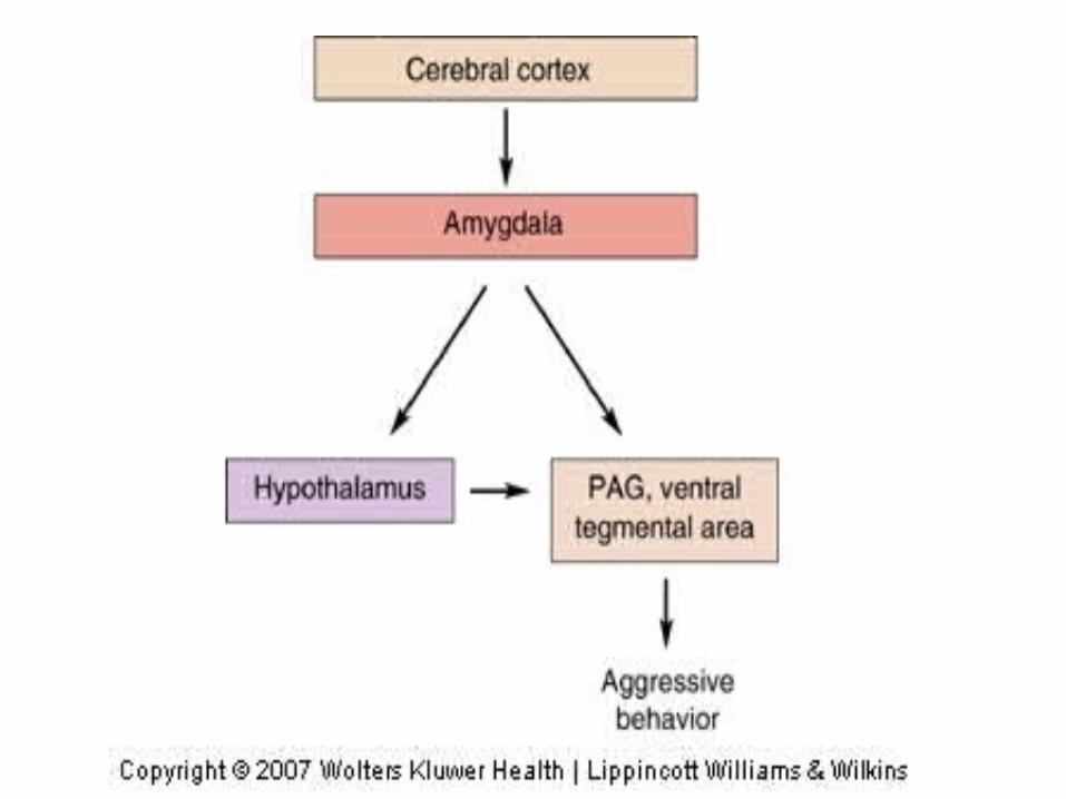
• Aggressive symptoms- orbitofrontal cortex and amygdala
HYPOTHALAMUS
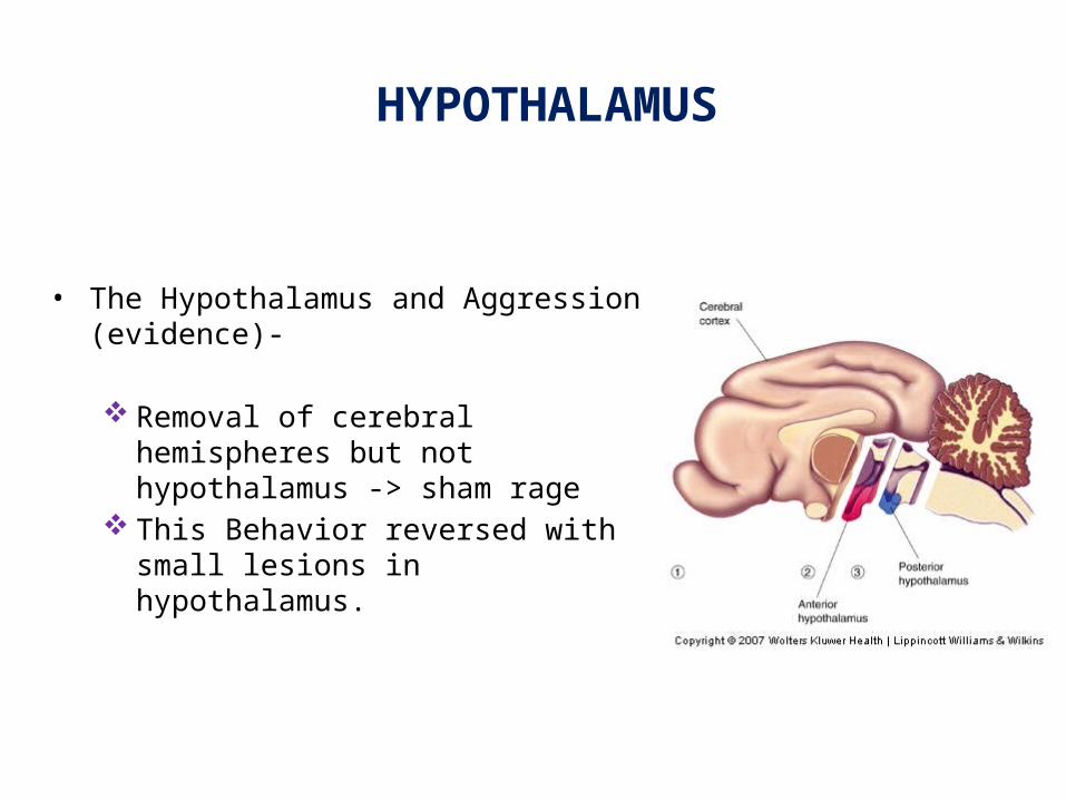
• The Hypothalamus and Aggression (evidence)-
Removal of cerebral hemispheres but not hypothalamus -> sham rage
This Behavior reversed with small lesions in hypothalamus.
HYPOTHALAMUS
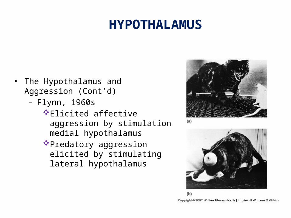
• The Hypothalamus and Aggression (Cont’d)– Flynn, 1960s
Elicited affective aggression by stimulation medial hypothalamus
Predatory aggression elicited by stimulating lateral hypothalamus
PREFRONTAL CORTEX
Emotional symptoms such as happiness and sadness are regulated by prefrontal cortex and amygdala.
Left prefrontal cortex appears to lift the mood.
Activation of the right prefrontal cortex causes depression.
Lesion to the right prefrontal area may produce laughter, euphoria, and moria or witzelsucht, a tendency to joke and make puns.
In treating depression, rTMS therapy targets the area left dorsolateral prefrontal cortex (DLPFC). The DLPFC is readily accessible to the magnetic field and is highly interconnected with limbic structures, which plays a dominant role in mood modulation and major depression.
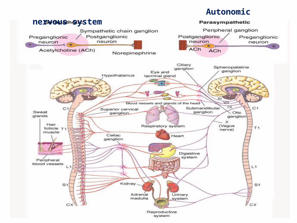
Autonomic nervous system
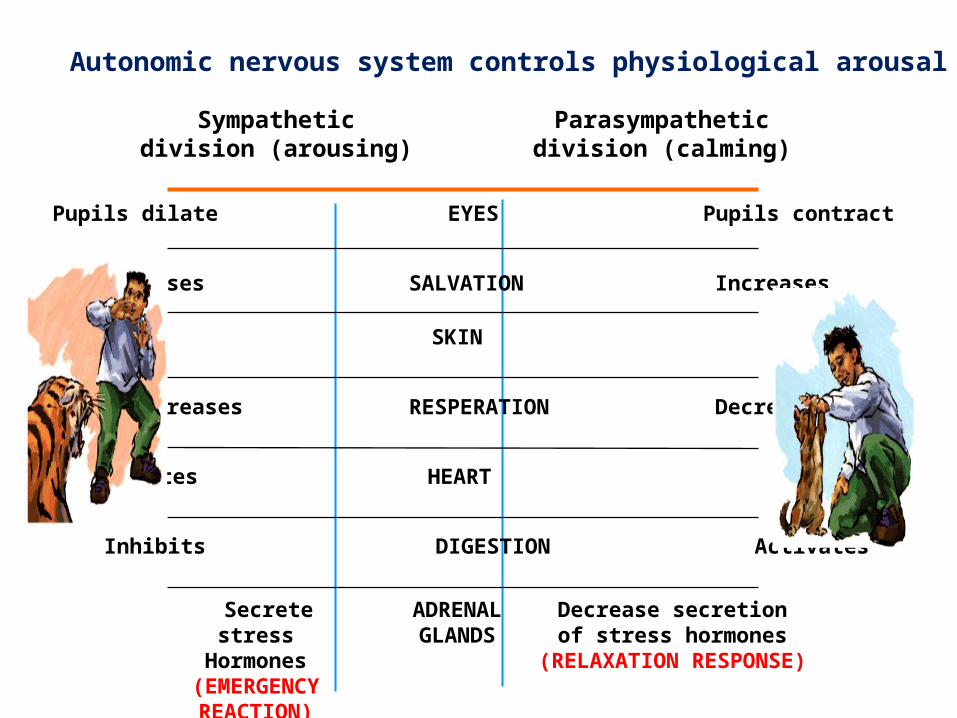
Autonomic nervous system controls physiological arousal
Sympatheticdivision (arousing)
Parasympatheticdivision (calming)
Pupils dilate EYES Pupils contract
Decreases SALVATION Increases
Perspires SKIN Dries
Increases RESPERATION Decreases
Accelerates HEART Slows
Inhibits DIGESTION Activates
Secrete stressHormones
(EMERGENCY REACTION)
ADRENALGLANDS
Decrease secretionof stress hormones
(RELAXATION RESPONSE)
• Vagus nerve stimulation (VNS) for treatment-resistant depressions- Little is understood about exactly how vagal nerve stimulation modulates
mood but proposed mechanisms include alteration of norepinephrine release by projections of solitary tract to the locus coeruleus, elevated levels of inhibitory GABA related to vagal stimulation and inhibition of aberrant cortical activity by reticular system activation.
Autonomic nervous system controls physiological arousal
AUTONOMIC&ENDOCRINE RESPONSES TO EMOTION Limbic stimulation causes changes in respiration & blood pressure
Hypothalamic autonomic responses are triggered by a complex phenomenon mediated by the cortical and limbic structures.
The fear and rage responses mediated by the limbic system cause stimulation of various parts of the hypothalamus, especially the lateral areas and produce diffuse sympathethic Discharge.
The physical symptoms of panic attacks can be reproduced by Carbon dioxide, Yohimbine, and Caffeine and Epinephrine administration.
The massive sympathetic discharge during stress is called the flight or fight response“. Stress via cortical and limbic connections causes release of corticotropin-releasing hormone (CRH) from the paraventricular nuclei of the hypothalamus. CRH release mediates endocrine and immune response
EMOTIONAL MEMORY
Emotion has powerful influence on learning and memory
Amygdala, in conjunction with prefrontal cortex &medial temporal lobe, is involved in consolidation and retrieval of emotional memories.
Amygdala, prefrontal cortex and hippocampus are also involved in the acquisition, extinction and recovery of fears to cues and contexts.
Hippocampus is critical for long-term,declarative memory storage
Propranolol impairs memory for emotionally provocative story but not for emotionally neutral story.
Chemical Neurotransmitters involved in Emotions
• Monoamine neurotansmitters – Norepinephrine, Serotonine, Dopamine.
• Aminoacid transmitters – GABA, Glutamate.
• Peptide neurotransmitters – CRH, Neuropeptide Y, Substance P, Opioid.
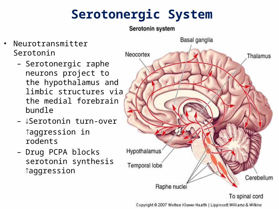
Serotonergic System
• Neurotransmitter Serotonin – Serotonergic raphe neurons
project to the hypothalamus and limbic structures via the medial forebrain bundle
– Serotonin turn-over aggression in rodents– Drug PCPA blocks serotonin
synthesis aggression
Serotonergic System 5HT 1A receptor knock out mice show a marked elevation of anxiety and fear behaviour.
Postsynaptic 5HT 1A receptor gene expression is under tonic inhibition by adrenal steroids.
Downregulation of 5HT 1A receptor in response to chronic stress.
Upregulation of 5HT 2A receptor during chronic stress. Chronic administration 5HT 1A receptor partial agonist and 5HT 2A receptor antagonist
exert anxiolytic effect.
5HT 1A receptor agonist and 5HT 2A receptor antagonist decrease rodent aggression.
Drugs such as Risperidone actually have more antagonist effect at 5HT 2A receptors than D2 receptors.
Depressed patient have reduced concentration of serotonine metabolite 5-HIAA in CSF.
Noradrenergic System
Norepinephrine (NE) released primarily from locus coeruleus(LC) which located in pons.
Exposure to acute stress/fear results in increase in NE in LC, hypothalamus, hippocampus, amygadala, and cerebral cortex.
Repeated exposure to stress/chronic stress- decreased release of NE in LC. (learned helplessness)
Patients with PTSD and panic disorder show evidence of increased NE activity.
Mice lacking Alpha-2 adrenergic receptors have hightened autonomic activation and concomitant anxiety behaviour.
Corticotropin relasing hormone (CRH) system
Central nucleus of amygdala contains CRH.
Axons of central nucleus cells target locus coeruleus neurons (which have CRH receptors and contain NE).
In animal administration of CRH in cerebral ventricles effectively induces anxiety responses, including hypervigilance, enhancement of the freezing posture, and decreased exploration in unfamiliar situations.
γ-Amino Butyric Acid and the Benzodiazepine System- Role of GABA and benzodiazepine receptors in anxiety is well documented in animal studies.
Several studies of panic disorder patients have shown reduced GABA-A and benzodiazepine binding, using PET and SPECT imaging, in areas such as the cortex, hippocampus, and insula when compared to controls.
Dopaminergic System- Inhibition in nucleus accumbens dopamine activity results in abnormalities in motivation and reward mechanisms.
Glutamate system- NMDA receptors are prominently involved in the conditioning of fear learning.
Blockade of the NMDA receptor with antagonists such as APV can block fear acquisitions, and some studies show that it may even block expression.
.
Neuropeptide Y- NPY has anxiolytic effects. NPY has been shown to be involved in fear consolidation, with
preclinical studies showing that the administration of NPY impairs the retention of traumatic memories and reduces anxiety during stressful tasks
Recently it has been observed that the neuropeptide cholecystokinin (CCK) is involved in panic disorders.(antagonists of CCK-B receptor have anxiolytic effect).
Concluding Remarks
• Neural Pathways
Experience, expression of emotion involves widespread activity in the nervous system from cortex to ANS as well as: limbic structures, hypothalamus, amygdala
Structures involved in emotions have other functions, including learning and memory
References (1) Stephan B. Hamann, Ralph Adolphs : Normal recognition of emotional similarity between facial expressions
following bilateral amygdala damage Neuropsychologia 37 (1999) 1135±1141.(2) Stahl’s Essential psychopharmacology – third edition.(3) Kaplan and Sadock’s comprehensive textbook of psychiatry.9th edition.(4) Fish psychopathology 3rd edition(5) Gaul C Jordan B, Wustmann T, Preuss UW [Klüver-Bucy syndrome in humans]. Nervenarzt. 2007
Jul;78(7):821-3.(6) Anne L. etal Klüver-Bucy Syndrome After Bilateral Selective Damage of Amygdala and Its Cortical
Connections, The Journal of Neuropsychiatry and Clinical Neurosciences 1998;10:354-358.(7) Gothard k etal. Neural Responses to Facial Expression and Face Identity in the Monkey Amygdala. PresS. J
Neurophysiol (November 8, 2006). doi:10.1152/jn.00714.2006 .(8) Guyton and Hall Textbook of Medical Physiology, 12th Edition(9) Lin D. Nature, 2011 Feb 10;470(7333):221-6.Functional identification of an aggression locus in the mouse
hypothalamus
.(10) Neuroscience. 2nd edition.Purves D, Augustine GJ, Fitzpatrick D, et al., editors.Sunderland (MA): Sinauer
Associates; 2001(11) Motivational properties of hypothalamic aggression in cats. Roberts, Warren W.; Kiess, Harold O. Journal of
Comparative and Physiological Psychology, Vol 58(2), Oct 1964, 187-193. doi: 10.1037/h0042377(12) Rush A etal, Vagusnervestimulation (VNS) for treatment-resistantdepressions: a multicenter study.
Biological Psychiatry Volume 47, Issue 4, 15 February 2000, Pages 276–286.(13) Rapid-rate transcranial magnetic stimulation of left dorsolateral prefrontal cortex in drug-
resistant depression,The Lancet Volume 348, Issue 9022, 27 July 1996, Pages 233–237.(14) Halen S. etal Deep Brain Stimulation for Treatment-Resistant Depression, NEURON, Volume 45, Issue 5, 3 March 2005, Pages 651–660.(15) Johansen H. etal Anatomical Connectivity of the Subgenual Cingulate Region Targeted with Deep Brain
Stimulation for Treatment-Resistant Depression. Oxford Journals Life Sciences & Medicine, Cerebral Cortex, Volume 18, Issue 6Pp. 1374-1383
(16) Mary L Phillips. etal, Neurobiology of emotion perception I: the neural basis of normalemotion perception, Biological Psychiatry Volume 54, Issue 5, 1 September 2003, Pages 504–514
(17) Markus Kosel, M.D., and Thomas E. Schlaepfer, M.D. Mechanisms and State of the Art of Vagus Nerve Stimulation, The Journal of ECT, 18(4):189–192 © 2002 Lippincott Williams & Wilkins, Inc., Philadelphia