Neurobiology of Aging · PDF fileThese findings raise the possibility that EHT may make a...
12
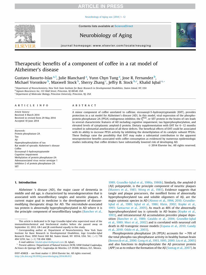
Therapeutic benefits of a component of coffee in a rat model of Alzheimer’s disease Gustavo Basurto-Islas a, 1 , Julie Blanchard a , Yunn Chyn Tung a , Jose R. Fernandez b , Michael Voronkov b , Maxwell Stock b , Sherry Zhang c , Jeffry B. Stock b, c , Khalid Iqbal a, * a Department of Neurochemistry, New York State Institute for Basic Research in Developmental Disabilities, Staten Island, NY, USA b Signum Biosciences Inc, 133 Wall Street, Princeton, NJ 08540, USA c Department of Molecular Biology, Princeton University, Princeton, NJ, USA article info Article history: Received 4 March 2014 Received in revised form 29 May 2014 Accepted 10 June 2014 Keywords: Protein phosphatase-2A Tau Ab Hyperphosphorylation of tau Rat model of sporadic Alzheimer’s disease Coffee Eicosanoyl-5-hydroxytryptamide Cognitive impairment Methylation of protein phosphatase-2A Adenoassociated virus vector serotype 1 Inhibitor-2 of protein phosphatase-2A SET abstract A minor component of coffee unrelated to caffeine, eicosanoyl-5-hydroxytryptamide (EHT), provides protection in a rat model for Alzheimer’s disease (AD). In this model, viral expression of the phospho- protein phosphatase 2A (PP2A) endogenous inhibitor, the I PP2A 2 , or SET protein in the brains of rats leads to several characteristic features of AD including cognitive impairment, tau hyperphosphorylation, and elevated levels of cytoplasmic amyloid-b protein. Dietary supplementation with EHT for 6e12 months resulted in substantial amelioration of all these defects. The beneficial effects of EHT could be associated with its ability to increase PP2A activity by inhibiting the demethylation of its catalytic subunit PP2Ac. These findings raise the possibility that EHT may make a substantial contribution to the apparent neuroprotective benefits associated with coffee consumption as evidenced by numerous epidemiologic studies indicating that coffee drinkers have substantially lowered risk of developing AD. Ó 2014 Elsevier Inc. All rights reserved. 1. Introduction Alzheimer ’s disease (AD), the major cause of dementia in middle and old age, is characterized by neurodegeneration that is associated with neurofibrillary tangles and neuritic plaques. A current major goal in medicine is the development of disease- modifying therapeutic drugs for AD. The microtubule-associated tau protein is abnormally hyperphosphorylated in AD where it is the principle component of neurofibrillary tangles (Bancher et al., 1989; Grundke-Iqbal et al., 1986a, 1986b). Similarly, the amyloid-b (Ab) polypeptide, is the principle component of neuritic plaques (Masters et al., 1985; Wong et al., 1985). Evidence suggests that tangle and plaque precursors, the nonfibril forms of abnormally hyperphosphorylated tau and soluble oligomers of Ab, are the major cytotoxic species in AD (Alonso et al., 1994, 2010; Grundke- Iqbal et al., 1989; Iqbal et al., 1986; Klein, 2002; Kopke et al., 1993; Santacruz et al., 2005). As much as 40% of the abnormally hyperphosphorylated tau is cytosolic in AD brains (Kopke et al., 1993), and intraneuronal Ab accumulation precedes plaque depo- sition (Bancher et al., 1989; Cataldo et al., 2004; Grundke-Iqbal et al., 1989; Mori et al., 2002) and is correlated with neuronal cell death in AD transgenic mouse models (Espana et al., 2010; Gandy et al., 2010; Oddo et al., 2003). Phosphoprotein phosphatase 2A (PP2A) accounts for w70% of the total phospho-tau phosphatase activity in healthy human brain (Bennecib et al., 2000; Gong et al., 1993, 1995, 2000; Liu et al., 2005) and also functions to dephosphorylate the Ab precursor protein (APP) so as to reduce the formation of the Ab (Sontag et al., 2007). In This article is dedicated to Dr Inge Grundke-Iqbal who supervised most of the immunohistochemical and biochemical studies before she passed away on September 22, 2012. GB-I and JB contributed equally to this study. * Corresponding author at: Department of Neurochemistry, New York State Institute for Basic Research in Developmental Disabilities, Inge Grundke-Iqbal Research Floor, 1050 Forest Hill Rd, Staten Island, NY 10314, USA. Tel.: (þ1) 718 494 5259; fax: (þ1) 718 494 1080. E-mail address: [email protected] (K. Iqbal). 1 Present address: Department of Natural Sciences DCNI, UAM Unidad Cuajimalpa, Av.Vasco de Quiroga 4871, Cuajimalpa de Morelos, C.P. 05300, México, D.F., Mexico. Contents lists available at ScienceDirect Neurobiology of Aging journal homepage: www.elsevier.com/locate/neuaging 0197-4580/$ e see front matter Ó 2014 Elsevier Inc. All rights reserved. http://dx.doi.org/10.1016/j.neurobiolaging.2014.06.012 Neurobiology of Aging xxx (2014) 1e12
Transcript of Neurobiology of Aging · PDF fileThese findings raise the possibility that EHT may make a...
lable at ScienceDirect
Neurobiology of Aging xxx (2014) 1e12
Contents lists avai
Neurobiology of Aging
journal homepage: www.elsevier .com/locate/neuaging
Therapeutic benefits of a component of coffee in a rat model ofAlzheimer’s disease
Gustavo Basurto-Islas a,1, Julie Blanchard a, Yunn Chyn Tung a, Jose R. Fernandez b,Michael Voronkov b, Maxwell Stock b, Sherry Zhang c, Jeffry B. Stock b,c, Khalid Iqbal a,*aDepartment of Neurochemistry, New York State Institute for Basic Research in Developmental Disabilities, Staten Island, NY, USAb Signum Biosciences Inc, 133 Wall Street, Princeton, NJ 08540, USAcDepartment of Molecular Biology, Princeton University, Princeton, NJ, USA
a r t i c l e i n f o
Article history:Received 4 March 2014Received in revised form 29 May 2014Accepted 10 June 2014
Keywords:Protein phosphatase-2ATauAbHyperphosphorylation of tauRat model of sporadic Alzheimer’s diseaseCoffeeEicosanoyl-5-hydroxytryptamideCognitive impairmentMethylation of protein phosphatase-2AAdenoassociated virus vector serotype 1Inhibitor-2 of protein phosphatase-2ASET
This article is dedicated to Dr Inge Grundke-Iqbalimmunohistochemical and biochemical studies beSeptember 22, 2012. GB-I and JB contributed equally* Corresponding author at: Department of Neuro
Institute for Basic Research in Developmental DisaResearch Floor, 1050 Forest Hill Rd, Staten Island, N494 5259; fax: (þ1) 718 494 1080.
E-mail address: [email protected] (K. Iqb1 Present address: Department of Natural Sciences D
Av.Vasco de Quiroga 4871, Cuajimalpa de Morelos, C.P.
0197-4580/$ e see front matter � 2014 Elsevier Inc. Ahttp://dx.doi.org/10.1016/j.neurobiolaging.2014.06.012
a b s t r a c t
A minor component of coffee unrelated to caffeine, eicosanoyl-5-hydroxytryptamide (EHT), providesprotection in a rat model for Alzheimer’s disease (AD). In this model, viral expression of the phospho-protein phosphatase 2A (PP2A) endogenous inhibitor, the IPP2A2 , or SET protein in the brains of rats leadsto several characteristic features of AD including cognitive impairment, tau hyperphosphorylation, andelevated levels of cytoplasmic amyloid-b protein. Dietary supplementation with EHT for 6e12 monthsresulted in substantial amelioration of all these defects. The beneficial effects of EHT could be associatedwith its ability to increase PP2A activity by inhibiting the demethylation of its catalytic subunit PP2Ac.These findings raise the possibility that EHT may make a substantial contribution to the apparentneuroprotective benefits associated with coffee consumption as evidenced by numerous epidemiologicstudies indicating that coffee drinkers have substantially lowered risk of developing AD.
� 2014 Elsevier Inc. All rights reserved.
1. Introduction
Alzheimer ’s disease (AD), the major cause of dementia inmiddle and old age, is characterized by neurodegeneration that isassociated with neurofibrillary tangles and neuritic plaques. Acurrent major goal in medicine is the development of disease-modifying therapeutic drugs for AD. The microtubule-associatedtau protein is abnormally hyperphosphorylated in AD where it isthe principle component of neurofibrillary tangles (Bancher et al.,
who supervised most of thefore she passed away onto this study.chemistry, New York Statebilities, Inge Grundke-IqbalY 10314, USA. Tel.: (þ1) 718
al).CNI, UAM Unidad Cuajimalpa,05300, México, D.F., Mexico.
ll rights reserved.
1989; Grundke-Iqbal et al., 1986a, 1986b). Similarly, the amyloid-b(Ab) polypeptide, is the principle component of neuritic plaques(Masters et al., 1985; Wong et al., 1985). Evidence suggests thattangle and plaque precursors, the nonfibril forms of abnormallyhyperphosphorylated tau and soluble oligomers of Ab, are themajor cytotoxic species in AD (Alonso et al., 1994, 2010; Grundke-Iqbal et al., 1989; Iqbal et al., 1986; Klein, 2002; Kopke et al.,1993; Santacruz et al., 2005). As much as 40% of the abnormallyhyperphosphorylated tau is cytosolic in AD brains (Kopke et al.,1993), and intraneuronal Ab accumulation precedes plaque depo-sition (Bancher et al., 1989; Cataldo et al., 2004; Grundke-Iqbalet al., 1989; Mori et al., 2002) and is correlated with neuronal celldeath in AD transgenic mouse models (Espana et al., 2010; Gandyet al., 2010; Oddo et al., 2003).
Phosphoprotein phosphatase 2A (PP2A) accounts for w70% ofthe total phospho-tau phosphatase activity in healthy human brain(Bennecib et al., 2000; Gong et al., 1993,1995, 2000; Liu et al., 2005)and also functions to dephosphorylate the Ab precursor protein(APP) so as to reduce the formation of the Ab (Sontag et al., 2007). In
G. Basurto-Islas et al. / Neurobiology of Aging xxx (2014) 1e122
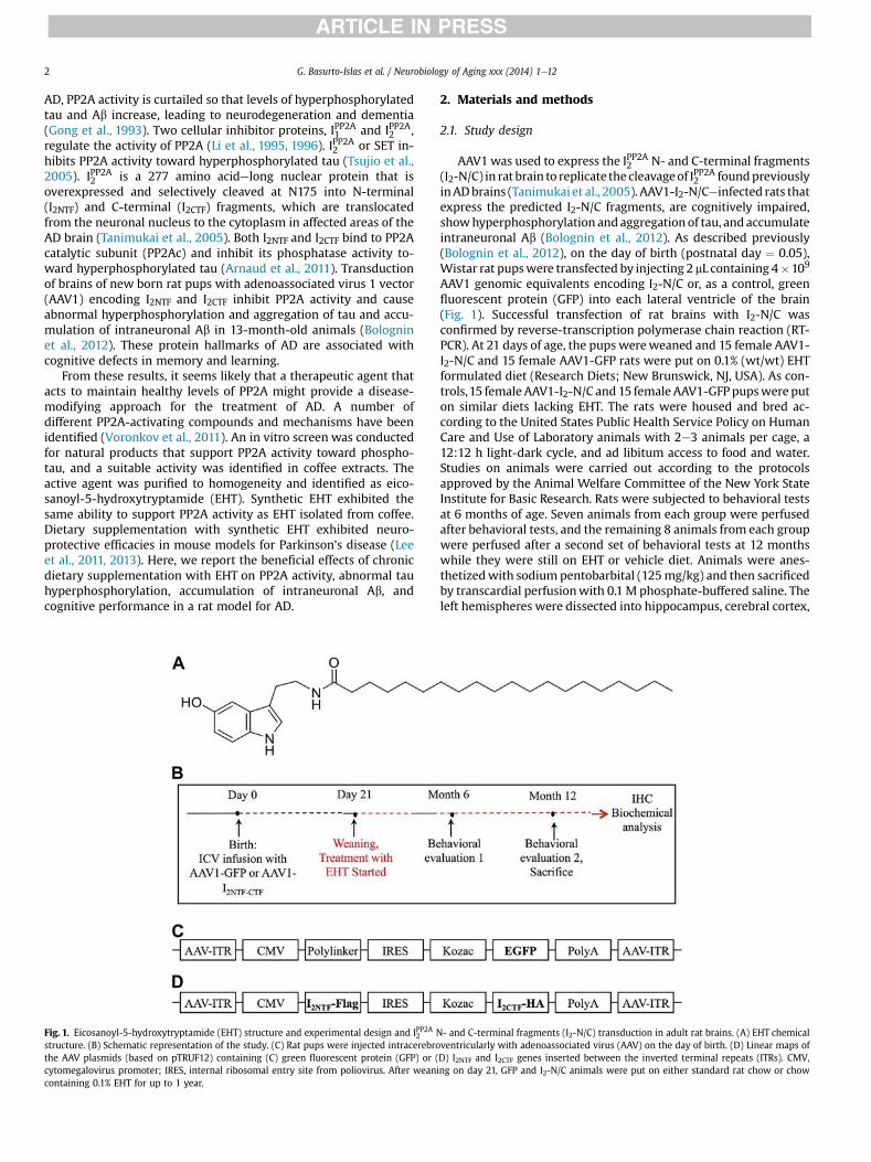
AD, PP2A activity is curtailed so that levels of hyperphosphorylatedtau and Ab increase, leading to neurodegeneration and dementia(Gong et al., 1993). Two cellular inhibitor proteins, IPP2A1 and IPP2A2 ,regulate the activity of PP2A (Li et al., 1995, 1996). IPP2A2 or SET in-hibits PP2A activity toward hyperphosphorylated tau (Tsujio et al.,2005). IPP2A2 is a 277 amino acidelong nuclear protein that isoverexpressed and selectively cleaved at N175 into N-terminal(I2NTF) and C-terminal (I2CTF) fragments, which are translocatedfrom the neuronal nucleus to the cytoplasm in affected areas of theAD brain (Tanimukai et al., 2005). Both I2NTF and I2CTF bind to PP2Acatalytic subunit (PP2Ac) and inhibit its phosphatase activity to-ward hyperphosphorylated tau (Arnaud et al., 2011). Transductionof brains of new born rat pups with adenoassociated virus 1 vector(AAV1) encoding I2NTF and I2CTF inhibit PP2A activity and causeabnormal hyperphosphorylation and aggregation of tau and accu-mulation of intraneuronal Ab in 13-month-old animals (Bologninet al., 2012). These protein hallmarks of AD are associated withcognitive defects in memory and learning.
From these results, it seems likely that a therapeutic agent thatacts to maintain healthy levels of PP2A might provide a disease-modifying approach for the treatment of AD. A number ofdifferent PP2A-activating compounds and mechanisms have beenidentified (Voronkov et al., 2011). An in vitro screen was conductedfor natural products that support PP2A activity toward phospho-tau, and a suitable activity was identified in coffee extracts. Theactive agent was purified to homogeneity and identified as eico-sanoyl-5-hydroxytryptamide (EHT). Synthetic EHT exhibited thesame ability to support PP2A activity as EHT isolated from coffee.Dietary supplementation with synthetic EHT exhibited neuro-protective efficacies in mouse models for Parkinson’s disease (Leeet al., 2011, 2013). Here, we report the beneficial effects of chronicdietary supplementation with EHT on PP2A activity, abnormal tauhyperphosphorylation, accumulation of intraneuronal Ab, andcognitive performance in a rat model for AD.
Fig. 1. Eicosanoyl-5-hydroxytryptamide (EHT) structure and experimental design and IPP2A2 Nstructure. (B) Schematic representation of the study. (C) Rat pups were injected intracerebrothe AAV plasmids (based on pTRUF12) containing (C) green fluorescent protein (GFP) or (cytomegalovirus promoter; IRES, internal ribosomal entry site from poliovirus. After weancontaining 0.1% EHT for up to 1 year.
2. Materials and methods
2.1. Study design
AAV1 was used to express the IPP2A2 N- and C-terminal fragments(I2-N/C) in rat brain to replicate the cleavageof IPP2A2 foundpreviouslyinADbrains (Tanimukai et al., 2005). AAV1-I2-N/Ceinfected rats thatexpress the predicted I2-N/C fragments, are cognitively impaired,showhyperphosphorylation and aggregation of tau, and accumulateintraneuronal Ab (Bolognin et al., 2012). As described previously(Bolognin et al., 2012), on the day of birth (postnatal day ¼ 0.05),Wistar rat pupswere transfected by injecting 2 mL containing 4�109
AAV1 genomic equivalents encoding I2-N/C or, as a control, greenfluorescent protein (GFP) into each lateral ventricle of the brain(Fig. 1). Successful transfection of rat brains with I2-N/C wasconfirmed by reverse-transcription polymerase chain reaction (RT-PCR). At 21 days of age, the pups wereweaned and 15 female AAV1-I2-N/C and 15 female AAV1-GFP rats were put on 0.1% (wt/wt) EHTformulated diet (Research Diets; New Brunswick, NJ, USA). As con-trols,15 female AAV1-I2-N/C and 15 female AAV1-GFPpupswere puton similar diets lacking EHT. The rats were housed and bred ac-cording to the United States Public Health Service Policy on HumanCare and Use of Laboratory animals with 2e3 animals per cage, a12:12 h light-dark cycle, and ad libitum access to food and water.Studies on animals were carried out according to the protocolsapproved by the Animal Welfare Committee of the New York StateInstitute for Basic Research. Rats were subjected to behavioral testsat 6 months of age. Seven animals from each group were perfusedafter behavioral tests, and the remaining 8 animals from each groupwere perfused after a second set of behavioral tests at 12 monthswhile they were still on EHT or vehicle diet. Animals were anes-thetizedwith sodium pentobarbital (125mg/kg) and then sacrificedby transcardial perfusionwith 0.1 M phosphate-buffered saline. Theleft hemispheres were dissected into hippocampus, cerebral cortex,
- and C-terminal fragments (I2-N/C) transduction in adult rat brains. (A) EHT chemicalventricularly with adenoassociated virus (AAV) on the day of birth. (D) Linear maps ofD) I2NTF and I2CTF genes inserted between the inverted terminal repeats (ITRs). CMV,ing on day 21, GFP and I2-N/C animals were put on either standard rat chow or chow

G. Basurto-Islas et al. / Neurobiology of Aging xxx (2014) 1e12 3
and subcortical structures and kept at �80 �C for biochemicalanalysis, whereas for immunohistochemical investigation, the righthemispheres were immerse-fixed for 48 hours in 4% para-formaldehyde in phosphate-buffered saline, cryoprotected in 30%sucrose, and then cut in 40 mm sagittal sections using a freezing-sliding microtome.
2.2. Recombinant plasmid production and vector packing
AAV1-I2-N/C and AAV1-GFP were generated as described pre-viously (Bolognin et al., 2012; Wang et al., 2010). Briefly, the
plasmid pEGFP� N3=IPP2A2 (Tsujio et al., 2005) was used as a tem-plate to generate by PCR I2-N/C encoding complementary DNAs(cDNAs). After verification by DNA sequencing, the cDNA fragmentswere cloned into the multicloning site of the AAV viral genomecontaining plasmid pTRUF12. Expression was driven by the cyto-megalovirus promoter/enhancer. Serotype 1 virus was produced(Henckaerts et al., 2009), and titers were calculated from standardcurves generated from pTRUF12 as previously described(Zolotukhin et al., 2002).
2.3. Western blots
Rat hippocampus was homogenized to 10% (wt/vol) final con-centration in cold buffer containing 50 mM Tris-HCl (pH 7.4), 8.5%sucrose, 2 mM EDTA, 2 mM EGTA,10mM b-mercaptoethanol, 5 mMbenzamidine, 0.5 mM AEBSF, 4 mg/mL pepstatin A, 10 mg/mL each ofaprotinin and leupeptin, 20 mM b-glycerophosphate, 100 mM so-dium fluoride, 1 mM sodium vanadate, and 100 nM okadaic acid(OA). Protein concentrations were determined by the modifiedLowry method (Bensadoun and Weinstein, 1976). Tissue homoge-nates were heated in Laemmli buffer and subjected to sodiumdodecyl polyacrylamide gel electrophoresis. Proteins were trans-ferred to polyvinylidine difluoride membrane of 0.45 mm pore size,and membranes were blocked with 5% nonfat dry milk. Thefollowing primary antibodies were used: anti-glyceraldehyde 3-phosphate dehydrogenase (1:2000; Santa Cruz Biotechnology,Santa Cruz, CA, USA); 92e to total tau (1:5000, Grundke-Iqbal et al.,1988); and phosphospecific tau antibodies tau pS199, tau pT205,tau pT212, tau pS214, tau pS396 (1:1000; BioSource, Camarillo, CA,USA), R145 to tau pS422 (1:3000, Tanaka et al., 1998), 12E8 to taupSer396/Ser404 (1:500, Seubert et al., 1995), 4D9 to methyl-PP2A(1:200; Princeton University, NJ, USA), 1D6 to unmethylated PP2A(Millipore), and 6A3 to total PP2A (Millipore). Immunoblots wereprobed with the corresponding anti-mouse or anti-rabbit horse-radish peroxidase secondary antibodies (1:5000; Jackson Immu-noResearch, West Grove, PA, USA) and detected using enhancedchemiluminescence reagents (Thermo Scientific, Rockford, IL, USA).Multi-Gauge V3 software (Fuji Photo Film, Tokyo, Japan) was usedto quantify the density of the protein bands in Western blots. Thequantified values were statistically analyzed with the nonpara-metric t test.
2.4. RT-PCR and quantitative PCR
Total RNA was extracted from cerebral cortex, with RNeasy plusmini kit (Qiagen, Valencia, CA, USA) according to themanufacturer’sinstructions. cDNA synthesis was performed using Super ScriptFirst-Strand Synthesis System kit (Invitrogen, Carlsbad, CA, USA).RT-PCR amplification was achieved in a thermocycler for 30 cycles:denaturation for 30 seconds at 95 �C, annealing for 30 seconds at 60�C, and polymerization for 30 seconds at 72 �C. The IPP2A2 N-termi-nal-FLAG primer sequence was the following: forward 50-gcaa-gaagcgattgaacaca-30 and reverse 50-gcagtgcctcttcatcttcc-30. The
amplification products were resolved on 1% agarose gels andquantified using the Molecular Imager System (Bio-Rad, Hercules,CA, USA).
2.5. Immunohistochemistry
Immunohistochemistry was performed on free-floating cryostatsagittal sections of right-brain hemispheres. The following anti-bodies were used at the indicated dilution: anti-Ab1e40 (1:200;Invitrogen, Camarillo, CA, USA) and Alexa 555econjugated goatanti-mouse and goat anti-rabbit IgG (H þ L) (1:500; MolecularProbes, Carlsbad, CA, USA) were used as secondary antibodies.Sections were analyzed using confocal microscope Nikon eclipse90i (Nikon, Melville, NY, USA). For quantitative analysis, the imageswere taken using �10 objective, 6 images into 5 mm depth throughthe z axis were scanned, and horizontal z sections were collectedand projected as superimposed stacks. The antibody staining wassemiquantitated by measuring mean fluorescence intensities(MFIs) with Image J software (US National Institutes of Health,Bethesda, MD, USA). MFI per square micrometer area was calcu-lated by dividing the MFI units by the area of outlined regions. TheCA3 region from 4 sections per brain and 4 animals per group wereused for fluorescence intensity and quantification of Ab1e40.
2.6. PP2A activity assay
PP2A activity toward phospho-tau was assayed as describedpreviously (Chohan et al., 2006). Briefly, 96-well plates were coatedfor 8 hours at room temperature with 60 mL of 35 mM NaHCO3 pH9.5, containing 8.0 mg/mL of a synthetic tau phosphopeptide inwhich Ser199 was phosphorylated. The coating solution wasremoved, and the wells were blocked with 150 mL of protein-freeblocking buffer (Pierce, Pittsburgh, PA, USA) at 4 �C overnight andthen washed with 50 mM Tris-HCl, pH 7. Phosphatase activity wasassessed with 60 mL per well of 0.15 mg tissue extract (preparedwithprotease but no phosphatase inhibitors) resuspended in reactionbuffer (20 mM b-mercaptoethanol, 2 mM EGTA, 2 mM MnCl2, and0.01 mg/mL bovine serum albumin) in the presence or absence of20 nMOA for 60minutes at 30 �C in a moist chamber. Eachwell wasthen incubated overnight at 4 �C with 75 mL of a monoclonal anti-body, tau-1 (1:25,000), specific for tau that is unphosphorylated atSer-198/199/202 (Grundke-Iqbal et al., 1986b). Plates were devel-oped with anti-mouse horseradish peroxidase secondary antibody(1:5000; Jackson ImmunoResearch) and tetramethylbenzidine.Development was monitored in a microtiter plate reader at 650 nmwith a 30-minute kinetic reading every 2 minutes. To determinePP2A activity, values in the presence of OAwere subtracted from thecorresponding values in the absence of OA.
2.7. Behavioral studies
Once a week, the condition of each animal was assessed bymeasuring body weight, rectal temperature, food consumption,grooming, physical state, and clasping reflex. After 6 and 12monthsof treatment, animals were subjected to hippocampal-dependentspatial memory tests using the water maze and object location,respectively. All the behavior procedures on animals were con-ducted in strict compliance with approved protocols from ourinstitutional Animal Welfare Committee.
2.8. Spatial reference memory evaluation at 6 months
The spatial reference memory task evaluated in a water mazeassesses hippocampal-dependent reference memory in rodents,requiring that rats use a spatial navigational strategy to find a fixed

G. Basurto-Islas et al. / Neurobiology of Aging xxx (2014) 1e124
submerged escape platform. The hippocampal system processesinformation about the relationships among distal environmentalcues into a spatial map where spatial coordinates of the submergedplatform are encoded (Morris et al., 1982). The hippocampus is alsocrucial for memory storage, consolidation, and restitution of thespatial information (Riedel et al., 1999). The procedure was per-formed in a 180-cm diameter circular tank. The pool was filled withwater (20 � 1 �C) made opaque by adding white nontoxic paint.Acquisition was started with the escape platform (14 cm diametersubmerged 1 cm below water surface) in the northwest quadrant,and each animal was given 90 seconds to find the platform. If the ratdid not find the platform in 90 seconds, it was gently guided to it. Atthe end of each trial, the rat was left on the platform for 20 seconds,dried, and then returned to its home cage until the next trial. Foursuch acquisition trials, 20 minutes apart, were given on each day for3 consecutive days. A test for retention (i.e., a probe trial [PT]) wasgiven 24 hours after the last day of training. During the PT, the ratwas allowed to swim in the tank without the escape platform for 60seconds. The measures of learning were the time and the distanceswum to reach the virtual escape platform. For PT, the number ofentries in the platform zone was recorded. Rat behavior in thewater maze was monitored by a Samsung digital camera (SDC4304) mounted to the ceiling and tracked and timed by SMART(PanLab/San Diego Instruments) version 2.0.14 software.
2.9. Object location evaluation
This task was performed after 12 months of treatment and wasused to measure hippocampal functioning because this brainstructure is critical for associating objects with locations (Malkovaand Mishkin, 2003). Animals were exposed to 2 similar objectsand they had to identify the spatial location of these 2 objects in anopen field as novel or familiar, based on the memory of an earlierexperience with one of the 2 different object locations. The familiarlocation was explored a shorter time than the novel locationbecause the spatial representation of the former was still availablein the memory. The test was developed in the classical open-fieldapparatus (i.e., a polyvinyl chloride square arena, 100 � 100 cm,with plexiglass walls, 70 cm high). The open field was placed in adifferent room from the experimenter. The open field was sur-mounted by a video camera connected to a computer for tracking.Before the object location test, animals received 6 sessions ofhabituation to the arena (2 session per day, 2 hours apart, 10 minuteper session). During habituation sessions, an object was placed inthe center of the arena. During the first habituation session, thetime of exploration of the object was measured to evaluate neo-phobia. Twenty-four hours after the last session of habituation, ratsperformed the object location test consisting of a sample phase anda test phase. During the sample phase, the rat was exposed to 2similar objects and was allowed to explore for 5 minutes. The testphase occurred 1 hour after the sample phase. One of the 2 identicalobjects was moved to a new location. To analyze cognitive perfor-mance, a discrimination index was calculated as follow: (timeexploring the new location e time exploring familiar location) �100/time exploring both locations. Rat behavior in the open fieldwas monitored by a Samsung digital camera (SDC 4304) mountedto the ceiling and tracked and timed by SMART (PanLab/San DiegoInstruments) version 2.0.14 software. Time spent close to eachobject was manually recorded by the experimenter.
2.10. General behavioral studies: monitoring of animals
During the period of the treatment, the individual condition ofeach animal was assessed every week by evaluating grooming and
physical state and by measuring body weight, rectal temperature,and food consumption.
2.11. Anxiety
Anxiety and exploratory activities were evaluated after 6months of treatment with EHT by allowing rats to freely explore anopen field for 20 minutes. The testing apparatus was a classic openfield (i.e., a polyvinyl chloride square arena of 100� 100 cm, with 70cm high plexiglass walls). The open field was placed in a part of theroom separated from the experimentator and the control stationwith a black opaque curtain. Rats were individually submitted to asingle 20-minute session. Because for rodents the middle of anonfamiliar arena is anxiogenic, anxiety was studied analyzing thetime spent in the middle of the arena during the first 5 minutes ofthe session. To assess exploratory activity, the total distance theanimals covered in the arena was tracked and measured. Datacollection was performed using tracking files of the experimentrecorded with SMART (PanLab/San Diego Instruments) version2.0.14 software.
2.12. Neurologic evaluation
After the first 6 months of treatment, rats were submitted to abattery of behavioral tests to perform a quantitative evaluation ofreflexes, muscle strength, and motor coordination. Using a scoringsystem adapted from Korenova et al. (2009), we were able tomeasure the consequences of the neurodegenerative processes onneurologic and neuromuscular functions (see SupplementaryTable S1).
2.12.1. The beam-walking testThree sorts of traversing segments were used (3 � 3 cm, 4 � 2
cm [traversing segment was 2 cm], and a round beam of 3.5 cmdiameter). All had the same length of 200 cm and were placed 75cm above the floor. Two training and 1 testing trials were per-formed. Traversing latency and number of hind-limb slips madeduring test performance were measured and scored according tothe predefined rating scale (Supplementary Table S1). The task wasrepeated at different sensitivity according to the following scheme:
� beam with a square section of 3 � 3 cm (day 1),� beamwith a rectangular cross-section of 4 � 2 cm (day 2), and� beam with a round cross-section of 3.5 cm diameter (day 3).
2.12.2. The prehensile traction testRats were allowed to grasp with their forepaws a horizontal
steel wire (3 mm in diameter) suspended 75 cm above a paddedsurface. Latency to fall from the wire was measured. The scoringconditions are described in Supplementary Table S1.
After 12 months of treatment, neurologic functions were eval-uated using a test for neuromuscular functions (i.e., the hind-limbextension reflex test), a test for muscle strength (i.e., the prehen-sile traction test), and a test for motor coordination (i.e., the foot-print test).
2.12.3. The hind-limb extension reflex testThe test measures the consequences of the neurodegenerative
processes on neurologic and neuromuscular functions. The scoringconditions are described in Supplementary Table S2.
2.12.4. The prehensile traction testThe test evaluates forelimb muscle strength. Rats were allowed
to grasp with their forepaws a horizontal steel wire (3 mm in

G. Basurto-Islas et al. / Neurobiology of Aging xxx (2014) 1e12 5
diameter) suspended 75 cm above a padded surface. Latency to fallfrom the wire was measured. The scoring conditions are describedin Supplementary Table S3.
2.12.5. The footprint testThe test evaluates motor coordination and synchrony by
examining gait during normal walking. For this test, the animalpaws are stainedwith nontoxic acrylic paint (forepawswith red andhind pawswith blue). The rat has towalk through a 12�12� 50 cmtransparent plexiglass tunnel over absorbent paper. The followingrecords were made from the walking tracks (see SupplementaryFig. S1): (1) stride length (distance between forepaw-forepaw andhind paw-hind paw), (2) gait width (distance between left and righthind paws), and (3) placement of hind paw relative to forepaw(distance between hind paw-forepaw in each step cycle).
3. Results
3.1. I2NTF and I2CTF genes were effectively expressed in rat brains
Rat pups were injected intracerebroventricularly on the day ofbirth with AAV1 containing N- and C-terminal fragments of thePP2A inhibitor, IPP2A2
�I2 � N=C
�or as control, AAV1-GFP, and after
weaning on day 21, GFP and I2-N/C animals were put on eitherstandard rat chow or chow containing 0.1% EHT for up to 1 year(Fig. 1). As shown previously (Bolognin et al., 2012), considering anFLAG-tag was encoded into the IPP2A2 N-terminal fragmentsequence, we were able to use IPP2A2 N-terminal fragment primersthat contain an FLAG sequence not homologous to endogenousIPP2A2 to confirm the successful transduction of the brain with theAAV1-I2-N/C. The AAV1-mediated protein expression was <5% ofthe endogenous I2PP2A level as reported previously (Wang et al.,2010). As expected, FLAG-tagged IPP2A2 N-terminal fragment se-quences were not found in GFP control rats.
3.2. EHT prevents I2-induced cognitive impairment in rats
The expression in rat brains of the highly active N- and C-ter-minal fragments of the PP2A inhibitor, IPP2A2
�I2 � N=C
�, led to
cognitive deficits, and dietary supplementation with EHT relievedthese deficiencies (Figs. 2 and 3). Hippocampal-dependent cogni-tive function was assessed at 6 months using the spatial reference
Fig. 2. Eicosanoyl-5-hydroxytryptamide (EHT) prevents the reference memory impairmenhippocampal-dependent spatial memory water-maze test. (A) Hippocampal functioning invariance [ANOVA], p < 0.01; Fisher post hoc test, p < 0.05); therefore, results of the trainingmaze task: training phase. I2-N/C rats treated with vehicle displayed delayed training perforreflecting hippocampal impairment that can be prevented with 6 months treatment with EHlocation less than other groups (Student t test, p ¼ 0.023), confirming hippocampal impair
memory task in thewatermaze (Fig. 2). Analysis of the swim speedsof the animals (Fig. 2A) revealed that AAV-I2-N/C rats swamsignificantly faster than other groups, independent of EHT supple-mentation (analysis of variance [ANOVA], p¼ 0.003; Fisher post hoctest, p < 0.049). Results of the training were therefore analyzed interms of distance covered to reach the submerged platform ratherthan time required (Fig. 2B). During training in the absence of EHTdietary supplementation, AAV-I2-N/C rats displayed delayed per-formance compared with other groups (Fig. 2B, ANOVA, p ¼ 0.038;Fisher post hoc test, p < 0.046). This finding showed that AAV-I2-N/C rats were impaired in the learning of the task compared withAAV-GFP rats, but that treatment with EHT rescued this impair-ment. During the PT, AAV-I2-N/C rats also displayed poor perfor-mance and visited the platform location significantly lessfrequently than AAV-GFP rats raised on control diets lacking EHT(Fig. 2C, Student t test, p ¼ 0.023). This confirmed the impairedability of AAV-I2-N/C rats to encode and memorize spatial infor-mation, that is, the spatial coordinates of the submerged platform.The AAV-I2-N/C rats treated with EHT visited the platform locationsimilarly to the AAV-GFP control animals, confirming that treat-ment of AAV-I2-N/C rats with EHT prevented spatial memoryimpairment.
After 12 months of EHT treatment, hippocampal cognitivefunction was evaluated using a different test: the object locationmemory task. During the sample phase of the test, all animalsspent similar time exploring the 2 objects in the arena (Fig. 3A,Student t test, p > 0.999). This result shows that the rat’s baselinepreferences for the objects and locations involved in the test werenot significantly affected by I2-N/C expression or EHT dietarysupplementation. During the test phase, control groups and AAV-I2-N/C rats treated with EHT displayed a discrimination ratio ofw70% showing that these animals tend to explore the new loca-tion more than the old. In contrast, AAV-I2-N/C rats that were nottreated with EHT presented a discrimination ratio close to 55%,showing that this group spent similar time in both locations. Theperformance of this group was statistically different from controland EHT-treated groups (Fig. 3B, Student t test, p < 0.020). Theseresults confirmed the hippocampal impairment associated withAAV-I2-N/C expression seen at 6 months in the water-maze taskand showed that 12 months of EHT treatment can prevent theimpairment of spatial information processing associated with I2-N/C expression.
t in rats expressing IPP2A2 N- and C-terminal fragments (I2-N/C) when tested by theI2-N/C rats displayed significantly faster swim speed than other groups (analysis ofphase were analyzed as distance covered to reach the submerged platform. (B) Water-
mance compared with other groups (ANOVA, p ¼ 0.038; Fisher post hoc test, p < 0.046)T. (C) Water-maze task: probe trial. I2-N/C rats treated with vehicle visited the platformment and its prevention by 6 months treatment with EHT. *p < 0.05.
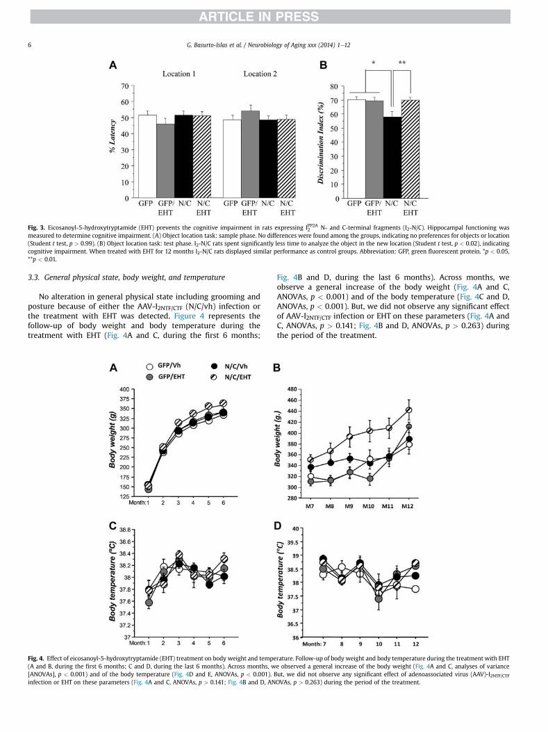
Fig. 3. Eicosanoyl-5-hydroxytryptamide (EHT) prevents the cognitive impairment in rats expressing IPP2A2 N- and C-terminal fragments (I2-N/C). Hippocampal functioning wasmeasured to determine cognitive impairment. (A) Object location task: sample phase. No differences were found among the groups, indicating no preferences for objects or location(Student t test, p > 0.99). (B) Object location task: test phase. I2-N/C rats spent significantly less time to analyze the object in the new location (Student t test, p < 0.02), indicatingcognitive impairment. When treated with EHT for 12 months I2-N/C rats displayed similar performance as control groups. Abbreviation: GFP, green fluorescent protein. *p < 0.05,**p < 0.01.
G. Basurto-Islas et al. / Neurobiology of Aging xxx (2014) 1e126
3.3. General physical state, body weight, and temperature
No alteration in general physical state including grooming andposture because of either the AAV-I2NTF/CTF (N/C/vh) infection orthe treatment with EHT was detected. Figure 4 represents thefollow-up of body weight and body temperature during thetreatment with EHT (Fig. 4A and C, during the first 6 months;
Fig. 4. Effect of eicosanoyl-5-hydroxytryptamide (EHT) treatment on body weight and tempe(A and B, during the first 6 months; C and D, during the last 6 months). Across months, w[ANOVAs], p < 0.001) and of the body temperature (Fig. 4D and E, ANOVAs, p < 0.001). Binfection or EHT on these parameters (Fig. 4A and C, ANOVAs, p > 0.141; Fig. 4B and D, AN
Fig. 4B and D, during the last 6 months). Across months, weobserve a general increase of the body weight (Fig. 4A and C,ANOVAs, p < 0.001) and of the body temperature (Fig. 4C and D,ANOVAs, p < 0.001). But, we did not observe any significant effectof AAV-I2NTF/CTF infection or EHT on these parameters (Fig. 4A andC, ANOVAs, p > 0.141; Fig. 4B and D, ANOVAs, p > 0.263) duringthe period of the treatment.
rature. Follow-up of body weight and body temperature during the treatment with EHTe observed a general increase of the body weight (Fig. 4A and C, analyses of varianceut, we did not observe any significant effect of adenoassociated virus (AAV)-I2NTF/CTFOVAs, p > 0.263) during the period of the treatment.

Fig. 5. General behavior in 6-month-old rats. (A) Assessment of neurologic examination (see Supplementary Table S1) quantitated as neuroscore. (B) Anxiety measured by timespent by an animal in the center of the arena. (C) Exploratory activity measured by distance covered exploring the arena. Eicosanoyl-5-hydroxytryptamide (EHT) treatment had nosignificant effect on any of the previously mentioned measures of general behavior.
G. Basurto-Islas et al. / Neurobiology of Aging xxx (2014) 1e12 7
3.4. Evaluation after 6 months of treatment with EHT
Figure 5A represents the neuroscore. Statistical analysis ofdata obtained from the assessment of neurologic examinationdid not reveal any significant difference among groups (Fig. 5A,Kruskal-Wallis test, p ¼ 0.380). This result indicates that, at 6months of age, AAV-I2NTF/CTF rats did not present any impairment inneurologic function and that treatment with EHT did not induceany neurologic side effects.
As shown in Fig. 5B, there was a tendency for AAV-I2NTF/CTF (N/C/vh) rats to visit less the center of the arena than other groups. Butstatistical analysis did not reveal any significant difference (Fig. 5B,ANOVA, p¼ 0.736) among groups. This tendency to explore less thecenter of the open field suggested that, as the pathology develops inAAV-I2NTF/CTF rats, anxiety levels would shift toward hyperanxiety.However, treatment with EHT restored anxiety to normal levels.
During the 20 minutes of free exploration, all groups coveredsimilar distance (Fig. 5C, ANOVA, p ¼ 0.852), suggesting that allanimals displayed similar level of exploration. No effect of thetreatment was observed.
3.5. Evaluation after 12 months of treatment with EHT
Figure 6A represents performance of rats in the hind-limbextension reflex test. Statistical analysis did not reveal any differ-ence between groups (Fig. 6A, Student t tests, p> 0.059). This resultindicates that, at 12 months of age, AAV-I2NTF/CTF rats did not pre-sent any deterioration of peripheral neurologic functions and thattreatment with EHT did not induce any peripheral neurologic sideeffects.
As shown in Fig. 6B, statistical analysis did not reveal any sig-nificant difference between groups in the prehensile traction test(Fig. 6B, Student t tests, p> 0.200). As represented in the regressionchart, correlation analysis of the body weight and the prehensiletraction score of the animals did not show any significant effectbetween body weight and performance in the test (Fig. 6C). Theseresults indicate that neither the AAV-I2NTF/CTF infection nor thetreatment with EHT induced any changes in forelimb musclestrength.
Finally, Fig. 6DeG represents analysis of different parameters forthe footprint test. Statistical analysis did not reveal any differencebetween groups in any parameter (Fig. 6DeG, Student t tests, p >
0.528). These analyses showed that neither the AAV-I2NTF/CTFinfection nor the treatment with EHT altered motor coordinationand synchrony.
3.6. Dietary supplementation with EHT blocks IPP2A2 inhibition ofPP2A and prevents tau hyperphosphorylation
I2-N/C fragments are known to bind to PP2Ac and inhibitphosphatase activity (Arnaud et al., 2011). We therefore comparedthe levels of PP2A activity toward phospho-tau in the brains of ratsraised with and without dietary EHT supplementation. On normaldiets, PP2A activity was significantly lower in rats that express I2-N/C than in GFP controls. EHT dietary supplementation completelyrescued the I2-N/Ceinduced PP2A deficiency (Fig. 7A and B).Neither I2-N/C expression nor EHT treatment caused any significantchanges in the level of total PP2Ac protein. These data confirm thatexpression of I2-N/C inhibits PP2A activity and that treatment withEHT can block I2-N/Ceinduced PP2A inhibition.
The principal form of PP2A that dephosphorylates phospho-taurequires carboxy methylation at its C-terminus (Tolstykh et al.,2000; Wu et al., 2000). Levels of PP2A methylation arecontrolled by a balance between the activities of 2 highlyconserved PP2A-specific enzymes: a methyl transferase, PPMT,that transfers methyl groups from S-adenosylmethionine to thePP2A carboxy terminus and a methyl esterase, PME, that deme-thylates PP2A. EHT was initially identified as a component incoffee extracts that inhibited the PP2A demethylation reaction(Lee et al., 2013). Dramatic increases in PP2A demethylation havebeen observed in brains from AD patients (Sontag et al., 2004). Theeffect of EHT on levels of PP2A methylation in I2-N/C and controlrats was therefore investigated, and it was found that EHT treat-ment significantly reduced the levels of demethylated PP2Ac in I2-N/C rats; a similar trend was seen in GFP controls (Fig. 7C and D).Together, these data suggest that the expression of I2-N/C de-creases PP2A activity and that the treatment with EHT that blocksPP2A demethylation (Lee et al., 2011) can rescue the I2-N/C inhi-bition of phosphatase activity.
Because PP2A is the major brain phospho-tau phosphatase(Bennecib et al., 2000; Gong et al., 2000; Liu et al., 2005), I2-N/Cinhibition of PP2A activity would be expected to cause tau hyper-phosphorylation. Western blot analysis of tau phosphorylationconfirmed this supposition. I2-N/C expression significantlydecreased the total tau level and increased tau hyper-phosphorylation compared with GFP control animals at T205, T212,S214, S262/356, and S396, but not at S199, a PP2A nonpreferred site(Liu et al., 2005) (Fig. 8). Treatment with EHT reduced I2-N/Ceinduced tau hyperphosphorylation at most of these sites; therewere no significant differences between EHT-treated experimentaland GFP control rats. These results indicate that I2-N/C expressioninduces abnormal hyperphosphorylation of tau through inhibition

Fig. 6. Lack of any side effects in eicosanoyl-5-hydroxytryptamide (EHT)etreated and control-treated rats at 12 months of age. Different tasks corresponding to general behaviorperformed at 12 months of age in the 4 groups of rats. (A) Clasping reflex score. (B and C) Prehensile traction score. (DeF) Stride length, gait, and placement in footprint test. (G)Latency to explore novel object in an arena in neophobia test. No significant differences among the 4 groups of animals were found in any of the previously mentioned generalbehavioral tests, indicating no adverse effect of EHT treatment.
G. Basurto-Islas et al. / Neurobiology of Aging xxx (2014) 1e128
of PP2A activity and that treatment with EHT ameliorates thispathology.
PP2A activity and the levels of tau hyperphosphorylation areknown to have a negative correlation at the phosphorylation sitesmost associated with pathology in AD brain (Liu et al., 2005). Asshown in Figs. 7 and 8, PP2A activity was significantly decreased inI2-N/C rats, tau hyperphosphorylation was coordinately increased,and both the changes in PP2A and tau phosphorylation wererescued in EHT-treated animals. To further establish that thehyperphosphorylation of tau that was observed in I2-N/C rats inthe present study was because of the inhibition of PP2A activity,we evaluated this possibility with Spearman nonparametric cor-relation analysis. We found a significant negative correlation be-tween the PP2A activity and the abnormal hyperphosphorylationof tau at T205 (p ¼ 0.0307), T212 (p ¼ 0.046), S262/356 (p ¼0.0002), S396 (p ¼ 0.0064), and S396/404 (p ¼ 0.0434) (Fig. 8).These data strongly support the hypothesis that EHT preventedI2-N/T-induced hyperphosphorylation of tau by maintaining
healthy levels of active PP2A in the presence of overexpressedI2-N/C.
3.7. EHT treatment reduces intraneuronal Ab accumulation
Reduced levels of PP2A activity induced by I2-N/C (Bologninet al., 2012) and by other means (Sontag et al., 2007) have previ-ously been shown to lead to increases in Ab load. The effect of EHTtreatment on the accumulation of intraneuronal Ab in I2-N/C ratswas therefore investigated. The immunohistochemical analysisrevealed a low number of intraneuronal Ab-positive cells in ratstreated with EHT compared with nontreated rats (Fig. 9). Theseresults suggest that I2-N/C expression promotes the amyloidogenicprocessing of APP and that this effect can be rescued by EHTtreatment. Our findings, though preliminary, are in agreement withthose of Sontag et al. (2007) who showed that increase in PP2Aactivity can lead to nonamyloidogenic processing of APP.

Fig. 7. Eicosanoyl-5-hydroxytryptamide (EHT) increases phosphoprotein phosphatase 2A (PP2A) activity in IPP2A2 N- and C-terminal fragments (I2-N/C) rats. (A) Western blotquantification of I2-N/C and green fluorescent protein (GFP) rat hippocampi showing the effect of EHT treatment on the level of total PP2A catalytic subunit (PP2Ac). (B) PP2Aactivity was recovered in I2-N/C rats treated with EHT, evaluated by PP2A phosphatase assay. (C) EHT increased PP2Ac methylation and (D) reduced PP2Ac demethylation in I2-N/Crats. (E) Representative blots shown for demethylated (1D6), methylated (4D9), and total PP2Ac (6A3) and glyceraldehyde 3-phosphate dehydrogenase (GAPDH). * p < 0.05. The dataare shown as mean � standard error, normalized by GAPDH for total PP2A.
G. Basurto-Islas et al. / Neurobiology of Aging xxx (2014) 1e12 9
4. Discussion
AD is multifactorial and involves several different etiopatho-genic mechanisms (Iqbal et al., 2005b). The familial form, whichaccounts for <1%, and the sporadic form, which represents theremaining >99% of the cases of AD, are histopathologically iden-tical. Although the familial form of AD has been associated withcertain mutations in APP and presenilins 1 and 2, and the inheri-tance of the APOE4 allelemarkedly increases the risk for the disease,the causes of the sporadic form of the disease are not understood.Nevertheless, the decrease of PP2A activity associated with an in-crease in the expression, the cleavage and the translocation of IPP2A2 ,and a decrease in the methylation of PP2Ac reported in AD brain(Bolognin et al., 2012; Gong et al., 1993, 1995; Sontag et al., 2004;Tanimukai et al., 2005; Wang et al., 2010) can lead both to tauand Ab pathologies. The present study shows that the over-expression and cleavage of IPP2A2 can lead to increases in PP2Acdemethylation, inhibition of PP2A activity, tau hyper-phosphorylation, Ab expression, and cognitive impairment and thatall these changes are significantly reversed by a minor componentof coffee, EHT.
The AAV1-I2NTF-CTF rat model used in the present study wasdeveloped in our laboratory (Bolognin et al., 2012). Comparedwith transgenic animals, one of the major advantages of the
viral gene transfer technology used to generate the I2-N/C ratmodel is that long-term transgene expression is achievedwithout affecting the genetic background of the animal (Lawloret al., 2007). Rats injected with AAV serotype 1 vector encodingthe 2 fragments of IPP2A2 showed a marked reduction of PP2Aactivity beginning at 4 months of age (Bolognin et al., 2012). Thepresent study extended these findings to 13 months of age,when the cytosolic tau and Ab pathologies were evident, makingthis model appropriate to study the effect of changes in PP2Aactivity on AD-type changes. EHT was administered to rats fromthe age of 21 days to 13 months. Compared with nontreatedanimals, a decrease in demethylation of PP2Ac was observed inEHT-treated rats, indicating the efficacy of the compound overchronic long-term use. These results corroborate previous re-ports showing EHT efficacy in both attenuation of a-synuclei-nopathies in vivo after a 9-month treatment and inhibition ofPP2A demethylation in vitro (Lee et al., 2011). Pharmacokineticstudies delivering [3H]-EHT intraperitoneally revealed that thiscompound crosses the blood brain barrier achieving levels suf-ficient to inhibit PP2A demethylation, indicating its feasibility forproof of concept studies (Lee et al., 2011). EHT was identified asthe major PP2A demethylation inhibitor in coffee (Lee et al.,2011) and was reported to be effective in preclinical studies(Lee et al., 2011, 2013).
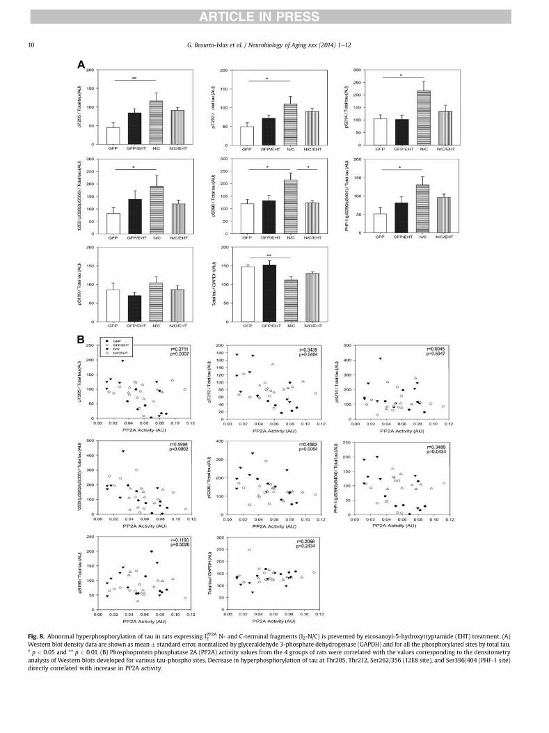
Fig. 8. Abnormal hyperphosphorylation of tau in rats expressing IPP2A2 N- and C-terminal fragments (I2-N/C) is prevented by eicosanoyl-5-hydroxytryptamide (EHT) treatment. (A)Western blot density data are shown as mean � standard error, normalized by glyceraldehyde 3-phosphate dehydrogenase (GAPDH) and for all the phosphorylated sites by total tau.* p < 0.05 and ** p < 0.01. (B) Phosphoprotein phosphatase 2A (PP2A) activity values from the 4 groups of rats were correlated with the values corresponding to the densitometryanalysis of Western blots developed for various tau-phospho sites. Decrease in hyperphosphorylation of tau at Thr205, Thr212, Ser262/356 (12E8 site), and Ser396/404 (PHF-1 site)directly correlated with increase in PP2A activity.
G. Basurto-Islas et al. / Neurobiology of Aging xxx (2014) 1e1210

Fig. 9. IPP2A2 N- and C-terminal fragments (I2-N/C) overexpression increases intra-neuronal amyloid b (Ab)1e40 load, which appears to be attenuated by eicosanoyl-5-hydroxytryptamide (EHT). (A) Representative micrographs of immunohistochemistry(IHC) from cerebral cortex showed Ab1e40 load increases in I2-N/C-treated (p < 0.05,n ¼ 3) but not EHT-treated rats. (B) IHC quantification of I2-N/C and green fluorescentprotein (GFP) rat hippocampi showing the effect of EHT treatment on the level ofAb1e40. EHT treatment appears to attenuate the Ab pathology (p ¼ 0.1, n ¼ 3 animalsper group; p < 0.01 for n ¼ 12 sections from 3 animals per group). *p < 0.05.
G. Basurto-Islas et al. / Neurobiology of Aging xxx (2014) 1e12 11
Epidemiologic studies have established a negative correlationbetween coffee consumption and the incidence of Parkinson’sdisease and AD (Barranco Quintana et al., 2007; Saaksjarvi et al.,2008). In the present study, we observed that rats expressing I2-N/C displayed increased PP2A demethylation and a decrease inPP2A activity and that treatment with EHT decreased PP2A deme-thylation and increased PP2A activity toward tau. Previous studiesshowed that methylation of PP2Ac affects phosphatase activity inpart by facilitating the binding of the regulatory Ba subunits to ACdimers (Tolstykh et al., 2000; Xu et al., 2008). It is not clear, how-ever, that the protective effects of EHT against IPP2A2 inhibitionderive entirely from inhibition of PP2A demethylation. The inhibi-tory effect of EHT on demethylation most likely stems from for-mation of a complex between EHT and PP2A that precludes theinteraction between PME and PP2A that is required for demethy-lation. Thus, the possibility cannot be excluded that the EHT/PP2Acomplex may also preclude the formation of inhibitory complexesbetween PP2A and IPP2A2 in much the same way that it appears toblock the interaction between PP2A and PME.
As a consequence of the decrease in PP2A activity in I2NTF-CTFrats, we observed a clear increase in tau hyperphosphorylation atmultiple sites. The association between these events was confirmedby correlation analysis, showing that decreases in PP2A activity arenegatively correlated with the hyperphosphorylation of tau atseveral sites, particularly the PP2A-dependent sites S262, T212,T205, and S396. S199 and S214 that are not preferred sites for PP2Adid not show any correlation. This analysis is consistent with pre-vious reports in AD cases (Liu et al., 2005). PP2A regulates phos-phorylation of tau both directly and by regulating the activities ofseveral tau protein kinases (Iqbal et al., 2005a). The effect of I2-N/Ccan be either direct by inhibiting the PP2A activity as we previouslydemonstrated (Arnaud et al., 2011) or through a downregulation ofBa subunit PP2A holoenzyme that specifically regulates the phos-phorylation of tau (Sontag et al., 2007; Xu et al., 2008).
In addition to tau pathology, we also observed an accumulationof intraneuronal Ab in I2-N/C rats, which was attenuated in animalstreated with EHT. These beneficial effects of EHT treatment maytarget early Ab pathologic mechanisms through an increase in PP2Aactivity. PP2A demethylation, for instance, has been reported to beassociated with a concomitant decrease in the steady-state releaseof neuroprotective APPa phosphorylated species and increasedsecretion of b- and g-secretaseecleaved APP fragments, inducing ashift in APP processing toward the amyloidogenic pathway (Sontaget al., 2007). Increased APP phosphorylation, either directly throughdecreased activity of PP2A toward phospho-APP or indirectlythrough reduced phosphatase activity toward phospho-JNK, canresult in increased Ab production (Colombo et al., 2009). Thus,activation of PP2A by small molecules such as EHT, offers a newtherapeutic approach for the prevention and treatment of AD andother neurodegenerative disorders, including tauopathies such asfrontotemporal dementias (Voronkov et al., 2011).
Disclosure statement
Conflicts of interest disclosure: Signum Biosciences has a USpatent on the EHTcompound used for this study. The animal studiesconform to National Institutes of Health guidelines and wereapproved by our institutional Institutional Animal Care and UseCommittee.
Acknowledgements
We thank Dr Ezzat El-Akkad for help in the preparation of thefigures and Ms Janet Murphy for secretarial assistance. Studiesdescribed in this article were supported in part by the New YorkState Office of People with Developmental Disabilities, NationalInstitutes of Health grant AG019158, and a research grant from(Signum Biosciences Inc, Princeton, NJ, USA).
Appendix A. Supplementary data
Supplementary data associated with this article can be found, inthe online version, at http://dx.doi.org/10.1016/j.neurobiolaging.2014.06.012.
References
Alonso, A.D., Zaidi, T., Grundke-Iqbal, I., Iqbal, K., 1994. Role of abnormally phos-phorylated tau in the breakdown of microtubules in Alzheimer disease. Proc.Natl. Acad. Sci. U.S.A 91, 5562e5566.
Alonso, A.D., Di Clerico, J., Li, B., Corbo, C.P., Alaniz, M.E., Grundke-Iqbal, I., Iqbal, K.,2010. Phosphorylation of tau at thr212, thr231, and ser262 combined causesneurodegeneration. J. Biol. Chem. 285, 30851e30860.
Arnaud, L., Chen, S., Liu, F., Li, B., Khatoon, S., Grundke-Iqbal, I., Iqbal, K., 2011.Mechanism of inhibition of pp2a activity and abnormal hyperphosphorylationof tau by i(2)(pp2a)/set. FEBS Lett. 585, 2653e2659.
Bancher, C., Brunner, C., Lassmann, H., Budka, H., Jellinger, K., Wiche, G.,Seitelberger, F., Grundke-Iqbal, I., Iqbal, K., Wisniewski, H.M., 1989. Accumula-tion of abnormally phosphorylated tau precedes the formation of neurofibril-lary tangles in Alzheimer’s disease. Brain Res. 477, 90e99.
Barranco Quintana, J.L., Allam, M.F., Serrano Del Castillo, A., Fernandez-CrehuetNavajas, R., 2007. Alzheimer’s disease and coffee: a quantitative review. Neurol.Res. 29, 91e95.
Bennecib, M., Gong, C., Wegiel, J., Lee, M.H., Grundke-Iqbal, I., Iqbal, K., 2000. In-hibition of protein phosphatases and regulation of tau phosphorylation in ratbrain. Alzheimer’s Rep. 3, 295e304.
Bensadoun, A., Weinstein, D., 1976. Assay of proteins in the presence of interferingmaterials. Anal. Biochem. 70, 241e250.
Bolognin, S., Blanchard, J., Wang, X., Basurto-Islas, G., Tung, Y.C., Kohlbrenner, E.,Grundke-Iqbal, I., Iqbal, K., 2012. An experimental rat model of sporadic Alz-heimer’s disease and rescue of cognitive impairment with a neurotrophicpeptide. Acta Neuropathol. 123, 133e151.
Cataldo, A.M., Petanceska, S., Terio, N.B., Peterhoff, C.M., Durham, R., Mercken, M.,Mehta, P.D., Buxbaum, J., Haroutunian, V., Nixon, R.A., 2004. Abeta localization

G. Basurto-Islas et al. / Neurobiology of Aging xxx (2014) 1e1212
in abnormal endosomes: association with earliest abeta elevations in AD andDown syndrome. Neurobiol. Aging 25, 1263e1272.
Chohan, M.O., Khatoon, S., Iqbal, I.G., Iqbal, K., 2006. Involvement of i2pp2a in theabnormal hyperphosphorylation of tau and its reversal by memantine. FEBSLett. 580, 3973e3979.
Colombo, A., Bastone, A., Ploia, C., Sclip, A., Salmona, M., Forloni, G., Borsello, T.,2009. Jnk regulates app cleavage and degradation in a model of Alzheimer’sdisease. Neurobiol. Dis. 33, 518e525.
Espana, J., Gimenez-Llort, L., Valero, J., Minano, A., Rabano, A., Rodriguez-Alvarez, J.,LaFerla, F.M., Saura, C.A., 2010. Intraneuronal beta-amyloid accumulation in theamygdala enhances fear and anxiety in Alzheimer’s disease transgenic mice.Biol. Psychiatry 67, 513e521.
Gandy, S., Simon, A.J., Steele, J.W., Lublin, A.L., Lah, J.J., Walker, L.C., Levey, A.I.,Krafft, G.A., Levy, E., Checler, F., Glabe, C., Bilker, W.B., Abel, T., Schmeidler, J.,Ehrlich, M.E., 2010. Days to criterion as an indicator of toxicity associated withhuman Alzheimer amyloid-beta oligomers. Ann. Neurol. 68, 220e230.
Gong, C.X., Singh, T.J., Grundke-Iqbal, I., Iqbal, K., 1993. Phosphoprotein phosphataseactivities in Alzheimer disease brain. J. Neurochem. 61, 921e927.
Gong, C.X., Shaikh, S., Wang, J.Z., Zaidi, T., Grundke-Iqbal, I., Iqbal, K., 1995. Phos-phatase activity toward abnormally phosphorylated tau: decrease in Alzheimerdisease brain. J. Neurochem. 65, 732e738.
Gong, C.X., Lidsky, T., Wegiel, J., Zuck, L., Grundke-Iqbal, I., Iqbal, K., 2000. Phos-phorylation of microtubule-associated protein tau is regulated by proteinphosphatase 2a in mammalian brain. Implications for neurofibrillary degener-ation in Alzheimer’s disease. J. Biol. Chem. 275, 5535e5544.
Grundke-Iqbal, I., Iqbal, K., Quinlan, M., Tung, Y.C., Zaidi, M.S., Wisniewski, H.M.,1986a. Microtubule-associated protein tau. A component of Alzheimer pairedhelical filaments. J. Biol. Chem. 261, 6084e6089.
Grundke-Iqbal, I., Iqbal, K., Tung, Y.C., Quinlan, M., Wisniewski, H.M., Binder, L.I.,1986b. Abnormal phosphorylation of the microtubule-associated protein tau(tau) in Alzheimer cytoskeletal pathology. Proc. Natl. Acad. Sci. U.S.A 83,4913e4917.
Grundke-Iqbal, I., Vorbrodt, A.W., Iqbal, K., Tung, Y.C., Wang, G.P., Wisniewski, H.M.,1988. Microtubule-associated polypeptides tau are altered in Alzheimer pairedhelical filaments. Brain Res. 464, 43e52.
Grundke-Iqbal, I., Iqbal, K., George, L., Tung, Y.C., Kim, K.S., Wisniewski, H.M., 1989.Amyloid protein and neurofibrillary tangles coexist in the same neuron inAlzheimer disease. Proc. Natl. Acad. Sci. U.S.A 86, 2853e2857.
Henckaerts, E., Dutheil, N., Zeltner, N., Kattman, S., Kohlbrenner, E., Ward, P.,Clement, N., Rebollo, P., Kennedy, M., Keller, G.M., Linden, R.M., 2009. Site-specific integration of adeno-associated virus involves partial duplication of thetarget locus. Proc. Natl. Acad. Sci. U.S.A 106, 7571e7576.
Iqbal, K., Grundke-Iqbal, I., Zaidi, T., Merz, P.A., Wen, G.Y., Shaikh, S.S.,Wisniewski, H.M., Alafuzoff, I., Winblad, B., 1986. Defective brain microtubuleassembly in Alzheimer’s disease. Lancet 2, 421e426.
Iqbal, K., Alonso, A., Chen, S., Chohan, M.O., El-Akkad, E., Gong, C.X., Khatoon, S.,Li, B., Liu, F., Rahman, A., Tanimukai, H., Grundke-Iqbal, I., 2005a. Tau pathologyin Alzheimer disease and other tauopathies. Biochim. Biophys. Acta 1739,198e210.
Iqbal, K., Flory, M., Khatoon, S., Soininen, H., Pirttila, T., Lehtovirta, M., Alafuzoff, I.,Blennow, K., Andreasen, N., Vanmechelen, E., Grundke-Iqbal, I., 2005b. Sub-groups of Alzheimer’s disease based on cerebrospinal fluid molecular markers.Ann. Neurol. 58, 748e757.
Klein, W.L., 2002. Abeta toxicity in Alzheimer’s disease: globular oligomers (addls)as new vaccine and drug targets. Neurochem. Int. 41, 345e352.
Kopke, E., Tung, Y.C., Shaikh, S., Alonso, A.C., Iqbal, K., Grundke-Iqbal, I., 1993.Microtubule-associated protein tau. Abnormal phosphorylation of a non-pairedhelical filament pool in Alzheimer disease. J. Biol. Chem. 268, 24374e24384.
Korenova, M., Zilka, N., Stozicka, Z., Bugos, O., Vanicky, I., Novak, M., 2009. Neuro-scale, the battery of behavioral tests with novel scoring system for phenotypingof transgenic rat model of tauopathy. J. Neurosci. Methods 177, 108e114.
Lawlor, P.A., Bland, R.J., Das, P., Price, R.W., Holloway, V., Smithson, L., Dicker, B.L.,During, M.J., Young, D., Golde, T.E., 2007. Novel rat Alzheimer’s disease modelsbased on aav-mediated gene transfer to selectively increase hippocampal abetalevels. Mol. Neurodegener. 2, 11.
Lee, K.W., Chen, W., Junn, E., Im, J.Y., Grosso, H., Sonsalla, P.K., Feng, X., Ray, N.,Fernandez, J.R., Chao, Y., Masliah, E., Voronkov, M., Braithwaite, S.P., Stock, J.B.,Mouradian, M.M., 2011. Enhanced phosphatase activity attenuates alpha-synucleinopathy in a mouse model. J. Neurosci. 31, 6963e6971.
Lee, K.W., Im, J.Y., Woo, J.M., Grosso, H., Kim, Y.S., Cristovao, A.C., Sonsalla, P.K.,Schuster, D.S., Jalbut, M.M., Fernandez, J.R., Voronkov, M., Junn, E.,Braithwaite, S.P., Stock, J.B., Mouradian, M.M., 2013. Neuroprotective and anti-inflammatory properties of a coffee component in the mptp model of Parkin-son’s disease. Neurotherapeutics 10, 143e153.
Li, M., Guo, H., Damuni, Z., 1995. Purification and characterization of two potentheat-stable protein inhibitors of protein phosphatase 2a from bovine kidney.Biochemistry 34, 1988e1996.
Li, M., Makkinje, A., Damuni, Z., 1996. The myeloid leukemia-associated protein setis a potent inhibitor of protein phosphatase 2a. J. Biol. Chem. 271, 11059e11062.
Liu, F., Grundke-Iqbal, I., Iqbal, K., Gong, C.X., 2005. Contributions of protein phos-phatases pp1, pp2a, pp2b and pp5 to the regulation of tau phosphorylation. Eur.J. Neurosci. 22, 1942e1950.
Malkova, L., Mishkin, M., 2003. One-trial memory for object-place associations afterseparate lesions of hippocampus and posterior parahippocampal region in themonkey. J. Neurosci. 23, 1956e1965.
Masters, C.L., Simms, G., Weinman, N.A., Multhaup, G., McDonald, B.L.,Beyreuther, K., 1985. Amyloid plaque core protein in Alzheimer disease andDown syndrome. Proc. Natl. Acad. Sci. U.S.A 82, 4245e4249.
Mori, C., Spooner, E.T., Wisniewsk, K.E., Wisniewski, T.M., Yamaguch, H., Saido, T.C.,Tolan, D.R., Selkoe, D.J., Lemere, C.A., 2002. Intraneuronal abeta42 accumulationin Down syndrome brain. Amyloid 9, 88e102.
Morris, R.G., Garrud, P., Rawlins, J.N., O’Keefe, J., 1982. Place navigation impaired inrats with hippocampal lesions. Nature 297, 681e683.
Oddo, S., Caccamo, A., Shepherd, J.D., Murphy, M.P., Golde, T.E., Kayed, R.,Metherate, R., Mattson, M.P., Akbari, Y., LaFerla, F.M., 2003. Triple-transgenicmodel of Alzheimer’s disease with plaques and tangles: intracellular abeta andsynaptic dysfunction. Neuron 39, 409e421.
Riedel, G., Micheau, J., Lam, A.G., Roloff, E.L., Martin, S.J., Bridge, H., de Hoz, L.,Poeschel, B., McCulloch, J., Morris, R.G., 1999. Reversible neural inactivationreveals hippocampal participation in several memory processes. Nat. Neurosci.2, 898e905.
Saaksjarvi, K., Knekt, P., Rissanen, H., Laaksonen, M.A., Reunanen, A., Mannisto, S.,2008. Prospective study of coffee consumption and risk of Parkinson’s disease.Eur. J. Clin. Nutr. 62, 908e915.
Santacruz, K., Lewis, J., Spires, T., Paulson, J., Kotilinek, L., Ingelsson, M.,Guimaraes, A., DeTure, M., Ramsden, M., McGowan, E., Forster, C., Yue, M.,Orne, J., Janus, C., Mariash, A., Kuskowski, M., Hyman, B., Hutton, M., Ashe, K.H.,2005. Tau suppression in a neurodegenerative mouse model improves memoryfunction. Science 309, 476e481.
Seubert, P., Mawal-Dewan, M., Barbour, R., Jakes, R., Goedert, M., Johnson, G.V.,Litersky, J.M., Schenk, D., Lieberburg, I., Trojanowski, J.Q., Lee, V. M-Y., 1995.Detection of phosphorylated ser262 in fetal tau, adult tau, and paired helicalfilament tau. J. Biol. Chem. 270, 18917e18922.
Sontag, E., Hladik, C., Montgomery, L., Luangpirom, A., Mudrak, I., Ogris, E.,White III, C.L., 2004. Downregulation of protein phosphatase 2a carboxylmethylation and methyltransferase may contribute to Alzheimer diseasepathogenesis. J. Neuropathol. Exp. Neurol. 63, 1080e1091.
Sontag, E., Nunbhakdi-Craig, V., Sontag, J.M., Diaz-Arrastia, R., Ogris, E., Dayal, S.,Lentz, S.R., Arning, E., Bottiglieri, T., 2007. Protein phosphatase 2a methyl-transferase links homocysteine metabolism with tau and amyloid precursorprotein regulation. J. Neurosci. 27, 2751e2759.
Tanaka, T., Zhong, J., Iqbal, K., Trenkner, E., Grundke-Iqbal, I., 1998. The regulation ofphosphorylation of tau in sy5y neuroblastoma cells: the role of protein phos-phatases. FEBS Lett. 426, 248e254.
Tanimukai, H., Grundke-Iqbal, I., Iqbal, K., 2005. Up-regulation of inhibitors ofprotein phosphatase-2a in Alzheimer’s disease. Am. J. Pathol. 166, 1761e1771.
Tolstykh, T., Lee, J., Vafai, S., Stock, J.B., 2000. Carboxyl methylation regulatesphosphoprotein phosphatase 2a by controlling the association of regulatory bsubunits. EMBO J. 19, 5682e5691.
Tsujio, I., Zaidi, T., Xu, J., Kotula, L., Grundke-Iqbal, I., Iqbal, K., 2005. Inhibitors ofprotein phosphatase-2a from human brain structures, immunocytologicallocalization and activities towards dephosphorylation of the Alzheimer typehyperphosphorylated tau. FEBS Lett. 579, 363e372.
Voronkov, M., Braithwaite, S.P., Stock, J.B., 2011. Phosphoprotein phosphatase 2a: anovel druggable target for Alzheimer’s disease. Future Med. Chem. 3,821e833.
Wang, X., Blanchard, J., Kohlbrenner, E., Clement, N., Linden, R.M., Radu, A.,Grundke-Iqbal, I., Iqbal, K., 2010. The carboxy-terminal fragment of inhibitor-2of protein phosphatase-2a induces Alzheimer disease pathology and cognitiveimpairment. FASEB J. 24, 4420e4432.
Wong, C.W., Quaranta, V., Glenner, G.G., 1985. Neuritic plaques and cerebrovascularamyloid in Alzheimer disease are antigenically related. Proc. Natl. Acad. Sci.U.S.A 82, 8729e8732.
Wu, J., Tolstykh, T., Lee, J., Boyd, K., Stock, J.B., Broach, J.R., 2000. Carboxylmethylation of the phosphoprotein phosphatase 2a catalytic subunit promotesits functional association with regulatory subunits in vivo. EMBO J. 19,5672e5681.
Xu, Y., Chen, Y., Zhang, P., Jeffrey, P.D., Shi, Y., 2008. Structure of a protein phos-phatase 2a holoenzyme: insights into b55-mediated tau dephosphorylation.Mol. Cell. 31, 873e885.
Zolotukhin, S., Potter, M., Zolotukhin, I., Sakai, Y., Loiler, S., Fraites Jr., T.J.,Chiodo, V.A., Phillipsberg, T., Muzyczka, N., Hauswirth, W.W., Flotte, T.R.,Byrne, B.J., Snyder, R.O., 2002. Production and purification of serotype 1, 2, and 5recombinant adeno-associated viral vectors. Methods 28, 158e167.