Neurobiology of Aggression and - Allan Siegel
290
-
Upload
manuel-jose-rengifo -
Category
Documents
-
view
108 -
download
17
description
Social Psychology
Transcript of Neurobiology of Aggression and - Allan Siegel


Copyr
CRC PR ESSBoca Raton London New York Washington, D.C.
Allan Siegel
The Neurobiology ofAggression and Rage
ight © 2005 CRC Press LLC
TF1661_C000.fm Page ii Tuesday, October 26, 2004 10:31 AM
Copyr
This book contains information obtained from authentic and highly regarded sources. Reprinted materialis quoted with permission, and sources are indicated. A wide variety of references are listed. Reasonableefforts have been made to publish reliable data and information, but the author and the publisher cannotassume responsibility for the validity of all materials or for the consequences of their use.
Neither this book nor any part may be reproduced or transmitted in any form or by any means, electronicor mechanical, including photocopying, microfilming, and recording, or by any information storage orretrieval system, without prior permission in writing from the publisher.
The consent of CRC Press does not extend to copying for general distribution, for promotion, for creatingnew works, or for resale. Specific permission must be obtained in writing from CRC Press for suchcopying.
Direct all inquiries to CRC Press, 2000 N.W. Corporate Blvd., Boca Raton, Florida 33431.
Trademark Notice: Product or corporate names may be trademarks or registered trademarks, and areused only for identification and explanation, without intent to infringe.
Visit the CRC Press Web site at www.crcpress.com
© 2005 by CRC Press
No claim to original U.S. Government worksInternational Standard Book Number 0-415-30834-8
Library of Congress Card Number 2004057043Printed in the United States of America 1 2 3 4 5 6 7 8 9 0
Printed on acid-free paper
Library of Congress Cataloging-in-Publication Data
Siegel, Allan.The neurobiology of aggression and rage / Allan Siegel.
p. cm.Includes bibliographical references and index.ISBN 0-415-30834-8 (alk. paper)1. Aggressiveness--Physiological aspects. 2. Anger--Physiological aspects. 3. Neuropsychology. I. Title.[DNLM: 1. Neurobiology. 2. Aggression--physiology. 3. Rage--physiology. WL 102S571n 2004]QP401.S555 2004155.2′32—dc22 2004057043
ight © 2005 CRC Press LLC

TF1661_C000.fm Page iii Tuesday, October 26, 2004 10:31 AM
Copyr
Dedication
To the memory of Dr. John P. Flynn, whose dedication to scientific truth, scholarship, and ethical conduct represented the gold standard for those who were privileged to study
under his guidance.
“Who is strong? He who conquers his evil inclination, as it is said: ‘Better is one slow to anger than a strong man, and one who rules over his spirit than a conqueror of a city’ ”
[Ethics of the Fathers, Chapter 4]
ight © 2005 CRC Press LLC
TF1661_C000.fm Page v Tuesday, October 26, 2004 10:31 AM
Copyr
Preface
The basic goal of this book is to provide an up-to-date review and analysis of thebiological factors and processes that are involved in the expression and control ofrage and aggressive behavior. The major focus of the book is to provide an under-standing of the anatomical substrates of the major forms of aggression as well as toexamine their basic underlying physiological, neurochemical and genetic mecha-nisms. This analysis is determined mainly from animal research in order to illustratehow such knowledge helps us to understand the neurology of human aggression. Afinal chapter provides the author’s views on how the neurobiology of aggression andrage can be utilized to understand and control our own aggressive tendencies.
ight © 2005 CRC Press LLC
TF1661_C000.fm Page vii Tuesday, October 26, 2004 10:31 AM
Copyr
The Author
Allan Siegel, Ph.D., is professor of neurosciences and psychiatry in the Departmentsof Neurology & Neuroscience and Psychiatry at the New Jersey Medical School inNewark. For the past 39 years, Dr. Siegel’s research has been dedicated to theneurobiology of aggression and rage, including studies concerning neurophysiolog-ical and neurochemical mechanisms and neuroanatomical substrates for these pro-cesses. He has published over 260 papers and abstracts, including the Pretest Seriesin Neurosciences, and is co-authoring a textbook in neurosciences that is nearcompletion.
ight © 2005 CRC Press LLC
TF1661_C000.fm Page ix Tuesday, October 26, 2004 10:31 AM
Copyr
Acknowledgment
Portions of the research presented in this book were supported by NIH Grant NS07941 and by grants from the Harry Frank Guggenheim Foundation to the author.
ight © 2005 CRC Press LLC

TF1661_C000.fm Page xi Tuesday, October 26, 2004 10:31 AM
Copyr
Table of Contents
Chapter 1 What Is Aggression?
Animal Models of Aggression Other Models of AggressionIs Aggressive Behavior Bimodal?
Offensive and Defensive Behavior Offense Defense
Aggression Induced by Brain Stimulation in Rat: Limitations of Offense–Defense Dichotomy
Defensive Rage (Affective Defense) versus Predatory AttackDo Equivalents to Affective Defense and Predatory Attack
Exist in Humans?Affective DefensePredatory Attack
Hostile versus Instrumental AggressionSummaryReferences
Chapter 2 History of the Neurology of Aggression and Rage
Effects of Ablations Attempts to Identify Anatomical Locus of Rage Mechanism, and Notion of
“Sham Rage”More Selective Forebrain and Brainstem Lesions
HypothalamusMidbrain Periaqueductal GrayLimbic Structures
Stimulation of Regions Mediating Aggression and RageReferences
Chapter 3 The Neuroanatomy of Aggression and Rage
Neuroanatomical MethodsPathways Mediating Defensive Rage in Cats
HypothalamusMidbrain Periaqueductal Gray
Pathways Mediating Predatory Attack in Cats
ight © 2005 CRC Press LLC

TF1661_C000.fm Page xii Tuesday, October 26, 2004 10:31 AM
Copyr
Relationship between Predatory Attack and Defensive Rage: Linkage between Medial and Lateral Hypothalamus
Hypothalamic Aggression in RatsNature of Attack Response Attack Sites in RatsEfferent Pathways from Hypothalamus Mediating Attack in Rats
SummaryReferences
Chapter 4 Physiological Processes and Mechanisms
Diencephalon and BrainstemRelative Roles of Hypothalamus, Brainstem, and Other Regions in
Expression of Aggression and RageHypothalamusMidbrain PAG and Related Structures Evidence from Studies Conducted in RatsAggression in Other Species Induced by Brain Stimulation
Physiological Properties of Stimulation-Induced Aggression and RageModulation from Hypothalamus and Periaqueductal GrayOther Effects of Electrical Stimulation of Attack Sites in CatsNeurophysiological Relationships Other Regions Associated with Rage and AggressionRole of Sensory Processes in Aggressive Behavior
OlfactionTactile StimulationMotor Aspects of Predatory Attack Behavioral Properties of Attack BehaviorDiscriminative Properties of AggressionReinforcing Properties of Rage and AggressionAggression or Feeding?
Avoidance, Play, and Predatory AttackSummaryReferences
Chapter 5 Limbic System I: Behavioral, Anatomical, and Physiological Considerations
Hippocampal FormationAnatomical ConsiderationsRelationship to Aggression and Rage
Septal AreaAnatomical ConsiderationsRelationship to Aggression and Rage
Amygdala
ight © 2005 CRC Press LLC
TF1661_C000.fm Page xiii Tuesday, October 26, 2004 10:31 AM
Copyr
Anatomical ConsiderationsAfferent ConnectionsEfferent ConnectionsRelationship to Aggression and RageEffects of Seizures
Seizures Induced by Electrical Stimulation of AmygdalaEffects of Amygdaloid Seizures on Spontaneously
Elicited Attack Prefrontal Cortex and Cingulate Gyrus
BackgroundAnatomical ConsiderationsRelationship to Aggression and Rage
Related Structures of Limbic Forebrain: Substantia Innominata and Bed Nucleus of Stria TerminalisBed Nucleus of Stria Terminalis
Anatomical ConsiderationsRelationship to Aggression and Rage
Substantia InnominataAnatomical ConsiderationsRelationship to Aggression
Limbic System and Human AggressionSummaryReferences
Chapter 6 Limbic System II: Functional Neuroanatomy
Methods UsedStudy Results
Hippocampal Formation Principal Findings Conclusions
Septal AreaPrincipal Findings Conclusions
AmygdalaPrincipal Findings Basomedial NucleusCortical NucleusMedial NucleusAmygdalo-Hippocampal Area Bed Nucleus of Stria Terminalis Anterior Amygdaloid AreaLateral NucleusCentral NucleusBasolateral Nucleus
ight © 2005 CRC Press LLC

TF1661_C000.fm Page xiv Tuesday, October 26, 2004 10:31 AM
Copyr
Prepyriform Cortex Entorhinal CortexSubstantia InnominataConclusions
Prefrontal Cortex and Associated Thalamus NucleiPrincipal FindingsMedial Prefrontal, Sulcal, and Infralimbic CorticesMediodorsal Thalamic NucleusMidline Thalamic NucleiConclusions
Overall ConclusionsAbbreviations Used in FiguresReferences
Chapter 7 The Neurochemistry of Rage and Aggression
AcetylcholineSynthesis and RemovalDistribution and FunctionsRole of ACh in Aggression and Rage
Effects of Systemic Administration of Cholinergic AgentsEffects of Intracerebral Microinjections of
Cholinergic AgentsGenetic Approach
SummaryNorepinephrine
Synthesis and RemovalDistribution and FunctionsRole of Norepinephrine in Aggression and Rage
Effects of Systemic and Intracerebroventricular Administration of Noradrenergic Agents
Neurochemical MeasurementsSummary
DopamineSynthesis and RemovalDistribution and FunctionsRole of Dopamine in Aggression and Rage
Effects of Systemic and Intracerebroventricular Administration of Dopaminergic Agents
Neurochemical MeasurementsGenetic ApproachesIntracerebral Administration of Dopaminergic Compounds
SummarySerotonin
Synthesis and Removal
ight © 2005 CRC Press LLC
TF1661_C000.fm Page xv Tuesday, October 26, 2004 10:31 AM
Copyr
Distribution and FunctionsRole of Serotonin in Aggression and Rage
Lesions or Stimulation of Serotonergic NucleiNeurochemical Depletion of SerotoninDepletion of Serotonin Levels by DietGenetic Manipulation of Brain Serotonin LevelsPeripheral Administration of Serotonergic CompoundsCentral Administration of Serotonergic Compounds
Neurochemical Measurements and ApproachesSummary
PeptidesOpioid Peptides
Characterization Distribution and FunctionsRole of Enkephalins in Aggression and Rage
Studies Conducted in RodentsStudies Conducted in Cats
Systemic Administration of Opioid CompoundsIntracerebral Administration of Opioid Compounds
SummarySubstance P
Characterization, Distribution, and FunctionsRole of SP in Rage and Aggression
Cholecystokinin (CCK)Characterization, Distribution, and FunctionsRole of CCK in Defensive Rage BehaviorSummary
Amino AcidsExcitatory Amino Acids (Glutamate)
Synthesis and RemovalDistribution and FunctionsRole of Excitatory Amino Acids in Defensive
Rage Behavior Summary
Inhibitory Amino Acids (GABA) Synthesis and RemovalDistribution and FunctionsRole of Inhibitory Amino Acids in Defensive Rage and
Predatory Attack BehaviorSummary
Can Substances of Abuse, Psychotropic Drugs, and Antidepressant Drugs Influence Aggression?Substances of Abuse
Opioid PeptidesAlcohol: Linkage between Alcohol Consumption and
Violent Behavior
ight © 2005 CRC Press LLC

TF1661_C000.fm Page xvi Tuesday, October 26, 2004 10:31 AM
Copyr
Psychotropic and Antidepressant DrugsSummary
References
Chapter 8 Aggression and Hormonal Status M. Demetrikopoulos and A. Siegel
Testosterone and Aggressive BehaviorExperimental Manipulation of Testosterone LevelsSeasonal Variations in Testosterone LevelsTestosterone Levels during DevelopmentTestosterone’s Role in Human Aggression
Estrogen and Aggressive BehaviorExperimental Manipulation of Estrogen LevelsNaturally Occurring Variations in Estrogen Levels Estrogen Levels during Development
Progesterone and Aggressive BehaviorExperimental Manipulation of Progesterone LevelsProgesterone Levels during Development
Adrenal Steroids and Aggressive BehaviorModulation of Hormonal-Aggression Interactions by Ethanol and
Illicit DrugsComments and Conclusions References
Chapter 9 Aggression and Immune FunctionM. Demetrikopoulos and A. Siegel
Social Status Effects on ImmunityStable Hierarchical GroupsHierarchy FormationSummary
Effects of Aggression on Recipient’s Immune FunctionSocial DisruptionSocial ConfrontationSummary
Effects of Aggression on Perpetrator’s Immune FunctionSocial Isolation–Induced FightingResident–Intruder InteractionStrain Differences in Behavior and Immune FunctionAggression, Hostility, and Marital Conflict: Effects on
Immunity in HumansSummary
Can Immune Events Affect Aggression? Immune Challenge Effects
ight © 2005 CRC Press LLC

TF1661_C000.fm Page xvii Tuesday, October 26, 2004 10:31 AM
Copyr
Autoimmune Disorder EffectsPotential MediatorsSummary
Converging Neuronal CircuitryRole of Cytokines Role of Periaqueductal GraySecondary Stress EffectsSummary
Overall SummaryReferences
Chapter 10 Genetics and Aggression
Studies Conducted in AnimalsEarly Behavioral Studies
Recent Studies Genetic Effects on Neuroanatomic Circuitry and Neurotransmitter–
Receptor SystemsStudies Conducted in HumansSummaryReferences
Chapter 11 Future Directions and Perspectives
References
ight © 2005 CRC Press LLC
TF1661_C001.fm Page 1 Monday, October 18, 2004 8:04 AM
Copyr
1
What Is Aggression?Violence and rage have become major public health and social problems in theUnited States and many other countries around the world. In particular, during thepast few years there have been more than 3 million violent crimes committed in theUnited States annually (Reiss et al., 1994), resulting in costs of billions of dollarsto society. In addition, the homicide rate among adolescents increased 238% between1985 and 1994 (Hennes, 1998). In the year 2000, an estimated 1.4 million violentcrimes were reported (FBI Press release, Oct. 22, 2001).
Violence is influenced by cultural, environmental, and social forces that shapethe manner in which it is expressed (Eron, 1987). Nevertheless, as supported by asignificant body of data described below and in subsequent chapters, the neural basisof aggressive behavior in humans resembles that of animals, and the forms ofaggression seen in humans parallel those observed in animals. Thus, some importantquestions are:
1. What are the forms of aggression that are studied in animal research?2. How are they related to each other? 3. How do they relate to human forms of aggression?4. What are the similarities, as well as differences, in the neural mechanisms
that govern each of these forms of aggression studied in animals?
In this chapter, I attempt to address the first three questions, and consider the fourthin subsequent chapters.
In attempting to identify models of aggression, it is constructive to first try todefine what is meant by “aggressive” behavior. Perhaps, the most common definitionof aggressive behavior used is a variant of one that was described by Moyer (1968)who referred to aggression as “a behavior that causes (or leads to) harm, damageor destruction of another organism.” While this definition would seem to include awide variety of conditions normally associated with aggressive behavior, it does notnecessarily include the classes of behavior that are associated with “threat” or relatedforms of “affect” that are intimidating in nature, and that are designed to control thebehavior of others. The notion of threat can also be expanded to include categoriesof behavior linked under the general rubric of “hostility.” Here, we have to distinguishbetween two levels of definition — one based on principles of social psychologyand the other based on likely underlying neuronal mechanisms. In terms of socialpsychology, it is possible and even reasonable to postulate distinct behavioral orsocial mechanisms that identify “hostility” as a unique aggressive process relativeto “threat” and “rage” (Kingsbury et al., 1997). However, in terms of the neuralmechanisms that may underlie these behaviors, it is conceivable and perhaps likely
ight © 2005 CRC Press LLC
TF1661_C001.fm Page 2 Monday, October 18, 2004 8:04 AM
Copyr
that they are highly similar. In terms of the major themes of this book, whereverpossible there will be attempts to collapse these into categories that are likelysubserved by common behavioral and neuronal substrates.
At the animal level, aggressive responses may include threatening postures andvocalizations such as hissing and growling. At the human level, the outward expres-sion of aggressive behavior can reach much higher levels of complexity and, accord-ingly, may include a much wider spectrum of responses. In particular, aggressiveresponses can be manifest in varieties of ways, extending from physical assaultthrough verbal and postural expressions. Aggression can even be of a passive formin which the aggressive display is in the form of a deliberate failure of an individualto perform a given act or task that was requested of him.
ANIMAL MODELS OF AGGRESSION
From the discussion above, it would seem natural to assume that aggressive behavioris not a unitary phenomenon, but instead may reflect various processes capturedunder a single common theme referred to as “aggressive behavior.” Complicatingthis matter further is the fact that different forms of aggression are identified oper-ationally on the basis of experimental methodologies applied by various investigatorsto the study of aggression. In 1968, Moyer attempted to define types of aggressionlargely on the basis of these methodologies. In effect, what Moyer established were“operational” definitions of aggression based on the arrangement of various envi-ronmental conditions. Because of the common use of several methodologies andtypes of aggression, these categories of aggression described by Moyer are summa-rized below as a basis for further discussion.
1. Fear-induced aggression. In this form of aggression, the animal is placedin a position where it would like to escape, either because of the degreeof confinement or the presence of a threatening animal in its environment.In this situation, however, escape is denied to the animal, and the animalturns, instead, to attack the attacking animal following its attempt toescape.
2. Maternal aggression. This form of aggression is one that occurs in mostforms of mammalian species, but is used infrequently in aggressionresearch. In this model, the critical stimulus is the presence of an organismin proximity to the young. Here, the closer the mother is situated to boththe young and threatening organism, the more likely an attack.
3. Inter-male aggression. In this form of aggression, the presence of a maletriggers aggressive reactions on the part of another male. The frequencyof bouts that occur between a male and another male greatly outweighthose that occur between a male and a female or between two females.Inter-male aggression shares some similarities with the resident–intrudermodel (see below), but differs from it in that the interactions do not takeplace within the subject’s territory.
4. Irritable aggression. This form of aggression occurs in response to athreat, intimidation, or an environmental condition that is annoying or
ight © 2005 CRC Press LLC
TF1661_C001.fm Page 3 Monday, October 18, 2004 8:04 AM
Copyr
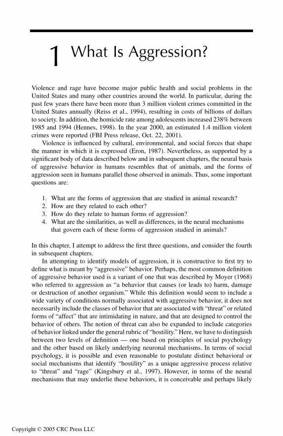
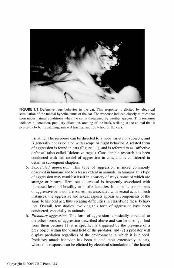
irritating. The response can be directed to a wide variety of subjects, andis generally not associated with escape or flight behavior. A related formof aggression is found in cats (Figure 1.1), and is referred to as “affectivedefense” (also called “defensive rage”). Considerable research has beenconducted with this model of aggression in cats, and is considered indetail in subsequent chapters.
5. Sex-related aggression. This type of aggression is more commonlyobserved in humans and to a lesser extent in animals. In humans, this typeof aggression may manifest itself in a variety of ways, some of which arestrange or bizarre. Here, sexual arousal is frequently associated withincreased levels of hostility or hostile fantasies. In animals, componentsof aggressive behavior are sometimes associated with sexual acts. In suchinstances, the aggressive and sexual aspects appear as components of thesame behavioral act, thus creating difficulties in classifying these behav-iors. Overall, few studies involving this form of aggression have beenconducted, especially in animals.
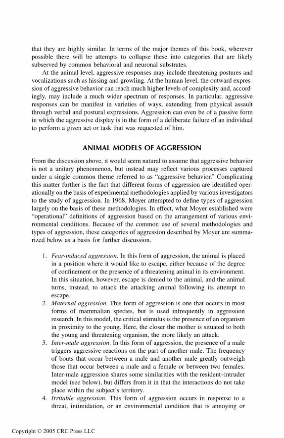
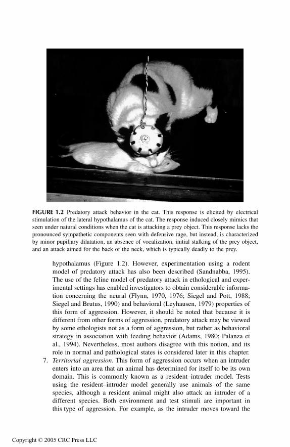
6. Predatory aggression. This form of aggression is basically unrelated tothe other forms of aggression described above and can be distinguishedfrom them because (1) it is specifically triggered by the presence of aprey object within the visual field of the predator, and (2) a predator willdisplay predation regardless of the environment in which it is placed.Predatory attack behavior has been studied most extensively in cats,where this response can be elicited by electrical stimulation of the lateral
FIGURE 1.1 Defensive rage behavior in the cat. This response is elicited by electricalstimulation of the medial hypothalamus of the cat. The response induced closely mimics thatseen under natural conditions when the cat is threatened by another species. This responseincludes piloerection, pupillary dilatation, arching of the back, striking at the animal that itperceives to be threatening, marked hissing, and retraction of the ears.
ight © 2005 CRC Press LLC
TF1661_C001.fm Page 4 Monday, October 18, 2004 8:04 AM
Copyr
hypothalamus (Figure 1.2). However, experimentation using a rodentmodel of predatory attack has also been described (Sandnabba, 1995).The use of the feline model of predatory attack in ethological and exper-imental settings has enabled investigators to obtain considerable informa-tion concerning the neural (Flynn, 1970, 1976; Siegel and Pott, 1988;Siegel and Brutus, 1990) and behavioral (Leyhausen, 1979) properties ofthis form of aggression. However, it should be noted that because it isdifferent from other forms of aggression, predatory attack may be viewedby some ethologists not as a form of aggression, but rather as behavioralstrategy in association with feeding behavior (Adams, 1980; Palanza etal., 1994). Nevertheless, most authors disagree with this notion, and itsrole in normal and pathological states is considered later in this chapter.
7. Territorial aggression. This form of aggression occurs when an intruderenters into an area that an animal has determined for itself to be its owndomain. This is commonly known as a resident–intruder model. Testsusing the resident–intruder model generally use animals of the samespecies, although a resident animal might also attack an intruder of adifferent species. Both environment and test stimuli are important inthis type of aggression. For example, as the intruder moves toward the
FIGURE 1.2 Predatory attack behavior in the cat. This response is elicited by electricalstimulation of the lateral hypothalamus of the cat. The response induced closely mimics thatseen under natural conditions when the cat is attacking a prey object. This response lacks thepronounced sympathetic components seen with defensive rage, but instead, is characterizedby minor pupillary dilatation, an absence of vocalization, initial stalking of the prey object,and an attack aimed for the back of the neck, which is typically deadly to the prey.
ight © 2005 CRC Press LLC
TF1661_C001.fm Page 5 Monday, October 18, 2004 8:04 AM
Copyr
periphery of the territorial demarcation, the likelihood of attack from theresident decreases. Several different types of intruders have been usedsuch as an unfamiliar male, an olfactory bulbectomized male, or a lactatingfemale. Each type of intruder generates a different probability of attackrelated to the sex of the resident. For example, a bulbectomized malemouse intruder will likely elicit attack from the resident male mouse,while a lactating female mouse intruder will induce attack more readilyfrom a female resident than a male resident mouse (Whalen and Johnson,1988).
OTHER MODELS OF AGGRESSION
Other models and operational definitions of aggression have been proposed that donot necessarily fit into the classification system proposed by Moyer. For example,one such model includes competition for a limited resource (Palanza et al., 1994).In this model, the subjects are placed into a situation in which they must interact toobtain basic commodities such as food and water, or to obtain access to a betterterritory or a mate. While this model may be viewed as a variation of the resi-dent–intruder model, it also differs from it in several ways. First, the resi-dent–intruder model involves interactions that take place in the resident’s territory,whereas in the competition model, the interaction takes place in an area that iscommon to both animals. Second, there is no need for competition to take placesince resources such as food and water are not limited. Another model is infanticide.Here, the subject is given access to pups and the variable measured is the death ofthe pups.
IS AGGRESSIVE BEHAVIOR BIMODAL?
Over the past few decades, a number of investigators have attempted to characterizeaggressive behavior in either animals or humans in a bimodal manner. In the animalliterature, the use of the terms offense and defense have been applied to describewhat appear to be opposing ends of a continuum (Adams, 1979). A second bimodalclassification scheme defines aggression along a continuum ranging from predatoryto affective defense behavior (Flynn, 1970; Siegel and Pott, 1988; Siegel and Brutus,1990). A third bimodal classification scheme was recently proposed by Kingsburyet al. (1997) for the study of human aggression. It suggests that aggressive behaviorcan be classified into two categories, which the author refers to as “factors.” Theseinclude instrumental aggression and hostile aggression. The following discussionsummarizes these classification schemes and approaches in the study of aggressivebehavior.
OFFENSIVE AND DEFENSIVE BEHAVIOR
Considerable effort has been expended, mainly through studies in rodents, in attempt-ing to characterize the terms “offensive” and “defensive” behaviors. In addition,several investigators have made attempts to identify the neuronal regions associated
ight © 2005 CRC Press LLC
TF1661_C001.fm Page 6 Monday, October 18, 2004 8:04 AM
Copyr
with each of these functions (see Adams, 1979; Blanchard et al., 1977a to 1977e;Blanchard and Blanchard, 1984; Mos et al., 1984). The neuronal concomitants ofaggression in rats will be considered in Chapter 3.
Offense
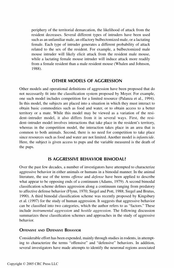
Offensive behavior in rats can typically be demonstrated with the use of the resi-dent–intruder model in which the alpha male (i.e., the resident) elicits the offensivebehavior (see Figure 1.3). Typically, offensive behavior is characterized primarilyas a form of threat together with a ritualized form of attack consisting of piloerection,a (lateral) bite and kick in which wounds are made mainly upon the back of theopponent. Since the wounds are basically superficial, it was suggested by Adams(1979) that offensive behavior provides a communicative function, namely, to servenotice of that animal’s dominance over the territory that it considers its own. How-ever, it has also been demonstrated that the site of the wounds is dependent on theenvironment of the animal. Most wounds involving largely the upper part of theback occur from rats housed individually in small cages, while rats housed in largercages with a female showed a smaller percentage of bites on the back with a greaterpercentage of bites directed at the head and belly (Mos et al., 1984).
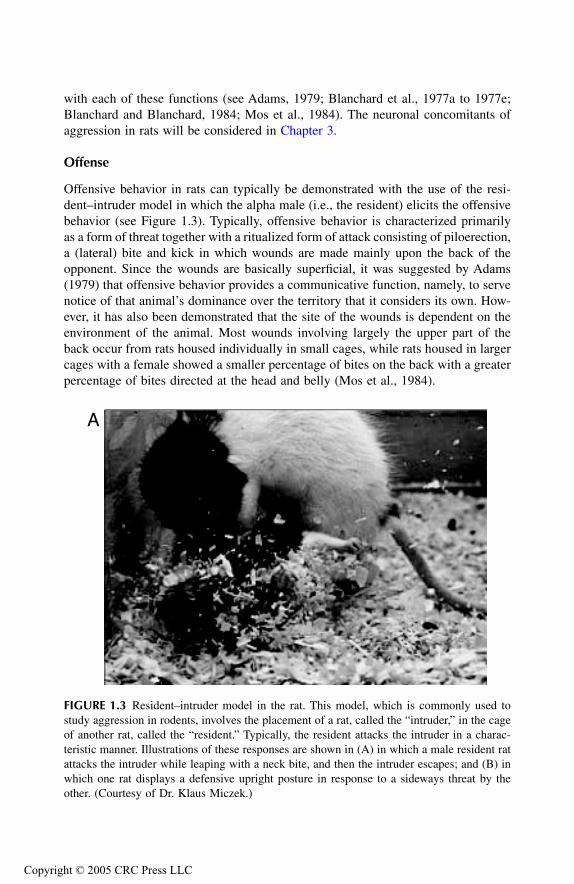
FIGURE 1.3 Resident–intruder model in the rat. This model, which is commonly used tostudy aggression in rodents, involves the placement of a rat, called the “intruder,” in the cageof another rat, called the “resident.” Typically, the resident attacks the intruder in a charac-teristic manner. Illustrations of these responses are shown in (A) in which a male resident ratattacks the intruder while leaping with a neck bite, and then the intruder escapes; and (B) inwhich one rat displays a defensive upright posture in response to a sideways threat by theother. (Courtesy of Dr. Klaus Miczek.)
A
ight © 2005 CRC Press LLC
TF1661_C001.fm Page 7 Monday, October 18, 2004 8:04 AM
Copyr
Experimental studies involving offensive behavior have not been conducted incats. However, a detailed ethological analysis of offensive behavior in felines hasbeen carried out (Leyhausen, 1979). According to Leyhausen, as the cat begins toapproach a conspecific, it draws itself high on its legs, stretching out its back andits head is pulled forward. The pupils are not dilated, but rather are slightly con-stricted, and the ears are turned outward in a sharp angle. As the offensive animalcontinues to approach the conspecific, it may elicit mewing and howling, whichmost likely constitutes a threat. It then raises its head, tilts it sideways while stillfocusing on the opponent, then turns the head from side to side, and as it passes theconspecific, attacks by attempting to bite the nape of the neck.
Defense
Most studies focused on aggressive behavior have involved the analysis of thebehavioral, anatomical, physiological, neurochemical, and neuropharmacologicalproperties of defensive behavior conducted principally in detail in both rodents andcats. In the rat, defense occurs in the presence of a predator, attacking conspecific,or any other stimulus that is perceived to be dangerous to the animal (Blanchardand Blanchard, 1989), and is sometimes alternatively referred to as “protectiveaggression” (Palanza et al., 1994). The behavioral characteristics of this responsemay include several of the following features: a lunge and bite attack, usually
FIGURE 1.3 (Continued)
B
ight © 2005 CRC Press LLC

TF1661_C001.fm Page 8 Monday, October 18, 2004 8:04 AM
Copyr
directed at the face or parts of the body, an upright posture, squealing, flight, freezing,submissive posture, and ultrasonic vocalization (Adams, 1979; Blanchard and Blan-chard, 1989). Several components of natural defense in the rodent are shown inFigure 1.3. It has also been reported that defensive behavior can be elicited byelectrical stimulation of the rat’s hypothalamus (Panksepp and Trowill, 1969). How-ever, brain stimulation–induced defense in the rat differs somewhat from that whichis induced under natural conditions. In particular, brain stimulation–induced defensein the rat includes a sudden retreat followed by attempts to escape by jumping outof the cage.
In the cat, defensive behavior occurs in the presence of another animal (eithera conspecific or an animal of another species) that is perceived to be a threat. Thebasic elements of this response include a flattening of the ears, a shrinking orlowering of the body, drawing in of the head, piloerection, hissing, pupillary dila-tation, and a stiffening of the tail that becomes motionless (Leyhausen, 1979). Quiteoften, such a response pattern is sufficient to cause the opponent to retreat, whichis the main objective of the defensive response. If the cat decides to attack, it doesso by striking with its paw. The paw strike is the primary means by which it signifiesits defense. If the opponent begins to attack, the cat eliciting defensive behavior willthen roll over on its back with its underside facing the opponent (Leyhausen, 1979).Such a response can interrupt the advance of the opponent. The continued use ofits forepaw may also be effective in warding off the opponent, especially if theforepaw strike lands on the head region of the opponent. If the opponent begins toattack, the cat that initially elicits defensive behavior may now change its strategyto that of offense as it attempts to scratch and bite the neck of the opponent. Asimilar pattern of responses is elicited by electrical stimulation of the cat’s medialhypothalamus or midbrain periaqueductal gray (PAG) (Hess and Brugger, 1943;Flynn, 1970; Chi and Flynn, 1971; Fuchs et al., 1985a, 1985b; Siegel and Pott, 1988;Siegel and Brutus, 1990).
AGGRESSION INDUCED BY BRAIN STIMULATION IN RAT: LIMITATIONS OF OFFENSE–DEFENSE DICHOTOMY
In contrast to cats, it is apparent that a variety of forms of aggression can be elicitedby electrical stimulation of several areas of the hypothalamus in rats (Kruk et al.,1979, 1980, 1983; Panksepp, 1971; Panksepp and Trowill, 1969; Woodworth, 1971).As noted above, elements of defensive behavior (Panksepp and Trowill, 1969) aswell as biting attack behavior (Woodworth, 1971) have been reported followingstimulation of the medial and lateral hypothalamus of the cat, respectively. In addi-tion, attack elicited from the intermediate hypothalamus (i.e., the region extendingfrom the lateral aspect of the ventromedial hypothalamus to the medial aspect ofthe lateral hypothalamus) produces a complex attack pattern that may include bitingof the head of the opponent, biting of the back accompanied by hind paw kickingof the flank, or a combination of jump attacks and clinch flights (Kruk et al., 1979,1980, 1983, 1990). The type of attack varies with the strain of rat. For example,head bites are dominant with the inbred strain, CPB-WEzob, while bites directed tothe back are common with random-bred albino Wistar CPB-WE rats.
ight © 2005 CRC Press LLC

TF1661_C001.fm Page 9 Monday, October 18, 2004 8:04 AM
Copyr
At the time of biting, the attacking rat may place its front paws around the neckor back of the opponent, while the hind limbs are placed in a position to kick theflank and other parts of the opponent’s body. If the opponent assumes an uprightposition, the attacking rat will modify its attacking pattern by eliciting jump attacks.In such an attack pattern, the attacking animal jumps a considerable distance toreach the opponent, at which time, it bites the head and kicks the body of theopponent with its hind paws. If the animal loses its balance after jumping, it maythen alter its attack to that of a clinch fight.
It is of interest to note that the attacking rat initially bites the head, then theupper and lower parts of the back, but not the ventral surface of the opponent (if itassumes a submissive position after it is attacked) (Kruk et al., 1983). This form ofhypothalamic attack may be directed against various targets. These include dominantand subordinate rats, dead and anesthetized rats, and mice. A male rat may attackanother male or a female, especially one that is not receptive (Kruk et al., 1984;Mos et al., 1987). In terms of the form of attack initiated, two additional facts shouldbe noted. The first is that the specific attack pattern is dependent on the behavior ofthe opponent, in which case the attacking animal may exhibit components of offenseand defense in response to the opponent. The second is that the pathways and neuralsystems mediating offense and defense, although not well delineated in the rat,presumably lie within overlapping regions of the forebrain and brainstem. Accord-ingly, stimulation of an attack site will likely activate regions that mediate offenseas well as defense.
In summary, the form of the attack is dependent on the (1) position of theopponent, including primarily the extent to which the head and back are exposed;(2) strategies adopted by the opponent; and (3) specific strain of the attacking animal.Consequently, the utility of applying the terms “offense” and “defense” in the contextof hypothalamically elicited aggressive behavior in the rat becomes questionable.Thus, it would appear to be difficult if not illogical to attempt to apply thesemotivational categories to define and classify hypothalamically elicited aggressionin the rat.
DEFENSIVE RAGE (AFFECTIVE DEFENSE) VERSUS PREDATORY ATTACK
One school of thought would suggest that aggressive forms of behavior could bereduced to two categories — affective defense and predatory attack. If this wereindeed the case, it should also be possible to reduce the seven types of aggressiondescribed by Moyer into these two categories. Indeed, categories of aggression thatinclude fear-induced, maternal, inter-male, sex-related, irritable, and territorialaggression may all share a similar common feature, namely, an aggressive responsebased on the presence of elements of fear and/or threat, which may be real orperceived. Thus, it is reasonable to re-label these categories of aggression under therubric “affective defense.”
Attention should be drawn to one possible source of confusion relating to thenomenclature that I am attempting to establish here. I just noted above that mixedcomponents of offense and defense may be present in the overall repertoire of theanimal in expressing aggressive behavior. Therefore, in the present analysis, while
ight © 2005 CRC Press LLC

TF1661_C001.fm Page 10 Monday, October 18, 2004 8:04 AM
Copyr
I may characterize the overall response patterns of six of seven Moyer categories asaffective defense, it is possible and indeed likely that the component of offense mayalso be represented here as well. In particular, in the resident–intruder model, mostauthors agree that elements of offense are clearly present in the behavior of theresident rat on the intruder even though the motivation underlying the aggressiveact may be one of perceived fear or threat to it by the intruder. Nevertheless, thatoffense may be present as a component of the behavioral repertoire of the animalis not inconsistent with the classification of the overall behavioral response asaffective defense. In contrast to affective defense, predatory attack is limited to asingle category of aggressive behavior, and therefore is far more restricted in itsoccurrence in nature.
DO EQUIVALENTS TO AFFECTIVE DEFENSE AND PREDATORY ATTACK EXIST IN HUMANS?
Affective Defense
To summarize our knowledge of affective defense behavior in animals, we mayconclude that affective defense occurs in response to a real or perceived threat. It isusually immediate following the sighting of the threat stimulus within the environ-ment, as thought and planning of the response appears to play little or no role in itsorganization. The response is also accompanied by marked sympathetic signs suchas marked increases in heart rate and blood pressure, piloerection, vocalization,lowering of the back, and retraction of the ears.
It is instructive to attempt to compare affective defense in animals with whatsome authors refer to as equivalent response forms in humans (Meloy, 1988, 1997;Vitiello et al., 1990). These authors characterize affective defense behavior as impul-sive, destructive to the object of the aggressor, and typically aversive to the aggressor.In addition, they further indicate that this response pattern includes poor modulationof behavior and high autonomic arousal. Questionnaire test items closely related toaffective defense encompass the following: aggression that was unplanned, an indi-vidual who was totally out of control during the aggressive act, aggression that hadno purpose, the person is exposed to physical harm during the time when he isaggressive, and an individual who damages his own property during the act ofaggression (Vitiello et al., 1990).
The characteristics associated with affective defense behavior in humans havebeen independently described in detail by Meloy (1988). Several of these charac-teristics clearly overlap with those described by Vitiello. Included are an intensesympathetic arousal, an affective attack based on a real or perceived (which couldbe delusional) threat to the person, and an immediate (i.e., impulsive) response tothe threat stimulus. Meloy extends his analysis by adding other properties of thisform of aggression. Specifically, the goal object of affective defense is to reduce oreliminate the threat object from the environment, and thus presumably reduce thelevel of tension. A second feature is that the person or animal displaying affectiveaggression can also easily show displacement of the perceived threat from one objectto another. For example, during the time period when affective defense is expressed,
ight © 2005 CRC Press LLC
TF1661_C001.fm Page 11 Monday, October 18, 2004 8:04 AM
Copyr
the aggressor may easily attack a third person who accidentally entered the roominstead of his original target. A third characteristic is that as a result of sympatheticarousal, the behavioral repertoire is typically limited in time to events of shortduration (usually lasting no longer than a few seconds or a minute). A fourthcharacteristic is that the aggressor elicits a ritualized or stereotyped posture display-ing defense and attack prior to the initiation of the actual attack. Such posturingcould take the form of clenching of the fists, other gestures, and the use of obscenelanguage. Ostensibly, the goal of such behavior is to intimidate the existing threatcarrier/object. Finally, the individual is able to subjectively experience the emotionalstate such as fear or anger that occurs during the time at which affective defense iselicited. In animals as well as humans, it is generally agreed that such states arebasically aversive.
These descriptions of affective defense behavior in humans bear a strikingresemblance to the term “episodic dyscontrol” that has been used by a variety ofinvestigators (see Monroe, 1978). According to Monroe, episodic dyscontrol is aterm that characterizes an “explosive personality,” reflecting the absence of impulsecontrol. This behavioral condition has often been associated with paranoia andaltered perceptual states, and presumably occurs in response to stimuli that evokefear, anger, or rage. (This response is currently referred to as an “intermittentexplosive disorder” in the psychiatric diagnostic classification scheme.) Monroeproposes that it results from an imbalance of urge control mechanisms in which oneof such mechanisms is overwhelmed by an intense drive or a poorly developedmechanism is overwhelmed by a normal urge. Episodic dyscontrol assumes thepresence of excessive neuronal discharges from limbic structures to subcorticalregions such as the hypothalamus and brainstem. In fact, this represents a centralthesis by Monroe, who provided extensive evidence in support of this view. Thisnotion as well as the likely neuronal mechanisms governing the expression andregulation of aggressive behavior will be more fully considered in a later chapter.
Predatory Attack
As we indicated above, predatory attack, which is common among a wide varietyof species, has as its major objective the procurement of food for the aggressor, andfor this reason, occurs across species. We also pointed out that few autonomic signsare present in this form of aggression aside from some mild pupillary dilatation.The response pattern is not associated with aversive properties, but is, instead,positively reinforcing. The question of importance here is whether a comparableform of behavior exists in humans. This question is addressed directly in the fol-lowing discussion.
A review of the literature reveals that overwhelming numbers of studies con-ducted in humans have concerned forms of aggressive behavior most closely linkedwith affective defense. In fact, a perusal of the test batteries constructed over theyears leads one to draw a similar conclusion. Almost all items taken from tests suchas the Buss–Durkee hostility scale (1957) and Bussand Perry (1992) relate to affec-tive forms of aggression rather than to predatory-like behavior. Nevertheless, a
ight © 2005 CRC Press LLC

TF1661_C001.fm Page 12 Monday, October 18, 2004 8:04 AM
Copyr
number of papers have been published over the past few years that have specificallyidentified the presence of predatory forms of violence in humans.
Perhaps, the most extensive description of human predatory aggression wasprovided by Meloy (1988, 1997). Of central importance in this description is thatthe characteristics of human predatory aggression contrast with those associatedwith affective defense. In particular, Meloy points out the virtual absence of sym-pathetic signs that are characteristic of affective defense. Because of the absence ofthese signs, it is often difficult to detect any response patterns that could be used topredict the onset of predatory aggression. It should also be noted that individualsdisplaying predatory violence might shift to affective aggression when the victim isin physical contact with the aggressor. The trigger for the shift in response patternsis the physical presence of the victim, which likely activates acute anxiety, fear, oranger reactions. It is further possible that the reverse sequence may take place,namely, that predatory aggression may follow affective aggression as a means ofcausing more punishment to the victim. This behavioral pattern may be particularlytrue with psychopaths who express sadistic impulses (Meloy, 1988, 1997).
A second characteristic of predatory aggression is that there appears to be littleconscious awareness of emotion. If there is any emotion at all, it is associated withpositive reinforcement, in which case the individual may have possessed feelingssuch as exhilaration. The aggressive act will also heighten self-esteem, resulting ina greater sense of self-confidence and sadistic pleasure. Such feelings contrastdramatically with affective defense, which is associated with aversive feelings.
A third property is that, similar to the cat, the behavior is purposeful and planned.The attack is purposeful in that the aggressor chooses the target, the manner ofattack, and the magnitude of the response. In this way, it parallels predatory attackin nonhuman species. One major difference, however, is that in nonhuman speciessuch as the cat, predatory attack is typically directed against an animal of anotherspecies. In humans, the attack is directed against other humans. The exception herewould be the human “sport” of hunting, which of course is directed against otherspecies. Concerning the motivation underlying such behaviors, in animals such asfelines, the purpose is to seek food. But, what is the motivation in humans? Certainly,it would seem that food-seeking behavior should play little or no role since food isgenerally available in supermarkets or local grocery stores. Meloy (1997) suggests,instead, that predatory behavior “may be used to gratify certain vengeful or retrib-utive fantasies. It may be subjectively experienced as a necessary behavior that wouldbe clinically assessed as compulsive.”
A fourth property described by Meloy is that there is no perceived threat. Instead,the aggressor rather than his responding to a threat by an opponent, which occursduring affective defense, actively seeks the target. Of interest here is that the aggres-sor’s active approach to the target can be considered a form of “stalking,” whichmay represent an homologous form of behavior to that elicited by the cat in its“stalking” of a prey object. A fifth property is that predatory aggression may betriggered by a variety of objectives such as gratification of sadistic desires andfantasies, and relief from compulsive drives. This contrasts with affective defensewhere there is a single objective of reducing the perceived threat.
ight © 2005 CRC Press LLC
TF1661_C001.fm Page 13 Monday, October 18, 2004 8:04 AM
Copyr
Meloy describes other properties of predatory aggression. There is limited or nodisplacement of the target of aggression in contrast to affective defense, whereconsiderable displacement may occur, as the aggressor will tenaciously pursue thevictim. The predatory response may take place over a period of minutes or extendover years and may be preceded or followed by some form of private rituals. Suchrituals may involve the selection of certain items of clothing, nationalistic or religioussymbols, weapons, or masks. The aggressor may anthropomorphize these objects inorder to fantasize control over them and thus provide a basis for exerting controlover the actual victim. In comparison to affective defense, there appears to be animportant cognitive component to the response in which fantasy may play a keyrole. The human predator also has the ability to focus on the target by filtering outother sensory information in much the same way that a cat will do in focusing onits prey object.
HOSTILE VERSUS INSTRUMENTAL AGGRESSION
There is still yet another way in which aggression can be conceptualized. In thisapproach, aggression can be viewed from the perspective of two models — instru-mental and hostile aggression (Aronson, 1992; Kingsbury et al., 1997). Instrumentalaggression can be understood in terms of operant conditioning. In this view, an actof aggression occurs because of the expectancy of the positive reinforcement orreward that is to follow. One can see from this model that the likelihood of com-mitting an aggressive act may increase as a function of social reinforcement ema-nating from an environment where gangs or mobs are present (Kingsbury et al.,1997). Alternatively, in cases that possibly involve certain types of brain-injured orpsychopathic personalities, such individuals may not experience negative or guiltfeelings after committing an act of aggression. Accordingly, the absence of negativereinforcement could increase the probability of aggression.
While the goal objective of instrumental aggression is the acquisition of someform of reward, in which harm inflicted on an individual serves as a tool for thisend, the goal objective of hostile aggression is to specifically harm another individual(Kingsbury et al., 1997). It is triggered by specific stimuli in the environment suchas the presence of a gun or other threatening objects, including a person, and isfacilitated by conditions that include negative reinforcement, fear, anxiety, andfrustration. Associated with hostile aggression is a state of heightened behavioraland autonomic arousal. Hostile aggression may occur in an individual who has justbeen assaulted or even in a person after perceiving that she was threatened or insulted.
One can argue that there are striking parallels between instrumental and hostileaggression, and predatory attack and affective defense behaviors, respectively. Thereare distinct similarities between instrumental aggression and predatory attack in thatthe goal of predatory attack in both animals and humans is positively reinforcing.In predatory attack, the response is planned with its purpose to achieve a specificgoal — a prey object for food (in animals) and a symbolic or practical objective (inhumans). In addition, few autonomic signs are present with instrumental aggression,as is the case with predatory attack. Similarities between hostile aggression andaffective defense include the following: both forms of aggression are activated by
ight © 2005 CRC Press LLC

TF1661_C001.fm Page 14 Monday, October 18, 2004 8:04 AM
Copyr
specific or perceived stimuli that are viewed as threatening; both occur in an impul-sive manner; both are directed at producing harm to the object of the attack; andboth are associated with marked autonomic signs.
SUMMARY
The purpose of this chapter was to consider the different ways in which aggressionhas been defined by investigators in the fields of both animal and human research.Moyer (1968) and others originally proposed seven categories of aggression. Othermodels of aggression, such as the competition model and infanticide, do not readilyfit into the classification scheme proposed by Moyer and may be viewed indepen-dently of them.
This chapter also considered the conceptual schemes proposed by various inves-tigators, which categorizes aggression in a bimodal manner. One commonly usedscheme is to categorize aggression in terms of offense and defense. The advantagesand disadvantages of the use of these terms were considered. The main advantageis that much work has been conducted in which behavioral properties of offense anddefense have been described and categorized. The disadvantage is that differentforms of aggressive behavior often include both components of offense and defense,thus complicating interpretation of the data, especially when attempts are made torelate such behaviors to specific neuronal regions and functions. A second catego-rization scheme is to classify aggressive behavior as either affective defense orpredatory attack. One strength of this approach is that it is possible to fit all of thecategories of aggression proposed by Moyer into either affective defense or predatoryattack. We pointed out that most of Moyer’s categories can fit within the rubric ofaffective defense. A second advantage of this categorization scheme is that humanaggression can also be described in terms of affective or predatory modes. Onepossible problematic feature with the use of this bimodal classification scheme isthat in humans, affective components of aggression may appear during behavioralsequences that would normally be classified as predatory. Because these forms ofaggression typically remain segregated in lower species such as cats, there may belogical as well as practical complications in attempting to apply these models in auniversal manner at the human level. Finally, the bimodal categorization of hostileand instrumental aggression was considered. Here, we also pointed out that hostileand instrumental aggression could be viewed as fitting into the categories of affectivedefense and predatory attack, respectively.
REFERENCES
Adams, D.B., Brain mechanisms for offense, defense, and submission, Behav. Brain Sci., 2,1979, 201–241.
Adams, D.B., Motivational systems of agonistic behavior in muroid rodents: a comparativereview and neural model, Aggress. Behav., 6, 1980, 295–346.
Aronson, E., The Social Animal, W.H. Freeman, New York, 1992.
ight © 2005 CRC Press LLC

TF1661_C001.fm Page 15 Monday, October 18, 2004 8:04 AM
Copyr
Blanchard, D.C. and R.J. Blanchard, Affect and aggression: an animal model applied to humanbehavior, in R.J. Blanchard and D.C. Blanchard, Eds., Advances in the Study ofAggression, vol. 1, Academic Press, New York, 1984, pp. 2–63.
Blanchard, D.C., R.J. Blanchard, L.K. Takahashi, T. Takahashi, Septal lesions and aggressivebehavior, Behav. Biol., 21, 1977a, 157–161.
Blanchard, D.C., R.J. Blanchard, T. Takahashi, M.J. Kelley, Attack and defensive behaviourin the albino rat, Anim. Behav., 25, 1977b, 622–634.
Blanchard, R.J. and D.C. Blanchard, Attack and defense in rodents as ethoexperimental modelsfor the study of emotion, Prog. Neuro-Psychopharmacol. Biol-Psychiatry, 13, 1989,S3–S14.
Blanchard, R.J., D.C. Blanchard, L.K. Takahashi, Reflexive fighting in the albino rat: aggres-sive or defensive behaviour? Aggress. Behav., 3, 1977c, 145–155.
Blanchard, R.J., L.K. Takahashi, D.C. Blanchard, The development of intruder attack incolonies of laboratory rats, Anim. Learn. Behav., 5, 1977d, 365–369.
Blanchard, R.J., L.K. Takahashi, K.K. Fukunaga, D.C. Blanchard, Functions of the vibrissaein the defensive and aggressive behavior of the rat, Aggress. Behav., 3, 1977e,231–240.
Buss, A.H. and A. Durkee, An inventory for assessing different kinds of hostility, J. Consult.Psychol., 21, 1957, 343–349.
Buss, A.H. and M. Perry, The aggression questionnaire, J. Personal. Soc. Psychol., 63, 1992,452–459.
Chi, C.C. and J.P. Flynn, Neural pathways associated with hypothalamically elicited attackbehavior in cats, Science, 171, 1971, 703–706.
Eron, L.D., The development of aggressive behavior from the perspective of a developingbehaviorism, Am. Psychologist, 42, 1987, 435–442.
Flynn, J.P., Neural basis of threat and attack, in R.G. Grenell and S. Gabay, Eds., BiologicalFoundations of Psychiatry, Raven Press, New York, 1976, pp. 111–133.
Flynn, J.P., H. Vanegas, W.E. Foote, S. Edwards, Neural mechanisms involved in a cat’s attackon a rat, in R. Whalen, Ed., The Neural Control of Behavior, Academic Press, NewYork, 1970, pp. 135–173.
Fuchs, S.A.G., H.M. Edinger, A. Siegel, The organization of the hypothalamic pathwaysmediating affective defense behavior in the cat, Brain Res., 330, 1985a, 77–92.
Fuchs, S.A.G., H.M. Edinger, A. Siegel, The role of the anterior hypothalamus in affectivedefense behavior elicited from the ventromedial hypothalamus of the cat, Brain Res.,330, 1985b, 93–108.
Hennes, H., A review of violence statistics among children and adolescents in the UnitedStates, Pediatr. Clin. North Am., 45, 1998, 269–280.
Hess, W.R. and M. Brugger, Das subkortikale Zentrum der affektiven Abwehrreaktion, Helv.Physiol. Pharmacol. Acta, 1, 1943, 33–52.
Kingsbury, S.J., M.T. Lambert, W. Hendrickse, A two-factor model of aggression, Psychiatry,60, 1997, 224–232.
Kruk, M.R., C.E. Van der Laan, J. Mos, A.M. Van der Poel, W. Meelis, B. Olivier, Comparisonof aggressive behaviour induced by electrical stimulation in the hypothalamus ofmale and female rats, Prog. Brain Res., 61, 1984, 303–314.
Kruk, M.R., C.E. Van der Laan, J. Van der Spuy, A.M.M. Van Erp, W. Meelis, Strain differencesin attack patterns elicited by electrical stimulation in the hypothalamus of maleCPBWEzob and CPBWI rats, Aggress. Behav., 16, 1990, 177–190.
Kruk, M.R. and A.M. Van der Poel, Is there evidence for a neural correlate of an aggressivebehavioural system in the hypothalamus of the rat? Prog. Brain Res., 53, 1980,385–390.
ight © 2005 CRC Press LLC
TF1661_C001.fm Page 16 Monday, October 18, 2004 8:04 AM
Copyr
Kruk, M.R., A.M. Van der Poel, T.P. De Vos-Frerichs, The induction of aggressive behaviourby electrical stimulation in the hypothalamus of male rats, Behaviour, 70, 1979,292–322.
Kruk, M.R., A.M. Van der Poel, W. Meelis, J. Hermans, P.G. Mostert, J. Mos, A.H.M. Lohman,Discriminant analysis of the localization of aggression-inducing electrode placementsin the hypothalamus of male rats, Brain Res., 260, 1983, 61–79.
Leyhausen, P., Cat Behavior: The Predatory and Social Behavior of Domestic and Wild Cats,Garland STPM Press, New York, 1979.
Meloy, J.R., The Psychopathic Mind: Origins, Dynamics, and Treatment, Jason Aronson, Inc.,Northvale, N.J., 1988.
Meloy, J.R., Predatory violence during mass murder, J. Forensic Sci., 42, 1997, 326–329.Monroe, R.R., Brain Dysfunction in Aggressive Criminals, Lexington Books, Lexington,
Mass., 1978.Mos, J., B. Olivier, J.H.C.M. Lammers, A.M. Van der Poel, M.R. Kruk, T. Zethof, Postpartum
aggression in rats does not influence threshold currents for EBS-induced aggression,Brain Res., 404, 1987, 263–266.
Mos, J., B. Olivier, R. Van Oorschot, H. Dijkstra, Different test situations for measuringoffensive aggression in male rats do not result in the same wound patterns, Physiol.Behav., 32, 1984, 453–456.
Moyer, K.E., Kinds of aggression and their physiological basis, Communic. Behav. Biol., 2,1968, 65–87.
Palanza, P., R.J. Rodgers, D. Della Seta, P.F. Ferrari, S. Parmigiani, Analysis of differentforms of aggression in male and female mice: ethopharmacological studies, in S.J.Cooper and C.A. Hendrie, Eds., Ethology and Psychopharmacology, John Wiley &Sons, New York, 1994, pp. 179–189.
Panksepp, J., Aggression elicited by electrical stimulation of the hypothalamus in albino rats,Physiol. Behav., 6, 1971, 321–329.
Panksepp, J. and J. Trowill, Electrically induced affective attack from the hypothalamus ofthe albino rat, Psychon. Sci., 16, 1969, 118–119.
Reiss, A.J., K.A. Miczek, J.A. Roth, Understanding and Preventing Violence, vol. 2, NationalAcademy Press, Washington, D.C., 1994, pp. vii–viii.
Sandnabba, N.K., Predatory aggression in male mice selectively bred for isolation-inducedintermale aggression, Behav. Genet., 25, 1995, 361–366.
Siegel, A. and M. Brutus, Neural substrates of aggression and rage in the cat, in A.N. Epsteinand A.R. Morrison, Eds., Progress in Psychobiology and Physiological Psychology,Academic Press, San Diego, Calif., 1990, pp. 135–233.
Siegel, A. and C.B. Pott, Neural substrate of aggression and flight in the cat, Prog. Neurobiol.,31, 1988, 261–283.
Vitiello, B., D. Behar, J. Hunt, D. Stoff, A. Ricciuti, Subtyping aggression in children andadolescents, J. Neuropsychiatry, 2, 1990, 189–192.
Whalen, R.E. and F. Johnson, Aggression in adult female mice: chronic testosterone treatmentinduces attack against olfactory bulbectomized male and lactating female mice, Phys-iol. Behav., 43, 1988, 17–20.
ight © 2005 CRC Press LLC
TF1661_C002.fm Page 17 Monday, October 4, 2004 8:40 AM
Copyr
2
History of the Neurology of Aggression and RageIn contrast to other areas of neuroscience research that have evolved only over thepast three decades, roots of the neurobiology of aggression extend back into the19th century. The basic objective of this chapter is to provide a historical backgroundof the neurobiology of aggression and rage. A major purpose in applying a historicalapproach is that it can provide understanding of the rationale and strategies employedby noted investigators during the early years of such research. In doing so, it maylikely offer insight into the directions that research in this Þeld generated morerecently.
Early periods of aggression research were characterized by attempts to determinethe role of broad regions of the brain in aggressive behavior. This was generallydone by observing the behavioral and physiological effects of gross ablation ofcertain regions in the cat (and occasionally dog). This approach was gradually reÞnedover later periods of time in which gross regional ablations were replaced by theuse of more focal lesions of brain regions, as well as by the application of electricalbrain stimulation. Electrical stimulation procedures have been applied principally tostudies in cats, but over time demonstrations of aggressive forms of behavior elicitedby electrical brain stimulation have been expanded to include chicken, ring dove,rat, and monkey. More recently, various investigators have administered drugs, eitherlocally into speciÞc regions of the brain, or peripherally, in order to induce aggressionor to modulate its occurrence. In this manner, such methodologies have given riseto the Þeld of neurobiology of aggressive behavior. Still other investigators havemanipulated endocrine function either by removal of speciÞc endocrine organs orby drug manipulation, and these procedures have provided the framework for thesubdiscipline of the endocrinology of aggression. The following discussion willattempt to provide from a historical perspective the growth and development of theÞeld of the neurobiology of aggression and rage.
EFFECTS OF ABLATIONS
Perhaps the Þrst recorded study of the neurobiology of aggression was carried outin dogs by Goltz (1892). In these preparations, all parts of the cerebral cortex wereeither removed or disconnected from the rest of the brain. Some additional damageinvolved parts of the neostriatum and dorsal aspect of diencephalon. The animalsdisplayed stereotyped barking and growling coupled with attempts to bite as wellas struggle when efforts were made to lift them out of their cages. When the animalswere moved back into their cages, they calmed down. Similar results were describedby Rothmann (1923), who showed that snarling and growling could be elicited in
ight © 2005 CRC Press LLC

TF1661_C002.fm Page 18 Monday, October 4, 2004 8:40 AM
Copyr
decorticate dogs by gently scratching their backs. Parallel observations were alsodescribed by Dusser de Barenne (1920), who demonstrated that two decorticate catswould elicit rage responses such as hissing, growling, spitting, and piloerection inresponse to innocuous stimulation. In fact, 15 years earlier, Woodworth and Sher-rington (1904) showed in their classical study that strong afferent nerve stimulationcould even elicit elements of a rage response in a decerebrate cat preparation. Thus,the results of these early studies indicated that the expression of rage behavior isnot dependent on an intact cerebral cortex, but that these functions are likelymediated through subcortical structures.
ATTEMPTS TO IDENTIFY ANATOMICAL LOCUS OF RAGE MECHANISM, AND NOTION OF
“SHAM RAGE”
If the mechanism underlying rage behavior is of subcortical origin, then a naturalquestion that one might be tempted to ask is: What is the level along the neuraxisof the brainstem or forebrain that is critical for the expression of this form ofaggression? This issue was directly addressed by a number of early investigators,including Cannon (Cannon, 1929; Cannon and Britton, 1925), Bard et al. (Bard,1939; Bard and McK. Rioch, 1937; Yasukochi, 1960), and McK. Rioch (1938).
Cannon (1929) believed that rage and related emotional responses were held incheck by the inhibitory actions of certain forebrain regions such as the cerebralcortex and thalamus. This was based on observations from their studies as well asthose described above. Cannon succeeded in removing the cortex of cats and sepa-rating it from the thalamus. Such preparations periodically displayed emotionalexcitement such as biting and clawing in spite of the removal of the cortex andunderlying thalamus. In a related set of studies, Bard (1928) sought to determinethe speciÞc region mediating rage by employing a series of progressive ablations oflevels of the cat forebrain. Rage responses were elicited by pinching the tip of thetail, foot, or the skin of the ßank and back, or by restraining the cat. Bard discoveredthat rage responses could be elicited as long as the posterior hypothalamus remainedintact. Failure to induce rage occurred only after ablations included the posteriorhypothalamus, which in effect, produced complete destruction of the hypothalamus.The signiÞcance of this Þnding was that it was the Þrst of its kind to demonstratethe importance of the hypothalamus in the expression of rage behavior.
As a result of their Þndings, Cannon and Britton (1925) introduced the term�sham rage,� which was later employed by Bard (1934) and many other investigatorsin later years. The following reasoning was given by these investigators for applyingthis term to their experimental animal preparations. First, it is reasonable to assumethat animals have a subjective experience associated with the expression of ragebehavior. Second, the behavioral pattern of the rage response in the decorticate (ordecerebrate) preparation is highly similar to that displayed by a normal, unoperatedanimal when exposed to a threatening stimulus. Such response patterns weredescribed in detail by McK. Rioch (1938). Therefore, on removal of the cerebralcortex and related regions, one may conclude that there is signiÞcant modiÞcation
ight © 2005 CRC Press LLC
TF1661_C002.fm Page 19 Monday, October 4, 2004 8:40 AM
Copyr
of the conscious aspects of emotion. These authors thus concluded that while thedecorticate cat is quite capable of expressing the behavioral components of the rageresponse, it is likely that consciousness and the subjective aspects of rage are nolonger present. Thus, in order to characterize the behavior in this experimentalpreparation, the term �sham rage� was employed.
Over the years, a number of difÞculties have arisen concerning the use andapplication of the term �sham rage.� One difÞculty is that it has been applied in aliberal manner to depict the rage response, even in experimental conditions wheresurgical ablation procedures were not employed. This problem suggests a theoreticalbias in the attempt to understand the neural basis for this response, namely, that onemay be easily led to conclude that the rage behavior (even in an intact animal) maynot be a true form of emotion. In fact, rage behavior, such as that elicited by electricalstimulation of the medial hypothalamus, is a true expression of emotional behaviorwhich closely mimics that which occurs in nature under conditions where a real orperceived threat is involved. (See Chapter 1 for further discussion of this model ofaggression.) A second difÞculty is that it gave rise to the notion that the rage responseis merely reßexive in nature, which implies that higher parts of the brain, includingthe cerebral cortex, normally have little or no role in the patterning mechanism forthis behavior. Unfortunately, to this date, our understanding of how the cerebralcortex and other higher regions of the brain interact with the hypothalamic andbrainstem mechanisms for rage remains largely incomplete. Nevertheless, it wouldbe very premature to rule out possible functions of higher regions of the brain inthe expression and regulation of rage behavior. In fact, further examination of thepossible role played by higher regions such as the limbic system are considered inChapters 5 and 6.
MORE SELECTIVE FOREBRAIN AND BRAINSTEM LESIONS
From the ablation studies described above and, in particular, those of Bard et al., itbecame apparent that the next step required for identifying the loci of the mechanismsregulating aggression would necessitate the application of methods that could moreselectively destroy or activate speciÞc regions of the brain. Such methods includedthe use of methods that produced more restrictive lesions and the application ofelectrical stimulation of speciÞc sites in the forebrain and brainstem.
Over different periods of time, investigators have sought to identify and placelesions in what they thought were regions of the brain that are critical for expressingrage. These regions can be divided into three categories: hypothalamus, periaque-ductal gray (PAG), and limbic structures. One of the Þrst studies employing deepbrain lesions was carried out and summarized by Ranson (1934).
HYPOTHALAMUS
In one study, large lesions were placed into the posterior aspect of the hypothalamusand adjoining aspects of the midbrain in cats (Ranson and Ingram, 1932). In a secondstudy, lesions were placed into similar regions of the hypothalamus in the monkey
ight © 2005 CRC Press LLC
TF1661_C002.fm Page 20 Monday, October 4, 2004 8:40 AM
Copyr
(Ranson, 1939). In both studies, such lesions produced a marked reduction in activitywith an absence of aggressive signs, and which culminated in somnolence. In laterstudies, Wang and Akert (1962) and Emmers et al. (1965) provided data consistentwith those described above by showing that decorticate cats whose thalami hadlargely been destroyed but whose hypothalami remained intact were capable ofdisplaying rage reactions in response to noxious stimulation. At about the same time,Carli et al. (1966) showed that lesions involving the posterior hypothalamus couldblock rage elicited by noxious stimulation. Studies such as these thus suggested thepossible critical importance of the posterior hypothalamus for expressing rage behav-ior. This line of research involving the use of selective lesions of the brain to blockaggression has continued into more recent times. An example of one of the manylesion studies that have been conducted over the past 40 years is that of Shivers andEdwards (1978) who provided evidence that lesions of the medial preoptic-anteriorhypothalamic region can eliminate the display of aggressive behavior in male micewhile having little or no effect on testicular hormone function. This study suggeststhat the importance of different parts of the hypothalamus may be species speciÞc.
In our discussion thus far, we have considered how lesions of speciÞc regionsof the hypothalamus could reduce or eliminate the expression of rage behaviorelicited by the onset of noxious stimulation. However, in a study conducted byWheatley (1944) and later conÞrmed and extended by Glusman (1974), it wasdiscovered that lesions placed bilaterally into the ventromedial hypothalamus of thecat could produce a permanently savage animal. Obviously, this Þnding was certainlynot predicted from the results of earlier ablation studies. Neither could it have beenpredicted from studies (described below) which demonstrated that rage can beelicited by electrical or chemical stimulation of similar regions of the hypothalamus.In attempting to account for this paradoxical Þnding, Glusman (1974) conducted aseries of experiments that tested several hypotheses. One hypothesis stated that alesion of the ventromedial nucleus produces a denervation supersensitivity of theremaining neurons. In support of this hypothesis, Glusman showed that lesions ofthe PAG (see below) produced a temporary loss of rage behavior. Glusman arguedthat the eventual return of rage behavior could be understood in terms of thedevelopment of a supersensitivity in more caudal links of the neuronal chain medi-ating this form of aggression. Glusman also suggested a further possibility �namely, that there occurred axonal sprouting from neurons in the hypothalamus thatmade functional connections with the PAG, thus providing the anatomical basis forthe activation of the Þnal common path subserving rage behavior.
A third possibility should also be mentioned here. Since it has been shown thatelectrolytic lesions produce an irritative focus around the lesion (Rabin, 1968, 1972;Reynolds, 1963), it is possible that such lesions ultimately generate a stimulation-like effect within the ventromedial nucleus. It is of interest to note that rage responsesin the human have been associated with the presence of a tumor in the ventromedialhypothalamus (Reeves and Plum, 1969). Here, too, one may speculate that themanifestation of rage may have been the result of a stimulation-like effect of thetumor within the ventromedial hypothalamus. Further evidence that lesions of theposterior hypothalamus can reduce aggression in humans was provided from severalreports by Sano et al. (Sano and Mayanagi, 1988; Sano et al., 1970). These authors
ight © 2005 CRC Press LLC

TF1661_C002.fm Page 21 Monday, October 4, 2004 8:40 AM
Copyr
initially identiÞed sites within the ergotropic triangle of the hypothalamus (i.e., theregion of the hypothalamus between the mammillary bodies and cerebral aqueduct),which, upon stimulation, produced a marked sympathetic activation. Subsequent tostimulation, bilateral lesions placed in this region resulted in a calming effect inmost patients over a 2-year period in which they were observed. This Þnding thusprovided additional evidence at the human level that underscores the importance ofthe posterior hypothalamus in mediating rage behavior.
MIDBRAIN PERIAQUEDUCTAL GRAY
In addition to the hypothalamus, a number of investigators have pointed to theimportance of the PAG in the expression of rage behavior. Most PAG studies haveinvolved the use of electrical or chemical stimulation, and are described below. Froma historical perspective, it is useful to note one of the Þrst studies that focused onthe importance of the PAG. Kelly et al. (1946) showed that central midbrain lesionsthat destroy the PAG and adjacent parts of the tegmentum could eliminate the facio-vocal component of the rage response in the cat. Moreover, this paper further showedthat destruction of the hypothalamus at the level of the mammillary bodies failed toeliminate growling, crying, and spitting following the application of tail pressure.The signiÞcance of this study is that it brought attention to the importance of thePAG as an essential component in the neural circuit mediating rage behavior in thecat.
LIMBIC STRUCTURES
Over the past half-century, an extensive literature has emerged in which lesions wereplaced into different limbic structures in order to assess their functions in aggressiveand related forms of behavior. Some of these studies will be examined in a laterchapter concerning the role of the limbic system and aggression. In this section, thebasic purpose is to indicate from a historical perspective several of the seminalpapers that provided the impetus for signiÞcant developments in this aspect ofaggression research.
Several laboratories have attempted to delineate the role of temporal lobe struc-tures in the regulation of rage behavior. In what has become one of the mostfrequently cited studies of its kind, Kluver and Bucy (1939) demonstrated that lesionsof the pyriform cortex and adjoining amygdaloid tissue produce a rather bizarresyndrome characterized by hypersexuality, abnormal docility, lack of recognition ofobjects, and marked oral tendencies. This constellation of symptoms has since cometo be known as �Kluver-Bucy syndrome.� For many years, this Þnding had a sig-niÞcant impact in thinking about how limbic structures relate to aggression. Inparticular, this report established the view that the amygdala normally serves as apotent facilitator of rage behavior. This view prevailed until Bard and Mountcastle(1948) conducted a study aimed at identifying the locus of the subcortical regionmediating rage and aggression in the decorticate cat. In contrast to the Kluver andBucy Þnding, Bard and Mountcastle observed a marked increase in rage behaviorfollowing removal of the amygdala in decorticate cats. From these observations,
ight © 2005 CRC Press LLC
TF1661_C002.fm Page 22 Monday, October 4, 2004 8:40 AM
Copyr
Bard and Mountcastle concluded that the amygdala serves as an inhibitory funnelthrough which the mechanism controlling rage is mediated. Thus, these two studiessuggested opposing views concerning how the amygdala related to the control ofrage and aggression. However, the contradictory conclusions concerning functionsof the amygdala generated from these studies provided an important stimulant thatgave rise to an extensive body of literature which utilized electrical stimulation andlesions of the amygdala in order to resolve this paradox. A more detailed analysisof these studies is considered in Chapters 5 and 6. Among the many other importantpapers that stimulated considerable research was a study by Brady and Nauta (1953),which established the importance of the septal area as a signiÞcant limbic structurein the regulation of aggression and rage. The essential feature of this study was thatrats sustaining lesions of the septal area became very irritable, were difÞcult tohandle, and showed hyperemotionality and heightened aggressiveness. This studyserved as a catalyst for many other investigations that utilized lesions or stimulationmethods to characterize the role of the septal area in the control of aggression andrage. Such studies have continued into the present.
STIMULATION OF REGIONS MEDIATING AGGRESSION AND RAGE
Two laboratories, one in Europe, and the other in United States, were instrumentalin the development and use of electrical stimulation for the study of aggression andrelated forms of behavior. In Europe, Hess and Brugger (1943) were the Þrst toexamine in a systematic manner the physiological and behavioral effects of electricalstimulation of the diencephalon. In this manner, they were able to identify the sitesin the medial hypothalamus of the cat from which rage behavior can be elicited.The response pattern that they observed from perifornical (medial) hypothalamicstimulation included hissing, spitting, growling, pupillary dilatation, retraction ofthe ears, the appearance of a bushy tail, and piloerection. Hess then coined the term�affective defense reaction� to describe this form of aggression. For his efforts inmapping the regions of the diencephalon from which rage and related responses canbe elicited by electrical stimulation, Hess received the Nobel Prize in 1949. It shouldbe added that the accuracy of his work (and those of other investigators at that time)was made all the more remarkable by the fact that the methods of electrical stimu-lation in the 1930s and 1940s were rather crude, as they lacked such features asconstant current conditions and isolation of electrical pulses.
At about the same time as Hess was conducting his research, Magoun andcolleagues (Magoun et al., 1937; Ranson and Magoun, 1939) adopted a similarapproach. These investigators were able to show response patterns in the cat follow-ing hypothalamic stimulation that were very similar to those described by Hess (Hessand Akert, 1955; Ranson and Magoun, 1939). In addition, they further demonstratedthat in decerebrate cats, signs of rage behavior could be elicited by stimulation ofthe ventral tegmental Þelds (Magoun et al., 1937).
A signiÞcant feature of the research conducted in both of these laboratories wasthat it brought attention to the value of electrical stimulation as a tool for studying
ight © 2005 CRC Press LLC

TF1661_C002.fm Page 23 Monday, October 4, 2004 8:40 AM
Copyr
the neurobiology of aggression. The following discussion brießy summarizes thedirection of this line of research over the past half-century.
The use of electrical brain stimulation for the study of aggressive behaviorfollowed various directions. In the period spanning the 1950s until approximately1970, numerous investigators applied this methodology in order to identify speciÞclimbic forebrain and brainstem structures that mediate rage and aggression. One ofthe Þrst investigators to identify the role of neural structures in mediating ragebehavior was Hunsperger (1956, 1959, 1969), who mapped out the sites in thehypothalamus, stria terminalis, and PAG in the cat by eliciting the behavior withelectrical stimulation. In addition, Hunsperger also identiÞed the functional relation-ship between the medial hypothalamus and PAG by demonstrating that rage elicitedfrom the medial hypothalamus can be blocked following the placement of a lesionin the PAG. Other investigators used electrical stimulation methods to elicit compo-nents of rage from the pontine tegmentum of the cat, thus providing evidencesuggesting that the pathway mediating rage descends through the pontine tegmentum(Bizzi et al., 1963; Kanai and Wang, 1962).
In what turned out be a landmark study, Wasman and Flynn (1962) published apaper which demonstrated that a directed attack against an anesthetized rat (i.e.,predatory attack) by a cat can be elicited along a rostral-ventral continuum extendingthrough much of the lateral or perifornical hypothalamus, while affective defense(rage) is elicited from sites along a similar rostral-ventral continuum within themedial hypothalamus. The discovery that two clearly different forms of aggressioncan be elicited from neighboring regions of hypothalamus demonstrated the strengthsinherent in the utilization of these models of aggression. It clearly provided the basisfor many other studies that were able to characterize the anatomical, physiological,and pharmacological properties (see Roberts and Kiess, 1964; Romaniuk, 1965; Satoand Wada, 1974; Siegel and Brutus, 1990; Siegel and Pott, 1987; Siegel et al., 1999;Yasukochi, 1960) of these forms of aggression, and gave considerable insight intothe neural bases of aggression in humans. Thus, our current understanding of theneural basis of aggression and rage is due in part to the impact of this and subsequentÞndings by Flynn et al. (Chi and Flynn, 1971a, 1971b; Flynn and Smith, 1979,1980a, 1980b). Several examples are brießy indicated here to illustrate how thismodel was used to gain valuable information about the neural bases of aggressionand rage. With respect to the anatomy of aggression, the laboratory of John Flynnwas the Þrst to apply neuroanatomical methods for the tracing of the neural pathwaysmediating aggression. This laboratory initially used the Fink-Heimer silver impreg-nation method of degenerating axons to trace the ascending and descending Þbersfrom predatory and affective attack sites in the hypothalamus (Chi and Flynn, 1971a,1971b), and later applied retrograde tracing procedures to identify afferent sourcesof Þbers that project to attack sites in the cat (Flynn and Smith, 1979, 1980a, 1980b).Other studies followed on this approach, using newer anterograde tracing methodsto further elucidate the pathways that mediate aggression and rage, as well as thosethat modulate it (see Siegel and Brutus, 1990, for details). More recent studies haveused immunocytochemistry coupled with retrograde tracing to characterize the neu-rochemical properties of neurons that mediate or modulate aggression and rage inthe cat (see Siegel et al., 1997a; Siegel and Shaikh, 1997b). The physiological
ight © 2005 CRC Press LLC

TF1661_C002.fm Page 24 Monday, October 4, 2004 8:40 AM
Copyr
mechanisms mediating aggression and rage in the cat have been examined in detailinitially by Flynn (1972; Flynn et al., 1970) in which the sensory and motor prop-erties of the attack response were identiÞed and characterized, and the modulatingproperties of the limbic system and related structures have been elucidated by Siegeland others (see Siegel and Brutus, 1990, for details). A number of early pharmaco-logical studies were conducted using these cat models of aggression that attemptedto test how various drugs such as benzodiazepines, antipsychotics, and speciÞcneurotransmitter agonists and antagonists affect aggressive behavior. A summary ofthese Þndings is provided in a review by Siegel and Pott (1988) and Siegel et al.(1999). A detailed analysis of the anatomical, physiological, and pharmacologicalproperties of aggression and rage are considered in Chapters 3 to 7.
This work also served as a stimulus for investigators to explore the nature ofstimulation induced aggression in other species such as rats (Kruk et al., 1979;Panksepp and Trowill, 1969); ring doves (Harwood and Vowles, 1967); chickens(Putkonen, 1966); and monkeys (Ciofalo and Malick, 1969; Lipp, 1978; Perachioand Alexander, 1975). Over the past 25 years, the body of research concerning thebiology of aggression has shifted signiÞcantly from cat models of aggression tothose of rodents. However, the most common approach to the study of aggressionin rodents has utilized the resident�intruder model rather than models employingbrain stimulation. This has been most apparent for studies examining both thepharmacology of aggression as well as hormones and aggression where an extensiveliterature has emerged over this period of time. An extensive review of the pharma-cology of aggression in rodents and other species has been provided by Miczek etal. (1994a, 1994b, 1994c), and the literature concerning hormones and aggressionhas been summarized by Siegel and Demetrikopoulos (1993). The pharmacology ofaggression and hormonal systems are considered in greater detail in Chapters 7 and8, respectively.
REFERENCES
Bard, P., A diencephalic mechanism for the expression of rage with special reference to thesympathetic nervous system, Am. J. Physiol., 84, 1928, 490�515.
Bard, P., On emotional expression after decortication with some remarks on certain theoreticalviews: Part 1, Psychol. Rev., 41, 1934, 309�329.
Bard, P., Central nervous mechanisms for emotional behavior patterns in animals, Res. Publ.Assoc. Res. Nerv. Ment. Dis., 19, 1939, 190�218.
Bard, P. and D. McK. Rioch, A study of four cats deprived of neocortex and additional portionsof the forebrain, Bull. Johns Hopkins Hosp., 60, 1937, 73�124.
Bard, P. and V.B. Mountcastle, Some forebrain mechanisms involved in expression of ragewith special reference to suppression of angry behavior, Yale J. Biol. Med., 14, 1948,362�399.
Bizzi, E., A. Malliani, J. Apelbaum, A. Zanchetti, Excitation and inhibition of sham ragebehavior by lower brain stem stimulation, Arch. Ital. Biol., 101, 1963, 614�631.
Brady, J.V. and W.J.H. Nauta, Subcortical mechanisms in emotional behavior: affectivechanges following septal forebrain lesions in the albino rat, J. Comp. Physiol. Psy-chol., 46, 1953, 339�346.
ight © 2005 CRC Press LLC
TF1661_C002.fm Page 25 Monday, October 4, 2004 8:40 AM
Copyr
Cannon, W.B., Bodily Changes in Pain, Fear, Hunger and Rage, Appleton-Century-Crofts,New York, 1929.
Cannon, W.B. and S.W. Britton, Studies on the conditions of activity in endocrine glands:XV. Pseudoaffective medulliadrenal secretion, Am. J. Physiol., 72, 1925, 283�294.
Carli, G., A. Malliani, A. Zanchetti, Lesioni selettive di varie strutture ipotalamiche e com-portamento spontaneo e provocato di falsa rabbia del gatto decorticato acuto, Boll.Della Soc. Ital. Di Biol. Speriment., 42, 1966, 291�294.
Chi, C.C. and J.P. Flynn, Neural pathways associated with hypothalamically elicited attackbehavior in cats, Science, 171, 1971a, 703�706.
Chi, C.C. and J.P. Flynn, Neuroanatomic projections related to biting attack elicited fromhypothalamus in cats, Brain Res., 35, 1971b, 49�66.
Ciofalo, V.B. and J.B. Malick, Evoked aggressive behavior in Cebus Apella: a New Worldprimate, Life Sci., 8, 1969, 1117�1122.
Dusser De Barenne, J.G., Recherches experimentales sur les fonctions du systeme nerveuxcentral, faites en particulier sur deux chats dont le neopallium avait ete enleve, Arch.Neer. Phys. del'Homme et des Animaux, 4, 1920, 31�123.
Emmers, R., R.W.M. Chun, G.H. Wang, Behavior and reßexes of chronic thalamic cats, Arch.Ital. Biol., 103, 1965, 178�193.
Flynn, J.P., Patterning mechanisms, patterned reßexes, and attack behavior in cats, NebraskaSymposium on Motivation, 20, 1972, 125�153.
Flynn, J.P. and D.A. Smith, Afferent projections related to attack sites in the pontine tegmen-tum, Brain Res., 164, 1979, 103�119.
Flynn, J.P. and D.A. Smith, Afferent projections to affective attack sites in cat hypothalamus,Brain Res., 194, 1980a, 41�51.
Flynn, J.P. and D.A. Smith, Afferent projections to quiet attack sites in cat hypothalamus,Brain Res., 194, 1980b, 29�40.
Flynn, J.P., H. Vanegas, W.E. Foote, S. Edwards, Neural mechanisms involved in a cat�s attackon a rat, in R. Whalen, Ed., The Neural Control of Behavior, Academic Press, NewYork, 1970, pp. 135�173.
Glusman, M., The hypothalamic �savage� syndrome, Res. Publ. Assoc. Res. Nerv. Ment. Dis.,52, 1974, 52�92.
Fr. Goltz, P., The dog without cerebrum, Pßuger�s Archiv fur die gesamte, Physiol. MenschenTiere, 51, 1892, 1�2.
Harwood, D. and D.M. Vowles, Defensive behaviour and the after effects of brain stimulationin the ring dove, Streptopelia Risoria, Neuropsychologia, 5, 1967, 345�366.
Hess, W.R. and K. Akert, Experimental data on role of hypothalamus in mechanism ofemotional behavior, Arch. Neurol. Psychiatry, 73, 1955, 127�129.
Hess, W. R. and M. Brugger, Das subkortikale Zentrum der affektiven Abwehrreaktion, Helv.Physiol. Pharmacol. Acta, 1, 1943, 33�52.
Hunsperger, R.W., Affektreactionen auf elektrische Reizung im Hirnstamm der Katze, Helv.Physiol. Acta, 14, 1956, 70�92.
Hunsperger, R.W., Les representation centrales des reactions affectives dans le cerveau anter-ieur et dans le tronc cerebral, Neuro-Chirurgie, 5, 1959, 207�233.
Hunsperger, R.W., Die Asynchrone und Lokal umschriebene Reizwirkung von Mittelfre-quenz-Dauerstromen im Hypothalamus der Katze, Exp. Brain Res., 9, 1969, 164�182.
Kanai, T. and S.C. Wang, Localization of the central vocalization mechanism in the brainstem of the cat, Exp. Neurol., 6, 1962, 426�434.
Kelly, A.H., L.E. Beaton, H.W. Magoun, A midbrain mechanism for facio-vocal activity, J.Neurophysiol., 9, 1946, 181�189.
ight © 2005 CRC Press LLC

TF1661_C002.fm Page 26 Monday, October 4, 2004 8:40 AM
Copyr
Kluver, H. and P.C. Bucy, Preliminary analysis of functions of the temporal lobes in monkeys,Arch. Neurol., Psychiatry, 42, 1939, 979�1000.
Kruk, M.R., A.M. Van der Poel, T.P. De Vos-Frerichs, The induction of aggressive behaviourby electrical stimulation in the hypothalamus of male rats, Behaviour, 70, 1979,292�322.
Lipp, H.P., Aggression and ßight behaviour of the Marmoset monkey Callithrix jacchus: anethogram for brain stimulation studies, Brain Behav. Evol., 15, 1978, 241�259.
Magoun, H. W., D. Atlas, E.H. Ingersoll, S.W. Ranson, Associated facial, vocal and respiratorycomponents of emotional expression: an experimental study, J. Neurol. Psychopathol.,17, 1937, 241�255.
McK. Rioch, D., Certain aspects of the behavior of decorticate cats, Psychiatry, 1, 1938,339�345.
Miczek, K.A., J.F. DeBold, M. Haney, J. Tidey, J. Vivian, E. Weerts, Alcohol, drugs of abuse,aggression, and violence, in A.J. Reiss and J.A. Roth, Eds., Understanding andPreventing Violence, vol. 3, National Academy Press, Washington, D.C., 1994a, pp.377�570.
Miczek, K.A., M. Haney, J. Tidey, J. Vivian, E. Weerts, Neurochemistry and pharmacother-apeutic management of aggression and violence, in A. Reiss, K. Miczek, J. Roth,Eds., Understanding and Preventing Violence, vol. 2, National Academy Press, Wash-ington, D.C., 1994b, pp. 245�514.
Miczek, K.A., E. Weerts, M. Haney, J. Tidey, Neurobiological mechanisms controlling aggres-sion: Preclinical developments for pharmacotherapeutic interventions, Neurosci.Biobehav. Rev., 18, 1994c, 97�110.
Panksepp, J. and J. Trowill, Electrically induced affective attack from the hypothalamus ofthe albino rat, Psychon. Sci., 16, 1969, 118�119.
Perachio, A.A. and M. Alexander, The neural bases of aggression and sexual behavior in theRhesus, in R.C. Bourne, Ed., The Rhesus Monkey, Academic Press, New York, 1975,pp. 381�409.
Putkonen, P.T.S., Attack elicited by forebrain and hypothalamic stimulation in the chicken,Experientia, 22, 1966, 405�407.
Rabin, B.M., Effects of lesions of the ventromedial hypothalamus on the electrical activityof the ventrolateral hypothalamus, Electroencephalogr. Clin. Neurophysiol., 25, 1968,344�350.
Rabin, B.M., Ventromedial hypothalamic control of food intake and satiety: a reappraisal,Brain Res., 43, 1972, 317�342.
Ranson, S.W., The hypothalamus: its signiÞcance for visceral innervation and emotionalexpression, Coll. Physicians Trans. (Phila.), 2, 1934, 222�242.
Ranson, S.W., Somnolence caused by hypothalamic lesions in the monkey, Arch. Neurol.,Psychiatry, 41, 1939, 1�23.
Ranson, S.W. and W.R. Ingram, Catalepsy caused by lesions between the mammillary bodiesand third nerve in the cat, Am. J. Physiol., 101, 1932, 690.
Ranson, S.W. and H. W. Magoun, The hypothalamus, Ergebn. Physiol., 41, 1939, 56�163.Reeves, A.G. and F. Plum, Hyperphagia, rage, and dementia accompanying a ventromedial
hypothalamic neoplasm, Arch. Neurol., 20, 1969, 616�624.Reynolds, R.W., Ventromedial hypothalamic lesions without hyperphagia, Am. J. Physiol.,
204, 1963, 60�62.Roberts, W.W. and H.O. Kiess, Motivational properties of hypothalamic aggression in cats,
J. Comp. Physiol. Psychol., 58, 1964, 187�193.Romaniuk, A., Representation of aggression and ßight reactions in the hypothalamus of the
cat, Acta Biol. Exp., 25, 1965, 177�186.
ight © 2005 CRC Press LLC
TF1661_C002.fm Page 27 Monday, October 4, 2004 8:40 AM
Copyr
Rothmann, H. Zusammenfassender Bericht uber den Rothmannschen groshirnlosen Hundnach klinischer und anatomischer Untersuchung, Zeit. gesamte, Neurol. Psychiatry,87, 1923, 247�313.
Sano, K. and Y. Mayanagi, Posteromedial hypothalamotomy in the treatment of violent,aggressive behaviour, Acta Neurochirurgica Suppl., 44, 1988, 145�151.
Sano, K., Y. Mayanagi, H. Sekino, M. Ogashiwa, B. Ishijima, Results of stimulation anddestruction of the posterior hypothalamus in man, J. Neurosurg., 33, 1970, 689�707.
Sato, M. Md. and J.A. Md. Wada, Hypothalamically induced defensive behavior and variousneuroactive agents, Folia Psychiatr. Neurol. Jpn., 28, 1974, 101�106.
Shivers, M. and D.A. Edwards, Hypothalamic destruction and mouse aggression, Physiol.Psychol., 6, 1978, 485�487.
Siegel, A. and M. Brutus, Neural substrates of aggression and rage in the cat, in A.N. Epsteinand A.R. Morrison, Eds., Progress in Psychobiology and Physiological Psychology,Academic Press, San Diego, Calif., 1990, pp. 135�233.
Siegel, A. and M. Demetrikopoulos, Hormones and aggression, in J. Shulkin, Ed., HormonallyInduced Changes in Mind and Brain, Academic Press, San Diego, Calif., 1993.
Siegel, A. and C. Pott, Neural regulation of hypothalamically-elicited aggression and ßightbehavior in the cat, Prog. Neurobiol., 31, 1987, 261�283.
Siegel, A. and C.B. Pott, Neural substrate of aggression and ßight in the cat, Prog. Neurobiol.,31, 1988, 261�283.
Siegel, A., T.A.P. Roeling, T.R. Gregg, M.R. Kruk, Neuropharmacology of brain-stimulation-evoked aggression, Neurosci. Biobehav. Rev., 23, 1999, 359�389.
Siegel, A., K.L. Schubert, M.B. Shaikh, Neurotransmitters regulating defensive rage behaviorin the cat, Neurosci. Biobehav. Rev., 21, 1997a, 733�742.
Siegel, A. and M.B. Shaikh, The neural bases of aggression and rage in the cat, Aggress. Viol.Behav., 2, 1997b, 241�271.
Wang, G.H. and K. Akert, Behavior and reßexes of chronic striatal cats, Arch. Ital. Biol., 100,1962, 48�85.
Wasman, M. and J.P. Flynn, Directed attack elicited from hypothalamus, Arch. Neurol., 6,1962, 220�227.
Wheatley, M.D., The hypothalamus and affective behavior in cats: a study of the effects ofexperimental lesions, with anatomic correlations, Arch. Neurol. Psychiatry, 52, 1944,296�316.
Woodworth, R. . and C.S. Sherrington, A pseudaffective reßex and its spinal path, J. Physiol.,31, 1904, 234�243.
Yasukochi, G., Emotional responses elicited by electrical stimulation of the hypothalamus incat, Folia Psychiatr. Neurol. Jpn., 14, 1960, 260�267.
ight © 2005 CRC Press LLC
3
The Neuroanatomy of Aggression and Rage
A basic question is whether it is necessary or even useful to attempt to understandthe neurobiology of aggression and rage by first determining their neuroanatomicalsubstrates. This is a philosophical question because a person who accepts thisapproach basically identifies with a strategy that is deemed necessary for under-standing the nature of the behavioral processes linked to aggression and rage. Thisapproach is adopted in this book and, hopefully, its advantages will be self-evidentfrom a perusal of this chapter.
In Chapter 2, I reviewed from a historical perspective the knowledge base ofthe brainstem and forebrain structures that play important if not critical roles in theorganization and expression of rage behavior. In this chapter, the primary focus ison the circuits in the brain that mediate the expression of defensive rage andpredatory attack behavior. The studies described in Chapter 2 provided the back-ground for the present studies. Accordingly, the conclusions of these studies againare summarized here as a means of providing the rational basis for the anatomicalstudies described below.
Results of ablation, lesion, and stimulation studies all point to the likelihoodthat the medial hypothalamus and periaqueductal gray (PAG) contain the key neu-ronal pools that are necessary for the expression of defensive rage behavior, andthat the lateral hypothalamus is the central structure for the expression of predatoryattack behavior.
NEUROANATOMICAL METHODS
The efferent pathways from the hypothalamus and regions of the PAG that mediateaggressive behavior were examined for the first time by Chi and Flynn (1971a,1971b) and at a later times by Fuchs et al. (1981, 1985a, 1985b) and Shaikh et al.(1987). The study by Chi and Flynn was unique because it was the first of its kindto use a functional approach to trace fiber pathways in an anterograde direction inthe central nervous system (CNS) with respect to behavioral systems. In their study,a stainless steel stimulating electrode was initially placed into a defensive rage orpredatory attack site in the medial or lateral hypothalamus, respectively. Afterresponses were firmly established, radio frequency lesions were placed at the tip ofthe electrode — the site at which the attack response was elicited. Following asuitable time period in which the damaged axons were allowed to degenerate, theanimal was sacrificed and brain tissue was processed histochemically by theFink–Heimer modification of the Nauta technique, which detects degenerating axonsand their preterminals. The strength of this approach is that it allows tracing of the
TF1661_C003.fm Page 29 Thursday, September 23, 2004 3:14 PM
Copyright © 2005 CRC Press LLC
pathways associated with structures from which attack behavior had been elicited.This approach is deficient in that the lesion could very well have damaged fibers“of passage” that may have been incidental to the behavioral process in question,and thus generate a “false positive” finding.
In later years, several other anterograde methods were also employed to tracethe functional pathways associated with aggression and rage. One method istritiated amino acid radioautography. In this approach, an attack site is identifiedby applying current through the cannula electrode. Then, a fine microsyringe isinserted through the barrel of the cannula electrode and a tritiated amino acid suchas
3
H-leucine or
3
H-proline is microinjected into the site at which the attackresponse had been elicited. The labeled amino acid is converted into protein bythe neuronal cell body, and is then transported down the axon to its terminals.Traditional autoradiographic procedures, employed to visualize the label overaxons, makes it possible to trace the pathway from the cell body to its terminalfield. The advantage of this method is that it eliminates the problem of labelingof fibers of passage. The possible deficiency is that, if the behavioral response iselicited as a result of activation of some fibers of passage, then this method wouldnot be able to identify such axons.
A third method is
14
C-2-deoxyglucose (2DG) autoradiography. In this method,2DG is systemically administered through a vessel such as the femoral vein and theattack response is elicited at different times over the following 45 minutes. In brief,the rational for the use of 2DG is as follows: 2DG is transported through theblood–brain barrier, and is metabolized through part of the pathway of glucosemetabolism at a known rate relative to that of glucose. When the glucose analogenters the cell, it competes with native glucose for phosphorylation by hexokinase.The resulting product is reduced to 2DG-6-phosphate where it cannot be furtherreduced. Thus, the labeled metabolite is essentially trapped in the cell, thus allowingthe pattern of distribution to be visualized with autoradiographic techniques(Sokoloff, 1977). The basic assumption here is that
the increased optical densitieson the autoradiographs are associated with higher concentrations of 2DG-6-phos-phate, and therefore with increased rates of glucose utilization.
Retrograde tracing techniques have also been used to identify the circuitryinvolved in the expression and modulation of aggression and rage. This method isbased on the knowledge that when a tracer such as horseradish peroxidase (HRP)or Fluoro-Gold is microinjected into the terminal arborizations of a given pathway,it is taken up into the terminals of the neurons and retrogradely transported back tothe cell body where it can be clearly identified under the microscope. In this manner,and depending on the strategy adopted, it is possible to identify the precise locationsof the cell bodies of origin of a given pathway linked to aggression or rage, if oneknows where the pathway terminates. For example, as indicated below, it is knownthat defensive rage is mediated over a pathway extending from parts of the medialhypothalamus to the PAG. Thus, if the retrograde label is microinjected into thePAG, one should be able to identify the loci of the cells in the medial hypothalamus,which project to the PAG. Another strategy that could be adopted with this methodis to determine the origins of the fiber pathways that project to a site from whichaggression or rage is elicited. This can easily be accomplished by microinjecting
TF1661_C003.fm Page 30 Thursday, September 23, 2004 3:14 PM
Copyright © 2005 CRC Press LLC

the retrograde label into an attack site, and then observing the locations of theretrogradely labeled neurons. Both of these approaches have been used in the analysisof the circuitry associated with aggression and rage.
PATHWAYS MEDIATING DEFENSIVE RAGE IN CATS
H
YPOTHALAMUS
The first study was carried out by Chi and Flynn (1971a), who used the Fink–Heimermodification of the Nauta technique for impregnating degenerating axons to tracefibers passing through the hypothalamus, which presumably participate in the expres-sion of defensive rage behavior. As noted above, this was achieved by initiallyimplanting a stimulating electrode into the medial hypothalamus just above theventromedial nucleus from which defensive rage could be elicited. Then, a lesionwas placed at the tip of the electrode and the degenerating axons were traced fromthe site of the lesion. The most significant finding from this study was that theinvestigators were able to trace degenerating fibers directly into the PAG. This findingwas to be confirmed in later studies (described below) where newer methodologieswere employed.
Investigators using a variety of techniques examined the study of the projectionsfrom the medial hypothalamus to the PAG. Several investigators used retrogradelabeling with HRP histochemistry. In one study, Bandler and McCulloch (1984)placed HRP injections into PAG sites from which defensive rage had been elicited.These authors observed one group of labeled neurons in the tuberal region of thehypothalamus. Other groups of labeled neurons were located in the ventromedial,dorsomedial, and anterior medial hypothalamus. There is some difficulty in assessingwhich groups of neurons are actually critical for the expression of defensive rage.For example, our experience has been that stimulation of the tuberal region doesnot elicit defensive rage. Therefore, it is likely that the tuberal region projection tothe PAG may underlie modulation by this region of hypothalamus on defensive rage.On the basis of further evidence provided below, the labeled cells situated in theanterior medial hypothalamus may constitute the most likely site for projection tothe PAG mediating defensive rage behavior.
In a study that was designed to directly determine more precisely the origin anddistribution of the pathway mediating the expression of defensive rage from thehypothalamus, Fuchs et al. (1985a, 1985b) conducted the following studies. Thestrategy here was to combine the methods of tritiated amino acid radioautographyand 2DG autoradiography. In the first phase of the study, cannula-electrodes wereimplanted into sites throughout the anterior–posterior extent of the medial hypothal-amus from which defensive rage could be elicited. Then, (
3
H) leucine was microin-jected into each of these sites in separate cats and the distribution of fibers in theseregions was determined. In a parallel manner, stimulation was applied to differentsites throughout the anterior–posterior extent of the medial hypothalamus in separatecats and the pathways activated by stimulation were identified following the systemicadministration of 2DG. Most interestingly, the distribution of label following micro-injections of tritiated amino acids was highly similar to the patterns of metabolic
TF1661_C003.fm Page 31 Thursday, September 23, 2004 3:14 PM
Copyright © 2005 CRC Press LLC
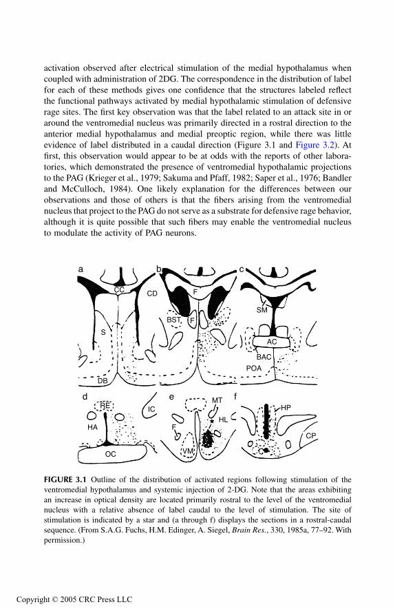
activation observed after electrical stimulation of the medial hypothalamus whencoupled with administration of 2DG. The correspondence in the distribution of labelfor each of these methods gives one confidence that the structures labeled reflectthe functional pathways activated by medial hypothalamic stimulation of defensiverage sites. The first key observation was that the label related to an attack site in oraround the ventromedial nucleus was primarily directed in a rostral direction to theanterior medial hypothalamus and medial preoptic region, while there was littleevidence of label distributed in a caudal direction (Figure 3.1 and Figure 3.2). Atfirst, this observation would appear to be at odds with the reports of other labora-tories, which demonstrated the presence of ventromedial hypothalamic projectionsto the PAG (Krieger et al., 1979; Sakuma and Pfaff, 1982; Saper et al., 1976; Bandlerand McCulloch, 1984). One likely explanation for the differences between ourobservations and those of others is that the fibers arising from the ventromedialnucleus that project to the PAG do not serve as a substrate for defensive rage behavior,although it is quite possible that such fibers may enable the ventromedial nucleusto modulate the activity of PAG neurons.
FIGURE 3.1
Outline of the distribution of activated regions following stimulation of theventromedial hypothalamus and systemic injection of 2-DG. Note that the areas exhibitingan increase in optical density are located primarily rostral to the level of the ventromedialnucleus with a relative absence of label caudal to the level of stimulation. The site ofstimulation is indicated by a star and (a through f) displays the sections in a rostral-caudalsequence. (From S.A.G. Fuchs, H.M. Edinger, A. Siegel,
Brain Res.
, 330, 1985a, 77–92. Withpermission.)
a b c
d e f
CC
S
DB
CD
BST
F
F
F
POA
BAC
AC
SM
HP
CP
HL
MT
VM
ICRE
HA
OC
TF1661_C003.fm Page 32 Thursday, September 23, 2004 3:14 PM
Copyright © 2005 CRC Press LLC
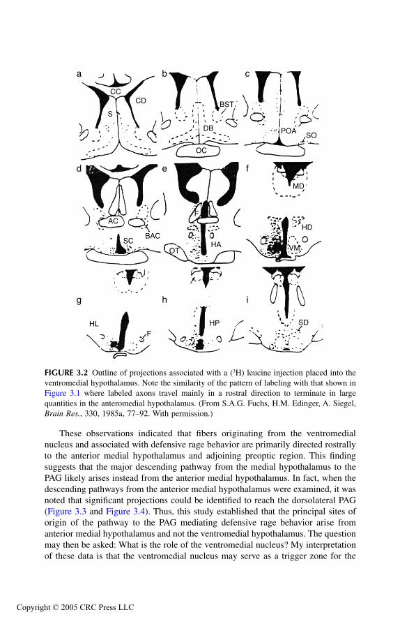
These observations indicated that fibers originating from the ventromedialnucleus and associated with defensive rage behavior are primarily directed rostrallyto the anterior medial hypothalamus and adjoining preoptic region. This findingsuggests that the major descending pathway from the medial hypothalamus to thePAG likely arises instead from the anterior medial hypothalamus. In fact, when thedescending pathways from the anterior medial hypothalamus were examined, it wasnoted that significant projections could be identified to reach the dorsolateral PAG(Figure 3.3 and Figure 3.4). Thus, this study established that the principal sites oforigin of the pathway to the PAG mediating defensive rage behavior arise fromanterior medial hypothalamus and not the ventromedial hypothalamus. The questionmay then be asked: What is the role of the ventromedial nucleus? My interpretationof these data is that the ventromedial nucleus may serve as a trigger zone for the
FIGURE 3.2
Outline of projections associated with a (
3
H) leucine injection placed into theventromedial hypothalamus. Note the similarity of the pattern of labeling with that shown inFigure 3.1 where labeled axons travel mainly in a rostral direction to terminate in largequantities in the anteromedial hypothalamus. (From S.A.G. Fuchs, H.M. Edinger, A. Siegel,
Brain Res.
, 330, 1985a, 77–92. With permission.)
a b c
d e f
g h i
CC
S
CD
OC
DB
BST
POASO
MD
F
HABAC
SC
AC
HLF
HP SD
OT VM
HD
TF1661_C003.fm Page 33 Thursday, September 23, 2004 3:14 PM
Copyright © 2005 CRC Press LLC
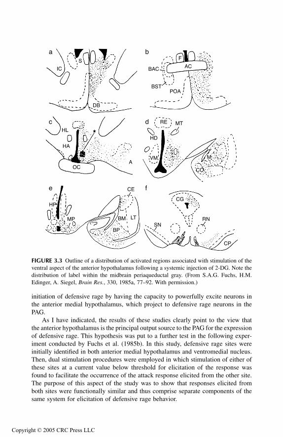
initiation of defensive rage by having the capacity to powerfully excite neurons inthe anterior medial hypothalamus, which project to defensive rage neurons in thePAG.
As I have indicated, the results of these studies clearly point to the view thatthe anterior hypothalamus is the principal output source to the PAG for the expressionof defensive rage. This hypothesis was put to a further test in the following exper-iment conducted by Fuchs et al. (1985b). In this study, defensive rage sites wereinitially identified in both anterior medial hypothalamus and ventromedial nucleus.Then, dual stimulation procedures were employed in which stimulation of either ofthese sites at a current value below threshold for elicitation of the response wasfound to facilitate the occurrence of the attack response elicited from the other site.The purpose of this aspect of the study was to show that responses elicited fromboth sites were functionally similar and thus comprise separate components of thesame system for elicitation of defensive rage behavior.
FIGURE 3.3
Outline of a distribution of activated regions associated with stimulation of theventral aspect of the anterior hypothalamus following a systemic injection of 2-DG. Note thedistribution of label within the midbrain periaqueductal gray. (From S.A.G. Fuchs, H.M.Edinger, A. Siegel,
Brain Res.
, 330, 1985a, 77–92. With permission.)
a b
c d
e f
IC
S
DB
BAC
BST
AC
F
POA
RE MT
HD
VM M
CO
AOC
HA
HL
HP
MP
CE
LTBM
BPSN
CG
RN
CP
TF1661_C003.fm Page 34 Thursday, September 23, 2004 3:14 PM
Copyright © 2005 CRC Press LLC
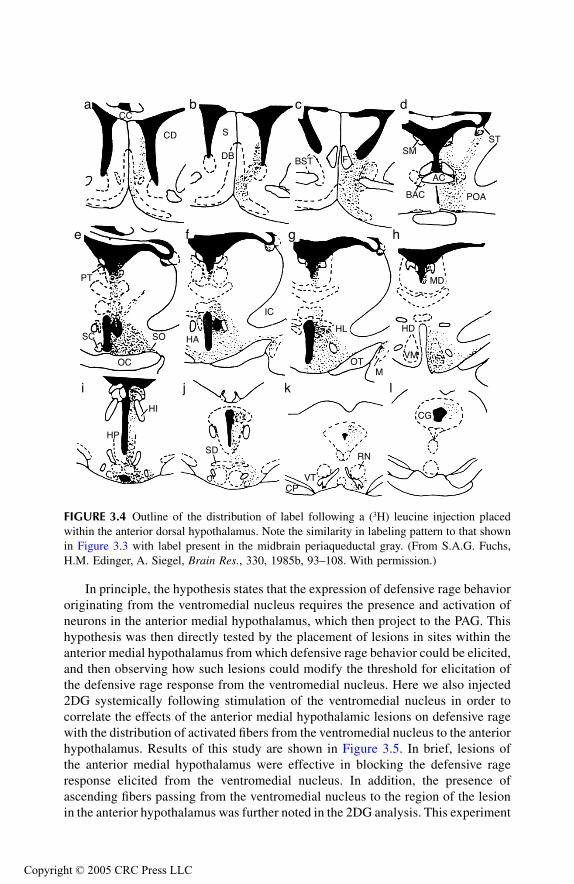
In principle, the hypothesis states that the expression of defensive rage behaviororiginating from the ventromedial nucleus requires the presence and activation ofneurons in the anterior medial hypothalamus, which then project to the PAG. Thishypothesis was then directly tested by the placement of lesions in sites within theanterior medial hypothalamus from which defensive rage behavior could be elicited,and then observing how such lesions could modify the threshold for elicitation ofthe defensive rage response from the ventromedial nucleus. Here we also injected2DG systemically following stimulation of the ventromedial nucleus in order tocorrelate the effects of the anterior medial hypothalamic lesions on defensive ragewith the distribution of activated fibers from the ventromedial nucleus to the anteriorhypothalamus. Results of this study are shown in Figure 3.5. In brief, lesions ofthe anterior medial hypothalamus were effective in blocking the defensive rageresponse elicited from the ventromedial nucleus. In addition, the presence ofascending fibers passing from the ventromedial nucleus to the region of the lesionin the anterior hypothalamus was further noted in the 2DG analysis. This experiment
FIGURE 3.4
Outline of the distribution of label following a (
3
H) leucine injection placedwithin the anterior dorsal hypothalamus. Note the similarity in labeling pattern to that shownin Figure 3.3 with label present in the midbrain periaqueductal gray. (From S.A.G. Fuchs,H.M. Edinger, A. Siegel,
Brain Res.
, 330, 1985b, 93–108. With permission.)
a b c d
e f g h
i j k l
CC
CD S
DB BST FSM
BAC
AC
ST
POA
MD
HD
VM
MOT
HL
IC
HASO
OC
SC
PT
HP
HI
SD
CPVT
RN
CG
TF1661_C003.fm Page 35 Thursday, September 23, 2004 3:14 PM
Copyright © 2005 CRC Press LLC
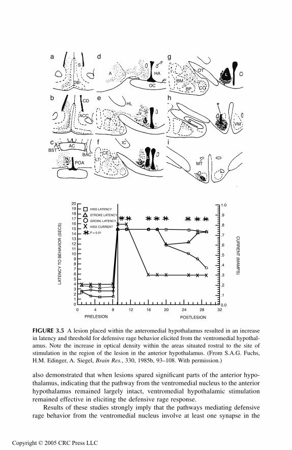
also demonstrated that when lesions spared significant parts of the anterior hypo-thalamus, indicating that the pathway from the ventromedial nucleus to the anteriorhypothalamus remained largely intact, ventromedial hypothalamic stimulationremained effective in eliciting the defensive rage response.
Results of these studies strongly imply that the pathways mediating defensiverage behavior from the ventromedial nucleus involve at least one synapse in the
FIGURE 3.5
A lesion placed within the anteromedial hypothalamus resulted in an increasein latency and threshold for defensive rage behavior elicited from the ventromedial hypothal-amus. Note the increase in optical density within the areas situated rostral to the site ofstimulation in the region of the lesion in the anterior hypothalamus. (From S.A.G. Fuchs,H.M. Edinger, A. Siegel,
Brain Res.
, 330, 1985b, 93–108. With permission.)
a
b
c
d
e
f
g
h
i
0123456789
1011121314151617181920
0 4 8 12 16 20 24 28 320.0
.1
.2
.3
.4
.5
.6
.7
.8
.9
1.0
PRELESION POSTLESION
LAT
EN
CY
TO
BE
HA
VIO
R (
SE
CS
)C
UR
RE
NT
(MA
MP
S)
XX XX
XX
XXXX
XX
XXXX XX
XX XX XX XX
HISS LATENCY
STROKE LATENCY
GROWL LATENCY
HISS CURRENT
P < 0.01
S
DB
A
OC
HA
F
BM
BP CO
OT
VM
HLCD
ACC
ACBST
POA
BACLT
CEM
MT
IC
TF1661_C003.fm Page 36 Thursday, September 23, 2004 3:14 PM
Copyright © 2005 CRC Press LLC

anterior hypothalamus and a second synapse in the PAG. This circuit is shown inFigure 3.6. Fuchs et al. (1985b) also point to the importance of the anterior hypo-thalamus in the process of defensive rage, as it occupies a key position within thebasal forebrain. It is strategically interposed between the limbic system, brainstem,tuberohypophyseal system and ventromedial nucleus. In this scheme, the anteriormedial hypothalamus could serve as a central integrating mechanism for defensiverage because of its direct inputs to the PAG, and because it receives important“modulating” inputs from such limbic regions as the septal area and amygdala.Moreover, the defensive rage response, which includes marked autonomic activitysuch as respiratory (Kastella et al., 1974) and cardiovascular changes (Stoddard-Apter et al., 1983), have also been linked to anterior hypothalamus. Thus, it isreasonable to conclude that further studies detailing the nature of the input–outputrelationships of the anterior medial hypothalamus might provide significant newinformation about the neural circuitry underlying defensive rage behavior.
M
IDBRAIN
P
ERIAQUEDUCTAL
G
RAY
The studies described above demonstrate that the primary target of medial hypotha-lamic neurons mediating defensive rage behavior is the dorsolateral aspect of thePAG. Thus, the next step in the analysis of the pathways mediating defensive ragewas to identify the projection pathways arising from sites in the PAG in cats fromwhich this form of aggressive behavior is elicited. The methods used were identicalto those described above for the study of hypothalamic pathways mediating defensiverage behavior (Shaikh et al., 1987).
In this study, defensive rage behavior was elicited by both electrical and chemical(i.e., D,L-homocysteic acid) stimulation, and these sites were located mainly in therostral half of the dorsolateral PAG — the principal receiving area of medial hypo-thalamic fibers mediating defensive rage. Then, either 2DG was injected systemicallyfollowing electrical stimulation of a defensive rage site in the PAG of an awake orlightly anesthetized cat preparation, or (
3
H) leucine was injected through the cannula-electrode into a defensive rage site within the PAG. Similar to findings observed inthe previous experiment, similar patterns of labeling were observed after each ofthe tracing procedures. From the PAG, labeled descending axons were followed into
FIGURE 3.6
Summary of the principal hypothalamic pathways mediating defensive ragebehavior. The organization of the descending pathway from the ventromedial hypothalamus(VM) to the brainstem involves neurons whose axons travel almost exclusively in a rostraldirection to terminate largely in the anterior medial hypothalamus (AH). The second-orderneurons of this pathway arise in the anterior medial hypothalamus and have more extensivedescending projections, the most significant of which supply the PAG (CG). (From S.A.G.Fuchs, H.M. Edinger, A. Siegel,
Brain Res.
, 330, 1985b, 93–108. With permission.)
VM AH
CG
TF1661_C003.fm Page 37 Thursday, September 23, 2004 3:14 PM
Copyright © 2005 CRC Press LLC
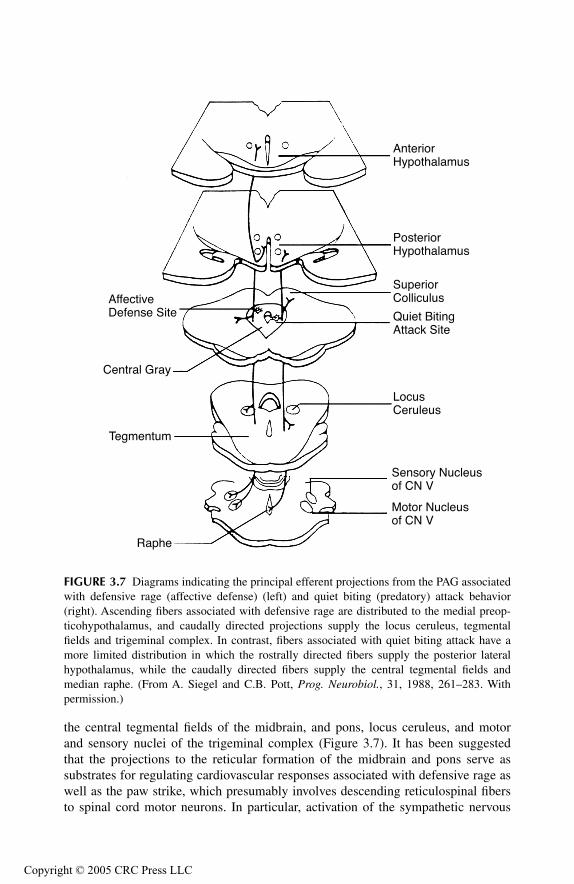
the central tegmental fields of the midbrain, and pons, locus ceruleus, and motorand sensory nuclei of the trigeminal complex (Figure 3.7). It has been suggestedthat the projections to the reticular formation of the midbrain and pons serve assubstrates for regulating cardiovascular responses associated with defensive rage aswell as the paw strike, which presumably involves descending reticulospinal fibersto spinal cord motor neurons. In particular, activation of the sympathetic nervous
FIGURE 3.7
Diagrams indicating the principal efferent projections from the PAG associatedwith defensive rage (affective defense) (left) and quiet biting (predatory) attack behavior(right). Ascending fibers associated with defensive rage are distributed to the medial preop-ticohypothalamus, and caudally directed projections supply the locus ceruleus, tegmentalfields and trigeminal complex. In contrast, fibers associated with quiet biting attack have amore limited distribution in which the rostrally directed fibers supply the posterior lateralhypothalamus, while the caudally directed fibers supply the central tegmental fields andmedian raphe. (From A. Siegel and C.B. Pott,
Prog. Neurobiol.
, 31, 1988, 261–283. Withpermission.)
AnteriorHypothalamus
PosteriorHypothalamus
SuperiorColliculus
Quiet BitingAttack Site
LocusCeruleus
Sensory Nucleusof CN V
Motor Nucleusof CN V
AffectiveDefense Site
Central Gray
Tegmentum
Raphe
TF1661_C003.fm Page 38 Thursday, September 23, 2004 3:14 PM
Copyright © 2005 CRC Press LLC
system may come about through several routes. One route might involve projectionsto the locus cereleus, whose descending fibers have been shown to pass to sympa-thetic neurons in the intermediolateral cell column of the thoracic and lumbar spinalcord (Nygren and Olson, 1977). Alternatively, other investigators have describedprojections from the PAG to the solitary nucleus (Bandler and Tork, 1987), whoseaxons pass via the ventrolateral medulla to the intermediolateral cell column of thethoracic and lumbar spinal cord. Concerning the projections to the trigeminal com-plex, this pathway would account for the jaw opening (digastric) reflex that charac-terizes the hissing component of the defensive rage response.
In addition to the descending projections from the PAG, there is also an ascendingprojection whose primary target is the rostro-caudal extent of the medial hypothal-amus. The unique feature of this projection is that the fibers pass to all regions ofthe medial hypothalamus associated with defensive rage behavior (Figure 3.7). Inthis manner, this ascending projection may serve as a substrate for a positive feedbackmechanism. Such a mechanism could increase the likelihood that defensive ragewould be expressed over an extended period of time following the presence of athreatening stimulus. It is reasonable to assume that by increasing duration of theresponse under dangerous conditions, survival value to the animal is likewiseincreased.
In summary, the anterior medial hypothalamus constitutes the primary output tothe PAG for the expression of defensive rage behavior. Inasmuch as only a fragmentof defensive rage components can be elicited below the level of the PAG, such asin the pons and medulla, it is probable that the PAG represents the lowest level ofthe CNS, where integration of somatomotor and autonomic components of this formof aggression takes place. These conclusions are summarized in Figure 3.7.
PATHWAYS MEDIATING PREDATORY ATTACK IN CATS
In Chapter 2, the sites in the hypothalamus from which predatory attack behaviorcould be elicited were identified (Wasman and Flynn, 1962). These and subsequentfindings (e.g., Siegel and Pott, 1988; Siegel and Brutus, 1990; and Siegel et al.,1999) demonstrated that predatory attacks in cats could be elicited from the rostro-caudal extent of the preopticohypothalamus.
After having mapped the regions of the hypothalamus associated with predatoryattack, Chi and Flynn (1971b) used the design described earlier in this chapter totrace the pathways mediating predatory attack. Again, predatory attack sites wereidentified and then a lesion was placed at the tip of the electrode. Following suitablesurvival times, animals were sacrificed and the brain processed by the Fink Heimermodification of the Nauta technique for identifying degenerating axons and theirterminals. The major finding of this study was the presence of a large bundle ofdescending fibers that could be traced into the ventral tegmental area, and a secondbundle that terminated in the midbrain PAG. In a second study based on thesefindings, Chi et al. (1976) used similar methods to trace descending fibers from theventral tegmental area in association with predatory attack behavior. The major
TF1661_C003.fm Page 39 Thursday, September 23, 2004 3:14 PM
Copyright © 2005 CRC Press LLC
finding of this study suggested a projection to motor and sensory nuclei of thetrigeminal nerve and to the motor nucleus of the facial nerve, which would play animportant part in the biting component of the attack. While some of the data shouldbe interpreted with caution because of the possibility of damage to fibers of passage,it is still likely that the projections from the lateral hypothalamus to the ventraltegmental area and PAG are important for the expression of predatory attack.
Several studies were conducted using retrograde tracing procedures, where HRPwas injected into the pontine tegmentum and ventral tegmental area of the midbrainfrom which components of attack could be elicited (Smith and Flynn, 1979; Schoelet al., 1981). The key point to note from both studies was that labeled cells wereidentified in the lateral perifornical hypothalamus — the region from which predatoryattack is typically elicited. Thus, this finding suggests the presence of a directprojection from the perifornical hypothalamus to the tegmentum of the midbrainand pons in association with predatory attack.
These findings were soon followed by an anterograde tracing study by Fuchs etal. (1981) in which the combined methods of 2DG and (
3
H)-leucine radioautographywere employed to re-examine the descending pathways from the perifornical andlateral hypothalamus to the brainstem. Again, the distribution of label from theperifornical hpothalamus was similar with both methodologies. In brief, fibers arisingfrom perifornical hypothalamus were traced caudally through the medial forebrainbundle into the ventral tegmental area. Additional groups of fibers passed dorsallyfrom the ventral tegmentum into the midbrain PAG and laterally into the centraltegmental fields. Other fibers could be followed further caudally into the lateraltegmental fields of the pons, locus ceruleus, and motor nucleus of the trigeminalnerve. When compared with the projections from the perifornical region, it wasobserved that fibers arising from more laterally situated aspects of the hypothalamus,from which predatory attack was also elicited, displayed a much more limitedprojection pattern. The key point here is that the principal projection was directedto the perifornical region.
This finding, taken together with the findings obtainedfrom the anterograde tracing methods and the retrograde labeling described bySmith and Flynn (1979, 1980a) and Schoel et al. (1981), provides support for theview that the perifornical region represents the central focus of the descendingprojection system of fibers for the expression of predatory attack behavior.
It shouldbe pointed out that the functional relationship between hypothalamic attack sitesand the midbrain tegmentum has also been documented by Huang and Flynn (1975).These authors showed that electrical stimulation of predatory attack sites in theawake cat generated excitation of neurons throughout this aspect of the midbrain.
A key challenge here is to determine the significance of these descending path-ways with respect to predatory attack. For example, it is possible that the projectionto the ventral tegmental area may play an important role by serving as the first neuronin a descending pathway to the lower brainstem tegmentum associated with predatoryattack (Bernston, 1973), and nuclei of cranial nerves V and VII associated with thejaw-closing reflex used in biting (Chi et al., 1976). However, the more significantpathway is likely the one that arises directly from the perifornical hypothalamus, andwhich projects directly to the PAG, pontine tegmentum and motor nucleus of thetrigeminal nerve. Such a projection enables the perifornical hypothalamus to directly
TF1661_C003.fm Page 40 Thursday, September 23, 2004 3:14 PM
Copyright © 2005 CRC Press LLC
control the masseteric reflex that is intrinsic to predatory attack. It is of interest tonote that neurophysiological support for this connection was established by Mac-Donnell and Flynn (1966) and by Landgren and Olsson (1980), who showed thathypothalamic stimulation can facilitate jaw reflexes in cats.
An ascending component of the perifornical region was also noted. Ascendingfibers from the perifornical hypothalamus could be followed rostrally into the pre-optic region, bed nucleus of the stria terminalis, and lateral septal area (Figure 3.8).The possible significance of this projection to limbic regions is that it may providethe basis for a feedback mechanism to the limbic structures, which play an importantrole in modulating the attack mechanism.
RELATIONSHIP BETWEEN PREDATORY ATTACK AND DEFENSIVE RAGE: LINKAGE BETWEEN MEDIAL AND
LATERAL HYPOTHALAMUS
It has been my experience over the course of conducting research on the neurobiologyof aggression in cats that when experimental procedures are introduced which resultin facilitation of one form of aggression, the same manipulations will cause sup-pression of the other form of aggression. A case in point, which is described ingreater detail in Chapter 4, concerns amygdaloid modulation of aggression and rage.It has been shown that stimulation of the medial amygdala facilitates defensive rage(Shaikh et al., 1993) and suppresses predatory attack (Han et al., 1996a, 1996b).Similar observations have been found following administration of drugs such asnaloxone (Brutus and Siegel, 1989) and cholecystokinin (CCK) compounds (Luo etal., 1998).
Evidence for the relationship between the medial and lateral hypothalamus wasoriginally provided by Millhouse (1973) in a Golgi study, which demonstrated lateralhypothalamic processes passing toward the medial hypothalamus in rodents, andfrom a retrograde tracing study by Smith and Flynn (1980b), who identified retro-grade-labeled neurons in the lateral hypothalamus following HRP injections thatwere placed at defensive rage sites in the medial hypothalamus of cats.
Several recent experiments have begun to systematically address this importantissue (Han et al., 1996b; Cheu and Siegel, 1998). One study considered the projec-tions from the medial to lateral hypothalamus as part of an overall analysis ofamygdaloid modulation of aggression and rage (Han et al., 1996a, 1996b), whichis considered in more detail in Chapter 4. For purposes of the present chapter, theanalysis is limited to the relationship between the medial and lateral hypothalamus.In one experiment, Han et al. (1996b) showed that microinjection of the GABAagonist, muscimol, into a predatory attack site suppressed this response in a dose-and time-dependent manner (Figure 3.9). This finding, coupled with pretreatmentwith the GABA
A
receptor antagonist, bicuculline, which blocked the suppressiveeffects of muscimol, demonstrated the presence of GABA
A
receptors in the regionof the lateral hypothalamus from which predatory attack is elicited. In a secondexperiment, it was initially shown that microinjection of the substance P agonist(Sar
9
,Met(O
2
)
11
)-substance P, into a site in the medial hypothalamus from which
TF1661_C003.fm Page 41 Thursday, September 23, 2004 3:14 PM
Copyright © 2005 CRC Press LLC
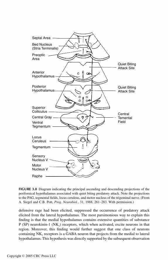
defensive rage had been elicited, suppressed the occurrence of predatory attackelicited from the lateral hypothalamus. The most parsimonious way to explain thisfinding is that the medial hypothalamus contains extensive quantities of substanceP (SP) neurokinin-1 (NK
1
) receptors, which when activated, excite neurons in thatregion. Moreover, this finding would further suggest that one class of neuronscontaining NK
1
receptors is a GABA neuron that projects from the medial to lateralhypothalamus. This hypothesis was directly supported by the subsequent observation
FIGURE 3.8
Diagram indicating the principal ascending and descending projections of theperifornical hypothalamus associated with quiet biting predatory attack. Note the projectionsto the PAG, tegmental fields, locus ceruleus, and motor nucleus of the trigeminal nerve. (FromA. Siegel and C.B. Pott,
Prog. Neurobiol.
, 31, 1988, 261–283. With permission.)
Septal Area
Bed Nucleus(Stria Terminalis)
PreopticArea
AnteriorHypothalamus
PosteriorHypothalamus
SuperiorColliculus
Central Gray
VentralTegmentum
LocusCeruleus
Tegmentum
SensoryNucleus V
MotorNucleus V
Raphe
Quiet BitingAttack Site
Quiet BitingAttack Site
CentralTementalField
TF1661_C003.fm Page 42 Thursday, September 23, 2004 3:14 PM
Copyright © 2005 CRC Press LLC
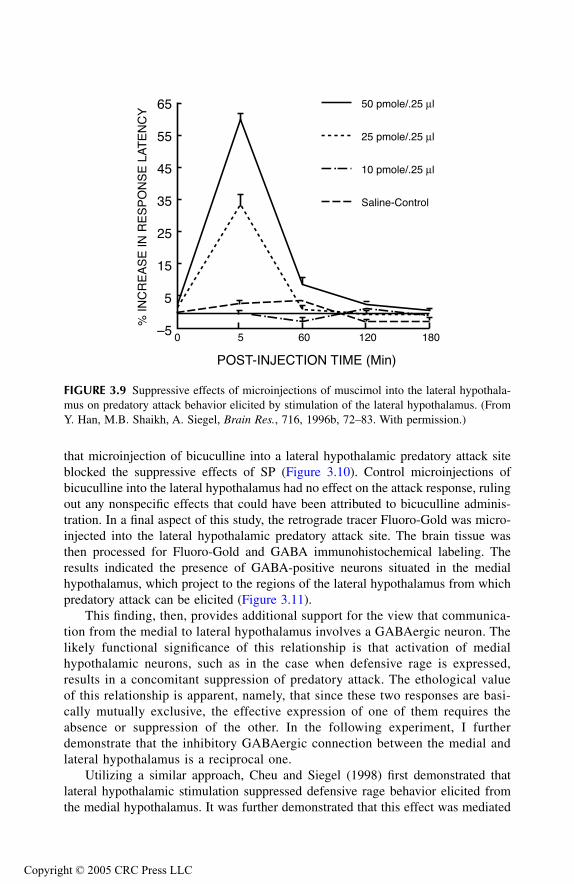
that microinjection of bicuculline into a lateral hypothalamic predatory attack siteblocked the suppressive effects of SP (Figure 3.10). Control microinjections ofbicuculline into the lateral hypothalamus had no effect on the attack response, rulingout any nonspecific effects that could have been attributed to bicuculline adminis-tration. In a final aspect of this study, the retrograde tracer Fluoro-Gold was micro-injected into the lateral hypothalamic predatory attack site. The brain tissue wasthen processed for Fluoro-Gold and GABA immunohistochemical labeling. Theresults indicated the presence of GABA-positive neurons situated in the medialhypothalamus, which project to the regions of the lateral hypothalamus from whichpredatory attack can be elicited (Figure 3.11).
This finding, then, provides additional support for the view that communica-tion from the medial to lateral hypothalamus involves a GABAergic neuron. Thelikely functional significance of this relationship is that activation of medialhypothalamic neurons, such as in the case when defensive rage is expressed,results in a concomitant suppression of predatory attack. The ethological valueof this relationship is apparent, namely, that since these two responses are basi-cally mutually exclusive, the effective expression of one of them requires theabsence or suppression of the other. In the following experiment, I furtherdemonstrate that the inhibitory GABAergic connection between the medial andlateral hypothalamus is a reciprocal one.
Utilizing a similar approach, Cheu and Siegel (1998) first demonstrated thatlateral hypothalamic stimulation suppressed defensive rage behavior elicited fromthe medial hypothalamus. It was further demonstrated that this effect was mediated
FIGURE 3.9
Suppressive effects of microinjections of muscimol into the lateral hypothala-mus on predatory attack behavior elicited by stimulation of the lateral hypothalamus. (FromY. Han, M.B. Shaikh, A. Siegel,
Brain Res.
, 716, 1996b, 72–83. With permission.)
–5
5
15
25
35
45
55
65
POST-INJECTION TIME (Min)
% IN
CR
EA
SE
IN R
ES
PO
NS
E L
AT
EN
CY
0 5 60 120 180
50 pmole/.25 µl
25 pmole/.25 µl
10 pmole/.25 µl
Saline-Control
TF1661_C003.fm Page 43 Thursday, September 23, 2004 3:14 PM
Copyright © 2005 CRC Press LLC

by GABA
A
receptors in the medial hypothalamus since bicuculline administrationinto the medial hypothalamic defensive rage site blocked the suppressive effects oflateral hypothalamic stimulation. This study also demonstrated through double-labeling procedures involving retrograde Fluoro-Gold labeling and immunocy-tochemical staining for GABA neurons that GABA positive neurons are present inthe lateral hypothalamus, which project to the medial hypothalamus. These recip-rocal relationships between the medial and lateral hypothalamus are illustrated inFigure 3.12, and further reinforce the dynamic interaction between defensive rageand predatory attack.
HYPOTHALAMIC AGGRESSION IN RATS
In attempting to develop an understanding of the nature of neural bases of aggressionand rage, it becomes useful to compare and contrast the forms of hypothalamicallyelicited aggression elicited from rats relative to those of cats. Likewise, it is alsoinstructive to compare the neuroanatomical circuitry for the respective forms ofaggression in rats and cats.
FIGURE 3.10
Blocking effects of bicuculline upon substance P induced suppression ofpredatory attack. Hatched bars represent effect of substance P (2.0 nM/0.25 l) infusion intothe medial hypothalamus following pretreatment with saline into the lateral hypothalamus.Closed bars reflect the effects of bicuculline (0.15 nM) pretreatment into lateral hypothalamicsites from which predatory attack was elicited upon suppression of this response followinginfusion of SP into the medial hypothalamus. (From Y. Han, M.B. Shaikh, A. Siegel,
BrainRes.
, 716, 1996b, 72–83. With permission.)
0
10
20
30
40
50
5 60 120 180
POST-INJECTION TIME (MIN)
% IN
CR
EA
SE
IN R
ES
PO
NS
E L
AT
EN
CY
SP +
Saline
SP +
Bicuculline
TF1661_C003.fm Page 44 Thursday, September 23, 2004 3:14 PM
Copyright © 2005 CRC Press LLC

N
ATURE
OF
A
TTACK
R
ESPONSE
Many investigators have shown that electrical stimulation of the rat hypothalamuscan produce diverse aggressive responses that may include affective attack (Kool-haas, 1978), quiet attacks (Panksepp, 1971), bite attacks (Woodworth, 1971), attackjumps, and clinch fights (Kruk et al., 1979; Lammers et al., 1988; Van der Poel etal., 1982). In recent years, a greater emphasis has been placed on the study ofelectrical stimulation–induced attack involving forms of biting attack of the neck,head, or back, coupled with paw kicks to the flank, clinch fights, and attack jumps(Kruk et al., 1979, 1983, 1984, 1990). In a jump attack, the attacker jumps towardthe opponent with the aim of biting its head. In a clinch fight, the attacker typicallybecomes locked with the top or back of the opponent, which often occurs when theattacker loses its footing after attempting to attack the target animal (Kruk et al.,1990). Stimulation-induced attack from the hypothalamus can be directed against adominant, subordinate, anesthetized, or dead rat. Sex does not seem to be a majorfactor, as a male can attack a female, while a female may attack another female ormale. Variety in the forms of attack is a function of factors such as the intensity of
FIGURE 3.11
Maps indicate the distribution of cells retrogradely labeled with Fluoro-Gold(open circles) microinjected into the lateral hypothalamus; GABA-immunopositive neurons(closed circles) and double-labeled neurons for both GABA and Fluoro-Gold (stars). Notethe presence of double-labeled neurons in the medial hypothalamus. (From Y. Han, M.B.Shaikh, A. Siegel,
Brain Res.
, 716, 1996b, 72–83. With permission.)
RE
HLHp
OT
RE
HL aHd
HVM
FxOT
Cl
TF1661_C003.fm Page 45 Thursday, September 23, 2004 3:14 PM
Copyright © 2005 CRC Press LLC
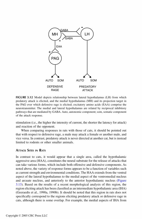
stimulation (i.e., the higher the intensity of current, the shorter the latency for attack)and reaction of the opponent.
When comparing responses in rats with those of cats, it should be pointed outthat with respect to defensive rage, a male may attack a female or another male, andvice versa. In contrast, predatory attack is never directed at another cat, but is insteadlimited to rodents or other smaller animals.
A
TTACK
S
ITES
IN
R
ATS
In contrast to cats, it would appear that a single area, called the hypothalamicaggressive area (HAA), constitutes the neural substrate for the release of attacks thatcan take various forms, which include both offensive and defensive components. Asnoted above, the variety of response forms appears to be a function of variables suchas current strength and environmental conditions. The HAA extends from the ventralaspect of the lateral hypothalamus to the medial aspect of the ventromedial nucleusand arcuate nucleus, and anteriorly to the anterior hypothalamic nucleus (Figure3.13). Based on the results of a recent morphological analysis of this region, theregion eliciting attack has been classified as an intermediate hypothalamic area (IHA)(Geeraedts et al., 1990a, 1990b). It should be noted that this region in rats does notspecifically correspond to the regions eliciting predatory attack or defensive rage incats, although there is some overlap. For example, the medial aspects of IHA from
FIGURE 3.12
Model depicts relationship between lateral hypothalamus (LH) from whichpredatory attack is elicited, and the medial hypothalamus (MH) and its projection target inthe PAG over which defensive rage is elicited; excitatory amino acids (EAA) comprise theneurotransmitter. The medial and lateral hypothalamus are related by reciprocal inhibitorypathways that are mediated by GABA. Auto, autonomic component; som, somatic componentof the attack response.
MH LH
PAG
GABA
GABA
EAA
-
-
SOMAUTO SOMAUTO
DEFENSIVERAGE
PREDATORYATTACK
+
TF1661_C003.fm Page 46 Thursday, September 23, 2004 3:14 PM
Copyright © 2005 CRC Press LLC
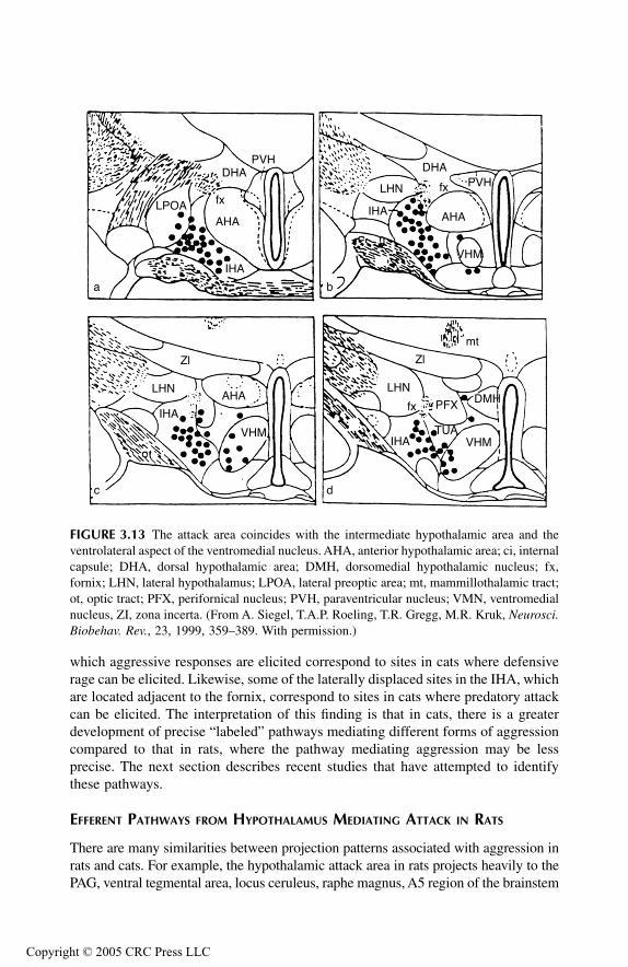
which aggressive responses are elicited correspond to sites in cats where defensiverage can be elicited. Likewise, some of the laterally displaced sites in the IHA, whichare located adjacent to the fornix, correspond to sites in cats where predatory attackcan be elicited. The interpretation of this finding is that in cats, there is a greaterdevelopment of precise “labeled” pathways mediating different forms of aggressioncompared to that in rats, where the pathway mediating aggression may be lessprecise. The next section describes recent studies that have attempted to identifythese pathways.
E
FFERENT
P
ATHWAYS
FROM
H
YPOTHALAMUS
M
EDIATING
A
TTACK
IN
R
ATS
There are many similarities between projection patterns associated with aggression inrats and cats. For example, the hypothalamic attack area in rats projects heavily to thePAG, ventral tegmental area, locus ceruleus, raphe magnus, A5 region of the brainstem
FIGURE 3.13
The attack area coincides with the intermediate hypothalamic area and theventrolateral aspect of the ventromedial nucleus. AHA, anterior hypothalamic area; ci, internalcapsule; DHA, dorsal hypothalamic area; DMH, dorsomedial hypothalamic nucleus; fx,fornix; LHN, lateral hypothalamus; LPOA, lateral preoptic area; mt, mammillothalamic tract;ot, optic tract; PFX, perifornical nucleus; PVH, paraventricular nucleus; VMN, ventromedialnucleus, ZI, zona incerta. (From A. Siegel, T.A.P. Roeling, T.R. Gregg, M.R. Kruk,
Neurosci.Biobehav. Rev.
, 23, 1999, 359–389. With permission.)
DHAPVH
fx
AHALPOA
IHA
a b
dc
DHAPVHfx
AHAIHA
LHN
VHM
Zl
LHN
IHA
AHA
VHM
fx
Zl
LHN
IHA VHM
PFX DMH
mt
TUA
TF1661_C003.fm Page 47 Thursday, September 23, 2004 3:14 PM
Copyright © 2005 CRC Press LLC
tegmentum, and nucleus of the solitary tract (Roberts and Nagel, 1996; Roeling et al.,1994). Projections to the various nuclei of the brainstem presumably serve to mediatethe autonomic and endocrine functions associated with aggressive reactions. One majorfunctional difference between the projections in rat and cat relates to the PAG. In thecat, destruction of the PAG blocks defensive rage behavior. In rats, similar destructionof the PAG only transiently reduces hypothalamically elicited attack, as well as aggres-sion induced by an intruder (Mos et al., 1983). This finding would suggest that otherprojections of the hypothalamic attack area are also significant.
Three other projections of the hypothalamic attack area should be noted. Thefirst is the ventromedial hypothalamic nucleus, a region from which attack can alsobe elicited. Projection to this region may be important for driving the attack mech-anism in much the same way as the projection to the dorsolateral PAG, where attackis also elicited. A second projection is the medial thalamus, and includes themediodorsal and parataenial nuclei. Although the significance of this projection isnot immediately apparent, it is reasonable to assume that this connection may beimportant because the mediodorsal nucleus projects directly to the frontal lobe andindirectly to the hypothalamus (Siegel et al., 1972). The frontal lobe is known toplay an important role in the regulation of emotional behavior and, therefore, thisprojection may serve as a feedback loop for limbic modulation of aggression byvirtue of frontal lobe projections to the hypothalamus via the mediodorsal nucleus.Likewise, the third projection involving fiber connections with the septal area pre-sumably also involves a feedback mechanism. The septal area is known to projectto the hypothalamus and modulate attack behavior (Meibach and Siegel, 1977;Krayniak et al., 1980; Brutus et al., 1984). Thus, the projection from the hypothal-amus to the dorsal septal area would provide the necessary feedback circuit. Here,activation of the hypothalamic attack area can excite the dorsal septal area, which,in turn, would modulate activity of the hypothalamic neurons governing attack inrats. Feedback circuits constitute an important feature of CNS function, and arecertainly present in sensory and motor systems. Accordingly, these feedback circuitsmay also constitute an important overall mechanism in regulating aggression. Therole of limbic system structures in the regulation of aggression and rage is consideredin detail in Chapter 5.
It should be pointed out that forms of aggression in rats other than biting attackhave not been systematically examined, and thus, there is no basis for a comparisonwith the circuits governing biting attack and affective forms of aggression in cats.Hopefully, future studies will be able to delineate the nature of specific pathwaysmediating different forms of aggression in rats. Such studies would allow for a moreeffective comparison of the similarities and differences between species concerningthese forms of aggression, and perhaps would provide new insights into the neuralbases of these behaviors.
SUMMARY
In this chapter, I have considered the anatomical circuitry governing defensive rageand predatory attack in cats and biting attack in rats. Studies in cats have revealedthat the key neuronal groups mediating defensive rage include the anterior medial
TF1661_C003.fm Page 48 Thursday, September 23, 2004 3:14 PM
Copyright © 2005 CRC Press LLC

hypothalamus and dorsolateral PAG, which receives its input from the anteriormedial hypothalamus. Projections from the PAG descend to a variety of lowerbrainstem nuclei that control the somatomotor and autonomic components of thedefensive rage response. With respect to predatory attack, the most important cellgroups essential for the expression of this response are situated in the perifornicalhypothalamus. Fibers from this region descend to nuclei such as the PAG, ventraltegmental area, tegmental fields of the midbrain and pons, locus ceruleus, and motornucleus of the trigeminal nerve. These pathways also provide the basis for thesomatomotor and autonomic components of this form of aggression. This chapteralso considered the relationship between the neural circuitry governing predatoryattack and defensive rage. It was discovered that reciprocal inhibitory connectionsare present between the lateral hypothalamic predatory attack zone and the medialhypothalamic defensive rage zone. The functional significance of these reciprocalconnections was considered in this chapter. Finally, the nature of the pathwaysgoverning biting attack in the rat was also considered, and was shown to parallelthose described in cats. The significance of identifying the anatomical circuitryunderlying various forms of aggression and rage is that by providing the basicsubstrates for their expression, a rational basis is established for advancing newhypotheses concerning the functional and neurochemical mechanisms that produceand control these responses.
REFERENCES
Bandler, R. and T. McCulloch, Afferents to a midbrain periaqueductal grey region involvedin the ‘defense reaction’ in the cat as revealed by horseradish peroxidase: II. Thediencephalon,
Behav. Brain Res.
, 13, 1984, 279–285.Bandler, R. and I. Tork, Midbrain periaqueductal grey region in the cat has afferent and
efferent connections with solitary tract nuclei,
Neurosci. Lett.
, 74, 1987, 1–6.Berntson, G.G., Attack, grooming, and threat elicited by stimulation of the pontine tegmentum
in cats,
Physiol. Behav.
, 11, 1973, 81–87.Brutus, M., M.B. Shaikh, A. Siegel, H.E. Siegel, An analysis of the mechanisms underlying
septal area control of hypothalamically elicited aggression in the cat,
Brain Res.
, 310,1984, 235–248.
Brutus, M. and A. Siegel, Effects of the opiate antagonist naloxone upon hypothalamicallyelicited affective defense behavior in the cat,
Behav. Brain Res.
, 33, 1989, 23–32.Cheu, J.W. and A. Siegel, GABA receptor mediated suppression of defensive rage behavior
elicited from the medial hypothalamus of the cat: role of the lateral hypothalamus,
Brain Res.
, 783, 1998, 293–304.Chi, C.C., R. Bandler, J.P. Flynn, Neuroanatomic projections related to biting attack elicited
from ventral midbrain in cats,
Brain Behav. Evol.
, 13, 1976, 91–110.Chi, C.C. and J.P. Flynn, Neural pathways associated with hypothalamically elicited attack
behavior in cats,
Science
, 171, 1971a, 703–706.Chi, C.C. and J.P. Flynn, Neuroanatomic projections related to biting attack elicited from
hypothalamus in cats,
Brain Res.
, 35, 1971b, 49–66.Fuchs, S.A.G., M. Dalsass, H.E. Siegel, A. Siegel, The neural pathways mediating quiet-
biting attack behavior from the hypothalamus in the cat: a functional autoradiographicstudy,
Aggress. Behav.
, 7, 1981, 51–67.
TF1661_C003.fm Page 49 Thursday, September 23, 2004 3:14 PM
Copyright © 2005 CRC Press LLC

Fuchs, S.A.G., H.M. Edinger, A. Siegel, The organization of the hypothalamic pathwaysmediating affective defense behavior in the cat,
Brain Res.
, 330, 1985a, 77–92.Fuchs, S.A.G., H.M. Edinger, A. Siegel, The role of the anterior hypothalamus in affective
defense behavior elicited from the ventromedial hypothalamus of the cat,
Brain Res.
,330, 1985b, 93–108.
Geeraedts, L.M.G., R. Nieuwenhuys, J.G. Veening, Medial forebrain bundle of the rat: III.Cytoarchitecture of the rostral, telencephalic, part of the medial forebrain bundle bednucleus,
J. Comp. Neurol.
, 294, 1990a, 507–536.Geeraedts, L.M.G., R. Nieuwenhuys, J.G. Veening, Medial forebrain bundle of the rat: IV.
Cytoarchitecture of the caudal, lateral hypothalamic, part of the medial forebrainbundle bed nucleus,
J. Comp. Neurol.
, 294, 1990b, 537–568.Han, Y., M.B. Shaikh, A. Siegel, Medial amygdaloid suppression of predatory attack behavior
in the cat: I. Role of a substance P pathway from the medial amygdala to the medialhypothalamus,
Brain Res.
, 716, 1996a, 59–71.Han, Y., M.B. Shaikh, A. Siegel, Medial amygdaloid suppression of predatory attack behavior
in the cat: II. Role of a GABAergic pathway from the medial to the lateral hypothal-amus,
Brain Res.
, 716, 1996b, 72–83.Huang, Y.H. and J.P. Flynn, Unit activities in the hypothalamus and midbrain during stimu-
lation of hypothalamic attack sites,
Brain Res.
, 93, 1975, 419–440.Kastella, K.G., H.A. Spurgion, G.K. Weiss, Respiratory related neurons in anterior hypothal-
amus of the cat,
Am. J. Physiol.
, 227, 1974, 710–713.Koolhaas, J.M. Hypothalamically induced intraspecific aggressive behaviour in the rat,
Exp.Brain Res.
, 32, 1978, 365–375.Krayniak, P., S. Weiner, A. Siegel, An analysis of the efferent connections of the septal area
in the cat,
Brain Res.
, 189, 1980, 15–29.Krieger, M.S., L.C.A. Conrad, D.W. Pfaff, An autoradiographic study of the efferent connec-
tions of the ventromedial nucleus of the hypothalamus,
J. Comp. Neurol.
, 183, 1979,785–816.
Kruk, M.R., C.E. Van der Laan, J. Mos, A.M. Van der Poel, W. Meelis, B. Olivier, Comparisonof aggressive behaviour induced by electrical stimulation in the hypothalamus ofmale and female rats,
Prog. Brain Res.
, 61, 1984, 303–314.Kruk, M.R., C.E. Van der Laan, J. Van der Spuy, A.M.M. Van Erp, W. Meelis, Strain differences
in attack patterns elicited by electrical stimulation in the hypothalamus of maleCPBWEzob and CPBWI rats,
Aggress. Behav.
, 16, 1990, 177–190.Kruk, M.R., A.M. Van der Poel, T.P. De Vos-Frerichs, The induction of aggressive behaviour
by electrical stimulation in the hypothalamus of male rats,
Behaviour
, 70, 1979,292–322.
Kruk, M.R., A.M. Van der Poel, W. Meelis, J. Hermans, P.G. Mostert, J. Mos, A.H.M. Lohman,Discriminant analysis of the localization of aggression-inducing electrode placementsin the hypothalamus of male rats,
Brain Res.
, 260, 1983, 61–79.Lammers, J.H.C.M., M.R. Kruk, W. Meelis, A.M. Van der Poel, Hypothalamic substrates for
brain stimulation-induced patterns of locomotion and escape jumps in the rat,
BrainRes.
, 449, 1988, 294–310.Landgren, S. and K.A. Olsson, The effect of electrical stimulation in the hypothalamus on
the monosynaptic jaw closing and the disynaptic jaw opening reflexes in the cat,
Exp.Brain Res.
, 39, 1980, 389–400.Luo, B., J.W. Cheu, A. Siegel, Cholecystokinin B receptors in the periaqueductal gray poten-
tiate defensive rage behavior elicited from the medial hypothalamus of the cat,
BrainRes.
, 796, 1998, 27–37.
TF1661_C003.fm Page 50 Thursday, September 23, 2004 3:14 PM
Copyright © 2005 CRC Press LLC
MacDonnell, M. and J.P. Flynn, Sensory control of hypothalamic attack,
Anim. Behav.
, 14,1966, 399–405.
Meibach, R. and A. Siegel, Efferent connections of the septal area in the rat: an analysisutilizing retrograde and anterograde transport methods,
Brain Res.
, 119, 1977, 1–20.Millhouse, O.E., Certain ventromedial hypothalamic afferents,
Brain Res.
, 55, 1973, 89–105.Mos, J., J.H.C.M. Lammers, A.M. Van der Poel, B. Bermond, W. Meelis, M.R. Kruk, Effects
of midbrain central gray lesions on spontaneous and electrically induced aggressionin the rat,
Aggress. Behav.
, 9, 1983, 133–155.Nygren, L.-G. and L. Olson, A new major projection from locus coeruleus: the main source
of noradrenergic nerve terminals in the ventral and dorsal columns of the spinal cord,
Brain Res.
, 132, 1977, 85–93.Panksepp, J. Aggression elicited by electrical stimulation of the hypothalamus in albino rats,
Physiol. Behav.
, 6, 1971, 321–329.Roberts, W.W. and J. Nagel, First-order projections activated by stimulation of hypothalamic
sites eliciting attack and flight in rats,
Behav. Neurosci.
, 110, 1996, 509–527.Roeling, T.A.P., J.G. Veening, M.R. Kruk, J.P.W. Peters, M.E.J. Vermelis, R. Nieuwenhuys,
Efferent connections of the hypothalamic “aggression area” in the rat,
Neuroscience
,59, 1994, 1001–1024.
Sakuma, Y. and D.W. Pfaff, Properties of ventromedial hypothalamic neurons with axons tomidbrain central gray,
Exp. Brain Res.
, 46, 1982, 292–300.Saper, C.L., Swanson, W. Cowan, The efferent connections of the ventromedial nucleus of
the hypothalamus of the rat,
J. Comp. Neurol.
, 169, 1976, 409–442.Schoel, W.M., C.A. Opsahl, J.P. Flynn, Afferent projections related to attack sites in the ventral
midbrain tegmentum of the cat,
Brain Res.
, 204, 1981, 269–282.Shaikh, M.B., J.A. Barrett, A. Siegel, The pathways mediating affective defense and quiet
biting attack behavior from the midbrain central gray of the cat: an autoradiographicstudy,
Brain Res.
, 437, 1987, 9–25.Shaikh, M.B., A. Steinberg, A. Siegel, Evidence that substance P is utilized in medial amygda-
loid facilitation of defensive rage behavior in the cat,
Brain Res.
, 625, 1993, 283–294.Siegel, A. and M. Brutus, Neural substrates of aggression and rage in the cat, in A.N. Epstein
and A.R. Morrison, Eds.,
Progress in Psychobiology and Physiological Psychology
,Academic Press, San Diego, Calif., 1990, pp. 135–233.
Siegel, A., J.P. Flynn, R. Bandler, Thalamic sites and pathways related to elicited attack,
BrainBehav. Evol.
, 6, 1972, 542–555.Siegel, A. and C.B. Pott, Neural substrate of aggression and flight in the cat,
Prog. Neurobiol.
,31, 1988, 261–283.
Siegel, A., T.A.P. Roeling, T.R. Gregg, M.R. Kruk, Neuropharmacology of brain-stimulation-evoked aggression,
Neurosci. Biobehav. Rev.
, 23, 1999, 359–389.Smith, D.A. and J.P. Flynn, Afferent projections to attack sites in the pontine tegmentum,
Brain Res.
, 164, 1979, 103–119.Smith, D.A. and J.P. Flynn, Afferent projections to affective attack sites in cat hypothalamus,
Brain Res.
, 194, 1980a, 41–51.Smith, D.A. and J.P. Flynn, Afferent projections to quiet attack sites in cat hypothalamus,
Brain Res.
, 194, 1980b, 29–40.Sokoloff, L. Relation between physiological function and energy metabolism in the central
nervous system,
J. Neurochem.
, 29, 1977, 13–26.Stoddard-Apter, S. B., Levin, A. Siegel, A sympathoadrenal and cardiovascular correlates of
aggressive behavior in the awake cat,
J. Auton. Nerv. Syst.
, 8, 1983, 343–360.
TF1661_C003.fm Page 51 Thursday, September 23, 2004 3:14 PM
Copyright © 2005 CRC Press LLC
Van der Poel, A.M., B. Olivier, J. Mos, M.R. Kruk, W. Meelis, J.H.M. Van Aken, Anti-aggressive effect of a new phenylpiperazine compound, DU27716, on hypothalami-cally induced behavioural activities,
Pharmacol. Biochem. Behav.
, 17, 1982, 147–153.Wasman, M. and J.P. Flynn, Directed attack elicited from hypothalamus,
Arch. Neurol.
, 6,1962, 220–227.
Woodworth, C.H., Attack elicited in rats by electrical stimulation of the lateral hypothalamus,
Physiol. Behav.
, 6, 1971, 345–353.
TF1661_C003.fm Page 52 Thursday, September 23, 2004 3:14 PM
Copyright © 2005 CRC Press LLC
4
Physiological Processes and Mechanisms
DIENCEPHALON AND BRAINSTEM
Various approaches to the study of the neurobiology of aggression and rage haveevolved. In this chapter, consideration is given to those studies that have attemptedto address the following questions:
1. What are the relative roles of the hypothalamus and brainstem in theexpression of aggression and rage?
2. Can aggression be elicited from regions other than the hypothalamus andbrainstem?
3. What are the physiological characteristics associated with electrical stim-ulation of attack sites in the hypothalamus and brainstem?
4. How does stimulation at attack sites affect the sensory and motor processesassociated with attack?
5. Which sensory processes are critical for the expression of attack and whichare not?
6. What is the key behavioral properties associated with attack?
Some of the key issues that have been studied include the specificity of the attackresponse, whether the attack response is positively or negatively reinforcing, andwhether there can be conditioning of the attack response. Several of these issueshave been addressed briefly in Chapters 2 and 3. However, because of their impor-tance, further consideration is given to them in this chapter.
RELATIVE ROLES OF HYPOTHALAMUS, BRAINSTEM, AND OTHER REGIONS IN EXPRESSION OF
AGGRESSION AND RAGE
As indicated in previous chapters, there have been two basic approaches to theimportance of specific regions in the brain in the expression of aggression and rage.One has been to place a selective lesion in a given region and then observe theeffects of that lesion on the propensity to elicit rage or aggression by stimulation oroccurring spontaneously in response to environmental conditions. The second hasbeen to use electrical or chemical stimulation of specific sites in the brain as a meansof eliciting an attack response. This approach would thus permit identification of agiven site as a component of a region associated with attack.
TF1661_C004.fm Page 53 Thursday, September 23, 2004 3:15 PM
Copyright © 2005 CRC Press LLC
H
YPOTHALAMUS
Studies conducted mainly in cats and rodents include those that have been directedat different hypothalamic and thalamic nuclei, related regions of the forebrain, andaspects of the brainstem, which include the periaqueductal gray (PAG) and adjoiningparts of the tegmentum. In studies using cats, several different approaches have beenused.
Ellison and Flynn (1968) asked a basic question: Is the hypothalamus necessaryfor the expression of rage and aggression? Their strategy was to create a surgicalisland of the hypothalamus by using a specialized knife. This knife was stereotaxi-cally placed into the hypothalamus and, by rotating it 360 degrees, much of hypo-thalamus was isolated from the rest of the brain. One set of experiments used catsthat spontaneously attacked rodents prior to the surgical isolation of the hypothala-mus, and demonstrated that the surgical procedure did not prevent spontaneouspredatory attack, in particular when the rodent approached the cat. Other cats dis-played no spontaneous attack after the surgical procedure. However, in these animals,a biting attack could be elicited by stimulation of the midbrain tegmentum lateralto the PAG. Likewise, cats did not show spontaneous defensive rage after isolationof the hypothalamus. However, defensive rage such as an attack on the experimentercould be elicited after pinching the tail. These authors thus concluded that aggressionand rage can be elicited in the absence of the hypothalamus. But they further statedthat normally the hypothalamus plays an important role in the expression of bothdefensive rage and predatory attack.
In addition to knife cuts that separated the hypothalamus from brainstem, Pax-inos and Bindra (1972) examined the effects of knife cuts separating the medialfrom lateral hypothalamus in rats, as determined by the number of bites deliveredto a mouse (as an index of aggression) or the degree to which a rat would bite aglove pushed against its snout (as an index of irritability). These authors reportedthat knife cuts, in particular at the level of the anterior hypothalamus, increasedlevels of irritability, and food and water intake, but failed to affect biting attack. If,as indicated in Chapter 1, it is assumed that irritability and defensive behavior reflectthe same or related processes, then these results are consistent with those providedby Cheu et al. (1998), which were described in Chapter 3. Cheu et al. providedevidence that the lateral hypothalamus can inhibit defensive rage in the cat by virtueof GABAergic neurons that project from the lateral to medial hypothalamus. There-fore, it is possible and perhaps likely that the increased irritability observed byPaxinos and Bindra may have resulted from the disruption of inhibitory GABAergicneurons that connect the lateral with the medial hypothalamus.
Another basic question concerns the relative importance of specific nuclei orregions of the hypothalamus in the expression of rage and aggression. Severalinvestigators have addressed this issue. In one group of studies, the effects of lesionsplaced in specific regions of the hypothalamus on predatory attack behavior weredetermined. Fonberg and Serduchenko (1980) placed bilateral lesions in the vent-rolateral posterior hypothalamic lesions in cats. These investigators observed thatsuch lesions did not diminish naturally occurring mouse-killing behavior, and thusare not critical for this form of aggression. However, these lesions did diminish
TF1661_C004.fm Page 54 Thursday, September 23, 2004 3:15 PM
Copyright © 2005 CRC Press LLC
mouse consumption, thus reflecting a neurally based distinction between predatoryattack and eating, a subject that will be considered later in this chapter.
In another study, Halliday and Bandler (1981) sought to determine the role ofthe anterior hypothalamus in predatory attack in cats. Here, knife cuts separatingthe anterior aspect from more posterior aspects of the hypothalamus blocked thisform of attack, which had been elicited from either the posterior hypothalamus orventral midbrain. The results of this study and the one described above indicate theimportance of the anterior hypothalamus in mediating predatory attack relative toother parts of the hypothalamus. These data suggest that the larger numbers ofneurons which integrate the attack response are situated in the anterior hypothalamus,and that the expression of this behavior is mediated over axons arising from thesecells that pass both caudally to the brainstem tegmentum and rostrally to the basalforebrain and limbic structures in the medial forebrain bundle.
It is apparent that the anterior hypothalamus-preoptic region plays an importantrole not only in the expression of predatory attack but in mediating affective formsof attack in several species as well. As indicated in Chapter 3, Fuchs et al. (1985a,1985b) showed that fibers from the ventromedial hypothalamus in association withdefensive rage project to the anterior medial hypothalamus-preoptic region. Theseauthors further demonstrated that lesions of the anterior medial hypothalamus-preoptic region block or significantly suppress this form of aggression. Likewise,bilateral lesions of the anterior hypothalamus-preoptic region decrease intermaleaggression in mice (Edwards et al., 1993). Thus, these studies demonstrated theimportance of the region of the anterior hypothalamus in different forms of aggres-sion across several species, including defensive rage and fighting behavior.
While the anterior hypothalamus is of obvious importance in the expression ofrage and aggression, Canteras et al. (1997) provided evidence that the dorsal pre-mammillary nucleus of the rat may also be of importance in defensive behavior forthis species. In this study, these authors first showed that c-Fos levels increasedsignificantly in the dorsal premammillary nucleus when the rat displayed defensivebehavior characterized by freezing, escape, and jump attacks in response to thepresence of a cat. Then, ibotenic acid lesions of the dorsal premammillary nucleuswere made. The lesions eliminated defensive behavior while exploratory behaviorwas increased, indicating that the suppressive effects were not due to a nonspecificblockade of motor responses. These authors suggested that the dorsal premammillarynucleus is involved in defensive behavior as a result of the inputs that it receivesfrom the anterior and ventromedial hypothalamus and because it projects to the PAG.
M
IDBRAIN
PAG
AND
R
ELATED
S
TRUCTURES
A number of studies, the majority of which were with cats, have attempted to assaythe importance of the PAG and adjoining regions of the brainstem tegmentum inaggression and rage. In an early study, Carli et al. (1963) elicited rage by stimulationof the hypothalamus in acutely prepared cats in which much of the forebrain hadpreviously been removed. Then, initially lesions were placed in the anterior aspectof the PAG but, surprisingly, they had little effect on the rage response. Other lesionsseparately involving either the medial or lateral aspects of the midbrain tegmentum
TF1661_C004.fm Page 55 Thursday, September 23, 2004 3:15 PM
Copyright © 2005 CRC Press LLC
also did not significantly impair the response. It was only when these investigatorsplaced extensive lesions involving much of midbrain tegmentum did they noticeimpairment of the rage response. They concluded that the pathways mediating rageare quite diffuse and likely involve a variety of descending pathways from thehypothalamus to the brainstem. However, as described below, there is a much largerbody of evidence pointing to the likelihood that defensive rage is mediated overdescending pathways in the cat that largely are directed to the PAG. In anotherattempt to use a lesion approach for identification of the neural pathways mediatingdefensive rage, Kanai and Wang (1962) elicited feline hissing associated with thisform of aggression by stimulation of sites either in the medial hypothalamus, PAG,and parts of the tegmentum of the midbrain and pons. They then placed discretelesions just ventral to the medial lemniscus at the level of the caudal medulla, andobserved that such lesions blocked vocalization elicited from the pontine tegmentum.
At approximately the same time, Skultety (1963) conducted an experiment inwhich he elicited defensive rage by stimulation of the anterior hypothalamus andthen placed extensive lesions in the PAG. In contrast to the above findings observedby Carli et al. (1963), Skultety found that such lesions produced extensive andprolonged blocking of the rage response. It was also shown that when stimulationwas applied to aspects of the PAG from which rage could be elicited and whichwere located caudal to the lesion, few or no effects of the lesions were noted, thusdemonstrating the anatomical and functional specificity of the lesions on hypotha-lamically elicited rage.
As described in Chapter 3 and in chapters to follow, there are other studies thatused lesion, stimulation, neuroanatomical, and neuropharmacological approaches todemonstrate the importance of the PAG in the expression of defensive rage. Inparticular, the research of Hunsperger is a case in point. Hunsperger (1956) dem-onstrated that defensive rage elicited by medial hypothalamic stimulation in the catwas initially blocked following a lesion of the PAG and, after several weeks, thecurrent threshold for elicitation of the response was still significantly elevated.Similarly, Berntson (1972) also observed the blockade of the vocalization componentof the defensive rage response following a lesion placed in the PAG of the cat. Incontrast, Mos et al. (1983) observed that PAG lesions did not block biting attacksin a rat (on another rat), although such lesions did raise the current threshold forelicitation of the attack response. This finding would suggest that in the rat, thepathways mediating biting attack do not pass exclusively through the PAG and doinvolve other regions of the midbrain tegmentum as well.
The use of lesions to study both predatory attack and rage was applied byBerntson (1972) who examined the effects of medial, lateral, and ventral tegmentallesions on hypothalamically elicited biting attack and components of rage. In thisstudy, it was shown that medial tegmental lesions blocked hypothalamically elicitedbiting attacks, while spontaneous attacks and reflexive biting appeared after lesionswere placed in the lateral tegmentum. Other lesions involving the dorsal tegmentalregion blocked the ear-flattening component of the rage response. These findingsled Berntson to conclude that different pathways passing through the brainstemmediate different sensorimotor components of the attack response.
TF1661_C004.fm Page 56 Thursday, September 23, 2004 3:15 PM
Copyright © 2005 CRC Press LLC

Additional evidence of involvement of the hypothalamus and midbrain PAG hasbeen determined in studies using electrical and chemical stimulation. The prepon-derance of studies described above involved the use of lesions to demonstrate theroles of the hypothalamus and PAG in rage and aggression. The studies describedbelow, considered together with experiments described in the previous chapters,further document the importance of the hypothalamus, PAG, and adjoining regionsin the expression of rage and aggression.
As previously noted, chemical stimulation of the hypothalamus such as the useof carbachol (see Golebiewski and Romaniuk, 1977) can induce rage behavior inthe cat. Likewise, Eckersdorf et al. (1987) initially showed that carbachol injectionsinto the anteromedial hypothalamus of the cat could induce rage behavior; and thenwhen injected into the same sites, the neurotoxin, kainic acid, from which rage waselicited and which acts on glutamate receptors. These authors concluded that mus-carinic receptors are either not located on the neurons responsible for the expressionof rage or that such neurons are insensitive to glutamate. Since most if not all neuronscontain glutamate receptors, it is somewhat difficult to understand these findings interms of the explanations proposed by these authors. But the results do indicate thelikely importance of cholinergic receptors in the excitation of hypothalamic neuronsassociated with defensive rage behavior in the cat.
As noted in Chapter 3, a number of studies employed chemical stimulation ofthe PAG as a means of demonstrating the importance of neurons in this neuropil inmediating defensive rage. In order to summarize this material, I will briefly reviewseveral of the papers that exhibited this phenomenon. In one earlier study,Golebiewski and Romaniuk (1977) placed injections of carbachol bilaterally intoeither the medial hypothalamus or PAG, and obtained defensive rage behavior fromchemical stimulation at either site. It was further noted that the response latencywas more rapid following microinjections into the PAG than hypothalamus, sug-gesting that microinjections of carbachol activated either more neurons or cholinergicreceptors in the PAG relative to the medial hypothalamus. When carbachol stimu-lation in the PAG was preceded by microinjections of hexamethonium, carbachol-induced rage from that region was blocked. In addition, a similar blockade was alsoobserved when rage was elicited from the hypothalamus, and hexamethonium wasinjected into the PAG. These findings are consistent with others described above orin previous chapters that demonstrated the functional linkage between the medialhypothalamus and PAG in the expression of defensive rage.
An extensive series of studies conducted by Bandler et al. (1982, 1984, 1985a,1985b, 1988, 1991, 1992) attempted to characterize the functional anatomical orga-nization of the PAG in both cats and rats. These studies made a number of contri-butions to our understanding of this region in the expression of defensive ragebehavior. The first was a demonstration that excitation of PAG neurons by excitatoryamino acids can induce defensive rage behavior. The second was that studies essen-tially provided a map of the sites and regions where defensive rage behavior iselicited. Here it was shown that the most effective sites where defensive rage iselicited are the dorsolateral aspect of the PAG, although adjoining regions can alsoelicit this response. That such sites extend in a caudal direction through the PAGled Bandler to propose that the PAG contains a columnar organization. The concept
TF1661_C004.fm Page 57 Thursday, September 23, 2004 3:15 PM
Copyright © 2005 CRC Press LLC
presented here, which was supported by experimental data, indicated that distinctsegments (i.e., dorsal, dorsolateral, lateral, and ventral divisions) of the PAG mediatedifferent functions as evidenced by their input–output relationships. As an exampleto illustrate this point, I indicated in Chapter 3 that the dorsolateral segment receivessignificant inputs from the anteromedial hypothalamus in association with defensiverage behavior (Fuchs et al., 1985a), and that this region provides a principal site oforigin of descending pathways to the lower brainstem mediating this form of aggres-sion (Shaikh et al., 1987).
The roles of the PAG and neighboring regions have also been studied with respectto predatory attack. Inselman and Flynn (1972) used the paradigm of dual stimulationof the preoptic region and more caudal aspects of the lateral hypothalamus todemonstrate that preoptic stimulation can facilitate the occurrence of predatory attackelicited from the lateral hypothalamus.
In several related studies, Proshansky and Bandler (1975) and Bandler (1979)used the findings of Chi and Flynn (1971) who had reported that descending fibersfrom the lateral hypothalamus in association with predatory attack behavior termi-nate or pass through the ventral tegmental area in extensive quantities. On the basisof this anatomical study, these investigators identified sites in the lateral hypothal-amus and ventral tegmental area from which predatory attack behavior could beelicited. In one experiment, lesions of attack sites in the ventral tegmental area wereshown to block predatory attack elicited from the lateral hypothalamus. This studyalso indicated that attack elicited from sites located in the anterior hypothalamuswere more effectively blocked than those situated in the posterior hypothalamus. Ina second study, Bandler (1979) demonstrated that qualitatively there exists a simi-larity in character of the predatory attack response elicited from the preoptic area,lateral hypothalamus, or ventral tegmental area. However, the current thresholdrequired for elicitation of the attack response was highest in the preoptic area,somewhat lower in the lateral hypothalamus, and lowest in the ventral tegmentalarea. In addition to the ventral tegmental area, Bandler (1977) further showed thatpredatory attack behavior could also be elicited by electrical stimulation of theventral aspect of the PAG. Moreover, the current threshold for elicitation of thisresponse from the ventral PAG was found to be three to four times lower than thatrequired to elicit biting attack from the lateral hypothalamus. These findings maybe interpreted to mean that each of these sites represents part of a functional circuitlinking the preoptic area of the
lateral hypothalamus with parts of the midbrain thatinclude the ventral tegmental area and PAG. The data also suggest that the pathwaysmediating predatory attack converge in the vicinity of the ventral tegmental area,thus enabling stimulation to activate more neurons associated with this responsethan at the level of the hypothalamus.
E
VIDENCE
FROM
S
TUDIES
C
ONDUCTED
IN
R
ATS
As described in earlier chapters, a variety of studies have been conducted in the ratin which brain stimulation was used to demonstrate the functional role of thehypothalamus in predatory and affective forms of attack in this species. In my attempt
TF1661_C004.fm Page 58 Thursday, September 23, 2004 3:15 PM
Copyright © 2005 CRC Press LLC
to document the importance of the hypothalamus in the expression of aggressivereactions across species, a number of these studies are briefly reviewed at this time.
Approximately 30 years ago, Panksepp (1971) was the first to report that pred-atory and affective forms of attack could be elicited from electrical stimulation ofthe hypothalamus. Panksepp differentiated predatory from affective attack on thebasis of whether a rat would attack a mouse (defined as predatory) or another rat(defined as affective). Panksepp observed that the sites yielding affective attack weresituated, in general, medial to those producing predatory attack, thus demonstratinga functional anatomical relationship parallel to that described for the cat. Morerecently, Roberts and Nagel (1996) reported that jump and prey kicking against micecould be elicited mainly from stimulation of the ventromedial hypothalamus. Onthe other hand, intraspecific aggression was reported following stimulation of thelateral hypothalamus (Koolhaas, 1978). Still a third set of studies (Kruk et al., 1981;Mos et al., 1982; Kruk et al., 1984) provided maps of the sites in the hypothalamusat which electrical stimulation could elicit biting attack of one rat on another rat inwhich it was indicated that such attack sites were located principally in the inter-mediolateral aspect of the hypothalamus. Thus, there appears to be some variationconcerning the loci of the sites in the hypothalamus from which intraspecific attackoccurs. However, the overall data presented from these studies suggest that it islikely that sites linked to this form of aggressive behavior are concentrated mainlyin the medial and intermediolateral aspect of the hypothalamus. The third set ofstudies also suggested that brain stimulation experiments can be used to determine(1) the neurophysiological properties of the response such as chronaxie and rheobase,(2) the modulatory effects of one region on another, and (3) that there seems to belittle or no sex differences with respect to intraspecific attack.
A
GGRESSION
IN
O
THER
S
PECIES
I
NDUCED
BY
B
RAIN
S
TIMULATION
As indicated previously, aggressive reactions have also been elicited from brainstimulation in other species. These findings are briefly reviewed here. In the lizard,stimulation of the reticular formation could elicit gular extension, which reflects acomponent of aggressive and defensive behavior (Sugarman and Demski, 1978).Since both defensive behavior and aggression could be elicited at the same site, itis suggested that at lower levels of the phylogenetic tree, the pathways for defenseand aggression are not as clearly separated as it has been with respect to felines.Stimulation of the hypothalamus in the avian brain could likewise elicit defensiveand threat postures (Putkonen, 1966, 1967) as well as a combination of threat andattack as indicated by pecking, wing raising, slapping, fending, and leaning (Har-wood and Vowles, 1967). A number of investigators have examined the effects ofelectrical stimulation on aggression and defense. In citing a few of these studies, ithas been shown that hypothalamic stimulation can elicit aggressive calls in both thesquirrel (Jürgens et al., 1967) and rhesus monkeys (Perachio and Alexander, 1975).Threat displays and short attacks can also be elicited from stimulation of the PAGin marmoset monkeys (Lipp and Hunsperger, 1978). Thus, in spite of wide speciesdifferences in brain structures, the hypothalamus and PAG play important roles inaggression and rage at different phylogenetic levels, leading Putkonen to conclude
TF1661_C004.fm Page 59 Thursday, September 23, 2004 3:15 PM
Copyright © 2005 CRC Press LLC
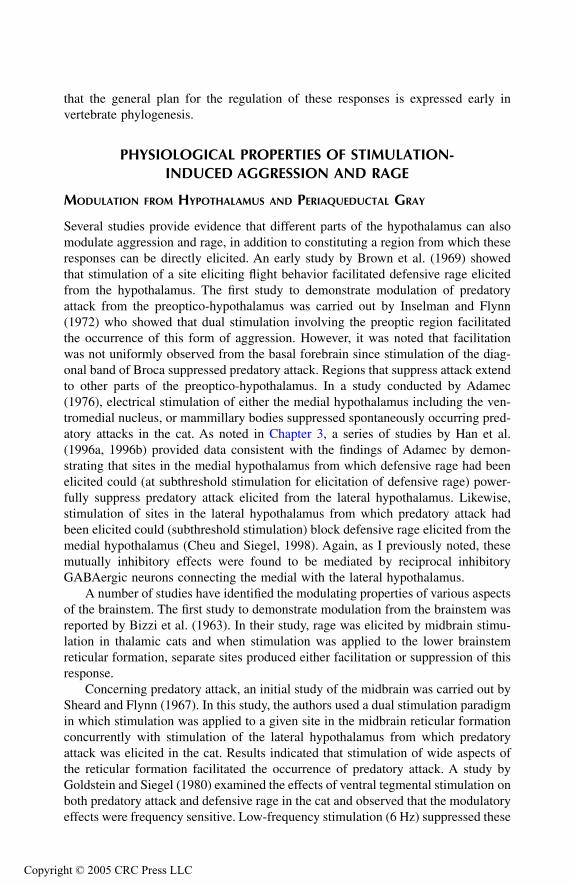
that the general plan for the regulation of these responses is expressed early invertebrate phylogenesis.
PHYSIOLOGICAL PROPERTIES OF STIMULATION-INDUCED AGGRESSION AND RAGE
M
ODULATION
FROM
H
YPOTHALAMUS
AND
P
ERIAQUEDUCTAL
G
RAY
Several studies provide evidence that different parts of the hypothalamus can alsomodulate aggression and rage, in addition to constituting a region from which theseresponses can be directly elicited. An early study by Brown et al. (1969) showedthat stimulation of a site eliciting flight behavior facilitated defensive rage elicitedfrom the hypothalamus. The first study to demonstrate modulation of predatoryattack from the preoptico-hypothalamus was carried out by Inselman and Flynn(1972) who showed that dual stimulation involving the preoptic region facilitatedthe occurrence of this form of aggression. However, it was noted that facilitationwas not uniformly observed from the basal forebrain since stimulation of the diag-onal band of Broca suppressed predatory attack. Regions that suppress attack extendto other parts of the preoptico-hypothalamus. In a study conducted by Adamec(1976), electrical stimulation of either the medial hypothalamus including the ven-tromedial nucleus, or mammillary bodies suppressed spontaneously occurring pred-atory attacks in the cat. As noted in Chapter 3, a series of studies by Han et al.(1996a, 1996b) provided data consistent with the findings of Adamec by demon-strating that sites in the medial hypothalamus from which defensive rage had beenelicited could (at subthreshold stimulation for elicitation of defensive rage) power-fully suppress predatory attack elicited from the lateral hypothalamus. Likewise,stimulation of sites in the lateral hypothalamus from which predatory attack hadbeen elicited could (subthreshold stimulation) block defensive rage elicited from themedial hypothalamus (Cheu and Siegel, 1998). Again, as I previously noted, thesemutually inhibitory effects were found to be mediated by reciprocal inhibitoryGABAergic neurons connecting the medial with the lateral hypothalamus.
A number of studies have identified the modulating properties of various aspectsof the brainstem. The first study to demonstrate modulation from the brainstem wasreported by Bizzi et al. (1963). In their study, rage was elicited by midbrain stimu-lation in thalamic cats and when stimulation was applied to the lower brainstemreticular formation, separate sites produced either facilitation or suppression of thisresponse.
Concerning predatory attack, an initial study of the midbrain was carried out bySheard and Flynn (1967). In this study, the authors used a dual stimulation paradigmin which stimulation was applied to a given site in the midbrain reticular formationconcurrently with stimulation of the lateral hypothalamus from which predatoryattack was elicited in the cat. Results indicated that stimulation of wide aspects ofthe reticular formation facilitated the occurrence of predatory attack. A study byGoldstein and Siegel (1980) examined the effects of ventral tegmental stimulation onboth predatory attack and defensive rage in the cat and observed that the modulatoryeffects were frequency sensitive. Low-frequency stimulation (6 Hz) suppressed these
TF1661_C004.fm Page 60 Thursday, September 23, 2004 3:15 PM
Copyright © 2005 CRC Press LLC

responses while high-frequency stimulation (60 Hz) was largely ineffective. It shouldbe noted that this finding is somewhat inconsistent with those reported by Bandler(1977) and Proshansky and Bandler (1975) who demonstrated the importance of theventral tegmental area in the expression of predatory attack. It is difficult to accountfor these inconsistent findings at this time. In a further study, Shaikh et al. (1985)provided a map of the sites in the midbrain from which predatory attack anddefensive rage could be modulated. It was discovered that stimulation of the medialand lateral aspects of the midbrain tegmentum differentially modulated predatoryattack and defensive rage elicited from the lateral and medial hypothalamus of thecat, respectively. Facilitation of predatory attack and suppression of defensive ragefollowed stimulation of the lateral tegmentum, while suppression of predatory attackand facilitation of defensive rage followed stimulation of its medial aspect. Theeffects of tegmental stimulation on predatory attack are consistent with thosedescribed by Sheard and Flynn (1967), and further point to the likelihood that thesites of interaction for midbrain tegmental modulation occurs in the hypothalamus,where fibers from this region are known to project.
O
THER
E
FFECTS
OF
E
LECTRICAL
S
TIMULATION
OF
A
TTACK
S
ITES
IN
C
ATS
Sledjeski and Flynn (1972) attempted to identify key characteristics of the behavioralprocess that is initiated by stimulation and which results in predatory attack behavior.Because they had noted that the autonomic components associated with stimulationof the attack site outlast the behavioral component, they specifically focused on theeffect of prior stimulation on excitability at attack sites. To address this question,they used 5 seconds of priming stimulation that were followed by a train of stimu-lation present until the attack occurred at 2, 4, 8, 16, and 32 seconds after the primingstimulation. These investigators observed that stimulation initiated at all but thelongest intervals after the priming stimulation led to a reduction in response latency.It was of interest to note that priming at other sites (from which attack could alsobe elicited in two of five cats after longer stimulation) were not effective in shorteningresponse latencies. Because of the localized nature of the excitatory effects bystimulation, it was concluded that electrical stimulation of attack sites in the hypo-thalamus excites specific parts of the central nervous system. These authors pointedout that this conclusion contrasts with a prior view — namely, that excitatoryhypothalamic stimulation results in an “affective” state such as anger, mood change,or drive, and that such a change in the psychological state of the animal determinesthe responsiveness of the central nervous system and organism.
In a related study, Bandler and Fatouris (1978) extended this approach to deter-mine whether the principal of “priming” could be applied to the study of attacksituated in different regions of the brain as a means of establishing the functionalneural relationship between two given regions. These authors observed that whenpriming was generated from the ventral tegmental area, response latencies for pred-atory attack following stimulation of the lateral hypothalamus were shortened. How-ever, when the procedures were reversed (i.e., priming stimulation was administeredto the lateral hypothalamus and attack elicited from the ventral tegmental area),little change in response latencies was observed. These authors suggested that their
TF1661_C004.fm Page 61 Thursday, September 23, 2004 3:15 PM
Copyright © 2005 CRC Press LLC

findings could be understood by the likelihood that the descending components ofthe medial forebrain bundle (which mediate predatory attack) are more tightlyarranged at the level of the ventral tegmental area than within the lateral hypothal-amus. In this manner, stimulation of the ventral tegmental area would excite greaterquantities of medial forebrain fibers than stimulation of the lateral hypothalamus.This study thus provides further evidence of the functional linkage between thelateral hypothalamus and ventral tegmental area with respect to predatory attackbehavior in the cat.
N
EUROPHYSIOLOGICAL
R
ELATIONSHIPS
Several studies have been conducted using single-cell recordings as a means ofidentifying neurons that are functionally related to aggression or rage. I have pre-viously described the findings of Adams (1968) who showed that single cells in thePAG of the cat respond selectively to the presence of a second cat at the time atwhich it is expressing defensive rage behavior, and the cat in question is respondingin kind. The results of two other experiments are described in this chapter. Oneexperiment carried out by Vanegas et al. (1970) is described later in this chapter inthe section concerning sensory processes. The second study, conducted by Huangand Flynn (1975), concerned analysis of single-cell responses identified in themidbrain in response to elicitation of predatory attack by stimulation of the lateralhypothalamus in the cat. The major findings reported were that stimulation at sitesthat elicited predatory attack affected more units, yielded a greater change in unitactivity, and significantly increased the firing rates in the dorsal tegmentum justlateral to the PAG as well as the PAG itself. Accordingly, these findings provideadditional evidence linking the PAG and adjoining regions of the tegmentum in thefunctional regulation of aggression and rage.
O
THER
R
EGIONS
A
SSOCIATED
WITH
R
AGE
AND
A
GGRESSION
There have been scattered reports in the literature that have indicated that rage and/oraggression may also be associated with regions other than the hypothalamus, PAG,and ventral tegmental area. With respect to defensive rage, several forebrain regionshave been implicated. In an early study, Emmers et al. (1965) was able to inducecats to elicit hissing and growling when provoked after forebrain ablation that sparedthe hypothalamus and amygdala. This finding would suggest that the thalamus isnot essential for the expression of defensive rage behavior. Fernandez de Molinaand Hunsperger (1962) reported that defensive rage in the cat could be elicited byelectrical stimulation of the medial amygdala, stria terminalis, medial hypothalamus,or PAG. An extensive body of literature documenting the roles of the hypothalamusand PAG in the expression of defensive rage behavior was described earlier. Whatis of interest in this study is the observation that amygdaloid or stria terminalisstimulation can induce defensive rage. One likely interpretation of these findings isthat medial amygdaloid stimulation directly excites the stria terminalis and thatstimulation of the stria terminalis drives the neurons in the medial hypothalamus
TF1661_C004.fm Page 62 Thursday, September 23, 2004 3:15 PM
Copyright © 2005 CRC Press LLC
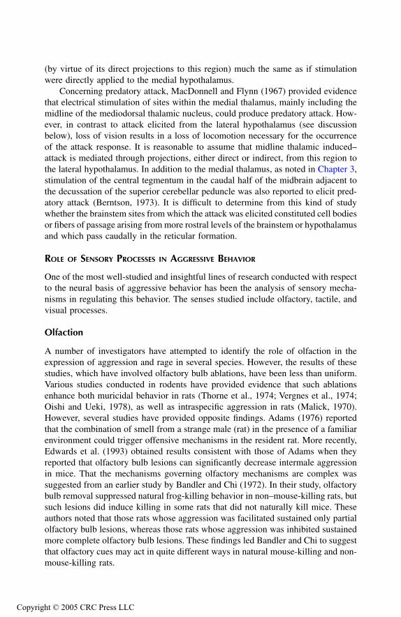
(by virtue of its direct projections to this region) much the same as if stimulationwere directly applied to the medial hypothalamus.
Concerning predatory attack, MacDonnell and Flynn (1967) provided evidencethat electrical stimulation of sites within the medial thalamus, mainly including themidline of the mediodorsal thalamic nucleus, could produce predatory attack. How-ever, in contrast to attack elicited from the lateral hypothalamus (see discussionbelow), loss of vision results in a loss of locomotion necessary for the occurrenceof the attack response. It is reasonable to assume that midline thalamic induced–attack is mediated through projections, either direct or indirect, from this region tothe lateral hypothalamus. In addition to the medial thalamus, as noted in Chapter 3,stimulation of the central tegmentum in the caudal half of the midbrain adjacent tothe decussation of the superior cerebellar peduncle was also reported to elicit pred-atory attack (Berntson, 1973). It is difficult to determine from this kind of studywhether the brainstem sites from which the attack was elicited constituted cell bodiesor fibers of passage arising from more rostral levels of the brainstem or hypothalamusand which pass caudally in the reticular formation.
R
OLE
OF
S
ENSORY
P
ROCESSES
IN
A
GGRESSIVE
B
EHAVIOR
One of the most well-studied and insightful lines of research conducted with respectto the neural basis of aggressive behavior has been the analysis of sensory mecha-nisms in regulating this behavior. The senses studied include olfactory, tactile, andvisual processes.
Olfaction
A number of investigators have attempted to identify the role of olfaction in theexpression of aggression and rage in several species. However, the results of thesestudies, which have involved olfactory bulb ablations, have been less than uniform.Various studies conducted in rodents have provided evidence that such ablationsenhance both muricidal behavior in rats (Thorne et al., 1974; Vergnes et al., 1974;Oishi and Ueki, 1978), as well as intraspecific aggression in rats (Malick, 1970).However, several studies have provided opposite findings. Adams (1976) reportedthat the combination of smell from a strange male (rat) in the presence of a familiarenvironment could trigger offensive mechanisms in the resident rat. More recently,Edwards et al. (1993) obtained results consistent with those of Adams when theyreported that olfactory bulb lesions can significantly decrease intermale aggressionin mice. That the mechanisms governing olfactory mechanisms are complex wassuggested from an earlier study by Bandler and Chi (1972). In their study, olfactorybulb removal suppressed natural frog-killing behavior in non–mouse-killing rats, butsuch lesions did induce killing in some rats that did not naturally kill mice. Theseauthors noted that those rats whose aggression was facilitated sustained only partialolfactory bulb lesions, whereas those rats whose aggression was inhibited sustainedmore complete olfactory bulb lesions. These findings led Bandler and Chi to suggestthat olfactory cues may act in quite different ways in natural mouse-killing and non-mouse-killing rats.
TF1661_C004.fm Page 63 Thursday, September 23, 2004 3:15 PM
Copyright © 2005 CRC Press LLC
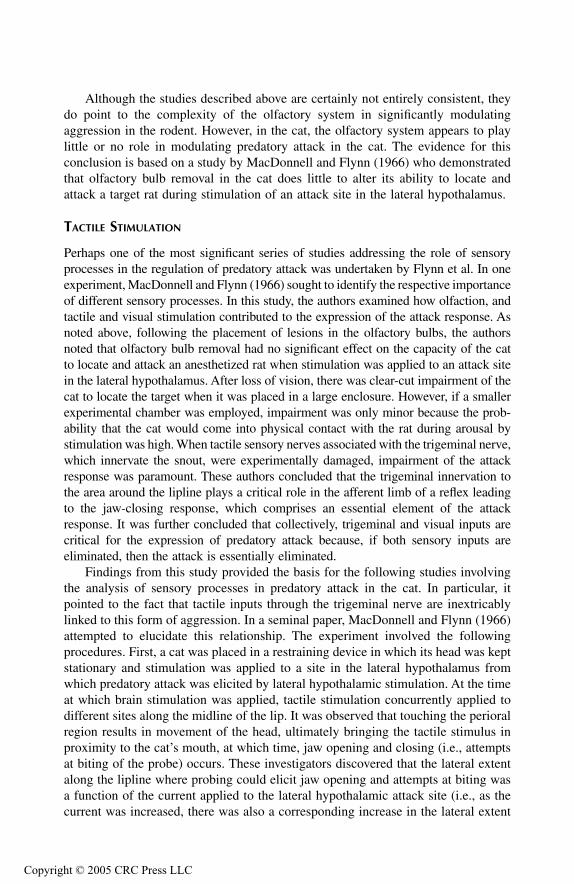
Although the studies described above are certainly not entirely consistent, theydo point to the complexity of the olfactory system in significantly modulatingaggression in the rodent. However, in the cat, the olfactory system appears to playlittle or no role in modulating predatory attack in the cat. The evidence for thisconclusion is based on a study by MacDonnell and Flynn (1966) who demonstratedthat olfactory bulb removal in the cat does little to alter its ability to locate andattack a target rat during stimulation of an attack site in the lateral hypothalamus.
T
ACTILE
S
TIMULATION
Perhaps one of the most significant series of studies addressing the role of sensoryprocesses in the regulation of predatory attack was undertaken by Flynn et al. In oneexperiment, MacDonnell and Flynn (1966) sought to identify the respective importanceof different sensory processes. In this study, the authors examined how olfaction, andtactile and visual stimulation contributed to the expression of the attack response. Asnoted above, following the placement of lesions in the olfactory bulbs, the authorsnoted that olfactory bulb removal had no significant effect on the capacity of the catto locate and attack an anesthetized rat when stimulation was applied to an attack sitein the lateral hypothalamus. After loss of vision, there was clear-cut impairment of thecat to locate the target when it was placed in a large enclosure. However, if a smallerexperimental chamber was employed, impairment was only minor because the prob-ability that the cat would come into physical contact with the rat during arousal bystimulation was high. When tactile sensory nerves associated with the trigeminal nerve,which innervate the snout, were experimentally damaged, impairment of the attackresponse was paramount. These authors concluded that the trigeminal innervation tothe area around the lipline plays a critical role in the afferent limb of a reflex leadingto the jaw-closing response, which comprises an essential element of the attackresponse. It was further concluded that collectively, trigeminal and visual inputs arecritical for the expression of predatory attack because, if both sensory inputs areeliminated, then the attack is essentially eliminated.
Findings from this study provided the basis for the following studies involvingthe analysis of sensory processes in predatory attack in the cat. In particular, itpointed to the fact that tactile inputs through the trigeminal nerve are inextricablylinked to this form of aggression. In a seminal paper, MacDonnell and Flynn (1966)attempted to elucidate this relationship. The experiment involved the followingprocedures. First, a cat was placed in a restraining device in which its head was keptstationary and stimulation was applied to a site in the lateral hypothalamus fromwhich predatory attack was elicited by lateral hypothalamic stimulation. At the timeat which brain stimulation was applied, tactile stimulation concurrently applied todifferent sites along the midline of the lip. It was observed that touching the perioralregion results in movement of the head, ultimately bringing the tactile stimulus inproximity to the cat’s mouth, at which time, jaw opening and closing (i.e., attemptsat biting of the probe) occurs. These investigators discovered that the lateral extentalong the lipline where probing could elicit jaw opening and attempts at biting wasa function of the current applied to the lateral hypothalamic attack site (i.e., as thecurrent was increased, there was also a corresponding increase in the lateral extent
TF1661_C004.fm Page 64 Thursday, September 23, 2004 3:15 PM
Copyright © 2005 CRC Press LLC

along the lipline from which this reflex could be elicited) (Figure 4.1). They furthernoted that upon using the distance from the midline of the lip where tactile stimu-lation elicited jaw opening as a measure, tactile stimulation of the side of the liplinecontralateral to that of hypothalamic stimulation was more effective than ipsilateralstimulation. These findings were interpreted to mean that hypothalamic stimulationat an attack site alters the trigeminal sensory fields along the lipline. The change inthe sensory fields in association with hypothalamic stimulation thus facilitates trig-gering of the reflex biting component of predatory attack by tactile stimulation.Therefore, when predatory attack occurs, either under natural conditions or byelectrical stimulation of the lateral hypothalamus, it is the tactile stimulation expe-rienced by the cat when its lipline touches the body of the prey object that activatesthe biting component and, when these trigeminal inputs are disrupted, as describedby MacDonnell and Flynn (1966), the biting component is also disrupted. It shouldbe noted that parallel findings were reported in a study in which electrical stimulationof the lateral hypothalamus of the rat was coupled with the application of tactilestimulation to the perioral region (Smith, 1972). Here, it was observed that onlywhen electrical stimulation of various sites in the lateral hypothalamus elicited aspecific behavior such as attack, eating, gnawing, or drinking tactile stimulation didtactile stimulation of the perioral region elicit turning toward the stimulus and biting.Thus, the role of sensory receptive field activation in elicitation of biting attackextends across species.
In a related study, Bandler and Flynn (1972) extended the findings of MacDon-nell and Flynn by demonstrating that light tactile stimulation of the forelimb involv-ing the fifth and sixth cervical dermatomes of the cat elicited striking by that limb
FIGURE 4.1
Extent of the “effective sensory fields” that were probed to elicit jaw openingand closing on a given trial. This field was generally probed from the lateral extent towardthe midline. (From C. Block, Ph.D. thesis, College of Medicine and Dentistry of New Jersey,Newark, N.J., 1974, in M. MacDonnell and J.P. Flynn,
Anim. Behav.
, 14, 1966, 399–405.With permission.)
TF1661_C004.fm Page 65 Thursday, September 23, 2004 3:15 PM
Copyright © 2005 CRC Press LLC

when combined with low-intensity stimulation of an attack site in the lateral hypo-thalamus. These authors further showed other similarities between the effects oftactile stimulation of the limb and that of the lipline with respect to attack behavior.These include the observations that (1) by increasing the intensity of stimulation,tactile stimulation of successive dermatomes (i.e., seventh and eighth cervical andfirst thoracic) could also elicit striking; and (2) the tactile receptive field on the sidecontralateral to hypothalamic stimulation was greater than the corresponding recep-tive field on the ipsilateral side. Thus, with respect to somatosensory stimulationand its role in initiating predatory attack, the same mechanism appears to be presentboth along the lipline and forelimb.
The phenomenon that sensory receptor activation can trigger the final componentof the attack response in the cat, namely, the biting reflex, is not limited to soma-tosensory stimulation, but includes visual inputs as well. Several studies addressedthe role of the visual system in predatory attack. In one study, Vanegas et al. (1970)recorded single unit responses from the visual cortex to oriented slits of light, edgesof a bar of light, and movement of a bar of light, as well as single cells spontaneouslyelicited or activated by diffuse light. These investigators reported that stimulationapplied to lateral hypothalamic predatory attack sites caused modification of theseunit responses. The implication from this study is that activation of neurons in thelateral hypothalamus in association with predatory attack can modify the firingpatterns of neurons in the visual cortex, which likely play an important role in thecat’s attempt to locate the target.
A second study that addressed the question of the role of vision in predationwas conducted by Bandler and Flynn (1971). In their study, predatory attack wasinitially elicited by electrical stimulation of the lateral hypothalamus of the cat. Then,an anesthetized mouse was placed in the visual field of either the eye ipsilateral orcontralateral to that of stimulation of the lateral hypothalamus in which the appear-ance of the mouse was available to one of the two eyes at a given time. The resultsrevealed that a cat lunged toward a mouse more frequently following stimulation ofa lateral hypothalamic attack site contralateral to the eye viewing the mouse thantoward a mouse presented to the ipsilateral eye. The authors concluded that thisfinding is consistent with previous studies described above. They further suggestedthat biting involves both a head turning component and another of mouth opening,which are manifest first along the lipline and limb on the side contralateral to thatof stimulation. As indicated above, Flynn et al. (1970) concluded from these studiesthat motivated (attack) behavior induced by brain stimulation engages the sensoryand motor systems to act in highly specific ways as “patterned” reflexes whoseultimate expression is the attack response.
I would like to point out that parallel tactile sensory modulation of other behav-ioral processes has also been documented. Wolgin and Teitelbaum (1978) placedlesions in the lateral hypothalamus of the cat and noted the presence of aphagia andadipsia, deficiencies in arousal, and
sensory neglect
. During recovery, these functionsreturned, including responsiveness to sensory stimuli. These observations led theseinvestigators to conclude that activation of sensory processes is essential for theoccurrence of feeding, much in the same way that they are important for the occur-rence of aggression.
TF1661_C004.fm Page 66 Thursday, September 23, 2004 3:15 PM
Copyright © 2005 CRC Press LLC
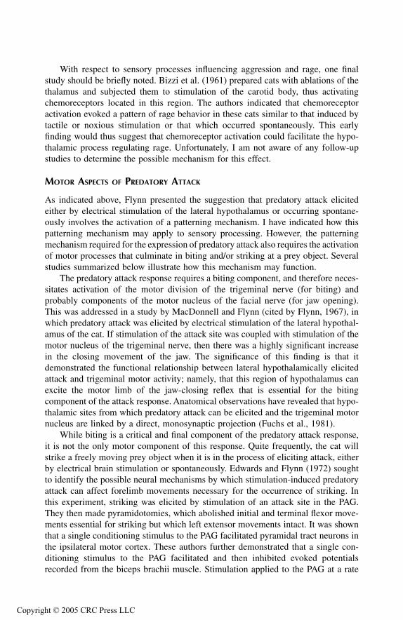
With respect to sensory processes influencing aggression and rage, one finalstudy should be briefly noted. Bizzi et al. (1961) prepared cats with ablations of thethalamus and subjected them to stimulation of the carotid body, thus activatingchemoreceptors located in this region. The authors indicated that chemoreceptoractivation evoked a pattern of rage behavior in these cats similar to that induced bytactile or noxious stimulation or that which occurred spontaneously. This earlyfinding would thus suggest that chemoreceptor activation could facilitate the hypo-thalamic process regulating rage. Unfortunately, I am not aware of any follow-upstudies to determine the possible mechanism for this effect.
M
OTOR
A
SPECTS
OF
P
REDATORY
A
TTACK
As indicated above, Flynn presented the suggestion that predatory attack elicitedeither by electrical stimulation of the lateral hypothalamus or occurring spontane-ously involves the activation of a patterning mechanism. I have indicated how thispatterning mechanism may apply to sensory processing. However, the patterningmechanism required for the expression of predatory attack also requires the activationof motor processes that culminate in biting and/or striking at a prey object. Severalstudies summarized below illustrate how this mechanism may function.
The predatory attack response requires a biting component, and therefore neces-sitates activation of the motor division of the trigeminal nerve (for biting) andprobably components of the motor nucleus of the facial nerve (for jaw opening).This was addressed in a study by MacDonnell and Flynn (cited by Flynn, 1967), inwhich predatory attack was elicited by electrical stimulation of the lateral hypothal-amus of the cat. If stimulation of the attack site was coupled with stimulation of themotor nucleus of the trigeminal nerve, then there was a highly significant increasein the closing movement of the jaw. The significance of this finding is that itdemonstrated the functional relationship between lateral hypothalamically elicitedattack and trigeminal motor activity; namely, that this region of hypothalamus canexcite the motor limb of the jaw-closing reflex that is essential for the bitingcomponent of the attack response. Anatomical observations have revealed that hypo-thalamic sites from which predatory attack can be elicited and the trigeminal motornucleus are linked by a direct, monosynaptic projection (Fuchs et al., 1981).
While biting is a critical and final component of the predatory attack response,it is not the only motor component of this response. Quite frequently, the cat willstrike a freely moving prey object when it is in the process of eliciting attack, eitherby electrical brain stimulation or spontaneously. Edwards and Flynn (1972) soughtto identify the possible neural mechanisms by which stimulation-induced predatoryattack can affect forelimb movements necessary for the occurrence of striking. Inthis experiment, striking was elicited by stimulation of an attack site in the PAG.They then made pyramidotomies, which abolished initial and terminal flexor move-ments essential for striking but which left extensor movements intact. It was shownthat a single conditioning stimulus to the PAG facilitated pyramidal tract neurons inthe ipsilateral motor cortex. These authors further demonstrated that a single con-ditioning stimulus to the PAG facilitated and then inhibited evoked potentialsrecorded from the biceps brachii muscle. Stimulation applied to the PAG at a rate
TF1661_C004.fm Page 67 Thursday, September 23, 2004 3:15 PM
Copyright © 2005 CRC Press LLC
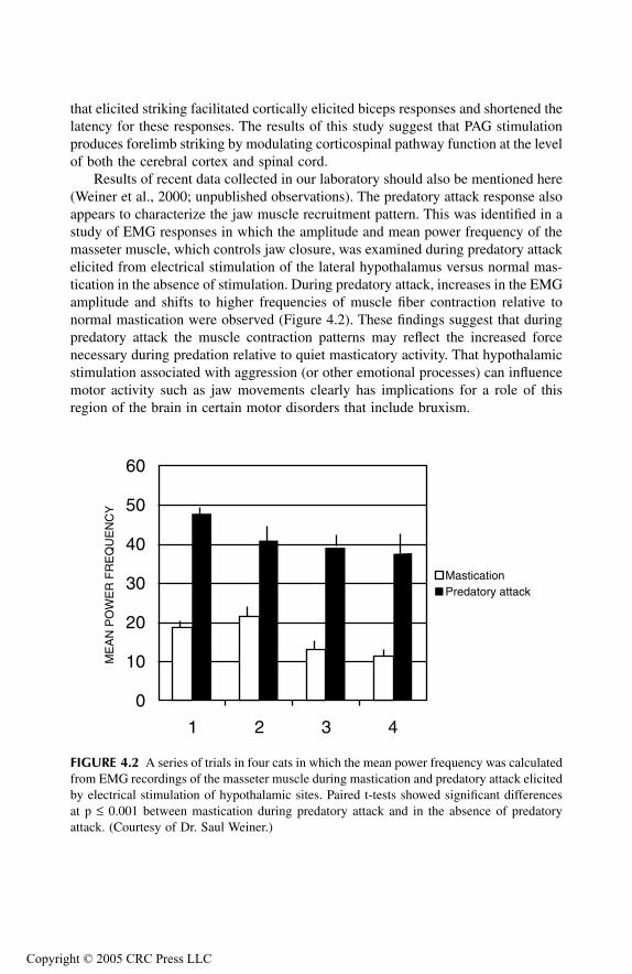
that elicited striking facilitated cortically elicited biceps responses and shortened thelatency for these responses. The results of this study suggest that PAG stimulationproduces forelimb striking by modulating corticospinal pathway function at the levelof both the cerebral cortex and spinal cord.
Results of recent data collected in our laboratory should also be mentioned here(Weiner et al., 2000; unpublished observations). The predatory attack response alsoappears to characterize the jaw muscle recruitment pattern. This was identified in astudy of EMG responses in which the amplitude and mean power frequency of themasseter muscle, which controls jaw closure, was examined during predatory attackelicited from electrical stimulation of the lateral hypothalamus versus normal mas-tication in the absence of stimulation. During predatory attack, increases in the EMGamplitude and shifts to higher frequencies of muscle fiber contraction relative tonormal mastication were observed (Figure 4.2). These findings suggest that duringpredatory attack the muscle contraction patterns may reflect the increased forcenecessary during predation relative to quiet masticatory activity. That hypothalamicstimulation associated with aggression (or other emotional processes) can influencemotor activity such as jaw movements clearly has implications for a role of thisregion of the brain in certain motor disorders that include bruxism.
FIGURE 4.2
A series of trials in four cats in which the mean power frequency was calculatedfrom EMG recordings of the masseter muscle during mastication and predatory attack elicitedby electrical stimulation of hypothalamic sites. Paired t-tests showed significant differencesat p
£
0.001 between mastication during predatory attack and in the absence of predatoryattack. (Courtesy of Dr. Saul Weiner.)
1 2 3 4
0
10
20
30
40
50
60
ME
AN
PO
WE
R F
RE
QU
EN
CY
MasticationPredatory attack
TF1661_C004.fm Page 68 Thursday, September 23, 2004 3:15 PM
Copyright © 2005 CRC Press LLC

B
EHAVIORAL
P
ROPERTIES
OF
A
TTACK
B
EHAVIOR
A variety of studies have been conducted over the past 3–4 decades that haveattempted to identify and characterize principal behavioral properties associated withaggression and rage most frequently elicited by electrical stimulation of the hypo-thalamus or PAG. These properties include: the capacity of the animal to identify aprey object and discriminate it from a non-prey object that has a similar appearance,the positive or negative reinforcing properties of different forms of attack, thepossible relationship to feeding behavior, and the relationship of predation to avoid-ance and play.
D
ISCRIMINATIVE
P
ROPERTIES
OF
A
GGRESSION
A basic question that naturally arises with respect to predatory attack behavior iswhether the attack is indiscriminate or directed to a specific prey object. This concernwas addressed directly by Levison and Flynn (1965). In their study, a number ofprey objects (or potential prey objects) were placed in a cage with a cat from whichpredatory attack behavior could be initiated by electrical stimulation of the lateralhypothalamus. The objects presented to the cat at any given time included ananesthetized rat, stuffed rat, and another object such as a hairy toy dog, Styrofoamblock, or foam rubber block. The frequency of attack on these objects followingbrain stimulation was then determined. These investigators demonstrated that thehighest frequency of attack was on the anesthetized rat with progressively fewerresponses on the stuffed rat and other objects (i.e., hairy toy dog, Styrofoam block,and foam rubber block). Surprisingly, a relatively higher response frequency of attackwas initiated in the presence of the hairy toy dog relative to the other inanimateobjects. These findings indicate the specificity of predatory attack, which is directedprimarily on a natural prey object (i.e., anesthetized rat) as opposed to a nonlivingobject. As these authors concluded, brain stimulation of an attack site is capable ofproducing behavior that limits the sensory objects triggering the completion of theattack response. Given the previous discussion, there are likely to be at least twosensory components active here. The first is the visual system, which allows the catto discern the differences between a live and stuffed rat. The second is somatosensorystimulation associated with the trigeminal nerve. As indicated above, tactile stimu-lation along the lipline is a sufficient stimulus to trigger the biting response, thusaccounting for the relative high frequencies of attack on the anesthetized rat andhairy toy dog.
R
EINFORCING
P
ROPERTIES
OF
R
AGE
AND
A
GGRESSION
Various studies conducted in several species have provided evidence that predatoryattack has positively reinforcing properties while defensive rage contains negativelyreinforcing properties. One of the first studies to address this question was carriedout by Roberts and Carey (1965). In their study, it was shown that when hypothalamicstimulation elicited gnawing behavior in rats, these objects could also act as rewards
TF1661_C004.fm Page 69 Thursday, September 23, 2004 3:15 PM
Copyright © 2005 CRC Press LLC
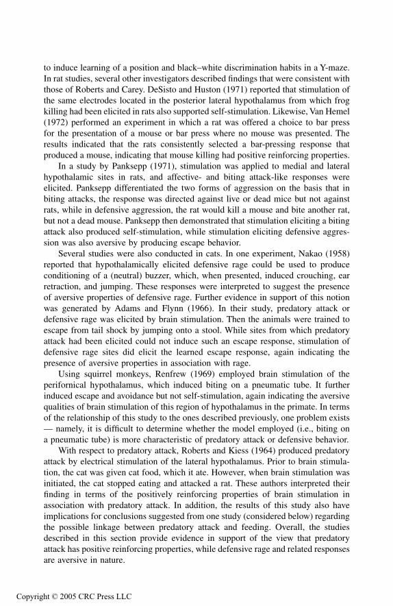
to induce learning of a position and black–white discrimination habits in a Y-maze.In rat studies, several other investigators described findings that were consistent withthose of Roberts and Carey. DeSisto and Huston (1971) reported that stimulation ofthe same electrodes located in the posterior lateral hypothalamus from which frogkilling had been elicited in rats also supported self-stimulation. Likewise, Van Hemel(1972) performed an experiment in which a rat was offered a choice to bar pressfor the presentation of a mouse or bar press where no mouse was presented. Theresults indicated that the rats consistently selected a bar-pressing response thatproduced a mouse, indicating that mouse killing had positive reinforcing properties.
In a study by Panksepp (1971), stimulation was applied to medial and lateralhypothalamic sites in rats, and affective- and biting attack-like responses wereelicited. Panksepp differentiated the two forms of aggression on the basis that inbiting attacks, the response was directed against live or dead mice but not againstrats, while in defensive aggression, the rat would kill a mouse and bite another rat,but not a dead mouse. Panksepp then demonstrated that stimulation eliciting a bitingattack also produced self-stimulation, while stimulation eliciting defensive aggres-sion was also aversive by producing escape behavior.
Several studies were also conducted in cats. In one experiment, Nakao (1958)reported that hypothalamically elicited defensive rage could be used to produceconditioning of a (neutral) buzzer, which, when presented, induced crouching, earretraction, and jumping. These responses were interpreted to suggest the presenceof aversive properties of defensive rage. Further evidence in support of this notionwas generated by Adams and Flynn (1966). In their study, predatory attack ordefensive rage was elicited by brain stimulation. Then the animals were trained toescape from tail shock by jumping onto a stool. While sites from which predatoryattack had been elicited could not induce such an escape response, stimulation ofdefensive rage sites did elicit the learned escape response, again indicating thepresence of aversive properties in association with rage.
Using squirrel monkeys, Renfrew (1969) employed brain stimulation of theperifornical hypothalamus, which induced biting on a pneumatic tube. It furtherinduced escape and avoidance but not self-stimulation, again indicating the aversivequalities of brain stimulation of this region of hypothalamus in the primate. In termsof the relationship of this study to the ones described previously, one problem exists— namely, it is difficult to determine whether the model employed (i.e., biting ona pneumatic tube) is more characteristic of predatory attack or defensive behavior.
With respect to predatory attack, Roberts and Kiess (1964) produced predatoryattack by electrical stimulation of the lateral hypothalamus. Prior to brain stimula-tion, the cat was given cat food, which it ate. However, when brain stimulation wasinitiated, the cat stopped eating and attacked a rat. These authors interpreted theirfinding in terms of the positively reinforcing properties of brain stimulation inassociation with predatory attack. In addition, the results of this study also haveimplications for conclusions suggested from one study (considered below) regardingthe possible linkage between predatory attack and feeding. Overall, the studiesdescribed in this section provide evidence in support of the view that predatoryattack has positive reinforcing properties, while defensive rage and related responsesare aversive in nature.
TF1661_C004.fm Page 70 Thursday, September 23, 2004 3:15 PM
Copyright © 2005 CRC Press LLC
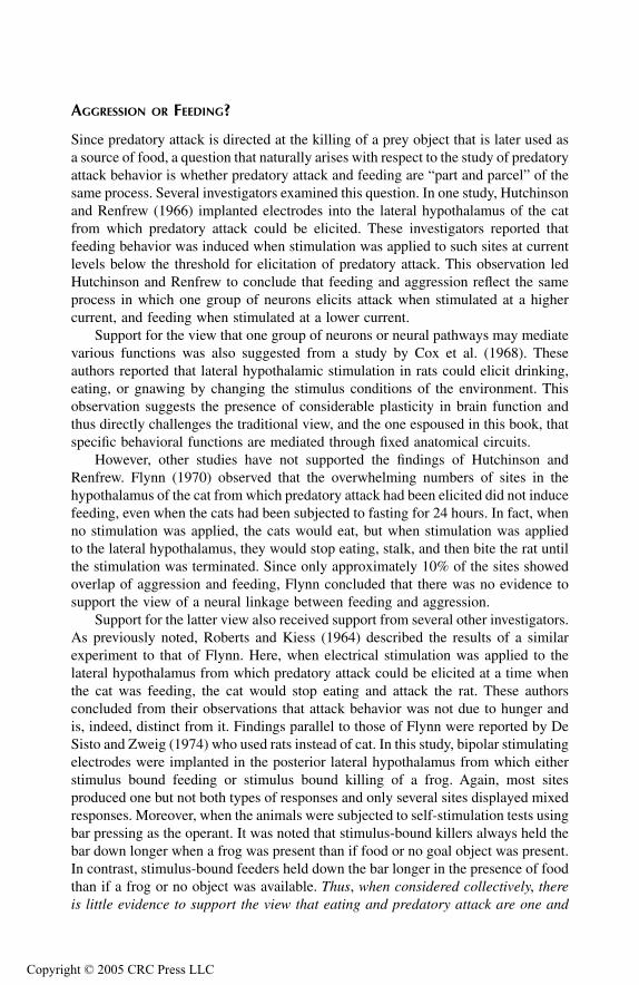
A
GGRESSION
OR
F
EEDING
?
Since predatory attack is directed at the killing of a prey object that is later used asa source of food, a question that naturally arises with respect to the study of predatoryattack behavior is whether predatory attack and feeding are “part and parcel” of thesame process. Several investigators examined this question. In one study, Hutchinsonand Renfrew (1966) implanted electrodes into the lateral hypothalamus of the catfrom which predatory attack could be elicited. These investigators reported thatfeeding behavior was induced when stimulation was applied to such sites at currentlevels below the threshold for elicitation of predatory attack. This observation ledHutchinson and Renfrew to conclude that feeding and aggression reflect the sameprocess in which one group of neurons elicits attack when stimulated at a highercurrent, and feeding when stimulated at a lower current.
Support for the view that one group of neurons or neural pathways may mediatevarious functions was also suggested from a study by Cox et al. (1968). Theseauthors reported that lateral hypothalamic stimulation in rats could elicit drinking,eating, or gnawing by changing the stimulus conditions of the environment. Thisobservation suggests the presence of considerable plasticity in brain function andthus directly challenges the traditional view, and the one espoused in this book, thatspecific behavioral functions are mediated through fixed anatomical circuits.
However, other studies have not supported the findings of Hutchinson andRenfrew. Flynn (1970) observed that the overwhelming numbers of sites in thehypothalamus of the cat from which predatory attack had been elicited did not inducefeeding, even when the cats had been subjected to fasting for 24 hours. In fact, whenno stimulation was applied, the cats would eat, but when stimulation was appliedto the lateral hypothalamus, they would stop eating, stalk, and then bite the rat untilthe stimulation was terminated. Since only approximately 10% of the sites showedoverlap of aggression and feeding, Flynn concluded that there was no evidence tosupport the view of a neural linkage between feeding and aggression.
Support for the latter view also received support from several other investigators.As previously noted, Roberts and Kiess (1964) described the results of a similarexperiment to that of Flynn. Here, when electrical stimulation was applied to thelateral hypothalamus from which predatory attack could be elicited at a time whenthe cat was feeding, the cat would stop eating and attack the rat. These authorsconcluded from their observations that attack behavior was not due to hunger andis, indeed, distinct from it. Findings parallel to those of Flynn were reported by DeSisto and Zweig (1974) who used rats instead of cat. In this study, bipolar stimulatingelectrodes were implanted in the posterior lateral hypothalamus from which eitherstimulus bound feeding or stimulus bound killing of a frog. Again, most sitesproduced one but not both types of responses and only several sites displayed mixedresponses. Moreover, when the animals were subjected to self-stimulation tests usingbar pressing as the operant. It was noted that stimulus-bound killers always held thebar down longer when a frog was present than if food or no goal object was present.In contrast, stimulus-bound feeders held down the bar longer in the presence of foodthan if a frog or no object was available.
Thus
,
when considered collectively
,
thereis little evidence to support the view that eating and predatory attack are one and
TF1661_C004.fm Page 71 Thursday, September 23, 2004 3:15 PM
Copyright © 2005 CRC Press LLC
the same process. In fact
,
the existing data support the opposite view
,
furthersuggesting a distinctiveness of the neural circuitry mediating each of these behav-ioral processes.
AVOIDANCE, PLAY, AND PREDATORY ATTACK
In the course of predatory attack, cats may engage in related behavior sometimesreferred to as “predatory play.” It typically involves repeated contact with the preyobject without injuring it. Although the response pattern resembles that of attack, itcan be viewed as constituting an independent behavioral pattern. Alternatively, itmay also be considered as falling along a continuum with predatory attack, wheredefense represents the end of the continuum in opposition to predatory attack. In astudy by Pellis et al. (1988), three lines of evidence were presented in support ofthe notion of a continuum (Figure 4.3). The first is that benzodiazepines administeredto cats shifted the direction of behavior along the continuum by increased killing ofmice which previously played with them and by increasing play in cats that previ-ously avoided mice. Second, lateral hypothalamic lesions initially disrupted attack,but during recovery, an escalation towards killing was observed in the absence ofplay and avoidance. When tail pinches were applied to cats, a similar pattern ofresults occurred in both killer and nonkiller cats. These investigators thus arguedthat predatory play in reality represents predatory behavior that has not yet pro-gressed to biting attack. They further suggested that this response may have survivalvalue by reducing the likelihood of injury to the animal, as it may be construed asconstituting a more cautious approach toward generating the ultimate attack on theprey object.
SUMMARY
In this chapter, I attempted to provide a review of the basic physiological andbehavioral properties associated with aggression and rage. Although this analysis isnot exhaustive, it does examine what I believe are the most important ones for theexpression of aggression and rage.
The first point made considered the importance of the hypothalamus, PAG, andrelated regions of the brainstem in mediating these forms of aggression as well asthe key interrelationships established both within and among these structures. I havetried to reinforce (based on previous chapters) the facts that the medial and lateralhypothalamus mediate defensive rage and predatory attack, and that the dorsolateralPAG constitutes a key brainstem component of the defensive rage response. It wasshown that while the hypothalamus is normally quite important for the expressionof aggression and rage, these responses might nevertheless take place after thehypothalamus has been disconnected from the rest of the brain. In contrast, destruc-tion of the PAG generally eliminates defensive rage behavior and tegmental lesionssignificantly suppress or block predatory attack in cats. Studies conducted in rodentsand other species have also pointed to the importance of the hypothalamus in theexpression of aggressive reactions characteristic of such species. However, with
TF1661_C004.fm Page 72 Thursday, September 23, 2004 3:15 PM
Copyright © 2005 CRC Press LLC
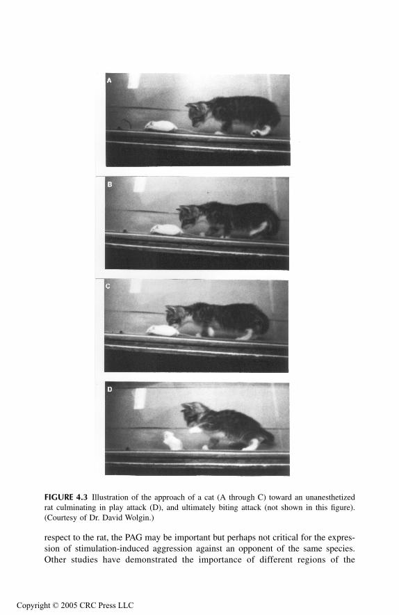
respect to the rat, the PAG may be important but perhaps not critical for the expres-sion of stimulation-induced aggression against an opponent of the same species.Other studies have demonstrated the importance of different regions of the
FIGURE 4.3
Illustration of the approach of a cat (A through C) toward an unanesthetizedrat culminating in play attack (D), and ultimately biting attack (not shown in this figure).(Courtesy of Dr. David Wolgin.)
TF1661_C004.fm Page 73 Thursday, September 23, 2004 3:15 PM
Copyright © 2005 CRC Press LLC
hypothalamus and brainstem tegmentum in modulating aggression and rage elicitedfrom other regions of the hypothalamus. These findings indicate important anatom-ical and functional relationships among various regions of hypothalamus such asthe excitatory interactions between the anterior and posterior hypothalamus andinhibitory interactions between the medial and lateral hypothalamus. Other studiesidentified the physiological properties that are characteristic of brain stimulation-induced aggression and rage.
A variety of studies have examined the role of different sensory processes inthe expression of aggression and rage in several species. It was concluded that whileolfaction may play an important (although not entirely consistent) role in rodentaggression, it appears to serve a less important role in feline aggression. Instead,the evidence is quite clear that vision and somatosensory inputs involving thetrigeminal system around the lipline constitute the necessary stimuli critical for theexpression of predatory aggression. Likewise, the motor components of aggressionhave also been examined. These studies have provided evidence that hypothalamicstimulation associated with predatory attack can significantly influence motor com-ponents of both the trigeminal nerve as well as spinal cord motor neurons thatmediate biting and striking of the prey object, respectively. It was proposed thatcollectively the effects of brain stimulation–induced attack produce a change in thepatterning of discharge of sensory and motor neurons, which ensure the occurrenceof the attack response.
The final section of this chapter considered several key behavioral propertiesassociated with aggression and rage: (1) discriminative properties of predatory attackthat allow cat to attack the appropriate prey object; (2) reinforcing properties fromwhich it was concluded that predatory attack is positively reinforcing, while defen-sive rage is negatively reinforcing; and (3) that predatory play represents a compo-nent that has not yet evolved into a full attack response and which may be viewedas lying along a continuum extending from predatory attack on one end to defensiverage on the other.
REFERENCES
Adamec, R.E., Hypothalamic and extrahypothalamic substrates of predatory attack. Suppres-sion and the influence of hunger,
Brain Res.
, 106, 1976, 57–69.Adams, D. and J.P. Flynn, Transfer of an escape response from tail shock to brain-stimulated
attack behavior,
J. Exp. Anal. Behav.
, 9, 1966, 401–408.Adams, D.B., Cells related to fighting behavior recorded from midbrain central gray neuropil
of cat,
Science
, 159, 1968, 894–896.Adams, D.B., The relation of scent marking, olfactory investigation, and specific postures in
the isolation-induced fighting of rats,
Behaviour
, 56, 1976, 286–297.Bandler, R., Predatory attack behavior in the cat elicited by preoptic region stimulation: a
comparison with behavior elicited by hypothalamic and midbrain stimulation,
Aggress. Behav.
, 5, 1979, 269–282.Bandler, R., Induction of ‘rage’ following microinjections of glutamate into midbrain but not
hypothalamus of cats,
Neurosci. Lett.
, 30, 1982, 183–188.
TF1661_C004.fm Page 74 Thursday, September 23, 2004 3:15 PM
Copyright © 2005 CRC Press LLC
Bandler, R., Identification of hypothalamic and midbrain periaqueductal grey neuronesmediating aggressive and defensive behaviour by intracerebral microinjections ofexcitatory amino acids, in R. Bandler, Ed.,
Modulation of Sensorimotor ActivityDuring Alterations in Behavioral States
, Alan R. Liss, Inc., New York, 1984, pp.369–391.
Bandler, R. and C.C. Chi, Effects of olfactory bulb removal on aggression: a reevaluation,
Physiol. Behav.
, 8, 1972, 207–211.Bandler, R. and A. Depaulis, Elicitation of intraspecific defence reactions in the rat from
midbrain periaqueductal grey by microinjection of kainic acid, without neurotoxic,
Neurosci. Lett.
, 88, 1988, 291–296.Bandler, R.A., Depaulis, M. Vergnes, Identification of midbrain neurones mediating defensive
behaviour in the rat by microinjections of excitatory amino acids,
Behav. Brain Res.
,15, 1985, 107–119.
Bandler, R. and D. Fatouris, Centrally elicited attack behavior in cats: post-stimulus excit-ability and midbrain-hypothalamic inter-relationships,
Brain Res.
, 153, 1978,427–433.
Bandler, R. and J.P. Flynn, Control of somatosensory fields for striking during hypothalam-ically elicited attack,
Brain Res.
, 38, 1972, 197–201.Bandler, R., S. Prineas, T. McCulloch, Further localization of midbrain neurones mediating
the defence reaction in the cat by microinjections of excitatory amino acids,
Neurosci.Lett.
, 56, 1985, 311–316.Bandler, R. and M.T. Shipley, Columnar organization in the midbrain periaqueductal gray:
modules for emotional expression,
Trends Neurosci.
, 17, 1994, 379–389.Bandler, R.J., Predatory behavior in the cat elicited by lower brain stem and hypothalamic
stimulation: a comparison,
Brain Behav. Evol.
, 14, 1977, 440–460.Bandler, R.J. and A. Depaulis, Midbrain periaqueductal gray control of defensive behavior
in the cat and the rat, in A. Depaulis and R.J. Bandler, Eds.,
The Midbrain Periaq-ueductal Gray Matter
, Plenum Press, New York, 1991, pp. 175–198.Berntson, G.G. Blockade and release of hypothalamically and naturally elicited aggressive
behaviors in cats following midbrain lesions,
J. Comp. Physiol. Psychol.
, 81, 1972,541–554.
Berntson, G.G. Attack, grooming, and threat elicited by stimulation of the pontine tegmentumin cats,
Physiol. Behav.
, 11, 1973, 81–87.Bizzi, E., A. Libretti, A. Malliani, A. Zanchetti, Reflex chemoceptive excitation of diencephalic
sham rage behavior,
Amer. J. Physiol.
, 200, 1961, 923–926.Bizzi, E., A. Malliani, J. Apelbaum, A. Zanchetti, Excitation and inhibition of sham rage
behavior by lower brain stem stimulation,
Arch. Ital. Biol.
, 101, 1963, 614–631.Boehrer, A., M. Vergnes, P. Karli, Interspecific aggressiveness and reactivity in mouse-killing
and nonkilling rats: compared effects of olfactory bulb removal and raphe lesions,
Aggress. Behav.
, 1, 1974, 1–16.Brown, J.L., R.W. Hunsperger, H.E. Rosvold, Interaction of defence and flight reactions
produced by simultaneous stimulation at two points in the hypothalamus of the cat,
Exp. Brain Res.
, 8, 1969, 130–149.Canteras, N.S., S. Chiavegatto, V. Do, L.W. Swanson, Severe reduction of rat defensive
behavior to a predator by discrete hypothalamic chemical lesions,
Brain Res. Bull.
,44, 1997, 297–305.
Carli, G., A. Malliani, A. Zanchetti, Midbrain course of descending pathways mediating shamrage behavior,
Exp. Neurol.
, 7, 1963, 210–223.
TF1661_C004.fm Page 75 Thursday, September 23, 2004 3:15 PM
Copyright © 2005 CRC Press LLC
Cheu, J.W. and A. Siegel, GABA receptor mediated suppression of defensive rage behaviorelicited from the medial hypothalamus of the cat: role of the lateral hypothalamus,
Brain Res.
, 783, 1998, 293–304.Chi, C.C. and J.P. Flynn, Neuroanatomic projections related to biting attack elicited from
hypothalamus in cats,
Brain Res.
, 35, 1971, 49–66.Cox, V.C., E.S. Valenstein, J.W. Kakolewski, Modification of motivated behavior elicited by
electrical stimulation of the hypothalamus,
Science
, 159, 1968, 1119–1121.Depaulis, A., K.A. Keay, R. Bandler, Longitudinal neuronal organization of defensive reac-
tions in the midbrain periaqueductal gray region of the rat,
Exp. Brain Res.
, 90, 1992,307–318.
DeSisto, M.J. and J.P. Huston, Aggression and reward from stimulating common sites in theposterior lateral hypothalamus of rats,
Commun. Behav. Biol.
, 6, 1971, 295–306.DeSisto, M.J. and M. Zweig, Differentiation of hypothalamic feeding and killing,
Physiol.Psychol.
, 2, 1974, 67–70.Eckersdorf, B., H. Golebiewski, M.K. Lewinska, Kainic acid lesions of the cat’s antero-medial
hypothalamus and emotional-defensive response evoked by carbachol injection to thesame loci,
Behav. Brain Res.
, 24, 1987, 161–166.Edwards, D.A., F.R. Nahai, P. Wright, Pathways linking the olfactory bulbs with the medial
preoptic anterior hypothalamus are important for intermale aggression in mice,
Phys-iol. Behav.
, 53, 1993, 611–615.Edwards, S.B. and J.P. Flynn, Corticospinal control of striking in centrally elicited attack
behavior,
Brain Res.
, 41, 1972, 51–65.Ellison, G.D. and J.P. Flynn, Organized aggressive behavior in cats after surgical isolation of
the hypothalamus,
Arch. Ital. Biol.
, 106, 1968, 1–20.Emmers, R., R.W.M. Chun, G.H. Wang, Behavior and reflexes of chronic thalamic cats,
Arch.Ital. Biol.
, 103, 1965, 178–193.Fernandez De Molina, A. and R.W. Hunsperger, Organization of the subcortical system
governing defence and flight reactions in the cat,
J. Physiol.
, 160, 1962, 200–213.Flynn, J.P., The neural basis of aggression in cats, in D.C. Glass, Ed.,
Neurophysiology andEmotion, Rockefeller University Press and Russell Sage Foundation, New York, 1967,pp. 40–60.
Flynn, J.P. and R. Bandler, Visual patterened reflex present during hypothalamically elicitedattack, Science, 171, 1971, 817–818.
Flynn, J.P. and M.F. MacDonnell, Control of sensory fields by stimulation of hypothalamus,Science, 152, 1966, 1406–1408.
Flynn, J.P. and M.F. MacDonnell, Sensory control of hypothalamic attack, Anim. Behav., 14,1966, 399–405.
Flynn, J.P., H. Vanegas, W.E. Foote, S. Edwards, Neural mechanisms involved in a cat’s attackon a rat, in R. Whalen, Ed., The Neural Control of Behavior, Academic Press, NewYork, 1970, pp. 135–173.
Fonberg, E. and V.M. Serduchenko, Predatory behavior after hypothalamic lesions in cats,Physiol. Behav., 24, 1980, 225–230.
Fuchs, S.A.G., M. Dalsass, H.E. Siegel, A. Siegel, The neural pathways mediating quiet-biting attack behavior from the hypothalamus in the cat: a functional autoradiographicstudy, Aggress. Behav., 7, 1981, 51–67.
Fuchs, S.A.G., H.M. Edinger, A. Siegel, The organization of the hypothalamic pathwaysmediating affective defense behavior in the cat, Brain Res., 330, 1985a, 77–92.
Fuchs, S.A.G., H.M. Edinger, A. Siegel, The role of the anterior hypothalamus in affectivedefense behavior elicited from the ventromedial hypothalamus of the cat, Brain Res.,330, 1985b, 93–108.
TF1661_C004.fm Page 76 Thursday, September 23, 2004 3:15 PM
Copyright © 2005 CRC Press LLC

Goldstein, J.M. and J. Siegel, Suppression of attack behavior in cats by stimulation of ventraltegmental area and nucleus accumbens, Brain Res., 183, 1980, 181–192.
Golebiewski, H. and A. Romaniuk, Midbrain interaction with the hypothalamus in expressionof aggressive behavior in cats, Acta Neurobiol. Exp., 37, 1977, 83–97.
Halliday, R. and R. Bandler, Anterior hypothalamic knife cut eliminates a specific componentof the predatory behaviour elicited by electrical stimulation of the posterior hypo-thalamus or ventral midbrain in the cat, Neurosci. Lett., 21, 1981, 231–236.
Han, Y., M.B. Shaikh, A. Siegel, Medial amygdaloid suppression of predatory attack behaviorin the cat: I. Role of a substance P pathway from the medial amygdala to the medialhypothalamus, Brain Res., 716, 1996a, 59–71.
Han, Y., M.B. Shaikh, A. Siegel, Medial amygdaloid suppression of predatory attack behaviorin the cat: II. Role of a GABAergic pathway from the medial to the lateral hypothal-amus, Brain Res., 716, 1996b, 72–83.
Harwood, D. and D.M. Vowles, Defensive behaviour and the after effects of brain stimulationin the ring dove (Streptopelia risoria), Neuropsychologia, 5, 1967, 345–366.
Huang, Y.H. and J.P. Flynn, Unit activities in the hypothalamus and midbrain during stimu-lation of hypothalamic attack sites, Brain Res., 93, 1975, 419–440.
Hunsperger, R.W., Role of substantia grisea centralis mesencephali in electrically-inducedrage reactions, Prog. Neurobiol., 1956, 289–294.
Hutchinson, R.R. and J.W. Renfrew, Stalking attack and eating behaviors elicited from thesame sites in the hypothalamus, J. Comp. Physiol. Psychol., 61, 1966, 360–367.
Inselman, B.R. and J.P. Flynn, Modulatory effects of preoptic stimulation on hypothalami-cally-elicited attack in cats, Brain Res., 42, 1972, 73–87.
Jurgens, U., M. Maurus, D. Ploog, P. Winter, Vocalization in the squirrel monkey, Saimirisciureus, elicited by brain stimulation, Exp. Brain Res., 4, 1967, 114–117.
Kanai, T. and S.C. Wang, Localization of the central vocalization mechanism in the brainstem of the cat, Exp. Neurol., 6, 1962, 426–434.
Koolhaas, J.M. Hypothalamically induced intraspecific aggressive behaviour in the rat, Exp.Brain Res., 32, 1978, 365–375.
Kruk, M.R., W. Meelis, A.M. Van der Poel, J. Mos, Electrical stimulation as a tool to tracephysiological properties of the hypothalamic network in aggression, in P.F. Brain andD. Benton, Eds., The Biology of Aggression, Sythoff and Noordhoff InternationalPublishers, The Netherlands, 1981, pp. 383–395.
Kruk, M.R., C.E. Van der Laan, J. Mos, A.M. Van der Poel, W. Meelis, B. Olivier, Comparisonof aggressive behaviour induced by electrical stimulation in the hypothalamus ofmale and female rats, Prog. Brain Res., 61, 1984, 303–314.
Levison, P.K. and J.P. Flynn, The objects attacked by cats during stimulation of the hypo-thalamus, Anim. Behav., 13, 1965, 217–220.
Lipp, H.P. and R.W. Hunsperger, Threat, attack and flight elicited by electrical stimulationof the ventromedial hypothalamus of the Marmoset monkey Callithrix Jacchus, BrainBehav. Evol., 15, 1978, 260–293.
MacDonnell, M. and J.P. Flynn, Sensory control of hypothalamic attack, Anim. Behav., 14,1966, 399–405.
MacDonnell, M.F. and J.P. Flynn, Attack elicited by stimulation of the thalamus and adjacentstructures of cats. Behaviour, 31, 1967, 185–202.
Malick, J.B., A behavioral comparison of three lesion-induced models of aggression in therat, Physiol. Behav., 5, 1970, 679–681.
Mos, J., M.R. Kruk, A.M. Van der Poel, W. Meelis, Aggressive behavior induced by electricalstimulation in the midbrain central gray of male rats, Aggress. Behav., 8, 1982,261–284.
TF1661_C004.fm Page 77 Thursday, September 23, 2004 3:15 PM
Copyright © 2005 CRC Press LLC
Mos, J., J.H.C.M. Lammers, A.M. Van der Poel, B. Bermond, W. Meelis, M.R. Kruk, Effectsof midbrain central gray lesions on spontaneous and electrically induced aggressionin the rat, Aggress. Behav., 9, 1983, 133–155.
Nakao, H., Emotional behavior produced by hypothalamic stimulation, Amer. J. Physiol., 194,1958, 411–418.
Oishi, R. and S. Ueki, Facilitation of muricide by dorsal norepinephrine bundle lesions inolfactory bulbectomized rats, Pharmacol. Biochem. Behav., 8, 1978, 133–136.
Panksepp, J., Aggression elicited by electrical stimulation of the hypothalamus in albino rats,Physiol. Behav., 6, 1971, 321–329.
Paxinos, G. and D. Bindra, Hypothalamic knife cuts: effects on eating, drinking, irritability,aggression, and copulation in the male rat, J. Comp. Physiol. Psychol., 79, 1972,219–229.
Pellis, S.M., D.P. O’Brien, V.C. Pellis, P. Teitelbaum, D.L. Wolgin, S. Kennedy, Escalationof feline predation along a gradient from avoidance through “play” to killing, Behav.Neurosci., 102, 1988, 760–777.
Perachio, A.A. and M. Alexander, The neural bases of aggression and sexual behavior in therhesus monkey, in R.C. Bourne, Ed., The Rhesus Monkey, Academic Press, New York,1975, pp. 381–409.
Proshansky, E. and R. Bandler, Midbrain-hypothalamic interrelationships in the control ofaggressive behavior, Aggress. Behav., 1, 1975, 135–155.
Putkonen, P.T.S., Attack elicited by forebrain and hypothalamic stimulation in the chicken,Experientia, 22, 1966, 1–5.
Putkonen, P.T.S., Electrical stimulation of the avian brain: behavioral and autonomic reactionsfrom the archistriatum, ventromedial forebrain and the diencephalon in the chicken,Ann. Acad. Sci. Fenn., 130, 1967, 9–89.
Renfrew, J.W. The intensity function and reinforcing properties of brain stimulation that elicitsattack, Physiol. Behav., 4, 1969, 509–515.
Roberts, W.W. and R.J. Carey, Rewarding effect of performance of gnawing aroused byhypothalamic stimulation in the rat, J. Comp. Physiol. Psychol., 59, 1965, 317–324.
Roberts, W.W. and H.O. Kiess, Motivational properties of hypothalamic aggression in cats,J. Comp. Physiol. Psychol., 58, 1964, 187–193.
Roberts, W.W. and J. Nagel, First-order projections activated by stimulation of hypothalamicsites eliciting attack and flight in rats, Behav. Neurosci., 110, 1996, 509–527.
Shaikh, M.B., J.A. Barrett, A. Siegel, The pathways mediating affective defense and quietbiting attack behavior from the midbrain central gray of the cat: an autoradiographicstudy, Brain Res., 437, 1987, 9–25.
Shaikh, M.B., M. Brutus, A. Siegel, H.E. Siegel, Topographically organized midbrain mod-ulation of predatory and defensive aggression in the cat, Brain Res., 336, 1985,308–312.
Sheard, M.H. and J.P. Flynn, Facilitation of attack behavior by stimulation of the midbrainof cats, Brain Res., 4, 1967, 324–333.
Skultety, F.M., Stimulation of periaqueductal gray and hypothalamus, Arch. Neurol., 8, 1963,38–50.
Sledjeski, M.B. and J.P. Flynn, Poststimulus excitability at attack sites in the cat’s hypothal-amus, Brain Res., 40, 1972, 516–522.
Smith, D.A., Increased perioral responsiveness: a possible explanation for the switching ofbehavior observed during lateral hypothalamic stimulation, Physiol. Behav., 8, 1972,617–621.
TF1661_C004.fm Page 78 Thursday, September 23, 2004 3:15 PM
Copyright © 2005 CRC Press LLC
TF1661_C005.fm Page 81 Monday, October 25, 2004 7:21 AM
Copyr
5
Limbic System I: Behavioral, Anatomical, and Physiological ConsiderationsIn this chapter, results are summarized of studies attempting to identify the relation-ships of a group of forebrain regions in aggression and rage referred to as “limbic”structures. The word “limbic” means “border” in Latin, and thus, the structurescomprising the limbic system are generally interposed between the overlying cere-bral cortex and underlying diencephalon. These structures include phylogeneticallyolder regions of the telencephalon and include the hippocampal formation, septalarea, and amygdala. In considering the limbic system, it is also useful to includethe prefrontal cortex and cingulate gyrus as components of this system because oftheir effects on visceral processes that parallel those of other limbic structures.
One of the defining properties of limbic structures is that they project directlyor indirectly to the hypothalamus or midbrain periaqueductal gray (PAG). In sodoing, limbic structures modulate the functions typically associated with the hypo-thalamus or PAG. The anatomical bases by which each of these limbic structurescan regulate visceral processes will be considered in detail in the next chapter. Whilethe major focus of the analysis considers how these limbic structures affect aggres-sion and rage, it should also be noted that several of these structures also mediateother processes such as short-term memory and epileptogenic activity that are appar-ently unrelated to functions of the hypothalamus or PAG.
As part of an overview of this system, it is useful to mention several featuresconcerning the input–output relationships of limbic structures, which perhaps canhelp to provide understanding of how these structures may modulate aggression andrage. As a general rule, limbic structures receive inputs from at least two sources:(1) either directly or indirectly from one or more sensory systems; and (2) monoam-inergic fiber systems. Limbic neurons then project directly or indirectly to thehypothalamus or midbrain PAG. A likely explanation of this organization is thatinputs to the hypothalamus and PAG from limbic structures serve to modulate theiroutputs, which are directed on somatic motor and autonomic neurons of the lowerbrainstem and spinal cord for the integration of the specific forms of emotionalbehavior comprising aggression and rage. To complete this overview, the possiblefeedback pathways that may come into play should also be noted. These includepathways from the hypothalamus and PAG that project to limbic structures (andcerebral cortex), as well as limbic structures that project to the cerebral cortex. In
ight © 2005 CRC Press LLC
TF1661_C005.fm Page 82 Monday, October 25, 2004 7:21 AM
Copyr
this manner, such feedback could serve to adjust the modulatory properties of limbicstructures, especially with respect to their receipt of new information from the cortexand monoamine neurons, which could affect the reaction to emotional stimuli andthe duration of the attack or rage response. Likewise, feedback to the cerebral cortexfrom the hypothalamus and limbic system could also provide the basis for the“patterning” mechanism governing the overall sensorimotor components of theexpression of the attack or rage response that were considered in Chapter 4.
On the basis of experimental evidence, the discussion below considers the extentto which each of the limbic and related structures is involved in the regulation ofaggression and/or rage behavior. In Chapter 6, evidence of limbic system involve-ment as determined from human studies and case histories will be further considered.This chapter will systematically examine each of the limbic structures by firstidentifying in brief their basic anatomical relationships and then their functionalproperties with respect to aggression and rage.
HIPPOCAMPAL FORMATION
ANATOMICAL CONSIDERATIONS
The hippocampal formation consists of the cornu Ammonis, dentate gyrus, andsubicular cortex. The cornu Ammonis refers to the hippocampus, which is a multi-layered primitive cortex whose primary cell type is the pyramidal cell. These cellsare arranged in a C-shaped fashion, which is interlocked with a C-shaped arrange-ment of the dentate gyrus. The dendrites of pyramidal cells receive inputs fromvarious sources. These include, in part, other regions of the hippocampus, entorhinalcortex, septal area, and brainstem monoamine neurons. Sensory information thatreaches the hippocampus through oligo- or multi-synaptic pathways includes olfac-tory, visual, and auditory messages. The axons of pyramidal cells project outsidethe hippocampal formation to such regions as entorhinal cortex, septal area, and thecontralateral hippocampus (Figure 5.1).
The dentate gyrus may also be viewed as primitive cortex, and has multiplelayers as well whose primary neuron is the granule cell. Axons of the dentate gyrusgenerally do not project outside of the hippocampal formation. Instead, the axon ofthe granule cell is called a mossy fiber that projects to parts of the cornu Ammonis.The subicular cortex is an extension of the cornu Ammonis and is interposed betweenthe cornu Ammonis and entorhinal cortex. Again, the principal cell type is a pyra-midal cell whose axons project both to entorhinal cortex, diencephalon (i.e., mam-millary bodies, medial hypothalamus, anteroventral thalamic nucleus) and septalarea (Krayniak et al., 1979; Meibach and Siegel, 1975, 1977a; Siegel and Tassoni,1971; Siegel et al., 1974, 1975) (Figure 5.2).
The key points to emphasize here are that (1) monoaminergic inputs provide theanatomical and functional bases underlying mood changes, which can thus affect orsculpture the hippocampal response to visual, auditory, olfactory stimuli and othercortical inputs; and (2) the hippocampal response to such inputs are reflected in termsof its direct or indirect outputs that modulate the functions of the hypothalamus.
ight © 2005 CRC Press LLC
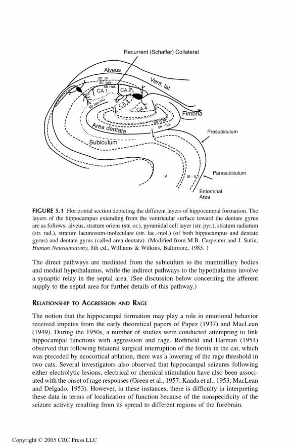
TF1661_C005.fm Page 83 Monday, October 25, 2004 7:21 AM
Copyr
The direct pathways are mediated from the subiculum to the mammillary bodiesand medial hypothalamus, while the indirect pathways to the hypothalamus involvea synaptic relay in the septal area. (See discussion below concerning the afferentsupply to the septal area for further details of this pathway.)
RELATIONSHIP TO AGGRESSION AND RAGE
The notion that the hippocampal formation may play a role in emotional behaviorreceived impetus from the early theoretical papers of Papez (1937) and MacLean(1949). During the 1950s, a number of studies were conducted attempting to linkhippocampal functions with aggression and rage. Rothfield and Harman (1954)observed that following bilateral surgical interruption of the fornix in the cat, whichwas preceded by neocortical ablation, there was a lowering of the rage threshold intwo cats. Several investigators also observed that hippocampal seizures followingeither electrolytic lesions, electrical or chemical stimulation have also been associ-ated with the onset of rage responses (Green et al., 1957; Kaada et al., 1953; MacLeanand Delgado, 1953). However, in these instances, there is difficulty in interpretingthese data in terms of localization of function because of the nonspecificity of theseizure activity resulting from its spread to different regions of the forebrain.
FIGURE 5.1 Horizontal section depicting the different layers of hippocampal formation. Thelayers of the hippocampus extending from the ventricular surface toward the dentate gyrusare as follows: alveus, stratum oriens (str. or.), pyramidal cell layer (str. pyr.), stratum radiatum(str. rad.), stratum lacunosum-moleculare (str. lac.-mol.) (of both hippocampus and dentategyrus) and dentate gyrus (called area dentata). (Modified from M.B. Carpenter and J. Sutin,Human Neuroanatomy, 8th ed., Williams & Wilkins, Baltimore, 1983. )
CA 1 CA 2
CA 3
CA 4
Alveus
Vent. lat.
str. orstr. pyr
str rad.
str gran
str lac-mo
str. molArea dentata
Subiculum
Fimbria
Presubiculum
Parasubiculum
EntorhinalArea
IV III - II I
Recurrent (Schaffer) Collateral
ight © 2005 CRC Press LLC
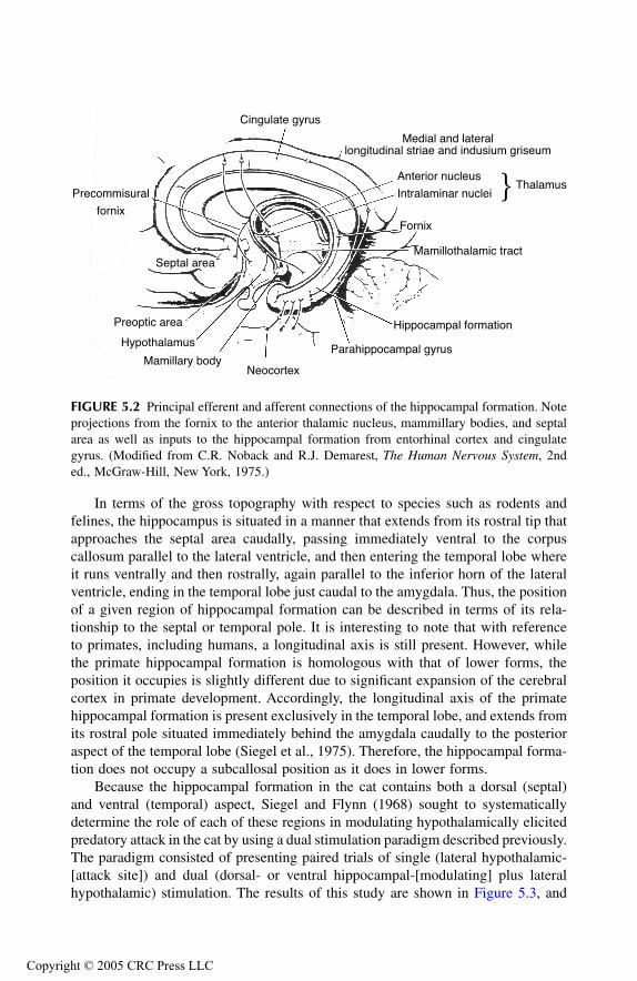
TF1661_C005.fm Page 84 Monday, October 25, 2004 7:21 AM
Copyr
In terms of the gross topography with respect to species such as rodents andfelines, the hippocampus is situated in a manner that extends from its rostral tip thatapproaches the septal area caudally, passing immediately ventral to the corpuscallosum parallel to the lateral ventricle, and then entering the temporal lobe whereit runs ventrally and then rostrally, again parallel to the inferior horn of the lateralventricle, ending in the temporal lobe just caudal to the amygdala. Thus, the positionof a given region of hippocampal formation can be described in terms of its rela-tionship to the septal or temporal pole. It is interesting to note that with referenceto primates, including humans, a longitudinal axis is still present. However, whilethe primate hippocampal formation is homologous with that of lower forms, theposition it occupies is slightly different due to significant expansion of the cerebralcortex in primate development. Accordingly, the longitudinal axis of the primatehippocampal formation is present exclusively in the temporal lobe, and extends fromits rostral pole situated immediately behind the amygdala caudally to the posterioraspect of the temporal lobe (Siegel et al., 1975). Therefore, the hippocampal forma-tion does not occupy a subcallosal position as it does in lower forms.
Because the hippocampal formation in the cat contains both a dorsal (septal)and ventral (temporal) aspect, Siegel and Flynn (1968) sought to systematicallydetermine the role of each of these regions in modulating hypothalamically elicitedpredatory attack in the cat by using a dual stimulation paradigm described previously.The paradigm consisted of presenting paired trials of single (lateral hypothalamic-[attack site]) and dual (dorsal- or ventral hippocampal-[modulating] plus lateralhypothalamic) stimulation. The results of this study are shown in Figure 5.3, and
FIGURE 5.2 Principal efferent and afferent connections of the hippocampal formation. Noteprojections from the fornix to the anterior thalamic nucleus, mammillary bodies, and septalarea as well as inputs to the hippocampal formation from entorhinal cortex and cingulategyrus. (Modified from C.R. Noback and R.J. Demarest, The Human Nervous System, 2nded., McGraw-Hill, New York, 1975.)
Cingulate gyrus
Medial and laterallongitudinal striae and indusium griseum
Anterior nucleus
Intralaminar nuclei � Thalamus
Fornix
Mamillothalamic tract
Hippocampal formation
Parahippocampal gyrus
NeocortexMamillary body
Hypothalamus
Preoptic area
Septal area
Precommisural
fornix
ight © 2005 CRC Press LLC
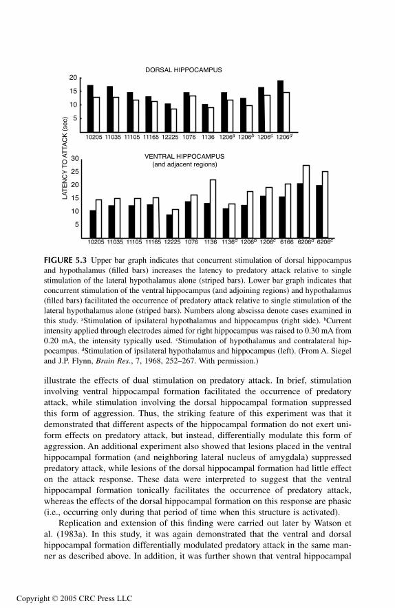
TF1661_C005.fm Page 85 Monday, October 25, 2004 7:21 AM
Copyr
illustrate the effects of dual stimulation on predatory attack. In brief, stimulationinvolving ventral hippocampal formation facilitated the occurrence of predatoryattack, while stimulation involving the dorsal hippocampal formation suppressedthis form of aggression. Thus, the striking feature of this experiment was that itdemonstrated that different aspects of the hippocampal formation do not exert uni-form effects on predatory attack, but instead, differentially modulate this form ofaggression. An additional experiment also showed that lesions placed in the ventralhippocampal formation (and neighboring lateral nucleus of amygdala) suppressedpredatory attack, while lesions of the dorsal hippocampal formation had little effecton the attack response. These data were interpreted to suggest that the ventralhippocampal formation tonically facilitates the occurrence of predatory attack,whereas the effects of the dorsal hippocampal formation on this response are phasic(i.e., occurring only during that period of time when this structure is activated).
Replication and extension of this finding were carried out later by Watson etal. (1983a). In this study, it was again demonstrated that the ventral and dorsalhippocampal formation differentially modulated predatory attack in the same man-ner as described above. In addition, it was further shown that ventral hippocampal
FIGURE 5.3 Upper bar graph indicates that concurrent stimulation of dorsal hippocampusand hypothalamus (filled bars) increases the latency to predatory attack relative to singlestimulation of the lateral hypothalamus alone (striped bars). Lower bar graph indicates thatconcurrent stimulation of the ventral hippocampus (and adjoining regions) and hypothalamus(filled bars) facilitated the occurrence of predatory attack relative to single stimulation of thelateral hypothalamus alone (striped bars). Numbers along abscissa denote cases examined inthis study. aStimulation of ipsilateral hypothalamus and hippocampus (right side). bCurrentintensity applied through electrodes aimed for right hippocampus was raised to 0.30 mA from0.20 mA, the intensity typically used. cStimulation of hypothalamus and contralateral hip-pocampus. dStimulation of ipsilateral hypothalamus and hippocampus (left). (From A. Siegeland J.P. Flynn, Brain Res., 7, 1968, 252–267. With permission.)
5
10
15
20
10205 11035 11105 11165 12225 1076 1136 1206a 1206b 1206c 1206d
10205 11035 11105 11165 12225 1076 1136 1136d 1206b 1206c 6166 6206d 6206c
5
10
15
20
25
30
DORSAL HIPPOCAMPUS
VENTRAL HIPPOCAMPUS
LAT
EN
CY
TO
AT
TAC
K (
sec)
(and adjacent regions)
ight © 2005 CRC Press LLC
TF1661_C005.fm Page 86 Monday, October 25, 2004 7:21 AM
Copyr
stimulation altered the trigeminal sensory fields along the lipline in association withthe expression of predatory attack over and above that which occurred with hypo-thalamic stimulation alone. Recall that the work of MacDonnell and Flynn (1966)demonstrated that following stimulation of lateral hypothalamic sites from whichpredatory attack was elicited resulted in an expansion of the sensory trigeminalfields around the lipline of the cat. Therefore, these authors interpreted their findingsto mean that ventral hippocampal stimulation directly (or indirectly through aninterneuron [see discussion below]) facilitates the lateral hypothalamic mechanismgoverning predatory attack behavior.
The relationship between the ventral hippocampal formation and the hypothal-amus was examined in the laboratory of Adamec (Adamec and Stark-Adamec,1983a, 1983b, 1983c, 1983d, 1986) in a totally different manner. Adamec initiallycategorized cats as being either naturally “(predatory) aggressive” or “defensive.”In the present series of studies, evoked potentials were then recorded in the ventro-medial hypothalamus (which presumably includes the mechanism for defensive ragebehavior) in response to single pulse stimulation of the amygdala (considered ingreater detail later in this chapter) or ventral hippocampal formation. Adamec dis-covered that the evoked potentials recorded in the ventromedial nucleus were ofgreater magnitude following amygdaloid stimulation and that smaller responses wererecorded following ventral hippocampal stimulation. In addition, Adamec found thatcats, which were more defensive, displayed reduced excitability with respect toevoked responses in the hippocampal formation to stimulation of the perforantpathway (i.e., a fiber bundle originating in entorhinal cortex that supplies the hip-pocampal formation) relative to cats, which were normally more predatory. It shouldbe further noted that Adamec described similar relationships following seizure dis-charges emanating from the ventral hippocampal formation, thereby implicatingepileptogenic activity in the modulation of aggression and rage. The role of seizureactivity in different parts of the amygdala of experimental animals is further con-sidered later in this chapter, and the effects of temporal lobe seizures on theseprocesses in humans will be considered.
The results of two additional studies are relevant and should be mentioned here.In one early study conducted by Wasman and Flynn (1966), after-discharges wereinduced in the dorsal hippocampus of cats and their effects on predatory attackduring the ictal (i.e., hippocampal stimulation) period. In brief, these investigatorsobserved that seizure discharges within the dorsal hippocampus delayed the onsetof the attack response, but did not completely block the expression of this response.These results were interpreted to mean that dorsal hippocampal seizures produceda generalized “motor” suppression of predatory attack similar to that observed whentested against other behavioral responses (e.g., learned responses and responses tonociceptive stimulation). These authors also concluded that hippocampal after-dis-charges did not prevent the animals from engaging in environmental contact withobjects of interest.
While the previous study examined the effects of dorsal hippocampal after-discharges on predatory attack, it did not address the issue of how hippocampal
ight © 2005 CRC Press LLC
TF1661_C005.fm Page 87 Monday, October 25, 2004 7:21 AM
Copyr
seizures may affect aggression or rage during the “interictal” period. This questionwas considered in a study by Griffith et al. (1987). In this study, seizures in thedorsal hippocampus of cats were induced by unilateral microinjections of kainicacid into this region. These injections produced acute periods of intense seizureactivity over the first few days, and were later followed by spontaneous complexpartial seizures lasting up to 4 months. Here, these investigators examined the effectsof defensive rage elicited from the medial hypothalamus and observed that rageresponse thresholds were lowered during the “interictal” period (when no seizuresoccurred). Moreover, mild stimulation was shown to induce “rage-like” responsesin otherwise normal and affectionate animals. These findings thus provide additionalevidence that activation of the dorsal hippocampus differentially affects predatoryattack and defensive rage similar to the effects described below associated with theseptal area and amygdala. One final point should be noted here: chronic seizureactivity involving the hippocampal formation can bring about long-term changes inthe propensity for the expression of defensive rage behavior. As discussed later inthis chapter, similar findings were observed following the induction of chronicseizures of the amygdala in cats. The most likely explanation for this phenomenonis that chronic seizure activity involving a limbic structure causes, over an extendedperiod of time, changes in the neurophysiological properties of the target neuronsfor such seizure activity. In this case, the most probable neuronal groups so affectedwould include extensive regions of the medial hypothalamus. This issue is alsoconsidered again later in this chapter.
These findings can be viewed as consistent with data provided above by Siegeland colleagues. This may be accomplished by integrating these two sets of studies.Here, it can be said that stimulation of the ventral hippocampal formation facilitatesthe occurrence of predatory attack, and that greater excitation of this region ofhippocampus occurs in association with predatory relative to defensive behavior.Moreover, stimulation of the ventral hippocampal formation results in smaller evokedpotentials in the ventromedial hypothalamus than sites in the amygdala that aretypically associated with suppression of predatory attack and facilitation of defensivebehavior. In relating the sets of data obtained from each of these laboratories, itshould be recalled that the medial and lateral hypothalamus are linked by reciprocalinhibitory connections. Thus, a reduced evoked potential observed in the ventrome-dial nucleus following ventral hippocampal stimulation obtained from an aggressivecat would be predicted. This conclusion is based on the principle of “disinhibition.”What this means is that a reduction in excitation of the ventromedial nucleus willresult in greater excitation in the lateral hypothalamus — the primary region of thebrain associated with predation. This is indeed due to a reduction in excitation ofthe inhibitory neurons passing from the medial to lateral hypothalamus, which thusreleases the lateral hypothalamus from inhibition by the medial hypothalamus. (SeeChapter 3, Figure 3.12, for description of the anatomical and functional relationshipbetween the medial and lateral hypothalamus.)
ight © 2005 CRC Press LLC
TF1661_C005.fm Page 88 Monday, October 25, 2004 7:21 AM
Copyr
SEPTAL AREA
ANATOMICAL CONSIDERATIONS
The histological appearance of the septal area is relatively similar in a variety ofspecies such as rodents, felines, and lower primates. In brief, the septal area consistsof different groups of cell groups, several of which are described in Figure 5.4. Thisfigure illustrates the loci of the principal nuclei shown in a frontal section takenthrough a midlevel of the septal area of the rat. The dorsal aspect of the septal areacontains the lateral septal nucleus, which occupies the lateral two-thirds of thisregion. The region medial to the lateral septal nucleus within the dorsal half of theseptal nucleus contains the medial septal nucleus. The medial septal nucleus becomescontinuous with the vertical and horizontal limbs of the diagonal band of Broca,which are situated in the ventral aspect of the septal area (Andy and Stephan, 1964;Krayniak et al., 1980; Meibach and Siegel, 1977b). It should be noted that in humans,the septal area is essentially located in a ventral position. The dorsal aspect in humansis limited to the septum pellucidum and, consequently, there are few neurons presentin this aspect.
Concerning the inputs and outputs of the septal area, the hippocampal formationconstitutes the primary afferent supply to the septal area (Krayniak et al., 1979;Meibach and Siegel, 1977a; Siegel and Tassoni, 1971; Watson et al., 1983b). Thisprojection arises from different fields of the cornu Ammonis and subiculum andproject topographically along the longitudinal axis of hippocampal formationthrough the precommissural fornix to the lateral septal area. In this manner, neuronsarising close to the septal pole of the hippocampal formation project to the medialaspect of the lateral septal nucleus, while progressively more distal aspects ofhippocampal formation that approach its temporal pole, project to progressivelymore lateral aspects of the lateral septal nucleus (Meibach and Siegel, 1977a; Siegeland Tassoni, 1971; Siegel et al., 1974, 1975).
In turn, the septal area projects principally to different nuclei of the hypothala-mus. The primary target within the hypothalamus of septal efferent fibers is theperifornical region and parts of the medial hypothalamus, including the mammillarybodies (Krayniak et al., 1980; Meibach and Siegel, 1977b; Stoddard-Apter andMacDonnell, 1980). The projection from the septal area can reach the hypothalamusfrom two groups of nuclei — the lateral septal nucleus and nuclei of the diagonalband (Krayniak et al., 1980). The key point to remember here is that the anatomicalrelationships of the septal area strongly suggest that one of its primary functions isto serve as a relay nucleus by which the hippocampal formation can indirectlymodulate hypothalamic processes (Figure 5.5). Studies describing the role of theseptal area in regulating aggression and rage are summarized below.
RELATIONSHIP TO AGGRESSION AND RAGE
Over the past 4 decades, many studies focused on the role of the septal area inemotional behavior, including aggression and rage. Typical of the approaches usedin research in this field, the earlier studies conducted sought to identify how lesionsand ablations of the septal area altered the expression of rage and aggression in
ight © 2005 CRC Press LLC
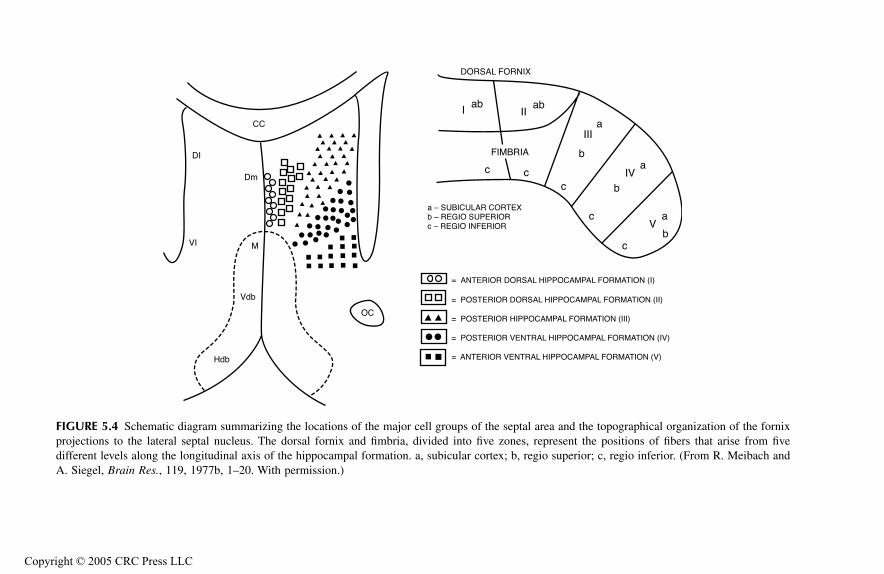
l area and the topographical organization of the fornix, represent the positions of fibers that arise from fiveregio superior; c, regio inferior. (From R. Meibach and
DORSAL FORNIX
abI
IIIa
IVa
aV
bc
c
b
b
cc
AL HIPPOCAMPAL FORMATION (I)
SAL HIPPOCAMPAL FORMATION (II)
OCAMPAL FORMATION (III)
TRAL HIPPOCAMPAL FORMATION (IV)
RAL HIPPOCAMPAL FORMATION (V)
TF
1661_C005.fm
Page 89 M
onday, October 25, 2004 7:21 A
M
Copyr
FIGURE 5.4 Schematic diagram summarizing the locations of the major cell groups of the septaprojections to the lateral septal nucleus. The dorsal fornix and fimbria, divided into five zonesdifferent levels along the longitudinal axis of the hippocampal formation. a, subicular cortex; b, A. Siegel, Brain Res., 119, 1977b, 1–20. With permission.)
CC
DI
Dm
VI M
Vdb
Hdb
OC
Iab
I
c
= ANTERIOR DORS
= POSTERIOR DOR
= POSTERIOR HIPP
= POSTERIOR VEN
= ANTERIOR VENT
FIMBRIA
a – SUBICULAR CORTEXb – REGIO SUPERIORc – REGIO INFERIOR
ight © 2005 CRC Press LLC
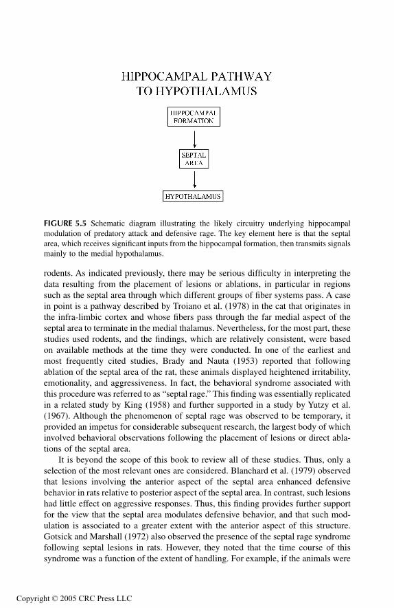
TF1661_C005.fm Page 90 Monday, October 25, 2004 7:21 AM
Copyr
rodents. As indicated previously, there may be serious difficulty in interpreting thedata resulting from the placement of lesions or ablations, in particular in regionssuch as the septal area through which different groups of fiber systems pass. A casein point is a pathway described by Troiano et al. (1978) in the cat that originates inthe infra-limbic cortex and whose fibers pass through the far medial aspect of theseptal area to terminate in the medial thalamus. Nevertheless, for the most part, thesestudies used rodents, and the findings, which are relatively consistent, were basedon available methods at the time they were conducted. In one of the earliest andmost frequently cited studies, Brady and Nauta (1953) reported that followingablation of the septal area of the rat, these animals displayed heightened irritability,emotionality, and aggressiveness. In fact, the behavioral syndrome associated withthis procedure was referred to as “septal rage.” This finding was essentially replicatedin a related study by King (1958) and further supported in a study by Yutzy et al.(1967). Although the phenomenon of septal rage was observed to be temporary, itprovided an impetus for considerable subsequent research, the largest body of whichinvolved behavioral observations following the placement of lesions or direct abla-tions of the septal area.
It is beyond the scope of this book to review all of these studies. Thus, only aselection of the most relevant ones are considered. Blanchard et al. (1979) observedthat lesions involving the anterior aspect of the septal area enhanced defensivebehavior in rats relative to posterior aspect of the septal area. In contrast, such lesionshad little effect on aggressive responses. Thus, this finding provides further supportfor the view that the septal area modulates defensive behavior, and that such mod-ulation is associated to a greater extent with the anterior aspect of this structure.Gotsick and Marshall (1972) also observed the presence of the septal rage syndromefollowing septal lesions in rats. However, they noted that the time course of thissyndrome was a function of the extent of handling. For example, if the animals were
FIGURE 5.5 Schematic diagram illustrating the likely circuitry underlying hippocampalmodulation of predatory attack and defensive rage. The key element here is that the septalarea, which receives significant inputs from the hippocampal formation, then transmits signalsmainly to the medial hypothalamus.
ight © 2005 CRC Press LLC
TF1661_C005.fm Page 91 Monday, October 25, 2004 7:21 AM
Copyr
handled once a day, they returned to normal within 6 days. When they were handled12 times on the first postoperative day, the syndrome persisted for a longer periodof time. This data demonstrated the importance of behavioral manipulations inregulating the septal rage syndrome as well as the interaction between neural andbehavioral processes. In contrast to the findings of Blanchard and colleagues, Lathemand Thorne (1974) reported that septal lesions enhanced aggressive behavior asmeasured by muricide, but that such effects were strain related. Here, the authorsreported that the primary effects were observed following lesions of Long-Evansstrain as opposed to Sprague Dawley rats. The difference in findings between thesetwo studies could possibly be accounted for on the basis of strain differences sinceBlanchard and colleagues employed the Wistar strain. This finding would furthersuggest that strain differences imply variations in the neural organization and pro-cessing of information from the septal area in each of these strains. However, it isnot possible to assess what these differences might constitute. That enhancement ofmuricide as a function of septal lesions is a temporary phenomenon is provided froma study by Miczek and Grossman (1972), and suggests that perhaps dissipation ofattack may reflect some kind of neural reorganization that takes place over the firstfew weeks following surgery.
Other approaches have been employed to determine the role of the rodent septalarea in aggression and rage. Here, several studies are cited. In one study, Albert andRichmond (1976) microinjected the anesthetic lidocaine into the lateral septalnucleus and observed an increase in reactivity and aggression (as measured byresponses to a sharp tap on the back, placement of a gloved hand near the snout,gentle prodding or grasping of the body, tail capture, vocalization, and biting).Microinjections placed in the region of the nucleus accumbens or adjoining regionsproduced similar results, while no effects followed microinjections into the caudatenucleus. This finding, which parallels those resulting from lesion studies describedabove, led these authors to suggest that septal lesions interfere with neural pathwaymediating aggression that pass from the hippocampal commissure through the lateralseptal area into the anterior aspect of the septal area and adjoining basal forebrain.It should be noted that the one pathway that would correspond to such a trajectoryis the precommissural fornix, which, as noted above, originates in the hippocampalformation. In a second approach, Bawden and Racine (1979) attempted to determinewhether kindling of the septal area (or amygdala) could affect aggressive reactionsin rats. This approach was employed as a means of comparing the effects describedby Adamec and colleagues (see discussion below) who demonstrated modulatingeffects of amygdaloid and hippocampal kindling on aggression and rage in the cat.In brief, Bawden and Racine reported that kindled seizures in the septal area oramygdala did not induce or inhibit muricide or ranicide, or modulate intraspecific(defensive) aggression. In contrast, stimulation of the septal area of the rat at currentlevels below that which could elicit seizures suppressed muricidal behavior (Potegalet al., 1980). It should be pointed out that similar findings were observed with septalstimulation in cats on predatory attack.
Fewer studies have been conducted in the cat. The studies described belowspecifically used electrical stimulation of different aspects of the septal area todetermine the role of this region on aggression and rage. One of the first studies
ight © 2005 CRC Press LLC
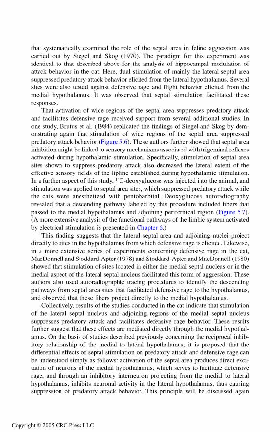
TF1661_C005.fm Page 92 Monday, October 25, 2004 7:21 AM
Copyr
that systematically examined the role of the septal area in feline aggression wascarried out by Siegel and Skog (1970). The paradigm for this experiment wasidentical to that described above for the analysis of hippocampal modulation ofattack behavior in the cat. Here, dual stimulation of mainly the lateral septal areasuppressed predatory attack behavior elicited from the lateral hypothalamus. Severalsites were also tested against defensive rage and flight behavior elicited from themedial hypothalamus. It was observed that septal stimulation facilitated theseresponses.
That activation of wide regions of the septal area suppresses predatory attackand facilitates defensive rage received support from several additional studies. Inone study, Brutus et al. (1984) replicated the findings of Siegel and Skog by dem-onstrating again that stimulation of wide regions of the septal area suppressedpredatory attack behavior (Figure 5.6). These authors further showed that septal areainhibition might be linked to sensory mechanisms associated with trigeminal reflexesactivated during hypothalamic stimulation. Specifically, stimulation of septal areasites shown to suppress predatory attack also decreased the lateral extent of theeffective sensory fields of the lipline established during hypothalamic stimulation.In a further aspect of this study, 14C-deoxyglucose was injected into the animal, andstimulation was applied to septal area sites, which suppressed predatory attack whilethe cats were anesthetized with pentobarbital. Deoxyglucose autoradiographyrevealed that a descending pathway labeled by this procedure included fibers thatpassed to the medial hypothalamus and adjoining perifornical region (Figure 5.7).(A more extensive analysis of the functional pathways of the limbic system activatedby electrical stimulation is presented in Chapter 6.)
This finding suggests that the lateral septal area and adjoining nuclei projectdirectly to sites in the hypothalamus from which defensive rage is elicited. Likewise,in a more extensive series of experiments concerning defensive rage in the cat,MacDonnell and Stoddard-Apter (1978) and Stoddard-Apter and MacDonnell (1980)showed that stimulation of sites located in either the medial septal nucleus or in themedial aspect of the lateral septal nucleus facilitated this form of aggression. Theseauthors also used autoradiographic tracing procedures to identify the descendingpathways from septal area sites that facilitated defensive rage to the hypothalamus,and observed that these fibers project directly to the medial hypothalamus.
Collectively, results of the studies conducted in the cat indicate that stimulationof the lateral septal nucleus and adjoining regions of the medial septal nucleussuppresses predatory attack and facilitates defensive rage behavior. These resultsfurther suggest that these effects are mediated directly through the medial hypothal-amus. On the basis of studies described previously concerning the reciprocal inhib-itory relationship of the medial to lateral hypothalamus, it is proposed that thedifferential effects of septal stimulation on predatory attack and defensive rage canbe understood simply as follows: activation of the septal area produces direct exci-tation of neurons of the medial hypothalamus, which serves to facilitate defensiverage, and through an inhibitory interneuron projecting from the medial to lateralhypothalamus, inhibits neuronal activity in the lateral hypothalamus, thus causingsuppression of predatory attack behavior. This principle will be discussed again
ight © 2005 CRC Press LLC
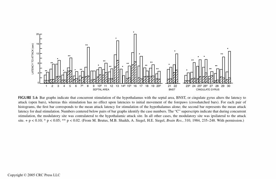
FIGUR
amus with the septal area, BNST, or cingulate gyrus alters the latency toattack initial movement of the forepaws (crosshatched bars). For each pair ofhistog n of the hypothalamus alone; the second bar represents the mean attacklatency ify the case numbers. The “C” superscripts indicate that during concurrentstimul site. In all other cases, the modulatory site was ipsilateral to the attacksite. + Siegel, H.E. Siegel,
Brain Res.
, 310, 1984, 235–248. With permission.)
17 18 19 20c 21 22BNST
23c 24 25c 26c 27 28 29 30CINGULATE GYRUS
TF
1661_C005.fm
Page 93 M
onday, October 25, 2004 7:21 A
M
Copyright ©
E 5.6 Bar graphs indicate that concurrent stimulation of the hypothal(open bars), whereas this stimulation has no effect upon latencies to
rams, the first bar corresponds to the mean attack latency for stimulatio for dual stimulation. Numbers centered below pairs of bar graphs ident
ation, the modulatory site was contralateral to the hypothalamic attack p < 0.10; * p < 0.05; ** p < 0.02. (From M. Brutus, M.B. Shaikh, A.
1 2 3 4 5 6 7c 8 9 10c 11 12 13 14c 15c 16SEPTAL AREA
0
4
8
12
16
20LA
TE
NC
Y T
O A
TTA
CK
(se
c)
+
2005 CRC Press LLC
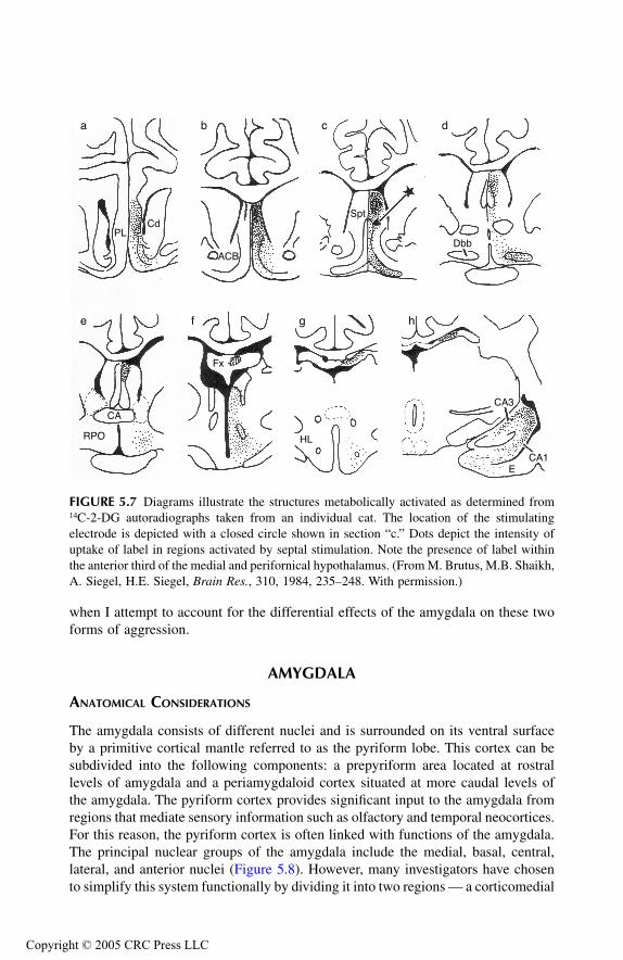
TF1661_C005.fm Page 94 Monday, October 25, 2004 7:21 AM
Copyr
when I attempt to account for the differential effects of the amygdala on these twoforms of aggression.
AMYGDALA
ANATOMICAL CONSIDERATIONS
The amygdala consists of different nuclei and is surrounded on its ventral surfaceby a primitive cortical mantle referred to as the pyriform lobe. This cortex can besubdivided into the following components: a prepyriform area located at rostrallevels of amygdala and a periamygdaloid cortex situated at more caudal levels ofthe amygdala. The pyriform cortex provides significant input to the amygdala fromregions that mediate sensory information such as olfactory and temporal neocortices.For this reason, the pyriform cortex is often linked with functions of the amygdala.The principal nuclear groups of the amygdala include the medial, basal, central,lateral, and anterior nuclei (Figure 5.8). However, many investigators have chosento simplify this system functionally by dividing it into two regions — a corticomedial
FIGURE 5.7 Diagrams illustrate the structures metabolically activated as determined from14C-2-DG autoradiographs taken from an individual cat. The location of the stimulatingelectrode is depicted with a closed circle shown in section “c.” Dots depict the intensity ofuptake of label in regions activated by septal stimulation. Note the presence of label withinthe anterior third of the medial and perifornical hypothalamus. (From M. Brutus, M.B. Shaikh,A. Siegel, H.E. Siegel, Brain Res., 310, 1984, 235–248. With permission.)
PLCd
a b c d
e f g h
ACB
Spt
Dbb
CA3
CA1E
HL
Fx
CA
RPO
ight © 2005 CRC Press LLC
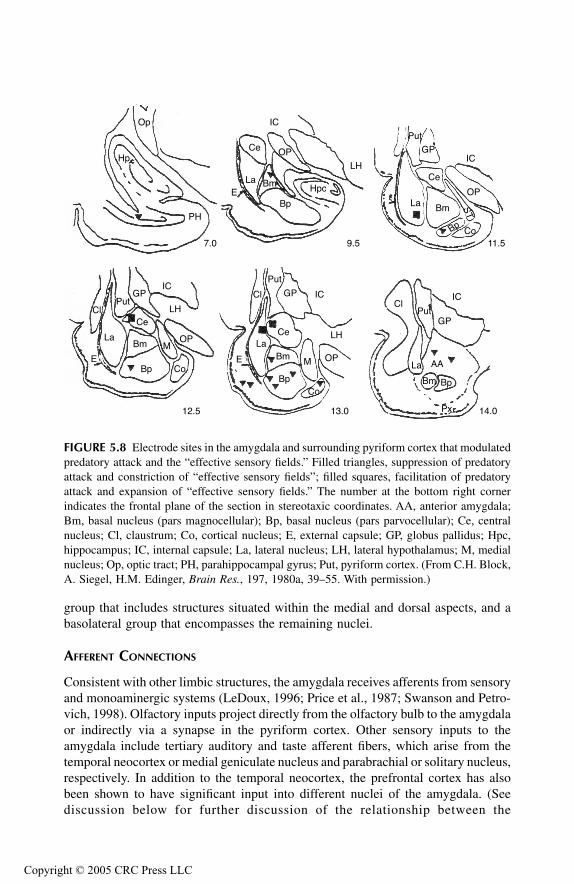
TF1661_C005.fm Page 95 Monday, October 25, 2004 7:21 AM
Copyr
group that includes structures situated within the medial and dorsal aspects, and abasolateral group that encompasses the remaining nuclei.
AFFERENT CONNECTIONS
Consistent with other limbic structures, the amygdala receives afferents from sensoryand monoaminergic systems (LeDoux, 1996; Price et al., 1987; Swanson and Petro-vich, 1998). Olfactory inputs project directly from the olfactory bulb to the amygdalaor indirectly via a synapse in the pyriform cortex. Other sensory inputs to theamygdala include tertiary auditory and taste afferent fibers, which arise from thetemporal neocortex or medial geniculate nucleus and parabrachial or solitary nucleus,respectively. In addition to the temporal neocortex, the prefrontal cortex has alsobeen shown to have significant input into different nuclei of the amygdala. (Seediscussion below for further discussion of the relationship between the
FIGURE 5.8 Electrode sites in the amygdala and surrounding pyriform cortex that modulatedpredatory attack and the “effective sensory fields.” Filled triangles, suppression of predatoryattack and constriction of “effective sensory fields”; filled squares, facilitation of predatoryattack and expansion of “effective sensory fields.” The number at the bottom right cornerindicates the frontal plane of the section in stereotaxic coordinates. AA, anterior amygdala;Bm, basal nucleus (pars magnocellular); Bp, basal nucleus (pars parvocellular); Ce, centralnucleus; Cl, claustrum; Co, cortical nucleus; E, external capsule; GP, globus pallidus; Hpc,hippocampus; IC, internal capsule; La, lateral nucleus; LH, lateral hypothalamus; M, medialnucleus; Op, optic tract; PH, parahippocampal gyrus; Put, pyriform cortex. (From C.H. Block,A. Siegel, H.M. Edinger, Brain Res., 197, 1980a, 39–55. With permission.)
Op
Hp
PH
7.0 9.5 11.5
12.5 13.0 14.0
IC
OP
LH
HpcBm
Bp
Ce
LaE
IC
OP
Bm
Bp
Ce
La
Co
ST
Put
GP
IC
Bm Bp
La
PutGP
Px
Cl
AA
IC
Bm
Bp
La
Put
GP
E
Co
Cl
Ce
OP
LH
M
IC
Bm
Bp
La
PutGP
ECo
ClCe
OP
LH
M
ight © 2005 CRC Press LLC
TF1661_C005.fm Page 96 Monday, October 25, 2004 7:21 AM
Copyr
prefrontal cortex and amygdala.) Noradrenergic, dopaminergic, and serotonergicinputs from brainstem neurons have also been demonstrated. Other regions that alsoproject to the amygdala include the substantia innominata (Femano et al., 1983),nuclei of the diagonal band of Broca (Krayniak et al., 1980; Meibach and Siegel,1977), and the medial thalamus (Ottersen and Ben-Ari, 1979).
EFFERENT CONNECTIONS
Axons arising from different nuclei of the amygdala have divergent projections to thetelencephalon, diencephalon, and brainstem. Amygdaloid projections to the prefrontalcortex complete a circuit which enables these two regions to be anatomically andfunctionally related through reciprocal pathways. Since both of these regions playimportant roles in the regulation and control of emotional behavior, which includeaggression and rage, these reciprocal pathways may reflect a mechanism by whichfeedback control of these processes may be maintained. In addition, the amygdaloidprojections to the prefrontal cortex may also serve to mediate olfactory signals to thecerebral cortex directly without involving a synapse in the thalamus, which is charac-teristic of all other sensory projections to the cerebral cortex.
With respect to the regulation of aggression and rage, perhaps the most significantprojections of the amygdala include those that supply the hypothalamus and midbrainPAG. There are various routes by which the amygdala can communicate with thehypothalamus. Direct pathways include the stria terminalis and the ventral amygdal-ofugal pathway. The stria terminalis arises mainly from neurons situated in the corti-comedial group of nuclei and the fibers project to the rostro-caudal extent of thepreoptico-medial hypothalamus. In contrast, the ventral amygdalofugal pathway arisesprincipally from the basolateral complex of amygdaloid nuclei and supplies the lateralhypothalamus and midbrain PAG. Thus, by virtue of these two pathways, the cortico-medial and basolateral nuclear complexes of the amygdala can directly regulate theactivity of neurons in the medial and lateral hypothalamus, respectively. The amygdalacan also modulate hypothalamic indirectly. It may do so by its projections to the bednucleus of the stria terminalis, which in turn projects to the intermediate regions ofthe hypothalamus proximal to the fornix as well as to the PAG.
RELATIONSHIP TO AGGRESSION AND RAGE
Of all the limbic structures, the amygdala has received the most attention concerningits possible relationship to aggressive behavior. In fact, one can easily argue that theliterature has clearly established that the amygdala and adjoining pyriform cortexdo play highly significant roles in the regulation of aggression and rage acrossspecies, including humans. The evidence spans four decades of research and involvesa variety of methodologies, such as the effects of regional brain ablation, localizedlesions, electrical and chemical stimulation, and seizure activity whose focusincludes the amygdala. However, a number of these studies and especially the earlierones were highly contradictory. Because of the very large numbers of studies thathave been conducted over this period of time, the discussion below does not attempt
ight © 2005 CRC Press LLC

TF1661_C005.fm Page 97 Monday, October 25, 2004 7:21 AM
Copyr
to review them all, but summarizes representative experiments that I believe havehad an impact on the field.
As an illustration of the conflicting findings of the early studies, both Kluverand Bucy (1939) and Schreiner and Kling (1953) placed amygdaloid lesions inmonkeys and cats, respectively, and each reported that such lesions produced tamingeffects in these species. In contrast, Bard and Mountcastle (1948) observed thatbilateral ablation of the amygdala of the cat produced ferocious behavior. Similarly,other investigators have also reported conflicting data following the placement oflesions most frequently conducted in cat or rat. For example, Shealy and Peele (1957)elicited nondirected rage behavior by amygdaloid stimulation, which also includedsalivation, sniffing, and gagging responses. They then placed lesions at these siteswhere stimulation was applied. The lesions were found to block the behavioralresponses observed from stimulation. Anatomical localization was difficult to assessbecause of the variability in the locus and general sizes of the lesions. There is oneadditional difficulty in interpreting the findings of this study. On the basis of thedescriptions of the behavioral responses associated with amygdaloid stimulation, itwas likely that stimulation-induced seizure activity resulting in the appearance ofrage behavior was secondary to seizure activity, and thus did not constitute “true”rage relative to the response patterns associated with medial hypothalamic or dorsalPAG stimulation. Nakao (1960) also observed that bilateral amygdaloid lesionsinvolving mainly the basal complex in cats reduced or eliminated rage and otheremotional responses elicited by the presence of a barking dog. In contrast, a morerecent study by Zagrodzka et al. (1998) demonstrated that lesions involving thecentral nucleus of amygdala in the cat increased defensive behavior to humans.
In two related studies, Zagrodzka and Fonberg (1977, 1978) examined the effectsof ventromedial amygdaloid lesions on predatory attack in the cat. The results ofboth of these studies revealed that such lesions abolished predatory attack whilehaving little effect on food intake. This finding suggests the specificity of effects ofamygdaloid lesions on aggression relative to other hypothalamic processes such asfeeding. In addition, the results of this and other studies described to this pointindicate that lesions of the amygdala appeared to have differential effects on defen-sive rage and predatory attack. Further discussion of these differential effects as wellas the implications is considered in the section concerning the effects of amygdaloidstimulation on aggression and rage.
Studies conducted in rats have also revealed varying results, which may relate toboth the model (and paradigm) employed as well as the locus of the amygdaloid lesion.For example, Allikmets and Ditrikh (1965) found that lesions placed in the basalamygdaloid complex blocked the expression of rage typically elicited by such pertur-bations as touching of its muzzle or back or lifting the rat by its tail. Using intermaleaggression as a model, Vochteloo and Koolhaas (1977) demonstrated that aggressionobserved by this paradigm could also be blocked when lesions are placed in the medialamygdala. However, these investigators qualified their conclusions about the effects ofsuch amygdaloid lesions by indicating that blockade only occurred when the animalswere exposed to at least four aggressive interactions. This finding would suggest theexistence of an interaction between brain mechanisms and environmental exposure inamygdaloid modulation of (at least certain forms of) aggression.
ight © 2005 CRC Press LLC

TF1661_C005.fm Page 98 Monday, October 25, 2004 7:21 AM
Copyr
Investigators have also examined the effects of amygdaloid lesions on mousekilling in rats. Lesions placed in different nuclei of the amygdala appear to haveproduced differential effects on this form of aggression. For example, mouse killingis induced or facilitated following lesions placed in either the medial (Karli et al.,1977; Vergnes, 1975) or central (Eclancher et al., 1975) nucleus or stria terminalis(Karli et al., 1977). In contrast, lesions placed in the lateral amygdala had the effectof reducing mouse killing in rats (Vergnes, 1976). In attempting to understand thenature of this form of aggression, it should be noted that it has been suggested thatthis model may be the equivalent of predatory attack in the cat. However, it mayalso be argued that the functional homology between predation in the cat and mousekilling in the rat is at best questionable, because the ethological objectives in eachsituation likely differ. Specifically, in the cat the ultimate goal of predation is theacquisition of food, while such an objective is not likely the objective in mousekilling. Thus, any attempt to equate this form of aggression with predation in thecat should be viewed with caution.
Studies conducted using electrical stimulation have been very useful in clarifyingthe roles of different nuclear groups of the amygdala in aggression and rage. Inparticular, Egger and Flynn (1963) published a seminal paper that contributed signif-icantly in clarifying some of the apparent contradictory evidence generated fromexperiments employing lesions or ablations. In their study, Egger and Flynn used dualstimulation of specific amygdaloid nuclei and hypothalamic sites from which predatoryattack behavior was elicited in the cat. Results of their study demonstrated that stim-ulation of the lateral amygdala, which included the lateral nucleus and lateral aspectof the basal complex, facilitated the occurrence of predatory attack, while stimulationof more medial aspects of the amygdala, which included the medial nucleus and medialaspects of the basal complex, suppressed predatory attack. Thus, this study was thefirst to demonstrate that different nuclei of the amygdala differentially modulate pred-atory attack behavior. In so doing, this study provided a possible explanation for thedivergent findings observed in studies employing lesions. On the basis of the resultsobtained by Egger and Flynn, the opposing effects observed by different investigatorsdescribed above may be accounted for on the basis that lesions associated with facil-itation of aggression primarily affected different nuclei than those associated withsuppression of aggression. In the discussion below, evidence is provided that suggestsanother level of complexity — namely, that the effects of perturbation of amygdaloidnuclei on aggression, either by stimulation or by lesions, are dependent on the natureof the aggressive response considered.
A number of other studies that built on the work of Egger and Flynn have servedto further clarify the relationships of various regions of the amygdala in the regulationof aggression and rage. Two studies examined various properties of amygdaloidmodulation of hypothalamically elicited predatory attack in the cat. In one study,Block et al. (1980a) initially demonstrated in a fashion similar to the findings ofEgger and Flynn that dual stimulation of separate regions of amygdala differentiallymodulated predatory attack elicited from the lateral hypothalamus of the cat (Figure5.8). More recent studies by Han et al. (1996a, 1996b) replicated and extended thefindings of Block et al., in part, by focusing on inhibitory role of the amygdala onpredatory attack. Collectively, from the results of these studies, stimulation at sites
ight © 2005 CRC Press LLC

TF1661_C005.fm Page 99 Monday, October 25, 2004 7:21 AM
Copyr
located in the basal complex, medial aspect of amygdala, including the medialnucleus, or pyriform cortex suppressed predatory attack, while stimulation at sitessituated in the central or lateral nuclei facilitated the occurrence of predatory attack(Figure 5.9). It would seem that such differential effects on predatory attack maybe best accounted for in terms of the efferent pathways from the amygdala to thehypothalamus and PAG. As already noted, the outputs from the medial and basalamygdala provide the origins of the stria terminalis that supply the medial hypo-thalamus. In contrast, the lateral and central amygdala provide the origins of theamygdalofugal pathways, including those that supply the PAG. The second aspectof the study by Block et al. demonstrated that the sensory trigeminal fields alongthe lipline of the cat were altered as a function of stimulation of sites in the amygdalathat suppressed or facilitated predatory attack. Similar to the findings observed withthe hippocampal formation and septal area, stimulation of inhibitory sites in theamygdala reduced the size of the effective sensory fields along the lipline, whilestimulation of amygdaloid sites that facilitated predatory attack increased the sizeof the effective sensory fields (Figure 5.10). Again, for reasons elaborated previouslyregarding the effects of stimulation of other limbic structures, these findings wouldalso suggest that the effects of amygdaloid stimulation on predatory attack weremediated directly on the mechanism related to the expression of this form of aggres-sion. Additional evidence in support of this notion is discussed below.
FIGURE 5.9 Bar graphs indicate that concomitant stimulation of the amygdala and hypo-thalamus alter latencies to predatory attack (open bar), whereas this stimulation has no effecton latencies to initial movement of the forepaws (hatched bar). For each pair of histograms,the first bar corresponds to the mean latency for single stimulation of the hypothalamus alone;the second bar represents the mean attack latency for dual stimulation. Numbers centeredbelow pairs of bar graphs identify the individual cats used in the study. + p < 0.10; * p <0.05; ** p < 0.01; *** p < 0.001. (A), inhibitory sites; (B), facilitatory sites. (From C.H.Block, A. Siegel, H.M. Edinger, Brain Res., 197, 1980a, 39–55. With permission.)
1 2 3 4 5 6 7 8 9 10 11 12 13 14 15 16 17
A B
0
5
10
15
RE
SP
ON
SE
LA
TE
NC
Y (
sec)
+
+
+
ight © 2005 CRC Press LLC

TF1661_C005.fm Page 100 Monday, October 25, 2004 7:21 AM
Copyr
Consider now the effects of stimulation of these regions of amygdala on defen-sive rage behavior elicited from the medial hypothalamus or PAG. Here, at leastfour studies are relevant. The first, conducted by Stoddard-Apter and MacDonnell(1980), was part of the same study employed by these authors for the study of theanatomical projections of septal sites that facilitated defensive rage. These investi-gators initially identified sites in the medial amygdala (with the aid of a cannula-electrode), which upon stimulation facilitated defensive rage in the cat elicited bymedial hypothalamic stimulation. Then the pathways from these sites were deter-mined using a radioactive tracer, which was microinjected through the cannula-electrode. The major pathway labeled was shown to be the stria terminalis in whichthe pathways were traced principally to the bed nucleus of the stria terminalis andmedial hypothalamus. Thus, this paper provided the first piece of evidence that themedial amygdala facilitates defensive rage by acting through a monosynaptic pro-jection to sites in the medial hypothalamus associated with this form of aggression.
Additional lines of evidence linking the amygdala with facilitation of defensiverage in the cat were provided in a study by Shaikh et al. (1993). While aspects of
FIGURE 5.10 Bar graphs indicate that concomitant stimulation of the amygdala and hypo-thalamus (hatched bar) alters the size of the “effective sensory field” established during singlestimulation of the hypothalamus (open bar). (A) Constriction following stimulation of inhib-itory sites. (B) Expansion following stimulation of facilitatory sites for both ipsilateral andcontralateral sensory fields. Numbers centered below pairs of bar graphs identify the individualcats used in the study. * p < 0.05; ** p < 0.01, *** p < 0.001. (From C.H. Block, A. Siegel,H.M. Edinger, Brain Res., 197, 1980a, 39–55. With permission.)
1
2
3
01 2 3 4 5 6 7 8 9 10 11 12 13 14 15 16 17
1
2
3
01 2 3 4 5 6 7 8 9 10 11 12 13 14 15 16 17
A B
DIS
TAN
CE
FR
OM
LIP
MID
LIN
E (
cm)
CO
NT
RA
LAT
ER
AL
IPS
ILA
TE
RA
L
ight © 2005 CRC Press LLC
TF1661_C005.fm Page 101 Monday, October 25, 2004 7:21 AM
Copyr
this and several other related studies are described in more detail in Chapter 7, otherfeatures of this study basically replicated the findings of Stoddard-Apter and Mac-Donnell. These include the observations that stimulation of the medial nucleus ofamygdala facilitates defensive rage elicited from the medial hypothalamus, and thatwhen using retrograde tracing methods, amygdaloid projections to sites in the medialhypothalamus from which defensive rage had been elicited arise mainly from themedial nucleus and adjoining aspects of the basal nuclear complex. (See Chapter 7for further details on the loci of amygdaloid nuclei that project to sites in thehypothalamus and PAG from rage and aggression are elicited.)
The studies described above focused on the facilitatory effects of amygdaloidstimulation on defensive rage behavior. In a separate study, Shaikh et al. (1991)showed that stimulation of central or lateral nucleus of amygdala powerfully sup-pressed defensive rage behavior in the cat elicited from stimulation of the dorsolateralPAG. Thus, similar to the findings described above with respect to amygdaloidmodulation of predatory attack, here too, defensive rage behavior is also differen-tially modulated from different nuclei of the amygdala as well. It should be noted,however, that the directionality of modulation by amygdaloid nuclei for predatoryattack appears to be the opposite to that for defensive rage behavior. Specifically,stimulation of the central or lateral nuclei of the amygdala facilitates predatoryattack, but suppresses defensive rage. In contrast, stimulation of the medial nucleusand adjoining regions of the basal nuclear complex suppresses predatory attack, butfacilitates defensive rage. Further evidence in support of the differential effects ofspecific amygdaloid nuclei on predatory attack and defensive rage behavior in thecat in association with seizure activity is provided below from the experiments ofBrutus et al. (1986).
Two other studies are worth noting here that involve amygdaloid stimulationand aggression conducted by Potegal et al. (1996a, 1996b). In one study, brief high-frequency stimulation was applied to the corticomedial amygdala of hamsters andthe effects of such stimulation on aggression measured at different times (i.e., 1 to90 minutes) following stimulation, using the resident–intruder design. These authorsobserved that amygdaloid (priming) stimulation facilitated aggressive responses inthe stimulated (resident) hamster by lowering attack latencies, the maximal effectoccurring 10 minutes following stimulation. In the second study, these authorsobserved that allowing a hamster a single “priming” attack on a conspecific (intruder)increased a transient increase in aggression by reducing the latency response forattack on a second intruder. Of particular interest was the fact that priming stimu-lation significantly enhanced c-fos expression in the corticomedial amygdala. Acareful analysis revealed that these increases in c-fos expression were specificallyassociated with aggressive reactions, and not with arousal or motor functions whenmeasured against locomotor activity. Thus, this study showed that in a species otherthan cat (i.e., hamster), activation of the corticomedial amygdala is associated withfacilitation of a defensive form of aggression. Therefore, this finding indicates thatthe corticomedial amygdala in both the hamster and cat functions in similar ways.
ight © 2005 CRC Press LLC
TF1661_C005.fm Page 102 Monday, October 25, 2004 7:21 AM
Copyr
EFFECTS OF SEIZURES
Seizures Induced by Electrical Stimulation of Amygdala
In one early study, Alonso-de Florida and Delgado (1958) stimulated the amygdalaof cats for 0.5 seconds every 5 seconds for 1 hour daily for 1 to 15 days. Theseauthors then reported that such stimulation increased playful and contact activitiesas well as increased or decreased rage responses when placed in a colony of cats.While the authors suggested that the directionality of the effects of seizures on ragebehavior is related to the anatomical locus, they did not specify the precise natureof this relationship other than to indicate that sites where seizures were initiatedincluded the corticomedial and basolateral components of amygdala. One significantfeature of this study is that it was the first to link seizures of the amygdala tomodulation of rage behavior. The authors also noted that the seizure paradigmresulted in the onset of spontaneous seizures, thus suggesting the presence of “kin-dling-like” phenomenon. The studies described below have clarified and extendedthis original finding.
The study by Brutus et al. (1986) differed from previous experiments describedabove in that it sought to determine the effects of seizure discharges generated fromdifferent nuclei of the amygdala on defensive rage and predatory attack elicited fromthe hypothalamus of the cat. Initially, these authors identified sites in the amygdalaand adjoining pyriform cortex from which subseizure stimulation modulated one orboth of these forms of aggression. Then, current thresholds for elicitation of either(or both) defensive rage and predatory attack were identified over a period of 1 to2 weeks. After these “baseline” thresholds were determined, three to five seizuresper day were initiated by stimulation of an amygdaloid modulating site for anadditional period of 1 to 4 weeks, which was then followed by a “post-seizure”period of 1 to 2 weeks when no seizures were induced.
The results of this study revealed that seizures induced at sites in the medialamygdala and pyriform cortex associated with facilitation of defensive rage atsubseizure levels of stimulation lowered the threshold for elicitation of defensiverage elicited from hypothalamic stimulation alone. In a parallel manner, seizuresinduced at sites in the central or lateral nuclei of amygdala associated with suppres-sion of defensive rage at subseizure levels of stimulation elevated the threshold forelicitation of defensive rage. In the post-seizure period, threshold responses forelicitation of defensive rage returned to baseline levels, thus demonstrating the potenteffects of seizure discharges at functionally identifiable sites in the amygdala ondefensive rage, as well as the fact that these seizures had not produced long-termfunctional damage to the stimulated region (Figure 5.11). Several cats were alsotested for the effects of seizure induction at sites that modulated predatory attackbehavior. In one cat, stimulation-induced seizures at a site in the basolateral nucleus(which facilitated predatory attack at subseizure levels of stimulation) resulted in anelevation of the threshold for elicitation of this form of aggression. In a second cat,pyriform cortex stimulation at subseizure levels suppressed predatory attack. Sei-zures induced at this site resulted in a lowering of response thresholds. This lattercase was of particular interest since subseizure stimulation had been shown to
ight © 2005 CRC Press LLC
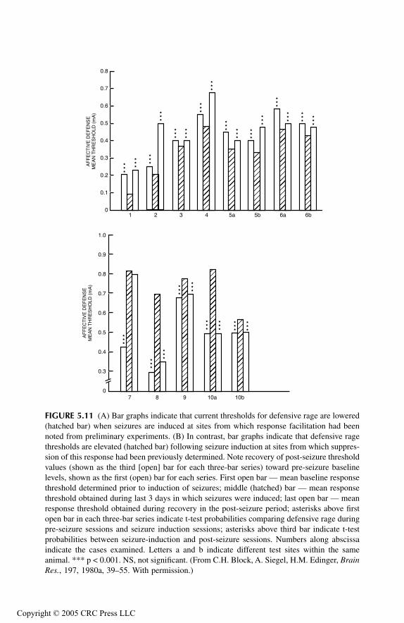
TF1661_C005.fm Page 103 Monday, October 25, 2004 7:21 AM
Copyr
FIGURE 5.11 (A) Bar graphs indicate that current thresholds for defensive rage are lowered(hatched bar) when seizures are induced at sites from which response facilitation had beennoted from preliminary experiments. (B) In contrast, bar graphs indicate that defensive ragethresholds are elevated (hatched bar) following seizure induction at sites from which suppres-sion of this response had been previously determined. Note recovery of post-seizure thresholdvalues (shown as the third [open] bar for each three-bar series) toward pre-seizure baselinelevels, shown as the first (open) bar for each series. First open bar — mean baseline responsethreshold determined prior to induction of seizures; middle (hatched) bar — mean responsethreshold obtained during last 3 days in which seizures were induced; last open bar — meanresponse threshold obtained during recovery in the post-seizure period; asterisks above firstopen bar in each three-bar series indicate t-test probabilities comparing defensive rage duringpre-seizure sessions and seizure induction sessions; asterisks above third bar indicate t-testprobabilities between seizure-induction and post-seizure sessions. Numbers along abscissaindicate the cases examined. Letters a and b indicate different test sites within the sameanimal. *** p < 0.001. NS, not significant. (From C.H. Block, A. Siegel, H.M. Edinger, BrainRes., 197, 1980a, 39–55. With permission.)
1 2 3 4 5a 5b 6a 6b
7 8 9 10a 10b0
0.3
0.4
0.5
0.6
0.7
0.8
0.9
1.0
0
0.1
0.2
0.3
0.4
0.5
0.6
0.7
0.8
AF
FE
CT
IVE
DE
FE
NS
EM
EA
N T
HR
ES
HO
LD (
mA
)
AF
FE
CT
IVE
DE
FE
NS
EM
EA
N T
HR
ES
HO
LD (
mA
)
ight © 2005 CRC Press LLC

TF1661_C005.fm Page 104 Monday, October 25, 2004 7:21 AM
Copyr
facilitate defensive rage, and seizures induced at this site lowered the threshold forthe expression of this response. Thus, these findings indicate the specificity of theeffects of seizure induction on each of these forms of aggressive behavior.
In a second related study, Siegel et al. (1986) focused on how chronic seizuresof the amygdala could affect defensive rage and predatory attack. In order to answerthis question, the following experiment was performed. Cannula-electrodes wereimplanted into various nuclei of the amygdala at sites where suppression or facili-tation of defensive rage could be obtained, as well as into the medial hypothalamusfrom which stimulation could elicit this response. Current response thresholds wereinitially determined on alternate days for a period of 2 weeks. Then, chronic seizureswere induced by the administration of cobalt powder injected through the cannula-electrode into a given amygdaloid site, and current threshold responses were againdetermined over the following 2 weeks. When this test period was completed,lidocaine hydrochloride (Xylocaine) was administered through an Alzet® minipumpinto the seizure focus over a 7-day period in order to significantly reduce or eliminatethe excitatory effects of the amygdaloid seizure focus.
Chronic seizures induced at sites associated with facilitation of defensive rage(as determined from the preliminary dual stimulation procedure of using subseizurestimulation at the amygdaloid site in question) resulted in a lowering of currentthreshold responses for defensive rage. When lidocaine was injected into these sites,the effects of the chronic seizures on defensive rage were reversed (Figure 5.12).These sites were generally located in the pyriform cortex or basal amygdaloidcomplex. In contrast, chronic seizures induced at sites associated with suppressionof defensive rage (i.e., the central or lateral nucleus) resulted in elevations of responsethresholds for this form of aggression. These effects were also reversed followinginjections of lidocaine into the inhibitory sites (Figure 5.13).
Collectively, the results of these studies indicate that seizures induced eitheracutely or chronically of the amygdala and related regions of cortex cause significantchanges in response thresholds for defensive rage. Moreover, whether the effects ofseizures reduced or elevated response thresholds was a function of the anatomicallocation of the induced seizures. In addition, the directionality of effects of theseizures is also linked to the form of aggression considered. As indicated above,seizures generated from the lateral or central nucleus of amygdala are associatedwith lowering of current thresholds for predatory attack and with elevations ofresponse thresholds for defensive rage. The opposite effects are observed followingseizures induced at more medial sites within the amygdala as well as pyriform cortex.
Effects of Amygdaloid Seizures on Spontaneously Elicited Attack
The discussion above considered how seizures of different aspects of the amygdalacould affect the expression of aggression and rage elicited by electrical stimulationof the hypothalamus. The question may be asked here how seizure activity involvingthe temporal lobe could affect these forms of aggression when they occur sponta-neously. This question was addressed in an extensive series of elegant studies byAdamec and colleagues.
ight © 2005 CRC Press LLC
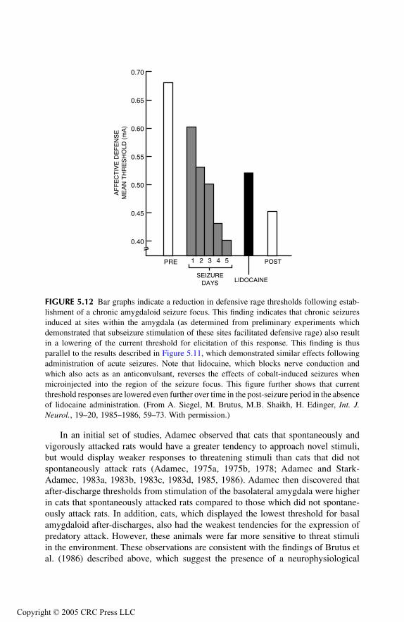
TF1661_C005.fm Page 105 Monday, October 25, 2004 7:21 AM
Copyr
In an initial set of studies, Adamec observed that cats that spontaneously andvigorously attacked rats would have a greater tendency to approach novel stimuli,but would display weaker responses to threatening stimuli than cats that did notspontaneously attack rats (Adamec, 1975a, 1975b, 1978; Adamec and Stark-Adamec, 1983a, 1983b, 1983c, 1983d, 1985, 1986). Adamec then discovered thatafter-discharge thresholds from stimulation of the basolateral amygdala were higherin cats that spontaneously attacked rats compared to those which did not spontane-ously attack rats. In addition, cats, which displayed the lowest threshold for basalamygdaloid after-discharges, also had the weakest tendencies for the expression ofpredatory attack. However, these animals were far more sensitive to threat stimuliin the environment. These observations are consistent with the findings of Brutus etal. (1986) described above, which suggest the presence of a neurophysiological
FIGURE 5.12 Bar graphs indicate a reduction in defensive rage thresholds following estab-lishment of a chronic amygdaloid seizure focus. This finding indicates that chronic seizuresinduced at sites within the amygdala (as determined from preliminary experiments whichdemonstrated that subseizure stimulation of these sites facilitated defensive rage) also resultin a lowering of the current threshold for elicitation of this response. This finding is thusparallel to the results described in Figure 5.11, which demonstrated similar effects followingadministration of acute seizures. Note that lidocaine, which blocks nerve conduction andwhich also acts as an anticonvulsant, reverses the effects of cobalt-induced seizures whenmicroinjected into the region of the seizure focus. This figure further shows that currentthreshold responses are lowered even further over time in the post-seizure period in the absenceof lidocaine administration. (From A. Siegel, M. Brutus, M.B. Shaikh, H. Edinger, Int. J.Neurol., 19–20, 1985–1986, 59–73. With permission.)
0.40
0.45
0.50
0.55
0.60
0.65
0.70
PRE 1 2 3 4 5
SEIZUREDAYS LIDOCAINE
POST
AF
FE
CT
IVE
DE
FE
NS
EM
EA
N T
HR
ES
HO
LD (
mA
)
ight © 2005 CRC Press LLC

TF1661_C005.fm Page 106 Monday, October 25, 2004 7:21 AM
Copyr
mechanism within parts of the amygdala relating epileptiform activity with aggres-sive behavior. Other experiments by Adamec, which are described below, providedfurther support for this notion.
In one set of studies, Adamec and Stark-Adamec (1983b, 1983c) reported thatafter-discharges involving other parts of the temporal lobe (i.e., ventral hippocampus)enhanced defensive responses in cats while increasing the latency for biting attack.The question may be raised here why after-discharges elicited from the ventralhippocampus should suppress predatory attack and facilitate defensive responseswhen subseizure stimulation of this region was reported to produce the oppositeresults (Siegel and Flynn, 1968; Watson et al., 1983a). Adamec provided an answerto this apparent contradiction by indicating that as a result of seizure dischargesinduced in the hippocampal formation, there is spread of neural activity to regionsof the amygdala such as the corticomedial and adjoining parts of the basal complexthat normally facilitate defensive responses and suppress predatory attack. Thisconclusion was based on the additional observation that seizures induced in theventral hippocampus in cats also resulted in an increase in the evoked responserecorded in the ventromedial hypothalamus of the same animals in response to basalamygdaloid stimulation.
In another set of studies, Adamec and Stark-Adamec (1986) showed that theamplitudes of evoked responses in the ventromedial nucleus were larger following
FIGURE 5.13 Bar graphs indicate an elevation in current threshold responses for defensiverage following establishment of a chronic seizure focus with cobalt in the amygdala. Notethat lidocaine administration reversed the effects of cobalt seizures, but that the responsethreshold tended to become elevated in the absence of lidocaine. (From A. Siegel, M. Brutus,M.B. Shaikh, H. Edinger, Int. J. Neurol., 19–20, 1985–1986, 59–73. With permission.)
0.10
0.20
0.30
0.40
0.50
0.60
0.70
PRE 1 2 3 4 5
SEIZUREDAYS LIDOCAINE
POST
AF
FE
CT
IVE
DE
FE
NS
EM
EA
N T
HR
ES
HO
LD (
mA
)
6
ight © 2005 CRC Press LLC
TF1661_C005.fm Page 107 Monday, October 25, 2004 7:21 AM
Copyr
amygdaloid stimulation in cats that were more defensive than the evoked responsesin cats that displayed more aggressiveness to a prey object. In a later study reportedby Adamec (1990), stimulation of amygdaloid sites that were shown to lower thesize of evoked potentials in the medial hypothalamus were also associated with botha reduction in the extent of defensive behavior as well as increases in predatoryattack expressed by these animals.
Collectively, the findings of Adamec suggest the following conclusions:
1. The likelihood that a cat will be more defensive or predatory is linked tothe threshold for elicitation of seizure discharges in specific regions ofthe amygdala.
2. The size of the evoked potential recorded in the ventromedial hypothal-amus following basal amygdaloid stimulation is related to the degree towhich the cat is likely to display defensive behavior, indicating the pres-ence of an underlying neurophysiological mechanism.
3. Long-term changes in defensive behavior may be mediated, in part, byseizure activity initiated in the ventral hippocampus, which is then trans-mitted to the basal amygdala, thus influencing the neural mechanismgoverning activity in the ventromedial hypothalamus.
4. The temporal lobe mechanism modulating the expression of defensivebehavior in a given direction has an opposing effect on predatory behavior.
Another important point to note here is that the differential effects of amygdaloidexcitation on defensive and predatory attack are entirely consistent with the studiesdescribed previously in this chapter that involved direct subseizure stimulation ofamygdaloid nuclei, as well as distinct regions of the medial and lateral hypothalamusas described in previous chapters. Moreover, the findings clearly reflect the similarityof the effects associated with the amygdaloid stimulation on these two forms ofaggression when these responses are elicited either by electrical brain stimulationof the hypothalamus or by natural “spontaneous” occurrence.
PREFRONTAL CORTEX AND CINGULATE GYRUS
BACKGROUND
The prefrontal cortex and (anterior) cingulate gyrus are considered together becausethey both share similar anatomical and functional properties. These include the factsthat: (1) both regions are characterized mainly by an agranular cortex; (2) bothregions project heavily to the mediodorsal thalamic nucleus; and (3) both regionssuppress predatory attack behavior. Details concerning these facts are presentedbelow together with a brief review of the literature. While it is beyond the scope ofthis chapter to consider the many important functions other than aggression andrage, suffice it to say that considerable research has been conducted on the prefrontalcortex, linking it to such processes as emotional behavior and related learning (Pujicet al., 1993; Sato, 1971), delayed responding (Wilcott, 1977), and schizophrenia(Winn, 1994). Thus, the prefrontal cortex may be viewed as a highly complex
ight © 2005 CRC Press LLC

TF1661_C005.fm Page 108 Monday, October 25, 2004 7:21 AM
Copyr
structure with respect to both its anatomical connections with other regions of thebrain and to functions that it mediates.
ANATOMICAL CONSIDERATIONS
A unique feature of the prefrontal cortex is that it receives inputs from all otherregions of the cerebral cortex and, in addition, it also receives major inputs fromthe mediodorsal thalamic nucleus and amygdala (Bacon, 1973; Divac et al., 1978).With respect to the efferent projections of the prefrontal cortex, the primary targetregions include the mediodorsal thalamic nucleus, amygdala, and midbrain PAG(Alexander and Fuster, 1973; An et al., 1998; Siegel et al., 1974, 1975, 1977). Otherinvestigators have also described connections from the prefrontal cortex directly tothe hypothalamus (Öngür et al., 1998), which may be inhibitory (Kita and Oomura,1982). From the descriptions provided above, one can see that there are variousways by which pathways originating from the prefrontal cortex may enter thehypothalamus. Nevertheless, the overwhelming numbers of fibers that pass to thediencephalon terminate in the mediodorsal thalamic nucleus (Figure 5.14). Thus, itis reasonable to conclude that these projections are the most likely ones over whichthe primary modulating effects of the prefrontal cortex are mediated. In fact, thisissue is addressed directly in several of the experiments described below (Siegel etal., 1972, 1977).
As noted above, a study of the projection pathways of the cingulate gyrusrevealed that its anterior aspect is highly similar to the prefrontal cortex with respectto its projection patterns (Siegel et al., 1973a). Namely, the primary thalamic pro-jection target of this region of cortex is the mediodorsal nucleus. In contrast, moreposterior aspects of the cingulate gyrus project to the anteroventral thalamic nucleus.
RELATIONSHIP TO AGGRESSION AND RAGE
The historical basis for the study of the role of the prefrontal cortex in emotionalbehavior presumably had its origin with the case reports of a New England railroadconstruction worker, Phineas Gage (Harlow, 1848, 1868). A heavy iron bar thatpenetrated his skull struck him, inflicting massive damage of the frontal region ofthe brain, in part, by destroying many of the anatomical connections between theprefrontal cortex and other regions of the cerebrum as well as with subcorticalstructures. Thus, the accident resulted in Gage becoming the first prefrontal lobot-omy. Following his accident, Gage displayed marked personality changes. Severalof these included child-like behavior in intellectual abilities and passions of a strongman. One of the implications of this observation is that the intact prefrontal cortexnormally inhibits various forms of emotional behavior, and that removal of suchinputs from the prefrontal cortex results in a release of its inhibitory influences. Onthe other hand, the display of temper tantrums by chimpanzees as a result of makingincorrect choices was reduced or eliminated following the cutting of the fiber tractsassociated with the prefrontal cortex (see studies cited in Fulton, 1952). In addition,after Moniz introduced prefrontal lobotomy in humans in 1936, Freeman and Watts(1942) reported favorable results from this procedure in treating aggressive
ight © 2005 CRC Press LLC
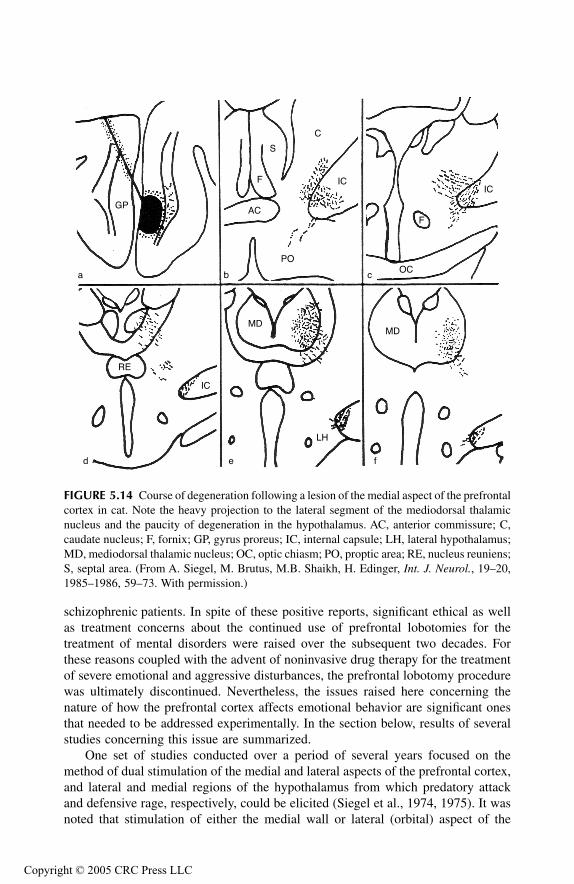
TF1661_C005.fm Page 109 Monday, October 25, 2004 7:21 AM
Copyr
schizophrenic patients. In spite of these positive reports, significant ethical as wellas treatment concerns about the continued use of prefrontal lobotomies for thetreatment of mental disorders were raised over the subsequent two decades. Forthese reasons coupled with the advent of noninvasive drug therapy for the treatmentof severe emotional and aggressive disturbances, the prefrontal lobotomy procedurewas ultimately discontinued. Nevertheless, the issues raised here concerning thenature of how the prefrontal cortex affects emotional behavior are significant onesthat needed to be addressed experimentally. In the section below, results of severalstudies concerning this issue are summarized.
One set of studies conducted over a period of several years focused on themethod of dual stimulation of the medial and lateral aspects of the prefrontal cortex,and lateral and medial regions of the hypothalamus from which predatory attackand defensive rage, respectively, could be elicited (Siegel et al., 1974, 1975). It wasnoted that stimulation of either the medial wall or lateral (orbital) aspect of the
FIGURE 5.14 Course of degeneration following a lesion of the medial aspect of the prefrontalcortex in cat. Note the heavy projection to the lateral segment of the mediodorsal thalamicnucleus and the paucity of degeneration in the hypothalamus. AC, anterior commissure; C,caudate nucleus; F, fornix; GP, gyrus proreus; IC, internal capsule; LH, lateral hypothalamus;MD, mediodorsal thalamic nucleus; OC, optic chiasm; PO, proptic area; RE, nucleus reuniens;S, septal area. (From A. Siegel, M. Brutus, M.B. Shaikh, H. Edinger, Int. J. Neurol., 19–20,1985–1986, 59–73. With permission.)
GP
a b c
d fe
AC
F
S
C
IC
POOC
F
IC
MDMD
LH
IC
RE
ight © 2005 CRC Press LLC
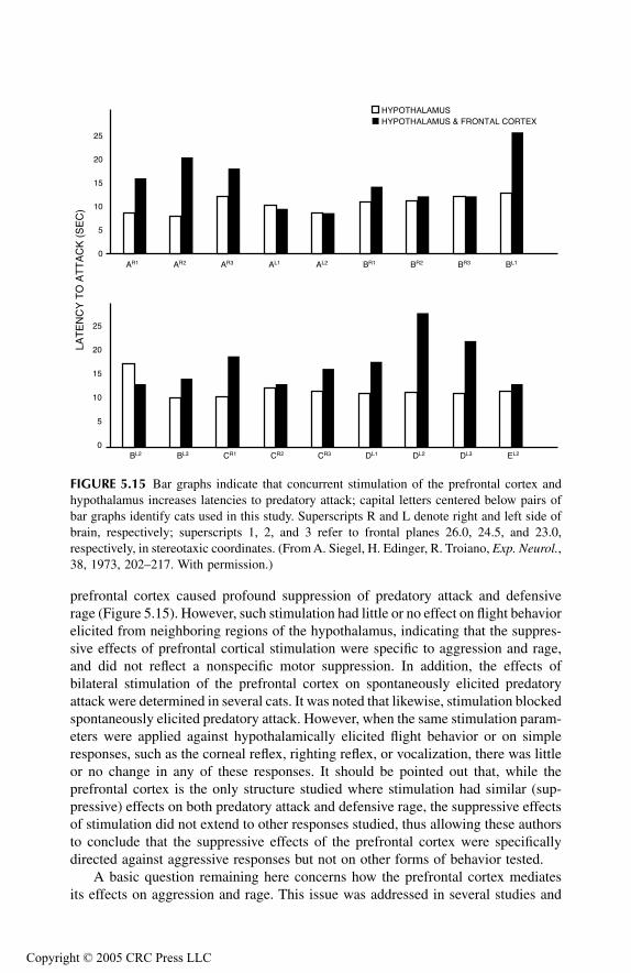
TF1661_C005.fm Page 110 Monday, October 25, 2004 7:21 AM
Copyr
prefrontal cortex caused profound suppression of predatory attack and defensiverage (Figure 5.15). However, such stimulation had little or no effect on flight behaviorelicited from neighboring regions of the hypothalamus, indicating that the suppres-sive effects of prefrontal cortical stimulation were specific to aggression and rage,and did not reflect a nonspecific motor suppression. In addition, the effects ofbilateral stimulation of the prefrontal cortex on spontaneously elicited predatoryattack were determined in several cats. It was noted that likewise, stimulation blockedspontaneously elicited predatory attack. However, when the same stimulation param-eters were applied against hypothalamically elicited flight behavior or on simpleresponses, such as the corneal reflex, righting reflex, or vocalization, there was littleor no change in any of these responses. It should be pointed out that, while theprefrontal cortex is the only structure studied where stimulation had similar (sup-pressive) effects on both predatory attack and defensive rage, the suppressive effectsof stimulation did not extend to other responses studied, thus allowing these authorsto conclude that the suppressive effects of the prefrontal cortex were specificallydirected against aggressive responses but not on other forms of behavior tested.
A basic question remaining here concerns how the prefrontal cortex mediatesits effects on aggression and rage. This issue was addressed in several studies and
FIGURE 5.15 Bar graphs indicate that concurrent stimulation of the prefrontal cortex andhypothalamus increases latencies to predatory attack; capital letters centered below pairs ofbar graphs identify cats used in this study. Superscripts R and L denote right and left side ofbrain, respectively; superscripts 1, 2, and 3 refer to frontal planes 26.0, 24.5, and 23.0,respectively, in stereotaxic coordinates. (From A. Siegel, H. Edinger, R. Troiano, Exp. Neurol.,38, 1973, 202–217. With permission.)
0
5
10
15
20
25
0
5
10
15
20
25
AR1 AR2 AR3 AL1 AL2 BR1 BR2 BR3 BL1
BL2 BL3 CR1 CR2 CR3 DL1 DL2 DL3 EL2
LAT
EN
CY
TO
AT
TA
CK
(S
EC
)
HYPOTHALAMUSHYPOTHALAMUS & FRONTAL CORTEX
ight © 2005 CRC Press LLC
TF1661_C005.fm Page 111 Monday, October 25, 2004 7:21 AM
Copyr
is briefly noted above. In one study (Siegel et al., 1974), a small lesion was placedat the tip of an electrode site from which prefrontal cortical stimulation had power-fully suppressed predatory attack, and the degenerating axons from this site weretraced to the diencephalon. As shown in Figure 5.14, the major distribution of fibersfrom this region of cortex was directed principally through the internal capsule tothe lateral half of the mediodorsal thalamic nucleus.
In a subsequent study (Siegel et al., 1977), the hypothesis that the suppressiveeffects of the prefrontal cortex are mediated primarily to the hypothalamus throughthe mediodorsal thalamic nucleus was tested directly. In this study, the suppressiveeffects of prefrontal cortical stimulation on hypothalamically elicited predatoryattack were initially demonstrated. Then, a lesion was placed in the mediodorsalthalamic nucleus and the effects of prefrontal cortical stimulation on predatory attackwere reassessed. The results showed that lesions of the anterior half of the mediodor-sal nucleus blocked the suppressive effects of the prefrontal cortex, but that lesionsof the posterior half of the mediodorsal nucleus but which spared the anterior aspectof this nucleus had little effect on suppression. Apparently, the locus of the lesionwas of importance because it was previously shown that the prefrontal cortical fibers,which project to the mediodorsal nucleus, supply mainly the anterior half of thisnucleus. Thus, lesions of the anterior aspect of the mediodorsal nucleus presumablywere more effective in blocking the suppressive effects of the prefrontal cortexbecause such lesions had a much greater effect in disrupting these afferent (prefron-tal) fibers than lesions of the posterior half of the mediodorsal nucleus.
With respect to the cingulate gyrus, a study by Siegel and Chabora (1971) useddual stimulation procedures to demonstrate that, similar to the prefrontal cortex,stimulation of the anterior cingulate gyrus suppresses predatory attack, while stim-ulation of more posterior aspects of this structure have little or no effect on this formof aggression. Based on the anatomical and brain stimulation findings, it may beconcluded that the suppressive effects of the anterior cingulate gyrus are mediatedthrough the mediodorsal nucleus to the lateral hypothalamus. Intuitively, it is under-standable that the posterior cingulate gyrus has little effect on predatory attackbecause its primary thalamic projection target — the anteroventral thalamic nucleus— is not known to project to the hypothalamus or have any effect on predatoryattack (MacDonnell and Flynn, 1967).
A further question was raised from these studies: How do signals from theprefrontal cortex and anterior cingulate gyrus reach the hypothalamus after theyterminate in the mediodorsal thalamic nucleus? This issue was addressed in severalearlier studies (Siegel et al., 1972, 1973b). These studies were based on the tracingof degenerating axons resulting from the placement of small lesions in the mediodor-sal thalamic nucleus and at different levels of the midline thalamus. The results,summarized in Figure 5.16, indicate that the pathway from the mediodorsal nucleusto the lateral hypothalamus is multisynaptic. It includes synapses in both caudal androstral aspects of the midline thalamus where the final neuron in this circuit arisesfrom the rostral aspect of the nucleus reuniens, and terminates mainly in the lateralhypothalamus.
An analysis of the role of the prefrontal region of the rat has also been carriedout by de Bruin (1983, 1990). These studies revealed several significant findings. In
ight © 2005 CRC Press LLC
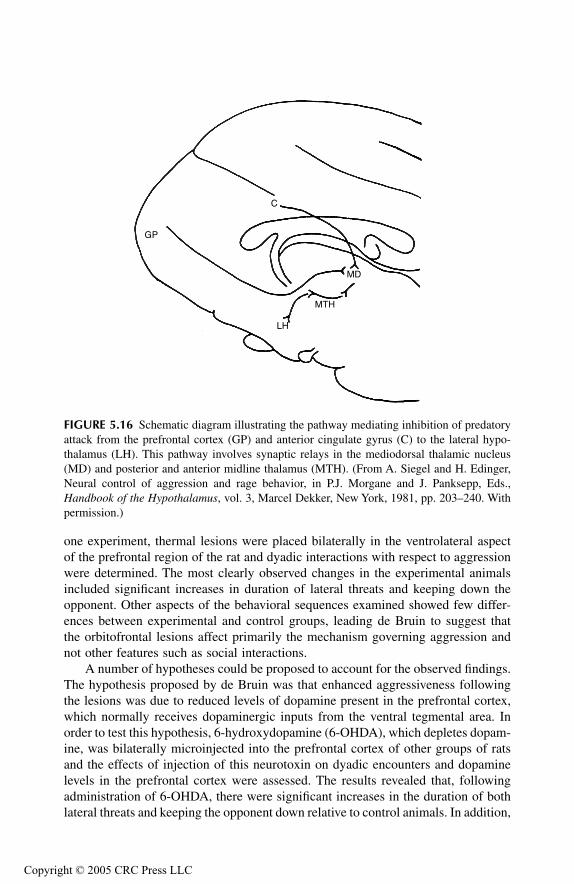
TF1661_C005.fm Page 112 Monday, October 25, 2004 7:21 AM
Copyr
one experiment, thermal lesions were placed bilaterally in the ventrolateral aspectof the prefrontal region of the rat and dyadic interactions with respect to aggressionwere determined. The most clearly observed changes in the experimental animalsincluded significant increases in duration of lateral threats and keeping down theopponent. Other aspects of the behavioral sequences examined showed few differ-ences between experimental and control groups, leading de Bruin to suggest thatthe orbitofrontal lesions affect primarily the mechanism governing aggression andnot other features such as social interactions.
A number of hypotheses could be proposed to account for the observed findings.The hypothesis proposed by de Bruin was that enhanced aggressiveness followingthe lesions was due to reduced levels of dopamine present in the prefrontal cortex,which normally receives dopaminergic inputs from the ventral tegmental area. Inorder to test this hypothesis, 6-hydroxydopamine (6-OHDA), which depletes dopam-ine, was bilaterally microinjected into the prefrontal cortex of other groups of ratsand the effects of injection of this neurotoxin on dyadic encounters and dopaminelevels in the prefrontal cortex were assessed. The results revealed that, followingadministration of 6-OHDA, there were significant increases in the duration of bothlateral threats and keeping the opponent down relative to control animals. In addition,
FIGURE 5.16 Schematic diagram illustrating the pathway mediating inhibition of predatoryattack from the prefrontal cortex (GP) and anterior cingulate gyrus (C) to the lateral hypo-thalamus (LH). This pathway involves synaptic relays in the mediodorsal thalamic nucleus(MD) and posterior and anterior midline thalamus (MTH). (From A. Siegel and H. Edinger,Neural control of aggression and rage behavior, in P.J. Morgane and J. Panksepp, Eds.,Handbook of the Hypothalamus, vol. 3, Marcel Dekker, New York, 1981, pp. 203–240. Withpermission.)
GP
C
LH
MTH
MD
ight © 2005 CRC Press LLC

TF1661_C005.fm Page 113 Monday, October 25, 2004 7:21 AM
Copyr
immunocytochemical staining of the brain tissue suggested that dopamine levelswere reduced in the prefrontal regions where microinjections of 6-OHDA wereplaced. Overall, these findings are consistent with the data described above in thecat and indicate that the prefrontal cortex normally provides significant inhibitionof aggressive reactions in several species. Moreover, these findings further suggestthat dopaminergic inputs to the prefrontal cortex are necessary for the prefrontalcortex to act in its “normal” manner by inhibiting the mechanism governing aggres-sion, which is presumably maintained at the level of the hypothalamus or PAG.
RELATED STRUCTURES OF LIMBIC FOREBRAIN: SUBSTANTIA INNOMINATA AND BED NUCLEUS OF
STRIA TERMINALIS
In addition to the limbic structures already considered, two additional nuclear groups— the bed nucleus of the stria terminalis (BNST) and substantia innominata — arereviewed here because of their anatomical connections with limbic structures, hypo-thalamus, or PAG, and because they have been shown to modulate aggression andrage.
BED NUCLEUS OF STRIA TERMINALIS
Anatomical Considerations
The BNST consists of a band of neurons that are located approximately at the levelof the anterior commissure just below the lateral ventricle, and at the confluence ofthe ventromedial and ventrolateral aspects of the caudate nucleus and septal area,respectively (see Chapter 6). That the BNST receives significant inputs mainly fromthe central nucleus of amygdala (and from other amygdaloid nuclei as well) is welldocumented from both anatomical (Beltramino et al., 1996; Petrovich and Swanson,1997; Sun et al., 1991) and electrophysiological (Dalsass and Siegel, 1987) studies.In turn, the BNST projects heavily to the region of the ventromedial hypothalamusimmediately below the fornix as determined from functional 2-DG analysis (Watsonet al., 1983c) and electrophysiology (Dalsass and Siegel, 1987), and to the PAG andrelated regions of the brainstem as determined from anatomical tracing methods(Holstege et al., 1985). Thus, the input–output relationships of the BNST endow itwith the capacity to receive information concerning visceral, autonomic, and emo-tional functions and to influence the primary regions of the diencephalon and brain-stem mediating rage and aggression. The results of an experiment described belowprovide evidence of BNST involvement in modulating rage and aggression.
Relationship to Aggression and Rage
In this study, a dual stimulation paradigm involved concurrent stimulation of theBNST and a medial or lateral hypothalamic site from which defensive rage orpredatory attack could be elicited, respectively (Shaikh et al., 1986). As shown inFigure 5.17, stimulation of the BNST facilitated the occurrence of defensive rage
ight © 2005 CRC Press LLC
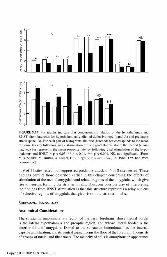
TF1661_C005.fm Page 114 Monday, October 25, 2004 7:21 AM
Copyr
in 9 of 11 sites tested, but suppressed predatory attack in 6 of 8 sites tested. Thesefindings parallel those described earlier in this chapter concerning the effects ofstimulation of the medial amygdala and related regions of the amygdala, which giverise to neurons forming the stria terminalis. Thus, one possible way of interpretingthe findings from BNST stimulation is that this structure represents a relay nucleusof selective regions of amygdala that give rise to the stria terminalis.
SUBSTANTIA INNOMINATA
Anatomical Considerations
The substantia innominata is a region of the basal forebrain whose medial borderis the lateral hypothalamus and preoptic region, and whose lateral border is theanterior third of amygdala. Dorsal to the substantia innominata lies the internalcapsule and striatum, and its ventral aspect forms the floor of the forebrain. It consistsof groups of nuclei and fiber tracts. The majority of cells is amorphous in appearance
FIGURE 5.17 Bar graphs indicate that concurrent stimulation of the hypothalamus andBNST alters latencies for hypothalamically elicited defensive rage (panel A) and predatoryattack (panel B). For each pair of histograms, the first (hatched) bar corresponds to the meanresponse latency following single stimulation of the hypothalamus alone; the second (cross-hatched) bar represents the mean response latency following dual stimulation of the hypo-thalamus and BNST. * p < 0.05; ** p < 0.01, *** p < 0.001. NS, not significant. (FromM.B. Shaikh, M. Brutus, A. Siegel, H.E. Siegel, Brain Res. Bull., 16, 1986, 179–182. Withpermission.)
** ** ** **
**** ******
* NS
NS
NS
NS
*
*
*
*
*
*
QU
IET
BIT
ING
AT
TA
CK
LA
TE
NC
Y (
sec)
AF
FE
CT
IVE
DE
FE
NS
E L
AT
EN
CY
(se
c)
0
2
4
6
8
0
2
4
6
8
A
B
ight © 2005 CRC Press LLC
TF1661_C005.fm Page 115 Monday, October 25, 2004 7:21 AM
Copyr
and does not form distinct nuclear groups. One group of cells, however, is large andquite prominent. It is situated near the base of the brain and is called the basalnucleus of Meynert. One characteristic of the basal nucleus and adjacent neurons isthat it has ubiquitous projections to wide regions of the cerebral cortex, hippocampalformation, and in particular, the amygdala (Femano et al., 1979). With respect tothis chapter, it is of special interest to note that the medial aspect of the substantiainnominata proximal to the preoptic region contains descending projections that arehighly similar to the preoptic region. In brief, such fibers enter the medial forebrainbundle and descend in this pathway, terminating at different levels throughout thelateral hypothalamus (Troiano and Siegel, 1978; Watson et al., 1983c). It is reason-able to suggest that this region of the substantia innominata may serve as an inter-neuron for transmission of signals from the lateral aspect of amygdala to the lateralhypothalamus.
Relationship to Aggression
As indicated above, the substantia innominata likely serves as a relay nucleus ofcertain cell groups of the amygdala such as the central and lateral nuclei, whichhave been shown to facilitate predatory attack. On this basis, it is reasonable toassume that the regions of substantia innominata receiving inputs from these regionsof amygdala would likewise facilitate this form of feline aggression. This hypothesiswas directly tested in a study by Block et al. (1980b). In this study, stimulatingelectrodes were placed into both the lateral and medial aspects of the substantiainnominata as well as the lateral hypothalamus from which predatory attack couldbe elicited. The methods employed were identical to the analysis of the modulatingeffects of the amygdaloid nuclei (Block et al., 1980a) and hippocampal formation(Watson et al., 1983a) on aggression and rage described earlier in the chapter.
The results indicate that stimulation of the medial half of the substantia innom-inata facilitated predatory attack and expanded the size of the trigeminal sensoryfields along the lipline activated by sensory stimulation. On the other hand, stimu-lation of the lateral aspect of the substantia innominata suppressed predatory attack,and caused a constriction of the same sensory fields. These findings indicate thatthe substantia innominata plays an important role in modulating predatory attack,and it appears to do so, at least in part, by acting on the trigeminal sensory fieldsthat control jaw closing and opening reflexes associated with predation. There arenevertheless a number of issues that remain unanswered from these studies:
How does the lateral aspect of the substantia innominata suppress predatoryattack?
To what extent are different regions of the substantia innominata critical foramygdaloid modulation of predatory attack?
How (if at all) would the substantia innominata modulate defensive ragebehavior?
Answers to these questions would certainly help to clarify our understanding of themechanisms by which the substantia innominata modulates aggressive reactions.
ight © 2005 CRC Press LLC
TF1661_C005.fm Page 116 Monday, October 25, 2004 7:21 AM
Copyr
LIMBIC SYSTEM AND HUMAN AGGRESSION
While most studies dealing with limbic system functions have been conducted inanimals, there has also been an increasing number of clinical reports detailing therelationship between limbic system dysfunctions and emotional disorders, includingrage and aggression. Some of these reports are summarized briefly below.
The aim of most studies has been uncovering the relationship between dys-functions associated with the temporal lobe and its underlying structures (i.e.,hippocampal formation and amygdala) and violence and aggression. In general,many of these individuals displayed marked outbursts of anger and violence towhat are considered innocuous statements by another individual. In addition, vio-lent episodes may even occur in such individuals in the absence of any clear-cutidentifiable threatening stimuli. Because of the nature of these characteristicresponses, such displays have been referred to as the “episodic dyscontrol syn-drome” by Monroe (1978). To site several examples of this syndrome, Martinius(1983) provided a report of a 14-year-old boy who brutally murdered an 8-year-old boy following an argument, and who was found to have a tumor bordering onthe hippocampal formation. This author further reported that a tumor was situatedin a similar region of hippocampal formation in Charles Whitman, the individualwho killed 16 people and wounded 32 others from a tower at the University ofTexas in Austin.
Other studies provided further support for the association between temporal lobedysfunction and violence and aggression. Serafetinides (1965) described the resultsof a study involving 100 medial temporal lobe patients, 36% of whom displayedovert physical aggressiveness. Following temporal lobectomy, long-term follow-upstudies resulted in significant reductions in aggressivity. Parallel findings were alsoreported in association with surgery involving the amygdala and adjoining peri-amygdaloid cortex (Hood et al., 1983). In more recent times, with the advent ofmagnetic resonance imaging techniques, Tonkonogy and Geller (1992) was able todemonstrate that lesions localized to the anterior inferior aspect of the temporal lobein 5 of 14 patients were associated with violent episodes of rage behavior.
Thus, these studies point to the likelihood that structures situated deep withinthe temporal lobe — namely, the hippocampal formation and amygdala — includingadjoining regions of the periamygdaloid, entorhinal, and prepyrifom cortices, playsignificant roles in the regulation of rage and aggression in a manner similar to thatdescribed above for these structures in regulating aggression in animals. It shouldalso be noted that other regions of the brain associated with the limbic system alsocontribute significantly to the organization and control of human aggression. Perhapsthe most important region studied here is the hypothalamus. A number of studieshave described the effects of tumors (Alpers, 1940; Reeves and Plum, 1969), hemato-mas (Berkovic et al., 1988), or cholinergic receptor dysfunction (Devinsky et al.,1992) in the hypothalamus on aggressive behavior. These studies demonstrated thatsuch disturbances of the hypothalamus resulted in displays of violence, rage, andaggression. These behaviors included stabbing, biting of the victim, and murder.These and related studies are summarized in Table 5.1.
ight © 2005 CRC Press LLC

TF1661_C005.fm Page 117 Monday, October 25, 2004 7:21 AM
Copyr
TABLE 5.1Violence in Humans Resulting from Dysfunction of Temporal Lobe and Hypothalamus
CitationType of
DysfunctionBehavioral
Manifestations Comment
Serafetinides, 1965
TLE, medial temporal sclerosis with tumors
Character disorders with aggressive outbursts
Reduction in aggression following temporal lobectomy
Malamud, 1967
Temporal lobe tumor Marked increase in rage and violence with little provocation
Tumors in nine patients displaying rage were located in temporal lobe
Ounsted, 1969 TLE Hyperkinetic syndrome with rage outbursts
Positive correlation between age, early onset, and occurrence of rage
Sweet et al., 1969
Temporal lobe tumor Two patients: one tried to kill his family; the other physically attacked family members
Symptoms had disappeared after removal of tumor
Mark and Sweet, 1974
TLE Violence induced with little provocation; patient stabbed stranger when he was accidentally bumped
Amygdaloid lesion reduced rage behavior
Falconer, 1973 TLE Interictal aggression Seven of 12 patients improved after unilateral temporal lobectomy
Hermann et al., 1980
TLE Psychopathic deviation and hypomania
Chronological age is inversely correlated with aggression scores
Delgado-Escueta et al., 1981
TLE Aggressive behavior associated with epileptic episodes
Aggression is not directed during seizure
Hood et al., 1983
TLE involving amygdala and periamygdala cortex
Increased aggression and irritable behavior
Amygdalectomy markedly reduced rage behavior
Martinius, 1983
Temporal lobe tumor Killed 16 people and wounded 32 before police shot him
Malignant tumor adjacent to lateral amygdala
Taylor, 1983 TLE Epileptic episodes and behavioral problems
Benefits of temporal lobectomy
Vaernet, 1983 TLE Hyperaggressive behavior Temporal lobe resection reduced aggression
Birmaher et al., 1993
Low serotonin Recurrent aggression behavior and impulsivity
Decreased serotonergic activity is associated with aggression; platelet IB is inversely correlated to degree of aggression
ight © 2005 CRC Press LLC
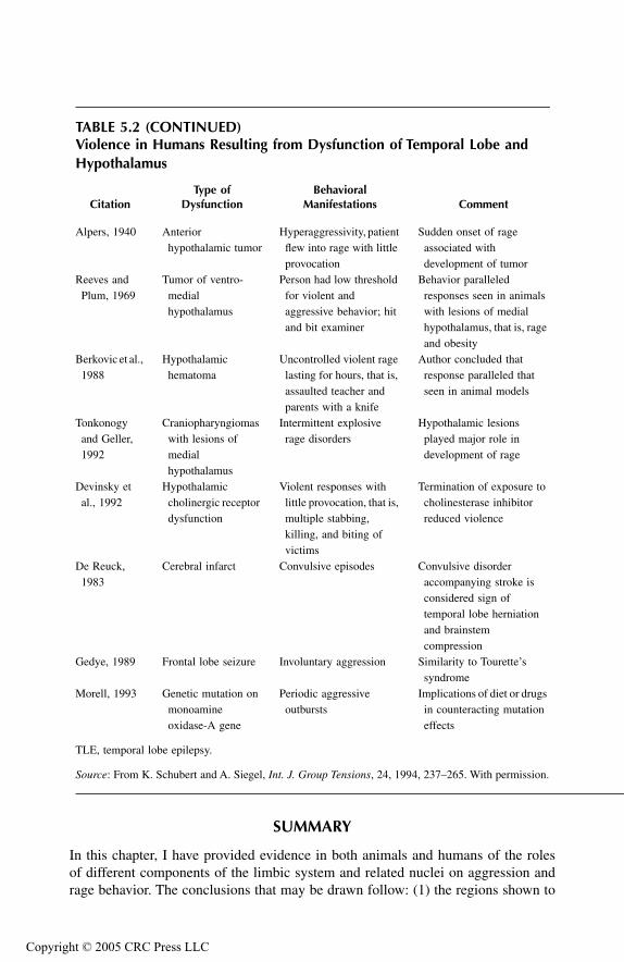
TF1661_C005.fm Page 118 Monday, October 25, 2004 7:21 AM
Copyr
SUMMARY
In this chapter, I have provided evidence in both animals and humans of the rolesof different components of the limbic system and related nuclei on aggression andrage behavior. The conclusions that may be drawn follow: (1) the regions shown to
TABLE 5.2 (CONTINUED)Violence in Humans Resulting from Dysfunction of Temporal Lobe and Hypothalamus
CitationType of
DysfunctionBehavioral
Manifestations Comment
Alpers, 1940 Anterior hypothalamic tumor
Hyperaggressivity, patient flew into rage with little provocation
Sudden onset of rage associated with development of tumor
Reeves and Plum, 1969
Tumor of ventro-medial hypothalamus
Person had low threshold for violent and aggressive behavior; hit and bit examiner
Behavior paralleled responses seen in animals with lesions of medial hypothalamus, that is, rage and obesity
Berkovic et al., 1988
Hypothalamic hematoma
Uncontrolled violent rage lasting for hours, that is, assaulted teacher and parents with a knife
Author concluded that response paralleled that seen in animal models
Tonkonogy and Geller, 1992
Craniopharyngiomas with lesions of medial hypothalamus
Intermittent explosive rage disorders
Hypothalamic lesions played major role in development of rage
Devinsky et al., 1992
Hypothalamic cholinergic receptor dysfunction
Violent responses with little provocation, that is, multiple stabbing, killing, and biting of victims
Termination of exposure to cholinesterase inhibitor reduced violence
De Reuck, 1983
Cerebral infarct Convulsive episodes Convulsive disorder accompanying stroke is considered sign of temporal lobe herniation and brainstem compression
Gedye, 1989 Frontal lobe seizure Involuntary aggression Similarity to Tourette’s syndrome
Morell, 1993 Genetic mutation on monoamine oxidase-A gene
Periodic aggressive outbursts
Implications of diet or drugs in counteracting mutation effects
TLE, temporal lobe epilepsy.
Source: From K. Schubert and A. Siegel, Int. J. Group Tensions, 24, 1994, 237–265. With permission.
ight © 2005 CRC Press LLC
TF1661_C005.fm Page 119 Monday, October 25, 2004 7:21 AM
Copyr
facilitate predatory attack include the ventral hippocampus, lateral aspects ofamygdala, and medial half of the substantia innominata; (2) the regions shown tosuppress predatory attack appear to be more widespread and include the prefrontalcortex, dorsal hippocampus, considerable parts of the septal area, lateral aspect ofthe substantia innominata, and medial amygdala, pyriform cortex, and BNST; (3) anumber of regions have been shown to differentially modulate aggression and rage,the most notable of which include the amygdala, BNST, and septal area. Researchhas shown that sites within these structures that facilitate predatory attack alsosuppress defensive rage, while sites that suppress predatory attack facilitate theoccurrence of defensive rage. The likely mechanism for this mechanism, discussedpreviously, suggests that direct or indirect inputs from these structures to the medialor lateral hypothalamus excite neurons governing defensive rage and predatoryattack, respectively, and that such excitation further causes a suppression of the otherform of attack by virtue of reciprocal inhibitory connections between the medialand lateral hypothalamus; and (4) limbic modulation of aggression and rage ismediated by direct or indirect connections from these regions to the hypothalamusor midbrain PAG. These functional anatomical relationships are further consideredin Chapter 6.
REFERENCES
Adamec, R.E., The behavioral bases of prolonged suppression of predatory attack in cats,Aggress. Behav., 1, 1975a, 297–314.
Adamec, R.E., The neural basis of prolonged suppression of predatory attack. I. Naturallyoccurring physiological differences in the limbic systems of killer and non-killer cats,Aggress. Behav., 1, 1975b, 315–330.
Adamec, R.E., Normal and abnormal limbic system mechanisms of emotive biasing, in K.E.Livingston and O. Hornykiewicz, Eds., Limbic Mechanisms, Plenum Press, New York,1978, pp. 405–455.
Adamec, R.E., Role of the amygdala and medial hypothalamus in spontaneous feline aggres-sion and defense, Aggress. Behav., 16, 1990, 207–222.
Adamec, R.E. and C. Stark-Adamec, Limbic control of aggression in the cat, Prog. Neuro-Psychopharmacol. Biol. Psychiatry, 7, 1983a, 505–512.
Adamec, R.E. and C. Stark-Adamec, Limbic kindling and animal behavior — implicationsfor human psychopathology associated with complex partial seizures, Biol. Psychi-atry, 18, 1983b, 269–293.
Adamec, R.E. and C. Stark-Adamec, Partial kindling and emotional bias in the cat: lastingaftereffects of partial kindling of the ventral hippocampus: I. Behavioral changes,Behav. Neural Biol., 38, 1983c, 205–222.
Adamec, R.E. and C. Stark-Adamec, Partial kindling and emotional bias in the cat: lastingaftereffects of partial kindling of the ventral hippocampus: II. Physiological changes,Behav. Neural Biol., 38, 1983d, 223–239.
Adamec, R.E. and C. Stark-Adamec, Kindling and interictal behavior: an animal model ofpersonality change, Psychiatr. J. (Ottawa), 10, 1985, 220–230.
Adamec, R.E. and C. Stark-Adamec, Partial kindling and behavioural change — some rulesgoverning behavioural outcome of repeated limbic seizures, Kindling 3, 1986,195–211.
ight © 2005 CRC Press LLC
TF1661_C005.fm Page 120 Monday, October 25, 2004 7:21 AM
Copyr
Albert, D.J. and S.E. Richmond, Hyperreactivity and aggressiveness following infusion oflocal anesthetic into the lateral septum or surrounding structures, Behav. Biol., 18,1976, 211–226.
Alexander, G.E. and J.M. Fuster, Effects of cooling prefrontal cortex on cell firing in thenucleus medialis dorsalis, Brain Res., 61, 1973, 93–105.
Allikmets, L. Kh. and M.E. Ditrikh, Effects of lesions of limbic system on emotional reactionsand conditioned reflexes in rats, Zh. Vyssh. Nerv. Deyatel'nosti imen. I.P. Pavlova,15, 1965, T1003–T1007.
Alonso-de Florida, F. and J.M.R. Delgado, Lasting behavioral and EEG changes in catsinduced by prolonged stimulation of amygdala, Am. J. Physiol., 193, 1958, 223–229.
Alpers, B.J., Personality and emotional disorders associated with hypothalamic lesions, Res.Publ. Assoc. Nerv. Ment. Dis., 20, 1940, 725–752.
An, X., R. Bandler, D. Öngür, J.L. Price, Prefrontal cortical projections to longitudinalcolumns in the midbrain periaqueductal gray in macaque monkeys, J. Comp. Neurol.,401, 1998, 455–479.
Andy, O.J. and H. Stephan, The Septum of the Cat, Charles C. Thomas, Springfield, Ill., 1964.Bacon, S.J., A.J.N. Headlam, P.L.A. Gabbott, A.D. Smith, Amygdala input to medial prefrontal
cortex, mPFC, in the rat: a light and electron microscope study, Brain Res., 720,1996, 211–219.
Bard, P. and V.B. Mountcastle, Some forebrain mechanisms involved in expression of ragewith special reference to suppression of angry behavior, Assoc. Res. Nerv. Ment. Dis.,27, 1948, 362–399.
Bawden, H.N. and R.J. Racine, Effects of bilateral kindling or bilateral sub-threshold stim-ulation of the amygdala or septum on muricide, ranacide, intraspecific aggressionand passive avoidance in the rat, Physiol. Behav., 22, 1979, 115–123.
Beltramino, C.A., M.S. Forbes, D.J. Swanson, G.F. Alheid, L. Heimer, Amygdaloid input totransiently tyrosine hydroxylase immunoreactive neurons in the bed nucleus of thestria terminalis of the rat, Brain Res., 706, 1996, 37–46.
Berkovic, S., F. Andermann, F. Melanson, R. Ethier, W. Feindell, P. Gloor, Hypothalamichematomas and ictal laughter: evolution of a characteristic epileptic syndrome anddiagnostic value of magnetic resonance imaging, Ann. Neurol., 23: 429–439, 1988.
Birmaher, B., M. Stanley, L. Greenhill, J. Twomey, A. Gavrilescu, H. Rabinovich, Plateletimipramine binding in children and adolescents with impulsive behavior, J. Am. Acad.Child Adolesc. Psychiatr., 29, 1993, 914–918.
Blanchard, D.C., R.J. Blanchard, E.M.C. Lee, S. Nakamura, Defensive behaviors in ratsfollowing septal and septal-amygdala lesions, J. Comp. Physiol. Psychol., 93, 1979,378–390.
Block, C.H., A. Siegel, H.M. Edinger, Effects of electrical stimulation of the amygdala upontrigeminal sensory fields established during hypothalamically-elicited quiet bitingattack in the cat, Brain Res., 197, 1980a, 39–55.
Block, C.H., A. Siegel, H.M. Edinger, Effects of stimulation of the substantia innominataupon attack behavior elicited from the hypothalamus in the cat, Brain Res., 197,1980b, 57–74.
Bobo, E.G. and M. Bonvallet, Amygdala and masserteric reflex. I. Facilitation, inhibition anddiphasic modifications of the reflex, induced by localized amygdaloid stimulation,Electroencephalogr. Clin. Neurophysiol., 39, 1975, 329–339.
Bonvallet, M. and E.G. Bobo, Amygdala and masseteric reflex, II. Mechanism of the diphasicmodifications of the reflex elicited from the “defence reaction area”: role of the spinaltrigeminal nucleus, Pars oralis, Electroencephalogr. Clin. Neurophysiol., 39, 1975,341–352.
ight © 2005 CRC Press LLC
TF1661_C005.fm Page 121 Monday, October 25, 2004 7:21 AM
Copyr
Brady, J.V. and W.J.H. Nauta, Subcortical mechanisms in emotional behavior: affectivechanges following septal forebrain lesions in the albino rat, J. Comp. Physiol. Psy-chol., 46, 1953, 339–346.
Brutus, M., M.B. Shaikh, A. Siegel, H. Edinger, Effects of experimental temporal lobe seizuresupon hypothalamically elicited aggressive behavior in the cat, Brain Res., 366, 1986,53–63.
Brutus, M., M.B. Shaikh, A. Siegel, H.E. Siegel, An analysis of the mechanisms underlyingseptal area control of hypothalamically elicited aggression in the cat, Brain Res., 310,1984, 235–248.
Carpenter, M.B. and J. Sutin, Human Neuroanatomy, 8th ed., Williams & Wilkins, Baltimore,1983.
Dalsass, M. and A. Siegel, The bed nucleus of the stria terminalis: electrophysiologicalproperties and responses to amygdaloid and hypothalamic stimulation, Brain Res.,425, 1987, 346–350.
de Bruin, J.P.C., Orbital prefrontal cortex, dopamine, and social agonistic behavior of maleLong Evans rats, Aggress. Behav., 16, 1990, 231–248.
de Bruin, J.P.C., H.G.M. Van Oyen, N. Van De Poll, Behavioural changes following lesionsof the orbital prefrontal cortex in male rats, Behav. Brain Res., 10, 1983, 209–232.
Delgado-Escueta, A.V., R.H. Mattson, L. King, E.S.Goldensohn, H. Spiegel, J.Madsen, P.Crandall, F. Dreifuss, R.J. Porter, New Eng. J. Med., 305, 1981, 711–716.
De Reuck, J.L., Neuropathology of Epilepsy Resulting from Cerebrovascular Disorders,presented at Advances in Epileptology, XIVth Epilepsy International Symposium, M.Parsonage et al., Eds., Raven Press, New York, 1983, pp. 95–98.
Devinsky, O., J. Kernan, D.M. Bear, Aggressive behavior following exposure to cholinesteraseinhibitors, J. Neuropsychiatry, 4, 1992, 189–194.
Divac, I., A. Kosmal, A. Bjorklund, O. Lindvall, Subcortical projections to the prefrontalcortex in the rat as revealed by the horseradish peroxidase technique, Neuroscience,3, 1978, 785–796.
Eclancher, F., P. Schmitt, P. Karli, Effets de lesions precoces de l’amygdale sur le developpe-ment de l’agressivite interspecifique du rat, Physiol. Behav., 14, 1975, 277–283.
Egger, M.D. and J.P. Flynn, Effects of electrical stimulation of the amygdala on hypothalam-ically elicited attack behavior in cats, J. Neurophysiol., 26, 1963, 705–720.
Falconer, M.A., Reversibility by temporal-lobe resection of the behavioral abnormalities oftemporal-lobe epilepsy, N. Engl. J. Med., 289, 1973, 451–455.
Femano, P.A., H.M. Edinger, A. Siegel, Evidence of a potent excitatory influence fromsubstantia innominata on basolateral amygdaloid units: a comparison with insular-temporal cortex and lateral olfactory tract stimulation, Brain Res., 177, 1979,361–366.
Femano, P.A., H.M. Edinger, A. Siegel, The effects of stimulation of substantia innominataand sensory receiving areas of the forebrain upon the activity of neurons within theamygdala of the anesthetized cat, Brain Res., 269, 1983, 119–132.
Freeman, W. and J.W. Watts, Psychosurgery, Intelligence, Emotion and Social BehaviorFollowing Prefrontal Lobotomy for Mental Disorders, Charles C. Thomas, Spring-field, Ill., 1942.
Fulton, J.F., The Frontal Lobes and Human Behavior, The Sherrington Lectures, No. 11,Charles C. Thomas, Springfield, Ill., 1952.
Gedye, A., Episodic rage and aggression attributed to frontal lobe seizures, J. Ment. Defic.Res., 33, 1989, 369–379.
Gotsick, J.E. and R.C. Marshall, Time course of the septal rage syndrome, Physiol. Behav.,9, 1972, 685–687.
ight © 2005 CRC Press LLC
TF1661_C005.fm Page 122 Monday, October 25, 2004 7:21 AM
Copyr
Green, J.D., C.D. Clemente, J. de Groot, Experimentally induced epilepsy in the cat withinjury of cornu ammonis, Arch. Neurol., Psychiatry, 78, 1957, 259–263.
Griffith, N., J. Engel, R. Bandler, Ictal and enduring interictal disturbances in emotionalbehaviour in an animal model of temporal lobe epilepsy, Brain Res., 400, 1987,360–364.
Han, Y., M.B. Shaikh, A. Siegel, Medial amygdaloid suppression of predatory attack behaviorin the cat: I. Role of a substance P pathway from the medial amygdala to the medialhypothalamus, Brain Res., 716, 1996a, 59–71.
Han, Y., M.B. Shaikh, A. Siegel, Medial amygdaloid suppression of predatory attack behaviorin the cat: II. Role of a GABAergic pathway from the medial to the lateral hypothal-amus, Brain Res., 716, 1996b, 72–83.
Harlow, J.M., Passage of an iron rod through the head., Boston Med. Surg. J., 39, 1848,389–393.
Harlow, J.M., Recovery from the passage of an iron bar through the head, Publ. Mass. Med.Soc., 2, 1868, 327–346.
Hermann, B.P., M.S. Schwartz, S. Whitman, W.E. Karnes, Aggression and epilepsy: seizure-type comparisons and high-risk variables, Epilepsia, 22, 1980, 691–698.
Holstege, G., L. Meiners, K. Tan, Projections of the bed nucleus of the stria terminalis to themesencephalon, pons, and medulla oblongata in the cat, Exp. Brain Res., 58, 1985,379–391.
Hood, T.W., J. Siegfried, H.G. Wieser, The role of stereotactic amygdalotomy in the treatmentof temporal lobe epilepsy associated with behavioral disorders, Appl. Neurophysiol.,46, 1983, 19–25.
Kaada, B.R., J. Jansen Jr., P. Andersen, Stimulation of the hippocampus and medial corticalareas in unanesthetized cats, Neurology 3, 1953, 844–857.
Karli, P., M. Vergnes, F. Eclancher, C. Penot, Involvement of amygdala in inhibitory controlover aggression in the rat: a synopsis, Aggress. Behav., 3, 1977, 157–162.
King, F.A., Effects of septal and amygdaloid lesions on emotional behavior and conditionedavoidance responses in the rat, J. Nerv. Ment. Dis., 126, 1958, 57–63.
Kita, H. and Y. Oomura, Evidence for a glycinergic cortico-lateral hypothalamic inhibitorypathway in the rat, Brain Res., 235, 1982, 131–136.
Kluver, H. and P.C. Bucy, Preliminary analysis of functions of the temporal lobes in monkeys,Arch. Neurol., Psychiatry, 42, 1939, 979–1000.
Krayniak, P.F., A. Siegel, R.C. Meibach, D. Fruchtman, M. Scrimenti, Origin of the fornixsystem in the squirrel monkey, Brain Res., 160, 1979, 401–411.
Krayniak, P.F., S. Weiner, A. Siegel, An analysis of the efferent connections of the septal areain the cat, Brain Res., 189, 1980, 15–29.
Latham, E.E. and B.M. Thorne, Septal damage and muricide: effects of strain and handling,Physiol. Behav., 12, 1974, 521–526.
LeDoux, J.E., The Emotional Brain, Simon & Schuster, New York, 1996.MacDonnell, M.F. and J.P. Flynn, Control of sensory fields by stimulation of hypothalamus,
Science, 152, 1966, 1406–1408.MacDonnell, M.F. and J.P. Flynn, Attack elicited by stimulation of the thalamus and adjacent
structures of cats, Behaviour, 31, 1967, 185–202.MacDonnell, M.F. and S. Stoddard-Apter, Effects of medial septal stimulation on hypotha-
lamically-elicited intraspecific attack and associated hissing in cats, Physiol. Behav.,21, 1978, 679–683.
MacLean, P.D., Psychosomatic disease and the “visceral brain,” Psychosom. Med., 11, 1949,338–353.
ight © 2005 CRC Press LLC

TF1661_C005.fm Page 123 Monday, October 25, 2004 7:21 AM
Copyr
MacLean, P.D. and J.M.R. Delgado, Electrical and chemical stimulation of frontotemporalportion of limbic system in the waking animal, EEG Clin. Neurophysiol., 5, 1953,91–100.
Malamud, N., Psychiatric disorder with intracranial tumors of limbic system, Arch. Neurol.,17, 1967, 113–123.
Mark, V.H. and W.H. Sweet, The role of limbic brain dysfunction in aggression, Res. Publ.Assoc. Nerv. Ment. Dis., 152, 1974, 186–200.
Martinius, J., Homicide of an aggressive adolescent boy with right temporal lesion: a casereport, Neurosci. Biobehav. Rev., 7, 1983, 419–422.
Meibach, R. and A. Siegel, The origin of fornix fibers which project to the mammillary bodiesin the rat: a horseradish peroxidase study, Brain Res., 88, 1975, 1–5.
Meibach, R. and A. Siegel, Efferent connections of the hippocampal formation in the rat,Brain Res., 124, 1977a, 197–224.
Meibach, R. and A. Siegel, Efferent connections of the septal area in the rat: an analysisutilizing retrograde and anterograde transport methods, Brain Res., 119, 1977b, 1–20.
Miczek, K.A. and S.P. Grossman, Effects of septal lesions on inter- and intraspecies aggressionin rats, J. Comp. Physiol. Psychol., 79, 1972, 37–45.
Monroe, R.R., Brain Dysfunction in Aggressive Criminals, Lexington Books, Lexington,Mass., 1978.
Morell, V., Evidence found for a possible “aggression gene”, Science, 260, 1993, 1722–1723.Nakao, H., Hypothalamic emotional reactivity after amygdaloid lesions in cats. Folia Psychiat.
Neurol. Jap., 14, 1960, 357–366.Noback, C.R. and R.J. Demarest, The Human Nervous System, 2nd ed., McGraw-Hill, New
York, 1975.Ottersen, O.P. and Y. Ben-Ari, Afferent connections to the amygdaloid complex of the rat and
cat: I. Projections from the thalamus, J. Comp. Neurol., 187, 1979, 401–424.Ounsted, C., Aggression and epilepsy rage in children with temporal lobe epilepsy, J. Psy-
chosom. Res., 13, 1969, 237–242.Öngür, D., X. An, J.L. Price, Prefrontal cortical projections to the hypothalamus in macaque
monkeys, J. Comp. Neurol., 401, 1998, 480–505.Papez, J.W., A proposed mechanism of emotion, Arch. Neurol. Psychiatry, 38, 1937, 725–743.Petrovich, G.D. and L.W. Swanson, Projections from the lateral part of the central amygdalar
nucleus to the postulated fear conditioning circuit, Brain Res., 763, 1997, 247–254.Potegal, M., C.F. Ferris, M. Hebert, J. Meyerhoff, L. Skaredoff, Attack priming in female
Syrian golden hamsters is associated with a c-fos-coupled process within the corti-comedial amygdala, Neuroscience, 75, 1996, 869–880.
Potegal, M., J.L. Gibbons, M. Glusman, Inhibition of muricide by septal stimulation in rats,Physiol. Behav., 24, 1980, 863–867.
Potegal, M., M. Hebert, M. DeCoster, J.L. Meyerhoff, Brief, high-frequency stimulation ofthe corticomedial amygdala induces a delayed and prolonged increase of aggressive-ness in male syrian golden hamsters, Behav. Neurosci., 110, 1996a, 401–412.
Potegal, M., C.F. Ferris, M. Hebert, J. Meyerhoff, L. Skaredoff, Attack priming in femaleSyrian golden hamsters is associated with a c-fos-coupled process within the corti-comedial amygdala, Neuroscience, 75, 1996b, 869–880.
Price, J.L., F.T. Russchen, D.G. Amaral, The limbic region II: The amygdaloid complex, inA. Bjorklund and T. Hokfelt, Eds., Handbook of Chemical Neuroanatomy, vol. 5,Elsevier, Amsterdam, 1987, pp. 279–388.
Pujic, Z., I. Matsumoto, P.A. Wilce, Extinction of emotional learning: contribution of medialprefrontal cortex, Neurosci. Lett., 162, 1993, 67–70.
ight © 2005 CRC Press LLC
TF1661_C005.fm Page 124 Monday, October 25, 2004 7:21 AM
Copyr
Reeves, A.G. and F. Plum, Hyperphagia, rage, and dementia accompanying a ventromedialhypothalamic neoplasm, Arch. Neurol., 20, 1969, 616–624.
Rothfield, L. and P.J. Harman, On the relation of the hippocampal-fornix system to the controlof rage responses in cats, J. Comp. Neurol., 101, 1954, 265–282.
Sato, M., Prefrontal cortex and emotional behaviors, Folia Psychiatr. Neurol. Jpn., 25, 1971,69–78.
Schubert, K. and A. Siegel, What animal studies have taught us about the neurobiology ofviolence, Int. J. Group Tensions, 24, 1994, 237–265.
Schreiner, L. and A. Kling, Behavioral changes following rhinencephalic injury in cat, J.Neurophysiol., 16, 1953, 643–659.
Serafetinides, E.A., Aggressiveness in temporal lobe epileptics and its relation to cerebraldysfunction and environmental factors, Epilepsia, 6, 1965, 33–42.
Shaikh, M.B., M. Brutus, A. Siegel, H.E. Siegel, Regulation of feline aggression by the bednucleus of stria terminalis, Brain Res. Bull., 16, 1986, 179–182.
Shaikh, M.B., C.-L. Lu, A. Siegel, Affective defense behavior elicited from the feline midbrainperiaqueductal gray is regulated by mu- and delta-opioid receptors, Brain Res., 557,1991, 344–348.
Shaikh, M.B., A. Steinberg, A. Siegel, Evidence that substance P is utilized in medial amygda-loid facilitation of defensive rage behavior in the cat, Brain Res., 625, 1993, 283–294.
Shealy, C. and T. Peele, Studies on amygdaloid nucleus of cat, J. Neurophysiol., 20, 1957,125–139.
Siegel, A., M. Brutus, M.B. Shaikh, H. Edinger, Effects of temporal lobe epileptiform activityupon aggressive behavior in the cat, Int. J. Neurol., 19–20, 1985–1986, 59–73.
Siegel, A. and J. Chabora, Effects of electrical stimulation of the cingulate gyrus upon attackbehavior elicited from the hypothalamus in the cat, Brain Res., 32, 1971, 169–177.
Siegel, A. and H. Edinger, Neural control of aggression and rage behavior, in P.J. Morganeand J. Panksepp, Eds., Handbook of the Hypothalamus, vol. 3, Marcel Dekker, NewYork, 1981, pp. 203–240.
Siegel, A., H. Edinger, M. Dotto, Effects of electrical stimulation of the lateral aspect of theprefrontal cortex upon attack behavior in cats, Brain Res., 93, 1975, 473–484.
Siegel, A., H. Edinger, A. Koo, Suppression of attack behavior in the cat by the prefrontalcortex: role of the mediodorsal thalamic nucleus, Brain Res., 127, 1977, 185–190.
Siegel, A., H. Edinger, H. Lowenthal, Effects of electrical stimulation of the medial aspectof the prefrontal cortex upon attack behavior in cats, Brain Res., 66, 1974, 467–479.
Siegel, A., H. Edinger, S. Ohgami, The topographical organization of the hippocampal pro-jection to the septal area: a comparative neuroanatomical analysis in the gerbil, rat,rabbit and cat, J. Comp. Neurol., 157, 1974, 359–378.
Siegel, A., H. Edinger, R. Troiano, The pathway from the mediodorsal nucleus of thalamusto the hypothalamus in the cat, Exp. Neurol., 38, 1973a, 202–217.
Siegel, A. and J.P. Flynn, Differential effects of electrical stimulation and lesions of thehippocampus and adjacent regions upon attack behavior in cats, Brain Res., 7, 1968,252–267.
Siegel, A., J.P. Flynn, R. Bandler, Thalamic sites and pathways related to elicited attack, BrainBehav. Evol., 6, 1972, 542–555.
Siegel, A., S. Ohgami, H. Edinger, The topographical organization of the hippocampal pro-jection to the septal area in the squirrel monkey, Brain Res., 99, 1975, 247–260.
Siegel, A. and D. Skog, Effects of electrical stimulation of the septum upon attack behaviorelicited from the hypothalamus in the cat, Brain Res., 23, 1970, 371–380.
ight © 2005 CRC Press LLC
TF1661_C005.fm Page 125 Monday, October 25, 2004 7:21 AM
Copyr
Siegel, A. and J.P. Tassoni, Differential efferent projections from the ventral and dorsalhippocampus of the cat, Brain Behav. Evol., 4, 1971, 185–200.
Siegel, A., R. Troiano, A. Royce, Differential efferent projections of the anterior and posteriorcingulate gyrus to the thalamus in the cat, Exp. Neurol., 38, 1973b, 192–201.
Stoddard-Apter, S.L. and M.F. MacDonnell, Septal and amygdalar efferents to the hypothal-amus which facilitate hypothalamically elicited intraspecific aggression and associ-ated hissing in the cat: an autoradiographic study, Brain Res., 193, 1980, 19–32.
Sun, N., L. Roberts, M.D. Cassell, Rat central amygdaloid nucleus projections to the bednucleus of the stria terminalis, Brain Res. Bull., 27, 1991, 1–12.
Swanson, L.W. and G.D. Petrovich, What is the amygdala? Trends Neurosci., 21, 1998,323–331.
Sweet, W.H., F. Ervin, V.H. Mark, The relationship of violent behaviour to focal cerebraldisease, in S. Garratini and E.B. Sigg, Eds., Aggressive Behavior, Excerta MedicaFoundation, Amsterdam, 1969, pp. 336–352.
Taylor, D.C., Child Behavioural Problems and Temporal Lobe Epilepsy, Proceedings,Advances in Epileptology, XVth Epilepsy International Symposium, 1983, pp.243–247.
Tonkonogy, J.M. and J.L. Geller, Hypothalamic lesions and intermittent explosive disorder,J Neuropsychiat. Clin. Neurosci., 4, 1992, 45–50.
Troiano, R., C. Boehme, and A. Siegel, A corthicothalamic projection involving the midlineseptum in the cat, Brain Res., 139, 1978, 348–353.
Troiano, R. and A. Siegel, Efferent connection of the basal forebrain in the cat: II. Thesubstantia innominata, Exp. Neurol., 61, 1978, 185–197.
Vaernet, K., Temporal Lobotomy in Children and Young Adults, Proceedings, Advances inEpileptology, XVth Epilepsy International Symposium, M. Parsonage et al., Eds.,Raven Press, New York, 1983, 255–261.
Vergnes, M., Controle amygdalien de comportements d'agression chez le rat, Physiol. Behav.,17, 1976, 439–444.
Vergnes, M., Declenchement de reactions d’agression interspecifique apres lesion amygdaliennechez le rat, Physiol. Behav., 17, 1975, 221–226.
Vochteloo, J.D. and J.M. Koolhaas, Medial amygdala lesions in male rats reduce aggressivebehavior: interference with experience, Physiol. Behav., 41, 1987, 99–102.
Wasman, M. and J.P. Flynn, Directed attack behavior during hippocampal seizures, Arch.Neurol., 14, 1966, 408–414.
Watson, R.E., H. Edinger, A. Siegel, An analysis of the mechanisms underlying hippocampalcontrol of hypothalamically-elicited aggression in the cat, Brain Res., 269, 1983a,327–345.
Watson, R.E., H.E. Edinger, A. Siegel, A 14C-2-deoxyglucose analysis of the functional neuralpathways of the limbic forebrain in the rat: III. The hippocampal formation, BrainRes. Brain Res. Rev., 5, 1983b, 133–176.
Watson, R.E., R. Troiano, J.J. Poulakos, S. Weiner, C.H. Block, A. Siegel, A (14C)2-deoxy-glucose analysis of the functional neural pathways of the limbic forebrain in the rat:I. The amygdala, Brain Res. Brain Res. Rev., 5, 1983c, 1–44.
Weiss, S.R.B., R.M. Post, P.W. Gold, G. Chrousos, T.L. Sullivan, D. Walker, A. Pert, CRF-induced seizures and behavior: interaction with amygdala kindling, Brain Res., 372,1986, 345–351.
Wilcott, R., Electrical stimulation in the prefrontal cortex and delayed response in the cat,Neuropsychologia, 15, 1977, 115–121.
ight © 2005 CRC Press LLC
TF1661_C005.fm Page 126 Monday, October 25, 2004 7:21 AM
Copyr
Winn, P., Schizophrenia research moves to the prefrontal cortex, Trends Neurosci., 17, 1994,265–268.
Yutzy, D.A., D.R. Meyer, P.M. Meyer, Effects of simultaneous septal and neo- or limbic-cortical lesions upon emotionality in the rat., Brain Res., 5, 1967, 452–458.
Zagrodzka, J., C.E. Hedberg, G.L. Mann, A.R. Morrison, Contrasting expressions of aggres-sive behavior released by lesions of the central nucleus of the amygdala duringwakefulness and rapid eye movement sleep without atonia in cats, Behav. Neurosci.,112, 1998, 589–602.
ight © 2005 CRC Press LLC
TF1661_C006.fm Page 127 Monday, October 18, 2004 8:20 AM
Copyr
6
Limbic System II: Functional NeuroanatomyChapter 5 focused on the anatomical, physiological, and behavioral properties oflimbic structures with respect to their roles in the regulation of aggression and ragebehavior. A large number of the studies that contributed to our knowledge linkingthe limbic system to aggression and rage were based on the application of mainlyelectrical, and to a lesser extent, chemical stimulation of these structures. Accord-ingly, a series of studies was conducted utilizing microstimulation of differentregions of the limbic system in conjunction with (14C)2-deoxyglucose (2-DG) auto-radiography. As pointed out in earlier chapters, a close relationship exists betweenfunctional activity and incorporation of 2-DG into neurons. For this reason, thismethod was used to identify the regions of the brain that are demonstrably activatedfollowing discrete stimulation of different sites within each of the limbic structures.This information was deemed valuable in providing insight into the anatomicalsubstrates involved in limbic modulation of aggression and rage behavior.
This chapter describes the results of mapping studies of the regions that aremetabolically activated in the rat brain following electrical stimulation of the dif-ferent components of the limbic system. Limbic structures from which stimulationwas applied and described below include the hippocampal formation, septal area,amygdala, and prefrontal cortex. It should be pointed out that this method was notintended to replace neuroanatomical methods. Instead, the results should be viewedas reflecting a means by which the major regions activated as a result of stimulationof a given site within the limbic system can be identified.
METHODS USED
The methods employed for these studies involving different components of the limbicsystem were basically identical and were described in Chapter 3. Details concerningthis methodology and related issues related to limbic system anatomy and functioncan be found in Brutus et al. (1984), Meibach and Siegel (1977), Siegel and Edinger(1976), Siegel et al. (1974, 1975), Siegel and Tassoni (1971), and Watson et al.(1982, 1983a, 1983b, 1985). In brief, adult rats were anesthetized with ketamine orpentobarbital, and a femoral vein was cannulated in which the patency of the cannulawas maintained with heparinized isotonic saline. The paradigm consisted of electricalstimulation, which was delivered continuously for periods of 30 seconds on and 30seconds off for both 5 minutes preceding the injection of 2-DG and for 45 minutesfollowing the injection of 2-DG. Current values were generally maintained at approx-imately 0.2 mA at 60 Hz as a balanced biphasic pulse with 1 millisecond per half-cycle duration. The findings presented below represent principally the results of
ight © 2005 CRC Press LLC

TF1661_C006.fm Page 128 Monday, October 18, 2004 8:20 AM
Copyr
stimulation at current intensities that did not induce any seizure activity. At thecompletion of the 45-minute stimulation procedure, the animal was sacrificed, thebrain was removed, and quickly frozen. Brain sections were cut and mounted oncoverslips, which were then serially mounted on cardboard. X-ray films were thenplaced over each cardboard array of sections with the emulsion side apposed to thetissue and sealed in a light-tight case in the darkroom. Brain sections were exposedfor a period of 4 weeks, and the x-rays were then developed. The optical densitiespresent on the x-rays were a direct function of the relative levels of 2-DG uptakeamong the structures in the brain, which, in turn, is a reflection of the degree ofmetabolic activation in these structures.
STUDY RESULTS
HIPPOCAMPAL FORMATION
Principal Findings
For analysis of the hippocampal formation, stimulation was applied in separateanimals at different sites along its longitudinal axis (Figure 6.1). Consider first thedistribution of 2-DG labeled regions following stimulation of the dorsal hippocam-pus of the rat, an example of which is shown in Figure 6.2. A principal finding wasthat stimulation of the dorsal hippocampal formation resulted in bilateral metabolicactivation of only the dorsal hippocampus and a restricted region of the dorsomedialaspect of the lateral septal nucleus (Figure 6.3). When stimulation was applied atmore posterior levels of the dorsal hippocampus, a similar pattern of labeling inthese regions was also observed. Further, it did not matter whether the stimulatingelectrode was placed in the regio inferior (CA3) or regio superior (CA1) of thedorsal hippocampus, since similar patterns of labeling resulted from stimulation ofeach of these regions.
Stimulation of sites at progressively more distal to the septal pole of the hip-pocampus (i.e., which corresponds to sites located progressively more proximal tothe temporal pole) resulted in activation patterns that were localized to progressivelymore ventrolateral aspects within the septal area (Figure 6.4). This functional topo-graphical organization of hippocampal outputs to the septal area corresponds quiteclosely to the anatomical findings described in Chapter 5 (Meibach and Siegel, 1977;Siegel and Edinger, 1976; Siegel et al., 1974, 1975; Siegel and Tassoni, 1971). It isof further interest to note that when stimulation was applied to the subicular cortexof either the dorsal or ventral hippocampal formation, prominent labeling within themammillary bodies was noted. In addition, stimulation of the ventral subicular cortexresulted in activation of the medial corticohypothalamic tract and its target regionin the ventromedial hypothalamus extending from the suprachiasmatic nucleus tothe arcuate region (Figure 6.4). Of further interest, stimulation of this region alsoproduced activation of the stria terminalis and amygdalo-hippocampal area, as wellas the medial, cortical, and basolateral amygdaloid nuclei. A summary of thesefindings is shown in Figure 6.5.
ight © 2005 CRC Press LLC
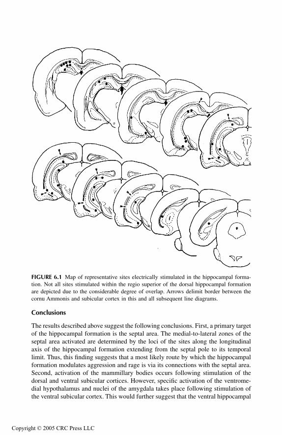
TF1661_C006.fm Page 129 Monday, October 18, 2004 8:20 AM
Copyr
Conclusions
The results described above suggest the following conclusions. First, a primary targetof the hippocampal formation is the septal area. The medial-to-lateral zones of theseptal area activated are determined by the loci of the sites along the longitudinalaxis of the hippocampal formation extending from the septal pole to its temporallimit. Thus, this finding suggests that a most likely route by which the hippocampalformation modulates aggression and rage is via its connections with the septal area.Second, activation of the mammillary bodies occurs following stimulation of thedorsal and ventral subicular cortices. However, specific activation of the ventrome-dial hypothalamus and nuclei of the amygdala takes place following stimulation ofthe ventral subicular cortex. This would further suggest that the ventral hippocampal
FIGURE 6.1 Map of representative sites electrically stimulated in the hippocampal forma-tion. Not all sites stimulated within the regio superior of the dorsal hippocampal formationare depicted due to the considerable degree of overlap. Arrows delimit border between thecornu Ammonis and subicular cortex in this and all subsequent line diagrams.
ight © 2005 CRC Press LLC

TF1661_C006.fm Page 130 Monday, October 18, 2004 8:20 AM
Copyr
formation could also modulate aggression and rage either by two additional routes.The first is by its direct connections with the hypothalamus, and the second is viaits activation of amygdaloid nuclei, which then project to the hypothalamus ormidbrain PAG.
SEPTAL AREA
Principal Findings
For this study, stimulation was applied within different zones of the lateral septalarea, medial septal nucleus, septofimbrial nucleus, and vertical and horizontal limbsof the diagonal band of Broca (Figure 6.6). Consider first the regions activatedfollowing stimulation of the intermediolateral aspect of the lateral septal nucleus(Figure 6.7). Two major patterns of activation should be noted. First, stimulation ofthe septal area activated wide regions of both the dorsal and ventral hippocampalformation bilaterally. The pattern of activation included all of the CA fields withminimal involvement of the subicular cortex. Additional label was also seen in theipsilateral basolateral nucleus of amygdala. A second pattern involved activation ofthe medial septal nucleus, vertical and horizontal limbs of the diagonal band ofBroca, preoptic region, and ventral and medial hypothalamus, including the mamil-lary bodies. Similar patterns of labeling (with the exception of the amygdala) wereseen after stimulation was applied to the septofimbrial nucleus (Figure 6.8). The
FIGURE 6.2 Autoradiographs displaying the distribution of (14C)2-DG labeled regions fol-lowing electrical stimulation of the regio superior at the level of the anterior dorsal hippoc-ampal formation. The white dot and star indicate the position of the stimulating electrode tip.The label present in the left parietal cortex is due to surgical trauma and passage of thestimulating electrode into the dorsal hippocampus. (From R.E. Watson, R. Troiano, J.J.Poulakos, S. Weiner, C.H. Block, A. Siegel, Brain Res. Rev., 5, 1983b, 1–44. With permission.)
ight © 2005 CRC Press LLC

TF1661_C006.fm Page 131 Monday, October 18, 2004 8:20 AM
Copyr
patterns of labeling observed following stimulation of the medial septal nucleus,vertical limb of the diagonal band of Broca and horizontal limb of the diagonal bandwere highly similar (Figures 6.9 and 6.10). Here, the basic sites of activation includedthe medial forebrain bundle and lateral hypothalamus.
Conclusions
Several important conclusions concerning functions of the septal area are evidentfrom these findings. As noted above, the first is that the lateral septal area serves asa relay of the hippocampal formation to the hypothalamus. This relay can be eitherthrough direct projections to the hypothalamus, or indirectly, via projections fromthe lateral septal nucleus to the vertical and horizontal limbs of the diagonal bandof Broca. The second is that the septal area can directly activate wide areas of thehippocampal formation. In this manner, the hippocampal formation and septal areamay be viewed as components of a single system. Namely, the hippocampal forma-tion can modulate the functions of the hypothalamus via its connections with theseptal area, and, in turn, the projections back to the hippocampal formation providea “feedback” mechanism for regulation of this circuit.
FIGURE 6.3 Distribution of 14C-2DG labeled regions following electrical stimulation of theregio superior at the level of the anterior dorsal hippocampal formation. The label shown inthis and subsequent line diagrams depicts only regions metabolically activated as a result ofthe applied stimulus and does not include regions normally associated with augmentedlabeling in control brains. (From R.E. Watson, R. Troiano, J.J. Poulakos, S. Weiner, C.H.Block, A. Siegel, Brain Res. Rev., 5, 1983b, 1–44. With permission.)
LS
DB
F
B
DF
Fl
MM
DG
F
SBPRO
CA1
CA3
VM
M
ight © 2005 CRC Press LLC

TF1661_C006.fm Page 132 Monday, October 18, 2004 8:20 AM
Copyr
AMYGDALA
Principal Findings
Stimulation was applied at sites within the principal nuclei of the amygdala as wellas the adjoining pyriform cortex (Figure 6.11).
Basomedial Nucleus
Following stimulation of the basomedial nucleus, activation was seen in both the striaterminalis and ventral amygdalofugal pathways. One band of activation extendedthrough the substantia innominata into the bed nucleus of the stria terminalis andadjoining septal area. A second band of activation could be seen passing caudally in anintermediolateral position within the ventral aspect of the hypothalamus to the level of
FIGURE 6.4 Distribution of label following electrical stimulation of the presubiculum at thelevel of the ventral hippocampal formation. Note that label is present over the ipsilateralforebrain and is extensive over the ventrolateral aspect of the lateral septal nucleus. Clearlydefined label is also present over the bed nucleus of the stria terminalis, medial corticohypo-thalamic tract, and ventromedial hypothalamus. There is also diffuse label over the medialhalf of the amygdala. (From R.E. Watson, R. Troiano, J.J. Poulakos, S. Weiner, C.H. Block,A. Siegel, Brain Res. Rev., 5, 1983b, 1–44. With permission.)
LS
AC
Fl
FB
SC
ST
L
BLBM
MEDH
VM
AR
CNAH
AHAMM
E
SBPAR
ight © 2005 CRC Press LLC
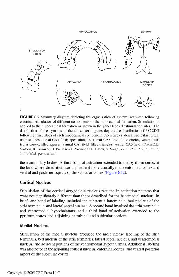
TF1661_C006.fm Page 133 Monday, October 18, 2004 8:20 AM
Copyr
the mammillary bodies. A third band of activation extended to the pyriform cortex atthe level where stimulation was applied and more caudally in the entorhinal cortex andventral and posterior aspects of the subicular cortex (Figure 6.12).
Cortical Nucleus
Stimulation of the cortical amygdaloid nucleus resulted in activation patterns thatwere not significantly different than those described for the basomedial nucleus. Inbrief, one band of labeling included the substantia innominata, bed nucleus of thestria terminalis, and lateral septal nucleus. A second band involved the stria terminalisand ventromedial hypothalamus; and a third band of activation extended to thepyriform cortex and adjoining entorhinal and subicular cortices.
Medial Nucleus
Stimulation of the medial nucleus produced the most intense labeling of the striaterminalis, bed nucleus of the stria terminalis, lateral septal nucleus, and ventromedialnucleus, and adjacent portions of the ventromedial hypothalamus. Additional labelingwas also noted in the adjoining cortical nucleus, entorhinal cortex, and ventral posterioraspect of the subicular cortex.
FIGURE 6.5 Summary diagram depicting the organization of systems activated followingelectrical stimulation of different components of the hippocampal formation. Stimulation isapplied to the hippocampal formation as shown in the panel labeled “stimulation sites.” Thedistribution of the symbols in the subsequent figures depicts the distribution of 14C-2DGfollowing stimulation of each hippocampal component. Open circles, dorsal subicular cortex;open squares, dorsal CA1 field; open triangles, dorsal CA3 field; filled circles, ventral sub-icular cortex; filled squares, ventral CA1 field; filled triangles, ventral CA3 field. (From R.E.Watson, R. Troiano, J.J. Poulakos, S. Weiner, C.H. Block, A. Siegel, Brain Res. Rev., 5, 1983b,1–44. With permission.)
STIMULATIONSITES
HIPPOCAMPUS SEPTUM
AMYGDALA HYPOTHALAMUS MAMILLARYBODIES
E
SBCA1
CA2
L
BL
C
ME
VM
MT
DB
AC
CM
ight © 2005 CRC Press LLC
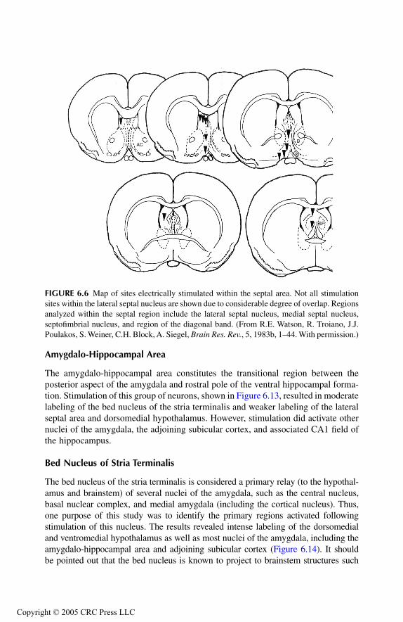
TF1661_C006.fm Page 134 Monday, October 18, 2004 8:20 AM
Copyr
Amygdalo-Hippocampal Area
The amygdalo-hippocampal area constitutes the transitional region between theposterior aspect of the amygdala and rostral pole of the ventral hippocampal forma-tion. Stimulation of this group of neurons, shown in Figure 6.13, resulted in moderatelabeling of the bed nucleus of the stria terminalis and weaker labeling of the lateralseptal area and dorsomedial hypothalamus. However, stimulation did activate othernuclei of the amygdala, the adjoining subicular cortex, and associated CA1 field ofthe hippocampus.
Bed Nucleus of Stria Terminalis
The bed nucleus of the stria terminalis is considered a primary relay (to the hypothal-amus and brainstem) of several nuclei of the amygdala, such as the central nucleus,basal nuclear complex, and medial amygdala (including the cortical nucleus). Thus,one purpose of this study was to identify the primary regions activated followingstimulation of this nucleus. The results revealed intense labeling of the dorsomedialand ventromedial hypothalamus as well as most nuclei of the amygdala, including theamygdalo-hippocampal area and adjoining subicular cortex (Figure 6.14). It shouldbe pointed out that the bed nucleus is known to project to brainstem structures such
FIGURE 6.6 Map of sites electrically stimulated within the septal area. Not all stimulationsites within the lateral septal nucleus are shown due to considerable degree of overlap. Regionsanalyzed within the septal region include the lateral septal nucleus, medial septal nucleus,septofimbrial nucleus, and region of the diagonal band. (From R.E. Watson, R. Troiano, J.J.Poulakos, S. Weiner, C.H. Block, A. Siegel, Brain Res. Rev., 5, 1983b, 1–44. With permission.)
ACB
DB
SF
ight © 2005 CRC Press LLC
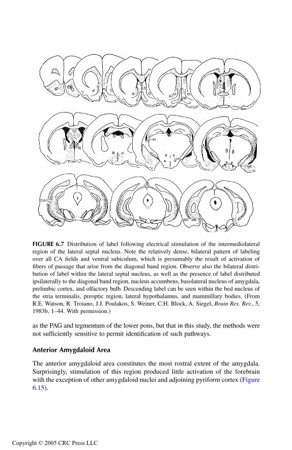
TF1661_C006.fm Page 135 Monday, October 18, 2004 8:20 AM
Copyr
as the PAG and tegmentum of the lower pons, but that in this study, the methods werenot sufficiently sensitive to permit identification of such pathways.
Anterior Amygdaloid Area
The anterior amygdaloid area constitutes the most rostral extent of the amygdala.Surprisingly, stimulation of this region produced little activation of the forebrainwith the exception of other amygdaloid nuclei and adjoining pyriform cortex (Figure6.15).
FIGURE 6.7 Distribution of label following electrical stimulation of the intermediolateralregion of the lateral septal nucleus. Note the relatively dense, bilateral pattern of labelingover all CA fields and ventral subiculum, which is presumably the result of activation offibers of passage that arise from the diagonal band region. Observe also the bilateral distri-bution of label within the lateral septal nucleus, as well as the presence of label distributedipsilaterally to the diagonal band region, nucleus accumbens, basolateral nucleus of amygdala,prelimbic cortex, and olfactory bulb. Descending label can be seen within the bed nucleus ofthe stria terminalis, preoptic region, lateral hypothalamus, and mammillary bodies. (FromR.E. Watson, R. Troiano, J.J. Poulakos, S. Weiner, C.H. Block, A. Siegel, Brain Res. Rev., 5,1983b, 1–44. With permission.)
BPL LS
ACDB
ME
LH
Fl
VC
FB
CA1
SB
ight © 2005 CRC Press LLC
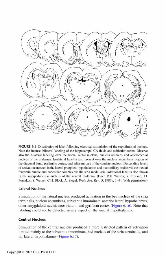
TF1661_C006.fm Page 136 Monday, October 18, 2004 8:20 AM
Copyr
Lateral Nucleus
Stimulation of the lateral nucleus produced activation in the bed nucleus of the striaterminalis, nucleus accumbens, substantia innominata, anterior lateral hypothalamus,other amygdaloid nuclei, neostriatum, and pyriform cortex (Figure 6.16). Note thatlabeling could not be detected in any aspect of the medial hypothalamus.
Central Nucleus
Stimulation of the central nucleus produced a more restricted pattern of activationlimited mainly to the substantia innominata, bed nucleus of the stria terminalis, andfar lateral hypothalamus (Figure 6.17).
FIGURE 6.8 Distribution of label following electrical stimulation of the septofimbrial nucleus.Note the intense, bilateral labeling of the hippocampal CA fields and subicular cortex. Observealso the bilateral labeling over the lateral septal nucleus, nucleus reuniens and anteromedialnucleus of the thalamus. Ipsilateral label is also present over the nucleus accumbens, region ofthe diagonal band, prelimbic cortex, and adjacent part of the caudate nucleus. Descending levelsof activation are seen in the lateral preoptico-hypothalamus and mammillary bodies via the medialforebrain bundle and habenular complex via the stria medullaris. Additional label is also shownin the interpeduncular nucleus of the ventral midbrain. (From R.E. Watson, R. Troiano, J.J.Poulakos, S. Weiner, C.H. Block, A. Siegel, Brain Res. Rev., 5, 1983b, 1–44. With permission.)
AC DB
SF
B
AM
AV
HB
LH
RE
CA1 CA3
IP
SBPAR
ight © 2005 CRC Press LLC

TF1661_C006.fm Page 137 Monday, October 18, 2004 8:20 AM
Copyr
Basolateral Nucleus
Stimulation of the basolateral nucleus resulted in activation patterns within both thestria terminalis and ventral amygdalofugal pathways. Strial activation was associatedwith intense labeling in the bed nucleus of the stria terminalis and labeling in thelateral septal nucleus, while ventral amygdalofugal pathway activation with labelingin the substantia innominata and nucleus accumbens. Little or no labeling wasdetected in the hypothalamus, subicular cortex, or hippocampus (Figure 6.18).
Prepyriform Cortex
Stimulation of the prepyriform cortex resulted in labeling of the posterior aspect ofthe olfactory bulb, substantia innominata, rostral part of the lateral septal nucleus,and infralimbic and prelimbic cortices. More intense labeling extended to mostamygdaloid nuclei except the central nucleus (Figure 6.19).
Entorhinal Cortex
Stimulation of the entorhinal cortex also produced limited patterns of labeling.Within the amygdala, labeling was restricted to the lateral and basolateral nuclei.Labeling was clearly seen in the posterior aspects of the subicular cortex and smallamounts in the lateral septal nucleus (Figure 6.20).
FIGURE 6.9 Distribution of label following electrical stimulation of the horizontal limb ofthe diagonal band. Label passes downstream in the medial forebrain bundle to the level ofthe mammillary bodies. Other label is diffuse and can be identified with the medial and lateralseptal nuclei. Little or no label could be identified within the hippocampal formation. (FromR.E. Watson, R. Troiano, J.J. Poulakos, S. Weiner, C.H. Block, A. Siegel, Brain Res. Rev., 5,1983b, 1–44. With permission.)
AC
LS
DB
MS
LH
ight © 2005 CRC Press LLC
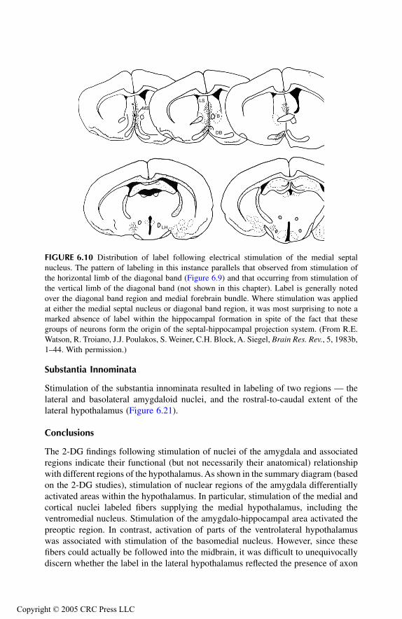
TF1661_C006.fm Page 138 Monday, October 18, 2004 8:20 AM
Copyr
Substantia Innominata
Stimulation of the substantia innominata resulted in labeling of two regions — thelateral and basolateral amygdaloid nuclei, and the rostral-to-caudal extent of thelateral hypothalamus (Figure 6.21).
Conclusions
The 2-DG findings following stimulation of nuclei of the amygdala and associatedregions indicate their functional (but not necessarily their anatomical) relationshipwith different regions of the hypothalamus. As shown in the summary diagram (basedon the 2-DG studies), stimulation of nuclear regions of the amygdala differentiallyactivated areas within the hypothalamus. In particular, stimulation of the medial andcortical nuclei labeled fibers supplying the medial hypothalamus, including theventromedial nucleus. Stimulation of the amygdalo-hippocampal area activated thepreoptic region. In contrast, activation of parts of the ventrolateral hypothalamuswas associated with stimulation of the basomedial nucleus. However, since thesefibers could actually be followed into the midbrain, it was difficult to unequivocallydiscern whether the label in the lateral hypothalamus reflected the presence of axon
FIGURE 6.10 Distribution of label following electrical stimulation of the medial septalnucleus. The pattern of labeling in this instance parallels that observed from stimulation ofthe horizontal limb of the diagonal band (Figure 6.9) and that occurring from stimulation ofthe vertical limb of the diagonal band (not shown in this chapter). Label is generally notedover the diagonal band region and medial forebrain bundle. Where stimulation was appliedat either the medial septal nucleus or diagonal band region, it was most surprising to note amarked absence of label within the hippocampal formation in spite of the fact that thesegroups of neurons form the origin of the septal-hippocampal projection system. (From R.E.Watson, R. Troiano, J.J. Poulakos, S. Weiner, C.H. Block, A. Siegel, Brain Res. Rev., 5, 1983b,1–44. With permission.)
MS
LS
B
DB
LH
ight © 2005 CRC Press LLC
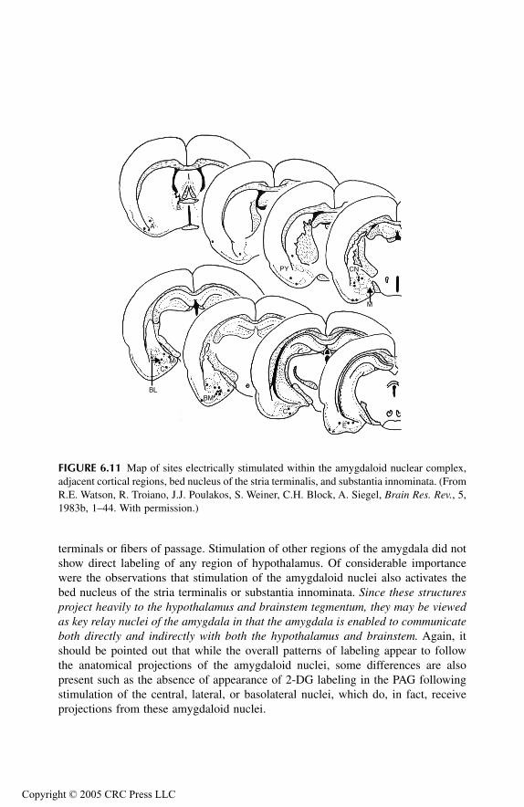
TF1661_C006.fm Page 139 Monday, October 18, 2004 8:20 AM
Copyr
terminals or fibers of passage. Stimulation of other regions of the amygdala did notshow direct labeling of any region of hypothalamus. Of considerable importancewere the observations that stimulation of the amygdaloid nuclei also activates thebed nucleus of the stria terminalis or substantia innominata. Since these structuresproject heavily to the hypothalamus and brainstem tegmentum, they may be viewedas key relay nuclei of the amygdala in that the amygdala is enabled to communicateboth directly and indirectly with both the hypothalamus and brainstem. Again, itshould be pointed out that while the overall patterns of labeling appear to followthe anatomical projections of the amygdaloid nuclei, some differences are alsopresent such as the absence of appearance of 2-DG labeling in the PAG followingstimulation of the central, lateral, or basolateral nuclei, which do, in fact, receiveprojections from these amygdaloid nuclei.
FIGURE 6.11 Map of sites electrically stimulated within the amygdaloid nuclear complex,adjacent cortical regions, bed nucleus of the stria terminalis, and substantia innominata. (FromR.E. Watson, R. Troiano, J.J. Poulakos, S. Weiner, C.H. Block, A. Siegel, Brain Res. Rev., 5,1983b, 1–44. With permission.)
B
A
PY CN
M
E
C
BM
L
BL
MC
ight © 2005 CRC Press LLC
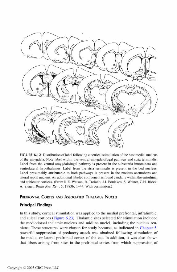
TF1661_C006.fm Page 140 Monday, October 18, 2004 8:20 AM
Copyr
PREFRONTAL CORTEX AND ASSOCIATED THALAMUS NUCLEI
Principal Findings
In this study, cortical stimulation was applied to the medial prefrontal, infralimbic,and sulcal cortices (Figure 6.23). Thalamic sites selected for stimulation includedthe mediodorsal thalamic nucleus and midline nuclei, including the nucleus reu-niens. These structures were chosen for study because, as indicated in Chapter 5,powerful suppression of predatory attack was obtained following stimulation ofthe medial or lateral prefrontal cortex of the cat. In addition, it was also shownthat fibers arising from sites in the prefrontal cortex from which suppression of
FIGURE 6.12 Distribution of label following electrical stimulation of the basomedial nucleusof the amygdala. Note label within the ventral amygdalofugal pathway and stria terminalis.Label from the ventral amygdalofugal pathway is present in the substantia innominata andventrolateral hypothalamus. Label from the stria terminalis is present in the bed nucleus.Label presumably attributable to both pathways is present in the nucleus accumbens andlateral septal nucleus. An additional labeled component is found caudally within the entorhinaland subicular cortices. (From R.E. Watson, R. Troiano, J.J. Poulakos, S. Weiner, C.H. Block,A. Siegel, Brain Res. Rev., 5, 1983b, 1–44. With permission.)
ACS
VMBL
MBM
L
CNP
B
SI
ST
PYC
LH
ESB
ight © 2005 CRC Press LLC

TF1661_C006.fm Page 141 Monday, October 18, 2004 8:20 AM
Copyr
attack had been elicited projected to the mediodorsal nucleus, and that fibers fromthe mediodorsal nucleus pass to the anterior lateral hypothalamus via a chain ofneurons located in the midline thalamus. Thus, this study was designed to deter-mine whether similar pathways would be activated following stimulation of theprefrontal cortex and medial thalamic nuclei, or whether different pathways wouldbe involved by stimulation of these sites.
FIGURE 6.13 Distribution of label following electrical stimulation of the amygdalo-hippo-campal area. Label is present primarily along the stria terminalis and its primary target nuclei— the bed nucleus of the stria terminalis and anteromedial hypothalamus. Label is also presentin the substantia innominata, nucleus accumbens, and lateral septal nucleus. Caudally, labelis present over the subiculum and adjacent entorhinal cortex.
AC
S
D
B
MBM
BL
CN
L
PSIA
CSB
E
ight © 2005 CRC Press LLC
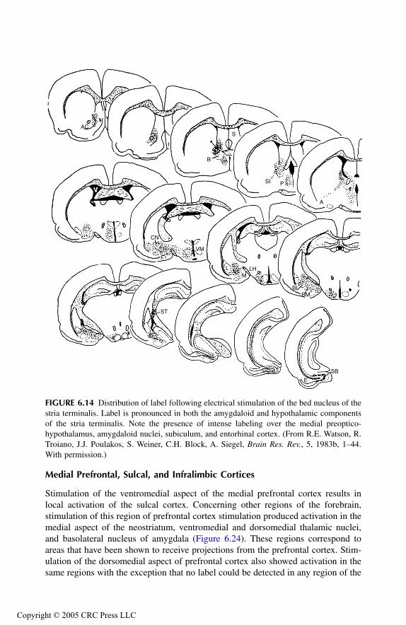
TF1661_C006.fm Page 142 Monday, October 18, 2004 8:20 AM
Copyr
Medial Prefrontal, Sulcal, and Infralimbic Cortices
Stimulation of the ventromedial aspect of the medial prefrontal cortex results inlocal activation of the sulcal cortex. Concerning other regions of the forebrain,stimulation of this region of prefrontal cortex stimulation produced activation in themedial aspect of the neostriatum, ventromedial and dorsomedial thalamic nuclei,and basolateral nucleus of amygdala (Figure 6.24). These regions correspond toareas that have been shown to receive projections from the prefrontal cortex. Stim-ulation of the dorsomedial aspect of prefrontal cortex also showed activation in thesame regions with the exception that no label could be detected in any region of the
FIGURE 6.14 Distribution of label following electrical stimulation of the bed nucleus of thestria terminalis. Label is pronounced in both the amygdaloid and hypothalamic componentsof the stria terminalis. Note the presence of intense labeling over the medial preoptico-hypothalamus, amygdaloid nuclei, subiculum, and entorhinal cortex. (From R.E. Watson, R.Troiano, J.J. Poulakos, S. Weiner, C.H. Block, A. Siegel, Brain Res. Rev., 5, 1983b, 1–44.With permission.)
AC
B
S
SI P
A
BMBL
LHM
L
VM
CN
ST
E
SB
ight © 2005 CRC Press LLC

TF1661_C006.fm Page 143 Monday, October 18, 2004 8:20 AM
Copyr
amygdala (Figure 6.25). Stimulation of the sulcal cortex, shown in Figure 6.26,resulted in a slightly different pattern of activation. The primary regions activatedby sulcal cortical stimulation included the medial prefrontal cortex, medial septal-diagonal band region, ventromedial thalamic nucleus (but not the dorsomedialnucleus), and prepyriform and pyriform cortices. Stimulation of the infralimbiccortex resulted in labeling throughout much of the septal area and mediodorsalthalamic nucleus. Diffuse labeling was noted over much of the caudate nucleus andnucleus accumbens (Figure 6.27). No label could be detected in the amygdala orany part of the hypothalamus. Thus, from the studies described above, it may beconcluded that the primary regions activated by prefrontal cortical stimulationinclude the neostriatum and the medial thalamus. In the studies described below,the regions activated following stimulation of the mediodorsal thalamic nucleus andmidline nuclei are identified.
FIGURE 6.15 Distribution of label following electrical stimulation of the anterior amygda-loid area. Labeling throughout the forebrain is sparse, and is restricted to the basolateralamygdala, adjacent prepyriform area, and the temporal cortical component of the anteriorcommissure. (From R.E. Watson, R. Troiano, J.J. Poulakos, S. Weiner, C.H. Block, A. Siegel,Brain Res. Rev., 5, 1983b, 1–44. With permission.)
AC
S
P
ACM
CN
M
BM
PY
SIA
L
BLC
ight © 2005 CRC Press LLC
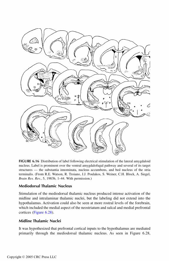
TF1661_C006.fm Page 144 Monday, October 18, 2004 8:20 AM
Copyr
Mediodorsal Thalamic Nucleus
Stimulation of the mediodorsal thalamic nucleus produced intense activation of themidline and intralaminar thalamic nuclei, but the labeling did not extend into thehypothalamus. Activation could also be seen at more rostral levels of the forebrain,which included the medial aspect of the neostriatum and sulcal and medial prefrontalcortices (Figure 6.28).
Midline Thalamic Nuclei
It was hypothesized that prefrontal cortical inputs to the hypothalamus are mediatedprimarily through the mediodorsal thalamic nucleus. As seen in Figure 6.28,
FIGURE 6.16 Distribution of label following electrical stimulation of the lateral amygdaloidnucleus. Label is prominent over the ventral amygdalofugal pathway and several of its targetstructures — the substantia innominata, nucleus accumbens, and bed nucleus of the striaterminalis. (From R.E. Watson, R. Troiano, J.J. Poulakos, S. Weiner, C.H. Block, A. Siegel,Brain Res. Rev., 5, 1983b, 1–44. With permission.)
SC
AC S
D
CN
LH
L
P
A
PY
BSI
M
BL
BM C
E
SB
ight © 2005 CRC Press LLC
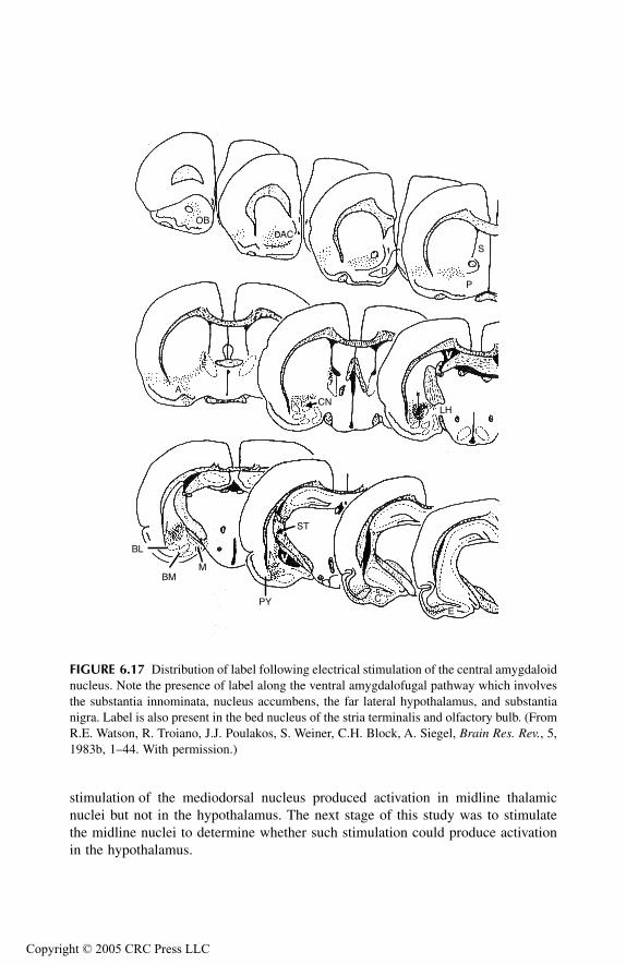
TF1661_C006.fm Page 145 Monday, October 18, 2004 8:20 AM
Copyr
stimulation of the mediodorsal nucleus produced activation in midline thalamicnuclei but not in the hypothalamus. The next stage of this study was to stimulatethe midline nuclei to determine whether such stimulation could produce activationin the hypothalamus.
FIGURE 6.17 Distribution of label following electrical stimulation of the central amygdaloidnucleus. Note the presence of label along the ventral amygdalofugal pathway which involvesthe substantia innominata, nucleus accumbens, the far lateral hypothalamus, and substantianigra. Label is also present in the bed nucleus of the stria terminalis and olfactory bulb. (FromR.E. Watson, R. Troiano, J.J. Poulakos, S. Weiner, C.H. Block, A. Siegel, Brain Res. Rev., 5,1983b, 1–44. With permission.)
OB
AC
D
S
P
LHCN
A
BL
BMM
ST
PY CE
ight © 2005 CRC Press LLC
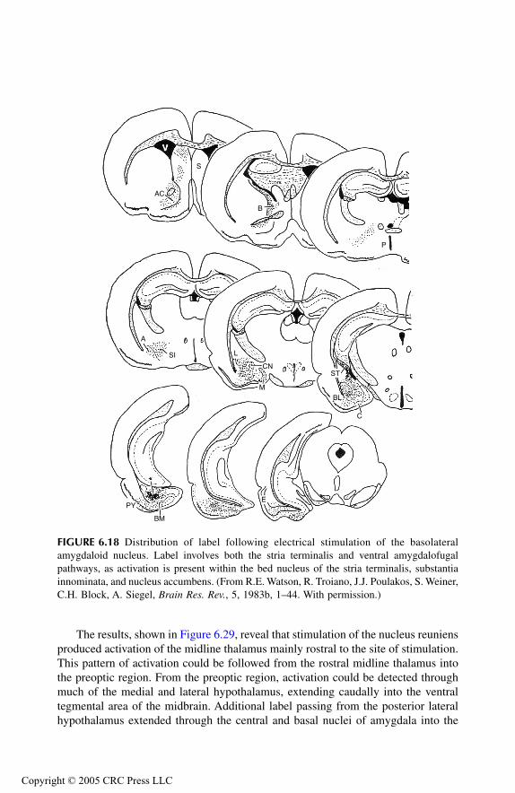
TF1661_C006.fm Page 146 Monday, October 18, 2004 8:20 AM
Copyr
The results, shown in Figure 6.29, reveal that stimulation of the nucleus reuniensproduced activation of the midline thalamus mainly rostral to the site of stimulation.This pattern of activation could be followed from the rostral midline thalamus intothe preoptic region. From the preoptic region, activation could be detected throughmuch of the medial and lateral hypothalamus, extending caudally into the ventraltegmental area of the midbrain. Additional label passing from the posterior lateralhypothalamus extended through the central and basal nuclei of amygdala into the
FIGURE 6.18 Distribution of label following electrical stimulation of the basolateralamygdaloid nucleus. Label involves both the stria terminalis and ventral amygdalofugalpathways, as activation is present within the bed nucleus of the stria terminalis, substantiainnominata, and nucleus accumbens. (From R.E. Watson, R. Troiano, J.J. Poulakos, S. Weiner,C.H. Block, A. Siegel, Brain Res. Rev., 5, 1983b, 1–44. With permission.)
S
AC
B
P
SI
A
L
CN
M
ST
BL
C
E
BM
PY
ight © 2005 CRC Press LLC
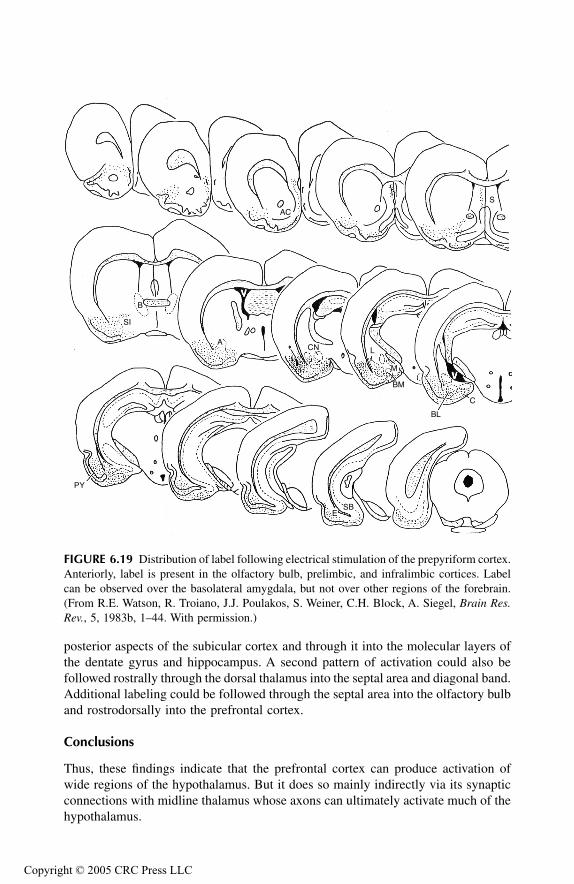
TF1661_C006.fm Page 147 Monday, October 18, 2004 8:20 AM
Copyr
posterior aspects of the subicular cortex and through it into the molecular layers ofthe dentate gyrus and hippocampus. A second pattern of activation could also befollowed rostrally through the dorsal thalamus into the septal area and diagonal band.Additional labeling could be followed through the septal area into the olfactory bulband rostrodorsally into the prefrontal cortex.
Conclusions
Thus, these findings indicate that the prefrontal cortex can produce activation ofwide regions of the hypothalamus. But it does so mainly indirectly via its synapticconnections with midline thalamus whose axons can ultimately activate much of thehypothalamus.
FIGURE 6.19 Distribution of label following electrical stimulation of the prepyriform cortex.Anteriorly, label is present in the olfactory bulb, prelimbic, and infralimbic cortices. Labelcan be observed over the basolateral amygdala, but not over other regions of the forebrain.(From R.E. Watson, R. Troiano, J.J. Poulakos, S. Weiner, C.H. Block, A. Siegel, Brain Res.Rev., 5, 1983b, 1–44. With permission.)
AC
S
C
BL
M
BM
LCNA
B
SI
PY
ESB
ight © 2005 CRC Press LLC

TF1661_C006.fm Page 148 Monday, October 18, 2004 8:20 AM
Copyr
OVERALL CONCLUSIONS
Results of the 2-DG studies reinforce the conclusions suggested in Chapter 5 con-cerning a basic functional property of the limbic system — its relationship to thehypothalamus. The feature common to each of the limbic structures is that whenstimulation is applied, there is subsequent direct or indirect activation of the hypo-thalamus. These findings thus provide further evidence that each of these limbicstructures can modulate hypothalamic functions (such as aggression and rage) and,in addition, they identify the pathways that likely mediate these effects.
FIGURE 6.20 Distribution of label following electrical stimulation of the entorhinal cortex.Label is prominent over the basolateral nucleus, subiculum, and to a lesser extent, the septalarea. (From R.E. Watson, R. Troiano, J.J. Poulakos, S. Weiner, C.H. Block, A. Siegel, BrainRes. Rev., 5, 1983b, 1–44. With permission.)
B
S
C
BM
M
PY
C
CN
L
BL
E SB
ight © 2005 CRC Press LLC
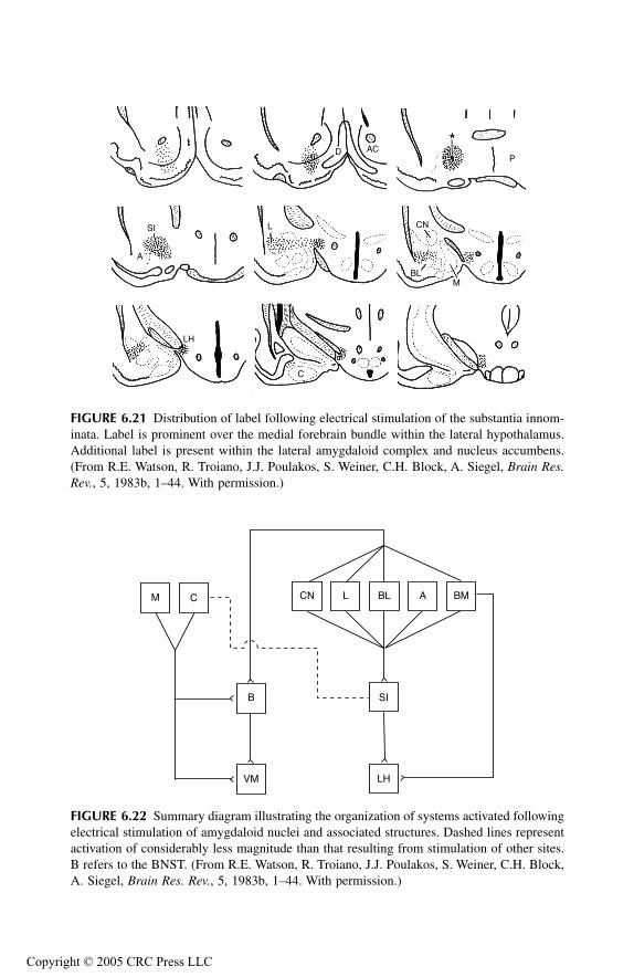
TF1661_C006.fm Page 149 Monday, October 18, 2004 8:20 AM
Copyr
FIGURE 6.21 Distribution of label following electrical stimulation of the substantia innom-inata. Label is prominent over the medial forebrain bundle within the lateral hypothalamus.Additional label is present within the lateral amygdaloid complex and nucleus accumbens.(From R.E. Watson, R. Troiano, J.J. Poulakos, S. Weiner, C.H. Block, A. Siegel, Brain Res.Rev., 5, 1983b, 1–44. With permission.)
FIGURE 6.22 Summary diagram illustrating the organization of systems activated followingelectrical stimulation of amygdaloid nuclei and associated structures. Dashed lines representactivation of considerably less magnitude than that resulting from stimulation of other sites.B refers to the BNST. (From R.E. Watson, R. Troiano, J.J. Poulakos, S. Weiner, C.H. Block,A. Siegel, Brain Res. Rev., 5, 1983b, 1–44. With permission.)
ACP
D
A
SI L CN
BLM
C
LH
M C CN L BL A BM
SIB
VM LH
ight © 2005 CRC Press LLC

TF1661_C006.fm Page 150 Monday, October 18, 2004 8:20 AM
Copyr
FIGURE 6.23 Map of sites stimulated in the prefrontal and adjacent cortical tissue and inassociated portions of the medial thalamus, including the mediodorsal nucleus and nucleusreuniens. (From M. Brutus, R.E. Watson, M.B. Shaikh, H.E. Siegel, S. Weiner, A. Siegel,Brain Res., 310, 1984, 279–293. With permission.)
a b c d
e f g h
PC
SC
ILAV
RE
AM
MDV
ight © 2005 CRC Press LLC

TF1661_C006.fm Page 151 Monday, October 18, 2004 8:20 AM
Copyr
FIGURE 6.24 Distribution of label following electrical stimulation of the ventromedialaspect of the prefrontal cortex. Note the presence of label within adjacent regions of sulcalcortex, dorsomedial aspect of the head of the caudate nucleus, basolateral amygdaloid nucleus,medial segment of the mediodorsal nucleus, and reticular thalamic nucleus. No label wasobserved in any region of the preoptico-hypothalamus. (From M. Brutus, R.E. Watson, M.B.Shaikh, H.E. Siegel, S. Weiner, A. Siegel, Brain Res., 310, 1984, 279–293. With permission.)
PC
SC AC
SM
ICCP
AV
R
MD
PT
VMT
HBPF
BL
ight © 2005 CRC Press LLC
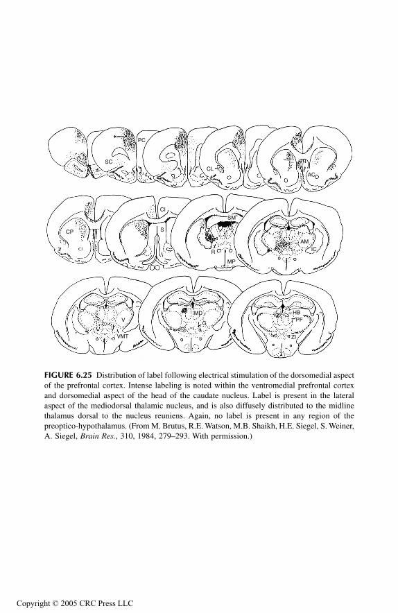
TF1661_C006.fm Page 152 Monday, October 18, 2004 8:20 AM
Copyr
FIGURE 6.25 Distribution of label following electrical stimulation of the dorsomedial aspectof the prefrontal cortex. Intense labeling is noted within the ventromedial prefrontal cortexand dorsomedial aspect of the head of the caudate nucleus. Label is present in the lateralaspect of the mediodorsal thalamic nucleus, and is also diffusely distributed to the midlinethalamus dorsal to the nucleus reuniens. Again, no label is present in any region of thepreoptico-hypothalamus. (From M. Brutus, R.E. Watson, M.B. Shaikh, H.E. Siegel, S. Weiner,A. Siegel, Brain Res., 310, 1984, 279–293. With permission.)
SC
PC
CLAC
AMIC
MP
R
SM
CI
SCP
V
VMT
MD
G
HBPF
ZI
ight © 2005 CRC Press LLC
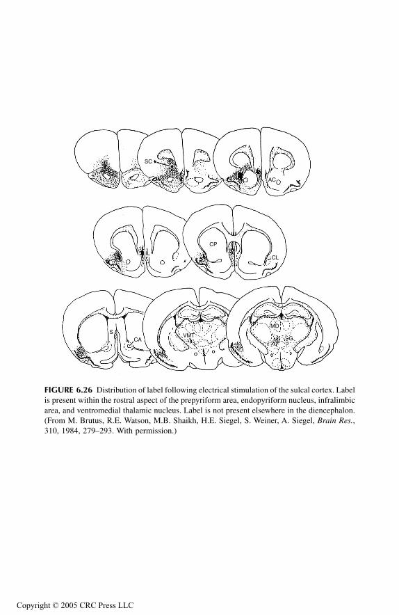
TF1661_C006.fm Page 153 Monday, October 18, 2004 8:20 AM
Copyr
FIGURE 6.26 Distribution of label following electrical stimulation of the sulcal cortex. Labelis present within the rostral aspect of the prepyriform area, endopyriform nucleus, infralimbicarea, and ventromedial thalamic nucleus. Label is not present elsewhere in the diencephalon.(From M. Brutus, R.E. Watson, M.B. Shaikh, H.E. Siegel, S. Weiner, A. Siegel, Brain Res.,310, 1984, 279–293. With permission.)
SC
AC
IL
CP
CL
CAS
VMT
MD
G
ight © 2005 CRC Press LLC
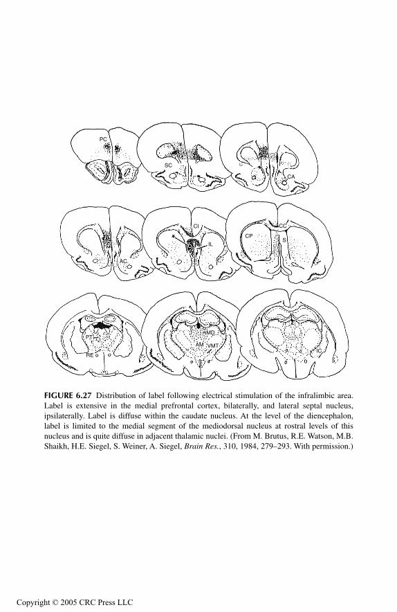
TF1661_C006.fm Page 154 Monday, October 18, 2004 8:20 AM
Copyr
FIGURE 6.27 Distribution of label following electrical stimulation of the infralimbic area.Label is extensive in the medial prefrontal cortex, bilaterally, and lateral septal nucleus,ipsilaterally. Label is diffuse within the caudate nucleus. At the level of the diencephalon,label is limited to the medial segment of the mediodorsal nucleus at rostral levels of thisnucleus and is quite diffuse in adjacent thalamic nuclei. (From M. Brutus, R.E. Watson, M.B.Shaikh, H.E. Siegel, S. Weiner, A. Siegel, Brain Res., 310, 1984, 279–293. With permission.)
PC
SC
CA
CC
SCP
IL
CI
AC
RE
PTMD
AM VMTIC
ight © 2005 CRC Press LLC
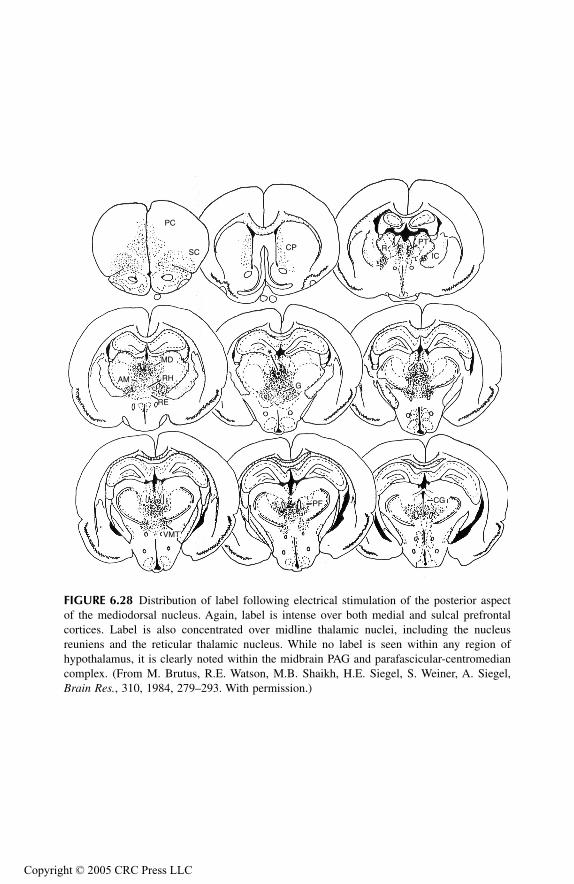
TF1661_C006.fm Page 155 Monday, October 18, 2004 8:20 AM
Copyr
FIGURE 6.28 Distribution of label following electrical stimulation of the posterior aspectof the mediodorsal nucleus. Again, label is intense over both medial and sulcal prefrontalcortices. Label is also concentrated over midline thalamic nuclei, including the nucleusreuniens and the reticular thalamic nucleus. While no label is seen within any region ofhypothalamus, it is clearly noted within the midbrain PAG and parafascicular-centromediancomplex. (From M. Brutus, R.E. Watson, M.B. Shaikh, H.E. Siegel, S. Weiner, A. Siegel,Brain Res., 310, 1984, 279–293. With permission.)
PC
SCCP R
PT
IC
G
MD
RH
RE
AM
VMT
PF CG
ight © 2005 CRC Press LLC

TF1661_C006.fm Page 156 Monday, October 18, 2004 8:20 AM
Copyr
FIGURE 6.29 Distribution of label following electrical stimulation of the nucleus reuniens.Note the presence of intense label over the medial prefrontal cortex, area infraradiata, olfactorybulbs, septal area, diagonal band nuclei, anterior thalamic nuclear complex, amygdalo-hip-pocampal area, basolateral amygdaloid nucleus, and midbrain midbrain periaqueductal gray.Significantly, label is extensive throughout both lateral and medial preoptico-hypothalamus,indicating the existence of a massive functional projection from the thalamus to the hypo-thalamus (and indirectly from the prefrontal cortex) that likely modulates functions of thehypothalamus such as aggression and rage. (From M. Brutus, R.E. Watson, M.B. Shaikh,H.E. Siegel, S. Weiner, A. Siegel, Brain Res., 310, 1984, 279–293. With permission.)
PC
OB
IR
IL
AC
CL
S
CI
FX
PY
MDAM
VMT
IC RE
PG
AVRPT
LP
B
MP
LT
G
LH
VM
ZIPH
ST
ME
CA1
CA3
FDG
CG
VTAHA
RS
PS
E
DS
VS
CA1
DG
ight © 2005 CRC Press LLC

TF1661_C006.fm Page 157 Monday, October 18, 2004 8:20 AM
Copyr
ABBREVIATIONS USED IN FIGURES
A, anterior hypothalamic areaAC, or CA, anterior commissure or nucleus accumbensACM, anterior commissureAH, anterior hypothalamusAHA, amygdalo-hippocampal areaAM, anteromedial thalamic nucleusAR, arcuate nucleus of hypothalamusAV, anteroventral thalamic nucleusB, bed nucleus of stria terminalisBL, basolateral nucleus of amygdalaBM, basomedial nucleus of amygdalaCA1, CA3, cornu Ammonis fields of hippocampusC, cortical nucleus of amygdalaCC, corpus callosumCG, midbrain periaqueductal, central, grayCI, cingulate cortexCL, claustrumCN, central nucleus of amygdalaCP, caudate-putamen, neostriatumD or DB, diagonal band of BrocaDF, dorsal fornixDG, dentate gyrus of hippocampusDH, dorsal hypothalamusDS, dorsal subiculumE, entorhinal cortexF or FX, fornixFI, fimbriaG, nucleus gelatinosus of thalamusHB, habenular complexIC, internal capsuleIL, infralimbic cortexIP, interpeduncular nucleusIR, infraradiate cortexL, lateral nucleus of amygdalaLH, lateral hypothalamusLP, lateral preoptic areaLS, lateral septal nucleusLT, lateral thalamic nucleusM or MM, mammillary bodiesM or ME, medial nucleus of amygdalaMD, mediodorsal thalamic nucleusME, median eminenceMP, medial preoptic areaMS, medial septal nucleus
ight © 2005 CRC Press LLC
TF1661_C006.fm Page 158 Monday, October 18, 2004 8:20 AM
Copyr
MT, mammillothalamic tractOB, olfactory bulbP, preoptic areaPAR, parasubiculumPC, prefrontal cortex, medial aspectPF, parafascicular thalamic nucleusPH, posterior hypothalamusPL, prelimbic cortexPRO, prosubiculumPS, posterior subiculumPT, parataenial thalamic nucleusPV, periventricular gray of hypothalamusPY, pyriform cortexR, reticular thalamic nucleusRE, nucleus reuniensRH, rhomboidal thalamic nucleusRS, retrosplenial cortexS, septal areaSB, subiculumSC, suprachiasmatic nucleus or sulcal cortexSF, septofimbrial nucleusSI, substantia innominataSM, stria medullarisST, stria terminalisV, ventricle or ventral thalamic nucleusVC, ventral hippocampal commissureVM, ventromedial nucleus of hypothalamusVMT, ventromedial thalamic nucleusVS, ventral subiculumZI, zona incerta
REFERENCES
Brutus, M., R.E. Watson, M.B. Shaikh, H.E. Siegel, S. Weiner, A. Siegel, A (14C)2-deoxy-glucose analysis of the functional neural pathways of the limbic forebrain in the rat.IV. A pathway from the prefrontal cortical-medial thalamic system to the hypothal-amus, Brain Res., 310, 1984, 279–293.
Meibach, R. and A. Siegel, Efferent connections of the hippocampal formation in the rat,Brain Res., 124, 1977, 197–224.
Siegel, A., H. Edinger, The organization of the hippocampal septal axis, in J. De France, Ed.,International Symposium on the Septal Nuclei, Wayne State University Press, Detroit,Mich., 1976, pp. 241–250.
Siegel, A., H. Edinger, S. Ohgami, The topographical organization of the hippocampal pro-jection to the septal area: a comparative neuroanatomical analysis in the gerbil, rat,rabbit and cat, J. Comp. Neurol., 157, 1974, 359–378.
ight © 2005 CRC Press LLC
TF1661_C006.fm Page 159 Monday, October 18, 2004 8:20 AM
Copyr
Siegel, A., S. Ohgami, H. Edinger, The topographical organization of the hippocampal pro-jection to the septal area in the squirrel monkey, Brain Res., 99, 1975, 247–260.
Siegel, A. and J.P. Tassoni, Differential efferent projections from the ventral and dorsalhippocampus of the cat, Brain Behav. Evol., 4, 1971, 185–200.
Watson, R.E., H.E. Edinger, A. Siegel, A 14C-2-deoxyglucose analysis of the functional neuralpathways of the limbic forebrain in the rat: III. The hippocampal formation, BrainRes. Rev., 5, 1983a, 133–176.
Watson, R.E., A. Siegel, H.E. Siegel, A (14C)2-deoxyglucose analysis of the functional neuralpathways of the limbic forebrain in the rat: V. The septal area, Brain Res., 346, 1985,89–107.
Watson, R.E., R. Troiano, J.J. Poulakos, S. Weiner, C.H. Block, A. Siegel, A (14C)2-deoxy-glucose analysis of the functional neural pathways of the limbic forebrain in the rat:I. The amygdala, Brain Res. Rev., 5, 1983b, 1–44.
Watson, R.E., R. Troiano, J.J. Poulakos, S. Weiner, A. Siegel, A 14 C-2-deoxyglucose analysisof the neural pathways of the limbic forebrain in the rat: II. The hypothalamus, BrainRes. Bull., 8, 1982, 459–476.
ight © 2005 CRC Press LLC

TF1661_C007.fm Page 161 Monday, October 18, 2004 8:35 AM
Copyr
7
The Neurochemistry of Rage and AggressionOf all the areas of investigation concerning the neurobiology of aggression and rage,studies involving neurochemistry and neuropharmacology have attracted the lion’sshare of attention as seen in the large numbers of research papers published in thisfield. A goal of this chapter is to provide a representative sampling of the literature,which typically reflects the major conclusions regarding the relationships of thevarious neurotransmitters to aggression and rage.
Investigators have used diverse approaches in their attempts to relate neurochem-ical processes and mechanisms to states of rage and aggression. The most commonapproach has been to examine the effects on aggression of peripheral administrationof a drug, which could be a specific or nonspecific receptor agonist or antagonist,or some other class of drug, such as a tranquilizer, anxiolytic, hallucinogen, or drugof abuse. Related approaches that have been used less frequently involved studiestesting the effects of similar drugs when applied intracerebroventricularly or intrac-erebrally into specific brain sites.
Another approach involved the attempt to determine how aggressive reactionsmay alter brain chemistry. Initially, these studies examined whole brain neurochem-ical changes following aggressive encounters. Later studies used more refined meth-ods such as the “punch” technique to sample tissue from selected regions of thebrain; and, most recently, there has been some attempt to use microdialysis to analyzethe effects of aggressive responses on highly localized regions of the brain. Anadditional approach has been to use newer genetic methods involving transgenicanimals such as mice in which the gene expressing a specific neurotransmitterreceptor is made defective. Such a preparation is referred to as a “knockout” andthe role of the receptor in question has been suggested by the extent to which theexperimental preparation displays aggression relative to control animals.
My objective in this chapter is to review and summarize the findings of studiesrepresentative of the research strategies described above with respect to the differentneurotransmitters and various classes of drugs. Two basic questions addressedinclude: (1) What are the roles of different neurotransmitters and their receptors?(2) Where in the brain do they act in regulating aggression and rage?
In attempting to answer these questions, the strengths as well as weaknesses ofthese approaches are briefly noted here. Concerning the use of systemic injectionsof drugs, one positive feature is that it provides an overall assessment of the role ofthe neurotransmitter in question in aggressive behavior. A second advantage is thatit follows the clinical route of drugs such as carbamazepine, anxiolytic, and antide-pressive compounds. However, this approach raises several concerns. One is that itis not readily discernible whether the effects of drug administration by this route
ight © 2005 CRC Press LLC

TF1661_C007.fm Page 162 Monday, October 18, 2004 8:35 AM
Copyr
are mediated by peripheral or central mechanisms. A second issue is that systemicadministration of drugs provides little or no information concerning where in thebrain the drug may be acting to produce its effects on aggressive behavior. A thirdconcern is the specificity of the effects resulting from systemic administration ofdrugs. In particular, if the drug suppresses a given form of aggression, a number ofcontrol experiments are required to determine whether the suppressive effects gen-erated are specific to the form of aggressive behavior examined, or whether theeffects are nonspecific and common to wide varieties of behaviors.
There is an important advantage associated with the approach using microinjec-tions of drugs into specific regions of the brain. Specifically, it may enable theinvestigator to identify the receptor–neurotransmitter mechanism operative at a givensite in the brain to alter a behavioral process. However, similar to the problemsassociated with systemic administration of drugs, other concerns relate to intrace-rebral administration of drugs as well that require the employment of careful controlsin the design of the experiment. For example, one aspect of the experimental designshould be included to control for difficulties in interpretation of the data due todiffusion of the drug from the site of delivery. This may be achieved by the placementof microinjections into neighboring regions and by comparing the effects obtainedfrom microinjections at this site with those obtained following microinjections intothe original site of interest. A second concern, similar to that described above forperipheral administration of drugs, is that the compound may have a nonspecificeffect on the behavioral response under examination, such as causing general inhi-bition of all motor responses. Here, it would be useful for the investigator to comparethe effects of intracerebral drug delivery on several different types of behavior inorder to rule out this possibility. A third concern is the limited selectivity of variousreceptor agonists and antagonists, especially when used at higher doses where theselective properties can be somewhat diminished. Investigators can cope with thisissue through the generation of dose–response curves and by pretreating a given sitewith a selective receptor antagonist prior to administration of the selective agonistfor that drug.
ACETYLCHOLINE
SYNTHESIS AND REMOVAL
Acetylcholine (ACh) is a small-molecule neurotransmitter. It is synthesized fromacetylcoenzyme-A and choline in the following way. Initially, pyruvate is derivedfrom glucose (which reaches the nerve terminal by passive transport) by glycolysisand is transported into the mitochondria. Acetylcoenzyme-A is formed from thecombination of coenzyme-A found in the mitochondria and an acetyl group derivedfrom pyruvic acid, and is transported back into the cytoplasm. The second compo-nent, choline, is present in the plasma and enters the nerve terminal via activetransport. The synthesis of choline and acetylcoenzyme-A takes place in the cyto-plasm of the nerve terminal by the enzyme, choline acetyltransferase. ACh is thentransported into the synaptic vesicles where it is stored.
ight © 2005 CRC Press LLC
TF1661_C007.fm Page 163 Monday, October 18, 2004 8:35 AM
Copyr
ACh is removed as a result of the actions of the enzyme, acetylcholinesterase,which is synthesized in the endoplasmic reticulum of cell body, and is found on theouter surface of the nerve terminal. Acetylcholinesterase then hydrolyzes ACh inthe extracellular spaces, thus reducing its concentration, and leading to dissociationfrom its receptor-binding site. Choline is now released, which enables it to re-enterthe nerve and be used again for the synthesis of ACh.
DISTRIBUTION AND FUNCTIONS
Cholinergic neurons are found widely throughout the brain and spinal cord. Exam-ples of specific groups of neurons have been clearly found in such regions as thebasal nucleus of Meynert of rostral forebrain, pedunculopontine region of the pontinetegmentum, cerebral cortex, neostriatum, hippocampal formation, and ventral horncells of the spinal cord. ACh is generally considered to be an excitatory neurotrans-mitter and plays an important role in the sleep–wakefulness cycle, visceral functionsinvolving the hypothalamus and limbic structures, as well as certain disease statessuch as in Alzheimer’s disease, which is linked to atrophy of basal forebrain cho-linergic neurons and ACh levels in the cortex.
ROLE OF ACH IN AGGRESSION AND RAGE
Effects of Systemic Administration of Cholinergic Agents
A number of early studies used the systemic administration of cholinergic drugs inorder to determine the role of ACh in aggressive behavior. Most of these studieswere conducted in either rodents or cats, and provide evidence that cholinergic agentsgenerally have facilitatory effects on rage and rage-related processes. This is notparticularly surprising in view of the fact that, as indicated above, ACh is viewedas an excitatory neurotransmitter.
In studies conducted in mice, muscarinic antagonists atropine or scopolaminereduce or eliminate threat attack (Valzelli et al., 1967; Barnett et al., 1971). A parallelstudy conducted in rats showed similar results following administration of scopola-mine (Van der Poel and Remmelts, 1971). However, not all experiments yieldedconsistent findings. When rats were tested for defensive behavior after exposure toa predator cat and scopolamine delivery, major changes were not apparent (Rodgerset al., 1990). These authors concluded that there is a lack of significant involvementof muscarinic mechanisms in the regulation of defensive patterns in rats. A similarexperiment conducted in the cat revealed that with respect to predatory attack,scopolamine administration causes an elevation of the threshold for this response(Katz and Thomas, 1975).
Consistent with the general pattern of the findings described above, a numberof studies examined the effects of systemic administration of cholinergic agonistson aggressive behavior. Gay and Leaf (1976) administered pilocarpine, a potentmuscarinic agonist, and observed an induction in mouse killing in different strainsof rats. Likewise, systemic injections of muscarinic agonists in facilitated defensiverage in the cat (George et al., 1962; Zablocka and Esplin, 1964; Leslie, 1965).
ight © 2005 CRC Press LLC
TF1661_C007.fm Page 164 Monday, October 18, 2004 8:35 AM
Copyr
In addition, Berntson and Leibowitz (1973, 1976) also observed that administrationof the muscarinic agonist, arecoline, can induce components of both biting attackand defensive rage in the cat.
Effects of Intracerebral Microinjections of Cholinergic Agents
The second approach used to examine the effects of acetylcholine has been tomicroinject cholinergic compounds into specific regions of the brain normally asso-ciated with aggression and rage. In most cases, the primary target has been thehypothalamus. Here, the basic principle tested was that aggression and rage behaviorcan be induced or altered following cholinergic receptor activation or blockage inselective regions of the brain.
A number of studies conducted mainly in rats and cats examined the effects ofcentral cholinergic receptor activation on aggression or rage responses. One commonapproach has been to microinject the muscarinic cholinergic agonist, carbachol, intospecific regions of the hypothalamus or neighboring aspects of the forebrain. Oneproblem of interpretation of experiments that examine the effects of administrationof receptor agonists on a given behavioral response should be noted here. Receptor-mediated changes in a behavioral response do not demonstrate the presence of aspecific neurotransmitter, but only of the presence of the receptor in question andhow it may affect the behavioral response under examination. Nevertheless, studiesusing such an approach provide useful and perhaps important data concerning thefunctional relationships of the receptor under consideration to specific forms ofaggression. The remainder of this subsection summarizes a variety of studies thatprovide support for a role of ACh in mediating aggression and rage.
Several reports have shown that microinjections of carbachol into the anteriorhypothalamus of the rat can induce vocalizations similar to those that follow footshock (Brudzynski and Bihara, 1990; Brudzynski et al., 1991; Brudzynski, 1994).The authors indicated that the vocalizations represented forms of emotional excite-ment. Such excitement could be interpreted to reflect anxiety, distress or perhaps acomponent of defensive behavior. Several investigators have demonstrated thatmicroinjections of carbachol can also induce mouse killing in rats (Smith et al.,1970). Moreover, administration of atropine was shown to block predatory killingin killer rats (Smith et al., 1970).
Other investigators have attempted to link cholinergic receptor activation orblockade in several limbic structures to the regulation of aggression in the rat. Forexample, cholinergic stimulation of the septal area produces an increase in irritabilityin rats (Igic et al., 1970). In contrast, administration of scopolamine into the medialamygdala was shown to block mouse killing (Leaf et al., 1969). Likewise, cholinergicblockade (with scopolamine) in the basolateral amygdala and hippocampal formationreduced fighting in rats (Rodgers and Brown, 1976).
Parallel studies have been conducted in the cat involving intracerebral microin-jections of cholinergic agents placed mainly into the hypothalamus. One of the firstinvestigators to demonstrate that cholinergic activation of the feline hypothalamuswith carbachol can induce defensive rage responses was Myers (1964). Baxter(1968a, 1968b) expanded on these findings by first identifying hypothalamic
ight © 2005 CRC Press LLC
TF1661_C007.fm Page 165 Monday, October 18, 2004 8:35 AM
Copyr
defensive rage sites in the cat by electrical stimulation and then obtained the sameresponses after microinjecting carbachol into the same sites. Similar observationswere made in subsequent years by other investigators such as Allikmets (1974),Romaniuk et al. (1973, 1974), and later by Brudzynski (1981a, 1981b), Brudzynskiand Eckersdorf (1988), and Brudzynski et al. (1993) following cholinergic stimula-tion of either the hypothalamus or PAG. Several investigators characterized thereceptor properties of this response. Romaniuk et al. (1973) showed that pretreatmentwith the muscarinic antagonist, atropine, into the same medial hypothalamic sitewhere carbachol was injected, blocked the defensive rage response induced bycarbachol. However, the carbachol-induced rage response was not affected when thehypothalamic sites was pretreated with a nicotinic receptor antagonist (betamon).That the cholinergic-induced defensive rage response is mediated in part throughmuscarinic receptors was further supported by a subsequent study by Brudzynski(1981a) who showed that the rage response elicited by carbachol could not beinduced by nicotine.
The studies described above have examined the role of cholinergic receptors indefensive rage behavior induced directly by chemical stimulation of the medial hypo-thalamus. A somewhat different approach was used by Brudzynski et al. (1990) inorder to demonstrate the importance of muscarinic receptors in defensive rage. In thisstudy, defensive responses were induced by exposing a cat to a dog, and atropinesulfate was either administered systemically or injected bilaterally into the ventrome-dial aspect of the borderline region of the anterior hypothalamus and preoptic area.Pretreatment by the cholinergic antagonist was shown to block or suppress the naturallyoccurring defensive rage response, thus providing additional support for the role ofmuscarinic receptors in the expression of defensive rage behavior.
Genetic Approach
A different approach to the study of the role of cholinergic mechanisms in aggressionwas used by Pucilowski et al. (1991). These authors used two rodent lines. The first,called the Flinders Sensitive Line, were selectively bred for increased sensitivity tocholinergic agonists presumably due to increased muscarinic binding and ACh syn-thesis rates. The second, called the Flinders Resistant Line, appear to lack theseproperties. When these two groups of rats were compared with respect to shock-induced and apomorphine-induced fighting tests, the Flinders Sensitive Line wereconsiderably more aggressive. Interestingly, the authors also reported that theFlinders Sensitive Line rats displayed a higher pain threshold, indicating that onecould not attribute the increase in aggression to a greater sensitivity to pain. Thus,this finding provides additional evidence of cholinergic involvement in aggressivebehavior, which is consistent with the results of studies using classical pharmaco-logical approaches presented above.
SUMMARY
The studies described above offer data supporting a cholinergic (excitatory) mech-anism involved in the overall expression of defensive rage behavior. In particular,
ight © 2005 CRC Press LLC

TF1661_C007.fm Page 166 Monday, October 18, 2004 8:35 AM
Copyr
there is good evidence confirming the presence of muscarinic receptors in thisbehavior. That defensive rage behavior can be suppressed or eliminated by cholin-ergic receptor blockade provides support for the presence of cholinergic neurotrans-mitter function as well in this process. A central question that remains to be answeredis how cholinergic receptors and neurons are organized within the hypothalamusand elsewhere in the limbic-hypothalamic-midbrain axis in mediating the expressionof defensive rage behavior. In particular, it would be important to know whethercholinergic neurons in the medial hypothalamus serve as interneurons or whetherthey contain long-descending axons that directly mediate the output of defensiverage behavior.
NOREPINEPHRINE
SYNTHESIS AND REMOVAL
The stages of synthesis are briefly described here. Tyrosine (hydroxy-phenylalanine),present in food, is synthesized from phenylalanine. After tyrosine enters the neuronby active transport, it is first converted into dihydroxyphenalanine (DOPA) by theenzyme, tyrosine hydroxylase, and then converted into dopamine by the enzyme, L-amino acid decarboxylase (DOPA-decarboxylase). Dopamine is then actively trans-ported into storage vesicles where it is converted into norepinephrine by the enzyme,dopamine-β-hydroxylase (DBH). At this point, norepinephrine is released by exo-cytosis. After norepinephrine is released into the synaptic cleft, it is removed pri-marily by re-uptake into the synaptic terminal. Other mechanisms that somewhatcontribute include inactivation by catechol-O-methyltransferase (COMT) in theeffector cell or by destruction in the liver by COMT and monoamine oxidase (MAO).
DISTRIBUTION AND FUNCTIONS
The greatest concentration of noradrenergic neurons is located within the pons inthe locus ceruleus. Other groups of noradrenergic neurons are located in differentregions of the medulla. These neurons project to the forebrain, including the limbicsystem, diencephalon, and cerebral cortex. A well-known function of norepinephrineis that it is released from postganglionic sympathetic nerve terminals. Norepineph-rine plays important roles in many behavioral processes, such as in states of wake-fulness, cortical activation, and the treatment for depression.
ROLE OF NOREPINEPHRINE IN AGGRESSION AND RAGE
Effects of Systemic and Intracerebroventricular Administration of Noradrenergic Agents
A number of studies have been conducted in several species that have addressed thequestion of the relationship of norepinephrine to aggressive behavior. These studieshave addressed this problem through the use of peripheral or intracerebroventricular(icv) administration of drugs, which have either noradrenergic agonistic or antago-nistic properties. However, findings do not appear to be entirely consistent, especially
ight © 2005 CRC Press LLC
TF1661_C007.fm Page 167 Monday, October 18, 2004 8:35 AM
Copyr
when compared with other experiments using different methodological approaches(described below). A number of investigators employing systemic administration ofnoradrenergic agents have suggested that norepinephrine has an inhibitory effect onaggressive processes. For example, icv administration of norepinephrine (Geyer andSegal, 1974) seemed to suppress shock-induced fighting in rodents, and icv deliveryof 6-hydroxydopamine (6-OHDA), which depletes catecholamines, causes a perma-nent increase in irritability in rats. Likewise, chronic systemic treatment with meth-yldopa, which depletes norepinephrine (and presumably serotonin and dopamine),can induce aggressive behavior in mice (Dominic and Moore, 1971). In contrast,Kemble et al. (1991) showed that administration of the α2-adrenergic antagonist,yohimbine, inhibited isolation-induced aggression in mice. Likewise, several studieswhere amphetamine was administered facilitated brain stimulation–induced aggres-sion in cats (Sheard, 1967; Marini et al., 1979). Further confusing the picture herewas the observation that following catecholamine depletion by alpha methyl paraty-rosine (AMPT), reflexive biting remains intact (Katz and Thomas, 1975).
Neurochemical Measurements
One of the earlier approaches to the study of the relationship of neurotransmittersto aggressive behavior was to determine whether the expression of aggressive behav-ior could affect changes in regional neurotransmitter levels in the brain. In a seriesof experiments, Reis et al. (Reis and Gunne, 1965; Reis and Fuxe, 1968, 1969; Reis,1974) observed depletions of norepinephrine in the brainstem following the expres-sion of rage behavior in the cat, suggesting an inverse relationship between rage andnorepinephrine levels. In contrast, Lamprecht et al. (1972) reported that rats sub-jected to extended periods of immobilization stress were more aggressive as deter-mined from shock-induced fighting. Here, such animals revealed higher serum levelsof both dopamine beta hydroxylase as well as tyrosine hydroxylase, suggesting apossible relationship between catecholamine synthesis and aggressive behavior.
Other studies also examined the relationship of norepinephrine turnover andaggression. Welch and Welch (1965, 1968) showed that brain norepinephrine levelsof isolated mice (which tends to make them more aggressive) were higher than inaggregated ones. In addition, Eichelmann (1973) provided data consistent with thoseof Welch and Welch, suggesting that an increase in norepinephrine turnover rates isassociated with an increase in aggression as determined by the shock-induced aggres-sion paradigm. When aggression in rats is induced by olfactory bulbectomy (Ueki andYoshimura, 1981), there was an increase in norepinephrine content in the hypothalamusrelative to intact or sham operated rats. However, changes in regional brain norepi-nephrine levels as a result of fighting were not observed, leading these authors tosuggest that norepinephrine may not participate in mediating mouse-killing behavior.
Thus, as seen in the results described above, it is difficult to draw conclusionsconcerning the unity of the effects of norepinephrine on aggression and rage. Severalfactors may account for these divergent conclusions. One is possibility that therelationships of norepinephrine to different forms of aggression may differ and thatclear-cut distinctions between affective (defensive), predatory, and perhaps otherforms of aggression may not have been clearly indicated. A second possibility
ight © 2005 CRC Press LLC
TF1661_C007.fm Page 168 Monday, October 18, 2004 8:35 AM
Copyr
involves the roles played by different subtypes of noradrenergic receptors. It ispossible that when different drugs are employed in various studies, different receptorsubtypes may have become activated, thus causing different actions on the attackmechanism.
A third approach involves the examination of the effects of direct infusion ofnoradrenergic compounds into specific regions of the brain on aggression and rage.Studies using this approach are described here.
Intracerebral InjectionsIn general, intracerebral injections of drugs have been directed mainly into regionsof the brain normally associated with aggression and rage in rats and cats. Theseinclude the hypothalamus and occasionally, into limbic structures such as theamygdala and septal area. A representative sampling of the studies conducted in thismanner follows.
The literature contains very few studies in which a transmitter was directlyinjected into a specific region of the brain. However, in an early study, Myers (1964)microinjected epinephrine directly into different regions of the hypothalamus of thecat, and observed drowsiness at low doses and sleep-like behavior at higher doses.In another study, Torda (1976) microinjected catecholamines, which included nore-pinephrine, epinephrine, and dopamine, into the ventromedial hypothalamus of therat. This author, likewise, reported inhibition of shock-induced aggression in ratsafter administration of epinephrine but, when a cocktail of dopamine and norepi-nephrine was injected into this region, facilitation of this form of aggression wasnoted. In this study, facilitatory effects were also reduced or eliminated followingmicroinjections of α and β adrenergic blocking agents or after catecholamine deple-tion by AMPT. On the other hand, Romaniuk and Golebiewski (1979) failed toobserve any changes in behavior following microinjections of norepinephrine intosites within the medial hypothalamus from which carbachol injections had eliciteddefensive rage in the cat.
The general underlying assumption on which these studies were based is thatnoradrenergic fibers project from the pontine tegmentum to the hypothalamus andlimbic structures and interact with noradrenergic receptors in these regions to mod-ulate aggression and rage behavior. In one study, the hypothesis that noradrenergicfibers facilitate defensive rage behavior in the cat at the level of the anterior hypo-thalamus was tested directly (Barrett et al., 1987, 1990). In order to test this hypoth-esis, stimulating electrodes were implanted into the region of the ventromedialhypothalamic nucleus and cannula-electrodes were implanted into the anteriormedial hypothalamus preoptic area, which receives major inputs from the ventro-medial nucleus (Figure 7.1). In this study, noradrenergic compounds were microin-jected into the anterior hypothalamic sites from which defensive rage could beelicited, and defensive rage was elicited by electrical stimulation at sites in theventromedial nucleus. Administration of either norepinephrine or the α-2 agonist,clonidine, into the anterior hypothalamic defensive rage site facilitated the occur-rence of this response when elicited from the ventromedial nucleus (Figure 7.2).These investigators further showed that pretreatment with the α-2 antagonist,yohimbine, blocked the effects of both norepinephrine and clonidine. In contrast,
ight © 2005 CRC Press LLC

TF1661_C007.fm Page 169 Monday, October 18, 2004 8:35 AM
Copyr
β-adrenergic compounds had little or no effect on the defensive rage response. Itwas concluded from this study that activation of α-2 adrenergic receptors in theanterior medial hypothalamus facilitates defensive rage in the cat.
While the majority of the studies considered the effects of the infusion of cate-cholaminergic compounds into the hypothalamus, several studies examined the effectsof microinjections of these drugs into limbic regions. When epinephrine was injectedinto the basolateral amygdala of the cat made aggressive by ventromedial hypothalamiclesions, a taming effect was observed (Mark et al., 1975). These observations wouldappear to suggest an inhibitory role for this transmitter in aggression. Consistent withthis finding was the observation that microinjections of α-adrenergic antagonists intothe ventral septal area of the rat were effective in increasing muricide and intermaleaggression (Albert and Richmond, 1977; Albert et al., 1979).
FIGURE 7.1 Loci of infusion cannulae and stimulating electrodes. Cannula electrode siteswithin the anterior hypothalamus where infusion of norepinephrine reduced hissing threshold(closed triangles facing up). Site where norepinephrine elevated hissing threshold (closedtriangle facing down). Site at which electrical stimulation failed to evoke hissing, and nore-pinephrine failed to alter hissing threshold (open circles). Sites within ventromedial hypo-thalamus from which defensive rage was elicited (closed circle). BNST, bed nucleus of striaterminalis; CA, anterior commissure; cc, corpus callosum; CI, internal capsule; DBB, diagonalband of Broca; Fx, fornix; Hvm, ventromedial nucleus of hypothalamus; RE, nucleus reuniens;TO, optic tract. (From J. Barrett, H. Edinger, A. Siegel, Brain Res., 525, 1990, 285–293. Withpermission.)
CC
Cd
DBB
GP
CI
CA
Cd
CC
GP
CI
CA
HAA
TO
BNST
Spt
Fx
CC
FxBNST
CI
fx
HA
TO
Fx
TO
RE
VA
VL
VM
fx
Hvm
Fx
TO
RE
VA
VL
VM
fx
Hvm
CI
En
CI
En
Fx
TO
REVA
fx
Hvm
CI
En
Cd
IAM
ight © 2005 CRC Press LLC
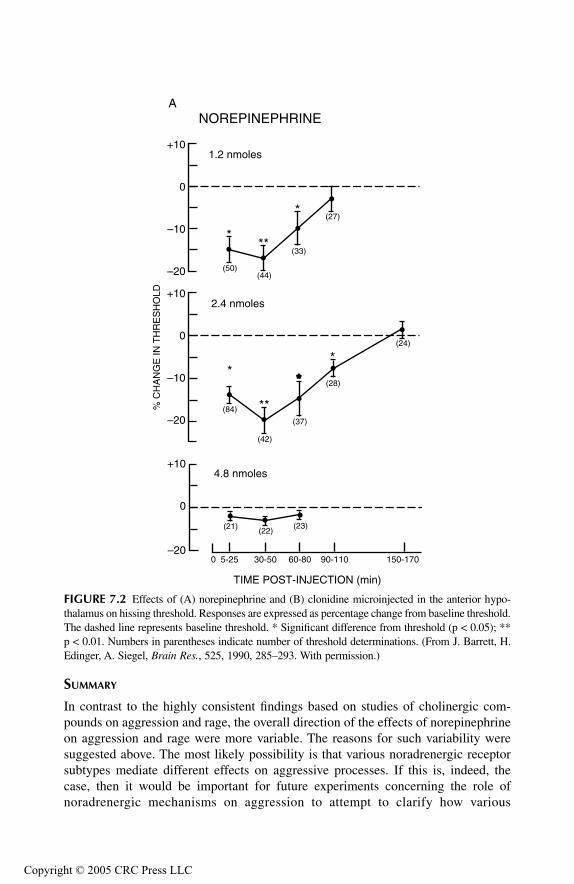
TF1661_C007.fm Page 170 Monday, October 18, 2004 8:35 AM
Copyr
SUMMARY
In contrast to the highly consistent findings based on studies of cholinergic com-pounds on aggression and rage, the overall direction of the effects of norepinephrineon aggression and rage were more variable. The reasons for such variability weresuggested above. The most likely possibility is that various noradrenergic receptorsubtypes mediate different effects on aggressive processes. If this is, indeed, thecase, then it would be important for future experiments concerning the role ofnoradrenergic mechanisms on aggression to attempt to clarify how various
FIGURE 7.2 Effects of (A) norepinephrine and (B) clonidine microinjected in the anterior hypo-thalamus on hissing threshold. Responses are expressed as percentage change from baseline threshold.The dashed line represents baseline threshold. * Significant difference from threshold (p < 0.05); **p < 0.01. Numbers in parentheses indicate number of threshold determinations. (From J. Barrett, H.Edinger, A. Siegel, Brain Res., 525, 1990, 285–293. With permission.)
–20
–10
0
+10
***
*
(50)(44)
(33)
(27)
1.2 nmoles
NOREPINEPHRINE
–20
–10
0
+10
*
**
*
(42)
(37)
(28)
(24)
2.4 nmoles
(84)
*
(21) (22)(23)
0 5-25 30-50 60-80 90-110 150-170–20
0
+104.8 nmoles
% C
HA
NG
E IN
TH
RE
SH
OLD
A
TIME POST-INJECTION (min)
ight © 2005 CRC Press LLC

TF1661_C007.fm Page 171 Monday, October 18, 2004 8:35 AM
Copyr
noradrenergic receptors can alter the different forms of aggression and rage. Oneaspect of this research does seem to be reasonably clear — namely, that activationof noradrenergic α-2 adrenergic receptors in the anterior medial hypothalamusfacilitates defensive rage in the cat. What is not known about this effect is whetheractivation of these receptors in the hypothalamus selectively affects aggressivebehavior, or whether they serve to facilitate other ongoing motivational processesas well. Again, it would be useful if future experiments could address this issue.
DOPAMINE
SYNTHESIS AND REMOVAL
The biosynthesis of dopamine is described above as the initial steps in the biosyn-thesis of norepinephrine (up to the point where dopamine is stored in vesicles). Likenorepinephrine, dopamine is removed mainly by re-uptake back into the terminalsof the presynaptic neuron. Similar to norepinephrine described above, dopaminemay also be removed by the action of COMT.
DISTRIBUTION AND FUNCTIONS
There are several major groups of dopaminergic neurons. One group arises from thesubstantia nigra and projects to the neostriatum. These fibers play an important role
FIGURE 7.2 (Continued).
CLONIDINE
–20
–10
0
*
** (42)
(44)
(21)
0.9 nmole
(65)
(36)
(35) (32)
0 5-25 30-50 60-80 90-110
–10
0
+101.8 nmoles
% C
HA
NG
E IN
TH
RE
SH
OLD
B
TIME POST-INJECTION (min)
+10
ight © 2005 CRC Press LLC

TF1661_C007.fm Page 172 Monday, October 18, 2004 8:35 AM
Copyr
in the regulation of motor functions associated with the basal ganglia. When suchinputs are damaged, Parkinson’s disease results, which is characterized by a tremorat rest, hypokinesia, and rigidity of the extremities. The other major group of fibershas two components. The first arises from the ventral tegmental area and projectsto the hypothalamus and limbic system (called the mesolimbic pathway) and thesecond passes to the frontal and cingulate gyri (called the mesocortical pathway).There is a third group of dopaminergic fibers that arises from the arcuate nucleusof the hypothalamus, and projects to the median eminence and anterior pituitarywhere it inhibits prolactin release. Dopamine mechanisms associated with themesolimbic and mesocortical pathways have been linked to various psychiatricconditions. In particular, disorders such as schizophrenia and anxiety have beenthought to be related to increased activity at dopaminergic synapses.
ROLE OF DOPAMINE IN AGGRESSION AND RAGE
A role for a dopaminergic mechanism in aggression was suggested in a studypublished by Bandler and Halliday (1982). In this study, predatory attack in the catwas diminished after lesions were placed in the region surrounding the medialforebrain. Such lesions were presumed to have damaged ascending dopaminergicfibers because the animals displayed sensory neglect, a phenomenon associated withdopamine loss (Marshall, 1979).
However, for the most part, more direct approaches were taken by most inves-tigators in examining the role of dopamine in aggression and rage, and they arebasically identical to those described above for the analysis of norepinephrine.Similar to the studies conducted with norepinephrine, the overwhelming numbersof studies conducted with dopaminergic compounds involved the systemic admin-istration of these drugs.
Effects of Systemic and Intracerebroventricular Administration of Dopaminergic Agents
A number of studies have attempted to assess the role of dopamine using indirectapproaches. For example, amphetamine, which releases biogenic amines from thenerve terminals, was employed in a number of studies. Since it is difficult todetermine whether amphetamine’s effects on aggression are due to release of dopam-ine or norepinephrine, the use of this drug to evaluate transmitter function is imper-fect at best. Not surprisingly, the results of these studies have been inconsistent.Several studies suggested facilitatory effects following amphetamine delivery. Theseincluded experiments by Hasselager et al. (1972) who reported that administrationof amphetamine to mice induced aggressive responses, and those of Lal et al. (1971)who showed that aggressive responses in rats induced by morphine withdrawal canbe potentiated by amphetamine. In the cat, methamphetamine, which is similar toamphetamine but which is a more potent stimulant, was shown to facilitate defensiverage elicited from the medial hypothalamus of the cat (Maeda et al., 1985). However,other investigators provided evidence that amphetamines suppress aggressiveresponses. Examples of such studies include the findings of Malick (1975) and Gay
ight © 2005 CRC Press LLC

TF1661_C007.fm Page 173 Monday, October 18, 2004 8:35 AM
Copyr
et al. (1975, 1977) who found that muricidal behavior in rats can be antagonized byamphetamines, and Barr et al. (1979) who reported similar results with the use ofphenethylamine, which acts somewhat like amphetamine.
A considerable numbers of studies have reported the effects of using of otherdopaminergic agents in the study of aggression and rage. The large majority of thesestudies provided evidence in support of the view that dopamine facilitates aggressionand rage. A number of these studies are indicated at this time. Messeri et al. (1975)were able to induce conspecific fighting in rats following peripheral administrationof the dopamine agonist, apomorphine. Direct intraventricular injections of dopam-ine can increase fighting in rats (Geyer and Segal, 1974). Consistent with thesefindings were the observations of Garmendia et al. (1992) who showed that admin-istration of the neuroleptic, clozapine, which acts as a dopamine D1 antagonist,caused a significant decrease in fighting in mice. Several other studies conducted inmice also indicated that dopamine receptors facilitate aggressive processes. Here,administration of the dopamine D2 agonist, LY 171555, increased defensive behavior(Cabib and Puglisi-Allegra, 1989), while delivery of the dopamine D2 antagonist,sulpiride, reduced this response (Puglisi-Allegra and Cabib, 1988).
When dopamine receptors were blocked by peripheral administration of chlor-promazine or haloperidol, hypothalamically elicited defensive rage behavior in thecat was suppressed (Maeda, 1976). Likewise, Maeda and Maki (1986) observed thatwhen electrolytic lesions of the basolateral amygdala of cats attenuated the facili-tatory effects of visual provocation on attack elicited from the hypothalamus, sub-sequent administration of apomorphine abolished the suppressive effects of thelesions. Two other studies conducted in the cat further elucidated the role of dopam-ine D2 receptors in facilitating aggressive processes. In one study, it was shown thatperipheral administration of haloperidol alone elevated thresholds for defensive ragebehavior, or if injected prior to apomorphine delivery, blocked the facilitatory effectsof the dopamine agonist (Sweidan et al., 1990). These authors further showed thatadministration of the dopamine D2 agonist, Quinpirol, facilitated defensive rage,while the dopamine D1 agonist, SKF 38393, was largely ineffective. They alsoshowed that the facilitatory effects of apomorphine were selectively blocked by thedopamine D2 antagonist, spiperone, but not with the selective dopamine D1 antag-onist, SCH 23390. In a parallel study, these procedures were employed to examinehow these dopamine receptors affect predatory attack in the cat (Shaikh et al., 1991b).The interesting finding associated with this study was that dopamine D2 receptorshad similar effects on predatory attack. In brief, dopamine D2 but not D1 receptoractivation facilitated predatory attack and that spiperone administration resulted inan elevation of predatory attack response thresholds. Thus, it appears that dopaminereceptors modulate both defensive rage and predatory attack in similar ways.
Not all findings supported a facilitatory role for dopamine on aggressive pro-cesses. Abe et al. (1987) reported that peripheral administration apomorphine inhib-ited muricide in rats. Thoa et al. (1972) and Nakamura and Thoenen (1972) admin-istered 6-OHDA by an intraventricular route in rats and observed an increase infootshock-induced fighting and irritability, respectively. However, as noted above,it is difficult to determine from this study whether the effects could be attributed tochanges in dopamine or norepinephrine levels in the brain. Consistent with these
ight © 2005 CRC Press LLC
TF1661_C007.fm Page 174 Monday, October 18, 2004 8:35 AM
Copyr
findings, Beleslin et al. (1985) observed that intracerebroventricular administrationof compounds, which have dopaminergic receptor antagonist properties, inducedaggressive displays in cats.
Neurochemical Measurements
As mentioned previously, another approach to the study of neurotransmitters andaggression involves examination of the tissue content of the neurotransmitter inquestion in specific regions of the brain of an animal in response to its elicitationof an aggressive response. A number of studies have been conducted using thisapproach, and a few of them with respect to dopamine are summarized here.
In one study, Tizabi et al. (1979) reported regional increases in dopamine levelsin the striatum of mice after isolation-induced aggressive behavior. Hadfield (1981,1983) conducted a series of studies in which he examined the regional neurochemicalkinetics as a result of acute fighting in isolated male mice. In contrast, his majorfinding was that Km and Vmax for dopamine were significantly increased in theprefrontal cortex but not in the neostriatum, pointing to the importance of themesocortical dopaminergic pathway in aggressive behavior.
Further support for a linkage between dopamine and prefrontal cortical mecha-nisms in aggression and rage come from several more recent studies. Cabib et al.(2000) observed that mice which were defeated tend to become more defensive.Subsequent interactions involving these mice were associated with increased dopam-ine metabolism in the prefrontal cortex. Using microdialysis measurements, van Erpand Miczek (2000) reported increases in dopamine levels in the prefrontal cortex(and nucleus accumbens) during aggressive encounters.
Genetic Approaches
Several reports used a genetic approach to test the role of dopamine in aggressivebehavior. Pradhan (1973) compared two strains of mice, one of which was bred forhigh levels of aggressiveness. Here, it was observed that the more aggressive grouphad higher levels of dopamine than the group that was not as aggressive. In a morerecent study, Lewis et al. (1994) also bred mice for high and low levels of aggression.These authors reported that treatment with the selective dopamine D1 receptoragonist, dihydrexidine, reduced isolation-induced aggression in the aggressive strainof rats, and that pretreatment with the selective dopamine D1 antagonist, SCH 23390,blocked the effects of dihydrexidine. While the findings of this study support therole of dopamine in aggressive behavior, it is not consistent with several others(described earlier as well as in the next section), which points to the importance ofdopamine D2 receptors in aggression and rage.
Intracerebral Administration of Dopaminergic Compounds
Since it is known that dopaminergic fibers project to wide areas of the forebrain,including the hypothalamus, a study was performed by Sweidan et al. (1991) todetermine what role dopaminergic innervation of the medial hypothalamus has inregulating defensive rage behavior in the cat. The rationale for this study was
ight © 2005 CRC Press LLC
TF1661_C007.fm Page 175 Monday, October 18, 2004 8:35 AM
Copyr
basically the same as that described above for the analysis of noradrenergic mech-anisms in the hypothalamus (Barrett et al., 1990). In brief, stimulating cannula-electrodes were initially implanted into sites within the ventromedial and anteriorhypothalamus, respectively, from which defensive rage could be elicited. Then,dopaminergic compounds were microinjected into defensive rage sites in the anteriorhypothalamus, and the effects of such drug administration on defensive rage elicitedfrom the ventromedial hypothalamus were assessed. Microinjections of either apo-morphine or of the selective dopamine D2 agonist, LY 171555, facilitated defensiverage behavior (Figure 7.3). However, microinjections of the selective dopamine D1
agonist, SKF 38393, had little effect on the attack response. Of further interest werethe observations that administration of either the nonselective dopamine antagonist,haloperidol, or the selective dopamine D2 antagonist, sulpiride, alone (but not theselective dopamine D1 antagonist, SCH 23390) into the anterior hypothalamus,elevated response thresholds, and blocked the effects of apomorphine or LY 171555when delivered prior to delivery of the agonist. These findings demonstrate thatdopaminergic stimulation in the anterior hypothalamus, acting via dopamine D2
receptors, facilitate defensive rage in the cat.
SUMMARY
Overall findings from the studies described above support the view that dopaminefacilitates various forms of aggression and rage behavior. Moreover, evidence waspresented which indicates that the facilitatory effects of dopamine are mediatedthrough dopamine D2 receptors in the anterior medial hypothalamus (and perhapselsewhere). As indicated in the discussion of norepinephrine, it is indeed possiblethat the dopaminergic effects on aggression and rage may not be selective for theseprocesses, but may serve to enhance a variety of motivational processes associatedwith hypothalamic processes. Answers to this and related questions can only beachieved through a systematic analysis of the manner in which dopaminergic mech-anisms may influence the varieties of functions of the hypothalamus and relatedregions of the brain.
SEROTONIN
SYNTHESIS AND REMOVAL
Since serotonin does not cross the blood–brain barrier, serotonin in the brain resultsfrom the synthesis in selective cell bodies in the brainstem. The substrate for serotoninsynthesis is dietary tryptophan. Tryptophan in plasma passes through the blood–brainbarrier. It is hydroxylated by tryptophan hydroxylase at the 5 position, then decarbox-ylated by L-amino acid decarboxylase, forming serotonin, and stored in vesicles forfuture release. Two mechanisms are involved in the removal of serotonin. One involvesre-uptake, which is facilitated by high-affinity re-uptake sites within the serotonin nerveterminals. The other is by metabolism. Serotonin is deaminated to 5-hydroxyindoleac-etaldehyde by monoamine oxidase, and is then oxidized to form 5-hydroxyindoleaceticacid by aldehyde dehydrogenase. It is ultimately excreted in the urine.
ight © 2005 CRC Press LLC

TF1661_C007.fm Page 176 Monday, October 18, 2004 8:35 AM
Copyr
FIGURE 7.3 Time course of the effects of microinjections of (A) apomorphine or vehicle,and (B) the selective dopamine D2 agonist, quinpirole (LY 171555), into the anterior hypo-thalamus on ventromedial hypothalamically elicited hissing. Each point represents the averagechange in hissing threshold current expressed as a percentage relative to preinjection baselinethreshold (BL). * p < 0.05 compared to vehicle. (From S. Sweidan, H. Edinger, A. Siegel,Brain Res., 549, 1991, 127–137. With permission.)
FIGURE 7.3 (Continued).
BL 1st 2 nd 3 rd 4 th 5 th 6 th
POST-INJECTION TIME PERIODS (IN HOURS)
VehicleAPO (50 ng/0.25 µl)APO (500 ng/0.25 µl)
–12.5
–10.0
–7.5
–5.0
–2.5
0
+2.5
% C
HA
NG
E IN
HIS
SIN
G T
HR
ES
HO
LDS
A
* *
BL 1st 2 nd 3 rd 4 th 5 th 6 th
POST-INJECTION TIME PERIODS (IN HOURS)
VehicleLY (50 ng/0.25 µl)LY (250 ng/0.25 µl)
–12.5
–10.0
–7.5
–5.0
–2.5
0
+2.5
% C
HA
NG
E IN
HIS
SIN
G T
HR
ES
HO
LDS
B
*
ight © 2005 CRC Press LLC
TF1661_C007.fm Page 177 Monday, October 18, 2004 8:35 AM
Copyr
DISTRIBUTION AND FUNCTIONS
Various groups of serotonin containing neurons are located mainly along the midlineof the medulla, pons, and midbrain. Neurons from the dorsal and median pontineand midbrain raphe nuclei project through the medial forebrain bundle to wide areasof the forebrain, including the diencephalon, limbic system, and cerebral cortex.Serotonin neurons arising from the medulla project to the dorsal horn of the spinalcord. The projections to the diencephalon and limbic system most likely modulatefunctions associated with these regions. These include visceral processes such asfeeding, drinking, endocrine, affective, thermoregulatory and autonomic functions,as well as aggression and rage, which are considered in depth below. Of considerableimportance has been the linkage between serotonin and emotional disorders, includ-ing depression, anxiety, and obsessive-compulsive disorders. In fact, one of the mostpopular pharmacological treatments for these disorders involve the use of serotoninre-uptake inhibitors to increase serotonin levels in the brain. Projections to thecerebral cortex have been shown to be associated with sleep and wakefulness anddescending projections to the spinal cord are related to the descending brainstempathway that inhibits pain impulses.
ROLE OF SEROTONIN IN AGGRESSION AND RAGE
Of all the neurotransmitters that have been investigated with respect to aggressivebehavior, none has received more attention than serotonin. Some studies consideredhow stimulation or electrolytic or neurochemical lesions of serotonergic neuronsaffect aggression and rage. However, the overwhelming numbers of studies haveused peripheral administration of serotonergic compounds, including drugs thatdeplete serotonin, and conducted mainly in rodents, to identify how serotonin recep-tor activation or blockade affects these processes. In contrast, few studies haveattempted to determine the effects of intracerebral microinjections of serotonergiccompounds. Other approaches have also been applied. One such approach involvedthe measurement of central or peripheral levels of serotonin and correlating thoselevels with changes in the propensity for the expression of the responses underconsideration. A second approach has examined the role of dietary manipulation oftryptophan levels and a third has considered how genetic factors affect aggressionand rage. The discussion below reviews a representative number of these studies inan attempt to gain an understanding of the overall role of serotonin receptors in theregulation of aggression and rage.
Lesions or Stimulation of Serotonergic Nuclei
As indicated by the large number of studies summarized below, most researchersstudying the serotonin system have manipulated this neurotransmitter by pharma-cological means. In contrast, several earlier investigators attempted to do the sameby examining the effects of either destruction (by the placement of lesions) oractivation (by electrical stimulation) of serotonin neurons and then observing theeffects of such procedures on aggression and rage.
ight © 2005 CRC Press LLC
TF1661_C007.fm Page 178 Monday, October 18, 2004 8:35 AM
Copyr
Several studies examined the effects of raphe lesions on aggression. One study,conducted by Sheard (1973), compared the effects of electrolytic lesions of the raphenuclei with those seen with p-chlorophenylalanine (PCPA), which depletes brainserotonin, on aggressive responsive in rats. Sheard reported that the increase inaggressive behavior seen following PCPA lesions was not seen after electrolyticlesions. In this study, the difference in effects following each of these procedurescould possibly have been attributed to the fact that a more complete depletion ofserotonin occurred after administration of PCPA. However, several other studies,positive effects of electrolytic lesions were reported. Vergnes et al. (1973, 1974)showed that destruction of the dorsal and medial raphe nuclei (but not lesions limitedto one of these two nuclei) induced mouse-killing behavior in rats. Jacobs and Cohen(1976) examined the effects of electrolytic lesions in the dorsal or median raphenuclei of rats, which resulted in significant reductions in forebrain serotonin levels,on a different form of aggression — that is, pain-elicited aggression. With respectto behavioral effects, dorsal raphe lesions produced long-lasting increases in thisform of aggression, while median raphe lesions were ineffective.
Concerning the effects of electrical stimulation of the raphe complex, Kostowskiet al. (1980) reported that such stimulation of the dorsal raphe nuclei markedlyreduced muricide in rats. This author went on to provide additional evidence ofserotonergic involvement in the modulation of aggression by demonstrating thatactivation of 5-HT receptors by quipazine, which is generally characterized as anonselective agonist, strongly decreased mouse-killing behavior.
Thus, the findings from lesion and stimulation studies suggest that serotoninsuppresses several different forms of aggressive behavior in rodents. The studiesdescribed below overall provide additional support for these initial findings.
Neurochemical Depletion of Serotonin
While depletion of serotonin can be achieved by the placement of lesions in thebrainstem that destroy large numbers of serotonin neurons, a more sophisticatedapproach applies a chemical agent that selectively depletes serotonin neurons byblocking synthesis or by acting as a neurotoxin. As indicated above, PCPA is sucha drug (as it prevents serotonin synthesis by inhibiting tryptophan hydroxylase), andhas been used widely to examine the effects of reductions in serotonin on variousforms of aggression as well as other behaviors. Another drug that has been used ina parallel manner is 5,7 dihydroxytryptamine (5,7 DHT), which acts as a selectiveserotonin neurotoxin. The findings using these drugs are summarized below in thissection of the chapter but reflect some inconsistencies among investigators. The basisfor such inconsistencies is considered at the end of this section.
Based on the data derived from lesions and stimulation described above, it wouldbe predicted at a simplistic level that depletion of serotonin should increase thepropensity for aggression. In fact, the majority of studies using neurochemicalmethods for depleting brain serotonin have yielded results consistent with thisprediction. Several independent investigations reported that serotonin depletioncaused by PCPA administration induced mouse killing in rats (Miczek et al., 1975;Vergnes, 1980; Valzelli et al., 1981). Likewise, increases in aggressive behavior in
ight © 2005 CRC Press LLC
TF1661_C007.fm Page 179 Monday, October 18, 2004 8:35 AM
Copyr
rats following PCPA treatment were also described by Sheard (Sheard, 1969, 1970)and later by Knutson et al. (1979). In contrast to these findings, Malick and Barnett(1976) induced aggression in mice by isolating them, and then observed that theaggressiveness was reduced by administration of PCPA. Similar approaches havealso been used by Katz and Thomas (1976) and by Ferguson et al. (1970) in severalstudies conducted in cats. Here, the results were parallel to the majority of studiesdescribed above in the rat; namely, that peripheral administration of PCPA resultedin potentiation of defensive rage and predatory attack, respectively.
With respect to 5,7-DHT, File and Deakin (1980) placed microinjections of thisneurotoxin into the dorsal and median raphe nuclei of rats. Animals with lesions ofthe median (but not the dorsal) raphe nucleus displayed increases in dominancebehaviors to intruders placed in the home cages. Similar facilitation of mouse-killingbehavior in rats was reported by Vergnes and Kempf (1982) following administrationof 5,7-DHT into the lateral hypothalamus. Several studies conducted in cats alsoproduced similar results. When 5,7-DHT was administered peripherally, cats showedan enhancement of growling induced by carbachol infusion into the hypothalamus(Koprowska and Romaniuk, 1997). These authors further showed that cats that werepreviously submissive displayed more dominance and fighting behavior after theplacement of the chemical lesions. Likewise, when 5,7-DHT lesions were placed inthe dorsal raphe complex, there was a consequent increase in growling induced bycarbachol injections into the hypothalamus. When viewed collectively, the evidenceindicates that reductions in serotonin levels by either PCPA or 5,7-DHT adminis-trations are associated with an increase in different forms of aggressive behavior.As previously noted, in this context, these data are clearly consistent with otherapproaches described above suggesting an inhibitory role of serotonin in the regu-lation of aggressive processes.
Depletion of Serotonin Levels by Diet
We have seen from the studies described above that a most common way of causingserotonin depletion involves the application of a neurochemical toxin or synthesisinhibitor. Since tryptophan is the primary amino acid essential for the synthesis ofserotonin and it is also commonly present in the diet, several investigators haveattempted to reduce serotonin levels by the use of a low-tryptophan diet given torats and mice over a period of weeks (Kantak et al., 1979a, 1979b; Vergnes andKempf, 1981).
In several studies, Kantak et al. (1979a, 1979b) examined the effects of admin-istration of a tryptophan-free diet on cricket killing in mice. Interestingly, theseauthors reported differential effects of reduced brain serotonin levels on isolation-induced killing and predatory responses. Specifically, there was a reduction in theduration of isolation-induced fighting, but an increase in the number of mice express-ing cricket killing coupled with decreased latencies to attack and to kill the mice.In another study, Vergnes and Kempf (1981) placed rats on a low-tryptophan dietfor several weeks, causing a 75% reduction in brain serotonin levels while cate-cholamine levels remained relatively the same. When rats were placed in an envi-ronment that allowed for interactions between rats and mice prior to initiation of
ight © 2005 CRC Press LLC

TF1661_C007.fm Page 180 Monday, October 18, 2004 8:35 AM
Copyr
dietary constraints, there was a subsequent suppression of mouse killing in theserats. However, after the rats were placed on the low-tryptophan diet for severalweeks, there was a subsequent increase in mouse killing that persisted even to theperiod after they were returned to a normal diet. Consistent with these findings isthe observation that peripheral administration of L-tryptophan results in inhibitionof muricide in rats (Broderick and Bridger, 1984). Thus, these observations providefurther support to the view presented thus far that serotonin has a suppressive effecton aggressive behavior.
Genetic Manipulation of Brain Serotonin Levels
There are several approaches to the study of genetic factors involved in aggressivebehavior. One source of support for a genetic basis comes from studies in whichanimals are specifically bred for high levels of aggression. In one study, Korte et al.(1996) showed a correlation between mice expressing high levels of aggression, asdemonstrated by short latency attacks, and elevated levels of 5-HT1A mRNA in thedorsal hippocampus relative to levels observed in low-aggressive mice that requiredlonger latencies to express attack. In essence, this study indicated that geneticselection for high offensive aggression also selects at the same time for increasedpostsynaptic 5-HT1A receptors in limbic and related regions of the forebrain. Inanother study, Lyons et al. (1999) explored the role of brain-derived neurotrophicfactor (BDNF), which has a trophic effect on 5-HT neurons in the central nervoussystem (CNS), on aggressiveness in mice. Here, animals that were bred for defi-ciencies in BDNF displayed losses in 5-HT axons and lower 5-HIAA/5-HT ratiosin the hypothalamus. These same animals also showed increases in intermale aggres-siveness as applied in the resident–intruder model.
An alternate strategy in recent years has been to employ knockout mice — thatis, mice created to be deficient in selected genes — to study the role of variousneurochemical systems on behavioral processes. As this procedure relates to thepresent discussion, one group of investigators, using homologous recombination,were able to create mutant knockout mice devoid of the 5-HT1B receptor (Saudouet al., 1994). These animals did not display any behavioral or development defects,but did exhibit heightened aggressiveness as determined from a resident–intruderparadigm. While certain pitfalls in the use of this methodology (similar to most otherexperimental methods) need to be addressed, this approach certainly represents anew and possibly heuristic direction in the study and analysis of the role of specificneurotransmitter receptors in aggression and rage. The use of knockout mice hasalso been applied in a recent study by Chiavegatto et al. (2001). Here, mice weregenetically engineered in order to disrupt the neuronal nitric acid sythase gene,which the authors believed inhibited aggression, by acting through 5-HT1A and 5-HT1B receptors. These authors showed that such genetic manipulation resulted indecrements in 5-HT turnover and reduced 5-HT1A and 5-HT1B receptor function. Tofurther test this possibility, agonists for 5-HT1A and 5-HT1B receptors were admin-istered systemically and caused a dramatic reduction in aggressive behavior whentested in a resident–intruder paradigm.
ight © 2005 CRC Press LLC
TF1661_C007.fm Page 181 Monday, October 18, 2004 8:35 AM
Copyr
Peripheral Administration of Serotonergic Compounds
As noted earlier, the most common approach to the study of serotonin receptors inaggression and rage has involved the examination of the effects of peripheral admin-istration of serotonin compounds. Because this literature is so extensive, and sinceit is not practical to review all of these studies here, a representative number, whichreflect the overall findings, are presented in this section.
Attempts to identify specific serotonin receptors that play important roles inregulating aggression and rage have yielded consistent findings in pointing to thefunctional importance of the 5-HT1A receptor. White et al. (1991) examined theeffects of administration of a variety of serotonergic compounds to mice in anisolation-induced aggression paradigm. They observed that 5-HT1A receptor agonistssuch as buspirone and gepirone reduced aggression, as did the 5-HT2 receptorantagonist, ritanserin, while the 5-HT3 receptor antagonist, zacopride (which alsohas some affinity for 5-HT4 receptors) had little effect on the attack response.Interestingly, administration of gepirone also attenuates low frequency vocalization,which likely communicates “affective states” in rodents (Vivian and Miczek, 1993),as well as defensive postures in rats (Blanchard, 1989). Further discussion of therole of 5-HT2 receptors in rage will be considered later in this section.
Other studies have attempted to test the role of 5-HT receptors more directly byusing more specific 5-HT receptor compounds. In a series of studies carried out byKruk and colleagues, and summarized in Olivier et al. (1990a), Kruk (1991), andSiegel et al. (1999), administration of “serenic” compounds such as fluprazine causedan elevation of threshold responses for hypothalamic stimulation-induced attack byone rat on another. Fluprazine delivery also was shown to inhibit intermale attackin mice (but not prey killing) (Parmigiani and Palanza, 1991). The key point to notehere is that these serenic drugs have a high affinity for 5-HT1 receptors, thereforeproviding additional support that this receptor plays an important role in serotoninsuppression of aggressive responses. Moreover, a number of other investigators haveobtained results that indicate that related behaviors such as isolation-induced aggres-sion (Sánchez et al., 1993), intermale aggression (Cologer-Clifford et al., 1996),offensive aggression (De Boer et al., 2000; Korzan et al., 2000), and defensivebehaviors associated with anxiety (Blanchard et al., 1993b) are similarly affected 5-HT1 receptor agonists. These findings thus suggest that with respect to differentcomponents of aggression and defense, the inhibitory effects of 5-HT1 receptoractivation are rather extensive.
Studies concerning the roles of other serotonin subtypes have been very few innumber, perhaps because of the absence of the lack of receptor specificity forreceptors other than 5-HT1 and possibly 5-HT2 receptors. In one study, m-chlorophe-nylpiperazine (m-CPP), a 5-HT receptor agonist, which has a high affinity for 5-HT2 receptors (and some affinity for 5-HT1C and 5-HT2C receptors) was injectedperipherally into male mice (Navarro et al., 1999). It was observed that isolation-induced aggression was decreased following administration of this drug. In anotherstudy, described below, opposing effects of delivery of this 5-HT2 agonist on defen-sive rage were noted. Thus, delineation of the functions of the 5-HT2 and other
ight © 2005 CRC Press LLC
TF1661_C007.fm Page 182 Monday, October 18, 2004 8:35 AM
Copyr
receptors on aggression and rage will require considerable more research coupledwith the development of more highly specific compounds.
Central Administration of Serotonergic Compounds
As noted above, peripheral administration of serotonergic drugs represents the pri-mary route of administration for the study of this neurotransmitter. Only a handfulof studies have considered the effects of delivery of serotonergic compounds indifferent regions of the brain, and they are reviewed in this section.
Numerous studies have been conducted in the rat. In one study, 5-HT1A agonistsincluding eltoprazine and 8-OH-DPAT were microinjected into the dorsal raphenucleus of rats and reduced aggression (Mos et al., 1993). These authors concludedthat these drugs activated serotonin 5-HT1 autoreceptors in the dorsal raphe, thusreducing serotonergic transmission and a nonspecific reduction in aggression. How-ever, the one difficulty with this interpretation of the data is that the larger numbersof findings described earlier indicate that increases (and not deceases) in serotonintransmission are linked to suppression of aggressive behaviors. It is difficult toprovide an adequate explanation of these data at the present time. Several studiesalso conducted in the rat have examined how serotonergic compounds microinjectedinto the amygdala may affect aggressive responses. In one study, Rodgers (1977)reported that bilateral administration of 5-HT into the cortico-medial amygdala(whose activation excites neurons in the medial hypothalamus) depressed fightinginduced by electric shock. Similar findings of suppression of aggressive postures inshock-induced fighting were observed by Pucilowski et al. (1985) following bilateralinjections of 5-HT or the nonselective agonist, quipazine, into the cortico-medialamygdala. This would suggest that the release of serotonin acts mainly on 5-HT1A
receptors in the medial amygdala to suppress neuronal activity and thus reduce itsexcitatory inputs into the medial hypothalamus which is associated with this formof aggression.
Microinjections of serotonergic compounds have also been placed in sites in thepreoptico-hypothalamus of the rodent and cat and in the PAG of the cat. In onestudy, the 5-HT1A agonist, 8-OH-DPAT, was infused into the preoptic region of themouse and decreased aggression (Cologer-Clifford et al., 1997). In a study conductedin the cat, Golebiewski and Romaniuk (1985) induced components of defensive rageby injecting carbachol into the medial hypothalamus. They then injected either 5-HT or the 5-HT antagonist, methysergide, into these sites and observed that 5-HTadministration suppressed the level of hissing and growling, while methysergideenhanced vocalization induced by carbachol delivery.
In a more recent study, Shaikh et al. (1997) sought to determine the effects ofserotonergic receptor activation in the PAG on hypothalamically elicited defensiverage in the cat. In this experiment, the 5-HT1A agonist, 8-OH-DPAT, was microin-jected into PAG sites from which defensive rage could also be elicited and was foundto powerfully suppress defensive rage elicited from the medial hypothalamus. Thespecificity of this effect was further demonstrated when pretreatment with a 5-HT1A
antagonist blocked the suppressive effects of the agonist. In contrast, administrationof the 5-HT2 agonist, m-CPP, facilitated the occurrence of the defensive rage
ight © 2005 CRC Press LLC

TF1661_C007.fm Page 183 Monday, October 18, 2004 8:35 AM
Copyr
response. These authors further showed by immunocytochemical methods that sero-tonin-positive fibers were present in the region of the PAG where the compoundswere infused.
Results of these studies generally support the view that 5-HT1A receptors inregions of the brain that are normally associated with the expression of rage behavioras well as regions linked to its potentiation serve to suppress this response. However,the finding that 5-HT2 receptor activation facilitates defensive rage indicates thepresence of a differentiation of function by these receptors on the rage mechanism.It would be of considerable interest to determine whether activation of these sero-tonin receptors in other regions such as the hypothalamus can also differentiallymodulate defensive rage.
NEUROCHEMICAL MEASUREMENTS AND APPROACHES
Several other approaches have been used to gain a better understanding of the roleof serotonin in aggression and rage. Studies summarized briefly here involvedindirect measurements of serotonin levels and were carried out in rodents, monkeys,and humans.
With respect to rodents, an early study demonstrated that decreased levels of 5-HT turnover and 5-HIAA in mice, which were subject to isolation, were alsocorrelated with heightened aggressiveness (Kostowski and Valzelli, 1974). In a morerecent study, mice were exposed to low levels of x-rays, and then, changes in brainturnover of 5-HT were correlated with their tendencies to display aggression. Theyobserved that rats which were irradiated had faster turnover rates of 5-HT and showeda calming effect in comparison to sham-irradiated animals which displayed lowerturnover rates and were more aggressive (Miyachi et al., 1994).
A somewhat different approach was taken by Higley et al. (1996a, 1996c) inwhich they studied monkeys in natural group settings while measuring 5-hydroxy-indole-acetic acid (5-HIAA), the major metabolite of serotonin. In several relatedstudies, female macaques with low levels of 5-HIAA in the cerebrospinal fluid (CSF)exhibited higher rates of spontaneous aggression, and were more likely to be removedfrom the group because of their wounding of other monkeys or for treatment ofinjuries. Moreover, animals that had a high ranking in the social group tended tohave lower levels of 5-HIAA in the CSF (Higley et al., 1996a, 1996c). In anotherstudy, it was shown that low concentrations of 5-HIAA in the CSF of monkeys,when quantified early in life, served as an effective biological predictor of futureexcessive levels of aggression (Higley et al., 1996b).
There is rather extensive literature concerning similar studies conducted inhumans in which 5-HIAA in blood was used as an index of serotonin levels andcompared to levels of aggressiveness. Because of the size of this literature, thegeneral findings will be briefly summarized at this time. It is interesting to note thatstudies by Coccaro et al. have shown a similar relationship between 5-HIAA levelsand aggression to what was described above in monkeys (Coccaro, 1989; Coccaroet al., 1990, 1997). The general observation from these and related studies indicatethat there is a reduction in 5-HIAA levels in individuals who are classified asantisocial. Related characteristics such as impulsive behavior and dyscontrol were
ight © 2005 CRC Press LLC
TF1661_C007.fm Page 184 Monday, October 18, 2004 8:35 AM
Copyr
also linked to low 5-HIAA levels. In their recent review of this literature, Moore etal. (2002) reached a similar conclusion and further suggested that reduced serotoninlevels may be characteristic of a wide variety of individuals who express antisocialbehavior, even though they may commit different types of crimes or may be asso-ciated with different psychiatric diagnoses.
SUMMARY
Studies summarized in this section involve various species, including humans, anda variety of methodologies. In spite of the diverse approaches used, it is of interestto note that the findings from these studies seem to point to the same conclusion,namely, that serotonin has a suppressive effect on aggression, rage, and relatedprocesses. It also appears that this phenomenon extends over different species,ranging from rodent to humans. The studies further suggest that different serotoninreceptors do not function in the same way. The literature clearly indicates that 5-HT1A receptor activation is associated with suppression of aggression and rage, whilethe more limited data regarding 5-HT2 receptors suggest that activation of thisreceptor potentiates these same processes. Further studies are definitely required toconfirm the role of 5-HT2 receptors, as well as to identify the roles of other serotoninreceptors.
PEPTIDES
Many peptides have been identified. However, only a few have been implicated inthe regulation and control of aggression and rage behavior. These include opioidpeptides, substance P (SP), and cholecystokinin (CCK). With respect to aggressionand rage, few studies have been conducted concerning SP and CCK, while greaterattention has been given to the role of opioid peptides. The discussion below providesan overview of the studies conducted on opioid peptides and summarizes the fewstudies concerning other peptides.
OPIOID PEPTIDES
CHARACTERIZATION
The three classes of opioid peptides include β-endorphins, enkephalins, and dynorphins.β-endorphins are derived from pre-proopiomelanocortin, which is present in the
rough endoplasmic reticulum of cells in the anterior and intermediate lobes of thepituitary as well as in the arcuate nucleus of the hypothalamus. The propeptide,proopiomelancortin, is generated after removal of the signal peptide from the pre-peptide within the endoplasmic reticulum. It is then packaged into vesicles in theGolgi apparatus of the cell and transported down the axon. In the axon terminal,further proteolytic processing occurs, resulting in the formation of the active pep-tides, β-lipotropin and ACTH. Beta-lipotropin is cleaved into β-endorphin and β-lipotropin These endorphins are packaged in synaptic vesicles and released byexocytosis as neurotransmitters.
ight © 2005 CRC Press LLC
TF1661_C007.fm Page 185 Monday, October 18, 2004 8:35 AM
Copyr
Enkephalins are formed in the following way: the pre-propeptide, pre-proen-kephalin A, is present in the rough endoplasmic reticulum of neurons in differentregions of the brain. When the signal peptide from the propeptide, proenkephalinA, is removed, the propeptide, proenkephalin A, is generated and packaged invesicles in the Golgi apparatus. The vesicles are then transported down the axon tothe synaptic terminal and further proteolytic processing at this site results in theformation of the active peptides, methionine and leucine enkephalin. Similar to β-endorphin, these enkephalins are packaged in vesicles in the synapse and releasedas neurotransmitters by exocytosis.
Dynorphins are derived from prodynorphin peptides, which are C-terminalextended forms of Leu5-enkephalin. In general, the actions of the opioid peptidesend by the presence of peptidases, which lead to their degradation.
DISTRIBUTION AND FUNCTIONS
Enkephalinergic neurons are found in different areas of the CNS. These regionsinclude the spinal cord and PAG, where they contribute to the modulation of painimpulses; hypothalamus and limbic structures, where they are involved in processessuch as temperature regulation, blood pressure, feeding, sexual behavior, and aggres-sion and rage (discussed below); and basal ganglia, where they contribute to thecontrol of motor functions.
ROLE OF ENKEPHALINS IN AGGRESSION AND RAGE
Studies Conducted in Rodents
Most studies concerning opioid peptides have been conducted in rodents andinvolved systemic administration of drugs. The general pattern of findings suggeststhat they have antiaggressive properties (Gianutsos and Lal, 1978). One line ofsupporting evidence comes from experiments showing that morphine withdrawalinduces aggression (Thor, 1971; Davis and Khalsa, 1971; Moyer and Crabtree, 1973;Gianutsos and Lal, 1976; Gianutsos et al., 1976; Gianutsos and Lal, 1978). Incontrast, findings from a number of investigators who have used the shock-inducedand predatory aggression models have not been as clear-cut as the data obtainedfrom morphine administration. For example, several authors studying the effects ofopioid peptides on shock-induced aggression have reported potentiation of aggres-sion following naloxone administration (Fanselow et al., 1980; Puglisi-Allegra etal., 1982; Tazi et al., 1983), while no direct effects were observed in a third study(Rodgers and Hendrie, 1982). With respect to predatory attack in rodents, two paperssuggested that there was little relationship between opioid blockade (by naloxone)and prey killing (Kromer and Dum, 1980; Walsh et al., 1984).
It is difficult to account for the variations in effects of opioid peptides on a givenmodel of aggression. However, one may conjecture that the directionality of theeffects of opioids may be related to a specific form of aggression. A possible reasonfor thinking along these lines is that predatory attack and defensive rage are asso-ciated with positive and negative reinforcing properties, respectively, and that bothforms of aggression are also associated with different anatomical pathways. In this
ight © 2005 CRC Press LLC
TF1661_C007.fm Page 186 Monday, October 18, 2004 8:35 AM
Copyr
manner, differential effects of opioid peptides may be linked to the different phys-iological and anatomical properties of each of these forms of aggression.
STUDIES CONDUCTED IN CATS
Systemic Administration of Opioid Compounds
Three studies addressed the question of how nonspecific opioid receptor blockadewould affect defensive rage and predatory attack (Shaikh and Siegel, 1989; Brutusand Siegel, 1989). In one earlier study, fighting was induced in cats by delivery ofcarbachol into the lateral ventricles (Kromer and Dum, 1980). These authors thentested the role of opioid receptor activation by injecting morphine (which activatesprimarily µ receptors) into the lateral ventricles and observed an attenuation of thefighting. This study, similar to others described above, provided additional supportto the view that opioid receptors mediate suppression of aggressive responses.
In two other studies, systemic administration of naloxone resulted in a dose-dependent decrease in the threshold for defensive rage elicited from either thehypothalamus (Brutus and Siegel, 1989) or PAG (Shaikh and Siegel, 1989). Incontrast, when tested against predatory attack elicited from the lateral hypothalamus,naloxone suppressed this response while having little or no effect on contralateralcircling elicited from other regions of the hypothalamus (Brutus and Siegel, 1989).Thus, on the basis of these findings, there is consistent evidence that opioid receptorsmediate suppression of defensive rage behavior, but it also suggested that thesereceptors may in fact facilitate predatory attack behavior. In view of the relativepaucity of data obtained with respect to opioid peptides and predatory attack, itwould seem quite useful for future studies to replicate and extend these findings.The importance of such a study would be that it could help to establish whetheropioid receptors have uniform or differential effects on different forms of aggressivebehavior.
Intracerebral Administration of Opioid Compounds
Several basal forebrain structures, which include the bed nucleus of the stria termi-nalis and nucleus accumbens, have been implicated in the regulation of rage andaggression. Evidence for this conclusion was presented in Chapters 5 and 6. Inseveral experiments, the question was asked whether opioid receptor activation inthese regions could affect these forms of aggression (Brutus et al., 1988, 1989). Therationale for these studies was based on several lines of evidence. The first is thatboth regions have been shown to modulate aggression and rage (Block et al., 1980b;Shaikh et al., 1986); and the second is that both regions receive opioidergic fibersthat appear to arise from the central nucleus of amygdala (Uhl et al., 1978). Theparadigm for the studies was to initially establish stable baseline response thresholdselicited by stimulation of the medial hypothalamus. Then, the nonspecific enkephalinagonist, D-ala2-met5-enkephalinamide (DAME) was microinjected into these sitesin separate experiments, and the effects of administration of this drug on defensiverage were assessed. The results revealed a dose–response suppression of defensiverage following delivery of this agonist into each of these regions. Moreover, the
ight © 2005 CRC Press LLC
TF1661_C007.fm Page 187 Monday, October 18, 2004 8:35 AM
Copyr
suppressive effects of DAME were blocked by pretreatment with naloxone into thesesites, indicating that these effects were mediated through opioid receptors.
From these studies, one may conclude that enkephalinergic neurons arising fromthe central nucleus of amygdala project to the bed nucleus of the stria terminalisand nucleus accumbens. When opioid receptors in these regions are activated, thereis suppression of the defensive rage response elicited from the medial hypothalamus.It is of interest to recall that stimulation of the bed nucleus facilitates defensive rage,presumably by fibers that project directly to either the medial hypothalamus or PAG.Thus, it may be concluded that the mechanism for modulation here involves centralnucleus–induced opioid suppression of neurons in the bed nucleus, which normallyfacilitates defensive rage behavior. As discussed below, the inhibitory actions of theopioid projections from the central nucleus on defensive rage are quite powerful andinvolve yet another, and perhaps more critical input to the PAG.
Thus far, the studies described above focused on opioid receptor activation oflimbic forebrain structures as they impacted on defensive rage elicited from themedial hypothalamus. In the remainder of this section, results of a number of studiesconducted with respect to the PAG are reviewed as they provide evidence that opioidreceptors in this structure play an important role in modulating defensive ragebehavior.
One such study involved the implantation of a stimulating electrode in the medialhypothalamus from which defensive rage could be elicited and a cannula-electrodein the PAG that would either suppress or facilitate defensive rage (Pott et al., 1986).When naloxone was microinjected into these inhibitory (but not facilitatory) sites,the inhibitory effects of PAG stimulation on hypothalamically elicited defensive ragewas blocked. In addition, infusion of DAME into the inhibitory sites in the PAGsuppressed hypothalamically elicited defensive rage. These data can be interpretedto mean that electrical stimulation of an inhibitory site in the PAG-activated enkepha-linergic neurons arose from either the central nucleus of amygdala (see below) orfrom interneurons within the PAG itself. Simply put, when naloxone was infusedinto the inhibitory sites, it served to block enkephalins from interacting with opioidreceptors in the PAG to suppress defensive rage behavior.
It should be pointed out that there are also sites in the PAG that, when stimulated,can facilitate or suppress predatory attack elicited from the lateral hypothalamus(Weiner et al., 1991). The key point to note here is that these modulatory sites areaffected by naloxone, indicating that an enkephalinergic mechanism contributes tothe regulation of predatory attack within the PAG as well. From the studies describedabove, two important questions required attention. The first concerned the specificreceptor subtypes that mediate opioid functions within the PAG. The second con-cerned the origin of the opioidergic pathway that projects to the PAG and whichmediates suppression of defensive rage behavior. Answers to the first question wereaddressed in an experiment that involved examination of the effects of infusion ofopioid compounds through a cannula-electrode into a site within the PAG fromwhich defensive rage had been elicited (Shaikh et al., 1988, 1991a). In an initialstudy, the nonspecific opioid agonist, DAME, was microinjected into the PAG, andclearly suppressed defensive rage behavior elicited from that site, while having noeffect on circling behavior that was also elicited from the PAG. In addition, the
ight © 2005 CRC Press LLC
TF1661_C007.fm Page 188 Monday, October 18, 2004 8:35 AM
Copyr
suppressive effects of DAME were blocked following pretreatment with naloxoneat the same PAG site. In a subsequent study, specific opioid receptor agonists weremicroinjected into defensive rage sites in the PAG and their effects on defensiverage were identified. The results indicated that administration of the selectiveµ receptor agonist, morphiceptin, into the PAG powerfully suppressed defensive ragebehavior. Lesser magnitudes of suppression were noted following administration ofthe selective δ agonist, DPDPE. However, there were no changes in response thresh-olds following administration of the selective κ agonist, U-50488H. In this study,pretreatment with the selective µ and δ antagonists, β-funaltrexamine (β-FNA) andICI 174864, blocked the suppressive effects of morphiceptin and DPDPE, respec-tively. The significance of this study is that it indicated the potency of these tworeceptors in mediating suppression of defensive rage within the PAG.
From the studies that have just been described, one may conclude that theopioidergic fibers mediating suppression of defensive rage constitute interneuronswithin the PAG that are activated from an external source. An alternative interpre-tation of the data is that the opioidergic mechanism within the PAG involves opioidpeptides that are synthesized in cell bodies in a distal structure that projects to thePAG. When activated, these neurons would then suppress defensive rage. This latterpossibility was tested in the following experiment (Shaikh et al., 1993). In this study,it was hypothesized that the central and lateral nuclei of the amygdala served justsuch a function, since it is known that stimulation of this region suppresses defensiverage behavior (Brutus et al., 1986), and that these nuclei have been identified asenkephalinergic neurons in rodents (Uhl et al., 1978; Rao et al., 1987). Initially,electrical stimulation applied to the central or lateral nucleus caused powerful sup-pression of defensive rage elicited from the PAG. This suppression was so potentthat it, in fact, lasted for over 30 minutes. Then, dual stimulation including the centralor lateral nucleus was again applied following administration of nonselective (nalox-one) and specific µ (β-FNA) and δ (ICI 174864) opioid antagonists. The suppressiveeffects of central amygdaloid stimulation were completely blocked by naloxone orβ-FNA but not by ICI 174864. That the suppressive effects of the central and lateralnuclei on defensive rage are mediated through a descending pathway to the PAGand that act on opioid µ receptors constitutes the key conclusion suggested from thedata in this study (see Figure 7.4) Alternatively, it might be concluded from thesedata that central amygdaloid neurons are not opioidergic, but instead, project tointerneurons within the PAG that are opioidergic and then project to PAG neuronsassociated with the expression of defensive rage. In order to determine which ofthese possibilities is correct, the following additional experiment was conducted.The retrograde tracer, Fluoro-Gold, was microinjected into defensive rage siteswithin the PAG, and after a suitable survival period, the animals were sacrificed andthe brain tissue was processed for both Fluoro-Gold and met-enkephalin immunore-activity. The findings revealed the presence of considerable numbers of enkephalin-positive neurons situated within the central and lateral nuclei of amygdala, whichwere also labeled with Fluoro-Gold. These findings thus provided further supportfor the view that enkephalinergic neurons in these nuclei of amygdala project directlyto the PAG and suppress defensive rage behavior via an enkephalinergic mechanism(Figure 7.5).
ight © 2005 CRC Press LLC

TF1661_C007.fm Page 189 Monday, October 18, 2004 8:35 AM
Copyr
FIGURE 7.4 Open circles indicate the time course for amygdaloid induced suppression ofdefensive rage as determined by the percent change in response latencies from baseline valuesobtained prior to amygdaloid stimulation. Closed circles indicate the effects of drug delivery.Note that when the selective µ receptor antagonist, β-FNA, was administered at a dose of 0.2nM, it completely blocked the suppressive effects of amygdaloid stimulation (top panel). Apartial blockade was noted at a lower dose of this drug (middle panel), and the selectiveδ antagonist, ICI 174,864, failed to block the suppressive effects of amygdaloid stimulationwhen microinjected at a dose of 0.7 nM into the PAG (bottom panel). * p < 0.05. (FromShaikh et al., 1991c. With permission.)
0 20 40 60 80 100 120 140–200
0
200
400
600
800
1000
pre-injectionpost-injection(B–FNA 0.2 nM)
0 20 40 60 80 100 120 140–200
0
200
400
600
800
1000
pre-injectionpost-injection(B–FNA 0.5 nM)
0 20 40 60 80 100 120 140–200
0
200
400
600
800
1000
pre-injectionpost-injection(ICI 174.864 .07 nM)
TIME AFTER AMYGDALOID STIMULATION (min.)
% C
HA
NG
E IN
RE
SP
ON
SE
ight © 2005 CRC Press LLC
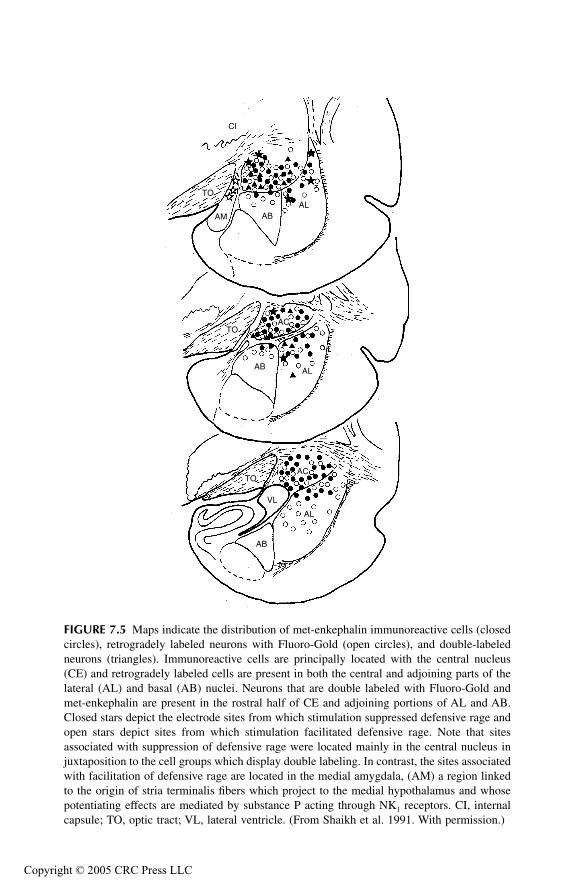
TF1661_C007.fm Page 190 Monday, October 18, 2004 8:35 AM
Copyr
FIGURE 7.5 Maps indicate the distribution of met-enkephalin immunoreactive cells (closedcircles), retrogradely labeled neurons with Fluoro-Gold (open circles), and double-labeledneurons (triangles). Immunoreactive cells are principally located with the central nucleus(CE) and retrogradely labeled cells are present in both the central and adjoining parts of thelateral (AL) and basal (AB) nuclei. Neurons that are double labeled with Fluoro-Gold andmet-enkephalin are present in the rostral half of CE and adjoining portions of AL and AB.Closed stars depict the electrode sites from which stimulation suppressed defensive rage andopen stars depict sites from which stimulation facilitated defensive rage. Note that sitesassociated with suppression of defensive rage were located mainly in the central nucleus injuxtaposition to the cell groups which display double labeling. In contrast, the sites associatedwith facilitation of defensive rage are located in the medial amygdala, (AM) a region linkedto the origin of stria terminalis fibers which project to the medial hypothalamus and whosepotentiating effects are mediated by substance P acting through NK1 receptors. CI, internalcapsule; TO, optic tract; VL, lateral ventricle. (From Shaikh et al. 1991. With permission.)
CI
TO
AM ABAL
TO
AB AL
AC
AC
AL
VL
TO
AB
ight © 2005 CRC Press LLC

TF1661_C007.fm Page 191 Monday, October 18, 2004 8:35 AM
Copyr
SUMMARY
The preponderance of evidence concerning the role of opioid peptides in aggressivebehavior indicates that activation of opioid receptors suppresses aggressive reactionsin general, and those associated with affective aggression (or defensive rage) inparticular. Of considerable importance is the role of opioid µ receptors, which appearto be of considerable significance within the PAG in mediating response suppression.In addition, it should be noted that specific groups of enkephalinergic neurons arisingfrom the central and lateral nuclei of amygdala project directly to the PAG andmediate their suppressive actions via µ opioid receptors in the PAG. Other opioidreceptors, which include δ and κ receptors, seem to be of lesser importance in theregulation of rage and aggression.
SUBSTANCE P
CHARACTERIZATION, DISTRIBUTION, AND FUNCTIONS
Substance P (SP) belongs to a family of tachykinins that also includes neurokinin Aand neurokinin B. Substance P is an undecapeptide and is found in different regionsof the brain such as the hypothalamus (see below), amygdala, basal ganglia, cerebralcortex, and dorsal horn of the spinal cord. Three kinds of neurokinin receptors havebeen identified: neurokinin-1 (NK1), neurokinin-2 (NK2), and neurokinin-3 (NK3). Itwas previously believed that SP interacts with NK1 receptors, neurokinin A with NK2
receptors, and neurokinin B with NK3 receptors. Although this view is somewhatoversimplified and, in fact, several of the neurokinins may interact with several neu-rokinin receptors, SP is generally believed to interact mainly with NK1 receptors.
SP is an excitatory neurotransmitter, producing depolarization of neurons for anextended period of time. The most well-known function of SP in the CNS is that itis released from the endings of first-order sensory axons mediating pain inputs intothe spinal cord. More recently, it has been implicated in affective processes andrecent evidence is presented below that links SP as an important neurotransmitterin the regulation of rage and aggression.
ROLE OF SP IN RAGE AND AGGRESSION
While considerable effort has been given to the study of the role of SP in the mediationof pain impulses at the level of the dorsal horn of the spinal cord, little attention hasbeen directed at other possible functions of this neurotransmitter associated with affec-tive processes. In recent years, however, several studies have been directed at examiningthis possible relationship. Results of these experiments are described in this section.
There are several reasons why attention was directed at SP’s role in rage andaggression. The first is that SP and NK1 receptors are present in regions of the brainassociated with rage and aggression, such as the amygdala, hypothalamus and PAG(Langevin and Emson, 1982; Warden and Young, 1988; Gallagher et al., 1992a,1992b). One of these regions, the medial amygdala, powerfully facilitates defensiverage and suppresses predatory attack (Stoddard-Apter and MacDonnell, 1980; Blocket al., 1980a). It is reasonable to assume that the mechanism underlying such
ight © 2005 CRC Press LLC
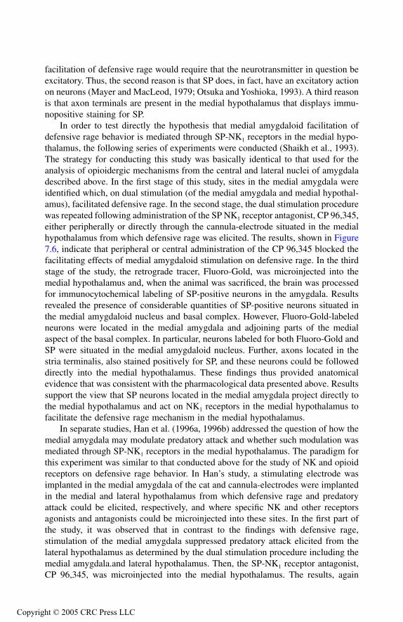
TF1661_C007.fm Page 192 Monday, October 18, 2004 8:35 AM
Copyr
facilitation of defensive rage would require that the neurotransmitter in question beexcitatory. Thus, the second reason is that SP does, in fact, have an excitatory actionon neurons (Mayer and MacLeod, 1979; Otsuka and Yoshioka, 1993). A third reasonis that axon terminals are present in the medial hypothalamus that displays immu-nopositive staining for SP.
In order to test directly the hypothesis that medial amygdaloid facilitation ofdefensive rage behavior is mediated through SP-NK1 receptors in the medial hypo-thalamus, the following series of experiments were conducted (Shaikh et al., 1993).The strategy for conducting this study was basically identical to that used for theanalysis of opioidergic mechanisms from the central and lateral nuclei of amygdaladescribed above. In the first stage of this study, sites in the medial amygdala wereidentified which, on dual stimulation (of the medial amygdala and medial hypothal-amus), facilitated defensive rage. In the second stage, the dual stimulation procedurewas repeated following administration of the SP NK1 receptor antagonist, CP 96,345,either peripherally or directly through the cannula-electrode situated in the medialhypothalamus from which defensive rage was elicited. The results, shown in Figure7.6, indicate that peripheral or central administration of the CP 96,345 blocked thefacilitating effects of medial amygdaloid stimulation on defensive rage. In the thirdstage of the study, the retrograde tracer, Fluoro-Gold, was microinjected into themedial hypothalamus and, when the animal was sacrificed, the brain was processedfor immunocytochemical labeling of SP-positive neurons in the amygdala. Resultsrevealed the presence of considerable quantities of SP-positive neurons situated inthe medial amygdaloid nucleus and basal complex. However, Fluoro-Gold-labeledneurons were located in the medial amygdala and adjoining parts of the medialaspect of the basal complex. In particular, neurons labeled for both Fluoro-Gold andSP were situated in the medial amygdaloid nucleus. Further, axons located in thestria terminalis, also stained positively for SP, and these neurons could be followeddirectly into the medial hypothalamus. These findings thus provided anatomicalevidence that was consistent with the pharmacological data presented above. Resultssupport the view that SP neurons located in the medial amygdala project directly tothe medial hypothalamus and act on NK1 receptors in the medial hypothalamus tofacilitate the defensive rage mechanism in the medial hypothalamus.
In separate studies, Han et al. (1996a, 1996b) addressed the question of how themedial amygdala may modulate predatory attack and whether such modulation wasmediated through SP-NK1 receptors in the medial hypothalamus. The paradigm forthis experiment was similar to that conducted above for the study of NK and opioidreceptors on defensive rage behavior. In Han’s study, a stimulating electrode wasimplanted in the medial amygdala of the cat and cannula-electrodes were implantedin the medial and lateral hypothalamus from which defensive rage and predatoryattack could be elicited, respectively, and where specific NK and other receptorsagonists and antagonists could be microinjected into these sites. In the first part ofthe study, it was observed that in contrast to the findings with defensive rage,stimulation of the medial amygdala suppressed predatory attack elicited from thelateral hypothalamus as determined by the dual stimulation procedure including themedial amygdala.and lateral hypothalamus. Then, the SP-NK1 receptor antagonist,CP 96,345, was microinjected into the medial hypothalamus. The results, again
ight © 2005 CRC Press LLC
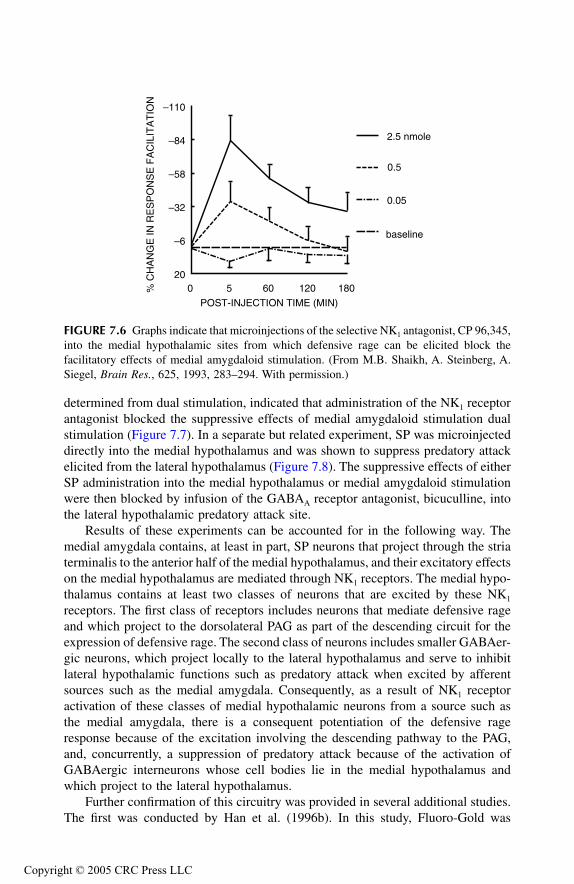
TF1661_C007.fm Page 193 Monday, October 18, 2004 8:35 AM
Copyr
determined from dual stimulation, indicated that administration of the NK1 receptorantagonist blocked the suppressive effects of medial amygdaloid stimulation dualstimulation (Figure 7.7). In a separate but related experiment, SP was microinjecteddirectly into the medial hypothalamus and was shown to suppress predatory attackelicited from the lateral hypothalamus (Figure 7.8). The suppressive effects of eitherSP administration into the medial hypothalamus or medial amygdaloid stimulationwere then blocked by infusion of the GABAA receptor antagonist, bicuculline, intothe lateral hypothalamic predatory attack site.
Results of these experiments can be accounted for in the following way. Themedial amygdala contains, at least in part, SP neurons that project through the striaterminalis to the anterior half of the medial hypothalamus, and their excitatory effectson the medial hypothalamus are mediated through NK1 receptors. The medial hypo-thalamus contains at least two classes of neurons that are excited by these NK1
receptors. The first class of receptors includes neurons that mediate defensive rageand which project to the dorsolateral PAG as part of the descending circuit for theexpression of defensive rage. The second class of neurons includes smaller GABAer-gic neurons, which project locally to the lateral hypothalamus and serve to inhibitlateral hypothalamic functions such as predatory attack when excited by afferentsources such as the medial amygdala. Consequently, as a result of NK1 receptoractivation of these classes of medial hypothalamic neurons from a source such asthe medial amygdala, there is a consequent potentiation of the defensive rageresponse because of the excitation involving the descending pathway to the PAG,and, concurrently, a suppression of predatory attack because of the activation ofGABAergic interneurons whose cell bodies lie in the medial hypothalamus andwhich project to the lateral hypothalamus.
Further confirmation of this circuitry was provided in several additional studies.The first was conducted by Han et al. (1996b). In this study, Fluoro-Gold was
FIGURE 7.6 Graphs indicate that microinjections of the selective NK1 antagonist, CP 96,345,into the medial hypothalamic sites from which defensive rage can be elicited block thefacilitatory effects of medial amygdaloid stimulation. (From M.B. Shaikh, A. Steinberg, A.Siegel, Brain Res., 625, 1993, 283–294. With permission.)
0 5 60 120 180
POST-INJECTION TIME (MIN)
20
–6
–32
–58
–84
–110
% C
HA
NG
E IN
RE
SP
ON
SE
FA
CIL
ITA
TIO
N2.5 nmole
0.5
0.05
baseline
ight © 2005 CRC Press LLC

TF1661_C007.fm Page 194 Monday, October 18, 2004 8:35 AM
Copyr
microinjected into lateral hypoothalamic predatory attack sites, and the brain tissuewas processed by immunocytochemical staining for GABA-positive neurons as wellas for Fluoro-Gold-positive neurons. Results showed the presence of neurons labeledfor both Fluoro-Gold and GABA situated in the medial hypothalamus, indicatingthat these GABAergic neurons project directly to the lateral hypothalamus. In severalfurther studies, Yao et al. (1999, 2001) used in situ hybridization to label NK1
receptors as well as GABA and glutamate-positive neurons in the hypothalamus.The findings of these studies demonstrated that indeed, NK1 receptors are situatedon both GABA and glutamate-positive neurons in the medial hypothalamus. Evi-dence presented in studies described below revealed that the glutamate neurons inthe medial hypothalamus project to the PAG, and as just noted, GABAergic neuronsproject to the lateral hypothalamus. The overall findings of these studies are sum-marized in Figure 7.9.
CHOLECYSTOKININ (CCK)
CHARACTERIZATION, DISTRIBUTION, AND FUNCTIONS
CCK, a gut hormone, is converted from procholecystokinin to peptides where it actson the gallbladder and liver. Smaller CCK-related peptides are found in the brain.An interesting feature of CCK is that it is frequently found co-localized with other
FIGURE 7.7 Effects of microinjections of CP 96,345 into the medial hypothalamus on medialamygdaloid suppression of predatory attack behavior elicited from the lateral hypothalamus.CP 96,345 blocked the suppressive effects of the medial amygdala on predatory attack in adose-dependent manner. The pre-injection baseline represents the percent increase in responselatency following dual stimulation whose value was then converted to 0%. Following drugor vehicle administration, decreases in the magnitude of medial amygdaloid modulation areexpressed as a negative percentage change and reflect an enhancement of the suppressiveeffects of medial amygdaloid stimulation. (From Y. Han, M.B. Shaikh, A. Siegel, Brain Res.,716, 1996a, 59–71. With permission.)
0 5 60 120 180
POST-INJECTION TIME (MIN)
–90
–70
–50
–30
10
% D
EC
RE
AS
E IN
RE
SP
ON
SE
SU
PP
RE
SS
ION
2.5 nM
0.5 nM
0.05 nM
Saline
–110
–10
ight © 2005 CRC Press LLC

TF1661_C007.fm Page 195 Monday, October 18, 2004 8:35 AM
Copyr
neurotransmitters. These include pro-enkephalin-derived peptides, SP, 5-HT, dopam-ine, neuropeptide Y, and GABA. CCK is found in wide regions of the brain thatinclude the cerebral cortex, diencephalon, brainstem, and dorsal root ganglion. Thereare two types of CCK receptors: CCKA and CCKB. It is generally accepted thatsatiety mechanisms are mediated via CCKA receptors and that reactions such asanxiety and panic attacks are mediated via CCKB receptors. It is also believed thatCCKB receptor activation serves to have excitatory effects on neuronal activity. Inthis section, evidence is presented supporting the view that defensive rage is alsopotentiated via CCKB receptors.
ROLE OF CCK IN DEFENSIVE RAGE BEHAVIOR
Behavioral experiments have focused mainly on the roles of CCK on feeding behav-ior and anxiety reactions. In contrast, very little research has been conducted relatingto its possible role in rage and aggression. The results of one such study are describedin this section.
Evidence had indicated that CCK fibers and terminals (Lanaud et al., 1989; Liuet al., 1994) as well as CCKB receptors (Mercer et al., 1996) are present in the PAG.Inasmuch as activation of CCKB receptors have been shown to induce anxietyreactions in rodents (Adamec et al., 1997), the hypothesis was proposed that CCKB
receptors in the PAG would potentiate defensive rage behavior elicited from themedial hypothalamus of the cat. In this study, stimulating electrodes were placed in
FIGURE 7.8 Effects of microinjections of (Sar9,Met(O2)11)-substance P into the medial hypo-thalamus on predatory attack elicited by single stimulation of the lateral hypothalamus. Pre-injection response latencies were converted to 0%. Following drug delivery, response latencieswere determined again over a 180-minute period. Increases in response latencies indicatedthe suppressive effects of drug infusion and are expressed as a positive percentage change onthe graph. Administration of the substance P agonist resulted in suppression of predatoryattack in a dose-dependent manner. (From Y. Han, M.B. Shaikh, A. Siegel, Brain Res., 716,1996a, 59–71. With permission.)
0 5 60 120 180
POST-INJECTION TIME (MIN)
–5
5
15
45
% IN
CR
EA
SE
IN R
ES
PO
NS
E L
AT
EN
CY
2.0 nM
1.0 nM
0.5 nM
control25
35
ight © 2005 CRC Press LLC
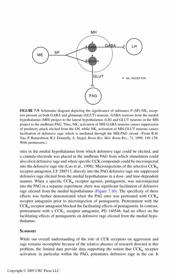
TF1661_C007.fm Page 196 Monday, October 18, 2004 8:35 AM
Copyr
sites in the medial hypothalamus from which defensive rage could be elicited, anda cannula-electrode was placed in the midbrain PAG from which stimulation couldalso elicit defensive rage and where specific CCK compounds could be microinjectedinto the defensive rage site (Luo et al., 1998). Microinjections of the selective CCKB
receptor antagonist, LY 288513, directly into the PAG defensive rage site suppresseddefensive rage elicited from the medial hypothalamus in a dose- and time-dependentmanner. When a specific CCKB receptor agonist, pentagastrin, was microinjectedinto the PAG in a separate experiment, there was significant facilitation of defensiverage elicited from the medial hypothalamus (Figure 7.10). The specificity of theseeffects was further demonstrated when the PAG sites was pretreated with CCKB
receptor antagonist prior to microinjection of pentagastrin. Pretreatment with theCCKB receptor antagonist blocked the facilitating effects of pentagastrin. In contrast,pretreatment with a CCKA receptor antagonist, PD 140548, had no effect on thefacilitating effects of pentagastrin on defensive rage elicited from the medial hypo-thalamus.
SUMMARY
While our overall understanding of the role of CCK receptors on aggression andrage remains incomplete because of the relative absence of research directed at thisproblem, the limited data provide data supporting the notion that CCKB receptoractivation, in particular within the PAG, potentiates defensive rage in the cat. It
FIGURE 7.9 Schematic diagram depicting the significance of substance P (SP)-NK1 recep-tors present on both GABA and glutamate (GLUT) neurons. GABA neurons from the medialhypothalamus (MH) project to the lateral hypothalamus (LH) and GLUT neurons in the MHproject to the midbrain PAG. Thus, NK1 activation of MH-GABA neurons causes suppressionof predatory attack elicited from the LH, while NK1 activation of MH-GLUT neurons causesfacilitation of defensive rage which is mediated through the MH-PAG circuit. (From R.H.Yao, P. Rameshwar, R.J. Donnelly, A. Siegel, Brain Res. Mol. Brain Res., 71, 1999, 149–158.With permission.)
GABA
GLUT
GABA
GLUT
PAG
MESP
MH
LH
NK1 RECEPTOR
ight © 2005 CRC Press LLC
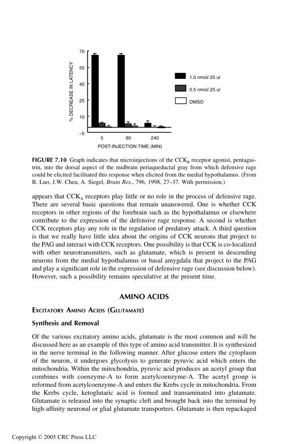
TF1661_C007.fm Page 197 Monday, October 18, 2004 8:35 AM
Copyr
appears that CCKA receptors play little or no role in the process of defensive rage.There are several basic questions that remain unanswered. One is whether CCKreceptors in other regions of the forebrain such as the hypothalamus or elsewherecontribute to the expression of the defensive rage response. A second is whetherCCK receptors play any role in the regulation of predatory attack. A third questionis that we really have little idea about the origins of CCK neurons that project tothe PAG and interact with CCK receptors. One possibility is that CCK is co-localizedwith other neurotransmitters, such as glutamate, which is present in descendingneurons from the medial hypothalamus or basal amygdala that project to the PAGand play a significant role in the expression of defensive rage (see discussion below).However, such a possibility remains speculative at the present time.
AMINO ACIDS
EXCITATORY AMINO ACIDS (GLUTAMATE)
Synthesis and Removal
Of the various excitatory amino acids, glutamate is the most common and will bediscussed here as an example of this type of amino acid transmitter. It is synthesizedin the nerve terminal in the following manner. After glucose enters the cytoplasmof the neuron, it undergoes glycolysis to generate pyruvic acid which enters themitochondria. Within the mitochondria, pyruvic acid produces an acetyl group thatcombines with coenzyme-A to form acetylcoenzyme-A. The acetyl group isreformed from acetylcoenzyme-A and enters the Krebs cycle in mitochondria. Fromthe Krebs cycle, ketoglutaric acid is formed and transaminated into glutamate.Glutamate is released into the synaptic cleft and brought back into the terminal byhigh-affinity neuronal or glial glutamate transporters. Glutamate is then repackaged
FIGURE 7.10 Graph indicates that microinjections of the CCKB receptor agonist, pentagas-trin, into the dorsal aspect of the midbrain periaqueductal gray from which defensive ragecould be elicited facilitated this response when elicited from the medial hypothalamus. (FromB. Luo, J.W. Cheu, A. Siegel, Brain Res., 796, 1998, 27–37. With permission.)
5 60 240–5
10
25
40
55
70
POST-INJECTION TIME (MIN)
% D
EC
RE
AS
E IN
LA
TE
NC
Y1.0 nmol/.25 ul
0.5 nmol/.25 ul
DMSO
ight © 2005 CRC Press LLC

TF1661_C007.fm Page 198 Monday, October 18, 2004 8:35 AM
Copyr
in the nerve terminal for subsequent release. In glial cells, glutamate is convertedinto glutamine by the enzyme glutamine synthase, and is then transported intoneighboring nerve terminals by glutamine transporters. Glutamine is then recon-verted back into glutamate by the enzyme, glutaminase (from mitochondria), and isstored in the nerve terminal for future release.
Distribution and Functions
Glutamate is found in many neural circuits in the brain such as in the efferent fibersof the cerebral cortex, and glutamate receptors are located on neurons throughoutthe CNS. Excitatory amino acids play a major role in a wide variety of processessuch as motor functions, learning, and memory. Glutamate is also believed to playroles in a number of neuropathological conditions such as amyotrophic lateralsclerosis and other conditions that lead to cell death by excitotoxicity.
Role of Excitatory Amino Acids in Defensive Rage Behavior
Perhaps the most important pathway governing defensive rage behavior in the catis that which arises from the medial hypothalamus and projects to the dorsolateralaspect of the PAG. Thus, knowledge of the neurotransmitter(s) associated with thispathway would be extremely important in our quest to gain an understanding of theneurochemistry of defensive rage behavior. The rationale for believing that excitatoryamino acids could be a good candidate here was based on previous anatomical datasuggesting that this pathway may contain glutamate (Beart et al., 1988, 1990; Beitz,1989).
On the basis of this evidence, several studies were conducted aimed at testingthe hypothesis that the function of defensive rage behavior was mediated throughexcitatory amino acid receptors in association with the pathway from the medialhypothalamus to the PAG in the cat (Lu et al., 1992; Schubert et al., 1996b). In aninitial study, the nonspecific excitatory amino acid antagonist, kyneurenic acid, wasmicroinjected into sites within the PAG from which defensive rage could be elicited.Administration of this agonist blocked medial hypothalamic facilitation of PAG-elicited defensive rage (Lu et al., 1992). When the specific NMDA antagonist, AP-7, was microinjected into the PAG, it also blocked medial hypothalamic facilitationof defensive rage. However, when the kainate and quisqualate receptor blocker,CNQX, was microinjected into the PAG in a similar to that of AP-7, it had little orno effect on this response. Further, infusion of NMDA directly into the PAG facil-itated defensive rage elicited from that site (Figure 7.11). A replication and extensionof this study was carried out by Schubert et al. (1996b). Here, it was observed thatmicroinjections of the NMDA antagonist, AP-7, into PAG defensive-rage sites,blocked medial hypothalamically elicited defensive rage. Another aspect of both ofthese studies combined retrograde labeling following microinjections of Fluoro-Goldinto the PAG and immunocytochemical labeling of glutamatergic neurons. Resultsindicated that double-labeled cells (for Fluoro-Gold and glutamate) were observedwithin the medial hypothalamus, mainly dorsal and rostral to the ventromedialnucleus. This finding provided direct support to the pharmacological data and to the
ight © 2005 CRC Press LLC

TF1661_C007.fm Page 199 Monday, October 18, 2004 8:35 AM
Copyr
hypothesis that glutamatergic neurons project from the medial hypothalamus to thePAG, a pathway that subserves the expression of defensive rage behavior and whosefunctions are mediated by NMDA receptors in the PAG.
In another study, evidence was provided indicating that basal amygdaloid facil-itation of defensive rage behavior is also mediated, in part, through NMDA receptors
FIGURE 7.11 (A) Microinjections of the selective NMDA receptor antagonist, AP7, intoperiaqueductal gray (PAG) sites from which defensive rage could be elicited suppressed thefacilitatory effects of medial hypothalamic stimulation upon this response in a dose-dependentmanner. Note that a higher dose of the CNQX, a non-NMDA receptor antagonist, had littleeffect on medial hypothalamic facilitation of this response. (B) Infusion of NMDA intodefensive rage sites in the PAG resulted in a dose-dependent facilitation of this response,mimicking the effects obtained with dual stimulation of the medial hypothalamus and PAG.(From C.-L. Lu, M.B. Shaikh, A. Siegel, Brain Res., 581, 1992, 123–132. With permission.)
FIGURE 7.11 (Continued).
2 nM AP70.5 nM AP70.1 nM AP74 nM CNOXBL
0 45 90 135 180POST-INJECTION TIME (MIN)
–20
–10
0
10
20
30
40
50
% C
HA
NG
E IN
LA
TE
NC
Y
A
1.0 nM0.5 nM0.1 nMBL
30POST-INJECTION TIME (MIN)
–60
–50
–40
–30
–20
–10
0
10
% C
HA
NG
E IN
LA
TE
NC
Y
B
0 5 15
ight © 2005 CRC Press LLC
TF1661_C007.fm Page 200 Monday, October 18, 2004 8:35 AM
Copyr
in the PAG (Shaikh et al., 1994). In brief, results indicated that dual stimulationinvolving the basal amygdala facilitated defensive rage elicited from the PAG.However, a key point in this study was that administration of the MDA receptorantagonist, AP-7, into the PAG reduced the facilitatory effects of amygdaloid stim-ulation by approximately 22%. This would mean that while NMDA receptors likelyplay a role in facilitating basal amygdaloid–induced facilitation of defensive rage,it is also reasonable to assume that other receptor mechanisms are involved in thesefacilitating effects as well.
Summary
As stated above, the most important limb of the circuit for defensive rage is the pathwayfrom the medial hypothalamus to the PAG. Results of the studies described aboveindicate that excitatory amino acids serve as a principal neurotransmitter in this path-way and that the excitatory functions are mediated by NMDA receptors in the PAG.These medial hypothalamic neurons contain NK1 receptors, which are excitatory andare typically driven by inputs such as the medial amygdala. In addition, one otherpathway associated with excitatory amino acids and defensive rage has been identified.This involves a projection from the basal complex of amygdala to the PAG whosefunctions are also partially mediated by NMDA receptors. However, another neu-rotransmitter or neurotransmitters are likely involved as well in mediating the facili-tating effects of this region of the amygdala on defensive rage.
INHIBITORY AMINO ACIDS (GABA)
Synthesis and Removal
The formation of GABA takes place by the α-decarboxylation of L-glutamate, asthe reaction is catalyzed by L-glutamic acid-1-decarboxylase (GAD) that is presentin GABAergic neurons. After GABA is released in the brain, it is taken up by asodium-dependent mechanism into presynaptic terminals and glia. GABA is gener-ally metabolized, forming glutamate and succinic semialdehyde by the enzymeGABA-oxoglutarate transaminase. Succinic semialdehyde is then converted intosuccinic acid by succinic semialdehyde dehydrogenase, and is further metabolizedin the Krebs cycle.
Distribution and Functions
GABA is found throughout the central but not peripheral nervous system. It isclassified as an inhibitory neurotransmitter and plays important roles in a numberof circuits mediating motor functions. Examples include the inhibitory functions ofthe Purkinje cell in the cerebellum and nuclei of the basal ganglia (caudate nucleus,putamen, and globus pallidus). Since drugs that inhibit GABA synthesis or releasefrequently have epileptogenic properties, and those such as valproic acid that inhibitthe enzyme, GABA-transaminase which metabolizes GABA, act as anticonvulsants.Reductions in GABA levels are believed to be an important factor in the genesis ofseizure activity.
ight © 2005 CRC Press LLC

TF1661_C007.fm Page 201 Monday, October 18, 2004 8:35 AM
Copyr
Role of Inhibitory Amino Acids in Defensive Rage and Predatory Attack Behavior
In studying the effects of GABA on aggressive processes, the general procedure hasbeen in most cases to use intracerebral injections of GABAergic compounds. Severalstudies testing the role of GABA in aggression were conducted in rats by microin-jecting GABA antagonists into the hypothalamus. Both studies produced the samegeneral results. When the GABA antagonist, picrotoxin, which acts mainly onGABAA receptors, was microinjected into the anterior hypothalamus, it was shownto induce offensive patterns of aggression in rats (Adams et al., 1993). Likewise,microinjections of a different GABAA receptor antagonist, bicuculline, into theventral hypothalamus of the rat also induced attack (Roeling et al., 1993). Thesedata would appear to suggest that GABA is tonically released onto neurons in themedial hypothalamus, and that aggression in these models reflect release of such aninhibitory mechanism. However, a somewhat different conclusion is drawn from thestudies described below that were conducted in the cat.
A number of studies have been conducted using defensive rage and predatoryattack models of aggression. In an early study, GABA was injected directly intodefensive rage sites in the medial hypothalamus and surprisingly, it was reportedthat response thresholds were actually lowered (Nakao et al., 1979). In a later study,a slightly different design was employed to determine the role of GABA receptorsin defensive rage (Shaikh and Siegel, 1990). Cannula-electrodes were implanted intosites in the PAG from which defensive rage could be elicited and where drugs couldbe microinjected. Administration of the GABA agonist, muscimol, into these sitesin very low doses (i.e., 12 pmol) suppressed defensive rage behavior elicited fromthat site and pretreatment with bicuculline at that site blocked the suppressive effectsof muscimol. Moreover, when tested against predatory attack elicited from theventral aspect of the PAG, muscimol was reported to have little or no effect on thisform of aggression, indicating the specificity of the effects of GABA on defensiverage at the level of the PAG. Although these findings indicate the importance ofGABA receptors in the PAG in suppressing defensive rage, they do not tell usanything about the organization of GABA neurons in this region. For example, arethe GABA neurons that modulate defensive rage behavior in the PAG interneurons,or do such neurons arise from other regions of the brainstem or forebrain? However,evidence from other studies would appear to suggest that there are many GABAergicneurons situated throughout the PAG in an uneven manner and likely serve mainlyas interneurons in this neuropil (Reichling and Basbaum, 1990, 1991).
Several studies carried out with respect to the role of GABA in the hypothalamushave begun to clarify the organization of the GABAergic system in modulatingdefensive rage and predatory attack. These studies were presented earlier in thischapter in another context and in Chapter 3, and will be reviewed at this time. Onestudy attempted to account for the fact that medial amygdaloid stimulation sup-presses predatory attack while facilitating defensive rage (Han et al., 1996a, 1996b).As I have already described, medial amygdaloid facilitation of defensive rage ismediated over a pathway contained in the stria terminalis, which arises in the medialamygdala and terminates throughout wide regions of the medial hypothalamus. The
ight © 2005 CRC Press LLC
TF1661_C007.fm Page 202 Monday, October 18, 2004 8:35 AM
Copyr
principal neurotransmitter mediating such facilitation is SP acting through NK1
receptors. These fibers then make contact with glutamate neurons in the medialhypothalamus, which project to the PAG. When these neurons are activated by inputssuch as from the medial amygdala, potentiation of the defensive rage responseensues. However, in order to account for the suppression of predatory attack by thesame region of amygdala, it was proposed that the same fibers of the stria terminalisthat make contact with descending glutamatergic fibers in the medial hypothalamusthat project to the PAG also make contact with GABA neurons in the medialhypothalamus. It was further proposed that these neurons act as inhibitory interneu-rons within the hypothalamus and project to the lateral hypothalamus. In this manner,stimulation of the medial amygdala would excite these GABAergic neurons in themedial hypothalamus, causing them to inhibit neurons in the lateral hypothalamusassociated with predatory attack.
This hypothesis was tested in several ways. As noted earlier in this chapter,administration of SP into medial hypothalamic defensive rage sites suppressedpredatory attack elicited from the lateral hypothalamus (Han et al., 1996a). Inanother experiment, it was initially shown that medial amygdaloid stimulationsuppressed predatory attack elicited from the lateral hypothalamus. Then, infusionof the GABAA antagonist, bicuculline, into the lateral hypothalamic attack siteblocked the suppressive effects of medial amygdaloid stimulation (Figure 7.12).Moreover, microinjections of the GABA agonist, muscimol, into lateral hypotha-lamic attack sites suppressed predatory attack and this suppression was, in turn,blocked with pretreatment with bicuculline (Han et al., 1996b). At the completionof these experiments, an anatomical study was conducted in which Fluoro-Goldwas microinjected into the lateral hypothalamic predatory attack site and, follow-ing sacrifice of the animal, the brain tissue was processed for immunocytochemicallabeling of GABA neurons. The results identified neurons in the medial hypothal-amus that were labeled for both Fluoro-Gold and GABA, indicating the presenceof GABA neurons in the medial hypothalamus that project to lateral hypothalamicpredatory attack sites. Recall that previously anatomical evidence was providedthat NK1 are present on both GABA- and glutamate-positive neurons in the medialhypothalamus (Yao et al., 1999, 2001). This anatomical evidence provides supportfor both the conclusions reached from both the double-labeling and pharmacolog-ical experiments described above — namely, that NK1 receptors situated on GABAneurons in the medial hypothalamus cause activation of these neurons, which, inturn, suppress their target neurons in the lateral hypothalamus, resulting in sup-pression of predatory attack.
The results of the experiments described above demonstrate the presence ofGABAergic interneurons that project from medial hypothalamic defensive rage sitesto the lateral hypothalamic predatory attack sites, and which function to suppresspredatory attack. A related question may be raised here: Are these GABAergicprojections reciprocal with respect to these regions and do they mediate similarfunctions? In order to test this hypothesis, a cannula-electrode was placed in adefensive rage site in the medial hypothalamus and a stimulating electrode wasplaced in a lateral hypothalamic predatory attack site (Cheu and Siegel, 1998). In
ight © 2005 CRC Press LLC
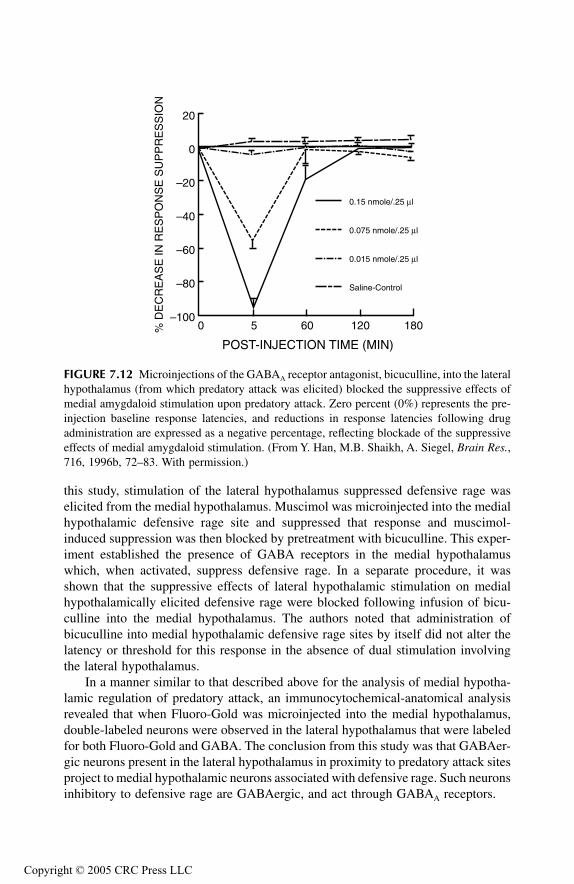
TF1661_C007.fm Page 203 Monday, October 18, 2004 8:35 AM
Copyr
this study, stimulation of the lateral hypothalamus suppressed defensive rage waselicited from the medial hypothalamus. Muscimol was microinjected into the medialhypothalamic defensive rage site and suppressed that response and muscimol-induced suppression was then blocked by pretreatment with bicuculline. This exper-iment established the presence of GABA receptors in the medial hypothalamuswhich, when activated, suppress defensive rage. In a separate procedure, it wasshown that the suppressive effects of lateral hypothalamic stimulation on medialhypothalamically elicited defensive rage were blocked following infusion of bicu-culline into the medial hypothalamus. The authors noted that administration ofbicuculline into medial hypothalamic defensive rage sites by itself did not alter thelatency or threshold for this response in the absence of dual stimulation involvingthe lateral hypothalamus.
In a manner similar to that described above for the analysis of medial hypotha-lamic regulation of predatory attack, an immunocytochemical-anatomical analysisrevealed that when Fluoro-Gold was microinjected into the medial hypothalamus,double-labeled neurons were observed in the lateral hypothalamus that were labeledfor both Fluoro-Gold and GABA. The conclusion from this study was that GABAer-gic neurons present in the lateral hypothalamus in proximity to predatory attack sitesproject to medial hypothalamic neurons associated with defensive rage. Such neuronsinhibitory to defensive rage are GABAergic, and act through GABAA receptors.
FIGURE 7.12 Microinjections of the GABAA receptor antagonist, bicuculline, into the lateralhypothalamus (from which predatory attack was elicited) blocked the suppressive effects ofmedial amygdaloid stimulation upon predatory attack. Zero percent (0%) represents the pre-injection baseline response latencies, and reductions in response latencies following drugadministration are expressed as a negative percentage, reflecting blockade of the suppressiveeffects of medial amygdaloid stimulation. (From Y. Han, M.B. Shaikh, A. Siegel, Brain Res.,716, 1996b, 72–83. With permission.)
0 5 60 120 180–100
–80
–60
–40
–20
0
20
POST-INJECTION TIME (MIN)
% D
EC
RE
AS
E IN
RE
SP
ON
SE
SU
PP
RE
SS
ION
Saline-Control
0.015 nmole/.25 µl
0.075 nmole/.25 µl
0.15 nmole/.25 µl
ight © 2005 CRC Press LLC
TF1661_C007.fm Page 204 Monday, October 18, 2004 8:35 AM
Copyr
Summary
Results of the experiments described above indicate a very fundamental and impor-tant relationship between the medial and lateral hypothalamus, which was discussedin Chapter 4. This relationship involves reciprocal connections between these tworegions of the hypothalamus involving neurons that are GABAergic. In this manner,activation of the lateral hypothalamus will suppress functions associated with themedial hypothalamus, and likewise, activation of the medial hypothalamus willinhibit functions of the lateral hypothalamus. As described in Chapter 4, this rela-tionship has important ethological significance; the importance is that it is notcompatible for both predatory attack and defensive rage to occur at the same time.Said otherwise, when one of these responses is predominant, the other must besuppressed in order for the predominant response to be most effective. This basicphysiological relationship between the medial and lateral hypothalamus most likelyhas important functional significance with respect to other processes associated withthese regions, such as feeding, drinking, temperature regulation, and endocrineregulation. It would be of great interest for investigators to be able to apply theseprinciples to the study of these functions as well. An overall summary of the basicrelationships of the amygdala, the hypothalamus and its intrinsic relationships, andPAG with respect to a number of the neurotransmitters described above are presentedin Figures 7.13 and 7.14.
CAN SUBSTANCES OF ABUSE, PSYCHOTROPIC DRUGS, AND ANTIDEPRESSANT DRUGS INFLUENCE
AGGRESSION?
SUBSTANCES OF ABUSE
Opioid Peptides
In our discussion of the role of opioid peptides in aggression and rage behaviorpresented earlier in this chapter, the general consensus reached was that opioidpeptides, which act mainly on µ and perhaps δ receptors, generally suppress rageand related responses. However, some investigators have reported a linkage betweenwithdrawal from morphine and aggression (Davis and Khalsa, 1971; Gianutsos andLal, 1978; Kantak and Miczek, 1986). Although the mechanism underlying morphinewithdrawal–induced aggression is not fully understood, it is possible that at leastone element of this process involves the absence of critical levels of enkephalins inthe CNS. Support for this notion comes, not only from the literature described earlierin this chapter, but also from a recent study (Konig et al., 1996). In this study, thepro-enkephalin gene was disrupted by the use of a homologous recombination ofembryonic stem cells, resulting in the generation of enkephalin-deficient mice. Suchmice displayed significant behavioral abnormalities, including elevated levels of bothanxiety and offensive components of aggression.
ight © 2005 CRC Press LLC
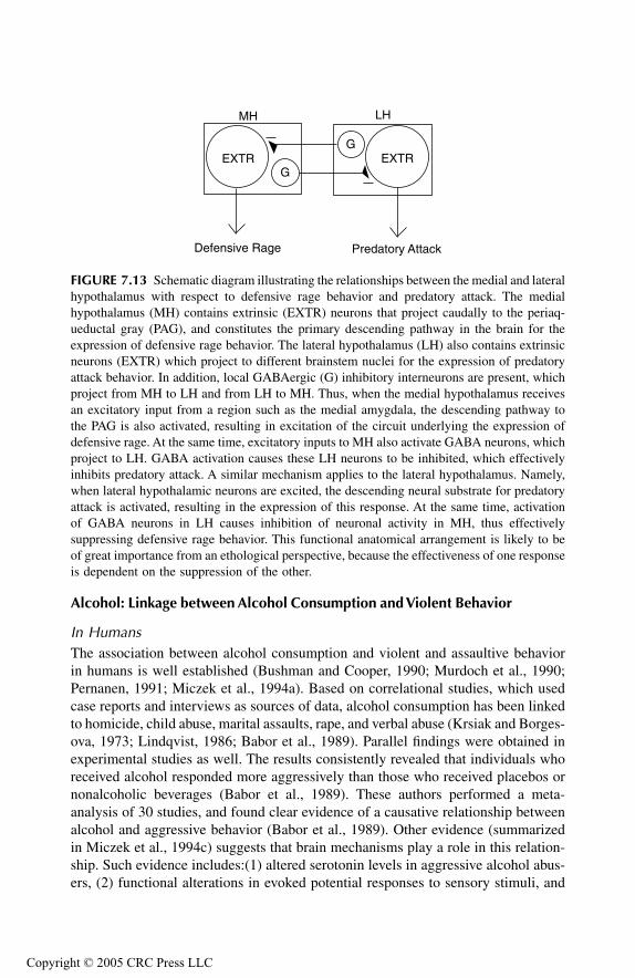
TF1661_C007.fm Page 205 Monday, October 18, 2004 8:35 AM
Copyr
Alcohol: Linkage between Alcohol Consumption and Violent Behavior
In HumansThe association between alcohol consumption and violent and assaultive behaviorin humans is well established (Bushman and Cooper, 1990; Murdoch et al., 1990;Pernanen, 1991; Miczek et al., 1994a). Based on correlational studies, which usedcase reports and interviews as sources of data, alcohol consumption has been linkedto homicide, child abuse, marital assaults, rape, and verbal abuse (Krsiak and Borges-ova, 1973; Lindqvist, 1986; Babor et al., 1989). Parallel findings were obtained inexperimental studies as well. The results consistently revealed that individuals whoreceived alcohol responded more aggressively than those who received placebos ornonalcoholic beverages (Babor et al., 1989). These authors performed a meta-analysis of 30 studies, and found clear evidence of a causative relationship betweenalcohol and aggressive behavior (Babor et al., 1989). Other evidence (summarizedin Miczek et al., 1994c) suggests that brain mechanisms play a role in this relation-ship. Such evidence includes:(1) altered serotonin levels in aggressive alcohol abus-ers, (2) functional alterations in evoked potential responses to sensory stimuli, and
FIGURE 7.13 Schematic diagram illustrating the relationships between the medial and lateralhypothalamus with respect to defensive rage behavior and predatory attack. The medialhypothalamus (MH) contains extrinsic (EXTR) neurons that project caudally to the periaq-ueductal gray (PAG), and constitutes the primary descending pathway in the brain for theexpression of defensive rage behavior. The lateral hypothalamus (LH) also contains extrinsicneurons (EXTR) which project to different brainstem nuclei for the expression of predatoryattack behavior. In addition, local GABAergic (G) inhibitory interneurons are present, whichproject from MH to LH and from LH to MH. Thus, when the medial hypothalamus receivesan excitatory input from a region such as the medial amygdala, the descending pathway tothe PAG is also activated, resulting in excitation of the circuit underlying the expression ofdefensive rage. At the same time, excitatory inputs to MH also activate GABA neurons, whichproject to LH. GABA activation causes these LH neurons to be inhibited, which effectivelyinhibits predatory attack. A similar mechanism applies to the lateral hypothalamus. Namely,when lateral hypothalamic neurons are excited, the descending neural substrate for predatoryattack is activated, resulting in the expression of this response. At the same time, activationof GABA neurons in LH causes inhibition of neuronal activity in MH, thus effectivelysuppressing defensive rage behavior. This functional anatomical arrangement is likely to beof great importance from an ethological perspective, because the effectiveness of one responseis dependent on the suppression of the other.
Defensive Rage Predatory Attack
MH LH
EXTR EXTRG
G
ight © 2005 CRC Press LLC

TF1661_C007.fm Page 206 Monday, October 18, 2004 8:35 AM
Copyr
(3) a possible common genetic basis for alcoholism and antisocial personality dis-order.
In Rodents, Monkeys, and CatsVarious animal studies have, in general, also provided evidence for potentiation ofaggressive behavior following alcohol consumption. Such effects were most clearlyobserved with respect to defensive behavior in rodents (Chance et al., 1973; Krsiak,1976; Miczek and O’Donnell, 1980; Blanchard et al., 1986, 1987, 1993a; Miczeket al., 1994b), and agonistic responses in a social setting in squirrel monkeys(Winslow et al., 1988; Weerts et al., 1993). However, not all studies have provideduniform results. Other investigators have shown that alcohol may, in fact, suppressaggressive behavior in rodents and primates (Krsiak and Borgesova, 1973; Crowleyet al., 1974; Lagerspetz and Ekqvist, 1978; Smoothy et al., 1982; Benton andSmoothy, 1984; Blanchard et al., 1986; Mos and Olivier, 1988). Moreover, severalof these studies showed that the effects in rodents appear to be dose dependent:suppression typically follows administration of relatively higher doses of alcohol(e.g., 2 g/kg). Dose-related effects were also observed in cats following administra-tion of ethanol: a low dose (0.37 g/kg) facilitated and a higher dose (1.50 g/kg)suppressed defensive rage behavior elicited by electrical stimulation of the medialhypothalamus (MacDonnell et al., 1971a, 1971b). In light of these observations, Iplan to use a wider dose range of ethanol than was used in our pilot experiments inorder to more fully examine its modulating effects. It has been further shown thatsuch variations in the effects of alcohol on aggressive behavior may be due toindividual differences in the actions of alcohol on the brain (Miczek et al., 1994b,
FIGURE 7.14 Summary of functional relationship of several key amygdaloid nuclei withrespect to how they regulate defensive rage and predatory attack. As summarized in Figure7.9, activation of the medial amygdala (ME) excites medial hypothalamic neurons (MH) viaa substance P (SP)-NK1 receptor mechanism. This drives the defensive rage mechanism byvirtue of its descending pathway to the periaqueductal gray (PAG), whose effects are mediatedlargely through excitatory amino acids (EAA) acting on NMDA receptors in the PAG. At thesame time, activation of MH causes suppression of LH and the predatory attack mechanism,as described in Figure 7.13. In contrast, activation of neurons in the central amygdala (CE)causes marked suppression of defensive rage via a direct pathway from CE to the PAG. Theseinhibitory effects are mediated through an enkephalinergic mechanism acting on µ-opioidreceptors in the PAG.
ight © 2005 CRC Press LLC
TF1661_C007.fm Page 207 Monday, October 18, 2004 8:35 AM
Copyr
1994c). These authors further speculated that several of the brain mechanismsmediating aggression and which are affected by ethanol include GABA, 5-HT, anddopaminergic systems.
In a more recent study, it was discovered that ethanol (administered peripherally)oppositely affects two forms of aggressive behavior by potentiating defensive rageand suppressing predatory attack (Schubert et al., 1996a). In this paper, it wassuggested that one mechanism underlying alcohol’s potentiation of defensive ragebehavior is that it acts on the descending pathway from the medial hypothalamusto the PAG. Recall that this pathway uses excitatory amino acids as a neurotrans-mitter. Confirmation of this hypothesis was obtained from data showing that thepotentiating effects of alcohol on defensive rage were blocked following adminis-tration of the NMDA antagonist, AP-7, into the PAG. The significance of this findingwas that it identified a specific component of the neural circuit for defensive ragebehavior over which the potentiating effects of alcohol are mediated.
Technical ConcernsAs noted above, progress has been made in demonstrating alcohol’s role in poten-tiating aggressive behavior and some data have further indicated a component of theneural circuit mediating these effects. In spite of the efforts made by various inves-tigators in this field, we still know very little about how alcohol affects brain cellsin modulating aggression as well as the overall circuitry and receptors implicatedin this process. There are several reasons that may account for the slow progresshere. One involves the route of administration. A common approach has been toemploy an intraperitoneal approach. Although successful experiments have beengenerated using this route, it does raise several concerns. In particular, it leaves openthe possibility that a component of the observed effects may be due to peripheralrather than central effects of alcohol delivery, raising an issue that is not easilysolved. The second is that peripheral administration may result in the absorption ofalcohol directly into fat pads, reducing the amount actually taken into the circulatorysystem. However, this latter concern can be partially handled by directly measuringlevels of blood alcohol. A second reason is that it is not easy to administer alcoholby mouth. It is somewhat unpleasant to the animal for it to be force-fed because ofthe restraint required as well as the fact that animals generally dislike the taste. Ifalcohol is included in a more pleasant liquid solution, the amount ingested remainsdependent on the animal’s decision. In this instance, there may be considerabledifferences in blood alcohol levels among different animals in the same as well asdifferent experimental and control conditions. A third reason is that, in contrast tothe many successful studies employing direct intracerebral administration of selec-tive receptor–neurotransmitter compounds into discrete regions of the brain (asdescribed in this chapter), such has not been the case with alcohol. The difficultystems in part from the fact that alcohol diffuses quite extensively when microinjectedinto the brain. Therefore, if low concentrations are injected, it is difficult to determinehow far it may spread, and it is also difficult if not impossible to tell whether theamount approaches physiological levels. The application of higher levels into thebrain raises the specter of introducing a toxic substance in brain cells. Hopefully,new approaches will be developed that will overcome these pitfalls.
ight © 2005 CRC Press LLC

TF1661_C007.fm Page 208 Monday, October 18, 2004 8:35 AM
Copyr
Psychotropic and Antidepressant Drugs
Over the years, various investigators have attempted to determine how antidepres-sants, tranquilizers, and psychotropic drugs affect rage and aggression. Typically,several different strategies have been employed. In one, several forms of aggressivebehavior were recorded as part of a battery of psychological and behavioral testsagainst different drugs. Here, the purpose of using a given drug was to determineits efficacy in treating a specific disorder. In a second strategy, the approach hasbeen to use a specific drug as a tool in order to learn how specific neurotransmitter—receptors may affect aggression and rage. Various studies using such drugs weredescribed earlier in this chapter. One caveat related to our interpretation of thesestudies is that many of them describe inhibitory effects of these drugs on aggressiveprocesses. The problem in interpreting such data is that it is difficult to discernwhether the inhibitory effects were due to specific actions of the drug on aggressivebehavior, or include a wider body of motor and behavioral processes. Unfortunately,few of these studies have addressed this issue.
Several early studies considered the role of several antidepressants, includingimipramine, desimipramine, and amytryptyline (Baxter, 1968b; Funderburk et al.,1970; Dubinsky and Goldberg, 1971; Dubinsky et al., 1971, 1973). The data fromthese studies suggested that imipramine and desimipramine suppress predatoryattack, but have little effect on defensive rage. However, in another study, it wasobserved that a lower dose of imipramine facilitated defensive rage, while a higherdose suppressed this response (Penaloza-Rojas et al., 1961). The antidepressantfluvoxamine was tested at a clinically relevant dose in rats, and was found to suppressbiting attack elicited by hypothalamic stimulation (Kruk et al., 1987; Olivier et al.,1990b; Kruk, 1991).
Various investigators have explored the role of benzodiazepine drugs acting onthe GABA receptor complex on aggression and rage in several species. In general,the effects of administration of this class of drugs, including diazepam, oxazepam,and etizolam, have been to suppress defensive rage and fighting in rats (Panksepp,1971); cats (Baxter, 1964; Malick, 1970; Funderburk et al., 1970; Fukuda andTsumagari, 1983); and monkeys (Delgado, 1973). In the rat, it may be that the effectsof this drug act mainly on the lateral threat component rather than on biting andfighting, per se. While suppression has been the main effect following administrationof this drug, in one study using rats, chlordiazepoxide was reported to facilitatepredatory attack (Kruk et al., 1979). However, the overall effects of benzodiazepineson aggression appear to mirror the inhibitory effects of GABA agonists on aggressionand defense.
Other drugs have also been tested for their possible effects on aggression andrage. In one study, carbamazepine, which resembles tricyclic antidepressants instructure, and has been used for the treatment of both epilepsy and mood disorders,was reported to powerfully suppress defensive rage in the cat while having little orno effect on predatory attack (Shaikh et al., 1987). In contrast, when tested in therat, carbamazepine appeared to have little effect on biting attack (Kruk et al., 1987).
ight © 2005 CRC Press LLC
TF1661_C007.fm Page 209 Monday, October 18, 2004 8:35 AM
Copyr
The general approach to the study of drugs and aggression has been to administersuch drugs and observe their effects on aggression and defense. In a recent seriesof studies, a somewhat different strategy has been adopted in order to identify therelationship between benzodiazepines and aggression (Adamec, 1993a; 1993b,1997). In these studies, systemic administration of the β-carboline and inversebenzodiazepine agonist, FG 7142 (N-methyl-β-carboline 3 carboxamide), generallypotentiated defensive behavior but suppressed predatory behavior in cats. Withrespect to defensive behavior, the facilitatory effects of FG 7142 were blockedfollowing treatment with the benzodiazepine antagonist, flumazenil.
It is of interest to point out that Adamec showed that the evoked potentialrecorded in the medial hypothalamus following stimulation of the basal or medialamygdala and administration of FG 7142 resulted in an increase in the size of theevoked potential. Since I indicated previously in this chapter and elsewhere that thestrial pathway from the medial amygdala to the medial hypothalamus mediates thefacilitatory effects of the medial amygdala, it would appear that the potentiatingeffects of FG 7142 can be explained in terms of the presumably excitatory actionsof this drug on neurons in the medial and basal amygdala that affect the medialhypothalamus via this pathway. One other feature of these findings, namely thedifferential effects of FG 7142 on naturally occurring defensive rage and predatoryattack, may also be accounted for in terms of our understanding of the anatomicalrelationships of the hypothalamus. The likely explanation for these effects is that ifFG 7142 has excitatory effects on medial and basal amygdaloid neurons, which giverise to the stria terminalis, the net effect will be excitation of the medial hypothalamusand potentiation of the defensive rage mechanism. Since I have described earlier inthis chapter and elsewhere that the medial hypothalamus contains GABAergic (inhib-itory) neurons that supply the lateral hypothalamus, it is appropriate to concludethat the suppressive effects of this drug on predatory attack is the result of indirectactivation of these GABAergic neurons by axons of the stria terminalis.
SUMMARY
In this section, I examined results of studies on several classes of drugs of abuse aswell as psychotropic and antidepressant drugs. The general findings suggest that opioidstend to suppress aggressive reactions, while alcohol potentiates rage responses. How-ever, individual differences appear to play a significant role in determining whetheralcohol will have a potentiating or inhibiting effect on aggressive behavior. Moreover,the effects may be further related to the type of aggression considered, as alcoholappears to potentiate defensive rage but inhibit predatory attack. With respect to psy-chotropic and antidepressant drugs, the overall picture is that these drugs suppressaggressive reactions. However, a basic difficulty with many of these studies in con-cluding that such drugs specifically affect aggression and rage is that one may not beable to factor out nonspecific suppressive effects of these drugs on many motor andother functions. Accordingly, at the present time, there is no known drug available thatacts specifically against one or more forms of aggressive behavior.
ight © 2005 CRC Press LLC
TF1661_C007.fm Page 210 Monday, October 18, 2004 8:35 AM
Copyr
REFERENCES
Abe, Y., T. Tadano, A. Yonezawa, K. Kisara, Suppressive effects of intraventricular injecteddopamine and nomifensine on muricide induced by thiamine deficiency, Pharmacol.Biochem. Behav., 26, 1987, 77–81.
Adamec, R.E., Transmitter systems involved in neural plasticity underlying increased anxietyand defense — implications for understanding anxiety following traumatic stress,Neurosci. Biobehav. Rev., 21, 1997, 755–765.
Adamec, R.E., Lasting effects of FG–7142 on anxiety, aggression and limbic physiology inthe cat, J. Psychopharmacol., 7, 1993a, 232–248.
Adamec, R.E., Partial limbic kindling — brain, behavior, and the benzodiazepine receptor,Physiol. Behav., 54, 1993b, 531–545.
Adamec, R.E., T. Shallow, J. Budgell, Blockade of CCKB but not CCKA receptors before andafter the stress of predator exposure prevents lasting increases in anxiety-like behav-ior: implications for anxiety associated with posttraumatic stress disorder, Behav.Neurosci., 111, 1997, 435–449.
Adams, D.B., W. Boudreau, C.W. Cowan, C. Kokonowski, K. Oberteuffer, K. Yohay, Offenseproduced by chemical stimulation of the anterior hypothalamus of the rat, Physiol.Behav., 53, 1993, 1127–1132.
Albert, D.J. and S.E. Richmond, Reactivity and aggression in the rat: induction by alpha-adrenergic blocking agents injected ventral to anterior septum but not into lateralseptum, J. Comp. Physiol. Psychol., 91, 1977, 886–896.
Albert, D.J., R.C.K. Wong, K.N. Brayley, H.C. Fibiger, Evaluation of adrenergic, cholinergicand dopaminergic involvement in the inhibition of hyperreactivity and interanimalaggression by the medial hypothalamus in the rat, Pharmacol. Biochem. Behav., 11,1979, 1–10.
Allikmets, L.H., Cholinergic mechanisms in aggressive behaviour, Med. Biol., 52, 1974,19–30.
Babor, T.F., H.R. Kranzler, R.J. Lauerman, Early detection of harmful alcohol consumption:Comparison of clinical laboratory and self report screening procedures, Addict.Behav., 14, 1989, 139–157.
Bandler, R. and R. Halliday, Lateralized loss of biting attack patterned reflexes followinginduction of contralateral sensory neglect in the cat: a possible role for the striatumin centrally elicited aggressive behaviour, Brain Res., 242, 1982, 165–177.
Barnett, A., J.B. Malick, R.I. Taber, Effects of antihistamines on isolation-induced fightingin mice, Psychopharmacologia, 19, 1971, 359–365.
Barr, G.A., J.L. Gibbons, W.H. Bridger, A comparison of the effects of acute and subacuteadministration of beta-phenylethylamine and d-amphetamine on mouse killing behav-ior of rats, Pharmacol. Biochem. Behav., 11, 1979, 419–422.
Barrett, J., H. Edinger, A. Siegel, Intrahypothalamic injections of norepinephrine facilitatefeline affective aggression via alpha-2 adrenoceptors, Brain Res., 525, 1990, 285–293.
Barrett, J.A., M.B. Shaikh, H. Edinger, A. Siegel, The effects of intrahypothalamic injectionsof norepinephrine upon affective defense behavior in the cat, Brain Res., 426, 1987,381–384.
Baxter, B.L. The effect of chlordiazepoxide on the hissing response elicited via hypothalamicstimulation, Life Sci., 3, 1964, 531–537.
Baxter, B.L., Elicitation of emotional behavior by electrical or chemical stimulation appliedat the same loci in cat mesencephalon, Exp. Neurol., 21, 1968a, 1–10.
Baxter, B.L., The effect of selected drugs on the “emotional” behavior elicited via hypotha-lamic stimulation, Int. J. Neuropharmacol., 7, 1968b, 47–54.
ight © 2005 CRC Press LLC

TF1661_C007.fm Page 211 Monday, October 18, 2004 8:35 AM
Copyr
Beart, P.M., L.S. Nicolopoulos, D.C. West, P.M. Headley, An excitatory amino acid projectionfrom ventromedial hypothalamus to periaqueductal gray in the rat: autoradiographicand electrophysiological evidence, Neurosci. Lett., 85, 1988, 205–211.
Beart, P.M., R.J. Summers, J.A. Stephenson, C.J. Cook, M.J. Christie, Excitatory amino acidprojections to the periaqueductal gray in the rat: a retrograde transport study utilizingd(3H)aspartate and, 3H,GABA, Neuroscience, 34, 1990, 163–176.
Beitz, A.J., Possible origin of glutamatergic projections to the midbrain periaqueductal grayand deep layer of the superior colliculus of the rat, Brain Res. Bull., 23, 1989, 25–35.
Beleslin, D.B. and R. Samardzic, Effects of parachlorophenylalanine and 5,6-dihydrox-ytryptamine on aggressive behaviour evoked by cholinomimetics and anticholinest-erases injected into the cerebral ventricles of conscious cats, Neuropharmacology,18, 1979, 251–257.
Beleslin, D.B., D. Jovanovic-Micic, N. Japundzic, A.M. Terzic, R. Samardzic, Behavioral,autonomic and motor effects of neuroleptic drugs in cats: motor impairment andaggression, Brain Res. Bull., 15, 1985, 353–356.
Benton, D. and R. Smoothy, The relationship between blood alcohol levels and aggressionin mice, Physiol. Behav., 33, 1984, 757–760.1.
Berntson, G.G., M.S. Beattie, J.M. Walker, Effects of nicotinic and muscarinic compoundson biting attack in the cat, Pharmacol. Biochem. Behav., 5, 1976, 235–239.
Berntson, G.G. and S.F. Leibowitz, Biting attack in cats: evidence for central muscarinicmediation, Brain Res., 51, 1973, 366–370.
Blanchard, R., Differential effects of benzodiazepines and 5-HT 1A agonists on defensivepatterns in wild rats, in P. Bevan, A. Cools, T. Archer, Eds., Behavioural Pharmacol-ogy of 5-HT, Lawrence Erlbaum Associates, Hillsdale, N.J., 1989, pp. 145–148.
Blanchard, R.J., C.D. Blanchard, K.J. Flannelly, K. Hori, Ethanol changes patterns of defen-sive behavior in wild rats, Physiol. Behav., 38, 1986, 645–650.
Blanchard, R.J., K. Hori, C. Blanchard, Ethanol effects on aggression of rats selected fordifferent levels of aggressiveness, Pharmacol. Biochem. Behav., 27, 1987, 641–644.
Blanchard, R.J., E.B. Yudko, C.D. Blanchard, Alcohol, aggression, and the stress of subordi-nation, J. Stud. Alcohol., 11, 1993a, 146–155.
Blanchard, R.J., E.B. Yudko, R.J. Rodgers, D.C. Blanchard, Defense system psychopharma-cology: an ethological approach to the pharmacology of fear and anxiety, Behav.Brain Res., 58, 1993b, 155–165.
Block, C.H., A. Siegel, H. Edinger, Effects of amygdaloid stimulation upon trigeminal sensoryfields established during hypothalamically-elicited quiet biting attack in the cat, BrainRes., 197, 1980a, 39–55.
Block, C.H., A. Siegel, H.M. Edinger, Effects of stimulation of the substantia innominataupon attack behavior elicited from the hypothalamus in the cat, Brain Res., 197,1980b, 57–74.
Broderick, P.A. and W.H. Bridger, A comparative study of the effect of L-tryptophan and itsacetylated derivative N-acetyl-L-tryptophan on rat muricidal behavior, Biol. Psychi-atry, 19, 1984, 89–94.
Brudzynski, S.M., Carbachol-induced agonistic behavior in cats: aggressive or defensiveresponse? Acta Neurobiol. Exp., 41, 1981a, 15–32.
Brudzynski, S.M., Growling component of vocalization as a quantitative index of carbachol-induced emotional-defensive response in cats, Acta Neurobiol. Exp., 41, 1981b,33–51.
Brudzynski, S.M. and F. Bihara, Ultrasonic vocalization in rats by cholinergic stimulation ofthe brain, Neurosci. Lett., 109, 1990, 222–226.
ight © 2005 CRC Press LLC
TF1661_C007.fm Page 212 Monday, October 18, 2004 8:35 AM
Copyr
Brudzynski, S.M. and B. Eckersdorf, Vocalization accompanying emotional-aversive responseinduced by carbachol in the cat, Neuropsychopharmacology, 1, 1988, 311–320.
Brudzynski, S.M., B. Eckersdorf, H. Golebiewski, Evidence for involvement of endogenousacetylcholine in emotional-aversive response in the cat, Prog. Neuro-Psychopharma-col., Biol. Psychiatry, 14, 1990, 807–812.
Brudzynski, S.M., B. Eckersdorf, H. Golebiewski, Emotional-aversive nature of the behavioralresponse induced by carbachol in cats, J. Psychiatr. Neurosci., 18, 1, 1993, 38–45.
Brudzynski, S.M., Ultrasonic vocalization induced by intracerebral carbachol in rats: local-ization and a dose–response study, Behav. Brain Res., 63, 1994, 133–143.
Brutus, M., M.B. Shaikh, A. Siegel, H. Edinger, Effects of experimental temporal lobe seizuresupon hypothalamically elicited aggressive behavior in the cat, Brain Res., 366, 1986,53–63.
Brutus, M. and A. Siegel, Effects of the opiate antagonist naloxone upon hypothalamicallyelicited affective defense behavior in the cat, Behav. Brain Res., 33, 1989, 23–32.
Brutus, M., S. Zuabi, A. Siegel, Effects of D-ala2-met5-enkephalinamide microinjectionsplaced into the bed nucleus of the stria terminalis upon affective defense behavior inthe cat, Brain Res., 473, 1988, 147–152.
Brutus, M., S. Zuabi, A. Siegel, Microinjection of D-ala2-met5-enkephalinamide placed intothe nucleus accumbens suppresses feline affective defense behavior, Exp. Neurol.,104, 1989, 55–61.
Bushman, B.J. and H.M. Cooper, Effects of alcohol on human aggression: an integrativeresearch review, Psychol. Bull., 107, 1990, 341–354.
Cabib, S., F.R. D’Amato, S. Puglisi-Allegra, D. Maestripieri, Behavioral and mesocorticolim-bic dopamine responses to nonaggressive social interactions depend on previoussocial experiences and on the opponent’s sex, Behav. Brain Res., 112, 2000, 13–22.
Cabib, S. and S. Puglisi-Allegra, Genotype-dependent modulation of LY 171555-induceddefensive behavior in the mouse, Psychopharmacology (Berlin), 97, 1989, 166–168.
Chance, M.R.A., R.A.J.H. Mackintosh, A.K. Dixon, The effects of ethyl alcohol on socialencounters between mice, J. Alcoholism, 8, 1973, 90–93.
Cheu, J.W. and A. Siegel, GABA receptor mediated suppression of defensive rage behaviorelicited from the medial hypothalamus of the cat: role of the lateral hypothalamus,Brain Res., 783, 1998, 293–304.
Chiavegatto, S., V.L. Dawsons, L.A. Mamounas, V.E. Koliatsos, T.M. Dawson, R.J. Nelson,Brain serotonin dysfunction accounts for aggression in male mice lacking neuronalnitric oxide synthase, Proc. Natl. Acad. Sci. U.S.A., 98, 2001, 1277–1281.
Coccaro, E.F., Central serotonin and impulsive aggression, Br. J. Psychiatr., 155 (Suppl. 8),1989, 52–62.
Coccaro, E.F., S. Gabriel, L.J. Siever, Buspirone challenge: Preliminary evidence for a rolefor central 5HT–1a receptor function in impulsive aggressive behavior in humans,Psychopharmacol. Bull., 26, 1990, 405.
Coccaro, E.F., R.J. Kavoussi, T.B. Cooper, R.L. Hauger, Central serotonin activity and aggres-sion: Inverse relationship with prolactin response to d-fenfluramine, but not CSF 5-HIAA concentration, in human subjects, Am. J. Psychiatr., 154, 1997, 1430–1435.
Cologer-Clifford, A., N.G. Simon, S.F. Lu, S.A. Smoluk, Serotonin agonist-induced decreasesin intermale aggression are dependent on brain region and receptor subtype, Phar-macol. Biochem. Behav., 58, 1997, 425–430.
Cologer-Clifford, A., S.A. Smoluk, N.G. Simon, Effects of serotonergic 1A and 1B agonistsin androgenic versus estrogenic systems for aggression, Ann. N.Y. Acad. Sci., 794,1996, 339–342.
ight © 2005 CRC Press LLC
TF1661_C007.fm Page 213 Monday, October 18, 2004 8:35 AM
Copyr
Crowley, T.J., A.J. Stynes, M. Hydinger, I.C. Kaufman, Ethanol, methamphetamine, pento-barbital, morphine, and monkey social behavior, Arch. Gen. Psychiatry, 31, 1974,829–838.
Davis, W.M. and J.H. Khalsa, Increased shock induced aggression during morphine with-drawal, part 1, Life Sci., 10, 1971, 1321–1327.
De Boer, S.F., M. Lesourd, E. Mocaër, J.M. Koolhaas, Somatodendritic 5-HT1A autoreceptorsmediate the anti-aggressive actions of 5-HT1A receptor agonists in rats: an ethophar-macological study with S-15535, alnespirone, and WAY-100635, Neuropsychophar-macology, 23, 2000, 20–33.
Delgado, J.M.R., Antiaggressive effects of chlordiazepoxide, in S. Garattini, E. Mussini, L.O.Randall, Eds., The Benzodiazepines, Raven Press, New York, 1973, pp. 419–432.
Dominic, J.A. and K.E. Moore, Behavior and brain contents of catecholamines in mice duringchronic administration of methyldopa, Neuropharmacology, 10, 1971, 565–570.
Dubinsky, B. and M.E. Goldberg, The effect of imipramine and selected drugs on attackelicited by hypothalamic stimulation in the cat, Neuropharmacology, 10, 1971,537–545.
Dubinsky, B., J.K. Karpowicz, M.E. Goldberg, Effects of tricyclic antidepessants on attackelicited by hypothalamic stimulation: Relation to brain biogenic amines, J. Pharma-col. Exp. Ther., 187, 1973, 550–557.
Dubinsky, B., K. Karpowicz, M.E. Goldberg, Effects of desipramine, imipramine and chlo-rimipramine on hypothalamically-induced attack in cats, Pharmacologist, 13, 1971, 1.
Eichelmann, B., The catecholamines and aggressive behavior, Neurosci. Res., 5, 1973,109–129.
Fanselow, M.S., R. Sigmundi, R.C. Bolles, Naloxone pretreatment enhances shock-elicitedaggression, Physiol. Psychol., 8, 1980, 369–371.
Ferguson, J., S. Henriksen, H. Cohen, G. Mitchell, J. Barchas, W. Dement, “Hypersexuality”and behavioral changes in cats caused by administration of p-chlorophenylalanine,Science, 168, 1970, 499–501.
File, S.E. and J.F.W. Deakin, Chemical lesions of both dorsal and median raphe nuclei andchanges in social and aggressive behaviour in rats, Pharmacol. Biochem. Behav., 12,1980, 855–859.
Fukuda, T. and T. Tsumagari, Effects of psychotropic drugs on the rage responses inducedby electrical stimulation of the medial hypothalamus in cats, Jpn. J. Pharmacol., 33,1983, 885–890.
Funderburk, W.H., M.H. Foxwell, M.W. Hakala, Effects of psychotherapeutic drugs on hypo-thalamic-induced hissing in cats, Neuropharmacology, 9, 1970, 1–7.
Gallagher, A.W., L.A. Chahl, A.M. Lynch, Distribution of substance P-like immunoreactivityin guinea pig central nervous system, Brain Res. Bull., 29, 1992, 199–207.
Garmendia, L., J.R. Sánchez, A. Azpiroz, P.F. Brain, V.M. Simón, Clozapine: strong antiag-gressive effects with minimal motor impairment, Physiol. Behav., 51, 1992, 51–54.
Gay, P.E. and R.C. Leaf, Rat strain differences in pilocarpine-induced mouse killing, Physiol.Psychol., 4, 1976, 28–32.
Gay, P.E., R.C. Leaf, F.B. Arble, Inhibitory effects of pre- and posttest drugs on mouse-killingby rats, Pharmacol. Biochem. Behav., 3, 1975, 33–45.
Gay, P.E., L.S. Potter, J.A. Consalvi, R.C. Leaf, The effects of d-amphetamine on prey killingand prey eating in the rat and mouse, Bull. Psychonom. Soc., 10, 1977, 385–388.
George, R., W.L. Haslett, D.J. Jenden, The central action of a metabolite of tremorine, LifeSci., 8, 1962, 361–363.
Geyer, M. and D. Segal, Shock-induced aggression: opposite effects of intraventricularlyinfused dopamine and norepinephrine, Behav. Biol., 10, 1974, 99–104.
ight © 2005 CRC Press LLC
TF1661_C007.fm Page 214 Monday, October 18, 2004 8:35 AM
Copyr
Gianutsos, G., M.D. Hynes, H. Lal, Enhancement of morphine-withdrawal and apomorphine-induced aggression by clonidine, Psychopharm. Comm., 2, 1976, 165–171.
Gianutsos, G. and H. Lal, Blockade of apomorphine-induced aggression by morphine orneuroleptics: differential alteration by antimuscarinics and naloxone, Pharmacol.Biochem. Behav., 4, 1976, 639–642.
Gianutsos, G. and H. Lal, Narcotic analgesics and aggression, Mod. Prob. Pharmacopsych.,13, 1978, 114–138.
Golebiewski, H. and A. Romaniuk, The participation of serotoninergic system in the regulationof emotional-defensive behavior evoked by intrahypothalamic carbachol injectionsin the cat, Acta Neurobiol. Exp., 45, 1985, 25–36.
Hadfield, M.G., Mesocortical vs nigrostriatal dopamine uptake in isolated fighting mice, BrainRes., 222, 1981, 172–176.
Hadfield, M.G., Dopamine: mesocortical vs nigrostriatal uptake in isolated fighting mice andcontrols, Behav. Brain Res., 7, 1983, 269–281.
Han, Y., M.B. Shaikh, A. Siegel, Medial amygdaloid suppression of predatory attack behaviorin the cat: I. Role of a substance P pathway from the medial amygdala to the medialhypothalamus, Brain Res., 716, 1996a, 59–71.
Han, Y., M.B. Shaikh, A. Siegel, Medial amygdaloid suppression of predatory attack behaviorin the cat: II. Role of a GABAergic pathway from the medial to the lateral hypothal-amus, Brain Res., 716, 1996b, 72–83.
Hasselager, E., Z. Rolinski, A. Randrup, Specific antagonism by dopamine inhibitors of itemsof amphetamine induced aggressive behaviour, Psychopharmacologia, 24, 1972,485–495.
Higley, J.D., S.T. King, M.F. Hasert, M. Champoux, S.J. Suomi, M. Linnoila, Stability ofinterindividual differences in serotonin function and its relationship to severe aggres-sion and competent social behavior in rhesus macaque females, Neuropsychophar-macology, 14, 1996a, 67–76.
Higley, J.D., P.T. Mehlman, S.B. Higley, B. Fernald, J. Vickers, S.G. Lindell, D.M. Taub,S.J. Suomi, M. Linnoila, Excessive mortality in young free-ranging male nonhumanprimates with low cerebrospinal fluid 5-hydroxyindoleacetic acid concentrations,Arch. Gen. Psychol., 53, 1996b, 537–543.
Higley, J.D., P.T. Mehlman, R.E. Poland, D.M. Taub, J. Vickers, S.J. Suomi, M. Linnoila,CSF testosterone and 5-HIAA correlate with different types of aggressive behaviors,Biol. Psychiatry, 40, 1996c, 1067–1082.
Igic, R., P. Stern, E. Basagic, Changes in emotional behaviour after application of cholinest-erase inhibitor in the septal and amygdala region, Neuropharmacology, 9, 1970,73–75.
Jacobs, B.L. and A. Cohen, Differential behavioral effects of lesions of the median or dorsalraphe nuclei in rats: open field and pain-elicited aggression, J. Comp. Physiol. Psy-chol., 90, 1976, 102–108.
Kantak, K.M., L.R. Hegstrand, B. Eichelman, Dietary tryptophan modulation and aggressivebehavior in mice, Pharmacol. Biochem. Behav., 12, 1979a, 675–679.
Kantak, K.M., L.R. Hegstrand, J. Whitman, B. Eichelman, Effects of dietary supplementsand a tryptophan-free diet on aggressive behavior in rats, Pharmacol. Biochem.Behav., 12, 1979b, 173–179.
Kantak, K.M. and K.A. Miczek, Aggression during morphine withdrawal: effects of methodof withdrawal, fighting experience, and social role, Psychopharmacology (Berlin),90, 1986, 451–456.
Katz, R.J. and E. Thomas, Effects of scopolamine and alpha-methylparatyrosine upon pred-atory attack in cats, Psychopharmacologia, 42, 1975, 153–157.
ight © 2005 CRC Press LLC

TF1661_C007.fm Page 215 Monday, October 18, 2004 8:35 AM
Copyr
Katz, R.J. and E. Thomas, Effects of para-chlorophenylalanine upon brain stimulated affectiveattack in the cat, Pharmacol. Biochem. Behav., 5, 1976, 391–394.
Kemble, E.D., M. Behrens, J.M. Rawleigh, B.M. Gibson, Effects of yohimbine on isolation-induced aggression, social attraction, and conspecific odor preference in mice, Phar-macol. Biochem. Behav., 40, 1991, 781–785.
Knutson, J.F., N.L. Kane, A.J. Schlosberg, D.J. Fordyce, K.J. Simansky, Influence of PCPA,shock level, and home-cage conditions on shock-induced aggression, Physiol. Behav.,23, 1979, 897–907.
Konig, M., A.M. Zimmer, H. Steiner, P.V. Holmes, J.N. Crawley, M.J. Brownstein, A. Zimmer,Pain responses, anxiety and aggression in mice deficient in pre-proenkephalin, Nature,383, 1996, 535–538.
Koprowska, M. and A. Romaniuk, Behavioral and biochemical alterations in median anddorsal raphe nuclei lesioned cats, Pharmacol. Biochem. Behav., 56, 1997, 529–540.
Korte, S.M., O.C. Meijer, E.R. De Kloet, B. Buwalda, J. Keijser, F. Sluyter, G. Van Oort-merssen, B. Bohus, Enhanced 5-HT1A receptor expression in forebrain regions ofaggressive house mice, Brain Res., 736, 1996, 338–343.
Korzan, W.J., T.R. Summers, C.H. Summers, Monoaminergic activities of limbic regions areelevated during aggression: influence of sympathetic social signaling, Brain Res.,870, 2000, 170–178.
Kostowski, W., O. Pucilowski, A. Plaznik, Effect of stimulation of brain serotonergic systemon mouse-killing behavior in rats, Physiol. Behav., 25, 1980, 161–165.
Kostowski, W. and L. Valzelli, Biochemical and behavioral effects of lesions of raphe nucleiin aggressive mice, Pharmacol. Biochem. Behav., 2, 1974, 277–280.
Kromer, W. and J.E. Dum, Mouse-killing in rats induces a naloxone-blockable increase innociceptive threshold, Eur. J. Pharmacol., 63, 1980, 195–198.
Krsiak, M., Effect of ethanol on aggression and timidity in mice, Psychopharmacology, 51,1976, 75–80.1.
Krsiak, M. and M. Borgesova, Effect of alcohol on behaviour of pairs of rats, Psychophar-macologia, 32, 1973, 201–209.
Kruk, M.R., Ethology and pharmacology of hypothalamic aggression in the rat, Neurosci.Biobehav. Rev., 15, 1991, 527–538.
Kruk, M.R., A.M. Van der Poel, T.P. De Vos-Frerichs, The induction of aggressive behaviourby electrical stimulation in the hypothalamus of male rats, Behaviour, 70, 1979,292–322.
Kruk, M.R., A.M. Van der Poel, J.H.C.M. Lammers, T. Hagg, A.M.D.M. de Hey, S. Oostwegel,Ethopharmacology of hypothalamic aggression in the rat, in B. Olivier, J. Mos, P.F.Brain, Eds., Ethopharmacology of Agonistic Behaviour in Animals and Humans,Martinus Nijhoff Publishers, Dordrecht, 1987, pp. 35–45.
Lagerspetz, K.M.J. and K. Ekqvist, Failure to induce aggression in inhibited and in geneticallynonaggressive mice through injections of ethyl alcohol, Aggress. Behav., 4, 1978,105–113.
Lal, H., J. O’Brien, S.K. Puri, Morphine-withdrawal aggression: sensitization by amphet-amines, Psychopharmacologia, 22, 1971, 217–223.
Lamprecht, F., B. Eichelman, N.B. Thoa, R.B. Williams, I.J. Kopin, Rat fighting behavior:serum dopamine-beta-hydroxylase and hypothalamic tyrosine hydroxylase, Science,177, 1972, 1214–1215.
Lanaud, P., T. Popovici, E. Normand, C. Lemoine, B. Bloch, B.P. Roques, Distribution ofCCK mRNA in particular region, hippocampus, periaqueductal grey and thalamus,of the rat by in situ hybridization, Neurosci. Lett., 104, 1989, 38–42.
ight © 2005 CRC Press LLC
TF1661_C007.fm Page 216 Monday, October 18, 2004 8:35 AM
Copyr
Langevin, H. and P.C. Emson, Distribution of substance P, somatostatin and neurotensin inthe human hypothalamus, Brain Res., 246, 1982, 65–69.
Leaf, R.C., L. Lerner, Z.P. Horovitz, Role of the amygdala in the pharmacological andendocrinological manipulation of aggression, in S. Garattini and E.B. Sigg, Eds.,Aggressive Behavior, Wiley, New York, 1969, pp. 120–131.
Leslie, G.B., Central stimulant properties of compounds with peripheral muscarinic properties,Nature, 208, 1965, 1291–1293.
M.H. Lewis, J.-L. Gariépy, P. Gendreau, D.E. Nichols, R.B. Mailman, Social reactivity andD1 dopamine receptors: studies in mice selectively bred for high and low levels ofaggression, Neuropsychopharmacology, 10, 1994, 115–122.
Lindqvist, C.U., Criminal homicide in northern Sweden 1970–1981: alcohol intoxication,alcohol abuse and mental disease, Int. J. Law Psychiatry, 8, 1986, 19–37.
Liu, H., S. Chandler, A.J. Beitz, M.T. Shipley, M.M. Behbehani, Characterization of the effectof cholecystokinin, CCK, on neurons in the periaqueductal gray of the rat: immuno-cytochemical and in vivo and in vitro electrophysiological studies, Brain Res., 642,1994, 83–94.
Lu, C.-L., M.B. Shaikh, A. Siegel, Role of NMDA receptors in hypothalamic facilitation offeline defensive rage elicited from the midbrain periaqueductal gray, Brain Res., 581,1992, 123–132.
Luo, B., J.W. Cheu, A. Siegel, Cholecystokinin B receptors in the periaqueductal gray poten-tiate defensive rage behavior elicited from the medial hypothalamus of the cat, BrainRes., 796, 1998, 27–37.
Lyons, W.E., L.A. Mamounas, G.A. Ricaurte, V. Coppola, S.W. Reid, S.H. Bora, C. Wihler,V.E. Koliatsos, L. Tessarollo, Brain-derived neurotrophic factor-deficient micedevelop aggressiveness and hyperphagia in conjunction with brain serotonergic abnor-malities, Proc. Natl. Acad. Sci. U.S.A., 96, 1999, 15239–15244.
MacDonnell, M.F., L. Fessock, S.H. Brown, Aggression and associated neural events in cats.Effects of p-chlorophenylalanine compared with alcohol, Q. J. Stud. Alcohol., 32,1971a, 748–763.
MacDonnell, M.F., L. Fessock, S.H. Brown, Ethanol and the neural substrate for affectivedefense in the cat, Q. J. Stud. Alcohol., 32, 1971b, 406–419.
Maeda, H., Effects of psychotropic drugs upon the hypothalamic rage response in cats, FoliaPsychiatr. Neurol. Jpn., 30, 1976, 539–546.
Maeda, H. and S. Maki, Dopaminergic facilitation of recovery from amygdaloid lesions whichaffect hypothalamic defensive attack in cats, Brain Res., 363, 1986, 135–140.
Maeda, H., T. Sato, S. Maki, Effects of dopamine agonists on hypothalamic defensive attackin cats, Physiol. Behav., 35, 1985, 89–92.
Malick, J.B., Effects of selected drugs on stimulus-bound emotional behavior elicited byhypothalamic stimulation in the cat, Arch. Int. Pharmacodyn. Ther., 186, 1970,137–141.
Malick, J.B., Differential effects of d- and l-amphetamine on mouse-killing behavior in rats,Pharmacol. Biochem. Behav., 3, 1975, 697–699.
Malick, J.B. and A. Barnett, The role of serotonergic pathways in isolation-induced aggressionin mice, Pharmacol. Biochem. Behav., 5, 1976, 55–61.
Marini, J.L., J.K. Walters, M.H. Sheard, Effects of d- and l-amphetamine on hypothalamically-elicited movement and attack in the cat, Agressologie, 20, 1979, 155–160.
Mark, V.H., I. Takada, H. Tsutsumi, H. Takamatsu, E. Toth, D.B. Mark, Effect of exogenouscatecholamines in the amygdala of a ‘rage’ cat, Appl. Neurophysiol., 38, 1975, 61–72.
ight © 2005 CRC Press LLC
TF1661_C007.fm Page 217 Monday, October 18, 2004 8:35 AM
Copyr
Marshall, J.F., Somatosensory inattention after dopamine-depleting intracerebral 6-OHDAinjections: spontaneous recovery and pharmacological control, Brain Res., 177, 1979,311–324.
Mayer, M.L. and N.K. MacLeod, The excitatory action of substance P and stimulation of thestria terminalis bed nucleus on preoptic neurones, Brain Res., 166, 1979, 206–210.
Mercer, L.D., P.M. Beart, M.K. Horne, D.I. Finkelstein, P. Carrive, G. Paxinos, On thedistribution of cholecystokinin B receptors in monkey brain, Brain Res., 738, 1996,313–318.
Messeri, P., B.E. Eleftheriou, A. Oliverio, Dominance behavior: a phylogenetic analysis inthe mouse, Physiol. Behav., 14, 1975, 53–58.
Miczek, K.A., J.L. Altman, J.B. Appel, W.O. Boggan, Para-chlorophenylalanine, serotoninand killing behavior, Pharmacol. Biochem. Behav., 3, 1975, 355–361.
Miczek, K.A., J.F. DeBold, M. Haney, J. Tidey, J. Vivian, E. Weerts, Alcohol, drugs of abuse,aggression, and violence, in A.J. Reiss and J.A. Roth, Eds., Understanding andPreventing Violence, vol. 3, National Academy Press, Washington, D.C., 1994a, pp.377–570.
Miczek, K.A., J.F. DeBold, A.M.M. Van Erp, Neuropharmacological characteristics of indi-vidual differences in alcohol effects on aggression in rodents and primates, Behav.Pharmacol., 5, 1994b, 407–421.
Miczek, K.A., M. Haney, J. Tidey, J. Vivian, E. Weerts, Neurochemistry and pharmacother-apeutic management of aggression and violence, in A. Reiss, K. Miczek, J. Roth,Eds., Understanding and Preventing Violence, vol. 2, National Academy Press, Wash-ington, D.C., 1994c, pp. 245–514.
Miczek, K.A. and J.M. O’Donnell, Alcohol and chlordiazepoxide increase suppressed aggres-sion in mice, Psychopharmacology (Berlin), 69, 1980, 39–44.
Miyachi, Y.H., Kasai, H. Ohyama, T. Yamada, Changes of aggressive behavior and brainserotonin turnover after very low-dose X-irradiation of mice, Neurosci. Lett., 175,1994, 92–94.
Moore, T.M., A. Scarpa, A. Raine, A meta-analysis of serotonin metabolite 5-HIAA andantisocial behavior, Aggress. Behav., 28, 2002, 299–316.
Mos, J. and B. Olivier, Differential effects of selected psychoactive drugs on dominant andsubordinate male rats housed in a colony, Neurosci. Res. Commun., 2, 1988, 29–36.
Mos, J., B. Olivier, M. Poth, R. Van Oorschot, H. Van Aken, The effects of dorsal rapheadministration of eltoprazine, TFMPP and 8-OH-DPAT on resident intruder aggres-sion in the rat, Eur. J. Pharmacol., 238, 1993, 411–415.
Moyer, K.E., and J.M. Crabtree, Sex differences in fighting and defense induced in rats byshock and d-amphetamine during morphine abstinence, Physiol. Behav., 11, 1973,337–343.
Murdoch, D., R.O. Pihl, D. Ross, Alcohol and crimes of violence: present issues, Int. J.Addict., 25, 1990, 1059–1075.
Myers, R.D., Emotional and autonomic responses following hypothalamic chemical stimula-tion, Can. J. Psychol., 18, 1964, 6–14.
Nakamura, K. and H. Thoenen, Increased irritability: a permanent change induced in the ratby intraventricular administration of 6-hydroxydopamine, Psychopharmacologia, 24,1972, 359–372.
Nakao, H., N. Tashiro, R. Kono, R. Araki, Effects of GABA and glycine on aggressive-defensereaction produced by electrical stimulation of the ventromedial hypothalamus in cats,in M. Itoh, Ed., Integrative Control Functions of the Brain, Elsevier, Amster-dam/Tokyo, 1979, pp. 332–334.
ight © 2005 CRC Press LLC
TF1661_C007.fm Page 218 Monday, October 18, 2004 8:35 AM
Copyr
Navarro, J.J., E. Maldonado, D. Beltrán, Effects of methyl-chlorophenylpiperazine, m-CPP,on isolation-induced aggression in male mice, Med. Sci. Res., 27, 1999, 661–662.
Olivier, B., J. Mos, D.L. Rasmussen, Behavioural pharmacology of the serenic, eltoprazine,in M. Raghoebar, B. Olivier, D.L. Rasmussen, J. Mos, Eds., Drug Metabolism andDrug Interactions, Freund, London, 1990a, pp. 31–83.
Olivier, B., J. Mos, M. Tulp, J. Schipper, S. DenDaas, G. Van Oortmerssen, Serotonergicinvolvement in aggressive behaviour in animals, in H.M. Van Praag, Ed., Monoam-inergic Regulation of Aggression and Impulse Control, ACNP Proceedings, Wash-ington, D.C., 1990b, pp. 79–137.
Otsuka, M. and K. Yoshioka, Neurotransmitter functions of mammalian tachykinins, Physiol.Rev., 73, 1993, 229–308.
Panksepp, J,.Drugs and stimulus-bound attack, Physiol. Behav., 6, 1971, 317–320.Parmigiani, S. and P. Palanza, Fluprazine inhibits intermale attack and infanticide, but not
predation, in male mice, Neurosci. Biobehav. Rev., 15, 1991, 511–513.Penaloza-Rojas, J.H., G. Bach-Y-Rita, H.F. Rubio-Chevannier, R. Hernandez-Peon, Effects
of imipramine upon hypothalamic and amygdaloid excitability, Exp. Neurol., 4, 1961,205–213.
Pernanen, K., Alcohol in Human Violence, Gilford Press, New York, 1991.Pott, C., S. Kramer, A. Siegel, Naloxone sensitivity of stimulation sites in the central gray
effective in modulating feline aggression, Physiol. Behav., 40, 1986, 207–213.Pradhan, S.N., Aggression and central neurotransmitters, Intl. Rev. Neurobiol., 18, 1973,
213–262.Pucilowski, O., B. Eichelman, D.H. Overstreet, A.H. Rezvani, D.S. Janowsky, Enhanced
affective aggression in genetically bred hypercholinergic rats, Neuropsychobiology,24, 1991, 37–41.
Pucilowski, O., A. Plaznik, W. Kostowski, Aggressive behavior inhibition by serotonin andquipazine injected into the amygdala in the rat, Behav. Neural Biol., 43, 1985, 58–68.
Puglisi-Allegra, S. and S. Cabib, Pharmacological evidence for a role of D2 dopaminereceptors in the defensive behavior of the mouse, Behav. Neural Biol., 50, 1988,98–111.
Puglisi-Allegra, S., A. Oliverio, P. Mandel, Effects of opiate antagonists on social and aggres-sive behavior of isolated mice, Pharmacol. Biochem. Behav., 17, 1982, 691–694.
Rao, R., M. Yamano, S. Shiosaka, A. Shinohara, M. Tohyama, Origin of leucine-enkephalinfibers and their two main afferent pathways in the bed nucleus of the stria terminalisin the rat, Exp. Brain Res., 65, 1987, 411–420.
Reichling, D.B. and A.I. Basbaum, Contribution of brainstem GABAergic circuitry todescending antinocicieptive controls: I. GABA-immunoreactive projection neuronsin the periaqueductal gray and nucleus raphe magnus, J. Comp. Neurol., 302, 1990,370–377.
Reichling, D.B. and A.I. Basbaum, Collateralization of periaqueductal gray neurons to fore-brain or diencephalon and to the medullary nucleus raphe magnus in the rat, Neuro-science, 42, 1991, 183–200.
Reis, D.J., Central neurotransmitters in aggression, Res. Publ. Assoc. Res. Neuro. Ment. Dis.,52, 1974, 119–148.
Reis, D.J. and K. Fuxe, Depletion of noradrenaline in brainstem neurons during sham ragebehaviour produced by acute brainstem transection in cat, Brain Res., 7, 1968,448–451.
Reis, D.J. and K. Fuxe, Brain norepinephrine: evidence that neuronal release is essential forsham rage behavior following brainstem transection in cat, Proc. Natl. Acad. Sci.U.S.A., 64, 1969, 108–112.
ight © 2005 CRC Press LLC

TF1661_C007.fm Page 219 Monday, October 18, 2004 8:35 AM
Copyr
Reis, D.J. and L.-M. Gunne, Brain catecholamines: Relation to the defense reaction evokedby amygdaloid stimulation in cat, Science, 149, 1965, 450–451.
Rodgers, R.J., The medial amygdala: serotonergic inhibition of shock-induced aggression andpain sensitivity in rats, Aggress. Behav., 3, 1977, 277–288.
Rodgers, R.J., D.C. Blanchard, L.K. Wong, R.J. Blanchard, Effects of scopolamine on anti-predator defense reactions in wild and laboratory rats, Pharmacol. Biochem. Behav.,36, 1990, 575–583.
Rodgers, R.J. and K. Brown, Amygdaloid function in the central cholinergic mediation ofshock-induced aggression in the rat, Aggress. Behav., 2, 1976, 131–152.
Rodgers, R.J. and C.A. Hendrie, Agonistic behaviour in rats: evidence for non-involvementof opioid mechanisms, Physiol. Behav., 29, 1982, 85–90.
Roeling, T.A.P., M.R. Kruk, R. Schuurmans, J.G. Veening, Behavioral responses of bicucullinemethiodide injections into the ventral hypothalamus of freely moving, socially inter-acting rats, Brain Res., 615, 1993, 121–127.
Romaniuk, A., S. Brudzynski, J. Gronska, The effect of chemical blockade of hypothalamiccholinergic system on defensive reactions in cats, Acta Physiol. Pol., 24, 1973,809–816.
Romaniuk, A., S. Brudzynski, J. Gronska, The effects of intrahypothalamic injections ofcholinergic and adrenergic agents on defensive behavior in cats, Acta Physiol. Pol.,25, 1974, 297–305.
Romaniuk, A. and H. Golebiewski, Adrenergic modulation of the hypothalamic cholinergicmechanism in the control of emotional-defensive behavior in the cat, Acta Neurobiol.Exp., 39, 1979, 313–326.
Sánchez, C., J. Arnt, J. Hyttel, E.K. Moltzen, The role of serotonergic mechanisms in inhibitionof isolation-induced aggression in male mice, Psychopharmacology (Berlin), 110,1993, 53–59.
Saudou, F., D.A. Amara, A. Dierich, M. Lemeur, S. Ramboz, L. Segu, M.C. Buhot, R. Hen,Enhanced aggressive behavior in mice lacking the 5ht1b receptor, Science, 265, 1994,1875–1878.
Schubert, K., M.B. Shaikh, Y. Han, L. Pohorecky, A. Siegel, Differential effects of ethanolon feline rage and predatory attack behavior: an underlying neural mechanism, Alco-hol. Clin. Exp. Res., 20, 1996a, 882–889.
Schubert, K., M.B. Shaikh, A. Siegel, NMDA receptors in the midbrain periaqueductal graymediate hypothalamically evoked hissing behavior in the cat, Brain Res., 726, 1996b,80–90.
Shaikh, M.B., M. Brutus, A. Siegel, H.E. Siegel, Regulation of feline aggression by the bednucleus of stria terminalis, Brain Res. Bull., 16, 1986, 179–182.
Shaikh, M.B., N.C. De Lanerolle, A. Siegel, Serotonin 5-HT 1A and 5-HT2/1C receptors in themidbrain periaqueductal gray differentially modulate defensive rage behavior elicitedfrom the medial hypothalamus of the cat, Brain Res., 765, 1997, 198–207.
Shaikh, M.B., H. Edinger, A. Siegel, Carbamazepine regulates feline aggression elicited fromthe midbrain periaqueductal gray, Pharmacol. Biochem. Behav., 30, 1987, 409–415.
Shaikh, M.B., C.-L. Lu, A. Siegel, Affective defense behavior elicited from the feline midbrainperiaqueductal gray is regulated by mu- and delta-opioid receptors, Brain Res., 557,1991a, 344–348.
Shaikh, M.B., C.-L. Lu, A. Siegel, D-2 dopamine receptor involvement in the regulation ofquiet biting attack behavior in the cat, Brain Res. Bull., 27, 1991b, 725–730.
Shaikh, M.B., C.-L. Lu, A. Siegel, An enkephalinergic mechanism involved in amygdaloidsuppression of affective defense behavior elicited from the midbain periaqueductalgray in the cat, Brain Res., 559, 1991c, 109–117.
ight © 2005 CRC Press LLC
TF1661_C007.fm Page 220 Monday, October 18, 2004 8:35 AM
Copyr
Shaikh, M.B., K. Schubert, A. Siegel, Basal amygdaloid facilitation of midbrain periaque-ductal gray elicited defensive rage behavior in the cat is mediated through NMDAreceptors, Brain Res., 635, 1994, 187–195.
Shaikh, M.B., A.B. Shaikh, A. Siegel, Opioid peptides within the midbrain periaqueductalgray suppress affective defense behavior in the cat, Peptides, 9, 1988, 999–1004.
Shaikh, M.B. and A. Siegel, Naloxone-induced modulation of feline aggression elicited frommidbrain periaqueductal gray, Pharmacol. Biochem. Behav., 31, 1989, 791–796.
Shaikh, M.B. and A. Siegel, GABA-mediated regulation of feline aggression elicited frommidbrain periaqueductal gray upon affective defense behavior in the cat, Brain Res.,507, 1990, 51–56.
Shaikh, M.B., A. Steinberg, A. Siegel, Evidence that substance P is utilized in medial amygda-loid facilitation of defensive rage behavior in the cat, Brain Res., 625, 1993, 283–294.
Sheard, M.H., The effects of amphetamine on attack behavior in the cat, Brain Res., 5, 1967,330–338.
Sheard, M.H., The effect of p-chlorophenylalanine on behavior in rats: relation to brainserotonin and 5-hydroxyindoleacetic acid, Brain Res., 15, 1969, 524–528.
Sheard, M.H., Behavioral effects of p-chlorophenylalanine in rats: inhibition by lithium,Commun. Behav. Biol., 5, 1970, 71–73.
Sheard, M.H., Brain serotonin depletion by p-chlorophenylalanine of lesions of raphe neuronsin rats, Physiol. Behav., 10, 1973, 809–811.
Siegel, A., T.A.P. Roeling, T.R. Gregg, M.R. Kruk, Neuropharmacology of brain-stimulation-evoked aggression, Neurosci. Biobehav. Rev., 23, 1999, 359–389.
Smith, D.E., M.B. King, B.G. Hoebel, Lateral hypothalamic control of killing: evidence fora cholinoceptive mechanism, Science, 167, 1970, 900–901.
Smoothy, R., N.J. Bowden, M.S. Berry, Ethanol and social behaviour in naive Swiss mice,Aggress. Behav., 8, 1982, 204–207.
Stoddard-Apter, S.L. and M.F. MacDonnell, Septal and amygdalar efferents to the hypothal-amus which facilitate hypothalamically elicited intraspecific aggression and associ-ated hissing in the cat: an autoradiographic study, Brain Res., 193, 1980, 19–32.
Sweidan, S., H. Edinger, A. Siegel, The role of D1 and D2 receptors in dopamine agonist-induced modulation of affective defense behavior in the cat, Physiol. Biochem. Behav.,36, 1990, 491–499.
Sweidan, S., H. Edinger, A. Siegel, D2 dopamine receptor-mediated mechanisms in the medialpreoptic-anterior hypothalamus regulate affective defense behavior in the cat, BrainRes., 549, 1991, 127–137.
Tazi, A., R. Dantzer, P. Mormede, M. Le Moal, Effects of post-trial administration of naloxoneand beta-endorphin on shock-induced fighting in rats, Behav. Neural Biol., 39, 1983,192–202.
Thoa, N.B., B. Eichelman, L.K.Y. Ng, Shock-induced aggression: effects of 6-hydroxydopam-ine and other pharmacological agents, Brain Res., 43, 1972, 467–475.
Thor, D., Amphetamine induced fighting during morphine withdrawal, J. Gen. Psychol., 84,1971, 245–250.
Tizabi, Y., N.B. Thoa, G.D. Maengwyn-Davies, I.J. Kopin, D.M. Jacobowitz, Behavioralcorrelation of catecholamine concentration and turnover in discrete brain areas ofthree strains of mice, Brain Res., 166, 1979, 199–205.
Torda, C., Effects of catecholamines on behavior, J. Neurosci. Res., 2, 1976, 193–202.Ueki, S. and H. Yoshimura, Regional changes in brain norepinephrine content in relation to
mouse-killing behavior by rats, Brain Res. Bull., 7, 1981, 151–155.
ight © 2005 CRC Press LLC

TF1661_C007.fm Page 221 Monday, October 18, 2004 8:35 AM
Copyr
Uhl, G.R., M.J. Kuhar, S.H. Synder, Enkephalin-containing pathway: amygdaloid efferentsin the stria terminalis, Brain Res., 149, 1978, 223–228.
Valzelli, L., S. Garattini, S. Bernasconi, A. Sala, Neurochemical correlates of muricidalbehavior in rats, Neuropsychobiology, 7, 1981, 172–178.
Valzelli, L., E. Giacalone, S. Garattini, Pharmacological control of aggressive behavior inmice, Eur. J. Pharmacol., 2, 1967, 144–146.
Van der Poel, A.M. and M. Remmelts, The effect of anticholinergics on the behaviour of therat in a solitary and in a social situation, Arch. Int. Pharmacodyn. Ther., 189, 1971,394–396.
Van Erp, A.M.M. and K.A. Miczek, Aggressive behavior, increased accumbal dopamine, anddecreased cortical serotonin in rats, J. Neurosci., 20, 2000, 9320–9325.
Vergnes, M., Induction du comportement d’agression rat-souris par la p-chlorophenylalanine:Role de L’amygdale, Physiol. Behav., 25, 1980, 353–356.
Vergnes, M. and E. Kempf, Tryptophan deprivation: effects on mouse-killing and reactivityin the rat, Physiol. Behav., 1981, 1–5.
Vergnes, M. and E. Kempf, Effect of hypothalamic injections of 5,7-dihydroxytryptamine onelicitation of mouse-killing in rats, Behav. Brain Res., 5, 1982, 387–397.
Vergnes, M., E. Kempf, G. Mack, Controle inhibiteur du comportement d’agression inter-specifique du rat: systeme serotoninergique du raphe et afferences olfactives, BrainRes., 70, 1974, 481–491.
Vergnes, M., G. Mack, E. Kempf, Lesions du raphe et reaction d’agression interspecifiquerat-souris: effets comportementaux et biochimiques, Brain Res., 57, 1973, 67–74.
Vivian, J.A. and K.A. Miczek, Diazepam and gepirone selectively attenuate either 20–32 or32–64 kHz ultrasonic vocalizations during aggressive encounters, Psychopharmacol-ogy (Berlin), 112, 1993, 66–73.
Walsh, M.L., R. White, D.J. Albert, Dissociation of prey killing and prey eating by naloxonein the rat, Pharmacol. Biochem. Behav., 21, 1984, 5–7.
Warden, M.K. and W. S. Young, Distribution of cells containing mRNA’s encoding substanceP and neurokinin B in the rat central nervous system, J. Comp. Neurol., 272, 1988,90–113.
Weerts, E.M., W. Tornatzky, K.A. Miczek, Prevention of pro-aggressive effects of alcohol inrats and squirrel monkeys by benzodiazepine receptor antagonists, Psychopharma-cology (Berlin), 111, 1993, 144–152.
Weiner, S., M.B. Shaikh, A.B. Shaikh, A. Siegel, Enkephalinergic involvement in periaque-ductal gray control of hypothalamically elicited predatory attack in the cat, Physiol.Behav., 49, 1991, 1099–1105.
Welch, A.S. and B.L. Welch, Effect of stress and para-chlorophenylalanine upon brain sero-tonin, 5-hydroxyindoleacetic acid and catecholamines in grouped and isolated mice,Biochem. Pharmacol., 17, 1968, 699–708.
Welch, B.L. and A.S. Welch, Effect of grouping on the level of brain norepinephrine in whiteSwiss mice, Life Sci., 4, 1965, 1011–1018.
White, S.M., R.F. Kucharik, J.A. Moyer, Effects of serotonergic agents on isolation-inducedaggression, Pharmacol. Biochem. Behav., 39, 1991, 729–736.
Winslow, J.T., J. Ellingboe, K.A. Miczek, Effects of alcohol on aggressive behavior in squirrelmonkeys: influence of testosterone and social context, Psychopharmacology, 95,1988, 356–363.
Yao, R.H., P. Rameshwar, R.J. Donnelly, A. Siegel, Neurokinin–1 expression and co-local-ization with glutamate and GABA in the hypothalamus of the cat, Brain Res. Mol.Brain Res., 71, 1999, 149–158.
ight © 2005 CRC Press LLC
TF1661_C007.fm Page 222 Monday, October 18, 2004 8:35 AM
Copyr
Yao, R.H., P. Rameshwar, T. Gregg, A. Siegel, Co-localization of NK1-receptor mRNA withglutamate immunopositivity in cat hypothalamic neurons by the combination of insitu hybridization and immunohistochemistry, Brain Res. Brain Res. Protoc., 7, 2001,154–161.
Zablocka, B. and D. Esplin, Central excitatory and neuromuscular paralyzant effects ofpilocarpine in cats, Arch. Int. Pharmacodyn. Thér., 147, 1964, 490–496.
ight © 2005 CRC Press LLC

TF1661_C008.fm Page 223 Monday, October 4, 2004 9:20 AM
Copyr
8
Aggression and Hormonal StatusM. Demetrikopoulos and A. Siegel
It is well established that various hormonal systems are critically involved in regu-lating aggressive behavior. Numerous review chapters (Siegel and Demetrikopoulos,1993; Barfield, 1984; Bouissou, 1983; Brain, 1977, 1978; Coe and Levine, 1983)as well as a monograph (Svare, 1983) were written on this subject before the early1990s. In this chapter, our review is limited to recent literature. As in our previousreview (Siegel and Demetrikopoulos, 1993), we will focus principally on studiesinvolving androgens and estrogens at different levels over the phylogenetic scale,and also include a brief review of experiments involving adrenal steroids. We explorehow aggression is affected by hormonal status by reviewing the experimental manip-ulation of hormones, seasonal variance of hormones, and the role that hormonesplay in development in order to understand the underlying mechanisms by whichhormones modulate aggressive responses.
TESTOSTERONE AND AGGRESSIVE BEHAVIOR
In the following section, we examine the relationship of testosterone to aggressivebehavior in various animal species, and discuss how seasonal changes and developmentmay affect both testosterone levels as well as aggressive behavior. Additionally, weassess the relationship between testosterone and aggressive behavior at the human level.
EXPERIMENTAL MANIPULATION OF TESTOSTERONE LEVELS
Experimental manipulation of testosterone levels can be accomplished through sys-temic administration of testosterone, castration, or intracerebral administration. Theeffects of testosterone on aggression are related to the gender and neuroendocrinestatus of participants as well as to environmental conditions. For example, attack ona lactating female is considered to be a female-typical form of aggressive behavior,while attack on an olfactory bulbectornized male is considered to be a male-typicalform of aggressive behavior. Castration of male mice leads to an increase in attackson lactating females (i.e., female-typical aggression) (Haug and Brain, 1983; Whalenand Johnson, 1987; Haug et al., 1986), which is suppressed by testosterone treatment(Haug and Brain, 1983; Whalen and Johnson, 1987). In contrast, testosterone treatmentof female ovariectomized mice increases attacks on lactating female targets (Whalenand Johnson, 1988). Castration of male mice leads to decreased attack of olfactory
ight © 2005 CRC Press LLC
TF1661_C008.fm Page 224 Monday, October 4, 2004 9:20 AM
Copyr
bulbectomized targets (i.e., male-typical aggression), and testosterone treatment leadsto an increase in this type of attack (Whalen and Johnson, 1987). Furthermore, ova-riectomized females given testosterone display increased male-typical aggressiontoward olfactory bulbectomized males (Simon et al., 1984; Simon and Masters, 1987;Whalen and Johnson, 1988). Thus, the effects of testosterone on these two models ofaggression are governed by both the target stimulus under study and the gender of theresident. Specifically, testosterone increases male-typical aggression in both male andfemale residents, while it decreases female-typical aggression in male residents andincreases female-typical aggression in female residents.
More recent studies have demonstrated that testosterone treatment not only increasesaggressive behavior by the subjects given testosterone, but also increases attacks ontreated subjects. A series of studies by Rhen and Crews (1999, 2000) and Rhen et al.(1999) examined the effects of administering testosterone to gonadectomized adultleopard geckos. They demonstrated that testosterone treatment of both genders elicitedmore attacks and increased aggressive behavior in male geckos. The result with femalegeckos was more complex since the aggressive displays were generally shorter anddepended on the duration of testosterone treatment. Similar results were found usingprepubertal male lambs (Ruiz-de-la-Torre and Manteca, 1999). They found that test-osterone-treated animals were more likely to be attacked in a familiar flock, and weremore likely to attack unfamiliar animals in a mixed flock. They suggested that thetestosterone was important for dominance development. This fits in well with a previousstudy by Albert et al. (1989), which demonstrated that when rats were subjected to acompetitive experience for palatable food, testosterone-dependent aggressive behaviorsensued that were characterized by lateral attacks and piloerection.
Modification of gonadotropin-releasing hormone (GnRH) is an indirect methodof manipulating testosterone levels. European ground squirrels were injected withGnRH before, during, and after mating season to determine behavioral effects, andto document changes in testosterone secretion. Testosterone and aggressive behaviorwere increased prior to female emergence by GnRH, but were not induced aftermating (Millesi et al., 2002). In a related study, male bulls were immunized againstGnRH at 4 months of age in an attempt to chemically castrate and manage theanimals. At 16 months, immunocastrated bulls demonstrated reduced aggressionlevels that were typical of steers (Price et al., 2003). Thus, modulation of the GnRHsystem was able to release context-dependent behaviors in the squirrels, and servedas an alternative to surgical castration in beef cattle, which further suggests hypo-thalamic involvement in testosterone-induced aggressive behaviors.
Since peripherally administered testosterone presumably acts through receptorspresent within the central nervous system (CNS), a number of studies have exploredthe central administration of testosterone. Albert et al. (1987a) demonstratedincreased aggression toward a male intruder when testosterone was implanted intothe medial hypothalamus of castrated male hooded rats. This effect was site specific,and was not obtained when testosterone was implanted either dorsally or anteriorto this site, nor when cholesterol was implanted into the medial hypothalamus.Similarly, Albert et al. (1987b) demonstrated that male hooded rats with electrolyticlesions of the medial hypothalamus showed lowered aggressiveness as measured bydeficits in attack, biting, and piloerection than sham-operated controls when
ight © 2005 CRC Press LLC

TF1661_C008.fm Page 225 Monday, October 4, 2004 9:20 AM
Copyr
presented with an unfamiliar male intruder. Additionally, Bermond et al. (1982)demonstrated that the threshold current for electrical stimulation of hypothalamic-induced biting behavior was higher in castrated subjects, which lack androgens intheir general circulation, than normal subjects. In addition, this increase in thresholdwas lowered following intramuscular testosterone propionate treatment. Collectively,these studies suggest that the hypothalamus is a central site critical for the develop-ment of aggressive behavior, and that testosterone acts as a neuromodulator in thisregion. The effects of experimental manipulation of testosterone on aggression inanimals are summarized in Table 8.1.
TABLE 8.1Effects of Experimental Manipulation of Testosterone on Aggression in Animals
Citation Species Results Obtained
Haug and Brain, 1983Whalen and Johnson, 1987Haug et al., 1986
Male mice Castration increased female-typical aggression
Haug and Brain, 1983Whalen and Johnson, 1987
Male mice T suppressed castration-induced aggression
Whalen and Johnson, 1988Whalen and Johnson, 1987
Female mice T increased female-typical aggressionMale mice Castration decreased male-typical
aggressionT increased male-typical aggression
Simon and Masters, 1987Whalen and Johnson, 1988
Female mice T increased male-typical aggression
Albert et al., 1987a Male rat Hypothalamic implant increased aggressionAlbert et al., 1987b Male rat Hypothalamic lesion decreased aggressionAlbert et al., 1989 Male rat T increased aggressionBermond et al., 1982 Male rat Hypothalamic stimulation increased
aggression; castration increased and T decreased hypothalamic current thresholds for aggression
Rhen and Crews, 1999 Leopard gecko T increased aggressive displays in females; T and DHT increased aggressive displays in males
Ruiz-de-la-Torre and Manteca, 1999
Rhen and Crews, 2000
Male lambs
Male leopard geckos
T increased aggression and caused evoked attacks
T increased aggression and caused evoked attacks
Rhen et al., 1999 Female leopard geckos T increased aggression and caused evoked attacks
Price et al., 2003Millesi et al., 2002
Male bullsMale squirrels
GnRH immunization decreased aggressionGnRH increased pre-mating season aggression
Note: T, testosterone.
ight © 2005 CRC Press LLC
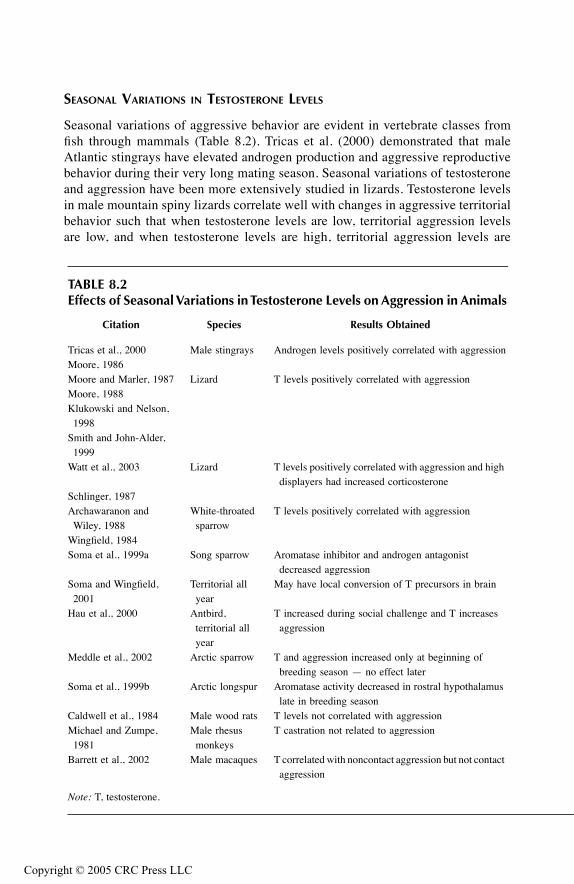
TF1661_C008.fm Page 226 Monday, October 4, 2004 9:20 AM
Copyr
SEASONAL VARIATIONS IN TESTOSTERONE LEVELS
Seasonal variations of aggressive behavior are evident in vertebrate classes fromfish through mammals (Table 8.2). Tricas et al. (2000) demonstrated that maleAtlantic stingrays have elevated androgen production and aggressive reproductivebehavior during their very long mating season. Seasonal variations of testosteroneand aggression have been more extensively studied in lizards. Testosterone levelsin male mountain spiny lizards correlate well with changes in aggressive territorialbehavior such that when testosterone levels are low, territorial aggression levelsare low, and when testosterone levels are high, territorial aggression levels are
TABLE 8.2Effects of Seasonal Variations in Testosterone Levels on Aggression in Animals
Citation Species Results Obtained
Tricas et al., 2000 Male stingrays Androgen levels positively correlated with aggressionMoore, 1986Moore and Marler, 1987 Lizard T levels positively correlated with aggressionMoore, 1988Klukowski and Nelson, 1998
Smith and John-Alder, 1999
Watt et al., 2003 Lizard T levels positively correlated with aggression and high displayers had increased corticosterone
Schlinger, 1987Archawaranon and Wiley, 1988
White-throated sparrow
T levels positively correlated with aggression
Wingfield, 1984Soma et al., 1999a Song sparrow Aromatase inhibitor and androgen antagonist
decreased aggressionSoma and Wingfield,
2001Territorial all year
May have local conversion of T precursors in brain
Hau et al., 2000 Antbird, territorial all year
T increased during social challenge and T increases aggression
Meddle et al., 2002 Arctic sparrow T and aggression increased only at beginning of breeding season — no effect later
Soma et al., 1999b Arctic longspur Aromatase activity decreased in rostral hypothalamus late in breeding season
Caldwell et al., 1984 Male wood rats T levels not correlated with aggressionMichael and Zumpe, 1981
Male rhesus monkeys
T castration not related to aggression
Barrett et al., 2002 Male macaques T correlated with noncontact aggression but not contact aggression
Note: T, testosterone.
ight © 2005 CRC Press LLC

TF1661_C008.fm Page 227 Monday, October 4, 2004 9:20 AM
Copyr
high (Moore, 1986). Moore and Marler (1987) also demonstrated that whiletestosterone implants in the nonbreeding season led to an increase in aggression,the aggression level was lower than that observed during the breeding seasondespite the fact that the testosterone levels were equivalent. When testosteroneimplants were given to castrated subjects during the breeding season, normalbreeding season levels of territorial aggression were observed (Moore, 1988).Similar seasonal relationships between testosterone and aggression have beenobtained with northern fence lizards (Smith and John-Alder, 1999; and Klukowskiand Nelson, 1998) and male Jacky dragons (Watt et al., 2003). Watt et al. dem-onstrated that elevated testosterone levels were the best predictor of territorialdisplay and that subjects with high levels of display demonstrated an acute risein corticosterone. Thus, territorial aggression during the breeding season is con-trolled by testosterone, and affected by a number of additional mechanisms. It islikely that during the breeding season, other neuroendocrine factors interact withincreased levels of testosterone to yield high levels of territorial aggression.
Several investigators have examined the effects of testosterone levels on aggres-sion in birds. Free-living birds that received testosterone capsule implants whichmaintained breeding level testosterone levels showed greater injury and had lowersurvival rates to the following year (Dufty, 1989). It was suggested that prolonged,elevated testosterone levels produced a risk factor by maintaining aggressive intra-male interactions that are normally evident only during the breeding season. Anearlier study by Wingfield (1984) suggests that this conclusion may be true. Free-living adult male song sparrows were subcutaneously implanted with testosteroneto maintain plasma testosterone at the springtime peak. Sparrows with the testoster-one implants showed higher levels of aggression than controls when challenged withtape recordings of conspecifics intruding into their territory.
Depending on the climatic conditions, birds demonstrate great variability in theirseasonal behavior. Seasonal variations in aggression are evident in the white-throatedsparrow, and these can be correlated with androgen levels (Schlinger, 1987) suchthat both androgen levels and aggression are increased in November but not inJanuary or March. Researchers have suggested that the increases in both androgenand aggression help establish flock formation and dominance hierarchies. Archa-waranon and Wiley (1988) examined whether the effects of androgens on aggressionand dominance in these sparrows were due to testosterone or to its metabolites. Thesubjects were subcutaneously implanted with testosterone, that is, its metabolites —androstenedione, 5α-dihydrotestosterone (DHT), androsterone, or estradiol. Test-osterone administration, or the combined treatment of its metabolites, produced themost potent effect on aggression and dominance. Further, when the conversion oftestosterone to both DHT and estradiol was blocked, aggression and dominancescores were lower compared to blockade of the conversion of testosterone to eitherDHT or estradiol, despite the fact that blockade of a single metabolite producedelevated testosterone levels. Thus, androgenic effects on aggression are significantlydependent on the conversion of testosterone to both androgenic and estrogenicmetabolites.
Some birds, such as the male song sparrows, fail to show seasonal behavioralchanges and demonstrate territorial behavior throughout the year. However, their
ight © 2005 CRC Press LLC
TF1661_C008.fm Page 228 Monday, October 4, 2004 9:20 AM
Copyr
nonbreeding-season aggressive behaviors appear to be testosterone independent,since they are unaffected by castration, and plasma testosterone levels are very lowduring this period. However, birds treated with an aromatase inhibitor and an andro-gen receptor antagonist demonstrated decreased aggressive behaviors (Soma et al.,1999a). Interestingly, plasma dehydroepiandrosterone is high in these subjects, andmay be converted locally in the brain into active sex steroids (Soma and Wingfield,2001). Similar mechanisms may be involved in the control of territorial aggressionin the tropical spotted antbird, which also displays year-long defensive behaviorsdespite low plasma testosterone levels (Hau et al., 2000). During periods of socialchallenge, these birds show elevated testosterone levels, and testosterone implantsincrease aggressive behaviors. Additionally, birds treated with an aromatase inhibitorand an androgen receptor antagonist demonstrated decreased aggressive behaviorsjust as the song sparrows did. Thus, these birds may also be converting testosteroneprecursors directly at their site of action or else use discrete testosterone secretionto avoid chronically elevated testosterone.
Due to extreme weather conditions, Arctic birds such as the Gambel’s white-crowned sparrow have a brief breeding season (unlike temperate birds), and onlydemonstrate high testosterone and luteinizing hormone early in the breeding season(Meddle et al., 2002). During incubation, simulated intrusions increased LH but nottestosterone, although testosterone could be elevated by injection of gonadotropin-releasing hormone, and testosterone implantation did not increase aggression inmales. However, corticosterone implants decreased aggression in response to asimulated intrusion during incubation. It is thought that modifications in the typicaleffects of these hormones are due to the fact that in such a harsh climate, interruptionsin parental care may adversely affect reproductive success. Results by Soma et al.(1999b) with another Arctic bird, the Lapland longspur, may provide a possiblemechanism for this lack of effect of testosterone, since they demonstrated thataromatase activity was decreased in the rostral hypothalamus in the late breedingseason. A recent review of hormonal behavioral interactions in Arctic birds exploresa variety of unique interrelationships of testosterone and aggression (Wingfield andHunt, 2002).
With mammals, the relationship among seasonal effects, testosterone, andaggression is more dynamic. Caldwell et al. (1984) castrated male wood rats afterpuberty, and demonstrated that seasonal increases in aggression could be indepen-dent of testosterone since the castrated subjects maintained their seasonal increasesin aggression. This finding essentially replicates an earlier result with rhesus mon-keys in which both castrated and intact subjects maintained their annual changes inaggression (Michael and Zumpe, 1981). The relationship between testosterone andaggression can also be more nuanced with higher animalsm as demonstrated bytestosterone being correlated with noncontact female-directed aggression, but notwith contact aggression in male Japanese macaques (Barrett et al., 2002).
TESTOSTERONE LEVELS DURING DEVELOPMENT
In mammals, sexual differentiation begins before birth and continues throughpuberty. Androgens are thought to play an important role in inducing prenatal
ight © 2005 CRC Press LLC
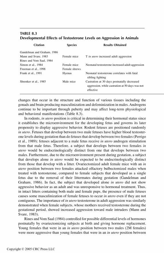
TF1661_C008.fm Page 229 Monday, October 4, 2004 9:20 AM
Copyr
changes that occur in the structure and function of various tissues including thegonads and brain producing masculinization and defeminization in males. Androgenscontinue to be important through puberty and may affect long-term physiologicaland behavioral manifestations (Table 8.3).
In rodents, in utero position is critical in determining their hormonal status sinceit establishes the microenvironment for the developing fetus and governs its laterpropensity to display aggressive behavior. Rodent fetuses are positioned randomlyin utero. Fetuses that develop between two male fetuses have higher blood testoster-one levels during gestation than do fetuses that develop between two females (Perrigoet al., 1989); fetuses adjacent to a male fetus receive in utero androgen stimulationfrom that male fetus. Therefore, a subject that develops between two females inutero would be endocrinologically distinct from one that develops between twomales. Furthermore, due to the microenvironment present during gestation, a subjectthat develops alone in utero would be expected to be endocrinologically distinctfrom those that develop with a litter. Ovariectomized adult female mice with an inutero position between two females attacked olfactory bulbectomized males whentreated with testosterone, compared to female subjects that developed as a singlefetus due to the removal of their littermates during gestation (Gandelman andGraham, 1986). In fact, the subject that developed alone in utero did not showaggressive behavior as an adult and was unresponsive to hormonal treatment. Thus,in intact litters containing both male and female pups, the presence of male fetusescauses some masculinization of female fetuses to occur in utero even if they are notcontiguous. The importance of in utero testosterone in adult aggression was similarlydemonstrated when female subjects, whose mothers received testosterone during thegestational period, showed increased aggression toward male intruders (Mann andSvare, 1983).
Rines and Vom Saal (1984) controlled for possible differential levels of hormonespostnatally by ovariectomizing subjects at birth and giving hormone replacement.Young females that were in an in utero position between two males (2M females)were more aggressive than young females that were in an in utero position between
TABLE 8.3Developmental Effects of Testosterone Levels on Aggression in Animals
Citation Species Results Obtained
Gandelman and Graham, 1986Mann and Svare, 1983 Female mice T in utero increased adult aggressionRines and Vom Saal, 1984Simon et al., 1984 Female mice Neonatal testosterone increased adult aggressionFreeman et al., 1998 Female shrewsFrank et al., 1991 Hyenas Neonatal testosterone correlates with fatal
sibling fightingShrenker et al., 1985 Male mice Castration at 30 days postnatally decreased
aggression, while castration at 50 days was not effective
ight © 2005 CRC Press LLC
TF1661_C008.fm Page 230 Monday, October 4, 2004 9:20 AM
Copyr
zero males (OM females). However, old OM females were aggressive, suggestingan age-related difference where OM females developed sensitivity to testosteronelater in life. Similarly, Perrigo et al. (1989) demonstrated that male mice thatdeveloped between two females in utero, and thus had lower circulating testosteronelevels during gestation, had increased infanticide both before and after mating. Thus,these studies indicate the importance of developmental factors that contribute to theorganization of aggressive behavior during the postnatal period. A more thoroughdiscussion of the subtleties of this area is beyond the scope of this chapter, but arecent review of this area has been conducted by Ryan and Vandenbergh (2002).
In addition to the critical time period during prenatal development, duringpostnatal development there is a temporal relationship between castration and thetendency for expression of aggressive behavior. Shrenker et al. (1985) demonstratedthat male mice castrated 30 days after birth were less aggressive than those castratedat 50 days after birth. In fact, subjects castrated at 50 days showed levels of aggres-sion similar to sham-operated controls. Similarly, female mice given testosterone atbirth behaved more aggressively when treated with testosterone as adults than didsubjects given estradiol neonatally and similarly tested with testosterone as adults(Simon et al., 1984). Neonatal testosterone treatment of female musk shrews alsoproduced more aggressive adults (Freeman et al., 1998). Additionally, early postnataleffects of testosterone are important in the early expression of fatal aggressivebehavior in neonatal spotted hyenas. During the first month following birth, test-osterone remained higher in males than females, while androstenedione, which waselevated in both sexes at birth, remained high in females and fell in males. Siblingfighting was most pronounced during this time period, and was directly related tohigh androgen levels (Frank et al., 1991).
There are critical time periods during development when circulating testosteronemust be present for appropriate interactions with neural structures associated withthe expression of aggressive reactions to take place, and thus ensure that suchresponses will remain present in the adult organism. Although this would seem tocontradict studies discussed earlier that used adult subjects in which testosteronewas shown to be effective in modulating aggression, once this critical developmentalperiod has passed, the presence or absence of circulating testosterone becomes lessimportant. However, while there are critical periods during which changes have themost dramatic and long-lasting effects, further manipulation of the system can occurat a later time as well. Whether such changes can significantly alter brain functionin adulthood has yet to be determined.
TESTOSTERONE’S ROLE IN HUMAN AGGRESSION
Various studies have been designed to assess the relationship between testosteroneand aggressive behavior in humans (Table 8.4). Several methodological difficultiesare apparent, including differences in the (1) nature of the populations sampled, (2)ages of the sample, (3) size of the sample considered, (4) measurement of testoster-one, and (5) kinds of behavioral processes investigated that are interpreted as beingaggressive in nature. A recent review of this area and its potential shortcomings wasconducted by Zitzmann and Nieschlag (2001). Nevertheless, a pattern appears to
ight © 2005 CRC Press LLC

TF1661_C008.fm Page 231 Monday, October 4, 2004 9:20 AM
Copyr
emerge from these studies that, while not entirely consistent, is supportive of thefindings obtained from the animal literature that demonstrate a positive correlationbetween testosterone levels and the presence of aggressive behavior.
Perhaps the most convincing data are derived from studies involving subjectsincarcerated in either a prison or mental institution. In an early study, Ehrenkranzet al. (1974) categorized prison inmates on the basis of their level of aggressiveness(as determined from a battery of psychological tests) and were thus able to distin-guish a chronically aggressive group from socially dominant and nonaggressiveindividuals. Testosterone levels were consistently higher in individuals displayingchronic aggressive behavior than in those inmates who were classified as eithersocially dominant or nonaggressive. Similarly, Dabbs et al. (1987) categorized prisoninmates in terms of whether they had committed violent crimes. Positive correlationswere obtained between individuals displaying high free salivary testosterone levelsand those who committed violent crimes. Moreover, individuals who had committednonviolent crimes had the lowest testosterone levels. Prisoners who were rated asbeing “tougher” by their peers as well as individuals who had committed nonviolentcrimes but had received punishment for disciplinary infractions also had high test-osterone levels. In a related study (Rada, 1976; Rada et al., 1983), the authorsclassified patients at the Atascadero State Hospital in California according to thedegree of violence exhibited during the commission of a rape and according to theirscores on the Buss-Durkee and Megargee Overcontrolled hostility inventories. Con-sistent with the findings described above, rapists who were judged to be most violent(i.e., those who beat their victims during the rape) had significantly higher plasmatestosterone levels than child molesters or normal subjects. Although individualcorrelations between testosterone levels and hostility ratings were not demonstrated,hostility rating scores were also higher for rapists than for normals. A more recentstudy also demonstrated a correlation between plasma testosterone and aggressiveacts in prisoners (Dolan et al., 2001). They also demonstrated that primary psycho-paths, as defined by the Special Hospital Assessment of Personality and Socializa-tion, had lower cortisol and higher testosterone concentrations. Similar findings havebeen reported in elderly demented patients such that free plasma testosterone waspositively correlated with aggression, while estrogen levels were negatively corre-lated with aggression (Orengo et al., 2002).
Similar positive correlations between aggressive behavior and high testosteronelevels were obtained from a number of studies on normal adolescents or youngadults. Testosterone levels were higher in college students who scored high forhostility and aggression as measured on a hostility inventory scale (Persky et al.,1971); serum testosterone levels were positively correlated with self-ratings of spon-taneous aggression and dominance in young adult males given standardized andprojective tests (Christiansen and Knussmann, 1987); and adolescent males whoresponded more vigorously to provocations and threats when given the OlweusAggression Inventory scale had high testosterone levels and were more irritable andmore likely to engage in aggressive behavior (Olweus et al., 1980, 1988). Further-more, Scaramella and Brown (1978) demonstrated that testosterone levels werepositively correlated with the response to threat in college hockey players; and Booth
ight © 2005 CRC Press LLC
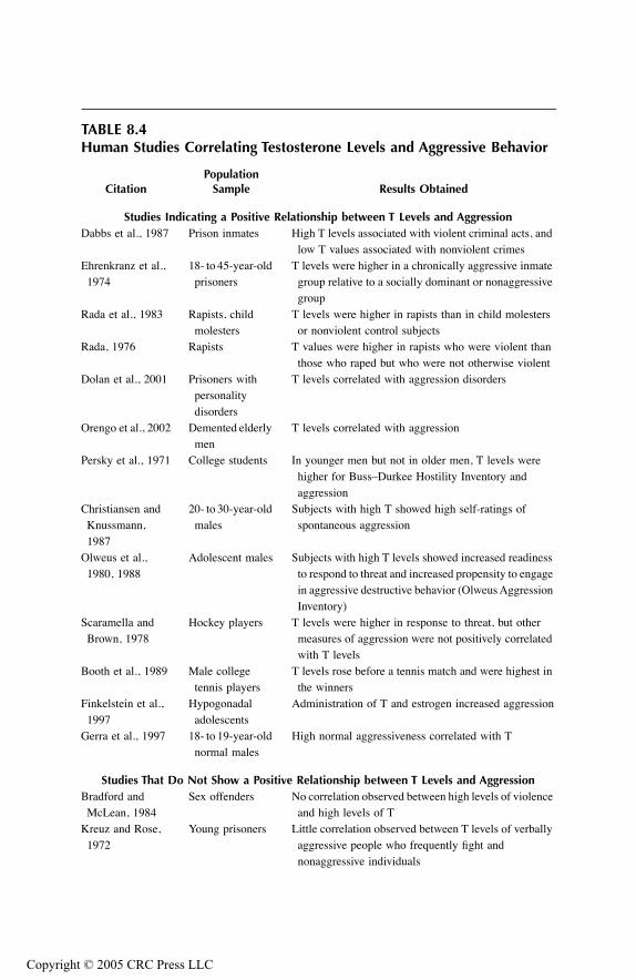
TF1661_C008.fm Page 232 Monday, October 4, 2004 9:20 AM
Copyr
TABLE 8.4Human Studies Correlating Testosterone Levels and Aggressive Behavior
CitationPopulation
Sample Results Obtained
Studies Indicating a Positive Relationship between T Levels and AggressionDabbs et al., 1987 Prison inmates High T levels associated with violent criminal acts, and
low T values associated with nonviolent crimesEhrenkranz et al., 1974
18- to 45-year-old prisoners
T levels were higher in a chronically aggressive inmate group relative to a socially dominant or nonaggressive group
Rada et al., 1983 Rapists, child molesters
T levels were higher in rapists than in child molesters or nonviolent control subjects
Rada, 1976 Rapists T values were higher in rapists who were violent than those who raped but who were not otherwise violent
Dolan et al., 2001 Prisoners with personality disorders
T levels correlated with aggression disorders
Orengo et al., 2002 Demented elderly men
T levels correlated with aggression
Persky et al., 1971 College students In younger men but not in older men, T levels were higher for Buss–Durkee Hostility Inventory and aggression
Christiansen and Knussmann, 1987
20- to 30-year-old males
Subjects with high T showed high self-ratings of spontaneous aggression
Olweus et al., 1980, 1988
Adolescent males Subjects with high T levels showed increased readiness to respond to threat and increased propensity to engage in aggressive destructive behavior (Olweus Aggression Inventory)
Scaramella and Brown, 1978
Hockey players T levels were higher in response to threat, but other measures of aggression were not positively correlated with T levels
Booth et al., 1989 Male college tennis players
T levels rose before a tennis match and were highest in the winners
Finkelstein et al., 1997
Hypogonadal adolescents
Administration of T and estrogen increased aggression
Gerra et al., 1997 18- to 19-year-old normal males
High normal aggressiveness correlated with T
Studies That Do Not Show a Positive Relationship between T Levels and AggressionBradford and McLean, 1984
Sex offenders No correlation observed between high levels of violence and high levels of T
Kreuz and Rose, 1972
Young prisoners Little correlation observed between T levels of verbally aggressive people who frequently fight and nonaggressive individuals
ight © 2005 CRC Press LLC

TF1661_C008.fm Page 233 Monday, October 4, 2004 9:20 AM
Copyr
et al. (1989) reported that testosterone levels rose just prior to a tennis match andwere highest in the winners of the matches.
Since correlational studies have numerous limitations, more recent studies havemanipulated testosterone and aggression levels in human subjects. Hypogonadaladolescents were treated with sex hormones at early, middle, and late pubertal doses.Both testosterone (in boys) and estrogen (in girls) increased physical aggressivebehaviors and aggressive impulses but not verbal aggressive behaviors as measuredby the Olweus Multifaceted Aggression Inventory (Finkelstein et al., 1997). Gerraet al. (1997) experimentally induced aggressiveness in psychophysically healthymale teenagers. Subjects with high-normal basal aggressiveness demonstrated ele-vated basal testosterone levels and elevated norepinephrine, growth hormone, andcortisol in response to the experimental induction of aggression.
A smaller number of studies have failed to provide data in support of a positiverelationship between high testosterone levels and aggressive behavior. Males incar-cerated for sex offenses that ranged from exhibitionism, fetishism, and pedophiliato more violent behavior (i.e., rape) did not demonstrate a significant correlationbetween sexual violence and testosterone levels (Bradford and McLean, 1984).However, this result may not conflict with Rada (1976) and Rada et al. (1983)described above since these authors demonstrated the dependence of other violentacts being committed on the victim, and Bradford and McLean did not mentionwhether the rapists in their study committed other violent acts on their victims.Kreuz and Rose (1972) examined young prisoners and observed little correlationbetween testosterone levels of verbally aggressive people who frequently fought andthose who were nonaggressive. Worthman and Konner (1987) followed the patternof change in testosterone levels of !Kung San men during the course of their huntand reported changes in testosterone levels were correlated more closely with thelength of exercise rather than with the success or failure of the hunt. Finally,investigators provided case study reports of individuals diagnosed with Klinefelter’ssyndrome, which is characterized, in part, by lower levels of testosterone and by the
TABLE 8.4 (CONTINUED)Human Studies Correlating Testosterone Levels and Aggressive Behavior
CitationPopulation
Sample Results Obtained
Worthman and Konner, 1987
!Kung San men Pattern of change in T levels is associated with prolonged exercise rather than the success or failure of the hunt
Sourial. and Fenton, 1988
Klinefelter’s syndrome
Case study — aggressive and sexual fantasies and impulses declined with T treatment in individual with low endogenous T levels
Raboch et al., 1987 Klinefelter’s syndrome
Case study — low T levels associated with sexually motivated murder
Note: T, testosterone.
ight © 2005 CRC Press LLC
TF1661_C008.fm Page 234 Monday, October 4, 2004 9:20 AM
Copyr
relative absence of secondary sex characteristics. Testosterone treatment resulted ina decline of aggressive impulses and sexual fantasies in one individual, which mayhave been due to the psychological benefit of secondary sex characteristic develop-ment following testosterone treatment (Sourial and Fenton, 1988). Raboch et al.(1987) reported that another individual with Klinefelter’s syndrome committed asexually motivated murder despite relatively low testosterone levels.
In spite of some negative data, the overall observations obtained from humanstudies support the presence of a relationship between endogenous testosterone levelsand the propensity to commit aggressive acts. However, other variables such asenvironmental conditions, the psychological state of the individual, and the type ofaggressive act committed appear to play more critical roles concerning whether apositive correlation will be obtained in humans.
ESTROGEN AND AGGRESSIVE BEHAVIOR
While the role of testosterone on aggressive behavior has been more widely studied,other hormones have also been shown to play an important part in the expressionof aggressive behavior. In the following section, the main findings of the effects ofestrogen on aggressive behavior will be summarized. It should be noted that theeffects of estrogen on aggressive behavior are much more inconsistent than theeffects of testosterone.
EXPERIMENTAL MANIPULATION OF ESTROGEN LEVELS
A number of studies have failed to demonstrate a direct effect of estrogen onaggression. For example, DeBold and Miczek (1984) found that residents are morelikely to attack (biting toward the neck, back, or flank, or a nip to the snout or face)intruders of the same sex than those of the opposite sex. This form of aggressivebehavior is gonadally dependent in males but not in females since castration of themale intruder led to a decrease in attack from male residents that could be reversedby testosterone replacement, while ovariectomy of female intruders did not changethe frequency of attack by female residents. Similarly, although baseline levels ofaggression were relatively high, chronic estradiol treatment failed to alter attackresponses in female resident hamsters that were ovariectomized, treated with varyingdoses of estradiol, and retested 3, 7, 10, and 14 days post-implantation (Meisel etal., 1988). However, when subsequently injected with progesterone, estradiol-treatedsubjects showed a reduction in aggression compared to cholesterol-treated controlsthat remained aggressive after progesterone treatment. Thus, inhibition of aggressionin female subjects is likely to be dependent on the combined effects of estradiol andprogesterone.
More recent studies have demonstrated mixed effects of estrogen on aggressivebehavior. For example, Stavisky et al. (1999) demonstrated that ovariectomizedfemale cynomolgus macaques had a two- to three-fold increase in aggressive behav-ior compared to sham or hormonally replaced subjects while affiliative behaviorswere unaffected. This effect is opposite that seen in spiny lizards that display reducedaggression following ovariectomy (Woodley and Moore, 1999).
ight © 2005 CRC Press LLC
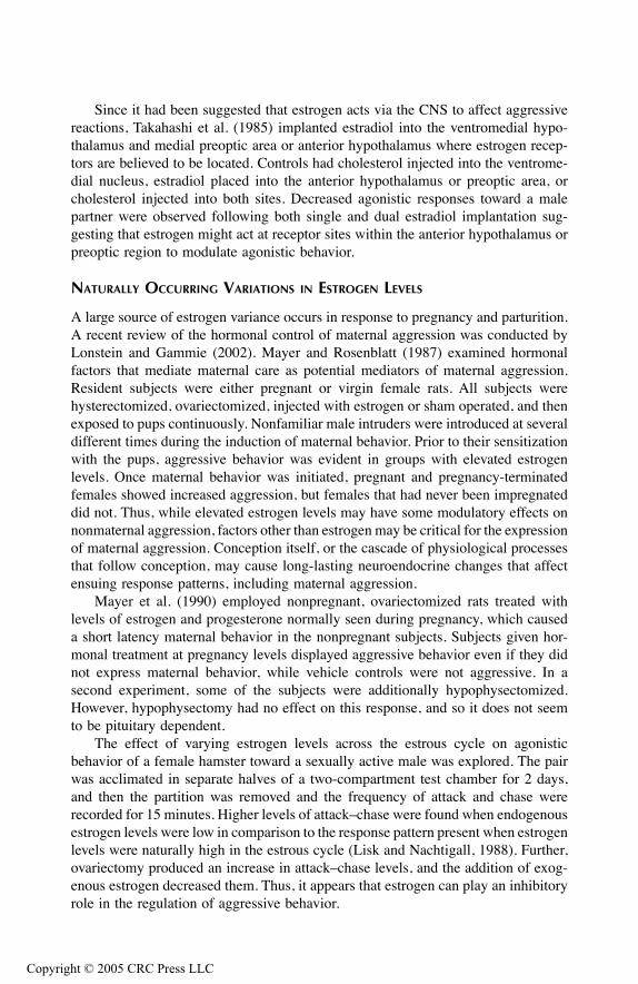
TF1661_C008.fm Page 235 Monday, October 4, 2004 9:20 AM
Copyr
Since it had been suggested that estrogen acts via the CNS to affect aggressivereactions, Takahashi et al. (1985) implanted estradiol into the ventromedial hypo-thalamus and medial preoptic area or anterior hypothalamus where estrogen recep-tors are believed to be located. Controls had cholesterol injected into the ventrome-dial nucleus, estradiol placed into the anterior hypothalamus or preoptic area, orcholesterol injected into both sites. Decreased agonistic responses toward a malepartner were observed following both single and dual estradiol implantation sug-gesting that estrogen might act at receptor sites within the anterior hypothalamus orpreoptic region to modulate agonistic behavior.
NATURALLY OCCURRING VARIATIONS IN ESTROGEN LEVELS
A large source of estrogen variance occurs in response to pregnancy and parturition.A recent review of the hormonal control of maternal aggression was conducted byLonstein and Gammie (2002). Mayer and Rosenblatt (1987) examined hormonalfactors that mediate maternal care as potential mediators of maternal aggression.Resident subjects were either pregnant or virgin female rats. All subjects werehysterectomized, ovariectomized, injected with estrogen or sham operated, and thenexposed to pups continuously. Nonfamiliar male intruders were introduced at severaldifferent times during the induction of maternal behavior. Prior to their sensitizationwith the pups, aggressive behavior was evident in groups with elevated estrogenlevels. Once maternal behavior was initiated, pregnant and pregnancy-terminatedfemales showed increased aggression, but females that had never been impregnateddid not. Thus, while elevated estrogen levels may have some modulatory effects onnonmaternal aggression, factors other than estrogen may be critical for the expressionof maternal aggression. Conception itself, or the cascade of physiological processesthat follow conception, may cause long-lasting neuroendocrine changes that affectensuing response patterns, including maternal aggression.
Mayer et al. (1990) employed nonpregnant, ovariectomized rats treated withlevels of estrogen and progesterone normally seen during pregnancy, which causeda short latency maternal behavior in the nonpregnant subjects. Subjects given hor-monal treatment at pregnancy levels displayed aggressive behavior even if they didnot express maternal behavior, while vehicle controls were not aggressive. In asecond experiment, some of the subjects were additionally hypophysectomized.However, hypophysectomy had no effect on this response, and so it does not seemto be pituitary dependent.
The effect of varying estrogen levels across the estrous cycle on agonisticbehavior of a female hamster toward a sexually active male was explored. The pairwas acclimated in separate halves of a two-compartment test chamber for 2 days,and then the partition was removed and the frequency of attack and chase wererecorded for 15 minutes. Higher levels of attack–chase were found when endogenousestrogen levels were low in comparison to the response pattern present when estrogenlevels were naturally high in the estrous cycle (Lisk and Nachtigall, 1988). Further,ovariectomy produced an increase in attack–chase levels, and the addition of exog-enous estrogen decreased them. Thus, it appears that estrogen can play an inhibitoryrole in the regulation of aggressive behavior.
ight © 2005 CRC Press LLC

TF1661_C008.fm Page 236 Monday, October 4, 2004 9:20 AM
Copyr
ESTROGEN LEVELS DURING DEVELOPMENT
As previously indicated, sexual differentiation begins during fetal life and continuesthrough puberty in mammals. In addition to androgens, estrogens are also importantin inducing changes in the structure and function of various tissues, such as the brainand gonads, which may be important in aggressive behavior. Males that developedbetween two female fetuses in utero had higher levels of estradiol in their amnioticfluid than males located between two male fetuses and were less aggressive as adults(Vom Saal et al., 1983). Possible differences in postnatal exposure to gonadalhormones were controlled by castrating the subjects at birth and giving hormonereplacement treatment. Variations in levels of aggression observed were the resultof developmental differences in brain tissue that occurred prenatally, due, in part,to the action of estrogen. However, prenatal exposure to estrogen — administeredto the mother during days 12 through 18 of gestation — did not appear to alter theexpression of aggressive behavior to chronic testosterone treatment given as adults(Gandelman et al., 1982). It can be assumed from the testosterone studies that duringgestation a critical time period exists for the development of aggression, and thisstudy may have failed to administer estrogen during the critical time window forthis hormone to be effective. Additionally, the in utero position findings are notlikely to be attributed solely to differences in estrogen levels, but are probably theresult of the interaction of the constellation of endocrine micro-changes. Such effectswould also include changes in androgen levels, as well as any neuroendocrine factorsthat interact with estrogens and androgens.
Female mice ovariectomized and administered androgen 90 to 140 days post-natally demonstrated increased aggression in neonatally treated controls, while post-natal estrogen administration was not effective (Simon and Whalen, 1987). Thismight have been predicted since the authors only tested aggressive behaviors bybiting or chasing of an olfactory bulbectomized male intruder, which is a male typicalform of aggression. Neonatal androgen treatment led to enhanced effectiveness ofsubsequent androgen, but not estrogen, treatment in eliciting aggression as adults.Similarly, neonatal estrogen exposure resulted in the expression of aggression whenthe subjects were subsequently given estrogenic, but not androgenic, stimulation asadults. Thus, responses to these compounds in adult subjects are dependent on theentire neuroendocrine history of the subject. Moreover, it should be further notedthat the differential effects of perinatal androgens and estrogens on aggression inadulthood are confounded by the fact that androgens such as testosterone are met-abolically converted (aromatized) to estradiol in the developing and adult mammalianbrain (Naftolin et al., 1975). Factors that affect this conversion have been shown toalter the organization (McEwen et al., 1977) as well as the activation (Harding,1986) of sexual and sexually dimorphic behavior.
PROGESTERONE AND AGGRESSIVE BEHAVIOR
The following section briefly reviews the relationship of progesterone to aggressivebehaviors. As with the experiments exploring the effects of estrogens on aggressivebehavior, this literature is not as consistent as the testosterone literature.
ight © 2005 CRC Press LLC
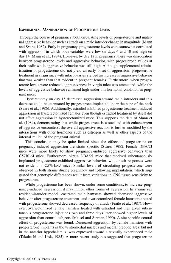
TF1661_C008.fm Page 237 Monday, October 4, 2004 9:20 AM
Copyr
EXPERIMENTAL MANIPULATION OF PROGESTERONE LEVELS
Through the course of pregnancy, both circulating levels of progesterone and mater-nal aggressive behavior such as attack on a male intruder change in magnitude (Mannand Svare, 1982). Early in pregnancy, progesterone levels were somewhat correlatedwith aggression in which both variables were low on days 6 and 10 and high onday 14 (Mann et al., 1984). However, by day 18 in pregnancy, there was dissociationbetween progesterone levels and aggressive behavior, with progesterone values attheir nadir while aggressive behavior was still high. Although supplemental admin-istration of progesterone did not yield an early onset of aggression, progesteronetreatment in virgin mice with intact ovaries yielded an increase in aggressive behaviorthat was weaker than that evident in pregnant females. Furthermore, when proges-terone levels were reduced, aggressiveness in virgin mice was attenuated, while thelevels of aggressive behavior remained high under this hormonal condition in preg-nant mice.
Hysterectomy on day 15 decreased aggression toward male intruders and thisdecrease could be attenuated by progesterone implanted under the nape of the neck(Svare et al., 1986). Additionally, estradiol inhibited progesterone treatment inducedaggression in hysterectomized females even though estradiol treatment by itself didnot affect aggression in hysterectomized mice. This supports the data of Mann etal. (1984), demonstrating that while progesterone is associated with enhancementof aggressive encounters, the overall aggressive reaction is further modified by theinteractions with other hormones such as estrogen as well as other aspects of theinternal milieu of the pregnant animal.
This conclusion may be quite limited since the effects of progesterone onpregnancy-induced aggression are strain specific (Svare, 1988). Female DBA/2Jmice were more likely to show pregnancy-induced aggressive behaviors thanC57BL6J mice. Furthermore, virgin DBA/2J mice that received subcutaneouslyimplanted progesterone exhibited aggressive behavior, while such responses werenot evident in C57BL/6J mice. Similar levels of circulating progesterone wereobserved in both strains during pregnancy and following implantation, which sug-gested that genotypic differences result from variations in CNS tissue sensitivity toprogesterone.
While progesterone has been shown, under some conditions, to increase preg-nancy-induced aggression, it may inhibit other forms of aggression. In a same sexresident–intruder model, castrated male hamsters showed decreased aggressivebehavior after progesterone treatment, and ovariectomized female hamsters treatedwith progesterone showed decreased frequency of attack (Fraile et al., 1987). How-ever, ovariectomized female hamsters treated with estradiol and then given subcu-taneous progesterone injections two and three days later showed higher levels ofaggression than control subjects (Meisel and Sterner, 1990). A site-specific centraleffect of progesterone was found. Decreased aggression by female hamsters withprogesterone implants in the ventromedial nucleus and medial preoptic area, but notin the anterior hypothalamus, was expressed toward a sexually experienced male(Takahashi and Lisk, 1985). A more recent study has suggested that progesterone
ight © 2005 CRC Press LLC
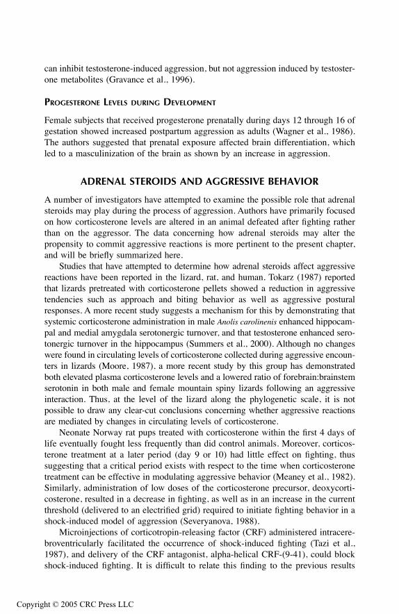
TF1661_C008.fm Page 238 Monday, October 4, 2004 9:20 AM
Copyr
can inhibit testosterone-induced aggression, but not aggression induced by testoster-one metabolites (Gravance et al., 1996).
PROGESTERONE LEVELS DURING DEVELOPMENT
Female subjects that received progesterone prenatally during days 12 through 16 ofgestation showed increased postpartum aggression as adults (Wagner et al., 1986).The authors suggested that prenatal exposure affected brain differentiation, whichled to a masculinization of the brain as shown by an increase in aggression.
ADRENAL STEROIDS AND AGGRESSIVE BEHAVIOR
A number of investigators have attempted to examine the possible role that adrenalsteroids may play during the process of aggression. Authors have primarily focusedon how corticosterone levels are altered in an animal defeated after fighting ratherthan on the aggressor. The data concerning how adrenal steroids may alter thepropensity to commit aggressive reactions is more pertinent to the present chapter,and will be briefly summarized here.
Studies that have attempted to determine how adrenal steroids affect aggressivereactions have been reported in the lizard, rat, and human. Tokarz (1987) reportedthat lizards pretreated with corticosterone pellets showed a reduction in aggressivetendencies such as approach and biting behavior as well as aggressive posturalresponses. A more recent study suggests a mechanism for this by demonstrating thatsystemic corticosterone administration in male Anolis carolinenis enhanced hippocam-pal and medial amygdala serotonergic turnover, and that testosterone enhanced sero-tonergic turnover in the hippocampus (Summers et al., 2000). Although no changeswere found in circulating levels of corticosterone collected during aggressive encoun-ters in lizards (Moore, 1987), a more recent study by this group has demonstratedboth elevated plasma corticosterone levels and a lowered ratio of forebrain:brainstemserotonin in both male and female mountain spiny lizards following an aggressiveinteraction. Thus, at the level of the lizard along the phylogenetic scale, it is notpossible to draw any clear-cut conclusions concerning whether aggressive reactionsare mediated by changes in circulating levels of corticosterone.
Neonate Norway rat pups treated with corticosterone within the first 4 days oflife eventually fought less frequently than did control animals. Moreover, corticos-terone treatment at a later period (day 9 or 10) had little effect on fighting, thussuggesting that a critical period exists with respect to the time when corticosteronetreatment can be effective in modulating aggressive behavior (Meaney et al., 1982).Similarly, administration of low doses of the corticosterone precursor, deoxycorti-costerone, resulted in a decrease in fighting, as well as in an increase in the currentthreshold (delivered to an electrified grid) required to initiate fighting behavior in ashock-induced model of aggression (Severyanova, 1988).
Microinjections of corticotropin-releasing factor (CRF) administered intracere-broventricularly facilitated the occurrence of shock-induced fighting (Tazi et al.,1987), and delivery of the CRF antagonist, alpha-helical CRF-(9-41), could blockshock-induced fighting. It is difficult to relate this finding to the previous results
ight © 2005 CRC Press LLC
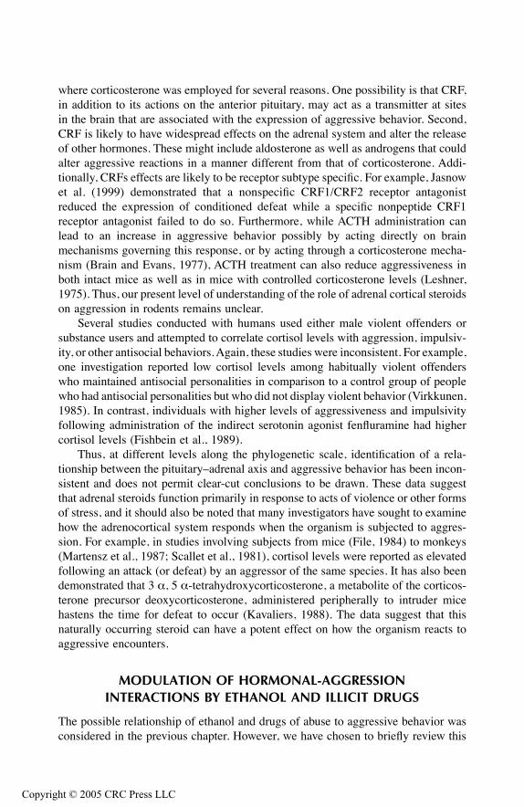
TF1661_C008.fm Page 239 Monday, October 4, 2004 9:20 AM
Copyr
where corticosterone was employed for several reasons. One possibility is that CRF,in addition to its actions on the anterior pituitary, may act as a transmitter at sitesin the brain that are associated with the expression of aggressive behavior. Second,CRF is likely to have widespread effects on the adrenal system and alter the releaseof other hormones. These might include aldosterone as well as androgens that couldalter aggressive reactions in a manner different from that of corticosterone. Addi-tionally, CRFs effects are likely to be receptor subtype specific. For example, Jasnowet al. (1999) demonstrated that a nonspecific CRF1/CRF2 receptor antagonistreduced the expression of conditioned defeat while a specific nonpeptide CRF1receptor antagonist failed to do so. Furthermore, while ACTH administration canlead to an increase in aggressive behavior possibly by acting directly on brainmechanisms governing this response, or by acting through a corticosterone mecha-nism (Brain and Evans, 1977), ACTH treatment can also reduce aggressiveness inboth intact mice as well as in mice with controlled corticosterone levels (Leshner,1975). Thus, our present level of understanding of the role of adrenal cortical steroidson aggression in rodents remains unclear.
Several studies conducted with humans used either male violent offenders orsubstance users and attempted to correlate cortisol levels with aggression, impulsiv-ity, or other antisocial behaviors. Again, these studies were inconsistent. For example,one investigation reported low cortisol levels among habitually violent offenderswho maintained antisocial personalities in comparison to a control group of peoplewho had antisocial personalities but who did not display violent behavior (Virkkunen,1985). In contrast, individuals with higher levels of aggressiveness and impulsivityfollowing administration of the indirect serotonin agonist fenfluramine had highercortisol levels (Fishbein et al., 1989).
Thus, at different levels along the phylogenetic scale, identification of a rela-tionship between the pituitary–adrenal axis and aggressive behavior has been incon-sistent and does not permit clear-cut conclusions to be drawn. These data suggestthat adrenal steroids function primarily in response to acts of violence or other formsof stress, and it should also be noted that many investigators have sought to examinehow the adrenocortical system responds when the organism is subjected to aggres-sion. For example, in studies involving subjects from mice (File, 1984) to monkeys(Martensz et al., 1987; Scallet et al., 1981), cortisol levels were reported as elevatedfollowing an attack (or defeat) by an aggressor of the same species. It has also beendemonstrated that 3 α, 5 α-tetrahydroxycorticosterone, a metabolite of the corticos-terone precursor deoxycorticosterone, administered peripherally to intruder micehastens the time for defeat to occur (Kavaliers, 1988). The data suggest that thisnaturally occurring steroid can have a potent effect on how the organism reacts toaggressive encounters.
MODULATION OF HORMONAL-AGGRESSION INTERACTIONS BY ETHANOL AND ILLICIT DRUGS
The possible relationship of ethanol and drugs of abuse to aggressive behavior wasconsidered in the previous chapter. However, we have chosen to briefly review this
ight © 2005 CRC Press LLC
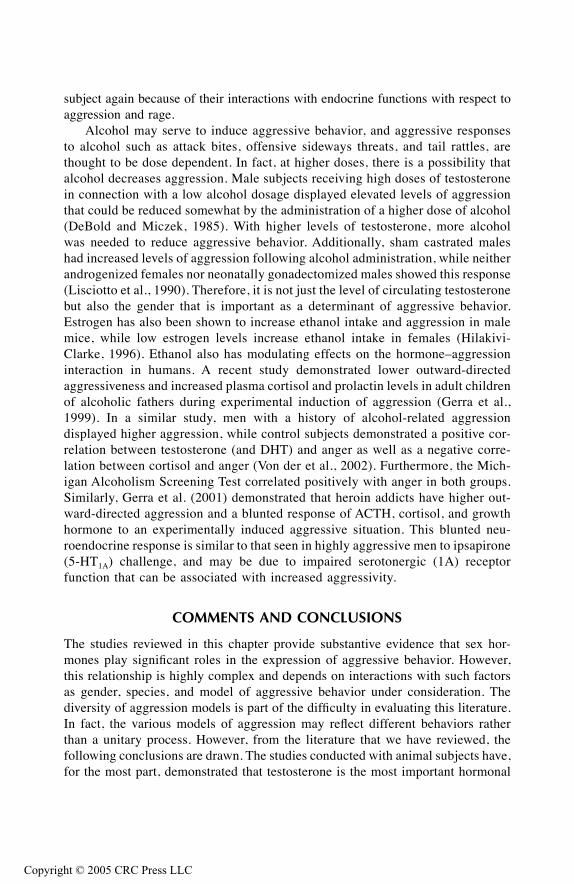
TF1661_C008.fm Page 240 Monday, October 4, 2004 9:20 AM
Copyr
subject again because of their interactions with endocrine functions with respect toaggression and rage.
Alcohol may serve to induce aggressive behavior, and aggressive responsesto alcohol such as attack bites, offensive sideways threats, and tail rattles, arethought to be dose dependent. In fact, at higher doses, there is a possibility thatalcohol decreases aggression. Male subjects receiving high doses of testosteronein connection with a low alcohol dosage displayed elevated levels of aggressionthat could be reduced somewhat by the administration of a higher dose of alcohol(DeBold and Miczek, 1985). With higher levels of testosterone, more alcoholwas needed to reduce aggressive behavior. Additionally, sham castrated maleshad increased levels of aggression following alcohol administration, while neitherandrogenized females nor neonatally gonadectomized males showed this response(Lisciotto et al., 1990). Therefore, it is not just the level of circulating testosteronebut also the gender that is important as a determinant of aggressive behavior.Estrogen has also been shown to increase ethanol intake and aggression in malemice, while low estrogen levels increase ethanol intake in females (Hilakivi-Clarke, 1996). Ethanol also has modulating effects on the hormone–aggressioninteraction in humans. A recent study demonstrated lower outward-directedaggressiveness and increased plasma cortisol and prolactin levels in adult childrenof alcoholic fathers during experimental induction of aggression (Gerra et al.,1999). In a similar study, men with a history of alcohol-related aggressiondisplayed higher aggression, while control subjects demonstrated a positive cor-relation between testosterone (and DHT) and anger as well as a negative corre-lation between cortisol and anger (Von der et al., 2002). Furthermore, the Mich-igan Alcoholism Screening Test correlated positively with anger in both groups.Similarly, Gerra et al. (2001) demonstrated that heroin addicts have higher out-ward-directed aggression and a blunted response of ACTH, cortisol, and growthhormone to an experimentally induced aggressive situation. This blunted neu-roendocrine response is similar to that seen in highly aggressive men to ipsapirone(5-HT1A) challenge, and may be due to impaired serotonergic (1A) receptorfunction that can be associated with increased aggressivity.
COMMENTS AND CONCLUSIONS
The studies reviewed in this chapter provide substantive evidence that sex hor-mones play significant roles in the expression of aggressive behavior. However,this relationship is highly complex and depends on interactions with such factorsas gender, species, and model of aggressive behavior under consideration. Thediversity of aggression models is part of the difficulty in evaluating this literature.In fact, the various models of aggression may reflect different behaviors ratherthan a unitary process. However, from the literature that we have reviewed, thefollowing conclusions are drawn. The studies conducted with animal subjects have,for the most part, demonstrated that testosterone is the most important hormonal
ight © 2005 CRC Press LLC

TF1661_C008.fm Page 241 Monday, October 4, 2004 9:20 AM
Copyr
variable involved in the induction of aggressive behavior. This response may begender specific and selective to the particular form of aggression measured, sug-gesting that the effects may not be totally generalizeable. Furthermore, it has beendemonstrated that there are specific critical times developmentally that are impor-tant for the manifestation of the effects of testosterone on the induction of aggres-sive behavior. The human data are less consistent, but still suggest that testosteroneaffects aggressive behavior. It is possible that at different levels along the phylo-genetic scale, other psychogenic variables begin to override the relationshipbetween testosterone and aggression.
With respect to other sex hormones, estrogen appears to oppose the effects oftestosterone and may inhibit aggressive responses, while the effects of progesteroneare less clear-cut and may be dependent on the interaction with other neuroendocrinefactors. The adrenal steroid literature fails to demonstrate that these hormones playa conclusive role in the regulation of aggressive behavior. Instead, adrenal steroidhormones may serve a more significant function with respect to processes associatedwith the defeat of an organism following an aggressive bout.
The possible mechanism by which sex hormones control aggressive reactionslikely involves the action of hormones that pass through the blood–brain barrier onselective neurons within the brain. These hormones will presumably modify therelease of specific neurotransmitters and may possibly affect the activity of secondmessenger systems as well. Since the overwhelming majority of the studies consid-ered involve models that may be classified as defensive aggression, it would seemthat two of the most critical neuroanatomical structures involved are the medialhypothalamus and midbrain periaqueductal gray (PAG). These structures are signif-icant because they are regions from which this form of aggression can be readilyelicited by electrical or chemical stimulation (Fuchs et al., 1985a, 1985b; Shaikh etal., 1987; Siegel and Pott, 1988; Siegel and Brutus, 1990). The medial hypothalamusplays a unique role in this process since (1) implantation of hormones into thisstructure can clearly modify the propensity for the occurrence of an attack response;(2) lesions of the medial hypothalamus result in an attenuation of aggressive behaviorsuch as maternal aggression (Hansen, 1989), which has been shown to be modifiedby hormonal treatment; and (3) sex hormone-concentrating cells can be found inhigh densities within the medial hypothalamus (Pfaff, 1968a, 1968b; Pfaff andKeiner, 1973). Although the medial hypothalamus and possibly the PAG wouldappear to be primary candidates where hormonal actions would most effectivelymodify aggressive behavior, other structures should also be considered. Theseinclude the limbic system structures — amygdala, hippocampal formation, septalarea, and prefrontal and cingulate cortices — which are characterized by theirmodulatory actions on aggressive behavior (Siegel and Edinger, 1981, 1983; Siegeland Brutus, 1990). It is suggested that sex hormone modulation of aggressivebehavior may likely result from its combined effects on both the medial hypothal-amus as well as limbic structures. This notion is depicted in Figure 8.1.
ight © 2005 CRC Press LLC
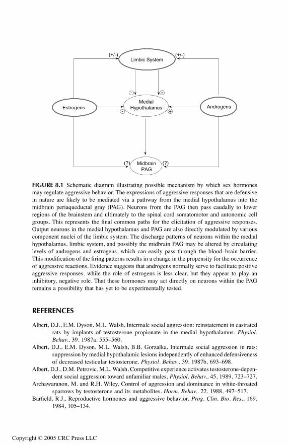
TF1661_C008.fm Page 242 Monday, October 4, 2004 9:20 AM
Copyr
REFERENCES
Albert, D.J., E.M. Dyson, M.L. Walsh, Intermale social aggression: reinstatement in castratedrats by implants of testosterone propionate in the medial hypothalamus, Physiol.Behav., 39, 1987a, 555–560.
Albert, D.J., E.M. Dyson, M.L. Walsh, B.B. Gorzalka, Intermale social aggression in rats:suppression by medial hypothalamic lesions independently of enhanced defensivenessof decreased testicular testosterone, Physiol. Behav., 39, 1987b, 693–698.
Albert, D.J., D.M. Petrovic, M.L. Walsh, Competitive experience activates testosterone-depen-dent social aggression toward unfamiliar males, Physiol. Behav., 45, 1989, 723–727.
Archawaranon, M. and R.H. Wiley, Control of aggression and dominance in white-throatedsparrows by testosterone and its metabolites, Horm. Behav., 22, 1988, 497–517.
Barfield, R.J., Reproductive hormones and aggressive behavior, Prog. Clin. Bio. Res., 169,1984, 105–134.
FIGURE 8.1 Schematic diagram illustrating possible mechanism by which sex hormonesmay regulate aggressive behavior. The expressions of aggressive responses that are defensivein nature are likely to be mediated via a pathway from the medial hypothalamus into themidbrain periaqueductal gray (PAG). Neurons from the PAG then pass caudally to lowerregions of the brainstem and ultimately to the spinal cord somatomotor and autonomic cellgroups. This represents the final common paths for the elicitation of aggressive responses.Output neurons in the medial hypothalamus and PAG are also directly modulated by variouscomponent nuclei of the limbic system. The discharge patterns of neurons within the medialhypothalamus, limbic system, and possibly the midbrain PAG may be altered by circulatinglevels of androgens and estrogens, which can easily pass through the blood–brain barrier.This modification of the firing patterns results in a change in the propensity for the occurrenceof aggressive reactions. Evidence suggests that androgens normally serve to facilitate positiveaggressive responses, while the role of estrogens is less clear, but they appear to play aninhibitory, negative role. That these hormones may act directly on neurons within the PAGremains a possibility that has yet to be experimentally tested.
Limbic System
Estrogens
Medial
Hypothalamus Androgens
Midbrain
PAG
- +
+-
(?)(?)
(+/-)(+/-)
ight © 2005 CRC Press LLC

TF1661_C008.fm Page 243 Monday, October 4, 2004 9:20 AM
Copyr
Barrett, G.M., K. Shimizu, M. Bardi, S. Asaba, A. Mori, Endocrine correlates of rank,reproduction, and female-directed aggression in male Japanese macaques, Macacafuscata, Horm. Behav., 42, 2002, 85–96.
Bermond, B., J. Mos, W. Meelis, A.M. VanDerPoel, M.R. Kruk, Aggression induced bystimulation of the hypothalamus: effects of androgens, Pharmacol. Biochem. Behav.,16, 1982, 41–45.
Booth, A., G. Shelley, A. Mazur, G. Tharp, R. Kittok, Testosterone, and winning and losingin human competition, Horm. Behav., 23, 1989, 556–571.
Bouissou, M.F., Androgens, aggressive behaviour and social relationships in higher mammals,Horm. Res., 18, 1983, 43–61.
Bradford, J. and D. McLean, Sexual offenders, violence and testosterone: a clinical study,Can. J. Psychiatry, 29, 1984, 335–343.
Brain, P.F., Hormones and aggression, Eden Press, Montreal, 1977.Brain, P.F., Hormones and aggression, Eden Press, Montreal, 1978.Brain, P.F. and A.E. Evans, Acute influences of some ACTH-related peptides on fighting and
adrenocortical activity in male laboratory mice, Pharmacol. Biochem. Behav., 7, 1977,425–433.
Caldwell, G.S., S.E. Glickman, E.R. Smith. Seasonal aggression independent of seasonaltestosterone in wood rats, Proc. Natl. Acad. Sci. U.S.A., 81, 1984, 5255–5257.
Christiansen, K. and R. Knussmann, Androgen levels and components of aggressive behaviorin men, Horm. Behav., 21, 1987, 170–180.
Coe, C.L. and S. Levine, Biology of aggression, Bull. Am. Acad. Psychiatry Law, 11, 1983,131–148.
Dabbs, J.M., R.L. Frady, T.S. Carr, N.F. Besch, Saliva, testosterone and criminal violence inyoung adult prison inmates, Psychosom. Med., 49, 1987, 174–182.
DeBold, L.F. and K.A. Miczek, Aggression persists after ovariectomy in female rats, Horm.Behav., 18, 1984, 177–190.
DeBold, J.F. and K.A. Miczek, Testosterone modulates the effects of ethanol on male mouseaggression, Psychopharmacology, 86, 1985, 286–290.
Dolan, M., I.M. Anderson, J.F. Deakin, Relationship between 5-HT function and impulsivityand aggression in male offenders with personality disorders, Br. J. Psychiatr., 178,2001, 352–359.
Dufty, A.M., Testosterone and survival: a cost of aggressiveness? Horm. Behav., 23, 1989,185–193.
Ehrenkranz, J., E. Bliss, M.H. Sheard, Plasma testosterone: correlation with aggressive behav-ior and social dominance in man, Psychosom. Med., 36, 1974, 469–475.
File, S.E., The stress of intruding: reduction by chlordiazepoxide, Physiol. Behav., 33, 1984,345–347.
Finkelstein, J.W., E.J. Susman, V.M. Chinchilli, S.J. Kunselman, M.R. D’Arcangelo, J.Schwab, LM. Demers, L.S. Liben, G. Lookingbill, H.E. Kulin, Estrogen or testoster-one increases self-reported aggressive behaviors in hypogonadal adolescents, J. Clin.Endocrin. Metab., 82, 1997, 2433–2438.
Fishbein, D.H., D. Lozovsky, J.H. Jaffe, Impulsivity, aggression, and neuroendocrineresponses to serotonergic stimulation in substance abusers, Biol. Psychiatry, 25, 1989,1049–1066.
Fraile, I.G., B.S. McEwen, D.W. Pfaff, Progesterone inhibition of aggressive behaviors inhamsters, Physiol. Behav., 39, 1987, 225–229.
Frank, L.G., S.E. Glickman, P. Light, Fatal sibling aggression, precocial development, andandrogens in neonatal spotted hyenas, Science, 252, 1991, 702–704.
ight © 2005 CRC Press LLC
TF1661_C008.fm Page 244 Monday, October 4, 2004 9:20 AM
Copyr
Freeman, L.M., T. Arora, E.F. Rissman, Neonatal androgen affects copulatory behavior in thefemale musk shrew, Horm. Behav., 34, 1998, 231–238.
Fuchs, S.A.G., H.M. Edinger, A. Siegel, The role of the anterior hypothalamus in affectivedefense behavior elicited from the ventromedial hypothalamus of the cat, Brain Res.,330, 1985a, 93–107.
Fuchs, S.A.G., H.M. Edinger, A. Siegel, The organization of the hypothalamic pathwaysmediating affective defense behavior in the cat, Brain Res., 330, 1985b, 77–92.
Gandelman, R. and S. Graham, Singleton female mouse fetuses are subsequently unresponsiveto the aggression-activating property of testosterone, Physiol. Behav., 37, 1986,465–467.
Gandelman, R, C. Peterson, H. Hauser, Mice: fetal estrogen exposure does not facilitate lateractivation of fighting by testosterone, Physiol. Behav., 29, 1982, 397–399.
Gerra, G., A. Zaimovic, A.P. Avanzini, B. Chittolini, G. Giucastro, R. Caccavari, M. Palladino,D. Maestri, C. Monica, R. Delsignore, F. Brambilla, Neurotransmitter-neuroendocrineresponses to experimentally induced aggression in humans: influence of personalityvariable, Psychiatr. Res., 66, 1997, 33–43.
Gerra, G., A. Zaimovic, M.A. Raggi, F. Giusti, R. Delsignore, S. Bertacca, F. Brambilla,Aggressive responding of male heroin addicts under methadone treatment: psycho-metric and neuroendocrine correlates, Drug Alcohol Depend., 65, 2001, 85–95.
Gerra, G., A. Zaimovic, R. Sartori, M.A. Raggi, C. Bocchi, U. Zambelli, M. Timpano, V.Sanichelli, R. Delsignore, F. Brambilla, Experimentally induced aggressiveness inadult children of alchoholics, ACOAs: preliminary behavioral and neuroendocrinefindings, J. Stud. Alcohol, 60, 1999, 776–783.
Gravance, C.G., P.J. Casey, M.J. Erpino, Progesterone does not inhibit aggression inducedby testosterone metabolites in castrated male mice, Horm. Behav., 30, 1996, 22–25.
Hansen, S., Medial hypothalamic involvement in maternal aggression of rats, Behav. Neuro-sci., 103, 1989, 1035–1046.
Harding, C., The role of androgen metabolism in the activation of male behavior, Ann. N.Y.Acad. Sci., 4, 1986, pp. 371–378.
Hau, M., M. Wikelski, K.K. Soma, J.C. Wingfield, Testosterone and year-round territorialaggression in a tropical bird, Gen. Comp. Endocrin., 117, 2000, 20–33.
Hau, M. and P.F. Brain, The effects of differential housing, castration and steroidal hormonereplacement on attacks directed by resident mice towards lactating intruders, Physiol.Behav., 30, 1983, 557–560.
Haug, M., L.F. Spetz, M.L. Ouss-Schlegel, D. Benton, P.F. Brain, Effects of gender, gona-dectomy and social status on attack directed towards female intruders by residentmice, Physiol. Behav., 37, 1986, 533–537.
Hilakivi-Clarke, L., Role of estradiol in alcohol intake and alcohol-related behaviors, J. Stud.Alcohol, 57, 1996, 162–170.
Jasnow, A.M., M.C. Banks, E.C. Owens, K.L. Huhman, Differential effects of two corticotro-pin-releasing factor antagonists on conditioned defeat in male Syrian hamsters,Mesocricetus auratus, Brain Res., 846, 1999, 122–128.
Kavaliers, M., Inhibitory influences of the adrenal steroid, 3alpha, 5alpha-tetrahydroxycorti-costerone on aggression and defeat-induced analgesia in mice, Psychopharmacology,95, 1988, 488–492.
Klukowski, M. and C.E. Nelson, The challenge hypothesis and seasonal changes in aggressionand steroids in male northern fence lizards, Sceloporus undulatus hyacinthinus, Horm.Behav., 33, 1998, 197–204.
Kreuz, L.E. and R.M. Rose, Assessment of aggressive behaviour and plasma testosterone ina young criminal population, Psychosom. Med., 34, 1972, 321–332.
ight © 2005 CRC Press LLC

TF1661_C008.fm Page 245 Monday, October 4, 2004 9:20 AM
Copyr
Leshner, A.I., A model of hormones and agonistic behavior, Physiol. Behav., 15, 1975,225–235.
Lisciotto, C.A., J.F. DeBold, K.A. Miczek, Sexual differentiation and the effects of alcoholon aggressive behavior in mice, Pharmacol. Biochem. Behav., 35, 1990, 357–362.
Lisk, R.D. and M.J. Nachtigall, Estrogen regulation of agonistic and proceptive responses inthe golden hamster, Horm. Behav., 22, 1988, 35–48.
Lonstein, J.S. and S.C. Gammie, Sensory, hormonal, and neural control of maternal aggressionin laboratory rodents, Neurosci. Biobehav. Rev., 26, 2002, 869–888.
Mann, M.A., C. Konen, B. Svare, The role of progesterone in pregnancy-induced aggressionin mice, Horm. Behav., 18, 1984, 140–160.
Mann, M.A. and B. Svare, Factors influencing pregnancy-induced aggression in mice, Behav.Neural Biol., 36, 1982, 242–258.
Mann, M.A. and B. Svare, Prenatal testosterone exposure elevates maternal aggression inmice, Physiol. Behav., 30, 1983, 503–507.
Martensz, N.D., S.V. Vellucci, L.M. Fuller, B.J. Everitt, E.B. Keverne, J. Herbert, Relationbetween aggressive behaviour and circadian rhythms in cortisol and testosterone insocial groups of talapoin monkeys, J. Endocrin., 115, 1987, 107–120.
Mayer, A.D., H.B. Ahdieh, J.S. Rosenblatt, Effects of prolonged estrogen–progesterone treat-ment and hypophysectomy on the stimulation of short-latency maternal behavior andaggression in female rats, Horm. Behav., 24, 1990, 152–173.
Mayer, A.D. and J.S. Rosenblatt, Hormonal factors influence the onset of maternal aggressionin laboratory rats, Horm. Behav., 21, 1987, 253–267.
McEwen, B.S., I. Lieberburg, C. Chaptal, L.C. Krey, Aromatization: important for sexualdifferentiation of the neonatal rat brain, Horm. Behav., 9, 1977, 249–263.
Meaney, M.J., J. Stewart, W.W. Beatty, The influence of glucocorticoids during the neonatalperiod on the development of play-fighting in Norway rat pups, Horm. Behav., 16,1982, 475–491.
Meddle, S.L., L.M. Romero, L.B. Astheimer, W.A. Buttemer, I.T. Moore, J.C. Wingfield,Steroid hormone interrelationships with territorial aggression in an Arctic-breedingsongbird, Horm. Behav., 42, 2002, 212–221.
Meisel, R.L. and M.R. Sterner, Progesterone inhibition of sexual behavior is accompaniedby an activation of aggression in female Syrian hamsters, Physiol. Behav., 47, 1990,415–417.
Meisel, R.L., M.R. Sterner, M.A. Diekman, Differential hormonal control of aggression andsexual behavior in female Syrian hamsters, Horm. Behav., 22, 1988, 453–466.
Michael, R.P. and D. Zumpe, Relation between the seasonal changes in aggression, plasmatestosterone and the photoperiod in male rhesus monkeys, Psychoneuroendocrinology,6, 1981, 145–158.
Millesi, E., I.E. Hoffmann, S. Steurer, M. Metwaly, J.P. Dittami, Vernal changes in thebehavioral and endocrine responses to GnRH application in male European groundsquirrels, Horm. Behav., 41, 2002, 51–58.
Moore, M.C., Elevated testosterone levels during nonbreeding season territoriality in a fall-breeding lizard, Sceloporus jarrovi, J. Comp. Physiol., 158, 1986, 159–163.
Moore, M.C., Circulating steroid hormones during rapid aggressive responses of territorialmale mountain spiny lizards, Sceloporus jarrovi, Horm. Behav., 21, 1987, 511–521.
Moore, M.C., Testosterone control of territorial behavior: tonic-release implants fully restoreseasonal and short-term aggressive responses in free-living castrated lizards, Gen.Comp. Endocrinol., 70, 1988, 450–459.
ight © 2005 CRC Press LLC

TF1661_C008.fm Page 246 Monday, October 4, 2004 9:20 AM
Copyr
Moore, M.C. and C.A. Marler, Effects of testosterone manipulations on nonbreeding seasonterritorial aggression in free-living male lizards, Sceloporus jarrovi, Gen. Comp.Endocrinol., 65, 1987, 225–232.
Naftolin, F., K.J. Ryan, I.J. Davies, V.V. Reddy, F. Flores, Z.I. Petro, M. Kuhn, The formationof estrogens by central neuroendocrine tissues, Rec. Prog. Horm. Res., 31, 1975,295–315.
Olweus, D., A. Mattsson, D. Schalling, H. Low, Testosterone, aggression, physical, andpersonality dimensions in normal adolescent males, Psychosom. Med., 42, 1980,253–269.
Olweus, D., A. Mattsson, D. Schalling, H. Low, Circulating testosterone levels and aggressionin adolescent males: a causal analysis, Psychosom. Med., 50, 1988, 261–272.
Orengo, C., M.E. Kunik, V. Molinari, K. Wristers, S.C. Yodofsky, Do testosterone levels relateto aggression in elderly men with dementia? J. Neuropsychiatr. Clin. Neurol., 14,2002, 161–166.
Perrigo, G., W.C. Bryant, F.S. Vom Saal, Fetal, hormonal and experiential factors influencingthe mating-induced regulation of infanticide in male house mice, Physiol. Behav., 46,1989, 121–128.
Persky, H., K.D. Smith, G.K. Basu, Relation of psychologic measures of aggression andhostility to testosterone production in man, Psychosom. Med., 33, 1971, 265–277.
Pfaff, D.W., Autoradiographic localization of radioactivity in the rat brain after injection oftritiated sex hormones, Science, 161, 1968a, 1355–1356.
Pfaff, D.W., Uptake of estradiol-17β-H3 in the female rat brain: An autoradiographic study,Endocrinology, 82, 1968b, 1149–1155.
Pfaff, D.W. and M. Keiner. Atlas of estradiol-concentrating cells in the central nervous systemof the female rat, J. Comp. Neurol., 151, 1973, 121–158.
Price, E.O., T.E. Adams, C.C. Huxsoll, R.E. Borgwardt, Aggressive behavior is reduced inbulls actively immunized against gonadotropin-releasing hormone, J. Anim. Sci., 81,2003, 411–415.
Raboch, J., H. Cerna, P. Zemek, Sexual aggressivity and androgens, Br. J. Psychiatry, 151,1987, 398–400.
Rada, R.T., Alcoholism and the child molester, Ann. N.Y. Acad. Sci., 273, 1976, 492–496.Rada, R.T., D.R. Laws, R. Kellner, L. Stivastava, G. Peake, Plasma androgens in violent and
nonviolent sex offenders, Bull. Am. Acad. Psychiatry Law, 11, 1983, 149–158.Rhen, T. and D. Crews, Embryonic temperature and gonadal sex organize male-typical sexual
and aggressive behavior in a lizard with temperature-dependent sex determination,Endocrinology, 140., 1999, 4501–4508.
Rhen, T. and D. Crews, Organization and activation of sexual and agonistic behavior in theleopard gecko, Neuroendocrinology, 71, 2000, 252–261.
Rhen, T., J. Ross, D. Crews, Effects of testosterone on sexual behavior and morphology inadult female leopard geckos Eublepharis macularius, Horm. Behav., 36, 1999,119–128.
Rines, J.P. and F.S. Vom Saal, Fetal effects on sexual behavior and aggression in young andold female mice treated with estrogen and testosterone, Horm. Behav., 18, 1984,117–129.
Ruiz-de-la-Torre, J.L. and X. Manteca, Effects of testosterone on aggressive behavior aftersocial mixing in male lambs, Physiol. Behav., 68, 1999, 1–15.
Ryan, B.C. and J.G. Vandenbergh, Intrauterine position effects, Neurosci. Biobehav. Rev., 26,2002, 665–678.
Scallet, A.C., S.J. Suomi, R.E. Bowman, Sex differences in adrenocortical response to con-trolled agonistic encounters in rhesus monkeys, Physiol. Behav., 26, 1981, 385–390.
ight © 2005 CRC Press LLC
TF1661_C008.fm Page 247 Monday, October 4, 2004 9:20 AM
Copyr
Scaramella, T.J. and W.A. Brown, Serum testosterone and aggressiveness in hockey players,Psychosom. Med., 40, 1978, 262–265.
Schlinger, B.A., Plasma androgens and aggressiveness in captive winter white-throated spar-rows, Zonotrichia albicollis, Horm. Behav., 21, 1987, 203–210.
Severyanova, L.A., Neuromodulator mechanism of the inhibitory influence of deoxycorticos-terone on the aggressive-defensive behavior of rats, Neurosci. Behav. Physiol., 18,1988, 486–492.
Shaikh, M.B., J.A. Barrett, A. Siegel, The pathways mediating affective defense and quietbiting attack behavior from the midbrain central gray of the cat: an autoradiographicstudy, Brain Res., 437, 1987, 9–25.
Shrenker, P., S.C. Maxson, B.E. Ginsburg, The role of postnatal testosterone in the develop-ment of sexually dimorphic behaviors in DBA/lBg mice, Physiol. Behav., 35, 1985,757–762.
Siegel, A. and M. Brutus, Neurosubstrates of aggression and rage in the cat, in A.N. Epsteinand A.R. Morrison, Eds., Progress in Psychobiology and Physiological Psychology,Academic Press, San Diego, Calif., 1990, pp. 135–233.
Siegel, A. and M.K. Demetrikopoulos, Hormones and aggression, in J. Shulkin, Ed., Hor-monally Induced Changes in Mind and Brain, Academic Press, San Diego, Calif.,1993, pp. 99–127.
Siegel, A. and H. Edinger, Neural control of aggression and rage, in P.J. Morgane and J.Panksepp, Eds., Handbook of the Hypothalamus, Marcel Dekker, New York, 1981,pp. 203–240.
Siegel, A. and H.M. Edinger, Role of the limbic system in hypothalamically elicited attackbehavior, Neurosi. Biobehav. Rev., 7, 1983, 395–407.
Siegel, A. and C.B. Pott, Neural substrate of aggression and flight in the cat, Prog. Neurobiol.,31, 1988, 261–283.
Simon, N.B., R. Gandelman, L.L. Gray, Endocrine induction of intermale aggression in mice:a comparison of hormonal regimens and their relationship to naturally occurringbehavior, Physiol. Behav., 33, 1984, 379–383.
Simon, N.G. and D.B. Masters, Activation of male-typical aggression by testosterone but notits metabolites in C57BL/6J female mice, Physiol. Behav., 41, 1987, 405–407.
Simon, N.G. and R.E. Whalen, Sexual differentiation of androgen-sensitive and estrogen-sensitive regulatory system for aggressive behavior, Horm. Behav., 21, 1987,493–500.
Smith, L.C. and H.B. John-Alder, Seasonal specificity of hormonal, behavioral, and colorationresponses to within- and between-sex encounters in male lizards, Sceloporus undu-latus, Horm. Behav., 36, 1999, 39–52.
Soma, K.K., R.K. Bindra, J. Gee, J.C. Wingfield, B.A. Schlinger, Androgen-metabolizingenzymes show region-specific changes across the breeding season in the brain of awild songbird, J. Neurobiol., 41, 1999b, 176–188.
Soma, K.K., K. Sullivan, J. Wingfield, Combined aromatase inhibitor and antiandrogentreatment decreases territorial aggression in a wild songbird during the nonbreedingseason, Gen. Comp. Endocrinol., 115, 1999a, 442–453.
Soma, K.K. and J.C. Wingfield, Dehydroepiandrosterone in songbird plasma: seasonal regu-lation and relationship to territorial aggression, Gen. Comp. Endocrinol., 123, 2001,144–155.
Sourial, N. and F. Fenton, Testosterone treatment of an XXYY male presenting with aggres-sion: a case report, Can. J. Psychol., 33, 1988, 846–850.
ight © 2005 CRC Press LLC
TF1661_C008.fm Page 248 Monday, October 4, 2004 9:20 AM
Copyr
Stavisky, R.C., T.C. Register, S.L. Watson, D.S. Weaver, J.R. Kaplan, Behavioral responsesto ovariectomy and chronic anabolic steroid treatment in female cynomolgusmacaques, Physiol. Behav., 66, 1999, 95–100.
Summers, C.H., E.T. Larson, P.J. Ronan, P.M. Hofmann, A.J. Emerson, K.J. Renner, Sero-tonergic responses to corticosterone and testosterone in the limbic system, Gen.Comp. Endocrinol., 117, 2000, 151–159.
Svare, B., Genotype modulates the aggression-promoting quality of progesterone in pregnantmice, Horm. Behav., 22, 1988, 90–99.
Svare, B., J. Miele, C. Kinsley, Mice: Progesterone stimulates aggression in pregnancy-terminated females, Horm. Behav., 20, 1986, 194–200.
Svare, B.B., Ed., Hormones and Aggressive Behavior, Plenum Press, New York, 1983.Takahashi, L.K. and R.D. Lisk, Diencephalic sites of progesterone action for inhibiting
aggression and facilitating sexual receptivity in estrogen-primed golden hamsters,Endocrinology, 116, 1985, 2393–2399.
Takahashi, L.K., R.D. Lisk, A.L. Burnett, Dual estradiol action in diencephalon and theregulation of sociosexual behavior in female golden hamsters, Brain Res., 359, 1985,194–207.
Tazi, A., R. Dantzer, M. Le Moal, J. Rivier, W. Vale, G.F. Koob, Corticotropin-releasing factorantagonist blocks stress-induced fighting in rats, Regul. Peptides, 18, 1987, 37–42.
Tokarz, R.R., Effects of corticosterone treatment on male aggressive behavior in a lizard,Anolis sagrei, Horm. Behav., 21, 1987, 358–370.
Tricas, T.C., K.P. Maruska, L.E. Rasmussen, Annual cycles of steroid hormone production,gonad development, and reproductive behavior in the Atlantic stingray, Gen. Comp.Endocrinol., 118, 2000, 209–225.
Virkkunen, M., Urinary free cortisol secretion in habitually violent offenders, Acta Psychiatr.Scand., 72, 1985, 40–44.
Vom Saal, F.S., W.M. Grant, C.W. McMullen, K.S. Laves, High fetal estrogen concentrations:correlation with increased adult sexual activity and decreased aggression in malemice, Science, 220, 1983, 1306–1308.
Von der, P.B., T. Sarkola, K. Seppa, C.J. Eriksson, Testosterone, 5 alpha-dihydrotestosteroneand cortisol in men with and without alcohol-related aggression, J. Stud. Alcohol, 63,2002, 518–526.
Wagner, C.K., C. Kinsley, B. Svare, Mice: postpartum aggression is elevated followingprenatal progesterone exposure, Horm. Behav., 20, 1986, 212–221.
Watt, M.J., G.L. Forster, J.M. Joss, Steroid correlates of territorial behavior in male jackydragons, Amphibolurus muricatus, Brain Behav. Evol., 61, 2003, 184–194.
Whalen, R.E. and F. Johnson, Individual differences in the attack behavior of male mice: afunction of attack stimulus and hormonal state, Horm. Behav., 21, 1987, 223–233.
Whalen, R.E. and F. Johnson, Aggression in adult female mice: chronic testosterone treatmentinduces attack against olfactory bulbectomized male and lactating female mice, Phys-iol. Behav., 43, 1988, 17–20.
Wingfield, J.C., Environmental and endocrine control of reproduction in the song sparrow,Melospiza melodia. 11. Agonistic interactions as environmental information stimu-lating secretion of testosterone, Gen. Comp. Endocrinol., 56, 1984, 417–424.
Wingfield, J.C. and K.E. Hunt, Arctic spring: hormone-behavior interactions in a severeenvironment, Comp. Biochem. Physiol., 132, 2002, 275–286.
Woodley, S.K. and M.C. Moore, Ovarian hormones influence territorial aggression in free-living female mountain spiny lizards, Horm. Behav., 35, 1999, 205–214.
ight © 2005 CRC Press LLC

TF1661_C008.fm Page 249 Monday, October 4, 2004 9:20 AM
Copyr
Worthman, C.M. and M.J. Konner, Testosterone levels change with subsistence hunting effortin !Kung San men, Psychoneuroendocrinology, 12, 1987, 449–458.
Zitzmann, M. and E. Nieschlag, Tesotosterone levels in healthy men and the relation tobehavioral and physical characteristics: facts and constructs, Eur. J. Endocrinol., 144,2001, 183–197.
ight © 2005 CRC Press LLC

TF1661_C009.fm Page 251 Monday, October 18, 2004 8:29 AM
Copyr
9
Aggression and Immune FunctionM. Demetrikopoulos and A. Siegel
The current literature suggests that there is a reciprocal interaction between aggressionand immune function such that aggressive behaviors affect immune system functioningand immune events contribute to aggressive behavior. Thus, the interplay of aggressionand immune function has been studied from a variety of vantage points, including (1)the role of social status on immune function, (2) the effect of an aggressive act on therecipient, (3) the effect of an aggressive act on the perpetrator, and (4) the effect ofimmune events on aggressive behaviors. Rather than providing an exhaustive reviewof these interactions, this chapter will use illustrative examples that represent the largerbody of knowledge. The objective of this chapter is to review the interplay of aggressionwith immune function with particular attention to these vantage points. While thediscussion of interspecific aggression may add further dimensions to this interaction,this chapter will specifically focus on intraspecific aggression. Following review ofthe interaction between aggression and immunity, we briefly review the convergingneuronal circuitry that may account for this phenomenon.
SOCIAL STATUS EFFECTS ON IMMUNITY
The literature describing the effects of social status on immunity serves as a generalintroduction to this discussion since these social issues are closely related to intraspe-cific aggressive interactions. Many mammals live in colonies, packs, troops, herds,tribes, or other social structures. Within these social structures there is often an ordersuch that animals within the group are part of a social hierarchy. Often, aggressiveinteractions are involved in the development and continuation of these social hier-archies. While a thorough review of this area is beyond the scope of this chapter,several representative examples are explored in order to examine the effect of socialstatus on immunity within a stable social hierarchy, and as a result of hierarchyformation in an unstable social situation. A comparison of results for these two typesof studies will provide suggestive evidence for the role that aggressive behaviorsplay in social status effects on immune function.
STABLE HIERARCHICAL GROUPS
One approach to examining the effects of social status on immune function involvesan experimentally developed stable social hierarchy in the laboratory. Once the
ight © 2005 CRC Press LLC
TF1661_C009.fm Page 252 Monday, October 18, 2004 8:29 AM
Copyr
hierarchy is well established, differences due to rank can be examined. Vekovish-cheva et al. (2000) examined the role of dominance in white mice on a variety ofphysiological functions, including pain threshold, thymus and spleen weights, andprimary antibody production, as well as behavioral measures. Through selectivemanipulations of the groups, they were able to form a number of linear hierarchieswhereby subject A dominated over subject B and C, and subject B dominated oversubject C. Despite differences in motor activity, there were no statistically significantdifferences in the basic physiology of dominant and subordinate mice in this stablelinear hierarchy. However, when more specific immunological measures were exam-ined, Stefanski et al. (2001) demonstrated that subdominant male rats had impairedimmune systems. This study used a despotic hierarchy with five pairs of subjects.In a despotic hierarchy, one subject dominates the others while the latter are relativelyequal and do not demonstrate aggression toward one another. In this model, sub-dominant males had decreased numbers of CD4 and CD8 T cells, as well asdecreased functional capacity of the T cells as measured by their proliferativecapacity to the mitogen ConA. In addition to changes in T cells, subdominantsubjects showed a marked enhancement of granulocyte numbers, which the authorssuggested was of adaptive value if wounding was likely. However, in this experi-mental paradigm wounding was not likely since dominance was largely maintainedby threat. It is likely that the immunological changes were related to the elevatedplasma epinephrine and norepinephrine in subdominant subjects, which may suggestthat these animals were somewhat stressed. The potential clinical significance ofthese changes was apparent in a study by Hessing et al. (1994) that demonstratedthat social status in pigs was critical in predicting morbidity and mortality followingchallenge by Aujeszky disease virus (also known as pseudorabies virus, which isfound mainly in swine). They found that morbidity and mortality were highest forsubordinate pigs in a stable social structure.
The experiments described thus far were conducted after the formation of astable hierarchy within the laboratory. Other researchers have used a naturalisticmodel of observing behavior in the wild. Naturalistic observation ensures that find-ings about dominance are not an artifact of experimentally manipulated socialhierarchy. In one such study, Sapolsky and Spencer (1997) studied a troop of wildbaboons in the Masai Mara National Reserve in Kenya. This troop had been habit-uated to observation for 4 years before the start of the study. The study examinedthe role of dominance hierarchies on anabolic hormone insulin-like growth factor-I (IGF-I), and found a suppression of IGF-I in subordinate male baboons. Thisfinding is germane to the current discussion since IGF-I has been implicated as apossible mediator in a variety of physiological and immunological functions.
A third model for studying the effect of dominance rank on immune function usesa comparison of baseline values to the response evoked by competition for limitedresources. It has been postulated that an important function of dominance hierarchiesis their use in determining access to resources. Boccia et al. (1992) used a competitivewater test, whereby the group was given access to a single waterspout following 24hours of deprivation. Subjects were observed after the water was turned back on todetermine the order in which the subjects drank, length of time they drank, anddisplacement of subjects from the spout. In addition to comparing the baseline values
ight © 2005 CRC Press LLC
TF1661_C009.fm Page 253 Monday, October 18, 2004 8:29 AM
Copyr
to the competitive water test condition, this study used pigtail and bonnet macaques,which exhibit differences in social behavior. Interestingly, dominance did not affectbaseline immune functioning in either species, although there were immunologicaldifferences between the two species such that pigtails showed higher natural killer(NK) cell function and lower lymphocyte activation than the bonnets. As expected,the pigtail macaques showed more agonistic behaviors than did the bonnet macaquesirrespective of the competition condition. During the competition task, the behaviorof the two species varied dramatically. The bonnet monkeys formed a line by domi-nance rank in order to wait for their turn to drink. In contrast, the pigtail monkeyscompeted for access to the water through the increased use of agonistic social inter-actions. Additionally, the pigtail macaques demonstrated an increase in NK activity,and a decrease in lymphocyte activity following the water competition task. Thesefinding for the pigtail macaques are similar to those found in the literature on aggressionand immune function in humans (see Aggression, Hostility, and Marital ConflictEffects on Immunity in Humans section).
HIERARCHY FORMATION
In addition to studying already established social hierarchies, a number of investigatorshave studied the immunological consequences of active hierarchy formation. Resultsfrom these studies have demonstrated that change in the social structure, and thesubsequent aggressivity resulting from social pressure to reestablish a hierarchy, pro-duce a variety of immune changes. For example, male guinea pigs put into socialsituations with unstable dominance had lower complement activity and engaged infrequent stand-threat interactions (Stefanski and Hendrichs, 1996). Generally, guineapigs are able to form stable dominance hierarchies within a very brief period of time,after which these stand-threat interactions disappear. Although plasma cortisol was notcorrelated with the changes in complement activity, the authors hypothesized that theprotracted aggressive interactions were evidence of a stressful environment.
Other investigators have demonstrated an initial concomitant rise in cortisol witha decrease in immune function in response to the establishment of a dominancehierarchy (Gust et al., 1991). Within 48 hours of the formation of a new social group,female rhesus monkeys were able to establish a dominance hierarchy with onlyminor fighting and no wounding or trauma. While the effects on cortisol levels wastransitory, the effects on CD4 and CD8 T cells persisted for more than 2 months,with the low ranking subjects showing significantly reduced numbers of T cells.Similar results were found in a more chronic model of social hierarchy disruptionusing male cynomolgus monkey subjects (Cohen et al., 1997). Subjects were putinto an unstable social situation for 15 months whereby they were reorganized intonew groups every month. Following 8 months of reorganization, the subjects wereexposed to an influenza virus and then subsequently infected with adenovirus after15 months of reorganization. Subjects exposed to the instability manipulation dem-onstrated increased aggressive behaviors. However, low social status, irrespective ofsocial reorganization, was associated with enhanced probability of infection and lessaggressive behaviors. Therefore, the reorganization per se was less important interms of immune competence than was social rank. Interestingly, subjects with lower
ight © 2005 CRC Press LLC
TF1661_C009.fm Page 254 Monday, October 18, 2004 8:29 AM
Copyr
rank showed enhanced cortisol responses to social reorganization, but these elevatedhormonal levels did not account for the changes in infection rates. The differencesin infection rates due to social rank may have clinical implications. Capitanio et al.(1998) studied this more directly by examining survival rates of male rhesusmacaques in unstable social environments that were exposed to simian immunode-ficiency virus (SIV). Animals with a disrupted social hierarchy survived significantlyshorter periods of time and engaged in more aggressive behaviors, while subse-quently engaging in less affiliative behaviors such as grooming. Furthermore, whenthe data were examined across social disruption conditions, subjects receiving moresocial threats had higher plasma SIV levels. These results correspond well with theprior experiment: since the authors chose the receipt of threats as the agonisticbehavior to study, it could be postulated that these were not the dominant subjects.
An interesting complication to the relationship of aggression, dominance, andimmunity was explored by Barnard et al. (1994). Contrary to the studies justdescribed, these authors demonstrated that dominant male mice were infected withthe parasite Babesia microti sooner and were slower to clear the infection. Theysuggested that this effect was due to the immunosuppressive effects of the elevatedtestosterone evident in the dominant mice.
SUMMARY
Because so many mammals demonstrate hierarchical group formation, it is temptingto postulate that there must be an advantage to this social structure and that main-tenance of a stable group should not be detrimental to the individuals within saidgroup. However, even within a stable hierarchy, most studies demonstrate thatsocially subordinate animals have suboptimal immune functioning (Table 9.1), whichmay be partially related to their rank imposing a chronic stress. During the formationof the hierarchy, socially subordinate animals also have suboptimal immune func-tioning, which puts them at a higher risk of opportunistic infections. Thus, subjectsthat engage in less aggressive behaviors tend to have lower immune competencies.
EFFECTS OF AGGRESSION ON RECIPIENT’S IMMUNE FUNCTION
When examining the effect of aggression on immune function, it is useful to distinguishbetween the recipient and perpetrator of the aggressive act. This factor is often relatedto various other factors including the dominance status of the individual, social context,and hormonal and neuroendocrine status at the time of the confrontation. A large bodyof literature focuses on the effect of aggression on the recipient’s immune function.While several models have been used to study this phenomenon, this review is limitedto the social disruption model, whereby an aggressive intruder is placed into anestablished cage of subjects, and to the social confrontation model, whereby the subjectserves as the intruder into an aggressive resident animal’s home cage. However, a largeproportion of this literature frames the findings in terms of the aggressive encounterbeing either a short-term or long-term stressor. Many of these papers do not focus onaggression per se but rather view it as a naturalist stressor. Nevertheless, a few selected
ight © 2005 CRC Press LLC
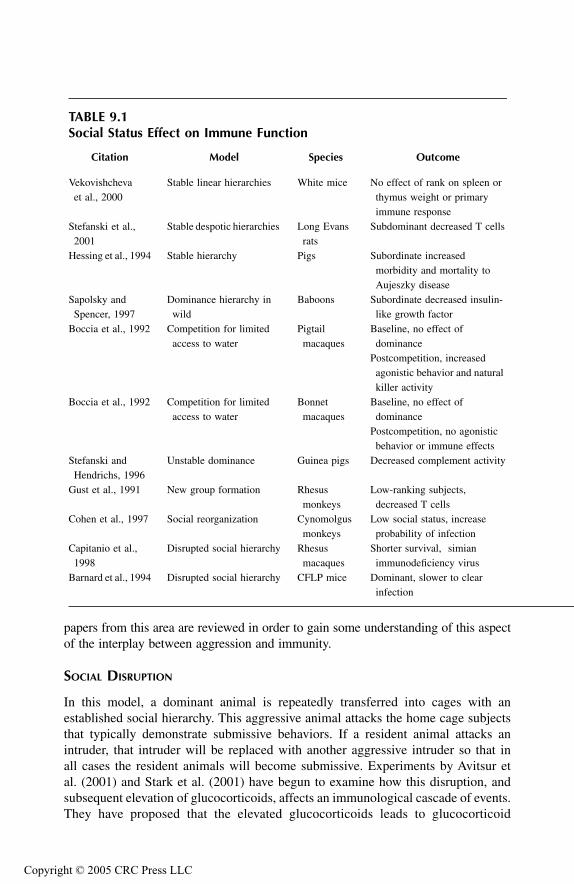
TF1661_C009.fm Page 255 Monday, October 18, 2004 8:29 AM
Copyr
papers from this area are reviewed in order to gain some understanding of this aspectof the interplay between aggression and immunity.
SOCIAL DISRUPTION
In this model, a dominant animal is repeatedly transferred into cages with anestablished social hierarchy. This aggressive animal attacks the home cage subjectsthat typically demonstrate submissive behaviors. If a resident animal attacks anintruder, that intruder will be replaced with another aggressive intruder so that inall cases the resident animals will become submissive. Experiments by Avitsur etal. (2001) and Stark et al. (2001) have begun to examine how this disruption, andsubsequent elevation of glucocorticoids, affects an immunological cascade of events.They have proposed that the elevated glucocorticoids leads to glucocorticoid
TABLE 9.1Social Status Effect on Immune Function
Citation Model Species Outcome
Vekovishcheva et al., 2000
Stable linear hierarchies White mice No effect of rank on spleen or thymus weight or primary immune response
Stefanski et al., 2001
Stable despotic hierarchies Long Evans rats
Subdominant decreased T cells
Hessing et al., 1994 Stable hierarchy Pigs Subordinate increased morbidity and mortality to Aujeszky disease
Sapolsky and Spencer, 1997
Dominance hierarchy in wild
Baboons Subordinate decreased insulin-like growth factor
Boccia et al., 1992 Competition for limited access to water
Pigtail macaques
Baseline, no effect of dominance
Postcompetition, increased agonistic behavior and natural killer activity
Boccia et al., 1992 Competition for limited access to water
Bonnet macaques
Baseline, no effect of dominance
Postcompetition, no agonistic behavior or immune effects
Stefanski and Hendrichs, 1996
Unstable dominance Guinea pigs Decreased complement activity
Gust et al., 1991 New group formation Rhesus monkeys
Low-ranking subjects, decreased T cells
Cohen et al., 1997 Social reorganization Cynomolgus monkeys
Low social status, increase probability of infection
Capitanio et al., 1998
Disrupted social hierarchy Rhesus macaques
Shorter survival, simian immunodeficiency virus
Barnard et al., 1994 Disrupted social hierarchy CFLP mice Dominant, slower to clear infection
ight © 2005 CRC Press LLC

TF1661_C009.fm Page 256 Monday, October 18, 2004 8:29 AM
Copyr
resistance in subordinate animals that may be important for wound healing and berelated to changes seen in humans during depression. This effect was particularlypronounced in macrophages, and was likely to develop in animals that showed lowlevels of social exploration prior to attack and high levels of submission followingattack. Thus, the behavioral profile of the animals, prior to their interaction in anagonistic encounter, was able to predict their behavior in this encounter as well aspredict the physiological consequences of the agonistic encounter. Hence, thereappears to be a social disposition that predisposes an individual animal to respondin a particular physiological way to an aggressive encounter.
SOCIAL CONFRONTATION
This model is often termed resident–intruder confrontation and has been widely usedas a social stress model to study a variety of physiological processes, includingchanges in immune function, cardiovascular issues, and stress-related digestiveissues. It has been likened to the naturalistic stressors that territorial animals wouldencounter in the wild. In this model, the subject acts as an intruder into an aggressiveanimal’s home cage. In general, the intruder animal is attacked and is often defeatedby the resident animal. The intruder may become submissive and cower away fromthe resident animal or it may become subdominant and show enhanced motor activity.Due to the complex nature of this manipulation, a variety of control groups arenecessary for this paradigm, including a group that experiences a novel environment.
While it is beyond the scope of this chapter to present a thorough review of thisliterature, a few papers have been selected that demonstrate some of the complexityof responses and variables that contribute to the richness of this model. For example,Dreau et al. (1999) used this model to examine the effect of social confrontation onimmune function. Subjects exposed to an aggressive resident animal five times aday for 2 days demonstrated decreases in thymocyte numbers, decreased in vitroresponse to LPS, and decreases in CD4 and CD8 T cells. Although not statisticallysignificant, the subjects also had increased in vivo Escherichia coli bacterial growth.
Stefanski and Engler (1998) sought to distinguish the effects of acute and chronicsocial confrontation on cellular immunity. The subjects were exposed to the aggres-sive resident for 2 hours or for 48 hours with either exposure time resulting inaggressive encounters to establish dominance. Many of the immunological variablesexamined were affected similary in both the chronic and acute exposure times.However, some of the impairment present following 2 hours of agonistic behaviorhad been resolved in the 48-hour group. Interestingly, the correlation that was presentat 2 hours between immune function and level of submissive behavior expressed bylosers was not present at 48 hours.
In order to better characterize immune status following social confrontation, Ste-fanski and Ben-Eliyahu (1996) examined the development of syngeneic tumor metasta-sis. Subjects experienced 7 hours of confrontation and received mammary tumor cells1 hour into the confrontation. The intruders had increased tumor retention that wasrelated to the level of submissive behaviors displayed. Furthermore, pretreatment with
ight © 2005 CRC Press LLC
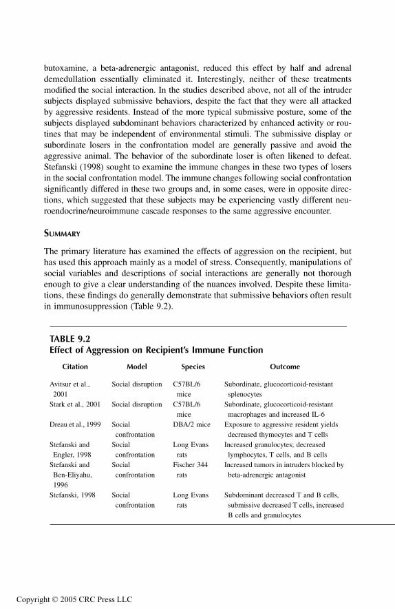
TF1661_C009.fm Page 257 Monday, October 18, 2004 8:29 AM
Copyr
butoxamine, a beta-adrenergic antagonist, reduced this effect by half and adrenaldemedullation essentially eliminated it. Interestingly, neither of these treatmentsmodified the social interaction. In the studies described above, not all of the intrudersubjects displayed submissive behaviors, despite the fact that they were all attackedby aggressive residents. Instead of the more typical submissive posture, some of thesubjects displayed subdominant behaviors characterized by enhanced activity or rou-tines that may be independent of environmental stimuli. The submissive display orsubordinate losers in the confrontation model are generally passive and avoid theaggressive animal. The behavior of the subordinate loser is often likened to defeat.Stefanski (1998) sought to examine the immune changes in these two types of losersin the social confrontation model. The immune changes following social confrontationsignificantly differed in these two groups and, in some cases, were in opposite direc-tions, which suggested that these subjects may be experiencing vastly different neu-roendocrine/neuroimmune cascade responses to the same aggressive encounter.
SUMMARY
The primary literature has examined the effects of aggression on the recipient, buthas used this approach mainly as a model of stress. Consequently, manipulations ofsocial variables and descriptions of social interactions are generally not thoroughenough to give a clear understanding of the nuances involved. Despite these limita-tions, these findings do generally demonstrate that submissive behaviors often resultin immunosuppression (Table 9.2).
TABLE 9.2Effect of Aggression on Recipient’s Immune Function
Citation Model Species Outcome
Avitsur et al., 2001
Social disruption C57BL/6 mice
Subordinate, glucocorticoid-resistant splenocytes
Stark et al., 2001 Social disruption C57BL/6 mice
Subordinate, glucocorticoid-resistant macrophages and increased IL-6
Dreau et al., 1999 Social confrontation
DBA/2 mice Exposure to aggressive resident yields decreased thymocytes and T cells
Stefanski and Engler, 1998
Social confrontation
Long Evans rats
Increased granulocytes; decreased lymphocytes, T cells, and B cells
Stefanski and Ben-Eliyahu, 1996
Social confrontation
Fischer 344 rats
Increased tumors in intruders blocked by beta-adrenergic antagonist
Stefanski, 1998 Social confrontation
Long Evans rats
Subdominant decreased T and B cells, submissive decreased T cells, increased B cells and granulocytes
ight © 2005 CRC Press LLC

TF1661_C009.fm Page 258 Monday, October 18, 2004 8:29 AM
Copyr
EFFECTS OF AGGRESSION ON PERPETRATOR’S IMMUNE FUNCTION
While the previous section reviewed the effects of aggression on the recipient, thecurrent section will review the effects of aggression on the perpetrator’s immunesystem. I attempt to describe what happens to an individual’s immune system as itengages in aggressive acts. This literature is quite broadly defined, and includes (1) anumber of models of aggression such as social isolation and resident–intruder models;(2) the relationship of aggressive behaviors to strain differences in immune function;and (3) the effects of aggressive behaviors in humans, including hostility and maritalconflict. Once again, rather than providing an exhaustive review of this area, theselected experiments highlight some important aspect of the larger literature.
SOCIAL ISOLATION–INDUCED FIGHTING
In this model of aggression, subjects are housed in isolation for some period of timeprior to reintroduction to a neutral group setting. Often, following the isolationhousing, subjects are kept in cages equipped with a transparent divider, so that theycan see the other subject but not physically interact with it prior to the experimentalencounter. These encounters generally involve an aggressive confrontation that ulti-mately leads to dominant and submissive behavioral development.
Gryazeva et al. (2001) examined the effect of multiple aggressive encounters onimmune function. They found that all participants in the aggressive encountersincreased the proportion of segmented neutrophils and CD4 and CD8 T cells in thespleen. Despite these general findings, there were additional immune changes thatdemonstrated divergence such that the winners and losers of these encounters hadopposite immunological outcomes. This finding agrees with that of Hardy et al.(1990), who demonstrated that the direction of functional T-cell changes was depen-dent on the dominance status of the fighting subject. Submissive mice had lower T-cell proliferation compared to dominant and nonfighter controls. In a second exper-iment, they used a test that was less likely to produce wounding. In this experiment,they found that dominant mice had elevated T-cell proliferation compared to sub-missive and nonfought controls. Thus, the physical consequences of fighting had aneffect independent of the psychological aspects of the aggressive encounter.
RESIDENT–INTRUDER INTERACTION
This model was described in the social confrontation subsection above, and isgenerally used to study the effects of aggression on the recipient’s immune systemsince the model normally uses a highly aggressive resident. As previously mentioned,in this model an intruder rat is introduced into a resident’s home cage. Kavelaars etal. (1999) used this model to study the effect of the attack latency of wild-typeresident rats on the immune function of the resident. Animals with short attacklatency were less resistant to experimental autoimmune encephalomyelitis (EAE).Thus, aggressive rats were more susceptible to EAE than nonaggressive rats. Thisfinding somewhat contradicts much of the immune/aggression literature in that itdemonstrates a detrimental outcome for the perpetrator of the aggressive act.
ight © 2005 CRC Press LLC

TF1661_C009.fm Page 259 Monday, October 18, 2004 8:29 AM
Copyr
Gasparotto et al. (2002) modified the basic resident–intruder preparation further byhaving both the residents and intruders individually housed prior to the socialconfrontation. Following social isolation, half of the subjects were moved into alarger environment 3 days before the confrontation experience to become essentiallya resident subject. This differs from the more classic resident–intruder model in thatthe resident is not very well established, and only obtains dominance in approxi-mately 65% of the dyads, whereas in the classic resident–intruder model, the residentis generally dominant. Subjects were socially challenged for 2 or 3 weeks andimmunologically challenged with sheep red blood cells (SRBC). The shorter chronicsocial challenge produced a depression in the primary immune response in submis-sive mice, while the longer chronic social challenge produced an enhancement ofthe primary immune response in dominant subjects. Thus, this distinction was similarto that seen in the less chronic confrontation that occurred in either the presence orabsence of wounding. It is possible that in the longer chronic confrontation, the levelof agonistic behavior was somewhat reduced or more stylized. However, this cannotbe determined from this study, since the authors did not address the nature or severityof the agonistic behaviors in the two conditions.
STRAIN DIFFERENCES IN BEHAVIOR AND IMMUNE FUNCTION
In addition to individual differences, many species have strains that differ greatly intheir levels of aggressive and submissive behaviors. In fact, some strains of micehave been selectively bred to produce high and low levels of aggressive behaviors.These selectively bred animals are models for understanding genetic contributionsto individual differences. Petitto et al. (1993, 1994) examined a number of immu-nological factors in ICR mice that were selectively bred for differences in levels ofsocial isolation–induced aggression. Selection was originally based on the frequencyof attacks following contact with a randomly bred conspecific. Although the selectionprogram was bidirectional, the high-aggressive line (NC900) does not differ fromthe unselected line. However, the low-aggressive line (NC100) exhibited enhancedfreezing and immobility that were characterized as social inhibition. Importantly,these lines only appear to differ behaviorally in social contexts, and are quite similarin terms of their other behavioral responses, including their response to novelty andfearfulness. In addition to the differences in aggressive behaviors, these mice havesignificant and biologically relevant changes in immune function. These sociallyinhibited mice had reduced NK activity and increased susceptibility to tumor devel-opment (Petitto et al., 1993). They also had lower T-cell proliferation and lower IL-2 and gamma interferon production (Petitto et al., 1994). Similarly, Devoino et al.(1993) demonstrated that strains of mice which differed in social isolation inducedaggression had correspondingly different immune responses following immunizationwith sheep red blood cells. The aggressive CBA strain had greater numbers of plaque-forming cells in response to aggressive encounters, while the plaque-forming cellsdecreased in the submissive C57BL mice compared to controls. Thus, associationof the aggressive behavior with the primary immune response to SRBC is dependenton the subject’s strain.
ight © 2005 CRC Press LLC
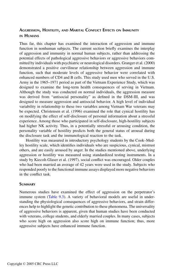
TF1661_C009.fm Page 260 Monday, October 18, 2004 8:29 AM
Copyr
AGGRESSION, HOSTILITY, AND MARITAL CONFLICT EFFECTS ON IMMUNITY IN HUMANS
Thus far, this chapter has examined the interaction of aggression and immunefunction in nonhuman subjects. The current section briefly examines the interplayof aggression and immunity in normal human subjects, rather than addressing thepotential effects of pathological aggressive behaviors or aggressive behaviors com-mitted by individuals with psychiatric or neurological disorders. Granger et al. (2000)demonstrated a positive curvilinear relationship between aggression and immunefunction, such that moderate levels of aggressive behavior were correlated withenhanced numbers of CD4 and B cells. This study used men who served in the U.S.Army in the 1965–1971 period as part of the Vietnam Experience Study, which wasdesigned to examine the long-term health consequences of serving in Vietnam.Although the study was conducted on normal individuals, the aggression measurewas derived from “antisocial personality” as defined in the DSM-III, and wasdesigned to measure aggression and antisocial behavior. A high level of individualvariability in relationship to these two variables among Vietnam War veterans maybe expected. Christensen et al. (1996) examined the role that cynical hostility hason modifying the effect of self-disclosure of personal information about a stressfulexperience. Among those who participated in self-disclosure, high-hostility subjectshad higher NK activity. Thus, in a potentially stressful or arousing condition, thepersonality variable of hostility predicts both the general status of arousal duringthe disclosure task and the immunological reaction to the task.
Hostility was measured in introductory psychology students by the Cook–Med-ley hostility scale, which identifies individuals who are suspicious, cynical, mistrustothers, and are easily aroused by anger. In the studies mentioned above, underlyingaggression or hostility was measured using standardized testing instruments. In astudy by Kiecolt-Glaser et al. (1997), social conflict was encouraged. Older coupleswho had been married an average of 42 years were used in the study. Subjects whoresponded poorly to the functional immune assays displayed more negative behaviorsin the conflict task.
SUMMARY
Numerous studies have examined the effect of aggression on the perpetrator’simmune system (Table 9.3). A variety of behavioral models are useful in under-standing the physiological consequences of aggressive behaviors, and strain differ-ences help to highlight the genetic contribution to these phenomena. The universalityof aggressive behaviors is apparent, given that human studies have been conductedwith veterans, college students, and elderly married couples. In many cases, subjectswho score high on aggression also score high on immune function; thus, moreaggressive subjects have enhanced immune function.
ight © 2005 CRC Press LLC
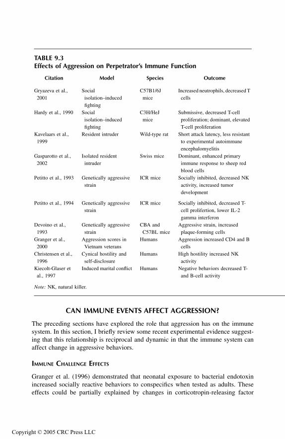
TF1661_C009.fm Page 261 Monday, October 18, 2004 8:29 AM
Copyr
CAN IMMUNE EVENTS AFFECT AGGRESSION?
The preceding sections have explored the role that aggression has on the immunesystem. In this section, I briefly review some recent experimental evidence suggest-ing that this relationship is reciprocal and dynamic in that the immune system canaffect change in aggressive behaviors.
IMMUNE CHALLENGE EFFECTS
Granger et al. (1996) demonstrated that neonatal exposure to bacterial endotoxinincreased socially reactive behaviors to conspecifics when tested as adults. Theseeffects could be partially explained by changes in corticotropin-releasing factor
TABLE 9.3Effects of Aggression on Perpetrator’s Immune Function
Citation Model Species Outcome
Gryazeva et al., 2001
Social isolation–induced fighting
C57B1/6J mice
Increased neutrophils, decreased T cells
Hardy et al., 1990 Social isolation–induced fighting
C3H/HeJ mice
Submissive, decreased T-cell proliferation; dominant, elevated T-cell proliferation
Kavelaars et al., 1999
Resident intruder Wild-type rat Short attack latency, less resistant to experimental autoimmune encephalomyelitis
Gasparotto et al., 2002
Isolated resident intruder
Swiss mice Dominant, enhanced primary immune response to sheep red blood cells
Petitto et al., 1993 Genetically aggressive strain
ICR mice Socially inhibited, decreased NK activity, increased tumor development
Petitto et al., 1994 Genetically aggressive strain
ICR mice Socially inhibited, decreased T-cell prolifertion, lower IL-2 gamma interferon
Devoino et al., 1993
Genetically aggressive strain
CBA and C57BL mice
Aggressive strain, increased plaque-forming cells
Granger et al., 2000
Aggression scores in Vietnam veterans
Humans Aggression increased CD4 and B cells
Christensen et al., 1996
Cynical hostility and self-disclosure
Humans High hostility increased NK activity
Kiecolt-Glaser et al., 1997
Induced marital conflict Humans Negative behaviors decreased T- and B-cell activity
Note: NK, natural killer.
ight © 2005 CRC Press LLC
TF1661_C009.fm Page 262 Monday, October 18, 2004 8:29 AM
Copyr
levels during neonatal development that had long-term social consequences. In asimilar study, Granger et al. (2001) explored the effect of early immune challengeon adult aggressive behaviors. When ICR neonatal mice were treated with endotoxin,adults from the high-aggressive line tended to show lower levels of aggressivebehaviors. Thus, an acute immune challenge was capable of modifying a geneticallydetermined behavioral expression.
AUTOIMMUNE DISORDER EFFECTS
Sakic et al. (1998) demonstrated that autoimmune, lupus-prone MRL-1pr mice showedreduced aggressiveness in the isolation-induced model of aggression. Several aspectsof aggressive behavior, including longer attack latencies, fewer fights, and shorterduration of fights were manifest. However, it is unclear how this immune activationproduced its effect since these subjects also had concomitant decreases in testosterone.
POTENTIAL MEDIATORS
Brain cytokines may represent a potential mediator for the effects of immune vari-ables on aggressive behaviors. This possibility was recently tested in a study byHassanain et al. (2003a, 2003b) in which the role of interleukin-1 beta (IL-1β) inthe hypothalamus on defensive rage behavior elicited from the periaqueductal gray(PAG) was assessed. IL-1β is derived from both the immune and nervous systems,and is endogenously present in the hypothalamus. It is thought to affect aggressionby the induction of serotonin release. In the first part of this study, it was shownthat microinjections of a 5-HT1A agonist into the medial hypothalamus suppresseddefensive rage behavior elicited from the PAG, while microinjections of a 5-HT2
agonist into the same region of the medial hypothalamus facilitated PAG-eliciteddefensive rage. In the second part of the study, microinjections of an IL-1β agonistinto the same region also facilitated PAG-elicited defensive rage. Of particularinterest was the third part of the study, which demonstrated that pretreatment witha 5-HT2 antagonist into the medial hypothalamus blocked the facilitatory effects ofIL-1β. Thus, this finding was the first to demonstrate that IL-1β in the medialhypothalamus can potentiate defensive rage behavior elicited from the PAG, an effectthat is mediated through a serotonergic mechanism.
SUMMARY
There is a growing body of literature demonstrating the reciprocal nature of the inter-action between aggression and immune functioning (Table 9.4). Thus, the findings ofthe effects of aggression on immunity and the effects of immunity on aggression mustbe placed within this larger framework. A careful examination of the effects of theimmune system on aggression may suggest critical developmental stages for aggressivebehavioral development, as well as pathways and neuroendocrine cascades for itsexpression. In addition, the discovery that cytokines in the brain can powerfully mod-ulate aggression and rage, and that such effects are mediated through classical neu-rotransmitter mechanisms may open an entirely new direction of research governingthe relationships among cytokines, aggression, and rage behavior.
ight © 2005 CRC Press LLC

TF1661_C009.fm Page 263 Monday, October 18, 2004 8:29 AM
Copyr
CONVERGING NEURONAL CIRCUITRY
ROLE OF CYTOKINES
The studies described above by Hassanain et al. (2003a, 2003b) have provided animportant clue concerning how centrally mediated cytokines may function withinexisting neuronal circuits that govern defensive rage behavior. The discovery of thepresence of IL-1β receptors in the medial hypothalamus, and the fact that its acti-vation can facilitate defensive rage, suggest that the functional effects of thesereceptors are mediated through their actions on medial hypothalamic neurons. Recallfrom earlier chapters that the major descending pathway from the medial hypothal-amus underlying the expression of defensive rage is one that projects to the PAGand whose functions are mediated principally by N-methyl-D-aspartate receptors inthe PAG. As noted above, IL-1β receptor facilitation of defensive rage appears tobe linked to 5-HT2 receptors, whose activation was shown to also facilitate defensiverage.
A significant feature of these findings is that they provide a basis for under-standing how other cytokines may modulate aggression and rage. Namely, wesuggest that different cytokines in the hypothalamus and related regions probablyact through classical neurotransmitter–receptors such as monoamines, amino acids,and neuropeptides to either excite or inhibit descending medial hypothalamic neu-rons, which project to the PAG and adjoining sites in the brainstem that mediate therage response. In view of the fact that this line of research is in its infancy, there isa basic need for us to strengthen our understanding of how different cytokines maymodulate aggression and rage and to establish the mechanisms underlying sucheffects.
TABLE 9.4Immune Event Effects on Aggression
Citation Model Species Outcome
Granger et al., 1996
Neonatal exposure to endotoxin
ICR mice Increased socially reactive behaviors as adults
Granger et al., 2001
Neonatal exposure to endotoxin
ICR mice selectively bred
Decreased aggressive behavior of genetically aggressive adults
Sakic et al., 1998 Autoimmune disorder Lupus-prone MRL-1pr mice
Reduced aggression in isolation-induced aggression
Hassanain et al., 2003a, 2003b
Midbrain periaqueductal gray–elicited defensive rage
Cats Medial hypothalamus 5-HT1A agonist suppressed rage; 5-HT2
agonist or IL-1β facilitated rage; IL-1β effect blocked by 5-HT2 antagonist
ight © 2005 CRC Press LLC

TF1661_C009.fm Page 264 Monday, October 18, 2004 8:29 AM
Copyr
ROLE OF PERIAQUEDUCTAL GRAY
As discussed in previous chapters, the PAG has a prominent role in the modulationof aggression, and there is a growing body of evidence which suggests that the PAGhas an important role in immune function. For example, Demetrikopoulos et al.(1994) demonstrated that stimulation of the rostral-dorsal PAG suppressed NK cellfunction, and then Demetrikopoulos and Zhang (1998) showed that stimulation ofthe rostral-ventral PAG enhanced NK cell activity. The role of the PAG in aggression-induced effects on immunomodulation may be directly related to the neural circuitryunderlying aggressive behaviors or may be due to the effects of secondary stress(reviewed in Demetrikopoulos et al., 1999).
SECONDARY STRESS EFFECTS
While a thorough review of the effects of stress on immune function are beyond thescope of this chapter, in this section I will briefly review the primary pathways thatmay be secondarily activated in response to aggressive encounters (Demetrikopouloset al., 1999). The classic stress response involves the following cascade: corticotro-pin-releasing factor is released from the hypothalamus, which leads to release ofadrenocorticotripic hormone from the anterior pituitary, which leads to release ofglucocorticoid from the adrenal cortex. While it is tempting to suggest that thissystem’s effects on immunity are simply due to the well-known immunosuppressiveeffect of glucocorticoid, an examination of this literature demonstrates that gluco-corticoids constitute one of various factors that regulate immune function. Anotheraspect of this hypothalamic-mediated stress response includes release of luteinizinghormone from the pituitary, which stimulates the testis to produce testosterone(Tewes, 1999), which leads to glucose uptake in muscles. Testosterone is criticallyimportant for aggression (see Chapter 8) and can be immunosuppressive.
The sympathetic nervous system is also critically involved with the effects ofstress on immunity and equally important in the study of aggression. It has beenwell documented that immune organs are directly innervated by the sympatheticnervous system and that many immune cells contain adrenergic receptors. Opiatesand serotonin are other neurotransmitter systems that may be important for theconvergence of aggression, stress, and immune function (Demetrikopoulos et al.,1999). For example, opiate-dependent immunosuppressive effects may be mediatedthrough the µ receptors in the caudal-ventral PAG. Furthermore, it has been sug-gested that the raphe nucleus serotonergic system is immunosuppressive.
SUMMARY
Various factors may be directly or indirectly involved in the effects of aggressionon immunity. There is a clear convergence in the literature of neuroanatomicalpathways, neurotransmitter systems, cytokines, and hormonal mediators that arecommon to the aggression and immune system literature.
ight © 2005 CRC Press LLC

TF1661_C009.fm Page 265 Monday, October 18, 2004 8:29 AM
Copyr
OVERALL SUMMARY
As a general rule, aggressive behaviors are correlated to increased immune functionin the perpetrator and decreased immune function in the losers. For the most part,in this chapter I did not attempt to look at more complex systems that would relateadditional variables such as the interplay of aggression, sex hormones, and immunefunction. A number of these three-part interactions may help to explain some of thecontradictions in the literature in this area. Future research may attempt to elucidatethe potential mediators of these phenomena, as well as explore the convergingneuronal circuitry that underlies the interaction between aggression and immunefunctions.
REFERENCES
Avitsur, R., J.L. Stark, J.F. Sheridan, Social stress induces glucocorticoid resistance in sub-ordinate animals, Horm. Behav., 39, 2001, 247–257.
Barnard, C.J., J.M. Behnke, J. Sewell, Social behaviour and susceptibility to infection inhouse mice, Mus musculus: effects of group size, aggressive behavior and status-related hormonal responses prior to infection on resistance to Babesia microti, Par-asitology, 108, 1994, 487–96.
Boccia, M.L., M.L. Laudenslager, C.L. Broussard, A.S. Hijazi, Immune responses followingcompetitive water tests in two species of macaques, Brain Behav. Immun., 6, 1992,201–213.
Capitanio, J.P., S.P. Mendoza, N.W. Lerche, W.A. Mason, Social stress results in alteredglucocorticoid regulation and shorter survival in simian acquired immune deficiencysyndrome, Proc. Natl. Acad. Sci. U.S.A., 95, 1998, 4714–4719.
Christensen, A.J., D.L. Edwards, M.A. John, S. Wiebe, E.G. Benotsch, L. McKelvey, M.Andrews, D.M. Lubaroff, Effect of verbal self-disclosure on natural killer cell activity:moderating influence of cynical hostility, Psychosom. Med., 58, 1996, 150–155.
Cohen, S., S. Line, S. Manuck, B.S. Rabin, E.R. Heise, J.R. Kaplan, Chronic social stress,social status, and susceptibility to upper respiratory infections in nonhuman primates,Psychosom. Med., 59, 1997, 213–221.
Demetrikopoulos, M.K., S.E. Keller, S.J. Schleifer. Stress effects on immune function inrodents, in Psychoneuroimmunology: an interdisciplinary introduction, M. Sched-lowski and U. Tewes, Eds., Kluwer Academic/Plenum Publishers, 1999, pp. 259–275.
Demetrikopoulos, M.K., A. Siegel, S.J. Schleifer, J. Obedi, S.E. Keller, Electrical stimulationof the dorsal midbrain periaqueductal gray suppresses peripheral blood natural killercell activity, Brain Behav. Immun., 8, 1994, 218–228.
Demetrikopoulos, M.K. and Z. Zhang, Rostal-ventral electrical stimulation of the midbrainperiaqueductal gray limits tumor metastasis in rats, Soc. Neurosci., 24, 1998, 737.
Devoino, L., E. Alperina, N. Kudryavrseva, N. Popova, Immune responses in male mice withaggressive and submissive behavior patterns: Strain differences, Brain Behav. Immun.,7, 1993, 91–96.
Dreau, D., G. Sonnenfeld, N. Fowler, D.S. Morton, M. Lyte, Effects of social conflict onimmune responses and E. coli growth within closed chambers in mice, Physiol.Behav., 67., 1999, 133–140.
ight © 2005 CRC Press LLC

TF1661_C009.fm Page 266 Monday, October 18, 2004 8:29 AM
Copyr
Gasparotto, O.C., Z.M. Ignacio, K. Lin, S. Goncalves, The effect of different psychologicalprofiles and timings of stress exposure on humoral immune response, Physiol. Behav.,76, 2002, 321–326.
Granger, D.A., A. Booth, D.R. Johnson, Human aggression and enumerative measures ofimmunity, Psychosom. Med., 62, 2000, 583–590.
Granger, D.A., K.E. Hood, N.A. Dreschel, E. Sergeant, A. Likos, Developmental effects ofearly immune stress on aggressive, socially reactive, and inhibited behaviors, Dev.Psychopathol., 13, 2001, 599–610.
Granger, D.A., K.E. Hood, S.C. Ikeda, C.L. Reed, M.L. Block, Neonatal endotoxin exposurealters the development of social behavior and the hypothalamic-pituitary-adrenal axisin selectively bred mice, Brain Behav. Immun., 10, 1996, 249–259.
Gryazeva, N.I., A.V. Shurlygina, L.V. Verbitskaya, E.V. Melnikova, N.N. Kudryavtseva, V.A.Trifakin, Changes in various measures of immune status in mice subject to chronicsocial conflict, Neurosci. Behav. Physiol., 31, 2001, 75–81.
Gust, D.A., T.P. Gordon, M.E. Wilson, A. Ahmed-Ansari, A.R. Brodie, H.M. McClure,Formation of a new social group of unfamiliar female rhesus monkeys affects theimmune and pituitary adrenocortical systems, Brain Behav. Immun., 5, 1991,296–307.
Hardy, C., J. Quay, S. Livnat, R. Ader, Altered T-lymphocyte response following aggressiveencounters in mice, Physiol. Behav., 47, 1990, 1245–1251.
Hassanain, M., S. Bhatt, A. Siegel, Differential modulation of feline defensive rage behaviorin the medial hypothalamus by 5-HT1A and 5-HT2 receptors, Brain Res., 981, 2003a,201–209.
Hassanain, M., S. Zalcman, S. Bhatt, A. Siegel, Interleukin-1 in the hypothalamus potentiatesfeline defensive rage: Role of 5-HT2 receptors, Neuroscience, 120, 2003b, 227–233.
Hessing, M.J., C.J. Scheepens, W.G. Schouten, M.J. Tielen, P.R. Wiepkema, Social rank anddisease susceptibility in pigs, Vet. Immunol. Immunopathol., 43, 1994, 337–387.
Kavelaars, A., C.J. Heijnen, R. Tennekes, J.E. Bruggink, J.M. Koolhaas. Individual behavioralcharacteristics of wild-type rats predict susceptibility to experimental autoimmuneencephalomyelitis, Brain Behav. Immun., 13, 1999, 279–286.
Kiecolt-Glaser, J.K., R. Glaser, J.T. Cacioppo, R.C. MacCallum, M. Snydersmith, K. Cheon-grag, W.B. Malarkey, Marital conflict in older adults: Endocrinological and immu-nological correlates, Psychosom. Med., 59, 1997, 339–349.
Petitto, J.M., D.T. Lysle, J. Gariepy, P.H. Clubb, R.B. Cairns, M.H. Lewis, Genetic differencesin social behavior: Relation to natural killer cell function and susceptibility to tumordevelopment, Neuropsychopharmacology, 8, 1993, 35–43.
Petitto, J.M., D.T. Lysle, J. Gariepy, M.H. Lewis, Association of genetic differences in socialbehavior and cellular immune responsiveness: effects of social experience, BrainBehav. Immun., 8, 1994, 111–122.
Sakic, B., L. Gurunlian, S.D. Denburg, Reduced aggressiveness and low testosterone levelsin autoimmune MRL-1pr males, Physiol. Behav., 63, 1998, 305–309.
Sapolsky, R.M. and E.M. Spencer, Insulin-like growth factor I is suppressed in sociallysubordinate male baboons, Am. J. Physiol., 273, 1997, R1336–R1351.
Stark, J.L., R. Avitsur, D.A. Padgett, K.A. Campbell, F.M. Beck, J.F. Sheridan, Social stressinduces glucocorticoid resistance in macrophages, Am. J. Physiol., 280, 2001,R1799–R1805.
Stefanski, V., Social stress in loser rats: opposite immunological effects in submissive andsubdominant males, Physiol. Behav., 63, 1998, 605–613.
Stefanski, V. and S. Ben-Eliyahu, Social confrontation and tumor metastasis in rats: defeatand B-adrenergic mechanisms, Physiol. Behav., 60, 1996, 277–282.
ight © 2005 CRC Press LLC

TF1661_C009.fm Page 267 Monday, October 18, 2004 8:29 AM
Copyr
Stefanski, V. and H. Engler, Effects of acute and chronic social stress on blood cellularimmunity in rats, Physiol. Behav., 64, 1998, 733–741.
Stefanski, V. and H. Hendrichs, Social confrontation in male guinea pigs: behavior, experience,and complement activity, Physiol. Behav., 60, 1996, 235–241.
Stefanski, V., G. Knopf, S. Schulz, Long-term colony housing in Long Evans rats: immuno-logical, hormonal, and behavioral consequences, J. Neuroimmunol., 114, 2001,122–130.
Tewes, U., Concepts in psychology. In Psychoneuroimmunology: an interdisciplinary intro-duction, in M. Schedlowski and U. Tewes, Eds., Kluwer Academic/Plenum Publish-ers, Dordrecht, 1999, pp. 93–111.
Vekovishcheva, O.Y., I.A. Sukhotina, E.E. Zvartau, Co-housing in a stable hierarchical groupis not aversive for dominant and subordinate individuals, Neurosci. Behav. Physiol.,30, 2000, 195–200.
ight © 2005 CRC Press LLC

10
Genetics and Aggression
A frequently asked question is: What role does genetics or heredity play in theexpression of behavior in animals and in humans? In effect, this question is address-ing one of the oldest issues confronting students of ethology and related fields ofanimal behavior, the “nature” versus “nurture” debate, that is, the relative contribu-tions of genetic and environmental factors to given behavioral processes. In thepresent context, this question relates directly to how genetic factors may influencerage and aggression. In this chapter, evidence is presented in support of geneticfactors that contribute to these forms of emotional behavior. Moreover, attempts aremade to relate the findings of these studies to the wider body of literature on theneurochemistry of rage and aggression.
STUDIES CONDUCTED IN ANIMALS
E
ARLY
B
EHAVIORAL
S
TUDIES
Numerous studies conducted in the first half of the 20th century provided the firstsupporting data on the relationship between genetic factors and aggressive behavior.In perhaps the first study of its kind, Yerkes (1913) compared wild and tame ratsand their F1 and F2 hybrids with respect to wildness, savageness, and timidity. Usingbehavioral measures including biting, exposing of the teeth, squealing, and jumping,it was concluded that savageness and timidity are heritable behavioral traits. Usinga slightly different approach, Utsurikawa (1917) compared inbred and randomlybred strains of albino rats with respect to aggressive tendencies. Savageness wasinduced after the investigator scratched the floor of the cage with a copper wire, andthe animals would bite viciously at the wire. The results showed that femalessurpassed males in vicious behavior, and that inbred animals were more savage thanthose that were bred randomly. A related approach was used by Coburn (1922) whoapplied Mendelian laws to determine the role of heredity in aggressive behavior.Here, the author used crosses in three generations of mice (i.e., tame vs. captured“wild” mice) and concluded that aggressive characteristics did have a hereditarybasis. Following this line of research, Keller
1942) made use of the principle of“pleiotropy,” which means that a single mutant gene can generate multiple, unrelatedeffects at clinical or phenotypical levels. In this particular study, Keller was able toshow the relationship between coat color and temperament by contrasting gray“wild” rats with black “tame” rats.
Scott (1940) conducted a series of studies in order to delineate more systemat-ically the behavioral characteristics of aggressive behavior in male mice. One of themajor findings of these studies was that different strains of mice displayed extreme
TF1661_C010.fm Page 269 Thursday, September 23, 2004 3:29 PM
Copyright © 2005 CRC Press LLC

differences in reactions toward their partners with respect to response latency forinitiating fights, thus supporting a general hereditary hypothesis for the expressionof aggression. Lagerspetz (1961) studied other characteristics of genetically manip-ulated offspring of mice. In this study, mice selectively bred over three generationsdisplayed motor activity that was greater in aggressive than in nonaggressive animals.In addition, it was noted that S3 descendants of aggressive mice were less emotion-ally reactive than controls.
Thus, the early behavioral studies described above provide evidence supportingthe view that hereditary factors do, in fact, play a role in the propensity for expressionof aggression and rage behavior.
RECENT STUDIES
In recent years, more sophisticated approaches to the study of genetics and emotionalbehavior and aggression have been employed. These approaches have focused on avariety of manipulations, including observations of the effects of chromosome alter-ations and mutations involving genetic deficiencies in specific neurotransmitter andrelated systems on aggressive behavior. The findings of these studies are describedbelow.
Sluyter et al. (1995) sought to determine the effects of different parts of the Ychromosome of wild house mice on aggression. The strategy employed was toestablish intercrosses between two selection lines for attack latency and their con-genic strains. This procedure resulted in F1 hybrids that carried the same autosomes,but differed in their X chromosome and the two different parts of their Y chromosome(referred to as an autosomal and nonrecombining part). The authors concluded thatboth parts of the Y chromosome contribute to changes in aggression with the majorchange associated with the nonrecombining part of the aggressive parent. In aseparate but related study, Sluyter et al. (1994) demonstrated that genetically moreaggressive mice were much more sensitive to apomorphine with respect to stereo-typed responses than nonaggressive mice.
The likely genetic disposition for aggressive behavior by specific strains of micewas used in a series of studies conducted by Sandnabba and colleagues (Sandnabba,1994; Sandnabba et al., 1994; Sandnabba and Korpela, 1994). After demonstratingthat various strains of mice can be selectively bred for high or low levels of aggres-sion, these investigators sought to determine the effects of several variables onaggressive behavior. In one study (Kouri et al., 1995), it was noted that male miceexposed to mating early in life had higher levels of aggression, and that geneticdisposition toward aggression was related to higher levels of sexual activity. In aparallel study (Schenberg et al., 1995), testosterone propionate given to a geneticallyaggressive strain of mice resulted in enhanced levels of aggression, and such levelswere reduced in isolated male mice of the same line. In contrast, mice that exhibitedlow levels of aggression were relatively unaffected by isolation or testosteronepropionate treatment. This finding suggests that when genes related to aggressionare present, the male hormone has an enhancing effect on aggression.
Recently, Brodkin et al. (2002) provided an elegant analysis that identified thenatural gene alleles that contribute to both individual and strain differences in
TF1661_C010.fm Page 270 Thursday, September 23, 2004 3:29 PM
Copyright © 2005 CRC Press LLC
aggression. These authors employed an outcross–backcross breeding protocol aswell as a genome-wide scan, which enabled them to identify an aggressive behaviorquantitative trait localized on distal chromosome 10 and the proximal chromosomeX. The authors suggested that candidate genes for aggression included dicylycerolkinase
a
subunit gene and the glutamate receptor subunit AMP-3 gene. The signif-icance of this finding is that it is the first of its kind to provide a genome-wide scanin a mammal, the net effect of which may lead to a greater clarification of the mousealleles that affect aggression as well as alleles that could account for individualdifferences in aggression within the same and other mammalian species.
GENETIC EFFECTS ON NEUROANATOMIC CIRCUITRY AND NEUROTRANSMITTER–
RECEPTOR SYSTEMS
Several of these studies were reviewed in Chapter 7, but are considered again becauseof their direct relevance to the current chapter.
A somewhat different approach was used by Guillot et al. (1994), who attemptedto relate various strains of mice to alterations in aggressive behavior as well as tovariations in selective neuroanatomic circuits. In this study, large strain differenceswith respect to aggressive tendencies were observed, which were strongly correlatedto differences in the size of the hippocampal and infrapyramidal mossy-fiber terminalfields. In particular, the authors found a large negative correlation between thepropensity to attack and the size of the hippocampal terminal field, leading theauthors to conclude that differences in aggressive tendencies are related to neuroan-atomic variations in specific brain structures, both of which are genetically deter-mined. While these authors have focused their attention on intrahippocampal path-ways, it would be of interest to determine whether similar changes might be observedin other neuronal groups that have also been classically associated with aggressionand rage.
Numerous studies have been conducted that have tried to relate genetic manip-ulations affecting selective neurotransmitter systems with respect to aggression andrage. In one of the first such studies, Saudou et al. (1994) attempted to determinethe effects of the role of 5-HT1B receptors in aggression in mice by producingmutant mice lacking this receptor. The authors reported that these animals showedno obvious developmental or behavioral deficits. With respect to aggressive behavior,the authors reported that when confronted with an intruder, these mutant miceattacked the intruder faster and more intensely than even wild-type mice. When themice were administered the 5-HT1A/1B agonist, it eliminated only the hyperloco-motor effects observed in these mutants, leading the authors to conclude that theprincipal basis for enhanced aggressive responses was due to the absence of the 5-HT1B receptor.
In a related study, Chiavegatto et al. (2001) used mice lacking the specific genesfor neuronal nitric oxide synthase, and the neural adhesion molecule, which isbelieved to affect 5-HT turnover and 5-HT receptor sensitivity. The results of thisstudy were consistent with the findings described above in that mice lacking nitric
TF1661_C010.fm Page 271 Thursday, September 23, 2004 3:29 PM
Copyright © 2005 CRC Press LLC

oxide synthase were highly aggressive. The authors concluded that the heightenedaggression was the result of a selective decrease in 5-HT turnover and a possibledeficiency in the 5-HT1A or 5-HT1B receptor in brain regions regulating aggression.
Chen et al. (1994) provided another piece of evidence related to these studies.In this study, it was shown that mice deficient in
a
-calcium-calmodulin-dependentkinase II resulted in an increase in defensive aggression without inducing any knowncognitive deficits. This author further reported that in these animals, there was areduction in 5-HT release in serotonergic neurons of the dorsal raphe. Such aninterpretation would clearly be consistent with the findings of the studies describedabove.
While the above studies are consistent with the large body of literature presentedin Chapter 7, there is a caveat about the general design and organization of thesestudies that should be noted. On the one hand, the use of genetic “knockout” micefor the study of aggression and other processes may result in significant progress inthis field. On the other hand, difficulties in interpretation of the data may result fromnonspecific effects generated from this approach, and thus may lead to false con-clusions. For example, it is possible that the absence of a given receptor–neurotrans-mitter may result in heightened aggression, but that the effect may be an indirecteffect due, perhaps, to loss of sleep or a greater sensitization to pain stimuli. Second,because the loss of a given receptor–neurotransmitter would affect all parts of thecentral nervous system, interpretation of such findings due to the effects on all brainsystems associated with the receptor in question is problematic. Nevertheless, andin spite of these problems, the novelty of the approach may provide new ways ofstudying the role of specific receptor systems in the regulation of aggression and rage.
STUDIES CONDUCTED IN HUMANS
The basic question raised from the studies conducted in animals is whether a geneticdisposition for aggression in humans exists. A number of studies have been con-ducted over the past decade that provide evidence supporting the view of a geneticlinkage to aggression and rage. Several of these findings are summarized here.
One line of investigation carried out by several investigators has been to use aco-twin approach in order to ascertain the presence or absence of a genetic dispo-sition for aggression. Gottesman and Goldsmith (1994) reported a higher correlationbetween identical twins than between fraternal twins with respect to aggression,leading these investigators to conclude that both genetic as well as environmentalfactors exist that contribute to the organization of aggressive behavior. Grove et al.(1990) compared identical twins reared apart at an early age and drew similarconclusions, which were based on scores determined from childhood and adultantisocial behavior, alcohol- and drug-related problems. Their findings showedwithin-scale cohesion (heritability) as measured by Chronbach’s coefficient
a
, inparticular for drug and antisocial scales.
Another approach has been to attempt to correlate neurochemical measurementswith antisocial behavior. In one study, Caspi et al. (2002) studied maltreated malechildren with a genotype of high levels of monamine oxidase A expression. It was
TF1661_C010.fm Page 272 Thursday, September 23, 2004 3:29 PM
Copyright © 2005 CRC Press LLC

observed that these children were more likely to develop antisocial behavior, sug-gesting that genotype can moderate sensitivity to environmental events. Lappalainenet al. (1998) observed that Finnish antisocial alcohol-dependent patients had asignificantly higher HTRIB-861C allele frequency than other Finns. The pairedlinkage of antisocial alcoholism to this allele suggested to these authors that thepredisposition to antisocial behavior and aggressiveness was clearly connected tothis allele. More recently, Underwood and Mann (2003) reviewed the literatureconcerning genetics, aggression, and antisocial behavior and, in general, reachedsimilar conclusions. However, a study by Nielsen et al. (1994) attempted to provideevidence of a linkage between the tryptophan hydroxylase (TPH) genotype and thepresence of SCF 5-HIAA in impulsive and nonimpulsive alcoholic violent offendersand healthy controls. These authors observed a relationship between TPH and 5-HIAA in the cerebrospinal fluid, but failed to show a similar relationship betweenthe TPH genotype and impulsive behavior.
Similar to animal studies, there are concerns associated with the design andorganization of studies linking genetic disposition to human aggression. There areseveral key issues relevant to these studies. One is the relatively small sample sizesused in a number of these studies. Another concerns differences in the environmentsof identical versus fraternal twins or of other control groups used. However, in spiteof these problems that are generic to these methods, the data obtained from suchstudies provide evidence consistent with the animal studies that reveal a linkagebetween differences in genotype and the propensity for elicitation of aggression andimpulsive behavior.
SUMMARY
The studies summarized in this chapter provide reasonable support for the existenceof a genetic basis for aggressive and impulsive behavior. As might be expected, thedata obtained from animal studies, including those employing knockout mice, pro-vide the most compelling evidence in support of a genetic connection. Likewise, thedata obtained from the majority of human studies provide less compelling but,nevertheless, supporting data consistent with the findings from animal studies. Poten-tial pitfalls associated with both animal and human studies were noted. In spite ofthese problems, these studies provide very promising data that could very well opennew lines of investigation that could reveal the genetic predispositions for elicitationof aggressive behavior.
REFERENCES
Brodkin, E.S., S.A. Goforth, A.H. Keene, J.A. Fossella, L.M. Silver, Identification of quan-titative trait loci that affect aggressive behavior in mice,
J. Neurosci.
, 22, 2002,1165–1170.
Caspi, A., J. McClay, T.E. Moffitt, J. Mill, J. Martin, I.W. Craig, A. Taylor, R. Poulton, Roleof genotype in the cycle of violence in maltreated children,
Science
, 297, 2002,851–854.
TF1661_C010.fm Page 273 Thursday, September 23, 2004 3:29 PM
Copyright © 2005 CRC Press LLC

Chen, C., D.G. Rainnie, R.W. Green, S. Tonegawa, Abnormal fear response and aggressivebehavior in mutant mice deficient for alpha-calcium-calmodulin kinase II,
Science
,266, 1994, 291–294.
Chiavegatto, S., V.L. Dawsons, L.A. Mamounas, V.E. Koliatsos, T.M. Dawson, R.J. Nelson,Brain serotonin dysfunction accounts for aggression in male mice lacking neuronalnitric oxide synthase,
Proc. Natl. Acad. Sci. U.S.A.
, 98, 2001, 1277–1281.Coburn, C.A., Heredity of wildness and savageness in mice,
Behav. Monogr.
, 4, 1922, 1–71.Gottesman, I.I. and H. Goldsmith, Developmental psychopathology of antisocial behavior:
inserting genes into its ontogenesis and epigenesis, in C.A. Nelson, Ed.,
Threats toOptimal Development: Integrating Biological
,
Psychological and Social Risk Factors
,Lawrence Erlbaum Associates, Hillsdale, N.J., 1994, pp. 69–104.
Grove, W.M., E.D. Eckert, L.L. Heston, T.J. Jr. Bouchard, N.L. Segal, D.T. Lykken, Herita-bility of substance abuse and antisocial behavior: a study of monozygotic twins raisedapart,
Biol. Psychiatry
, 27, 1990, 293–304.Guillot, P.-V., P.L. Roubertoux, W.E. Crusio, Hippocampal mossy fiber distributions and
intermale aggression in seven inbred mouse strains,
Brain Res.
, 660, 1994, 167–169.Keller, C.E., The association of the black, non-agouti, gene with behavior in the Norway rat,
J. Heredity
, 33, 1942, 371–384.Kouri, E.M., S.E. Lukas, H.G. Pope Jr., P.S. Oliva, Increased aggressive responding in male
volunteers following the administration of gradually increasing doses of testosteronecypionate,
Drug Alcohol Depend.
, 40, 1995, 73–79.Lagerspetz, K., Genetic and social causes of aggressive behaviour in mice,
Scand. J. Psychol.
,2, 1961, 167–173.
Lappalainen, J., J.C. Long, N. Ozaki, R.W. Robin, G.L. Brown, H. Naukkarinen, M.Virkkunen, M. Linnoila, D. Goldman, Linkage of antisocial alcoholism to the sero-tonin 5-HT1B receptor gene in 2 populations,
Arch. Gen. Psychiatry
,
55
, 1998,989–994.
Nielsen, D.A., D.V.M. Goldman, R. Tokola, R. Rawlings, M. Linnoila, Suicidality and 5-hydroxyindoeacetic acid concentration associated with a tryptophan hydroxylasepolymorphism,
Arch. Gen. Psychiatry
, 51, 1994, 34–38.Sandnabba, N.K., Predatory behaviour in females of two strains of mice selectively bred for
isolation-induced intermale aggression,
Behav. Processes
, 34, 1994, 93–100.Sandnabba, N.K. and S.R. Korpela,
E
ffects of early exposure to mating on adult sexualbehavior in male mice varying in their genetic disposition for aggressive behavior,
Aggress. Behav.
, 20, 1994, 429–439.Sandnabba, N.K., K.M. Lagerspetz, E. Jensen, Effects of testosterone exposure and fighting
experience on the aggressive behavior of female and male mice selectively bred forintermale aggression,
Horm. Behav.
, 28, 1994, 219–231.Saudou, F., D.A. Amara, A. Dierich, M. Lemeur, S. Ramboz, L. Segu, M.C. Buhot, R. Hen,
Enhanced aggressive behavior in mice lacking the 5ht1b receptor,
Science
, 265, 1994,1875–1878.
Schenberg, L.C., C.A.L. Brandao, E.C. Vasquez, Role of periaqueductal gray matter inhypertension in spontaneously hypertensive rats,
Hypertension
, 26, 1995, 1125–1128.J.P. Scott, Hereditary differences in social behavior, fighting of males, betwen two inbred
strains of mice,
Anat. Rec. Suppl.
, 79, 1940, 103.Sluyter, F., B.J. Meijeringh, G.A. Van Oortmerssen, J.M. Koolhaas, Studies on wild house
mice, VIII: postnatal maternal influences on intermale aggression in reciprocal F1’s,
Behav. Genet.
, 25, 1995, 367–370.
TF1661_C010.fm Page 274 Thursday, September 23, 2004 3:29 PM
Copyright © 2005 CRC Press LLC

Sluyter, F., G.A.Van Oortmerssen, J.M. Koolhaas, Studies on wild house mice VI: Differentialeffects of the Y chromosome on intermale aggression,
Aggress. Behav.
, 20, 1994,379–386.
Underwood, M.D. and J.J. Mann,
T
he neurochemical genetics of serotonin in aggression,impulsivity, and suicide, in M.P. Mattson, Ed.,
Neurobiology of Aggression: Under-standing and Preventing Violence
, Humana Press, Totowa, N.J., 2003, pp. 65–72.Utsurikawa, N., Temperamental differences between outbred and bred strains of the albino
rat,
J. Anim. Behav.
, 7, 1917, 111–129.Yerkes, R.M., The heredity of savageness and wildness in rats,
J. Anim. Behav.
, 7, 1913, 11–28.
TF1661_C010.fm Page 275 Thursday, September 23, 2004 3:29 PM
Copyright © 2005 CRC Press LLC
11
Future Directions and Perspectives
A basic objective of this book is to provide both historical and up-to-date selectivereviews of the major areas of research that comprise the neurobiology of aggressionand rage in both animals and humans. After having reviewed the literature, it isappropriate at this time to step back and address the following questions: (1) Whereshould we go with our research at this time? Can we identify what would appear tobe the most heuristic lines of research that should now be addressed? (2) Is it possibleto use our knowledge of the neurobiology of aggression and rage in order to developstrategies by which we can control our impulsive aggressive tendencies in responseto specific environmental events? The following discussion attempts to address thesebasic questions.
From our review and analysis of the literature, I would submit that the followinglines of research represent fertile areas of investigation for the present and over thenext decade. While I believe these areas of research to be important, the list is notintended in any sense to be exclusive.
As indicated in Chapter 3, a basic element essential to our understanding of theneurobiology of aggression and rage is knowledge of the underlying anatomicalsubstrates over which various forms of aggression are mediated. While substantialprogress along these lines has been made, there yet remain significant gaps in ourknowledge of these pathways. For both defensive rage and predatory attack, we havelittle understanding of the lower brainstem circuitry mediating these processes, whichinclude, in particular, the relationships of these pathways to cranial nerve nuclei andspinal cord motor and autonomic neurons that contribute to the expression of thesebehaviors. There are numerous contributions of this avenue of research. For one, itwould enable a comparison of the pathways mediating similar forms of aggressionin different species. By comparing the similarities as well as differences in thecircuitry among various species, it would provide new insights into the organizationand nature of the anatomic substrates for specific forms of aggression. In addition,such comparisons would provide a further basis by which knowledge of the recep-tor–neurotransmitter mechanisms situated at key synapses in the pathways thatmediate rage and aggression. This kind of information would provide a betterperspective of the commonality of neurotransmitters/receptors critical for the expres-sion of these behaviors, the significance of which could have an important impactto future studies involving the neurochemistry of aggression and rage.
Related to future studies concerning neuroanatomical approaches are those thatwill seek to generate a better understanding of the neurochemical mechanismsintegral to the expression and control of aggression and rage. Here, several lines ofresearch would appear to be of considerable importance. One direction clearly in
TF1661_C011.fm Page 277 Thursday, September 23, 2004 3:30 PM
Copyright © 2005 CRC Press LLC

need of investigation is to explore in greater detail the role of neuropeptides thatmay act either as neurotransmitters or as neuromodulators and to identify theirsynaptic loci within neuronal cell groups where their effects are mediated. We haveonly begun to explore several of these peptides, including substance P, cholecysto-kinin, and opioids. There are many others, such as gut peptides in addition tocholecystokinin, and peptides normally associated with the regulation of pituitaryfunction (i.e., releasing hormones). A second approach would be to explore howother peptides recently shown to be present in the central nervous system but whichhave just begun to be associated with the regulation of neurotransmitter functions.Examples of these include the role of cytokines, which was addressed briefly inChapter 9, and leptins, which have been linked to the regulation of feeding behavior.In particular, these studies should be aimed, in part, at identifying the neurotrans-mitter–receptor mechanisms through which their effects are mediated. A thirdapproach in need of exploration includes identification and analysis of the molecularmechanisms associated with the various neurotransmitters and neuromodulators thathave already been shown to play important roles in the regulation of aggression andrage. This approach could have important implications for the development of“serenic” drugs for the treatment of violence and aggression. A fourth approach isour need to understand: (1) what neurotransmitters are released during the rage oraggressive process, (2) the synaptic regions where they are released, and (3) whateffects blockade of their release at specific synaptic regions would have in regulatingthese behaviors.
Another area of investigation that deserves attention involves an aspect of thesubject of hormones and aggression. Although considerable knowledge has beenobtained concerning the roles of various sex hormones on aggression, we have verylittle understanding of the mechanisms underlying such hormonal modulation. Inparticular, we do not know where in the forebrain sex hormones may interact withneurotransmitter–receptors to modulate aggression and rage. Moreover, we also haveno idea what these neurotransmitter–receptors are or how they may affect the localneuronal populations. Studies addressing these issues are essential for us to gain anunderstanding of how hormones such as testosterone and estrogen affect aggressionand rage. Hopefully, in the next few years investigators will begin to focus on theseproblems.
An evolving area of research is the linkage between genetics and behavior. Withthe recent development of molecular methodologies, which have provided us withtools such as “knockout” mice, investigators are now in a position to be able toaddress in a different way the role of specific neurotransmitter–receptors in aggres-sion and rage. In fact, this represents a very novel approach by which a wide varietyof receptor systems could be studied for their effects on these processes. Indeed, asignificant advantage of this methodology is that it can conceptually allow one tostudy receptor systems that are unavailable for analysis by traditional neurochemicalor neuropharmacologic methods. Likewise, this methodology may also provide abetter understanding of the genetic factors that contribute to aggression and rage,which is somewhat lacking in the literature. However, for this approach to beeffective, several of the pitfalls and difficulties described in Chapter 10 have to beaddressed and overcome.
TF1661_C011.fm Page 278 Thursday, September 23, 2004 3:30 PM
Copyright © 2005 CRC Press LLC
Finally, there is also a need for further examination in a systematic manner toidentify the relationship between brain regions displaying impaired function andaggressive and impulsive tendencies in humans. With the development of newerand more sophisticated methods such as f-MRIs and PET scans that can be appliedto the study of human behavior, there is now a greater opportunity for investigatorsto be able to identify more precisely the neuronal cell groups and regions linkedto the expression of aggression, rage, and impulsivity. In addition, there is also aneed to develop better questionnaire and other testing techniques that can classifyindividuals more clearly in terms of the extent to which they fit into categories ofpredatory or defensive (affective) forms of aggression. Such efforts could thenprovide the basis for the design of experiments that could more clearly identify therelationship between transmitter levels and function and each of these forms ofaggression.
Can our knowledge of the neurobiology of aggression and rage help us asindividuals in reducing our tendencies to display aggressive, hostile, and impulsivebehavior? As indicated in the beginning of Chapter 1, violence has been and con-tinues to be a major concern in the United States and elsewhere. While a discussionof the subject of criminal activity and its control goes well beyond the scope of thisbook, the issue of our individual capacity to control and release ourselves fromanger, impulsivity, and hostility relates very much to what has been discussed thusfar. The question can thus be raised about whether the knowledge of the neurobiologyof aggression and rage that we have acquired can now be applied to develop strategiesto control these tendencies. In the following, I suggest how this objective may beaccomplished.
The primary assumption on which the following arguments are based is thatthere is (are) a neural basis (or bases) for establishing these strategies. One suchsubstrate, shown in Figure 11.1, focuses on the role of the prefrontal cortex. Severalfeatures unique to the prefrontal cortex make it a prime candidate in this context.One unique feature is that the prefrontal cortex receives inputs from all regions ofthe cerebral cortex, limbic structures, and the medial thalamus (Fuster, 1980). Suchinputs allow for the mediation and integration of all types of sensory and emotionalsignals. Second, the prefrontal cortex projects both directly and indirectly to thehypothalamus, midbrain periaqueductal gray, and limbic structures, the most notableof which is the amygdala. Thus, from a neuroanatomical perspective, the prefrontalcortex is capable of receiving sensory information, integrating it, and providing adescending output to regions controlling emotional behavior in response to thesesignals. Third, the prefrontal cortex is also known as a region associated with highercognitive functions as well as the regulation of emotional processes. Therefore, theseproperties of the prefrontal cortex endow it with the capacity to receive informationfrom the external environment and translate that information into both a cognitiveas well as emotional output. One other assumption concerning neurons in the pre-frontal cortex should be indicated; namely, that these neurons are capable of condi-tioning. This assumption is quite reasonable since neurons in many areas of thecortex and other regions of the brain such as the hippocampal formation and cere-bellum also have this property. If we can accept this analysis and the underlyingassumptions as reasonable, we can now postulate how we can utilize the unique
TF1661_C011.fm Page 279 Thursday, September 23, 2004 3:30 PM
Copyright © 2005 CRC Press LLC

properties of the prefrontal cortex to free ourselves from anger, hostility, and impul-sive behavior. The following behavioral strategies are predicated on these conclu-sions concerning the prefrontal cortex, and make use of the principles of behavioralconditioning techniques, some of which have been described in a somewhat differentcontext by Pliskin (1996).
In order for anger control strategies to be effective, it is necessary to firstacknowledge the importance of achieving this goal. While not everyone would agreethat such an objective is warranted or necessary, I would argue otherwise, and, insupport of this position, the advantages of reducing feelings of anger are presentedat this time. Examples of what anger reduction can achieve include
1. The positive feeling of experiencing self-improvement and control overone’s feelings
2. The ability to be able to learn from others3. A capacity to take responsibility for one’s actions and not to blame others
for actions taken4. The positive effect it has on one’s cardiovascular system by reducing the
likelihood of elevating blood pressure
FIGURE 11.1
(1) Afferent sources of input to prefrontal cortex from other regions of cerebralcortex; and (2) outputs of prefrontal cortex to limbic structures, hypothalamus, and midbrainperiaqueductal gray (PAG). The prefrontal cortex is unique in that it receives inputs frommost other aspects of the cerebral cortex, including primary and secondary sensory regions.In addition, it has the capacity to influence structures of the forebrain that mediate or regulateaggression and rage behavior. These include the hypothalamus and PAG, from which theseforms of behavior are expressed, and the limbic system, which powerfully modulates thehypothalamus and PAG and their associated behaviors.
Limbic
Structures
PAG
Hypothalamus
PFC
TF1661_C011.fm Page 280 Thursday, September 23, 2004 3:30 PM
Copyright © 2005 CRC Press LLC
5. The avoidance of a behavioral response that is inherently aversive innature. In fact, sages conveyed this position approximately 2000 yearsago with the saying: “Who is strong? He who conquers his evil inclination,as it is said: ‘Better is one slow to anger than a strong man, and one whorules over his spirit than a conqueror of a city’” (Ethics of the Fathers,Chapter 4). In other words, one’s strength should not be measured in termsof physical prowess, but in terms of one’s ability to control his passionsand show restraint.
What then are the conditioning techniques that can be applied to control anger?An answer to this question basically requires a multidimensional approach involvingthe utilization of internal and external sensory signals, including those present inthe environment. Concerning internal cues, one approach is to apply the principlesof “feedback” that have been used with some success in controlling autonomicfunctions. Here, it is important to develop sensitivity to the internal stimuli associatedat the time at which one’s anger is beginning to develop. By sensing increased bloodpressure and muscle tension in response to a given event, these sudden internalbiological changes can be used by an individual as warning signals to back off fromwhat he or she is about to do or say. In this way, such signals could serve asconditioning stimuli to induce a conditioned response (i.e., control of one’s angerand impulsivity).
In addition to utilizing internal clues, much more can be said about externalevents that drive people to anger. In addressing this issue, a slight digression isnecessary at this time. The question must first be asked: Is there a single factor morethan any other that contributes to states of anger? It may be suggested that there issuch a factor, and that factor is “arrogance” or “haughtiness,” which is endemic tomost of us. Obviously, the extent to which arrogance tends to affect responseprobabilities for anger, violence, and impulsive behavior varies among people. Ifour tendencies of arrogance or haughtiness are sources of anger, then the antidotefor this behavior is “humility.” The ability to develop humility is not simple, but israther a life-long process. It requires that people realize they are not perfect and thatthey can make mistakes like everyone else. It requires that one appreciates the beauty,importance, and value of other individuals, and thus see others in a positive light.By viewing the world in this way, we are enabled to establish a different perceptionof how to interpret events that might impact on us. In other words, an external eventthat could lead an individual to express anger could have a more neutral or dampeningeffect on her or his emotionality. This kind of change basically involves a condi-tioning process requiring that an individual must constantly remind himself to takea positive view of others, in particular when an aversive or provocative situationarises. Several approaches can be applied to enhance a sense of humility. One strategyis to take a few moments at bedtime to evaluate one’s conduct that day. If there wasa particular event or response that was unpleasant or inappropriate, recapitulationof these events could serve to reduce the likelihood that such a response is repeatedin the future. In essence, acknowledgment of the mistakes that we make facilitates,by virtue of a conditioning process, the ability to control impulsivity and anger.Through an introspective approach, people can develop a better sense of their own
TF1661_C011.fm Page 281 Thursday, September 23, 2004 3:30 PM
Copyright © 2005 CRC Press LLC
self-worth, which includes an accurate assessment of their strengths and weaknesses.This is also what humility is all about — knowing what we do well and what wedo not do well. Conscious awareness of our strengths and weaknesses would alsoserve as important conditioning stimuli in allowing us to avoid inappropriateresponses after experiencing a negative or aversive event.
A number of other conditioning procedures can be applied as well. Oneapproach, presented here as an example, relates to a principle referred to as “refram-ing.” This term is derived from the art world where a frame is a part of the picture.When the frame is changed, the appearance of the picture would also likely changeas well. In order to better understand this concept, we must first ask the question:Why is it that two different people react in diametrically opposed ways (i.e., onebecomes very angry and the other is nonresponsive) to a given event? The answerto this question relates to how each of these individuals perceives that event. It maybe argued that the individual who reacts with anger to a specific event is using aninappropriate frame about the events. This reaction may be linked to feelings ofinsecurity or feelings that were hurt as result of a previous experienced. The trickhere is for the individual to find ways of reframing such events. As noted above,one approach is to express humility, which is intrinsically bound up with modestyand a rational sense of self-worth. As a result, the individual may then look differentlyat a specific event, causing deflation of the expression of anger. Consider a situationthat is probably familiar to all of us, namely, experiencing an insult. There are severalways in which an individual may react to the insult. One way of dealing with theinsult is to elicit a verbal or physical assault on the person giving the insult, theeffect of which would be to create a much more difficult and painful situation at alater time. Alternatively, the person receiving the insult may consider two possibil-ities: the first is that the insult is so distant from reality and the person delivering itis suffering from hallucinations or another major perceptual deficit. If this is thecase, it would almost be silly to respond to someone who is suffering from such aperceptual disorder since a response becomes equally meaningless. The secondpossibility is that the person delivering the insult is correct in that the insult accuratelyreflects something negative in a statement or act by the individual at whom the insultis aimed. Friends are not likely to point out such personality or character flaws forfear of hurting the individual’s feelings. Thus, it is left up to the one who may notcare very much for the individual in question to express such an insult for the purposeof hurting his feelings or otherwise “putting him down.” If this were the case, howthen should the individual react? Again, he can express anger and more. Or, he canuse a reframing technique. He may think as follows: “The person delivering theinsult has done a favor for me. He, alone, has pointed out a personality flaw thatnobody else would dare to do. Therefore, I have to be thankful for what he has done,even though his motives were far from righteous. By interpreting the insulting eventin this manner, there is also no need to become angry or act impulsively since theindividual delivering the insult has been very helpful to me.” The key issue here isthat the principle of conditioning is now linked to the mechanism of reframing.Specifically, if the individual now is cognizant of this argument, the next time thathe experiences a situation where he is insulted, his thoughts of this argument willcome to mind, producing a reframe of the event, and thus result in a deflation of
TF1661_C011.fm Page 282 Thursday, September 23, 2004 3:30 PM
Copyright © 2005 CRC Press LLC

any emotional response that might otherwise have been generated by the elicitationof the insulting remarks. The same arguments can now be applied to many otherevents where anger may be aroused, such as not acting out “road rage” in responseto the poor driving of others, or reducing or eliminating the anger and irritation thatcan typically occur when stuck in the middle of a traffic jam.
The discussion presented above represents my views on how we can take theprinciples of neuroscience and behavioral conditioning linked to regional circuitryin the brain and use that information to develop ways of controlling anger, impul-sivity, and violent behavior. Obviously, there are many other approaches that can beapplied to the control of emotional behavior that have not been considered in thischapter, as such a discussion would have gone well beyond its scope and purpose.There are many excellent sources in the fields of psychology, sociology, and theologythat focus on this issue, and such reading is certainly recommended.
REFERENCES
Ethics of the Fathers
, Chapter 4, Mishna 1a.Fuster, J.M.,
The Prefrontal Cortex: Anatomy
, Raven Press, New York, 1980.Pliskin, Z.,
Anger! The Inner Teacher
, Artscroll, New York, 1996.
TF1661_C011.fm Page 283 Thursday, September 23, 2004 3:30 PM
Copyright © 2005 CRC Press LLC