Neuro-Ophthalmology - prime.edu.pkprime.edu.pk/4th_Year_Eye_Lectures/Neuro... ·...
54
Transcript of Neuro-Ophthalmology - prime.edu.pkprime.edu.pk/4th_Year_Eye_Lectures/Neuro... ·...

Neuro-OphthalmologyDisorders of the Optic Nerve.
DR. Faizur Rahman
Department of Ophthalmology
Peshawar Medical College,
Peshawar.
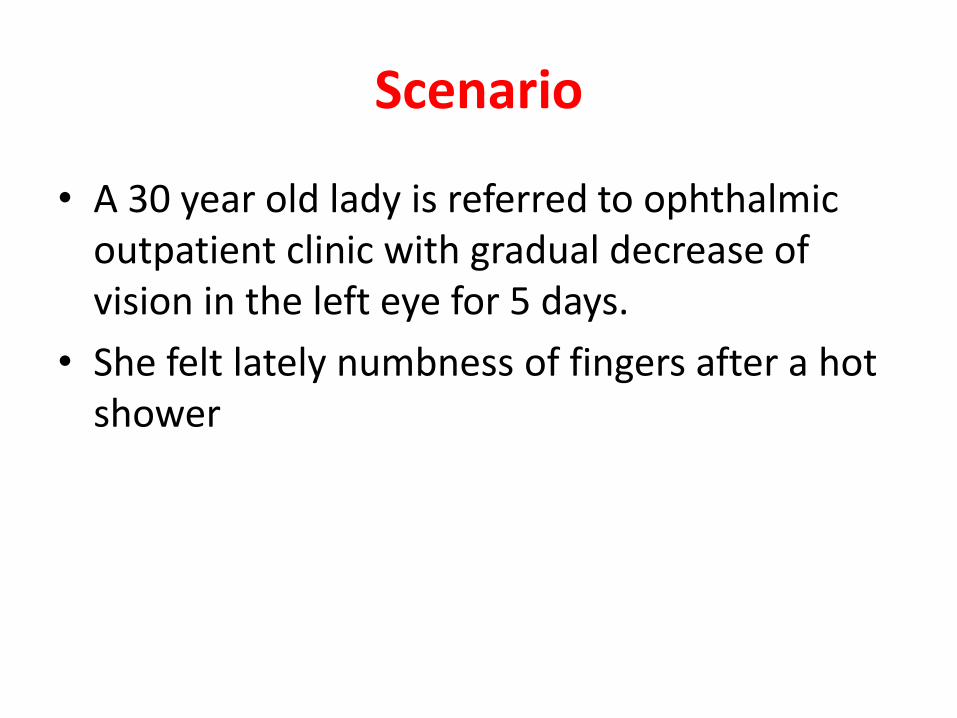
Scenario
• A 30 year old lady is referred to ophthalmic outpatient clinic with gradual decrease of vision in the left eye for 5 days.
• She felt lately numbness of fingers after a hot shower
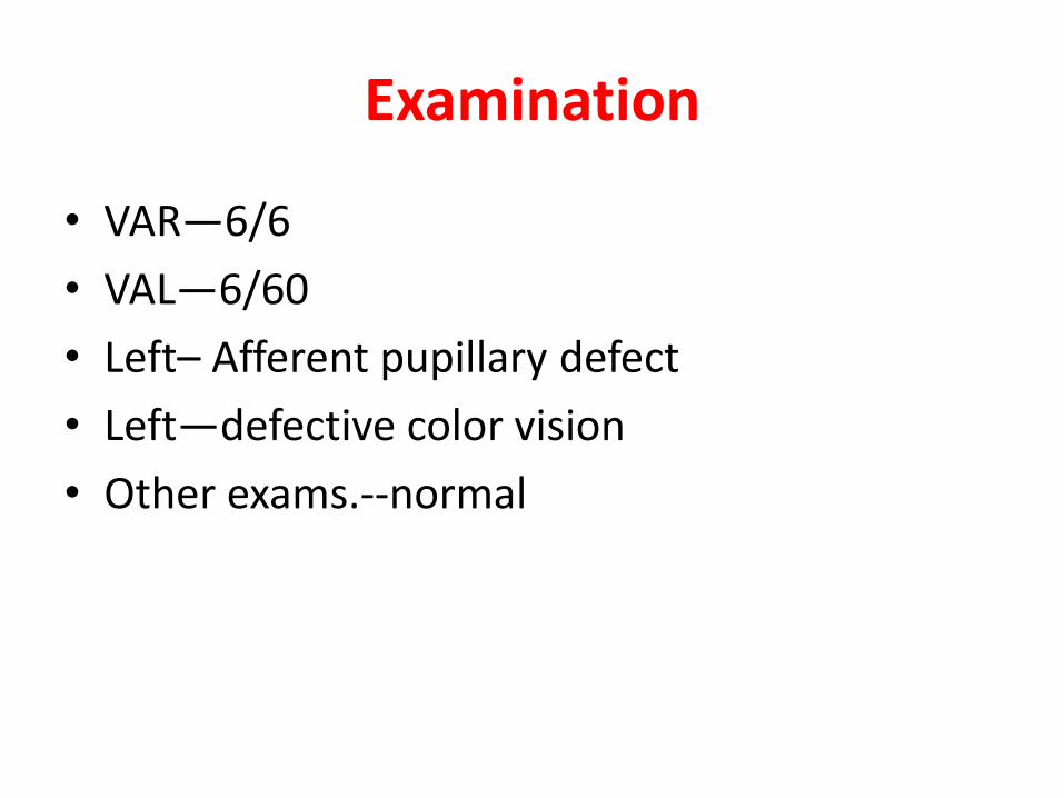
Examination
• VAR—6/6
• VAL—6/60
• Left– Afferent pupillary defect
• Left—defective color vision
• Other exams.--normal

Questions
• What is your most likely diagnosis?
• How to confirm your diagnosis?
• How to treat?

Applied Anatomy
50mm long from globe(Lamina caribrosa) to chiasma• Intraocular part : 1 mm long, 1.5 mm thick
Prelaminar zoneLaminar zonePot laminar zone
• Intra orbital: 25-30 mm long to the optic foramen3-4 mm thick, surrounded by annulus of Zinn
• Intra canalicular: 6 mm long, fixed to the canal • Intra cranial: 5-16 mm long (Av. 10 mm)

Applied Anatomy
– Contains 1.2 million axons, most of these synapse in the lateral geniculate body some reaches other centres
• 1/3 of fibres sub serves the central 50 of visual field

Surrounding sheaths
• Pia mater is the delicate innermost sheath containing blood vessels.
• Subarachnoid space is continuous with the cerebral subarachnoid space and contains CSF.
• Outer sheath comprises the arachnoid mater and the tougher dura mater. The latter is continuous with the sclera.
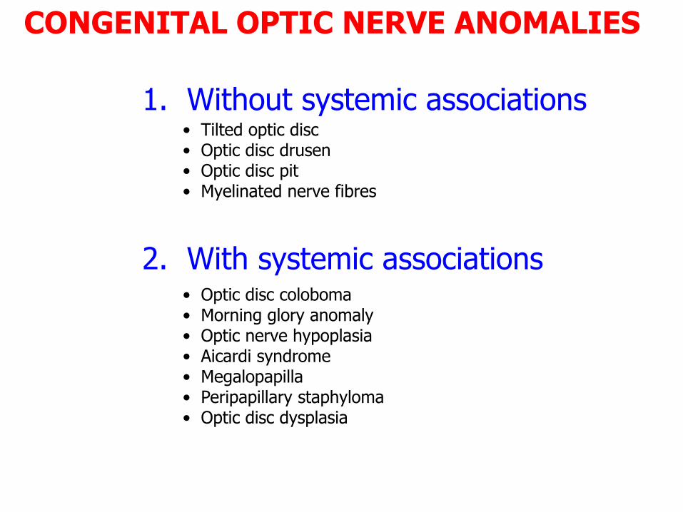
CONGENITAL OPTIC NERVE ANOMALIES
1. Without systemic associations
2. With systemic associations• Optic disc coloboma• Morning glory anomaly• Optic nerve hypoplasia• Aicardi syndrome• Megalopapilla• Peripapillary staphyloma• Optic disc dysplasia
• Tilted optic disc• Optic disc drusen• Optic disc pit• Myelinated nerve fibres
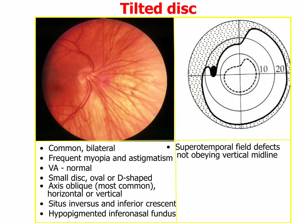
Tilted disc
• Common, bilateral• Frequent myopia and astigmatism• VA - normal• Small disc, oval or D-shaped• Axis oblique (most common),
horizontal or vertical• Situs inversus and inferior crescent• Hypopigmented inferonasal fundus
• Superotemporal field defectsnot obeying vertical midline

Optic disc drusen• Uncommon, bilateral and familial• Associations - RP, angioid streaks and Alagille syndrome• VA - usually normal
Buried drusen
• Absent optic cup
• Pink or yellow colour
• Indistinct ‘lumpy’ margins
• Anomalous branching
patterns
with premature branching
• Absent venous
engorgement
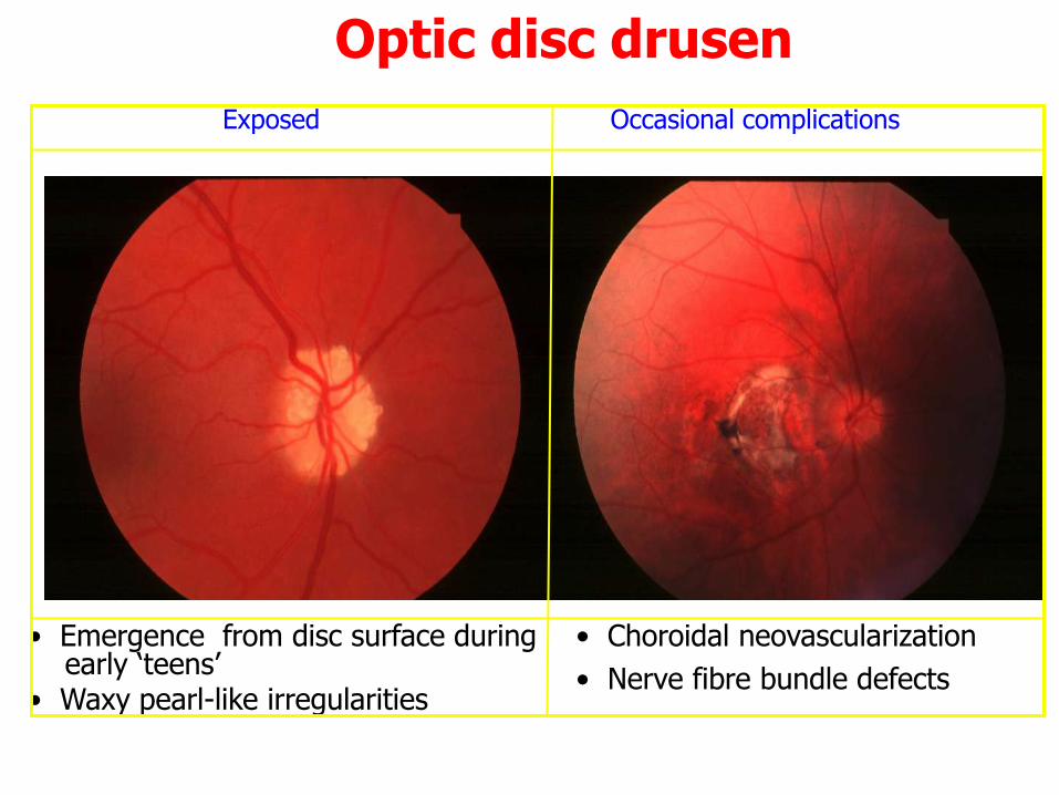
Optic disc drusen
Occasional complications
• Emergence from disc surface duringearly ‘teens’
• Waxy pearl-like irregularities
• Choroidal neovascularization
• Nerve fibre bundle defects
Exposed
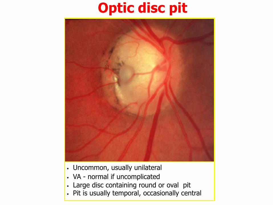
Optic disc pit
• Uncommon, usually unilateral
• VA - normal if uncomplicated
• Large disc containing round or oval pit• Pit is usually temporal, occasionally central

Myelinated nerve fibres
ExtensiveIsolated peripheral Peripapillary

Optic disc coloboma
• VA - decreased
• Rare, unilateral or bilateral
• Usually sporadic - occasionally dominant
• May be associated with other colobomas • Large disc with inferior excavation• Superior visual field defects
Signs Ocular associations

Occasional Systemic Associations of Optic Disc Coloboma
1. CNS malformation - basal encephalocele and cysts
2. Chromosomal anomalies - Patau syndrome (trisomy 13) and
cat-eye syndrome (trisomy 22)
3. ‘CHARGE’ - Coloboma, Heart defects, choanal Atresia, Retarded development, Genital and Ear anomalies
4. Other syndromes - Meckel-Gruber, Goltz, Lenz microphthalmos,Walker-Warburg and Goldenhar

Optic nerve hypoplasia
• Small disc surrounded by halo (double ring sign)
• Rare, unilateral or bilateral
• VA - variable according to severity
• Vessel normal calibre but may be tortuous
Occasional association
• De Morsier syndrome (septo-optic dysplasia)
• Absence of septum pellucidum and corpus callosum
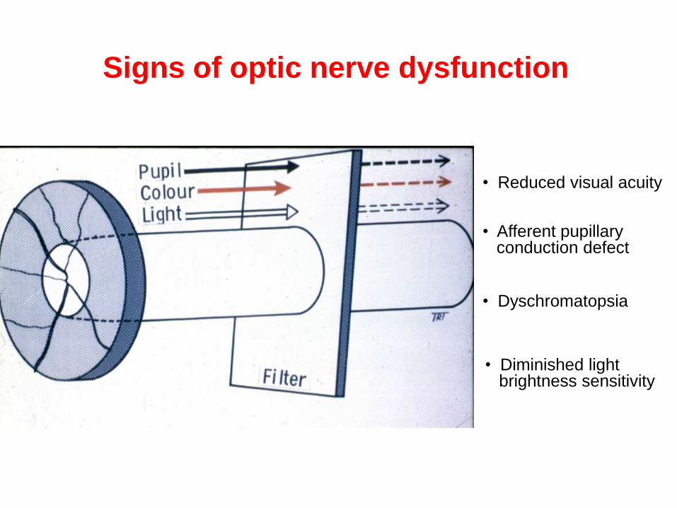
Signs of optic nerve dysfunction
• Reduced visual acuity
• Diminished light brightness sensitivity
• Dyschromatopsia
• Afferent pupillary conduction defect
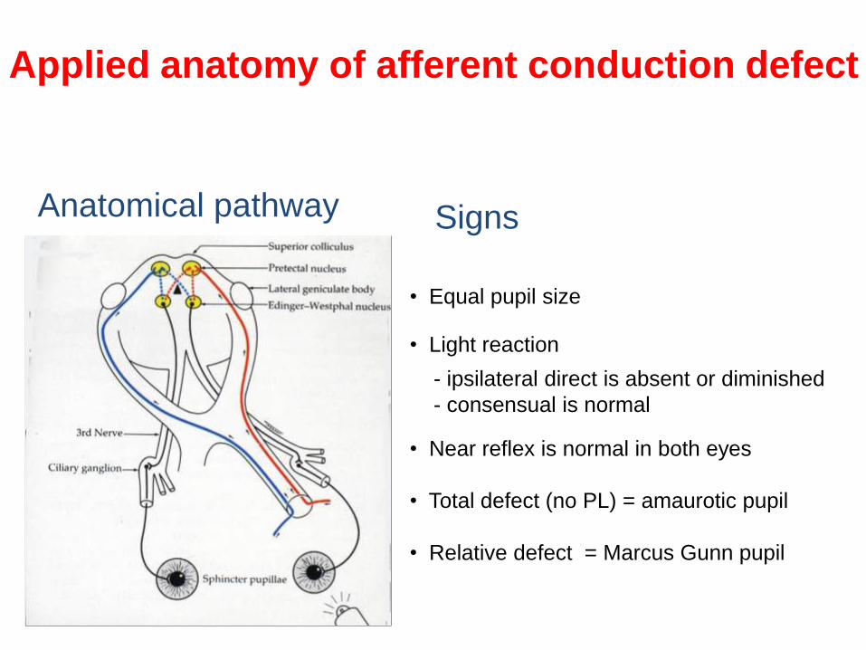
Applied anatomy of afferent conduction defect
Anatomical pathway Signs
• Equal pupil size
• Light reaction
- ipsilateral direct is absent or diminished
- consensual is normal
• Near reflex is normal in both eyes
• Total defect (no PL) = amaurotic pupil
• Relative defect = Marcus Gunn pupil

Optic disc changes
• Early compression
Normal
• Papillitis and neuroretinitis
Swelling
• Optic nerve sheath meningioma
Optico-ciliary shunts
• Compression
Atrophy

Visual field defects
Central scotoma Centrocaecal scotoma

Etiologic Classification of optic nerve disease
• Optic nerve abnormalities congenital – Hypoplasia– Dysplasia – Tilted disks– Myelinated nerve fibers
• Hereditary optic atrophy– Laber’s hereditary optic neuropathy
• Optic neuritis– Demyelinative – Immune-mediated– Direct infections– Granulomatous optic neuropathy– Contiguous inflammatory disease

Etiologic Classification of optic nerve disease
• Vascular (ischemic optic neuropathy)
– Nonarteritic anterior ischemic optic neuropathy
– Giant cell arteritis
– Systemic vasculitis
– Migraine
– Inherited coagulation defects
– Diabetic papillopathy
– Radiation optic neuropathy
– Sudden massive blood loss

Etiologic Classification of optic nerve disease
• Papilloedema
Space-occupying lesions
Blockage of ventricular system
Obstruction of CSF absorption
Benign intracranial hypertension(pseudotumour cerebri)
Diffuse cerebral oedema
Hypersecretion of CSF
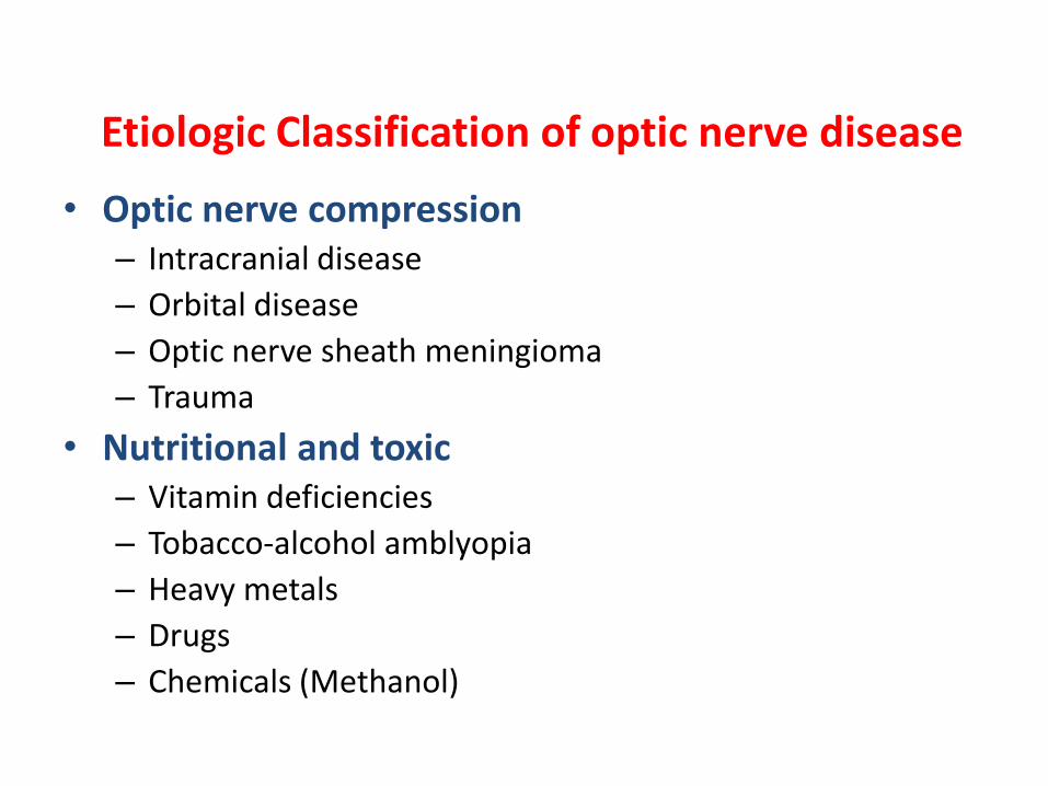
Etiologic Classification of optic nerve disease
• Optic nerve compression– Intracranial disease
– Orbital disease
– Optic nerve sheath meningioma
– Trauma
• Nutritional and toxic– Vitamin deficiencies
– Tobacco-alcohol amblyopia
– Heavy metals
– Drugs
– Chemicals (Methanol)

Etiologic Classification of optic nerve disease
• Optic nerve atrophy
– Primary optic atrophy• Following retrobulbar neuritis
• Compressive lesions such as tumours and aneurysms
• Hereditary optic neuropathies
• Toxic and nutritional optic neuropathies
• Degenerative retinal diseases
– Secondary optic atrophy• Chronic papilloedema
• Anterior ischeamic optic neuropathy
• Papillitis

Optic Neuritis
Ophthalmoscopic classification
Retrobulbar neuritis(normal disc)
Papillitis (hyperaemia and oedema)
Neuroretinitis (papillitisand macular star)

Optic Neuritis
• Aetiological Classification
– Demyelinating
– Parainfectious
– Infectious
– Autoimmune
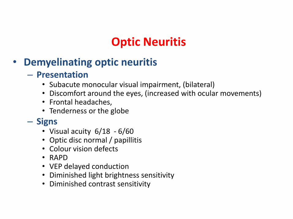
Optic Neuritis
• Demyelinating optic neuritis– Presentation
• Subacute monocular visual impairment, (bilateral)• Discomfort around the eyes, (increased with ocular movements) • Frontal headaches, • Tenderness or the globe
– Signs• Visual acuity 6/18 - 6/60• Optic disc normal / papillitis• Colour vision defects • RAPD• VEP delayed conduction • Diminished light brightness sensitivity • Diminished contrast sensitivity
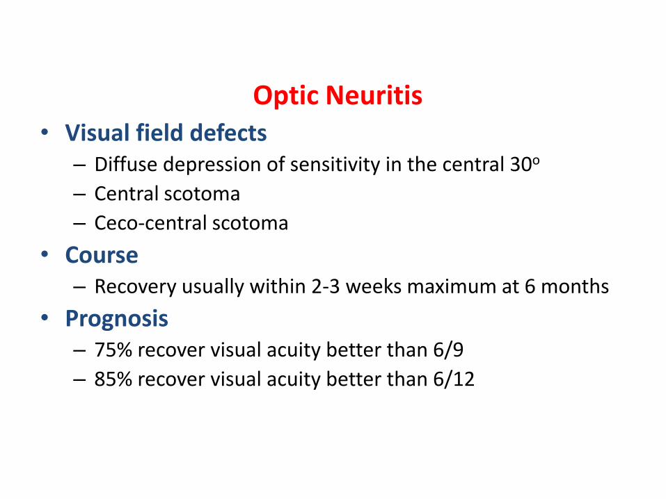
Optic Neuritis • Visual field defects
– Diffuse depression of sensitivity in the central 30o
– Central scotoma
– Ceco-central scotoma
• Course– Recovery usually within 2-3 weeks maximum at 6 months
• Prognosis– 75% recover visual acuity better than 6/9
– 85% recover visual acuity better than 6/12

Optic Neuritis
• Treatment – Indications
• If there is poor vision in the other eye
• If the visual acuity at the onset is less than 6/12
– Regimen• I/V methyl prednisone sodium 1g for 3 days
• Followed by oral prednisolone 1mg/kg daily x 11days
– Benefits • Delays further neurological events with MS by 2 years
• Hastens visual recovery from optic neuritis but does not appear to have any long term benefit on final visual acuity

TREATMENT
Acute Relapses of MS• For clinically significant symptoms/lesions, consider
methylprednisolone (Solumedrol), 250 mg IV q6h x 3-5 days followed by an oral prednisolone (Deltacortil) in tapering dose
• Steroids hasten the rate of recovery from acute exacerbations but have never been proved to improve overall outcome
• Plasmapheresis, ACTH, and cyclophosphamide are options for patients who fail methylprednisolonetherapy

TREATMENT
Interferon Therapy
• Interferon (1FN) therapy has produced almost equivalent levels of excitement and disagreement among neurologists regarding effectiveness and indications
• It is generally well tolerated, with flulike symptoms and injection site reactions being the most common reactions

TREATMENT
• Other agents include mitoxantrone, IV immuno-globulin, cyclophosphamide, cladribine and methotrexate

Papilloedema
1. Introduction
2. Classification of papilloedema
• Early
• Established (acute)
• Longstanding (chronic)
• Atrophic (secondary optic atrophy)
• Circulation of cerebrospinal fluid
• Causes of raised intracranial pressure
• Hydrocephalus

(a) Subarachnoid space
Circulation of cerebrospinal fluid
(b) Lateral ventricle
(c) Third ventricle
(d) Aqueduct
(e) Fourth ventricle
a
b
c
e
d

Space-occupying lesions
Blockage of ventricular system
Obstruction of CSF absorption
Benign intracranial hypertension(pseudotumour cerebri)
Diffuse cerebral oedema
Hypersecretion of CSF
Causes of Raised Intra-cranial Pressure
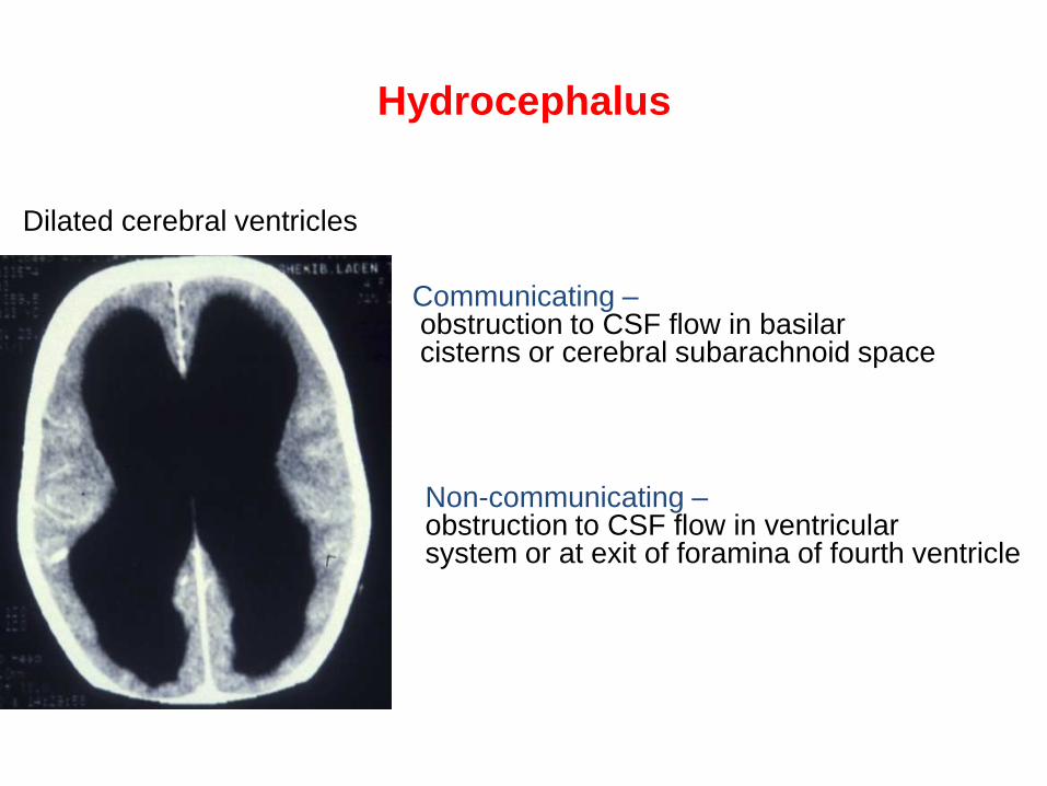
Hydrocephalus
Dilated cerebral ventricles
Communicating –obstruction to CSF flow in basilarcisterns or cerebral subarachnoid space
Non-communicating –obstruction to CSF flow in ventricularsystem or at exit of foramina of fourth ventricle

Early papilloedema
• VA - normal
• Mild disc hyperaemia
• Indistinct disc margins –
initially nasal
• Mild venous engorgement
• Normal optic cup
• Spontaneous venous
pulsation - absent
(also absent in 20% of
normal)

Established papilloedema (acute)
• VA - usually normal
• Severe disc elevation and hyperaemia
• Very indistinct disc margins
• Obscuration of small vessels on disc
• Marked venous engorgement
• Reduced or absent optic cup
• Haemorrhages + cotton-wool spots
• Macular star

Longstanding papilloedema (chronic)
• VA - variable
• Marked disc elevation
but less hyperaemia
• Disc margins - indistinct
• Variable venous engorgement
• Absent optic cup
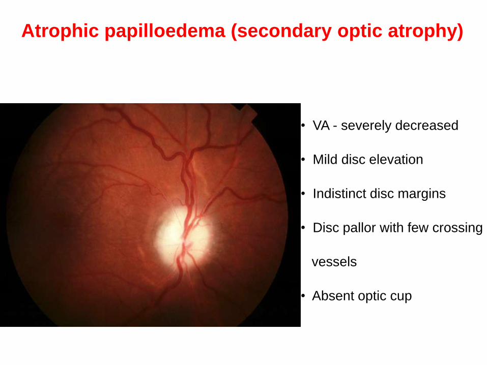
Atrophic papilloedema (secondary optic atrophy)
• VA - severely decreased
• Mild disc elevation
• Indistinct disc margins
• Disc pallor with few crossing
vessels
• Absent optic cup
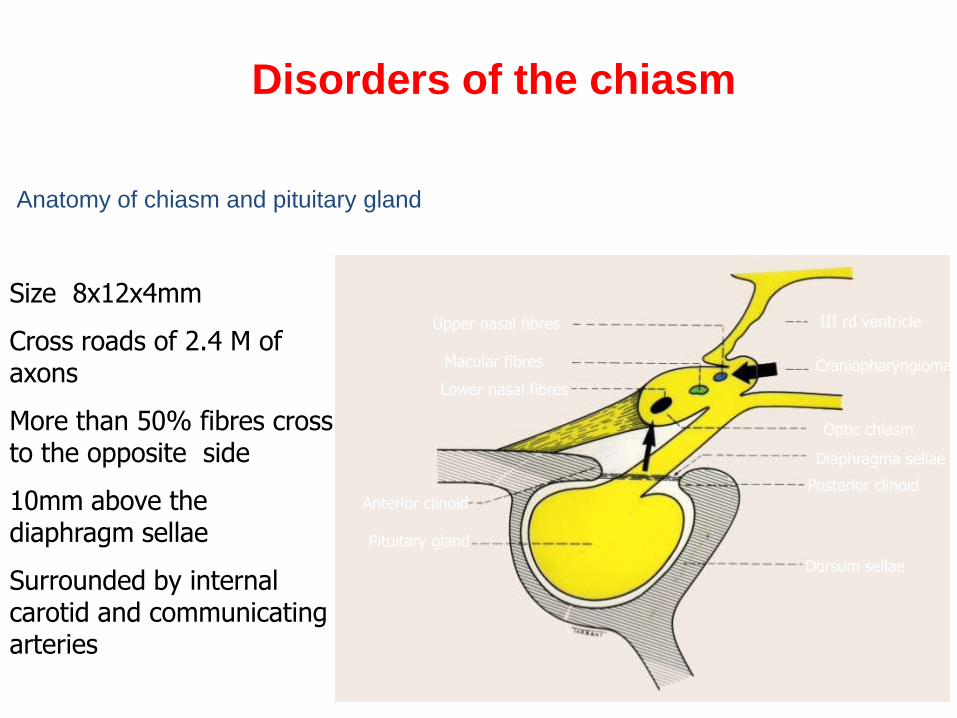
Anatomy of chiasm and pituitary gland
Upper nasal fibres
Macular fibres
Lower nasal fibres
Anterior clinoid
Pituitary gland
III rd ventricle
Craniopharyngioma
Optic chiasm
Diaphragma sellae
Posterior clinoid
Dorsum sellae
Disorders of the chiasm
Size 8x12x4mm
Cross roads of 2.4 M of axons
More than 50% fibres cross to the opposite side
10mm above the diaphragm sellae
Surrounded by internal carotid and communicating arteries

Normal anatomical variations
Central - 80%
Prefixed - 10% Postfixed - 10%
Disorders of the chiasm

Pituitary adenomas
Cushing syndrome
ACTH
Growth hormone
Acromegaly Gigantism
AmenorrhoeaInfertilityGalactorrhoea
HypoglandismImpotenceInfertilityGynaecomastiaGalactorrhoea
PROLACTINChromophobe

Acromegaly
Enlargement of hands and feet Enlargement of lower jaw

Acromegaly
• Facial coarseness• Hypertension, diabetes and
gonadal dysfunction
• Organomegaly• Carpal tunnel syndrome and
cardiomyopathy

Visual field defects in pituitary adenomas LE RE
HM
CF
Decussating fibresare most vunerable
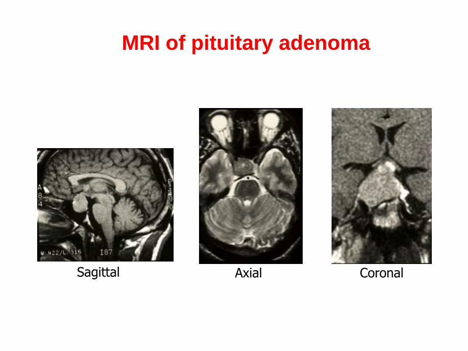
MRI of pituitary adenoma
Sagittal CoronalAxial

Treatment options for pituitary adenomas
RadiotherapySurgery
Transfr`ontal
Trans-sphenoidalBromocriptine

Craniopharyngioma
Presents
• In children with endocrine dysfunction
• In adults with visual field defects
LE RE
HM
CF
The posteriorly crossingfibres are most vunerable
Craniopharyngioma
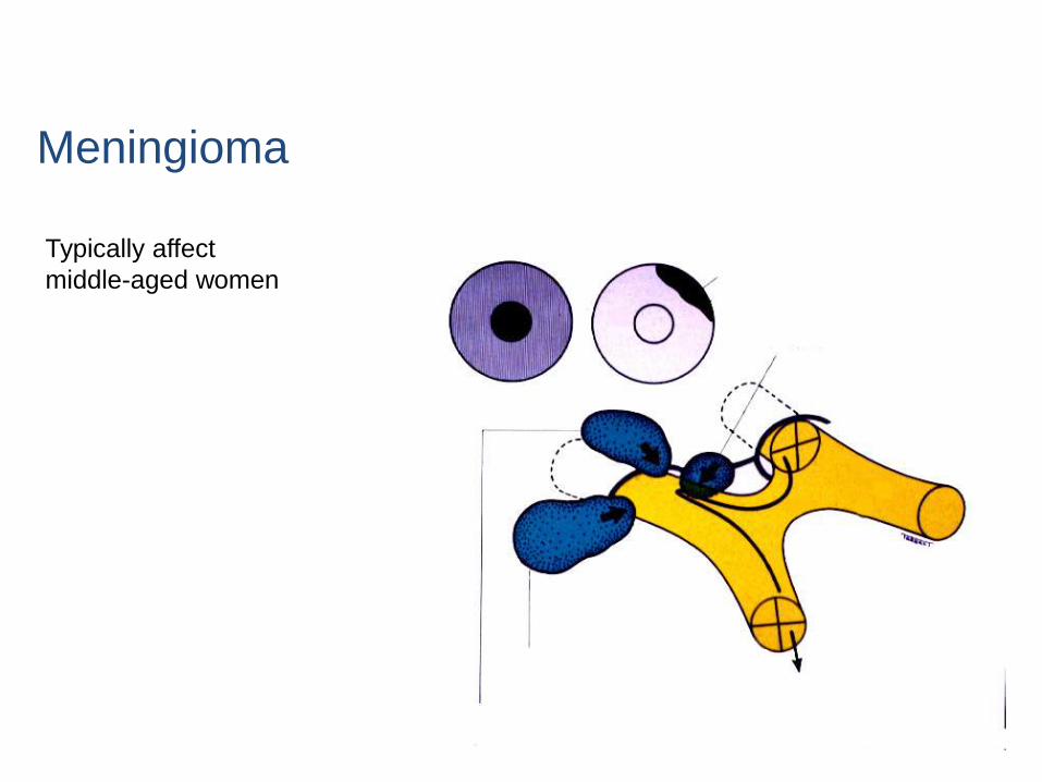
Meningioma
Typically affect
middle-aged women
LE REJunctional scotoma
Tuberculum Sellameningioma
Olfactory groove meningioma
Sphenoid ridge meningioma

THANK YOU

THANK YOU