Neuro-ophthalmic effects of stenting across the ophthalmic ... · treatment of wide-necked...
6
J Neurosurg 121:18–23, 2014 18 J Neurosurg / Volume 121 / July 2014 ©AANS, 2014 A NEURYSMS of the ophthalmic segment of the in- ternal carotid artery (ICA) provide unique ana- tomical challenges to those attempting surgical or endovascular treatment. 8,12 Achieving adequate visualiza- tion and proximal control for surgical clipping is difficult given the location of the ophthalmic artery branch point from the ICA. Therefore, endovascular methods have be- come more popular for the treatment of ophthalmic ICA aneurysms. 13,16 The proximity of ophthalmic ICA aneu- rysms to vital structures and blood supply of the visual pathway can lead to ophthalmic complications both pre- and postoperatively. Following endovascular and open surgical treatment of intracranial aneurysms, a variety of ophthalmic side Neuro-ophthalmic effects of stenting across the ophthalmic artery origin in the treatment of intracranial aneurysms Clinical article ROBERT S. HELLER, M.D., 1 CLAIRE M. LAWLOR, M.D., 2 THOMAS R. HEDGES III, M.D., 2 Y ANIK J. BABABEKOV , M.D., M.P.H., 2 MINA G. SAFAIN, M.D., 1 AND ADEL M. MALEK, M.D., PH.D. 1 1 Cerebrovascular and Endovascular Division, Department of Neurosurgery; and 2 Departments of Ophthalmology and Neurology, New England Eye Center, Tufts Medical Center and Tufts University School of Medicine, Boston, Massachusetts Object. The benefits of treating intracranial aneurysms in the region of the anterior visual pathways are well understood. However, the adverse effects of endovascular stenting across the ophthalmic artery have received little attention. The authors reviewed their experience with patients who had stents deployed across the ophthalmic artery origin. Methods. Patients’ medical charts and imaging studies were reviewed to identify all patients with a non–flow diverting stent deployed over the ophthalmic artery origin for the treatment of intracranial aneurysms. All patients with neuro-ophthalmic complaints were referred for formal ophthalmological evaluation. Results. A total of 104 consecutive patients with 106 aneurysms were identified to meet criteria for inclusion in the study cohort. Preoperatively, 30 patients (29%) described headache symptoms and 32 patients (31%) reported visual complaints. Of the patients with preoperative headaches, 15 (54%) of 28 patients for whom follow-up was available experienced improvement in their symptoms. Of the patients with preoperative visual complaints, improve- ment was noted in 11 (41%) of the 27 patients for whom follow-up was available, 9 (33%) of 27 patients reported no change in visual symptoms, and 7 (26%) of 27 patients reported progression of symptoms. Visual field defects developing posttreatment were noted to occur in 8 (7.7%) of 104 patients: 3 with immediate postoperative retinal in- farcts, 1 with perioperative hemianopia that resolved by the time of discharge, 1 with a subjective visual field defect, 1 with subjective migratory visual field defects, and 2 with nonspecific visual symptoms. Compressive symptoms from aneurysm mass effect were noted in 6 patients preoperatively, with 4 of those patients experiencing persistent worsening, resolution in 1 case, and no change in 1 case. One patient developed a novel cranial nerve palsy from mass effect in the immediate postoperative period. Conclusions. Deployment of stents across the ophthalmic artery origin for the treatment of intracranial aneu- rysms appears to be relatively safe with regard to visual outcomes. Neuro-ophthalmic complaint resolution rates were comparable to endovascular procedures that do not employ stents, with headache resolution rates comparable to coil-only aneurysm obliteration and low rates of retinal ischemic events. For patients presenting with mass effect, stent-assisted coiling appears to be less effective than microsurgery with decompression for relief of compressive symptoms. (http://thejns.org/doi/abs/10.3171/2014.3.JNS131493) KEY WORDS • intracranial stent • aneurysm coiling • migraine • retinal emboli • vascular disorders Abbreviation used in this paper: ICA = internal carotid artery. This article contains some figures that are displayed in color online but in black-and-white in the print edition.
Transcript of Neuro-ophthalmic effects of stenting across the ophthalmic ... · treatment of wide-necked...
J Neurosurg 121:18–23, 2014
18 J Neurosurg / Volume 121 / July 2014
©AANS, 2014
Aneurysms of the ophthalmic segment of the in-ternal carotid artery (ICA) provide unique ana-tomical challenges to those attempting surgical or
endovascular treatment.8,12 Achieving adequate visualiza-tion and proximal control for surgical clipping is difficult given the location of the ophthalmic artery branch point from the ICA. Therefore, endovascular methods have be-
come more popular for the treatment of ophthalmic ICA aneurysms.13,16 The proximity of ophthalmic ICA aneu-rysms to vital structures and blood supply of the visual pathway can lead to ophthalmic complications both pre- and postoperatively.
Following endovascular and open surgical treatment of intracranial aneurysms, a variety of ophthalmic side
Neuro-ophthalmic effects of stenting across the ophthalmic artery origin in the treatment of intracranial aneurysms
Clinical articleRobeRt S. HelleR, M.D.,1 ClaiRe M. lawloR, M.D.,2 tHoMaS R. HeDgeS iii, M.D.,2 Yanik J. bababekov, M.D., M.P.H.,2 Mina g. Safain, M.D.,1 anD aDel M. Malek, M.D., PH.D.1
1Cerebrovascular and Endovascular Division, Department of Neurosurgery; and 2Departments of Ophthalmology and Neurology, New England Eye Center, Tufts Medical Center and Tufts University School of Medicine, Boston, Massachusetts
Object. The benefits of treating intracranial aneurysms in the region of the anterior visual pathways are well understood. However, the adverse effects of endovascular stenting across the ophthalmic artery have received little attention. The authors reviewed their experience with patients who had stents deployed across the ophthalmic artery origin.
Methods. Patients’ medical charts and imaging studies were reviewed to identify all patients with a non–flow diverting stent deployed over the ophthalmic artery origin for the treatment of intracranial aneurysms. All patients with neuro-ophthalmic complaints were referred for formal ophthalmological evaluation.
Results. A total of 104 consecutive patients with 106 aneurysms were identified to meet criteria for inclusion in the study cohort. Preoperatively, 30 patients (29%) described headache symptoms and 32 patients (31%) reported visual complaints. Of the patients with preoperative headaches, 15 (54%) of 28 patients for whom follow-up was available experienced improvement in their symptoms. Of the patients with preoperative visual complaints, improve-ment was noted in 11 (41%) of the 27 patients for whom follow-up was available, 9 (33%) of 27 patients reported no change in visual symptoms, and 7 (26%) of 27 patients reported progression of symptoms. Visual field defects developing posttreatment were noted to occur in 8 (7.7%) of 104 patients: 3 with immediate postoperative retinal in-farcts, 1 with perioperative hemianopia that resolved by the time of discharge, 1 with a subjective visual field defect, 1 with subjective migratory visual field defects, and 2 with nonspecific visual symptoms. Compressive symptoms from aneurysm mass effect were noted in 6 patients preoperatively, with 4 of those patients experiencing persistent worsening, resolution in 1 case, and no change in 1 case. One patient developed a novel cranial nerve palsy from mass effect in the immediate postoperative period.
Conclusions. Deployment of stents across the ophthalmic artery origin for the treatment of intracranial aneu-rysms appears to be relatively safe with regard to visual outcomes. Neuro-ophthalmic complaint resolution rates were comparable to endovascular procedures that do not employ stents, with headache resolution rates comparable to coil-only aneurysm obliteration and low rates of retinal ischemic events. For patients presenting with mass effect, stent-assisted coiling appears to be less effective than microsurgery with decompression for relief of compressive symptoms.(http://thejns.org/doi/abs/10.3171/2014.3.JNS131493)
keY woRDS • intracranial stent • aneurysm coiling • migraine • retinal emboli • vascular disorders
Abbreviation used in this paper: ICA = internal carotid artery.
This article contains some figures that are displayed in color on line but in black-and-white in the print edition.
J Neurosurg / Volume 121 / July 2014
Ophthalmic sequelae of aneurysm stent coiling
19
effects have been described.17,20 Previous reports have identified compression of elements of the visual pathway by both mass effect and inflammation, as well as ische-mic sequelae.11,17 A recent study found new visual deficits in 33% of patients treated for ophthalmic segment ICA aneurysms, although the majority of the population stud-ied had undergone surgical clipping.9
The recent introduction of stent deployment for the treatment of wide-necked aneurysms not suitable for en-dovascular coiling alone has broadened the spectrum of intracranial aneurysms that can be treated in the neuro-interventional suite. While several studies have evaluated various ophthalmic sequelae as mentioned, a review of patients receiving intracranial stents for treatment of in-tracranial aneurysms has not been analyzed to determine the frequency, types, or causes of visual complications. The aim of this study is to document the frequency and types of ophthalmic complications that have occurred af-ter endovascular placement of a non–flow diverting stent across the ophthalmic artery origin to increase the under-standing of their relationship to the intervention.
MethodsAll patients who had Neuroform (Stryker) or Enter-
prise (Codman) stents placed for ruptured or unruptured intracranial aneurysms between January 2006 and Au-gust 2011 at our institution were identified using a pro-spectively maintained database. All patients with a stent in the ICA spanning the origin of the ophthalmic artery were included in the study. Exact stent location was deter-mined through the use of 3D subtraction angiography and 3-T MR angiography.
Patient medical charts, radiographic studies, and en-dovascular procedures were retrospectively reviewed for pertinent history. Variables recorded included patient age, sex, aneurysm location, stent location in relation to the ophthalmic artery origin, stent type, rupture status, previ-ous intracranial surgeries, clinical presentation, previous visual complaints, history of migraine, type of procedure, procedure date, and type and duration of anticoagulation. Visual complaints and migraine headaches were consid-ered distinct entities from the presenting symptom unless the visual complaint or migraine headache directly led to the discovery of the aneurysm. Determination of stent length and placement was made intraoperatively by the neurovascular team and was based on aneurysm location and neck diameter. Stent patency and ophthalmic artery flow were documented angiographically.
After aneurysm embolization, patients were sched-uled to return for follow-up at 3 months, 6 months, 1 year, and annually thereafter with angiography performed at 3 months and 1 year. At each visit, patients were screened for headaches and vision changes including decreased acuity, diplopia, blurred vision, and visual field defects. Patients who had visual complaints were referred to the neuro-ophthalmologist (T.R.H.) at our institution where they underwent complete ophthalmic examination in-cluding visual field testing and retinal imaging, fluores-cein angiography, and optical coherence tomography as needed.
Patients were screened for potential neuro-ophthal-mological visual complaints and migraine headaches prior to and after treatment. Visual complaints included in screening were the presence of blurred vision, diplopia, hemianopia, anisocoria, cranial nerve palsy, papillary dysfunction, ptosis, and visual migraines.
The current study was approved by the Institutional Review Board of Tufts Medical Center.
ResultsA total of 104 consecutive patients undergoing stent-
mediated coil embolization of 106 intracranial aneurysms requiring stents spanning the ophthalmic artery origin were included in the current study. Aneurysm locations were classified using an anatomical system with the distri-bution presented in Table 1. Two patients presenting with multiple aneurysms were treated with a single stent cover-ing the ophthalmic artery origin; both patients harbored an adjacent posterior communicating artery aneurysm in addition to a more proximal ophthalmic artery aneurysm.
The study population consisted of 93 female patients (89%) and 11 male patients (11%), with a mean age of 54.4 ± 13.7 years. Five patients (5%) presented with subarach-noid hemorrhage secondary to target aneurysm rupture. The presenting symptoms of all patients are detailed in Ta-ble 2. Stent selection for aneurysm obliteration was deter-mined on a case-by-case basis by the neurovascular team. Neuroform stents were deployed in 48 patients (46%) and Enterprise stents were deployed in 56 patients (54%).
Preoperatively, 30 patients (29%) described having migraine headaches. Neuro-ophthalmological visual com-plaints were reported in 32 patients (31%) (Table 3), with 8 patients undergoing formal neuro-ophthalmological evalu-ation preoperatively.
Clinical follow-up longer than 30 days postprocedure was available in 89 (86%) of 104 patients; 15 patients (14%) were lost to follow-up longer than 30 days. All pa-tients reporting neuro-ophthalmological complaints at a follow-up visit were directly referred for same-day neuro-ophthalmological evaluation at our institution. In sum, 46 patients underwent postoperative neuro-ophthalmologi-cal evaluation.
Follow-up was available in 28 of 30 patients with
TABLE 1: Location of aneurysms within the ICA treated with a stent covering the ophthalmic artery origin
Location No. of Aneurysms (%)
cavernous 16 (15)paraclinoid 32 (30)supraclinoid 13 (12)ophthalmic 33 (31)superior hypophysial 5 (5)posterior communicating artery 6 (6)fetal posterior cerebral artery 1 (1)total 106 (100)*
* Two patients presented with multiple intracranial aneurysms treated by the same stent. See text for details.
R. S. Heller et al.
20 J Neurosurg / Volume 121 / July 2014
preoperative migraine headaches. Of these 28 patients, postoperative monitoring demonstrated that 15 (54%) experienced improvement of their migraine symptoms, 1 (3%) had complete resolution of symptoms, 11 (39%) had no change in symptoms, and 2 (7%) had worsening of migraine symptoms (1 Enterprise case and 1 Neuroform case) experienced by increased frequency of migraine attacks. Development of new migraines following stent-mediated coil embolization of patients with no previous migraines occurred in 3 patients (1 Enterprise case and 2 Neuroform cases). Two of these patients developed new migraines within 30 days of stent deployment; the other patient developed new migraines 20 months after stent deployment. Development of new visual migraines in pa-tients with a history of migraine headaches occurred in 8 patients (3 Enterprise cases and 5 Neuroform cases).
Follow-up was available in 27 of 32 patients with preoperative neuro-ophthalmological visual complaints. Symptoms remained stable in 9 patients (33%), pro-gressed in 7 patients (26%), improved in 6 patients (22%), and resolved in 5 patients (19%). New visual symptoms posttreatment included retinal ischemic events in 3 pa-tients (2 Enterprise cases and 1 Neuroform case), all of which occurred immediately postoperatively (Fig. 1) with at least partial resolution of the visual field defect by dis-charge in each case (Fig. 2).
Compressive symptoms were observed in 7 patients
(3 Enterprise cases and 4 Neuroform cases). Six patients presented with cranial nerve compression symptoms pre-procedure. Persistent worsening of symptoms was noted in 4 patients: 1 case each of optic chiasm compression, optic nerve compression, oculomotor nerve palsy, and abducens nerve palsy. Intermittent worsening of an ab-ducens nerve palsy followed by resolution was observed in 1 case, and no change in an oculomotor nerve palsy was observed in the remaining case. Novel compressive symptoms were noted in a patient who developed an ocu-lomotor nerve palsy 3 days after Neuroform stent–medi-ated embolization of a cavernous ICA aneurysm.
New visual field defects were identified in 5 patients after stent deployment (4.8%). Acute deficits occurred in 2 patients, 1 of which occurred in the immediate post-operative period with complete hemianopia ipsilateral to the stent secondary to presumed vasospasm with ap-proximately 50% recovery before discharge. The second acute deficit occurred in a patient with a subjective com-plaint on the 1st postoperative day of a blind spot with unremarkable formal visual field testing. The third visual field defect was noted 3 months postoperatively in a pa-tient with migratory visual field defects and a concurrent relative afferent pupillary defect. Visual fields could not be formally tested because of poor patient cooperation. Visual field defects were deemed to be unrelated to the intracranial aneurysm and stent deployment in the final 2 patients, were caused by a chorioretinal scar and vitreous traction, and were identified at 2 months and 3 months postoperatively. Two patients reported nonspecific visual complaints at follow-up. One patient noted intermittent blurry vision in the eye ipsilateral to the stent 2 years af-ter stent deployment, and the other noted anisocoria upon waking from sleep, which was ultimately presumed to be caused by sympathetic nerve damage from the stent.
TABLE 2: Presenting symptoms in 104 patients
Symptoms No. of Patients (%)
peripheral vision changes 1 (1)oculomotor nerve palsy 2 (2)abducens nerve palsy 1 (1)subjective diplopia 1 (1)dizziness 5 (5)headache 34 (33)near syncope workup 1 (1)nonspecific visual complaints 1 (1)paresthesias 1 (1)pale optic disc 1 (1)progressive cognitive decline 1 (1)subarachnoid hemorrhage* 5 (5)stroke workup 5 (5)multiple sclerosis workup 2 (2)transient ischemic attack workup 4 (4)pulsing sensation in ears 2 (2)incidental/screening† 37 (36) other intracranial ruptured aneurysm 5total 104
* Patients designated as having a presentation of subarachnoid hemor-rhage were those with rupture of the aneurysm that was targeted with a stent deployed over the origin of the ophthalmic artery.† Aneurysms treated with stents deployed over the ophthalmic artery origin that were detected in the workup for the rupture of other intracra-nial aneurysms were designated as incidental.
TABLE 3: Preoperative neuro-ophthalmological complaints
Symptoms No. of Patients
decreased visual acuity 8decreased visual acuity w/ diplopia 1decreased visual acuity w/ photophobia 1oculomotor nerve palsy 2oculomotor nerve palsy w/ photophobia 1abducens nerve palsy 1loss of peripheral vision 1decreased pupillary reaction w/ floaters 1subjective diplopia 3idiopathic hemianopia 1hemianopia related to motor vehicle accident 1presumptive diagnosis of multiple sclerosis 1mydriasis 1ocular pain 1ptosis 1nonspecific complaints 3visual migraines 4total 32
J Neurosurg / Volume 121 / July 2014
Ophthalmic sequelae of aneurysm stent coiling
21
Clinical outcome analysis was performed using the modified Rankin Scale. Preoperative scores aver-aged 0.70 ± 0.62, and postoperative scores (using the last known condition for those patients lost to follow-up) aver-aged 0.64 ± 0.82.
DiscussionVisual dysfunction is well known to be associated
with intracranial aneurysms through mass effect and com-pression of visual pathway components. Treatment of these aneurysms is thus aimed at resolution of symptoms in addi-tion to aneurysm obliteration for rupture prevention.
Previous reports have shown a sustained benefit in headache pain scores following unruptured aneurysm treatment that was equivalent following either endovas-cular or microsurgical techniques.1,10,15 However, one study demonstrated that the use of stent assistance was not associated with improvement in headache symptoms in patients with pretreatment headaches or new head-aches following treatment (1 of 5 patients improved).19 The observed prevalence in the current series of migraine headaches (29%) and subsequent improvement of those headaches following stent-mediated embolization of an-eurysms in 54% of those patients echoes the reports of sustained benefit in pain scores following aneurysm oblit-eration while demonstrating that the benefit of aneurysm embolization on headache symptoms may indeed persist in the population of patients undergoing stent assistance for treatment of their aneurysms.
Several previous series have analyzed the relation-ship between intracranial aneurysms, their treatment, and the ensuing prevalence of visual symptoms. The preva-lence of pretreatment visual symptoms has been reported
to occur in 23%–32% of patients with ICA aneurysms in previously reported studies, a rate comparable to the 29% of patients with visual complaints at initial presentation in the current series.2,4,5
Resolution of visual symptoms caused by intracrani-al aneurysms following treatment has been reported to be more successful in instances in which the target aneurysm was treated within 3 months of visual symptom onset.2 The literature also provides evidence that microsurgery is superior to embolization with regard to resolution of visual symptoms, with one study reporting improvement or resolution of visual symptoms in 75% of aneurysms treated with microsurgery versus 38% of patients treated with embolization.18 This finding may be explained by the ability of microsurgery to decompress the aneurysm through a postclipping aneurysm puncture and decom-pression of the aneurysm, which relieves the mass effect of the aneurysm on the anterior visual pathways.3,12,21
Fulkerson et al.5 studied the treatment of 134 pa-tients, 97 of whom harbored unruptured aneurysms, and observed an incidence of pretreatment ophthalmological findings in 27.8% of unruptured aneurysms. New visual deficits were observed in 4.8% of patients undergoing craniotomy for microsurgical clipping without worsening
Fig. 2. A 63-year-old woman with a right-sided ophthalmic ICA an-eurysm was treated with stent-mediated coiling. A and B: Diagnostic cerebral angiography and 3D reconstruction demonstrating an ophthal-mic ICA aneurysm with the origin of the ophthalmic artery branching from the neck of the aneurysm. C: Intraoperative angiogram of the embolization procedure demonstrating persistence of flow through the ophthalmic artery during coiling of the aneurysm dome. D: Postop-erative Dyna-CT showing complete occlusion of the aneurysm from the circulation following stent-mediated coiling. E and F: Postoperatively the patient complained of visual field deficits in the right eye, due to retinal infarcts in the right eye (E, arrows) and absent in the left eye (F). The patient reported improvement of symptoms before discharge.
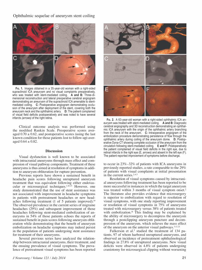
Fig. 1. Images obtained in a 35-year-old woman with a right-sided supraclinoid ICA aneurysm and no visual complaints preoperatively, who was treated with stent-mediated coiling. A and B: Three-di-mensional reconstruction and lateral preoperative cerebral angiogram demonstrating an aneurysm of the supraclinoid ICA amenable to stent-mediated coiling. C: Postoperative angiogram demonstrating occlu-sion of the aneurysm after deployment of the stent, covering both the aneurysm neck and the ophthalmic artery. D: The patient complained of visual field deficits postoperatively and was noted to have several infarcts (arrows) of the right retina.

R. S. Heller et al.
22 J Neurosurg / Volume 121 / July 2014
visual deficits in the 26 patients undergoing coil emboli-zation. Hoh et al.7 noted improvement of visual symptoms in 69% of patients following treatment of paraclinoid an-eurysms, while Heran et al.6 noted that visual symptoms improved in 50% of cases with the conclusion that endo-vascular therapy may not relieve anterior optic pathway compression.
In the current series, preoperative neuro-ophthalmo-logical complaints improved or resolved in 11 (41%) of 27 patients while symptoms remained stable in 33% of pa-tients and worsened in 26% of patients. Of the 6 patients with preoperative symptoms of mass effect on the ante-rior visual pathways, only 1 patient experienced resolu-tion of symptoms, a finding consistent with the literature indicating that microsurgery is a more effective treatment in reversing mass effect.
New neuro-ophthalmological complaints that were likely related to stent-mediated embolization of the target aneurysm occurred in a total of 8 patients (7.7%). Worst-case scenario analysis accounting for the 15 patients lost to follow-up with the assumption that all 15 cases experienced a visual complication would result in new neuro-ophthal-mological complaints in a total of 23 patients (22%).
Five patients experienced visual dysfunction in the immediate postoperative period from retinal infarcts in 3 patients, transient hemianopia in 1 patient, and subjective complaints in the remaining patient. These findings rep-resent a 4.8% rate of visual complications in the immedi-ate postoperative period for patients with stents deployed over the ophthalmic artery origin. Subsequent follow-up revealed neuro-ophthalmological findings in 3 additional patients that could not be objectively quantified: 1 patient with subjective migratory visual field defects, 1 patient with intermittently blurred vision ipsilateral to the stent, and 1 patient with anisocoria.
While 54% of patients (15 of 28) with a preoperative diagnosis of migraines for whom follow-up was avail-able experienced improvement in their symptoms, 8 pa-tients (29%) with a preoperative diagnosis of migraines developed new visual symptoms consistent with visual migraines. These findings could indicate that deployment of stents and coils within the intracranial circulation for aneurysm treatment could induce, among other possi-bilities, subtle inflammatory changes or alteration of the autonomic nervous system and cerebral hemodynamics. These changes could affect the susceptibility of a particu-lar aneurysm patient to improvement or resolution of his or her migraine symptoms. Further work remains to be done to determine the global effects of stent deployment within the intracranial circulation.
The effects of stent deployment within the ICA and covering the origin of the ophthalmic artery are not yet fully understood. A series of 19 patients receiving the Pipeline Embolization Device for the treatment of para-clinoid aneurysms demonstrated angiographic evidence of ophthalmic artery occlusion in 21% of patients after follow-up; no patients with ophthalmic artery occlusion developed visual symptoms.14 While there are significant differences between the Pipeline flow diversion device and the open-cell design Neuroform and closed-cell de-sign Enterprise stents used in the current series, this study
demonstrated that there remains an inherent risk associ-ated with device deployment over the ophthalmic artery.
Limitations of the current study include the retrospec-tive nature of this data set. As detailed, neuro-ophthalmic examinations were only obtained in those patients with subjective visual complaints; objective measurement of the incidence of neuro-ophthalmic events in the entire population could not be performed. Furthermore, without a prospective study design, visual outcomes were mea-sured in a subjective manner without a standardized tool for objective visual symptom monitoring.
Further research is warranted to more completely evaluate this risk, and whether anticoagulation regimens need to be altered to prevent thrombosis of the ophthal-mic artery. Although conclusions about the efficacy of this treatment over other available modalities cannot be drawn because of the retrospective nature of this study, at this time, stent deployment over the origin of the ophthal-mic artery appears to be safe. Rates of improvement in visual symptoms and improvement in preoperative head-aches appear comparable to other published studies along with a low rate of new neuro-ophthalmological events.
ConclusionsIn the current study, we carefully monitored visual
signs and symptoms of 104 patients who underwent en-dovascular treatment using stents across the ophthalmic artery origin to identify the safety of these procedures. Rates of resolution of neuro-ophthalmic complaints were comparative to endovascular procedures that do not use stents, with headache resolution rates comparative to coil-only aneurysm obliteration and low rates of retinal ischemic events. For patients presenting with mass effect, stent-assisted coiling appears to be less effective than mi-crosurgery with decompression for relief of compressive symptoms.
Acknowledgment
We would like to thank Courtney Peterson, P.A.-C., for her assistance.
Disclosure
During the conduct of this study, Dr. Malek received unre-stricted support from Codman, Stryker, Microvention-Terumo, Covidien-ev3, and Siemens for unrelated research.
Author contributions to the study and manuscript preparation include the following. Conception and design: Malek, Hedges. Acquisition of data: Lawlor, Bababekov, Safain. Analysis and inter-pretation of data: Malek, Heller, Lawlor. Drafting the article: Malek, Heller, Safain. Critically revising the article: all authors. Reviewed submitted version of manuscript: all authors. Approved the final version of the manuscript on behalf of all authors: Malek. Study supervision: Malek, Hedges.
References
1. Choxi AA, Durrani AK, Mericle RA: Both surgical clip-ping and endovascular embolization of unruptured intracra-nial aneurysms are associated with long-term improvement in self-reported quantitative headache scores. Neurosurgery 69:128–134, 2011

J Neurosurg / Volume 121 / July 2014
Ophthalmic sequelae of aneurysm stent coiling
23
2. Date I, Asari S, Ohmoto T: Cerebral aneurysms causing visual symptoms: their features and surgical outcome. Clin Neurol Neurosurg 100:259–267, 1998
3. de Oliveira JG, Borba LA, Rassi-Neto A, de Moura SM, Sanchez-Júnior SL, Rassi MS, et al: Intracranial aneurysms presenting with mass effect over the anterior optic pathways: neurosurgical management and outcomes. Neurosurg Focus 26(5):E3, 2009
4. Ferguson GG, Drake CG: Carotid-ophthalmic aneurysms: vi-sual abnormalities in 32 patients and the results of treatment. Surg Neurol 16:1–8, 1981
5. Fulkerson DH, Horner TG, Payner TD, Leipzig TJ, Scott JA, DeNardo AJ, et al: Results, outcomes, and follow-up of rem-nants in the treatment of ophthalmic aneurysms: a 16-year ex-perience of a combined neurosurgical and endovascular team. Neurosurgery 64:218–230, 2009
6. Heran NS, Song JK, Kupersmith MJ, Niimi Y, Namba K, Langer DJ, et al: Large ophthalmic segment aneurysms with anterior optic pathway compression: assessment of anatomi-cal and visual outcomes after endosaccular coil therapy. J Neurosurg 106:968–975, 2007
7. Hoh BL, Carter BS, Budzik RF, Putman CM, Ogilvy CS: Re-sults after surgical and endovascular treatment of paraclinoid aneurysms by a combined neurovascular team. Neurosur-gery 48:78–90, 2001
8. Javalkar V, Banerjee AD, Nanda A: Paraclinoid carotid aneu-rysms. J Clin Neurosci 18:13–22, 2011
9. Kanagalingam S, Gailloud P, Tamargo RJ, Subramanian PS, Miller NR: Visual sequelae after consensus-based treatment of ophthalmic artery segment aneurysms: the Johns Hopkins experience. J Neuroophthalmol 32:27–32, 2012
10. Kong DS, Hong SC, Jung YJ, Kim JS: Improvement of chronic headache after treatment of unruptured intracranial aneu-rysms. Headache 47:693–697, 2007
11. Mendez Roberts A, Grimes AL: Enlargement of internal ca-rotid artery aneurysm presenting with severe visual sequela: a case report and anatomy review. Optometry 80:76–82, 2009
12. Nanda A, Javalkar V: Microneurosurgical management of ophthalmic segment of the internal carotid artery aneurysms: single-surgeon operative experience from Louisiana State Uni-versity, Shreveport. Neurosurgery 68:355–371, 2011
13. Park HK, Horowitz M, Jungreis C, Kassam A, Koebbe C, Genevro J, et al: Endovascular treatment of paraclinoid aneu-
rysms: experience with 73 patients. Neurosurgery 53:14–24, 2003
14. Puffer RC, Kallmes DF, Cloft HJ, Lanzino G: Patency of the ophthalmic artery after flow diversion treatment of paracli-noid aneurysms. Clinical article. J Neurosurg 116:892–896, 2012
15. Qureshi AI, Suri MF, Kim SH, Olson K, Siddiqui AM, Yahia AM, et al: Effect of endovascular treatment on headaches in patients with unruptured intracranial aneurysms. Headache 43:1090–1096, 2003
16. Roy D, Raymond J, Bouthillier A, Bojanowski MW, Moumd-jian R, L’Espérance G: Endovascular treatment of ophthalmic segment aneurysms with Guglielmi detachable coils. AJNR Am J Neuroradiol 18:1207–1215, 1997
17. Schmidt GW, Oster SF, Golnik KC, Tumialán LM, Biousse V, Turbin R, et al: Isolated progressive visual loss after coiling of paraclinoid aneurysms. AJNR Am J Neuroradiol 28:1882–1889, 2007
18. Schuss P, Güresir E, Berkefeld J, Seifert V, Vatter H: Influence of surgical or endovascular treatment on visual symptoms caused by intracranial aneurysms: single-center series and systematic review. Clinical article. J Neurosurg 115:694–699, 2011
19. Schwedt TJ, Gereau RW, Frey K, Kharasch ED: Headache out-comes following treatment of unruptured intracranial aneu-rysms: a prospective analysis. Cephalalgia 31:1082–1089, 2011
20. Shinoda J, Ajimi Y, Yamada M, Onozuka S: Cortical blind-ness during coil embolization of an unruptured intracranial aneurysm—case report. Neurol Med Chir (Tokyo) 44:416–419, 2004
21. Tawk RG, Villalobos HJ, Levy EI, Hopkins LN: Surgical de-compression and coil removal for the recovery of vision after coiling and proximal occlusion of a clinoidal segment aneu-rysm: technical case report. Neurosurgery 58:E1217, 2006
Manuscript submitted July 11, 2013.Accepted March 11, 2014.Please include this information when citing this paper: pub-
lished online April 11, 2014; DOI: 10.3171/2014.3.JNS131493.Address correspondence to: Adel M. Malek, M.D., Ph.D., Depart-
ment of Neurosurgery, Tufts Medical Center, 800 Washington St., Boston, MA 02111. email: [email protected].