Neural circuitry of emotion regulation: Effects of …...Neural circuitry of emotion regulation:...
15
Neural circuitry of emotion regulation: Effects of appraisal, attention, and cortisol administration Sean T. Ma 1 & James L. Abelson 1 & Go Okada 2 & Stephan F. Taylor 1 & Israel Liberzon 1,3,4 Published online: 28 December 2016 # Psychonomic Society, Inc. 2016 Abstract Psychosocial well-being requires effective regula- tion of emotional responding in context of threat or stress. Neuroimaging studies have focused on instructed, volitional regulation (e.g., reappraisal or distancing), largely ignoring implicit regulation that does not involve purposeful effort to alter emotional experience. These implicit processes may or may not involve the same neural pathways as explicit regula- tory strategies. We examined the neurobiology of implicit emotional regulation processes and the impact of the stress hormone cortisol on these processes. Our study task employed composite pictures of faces and places to examine neural ac- tivity during implicit emotional processing (of emotional faces), while these responses were implicitly regulated by at- tention shift away from the emotionally evocative stimuli, and while subjects reflectively appraised their own emotional re- sponse to them. Subjects completed the task in an fMRI scan- ner after random assignment to receive placebo or hydrocor- tisone (HCT), an orally administered version of cortisol. Implicit emotional processing activated insula/IFG, dACC/dMPFC, midbrain and amygdala. With attention shifting, we saw diminished signal in emotion generating/ response regions (e.g., amygdala) and increased activations in task specific attention regions like parahippocampus. With appraisal of emotions, we observed robust activations in me- dial prefrontal areas, where activation is also seen in instructed reappraisal studies. We observed no main effects of HCT ad- ministration on brain, but males and females showed opposing neural effects in prefrontal areas. The data suggest that differ- ent types of emotion regulation utilize overlapping circuits, but with some strategy specific activation. Further study of the dimorphic sex response to cortisol is needed. Keywords Emotion . Regulation . Attention . Cognitive control . Prefrontal cortex Effective emotion regulation is crucial for psychosocial well- being (Berking & Wupperman, 2012; Eisenberg, 2000; Gross & Munoz, 1995). A growing body of neuroscientific work examines the psychological and neurobiological mechanisms that contribute to effective regulation of emotions (Gross, 1998; Ochsner & Gross, 2007). The bulk of this work studies explicit and volitional regulation processes, such as distancing and re- appraisal (McRae et al., 2010). In these studies, participants are asked to purposely alter responses to emotion-eliciting stimuli by cognitively separating themselves from the perceived cues (distancing) or by reinterpreting what they are seeing (reap- praisal), e.g., by reframing a gory scene as coming from a movie rather than reflecting real injury. These techniques re- duce emotional reactivity, increase neural activity in regulatory brain regions such as medial prefrontal cortex (mPFC), dorso- lateral PFC (dlPFC), and anterior cingulate cortex (ACC), and reduce activity in salience detection regions such as amygdala (Kompus, Hugdahl, Ohman, Marklund, & Nyberg, 2009; Ochsner & Gross, 2007). They are of considerable clinical Electronic supplementary material The online version of this article (doi:10.3758/s13415-016-0489-1) contains supplementary material, which is available to authorized users. * Israel Liberzon [email protected] 1 Department of Psychiatry, University of Michigan, Ann Arbor, MI 48109-2701, USA 2 Department of Psychiatry, Hiroshima University, Hiroshima, Japan 3 Mental Health Service, Veterans Affairs Ann Arbor Health System, Ann Arbor, MI 48105, USA 4 Department of Psychiatry, University of Michigan, 4250 Plymouth Road, Ann Arbor, MI 48109-2700, USA Cogn Affect Behav Neurosci (2017) 17:437–451 DOI 10.3758/s13415-016-0489-1
Transcript of Neural circuitry of emotion regulation: Effects of …...Neural circuitry of emotion regulation:...

Neural circuitry of emotion regulation: Effects of appraisal,attention, and cortisol administration
Sean T. Ma1 & James L. Abelson1& Go Okada2 & Stephan F. Taylor1 & Israel Liberzon1,3,4
Published online: 28 December 2016# Psychonomic Society, Inc. 2016
Abstract Psychosocial well-being requires effective regula-tion of emotional responding in context of threat or stress.Neuroimaging studies have focused on instructed, volitionalregulation (e.g., reappraisal or distancing), largely ignoringimplicit regulation that does not involve purposeful effort toalter emotional experience. These implicit processes may ormay not involve the same neural pathways as explicit regula-tory strategies. We examined the neurobiology of implicitemotional regulation processes and the impact of the stresshormone cortisol on these processes. Our study task employedcomposite pictures of faces and places to examine neural ac-tivity during implicit emotional processing (of emotionalfaces), while these responses were implicitly regulated by at-tention shift away from the emotionally evocative stimuli, andwhile subjects reflectively appraised their own emotional re-sponse to them. Subjects completed the task in an fMRI scan-ner after random assignment to receive placebo or hydrocor-tisone (HCT), an orally administered version of cortisol.Implicit emotional processing activated insula/IFG,dACC/dMPFC, midbrain and amygdala. With attention
shifting, we saw diminished signal in emotion generating/response regions (e.g., amygdala) and increased activationsin task specific attention regions like parahippocampus. Withappraisal of emotions, we observed robust activations in me-dial prefrontal areas, where activation is also seen in instructedreappraisal studies. We observed no main effects of HCT ad-ministration on brain, but males and females showed opposingneural effects in prefrontal areas. The data suggest that differ-ent types of emotion regulation utilize overlapping circuits,but with some strategy specific activation. Further study ofthe dimorphic sex response to cortisol is needed.
Keywords Emotion . Regulation . Attention . Cognitivecontrol . Prefrontal cortex
Effective emotion regulation is crucial for psychosocial well-being (Berking & Wupperman, 2012; Eisenberg, 2000; Gross& Munoz, 1995). A growing body of neuroscientific workexamines the psychological and neurobiological mechanismsthat contribute to effective regulation of emotions (Gross, 1998;Ochsner & Gross, 2007). The bulk of this work studies explicitand volitional regulation processes, such as distancing and re-appraisal (McRae et al., 2010). In these studies, participants areasked to purposely alter responses to emotion-eliciting stimuliby cognitively separating themselves from the perceived cues(distancing) or by reinterpreting what they are seeing (reap-praisal), e.g., by reframing a gory scene as coming from amovie rather than reflecting real injury. These techniques re-duce emotional reactivity, increase neural activity in regulatorybrain regions such as medial prefrontal cortex (mPFC), dorso-lateral PFC (dlPFC), and anterior cingulate cortex (ACC), andreduce activity in salience detection regions such as amygdala(Kompus, Hugdahl, Ohman, Marklund, & Nyberg, 2009;Ochsner & Gross, 2007). They are of considerable clinical
Electronic supplementary material The online version of this article(doi:10.3758/s13415-016-0489-1) contains supplementary material,which is available to authorized users.
* Israel [email protected]
1 Department of Psychiatry, University of Michigan, AnnArbor, MI 48109-2701, USA
2 Department of Psychiatry, Hiroshima University, Hiroshima, Japan3 Mental Health Service, Veterans Affairs Ann Arbor Health System,
Ann Arbor, MI 48105, USA4 Department of Psychiatry, University of Michigan, 4250 Plymouth
Road, Ann Arbor, MI 48109-2700, USA
Cogn Affect Behav Neurosci (2017) 17:437–451DOI 10.3758/s13415-016-0489-1
interest because they parallel techniques used in cognitive-behavioral therapies (Hannesdottir & Ollendick, 2007; Portoet al., 2009) and because these same brain regions may play apathophysiological role in mood/anxiety disorders (Shanget al., 2014). However, instructed regulatory efforts do not cap-ture all available Breal life^ regulation processes, some ofwhich are more implicit and do not involve volitional effortor techniques specifically designed to modulate emotion(Kuhn, Haggard, & Brass, 2014).
There are in fact other processes that can impact emotionalresponses without requiring explicit effort to alter emotionalexperience or expression. Attention allocation and appraisal ofemotion are two examples of ubiquitous, ecologically validprocesses that can impact emotional processing, and may bepotential points of intervention for enhancing emotional regu-lation capacities (Lieberman et al., 2007; S. F. Taylor, Phan,Decker, & Liberzon, 2003). Attentional control may be thesimplest example, with evidence that shifting attention awayfrom emotionally arousing components of a stimulus can re-duce emotional reactivity to it, with potential therapeutic value(Almeida et al., 2014; Britton et al., 2013). In the same vein,when emotionally arousing stimuli trigger emotional responses,a simple reflection or cognitive assessment of this response candiminish its intensity even if no volitional Bregulation^ wasintended. The intensity of feelings elicited can be reduced bysimply asking subjects to appraise their feelings (a form ofinternally directed cognitive appraisal) and choose a label forthem (Liberzon et al., 2000; Lieberman et al., 2007; Payer,Baicy, Lieberman, & London, 2012; S. F. Taylor et al., 2003).
Whether these processes should be defined as emotion reg-ulation can be debated, but additional data on the neural pro-cesses involved can help elucidate the nature of emotion andthe boundaries between emotion generation and emotion reg-ulation processes (Gross & Barrett, 2011). If attention shiftingand this type of appraisal can alter emotions and they do so viathe same neural pathways involved in more traditional regu-lation strategies, then they can perhaps be thought of as im-plicit emotion regulation strategies because they do not in-volve instructed, volitional efforts to directly alter emotionalresponses. Available evidence suggests that they may in factimpact emotional processing through similar pathways asmore explicit, volitional regulation strategies. Labeling, forexample, increases mPFC activity (Phan, Wager, Taylor, &Liberzon, 2002; S. F. Taylor et al., 2003), and simultaneouslyreduces amygdala activation (Critchley et al., 2000; Hariri,Bookheimer, & Mazziotta, 2000; Hariri, Mattay, Tessitore,Fera, & Weinberger, 2003; Liberzon et al., 2000; Liebermanet al., 2007; S. F. Taylor et al., 1998; S. F. Taylor et al., 2003),which parallels findings on explicit emotion regulation strate-gies (McRae et al., 2010). Attention shifting has not beenspecifically studied as an emotion regulation strategy, but at-tentional control involves activity in dlPFC and ACC, whichmodulate activity in salience processing regions like
amygdala (Bishop, 2008; Klumpp, Angstadt, & Phan,2012), again paralleling pathways involved in explicit emo-tion regulation. Clinically useful attention training can alsoincrease PFC activation and reduce activation in salience pro-cessing regions like amygdala (C. T. Taylor et al., 2014).
Attention and appraisal processes have relevance to psy-chiatric disorders like PTSD and social anxiety disorder,where they may be involved in pathophysiology (Britton,Lissek, Grillon, Norcross, & Pine, 2011; Elsesser, Freyth,Lohrmann, & Sartory, 2009) and might provide additionaltargets for intervention development (Britton et al., 2013;Britton et al., 2014; Browning, Holmes, Murphy, Goodwin,& Harmer, 2010). Identifying the pathways through whichthey work will help us understand their potential roles in bothpathophysiology and treatment and help determine whetherthey should be thought of as emotion regulation approacheswith therapeutic potential. If they alter emotional experience,via the same neural circuits as explicit, volitional emotionregulation techniques (Aron, Robbins, & Poldrack, 2004;Quirk & Gehlert, 2003)—they should perhaps be thought ofas emotion regulation processes and further studied as such.To further explore these processes, we adapted an attentionshifting task (Anderson, Christoff, Panitz, De Rosa, &Gabrieli, 2003) to incorporate two different implicit emotionregulation mechanisms, attention-shifting and appraisal ofemotions (using a labeling-like task), further modified to al-low us to examine the effects of these processes on knownemotion regulation pathways in the brain.
Emotion generation and emotion regulation are criticallyinvolved in adaptive behavioral responding to environmentalthreat and challenge. They are often called upon in situationsof acute stress, with important implications for organismicoutcomes. They influence and are influenced by activity inthe body’s central, neuroendocrine stress response system,the hypothalamic-pituitary adrenal (HPA) axis. The end prod-uct of HPA axis activation, cortisol, acutely influences a hostof neurocognitive processes relevant to fear, learning, andcoping (Buchanan & Lovallo, 2001; De Quervain, Aerni,Schelling, & Roozendaal, 2009; Payne et al., 2007). Theseeffects are likely mediated by neural circuits that includemPFC and amygdala (as well as hippocampus), where theglucocorticoid receptors are densely distributed (Sarrieauet al., 1986; Watzka et al., 2000). Cortisol modulation of func-tional connectivity between amygdala and mPFC (Henckens,van Wingen, Joels, & Fernandez, 2010; Veer et al., 2012), forexample, offers a potential pathway for its effects on emotionregulation. Furthermore, dysregulation of HPA axis activity iswell documented in numerous psychiatric disorders, includingdepression and PTSD (Yehuda, Giller, Southwick, Lowy, &Mason, 1991; Young & Korszun, 2010). The impact of stressand cortisol on emotion regulation—for example, the abilityof cortisol to buffer mood or alter attentional biases (Het &Wolf, 2007; Putman, Hermans, & van Honk, 2010; Van Peer
438 Cogn Affect Behav Neurosci (2017) 17:437–451

et al., 2007)—may be relevant to the pathophysiology of thesedisorders. It is thus of considerable potential interest to exam-ine the impact of stress hormones like cortisol on emotionregulation processes. We therefore included placebo-controlled administration of exogenous hydrocortisone(HCT, which raises cortisol levels) in our design, to allow apreliminary examination of how cortisol might influence theimpact of attention shifting and emotion labeling on both emo-tion expression and associated neural processes. Issue oftiming and dosing are complex in cortisol administration stud-ies. We elected to use a high dose and timing that would putmeasured cortisol levels in our subjects in a fairly stable, highstress range during completion of our tasks (Domes,Rothfischer, Reichwald, & Hautzinger, 2005). We expectedthat this effort to mimic a sustained stressful situation wouldenhance implicit emotional responses and diminish any po-tential Bemotion regulatory^ effects of shifting attention awayfrom the emotional stimuli.
Method
Participants and procedures
Forty individuals (20 females, 20 males; mean age = 22.8 ±5.4 years) were recruited from the local community and uni-versity populations. All were right-handed, healthy, unmedicat-ed, and without psychiatric conditions, as confirmed by theStructured Clinical Interview for the DSM-IV. All providedIRB approved, written informed consent. They signed consentat a screening session, agreeing to random assignment to re-ceive placebo or hydrocortisone (HCT), and they were in-formed about the fMRI task. They were also instructed to re-frain from caffeine or heavy exercise prior to the experimentalsession. They reported to the fMRI laboratory for study be-tween 11:00 a.m. and noon on a weekday, to control for diurnalvariation in endogenous cortisol levels. They were assigned toplacebo or HCT by constrained random assignment to insureequal sex representation (10 males, 10 females) in each group.Timing was structured to insure that subjects in the HCT grouphad reached peak cortisol prior to scanning and were in a win-dow of stable high levels while completing the task (see belowfor details). Subjects read magazines while waiting.
Stimuli and task
Picture stimuli were presented in the scanner using E-Primeand a rear-projection screen (Psychological Software Tools,Inc., Pittsburgh, PA). Pictures were composite face/scene im-ages comprised of 20 angry, 20 fearful, and 20 neutral expres-sions (Ekman & Friesen, 1976; Gur et al., 2002) superimposedon 20 building scenes (10 indoor, 10 outdoor; Klumpp et al.,2012; Sripada et al., 2013). To localize the face and place
processing brain areas, we used an additional 10 neutral facesand 10 indoor or outdoor scenes as noncomposite pictures.There were 80 unique gray-scale pictures in total.
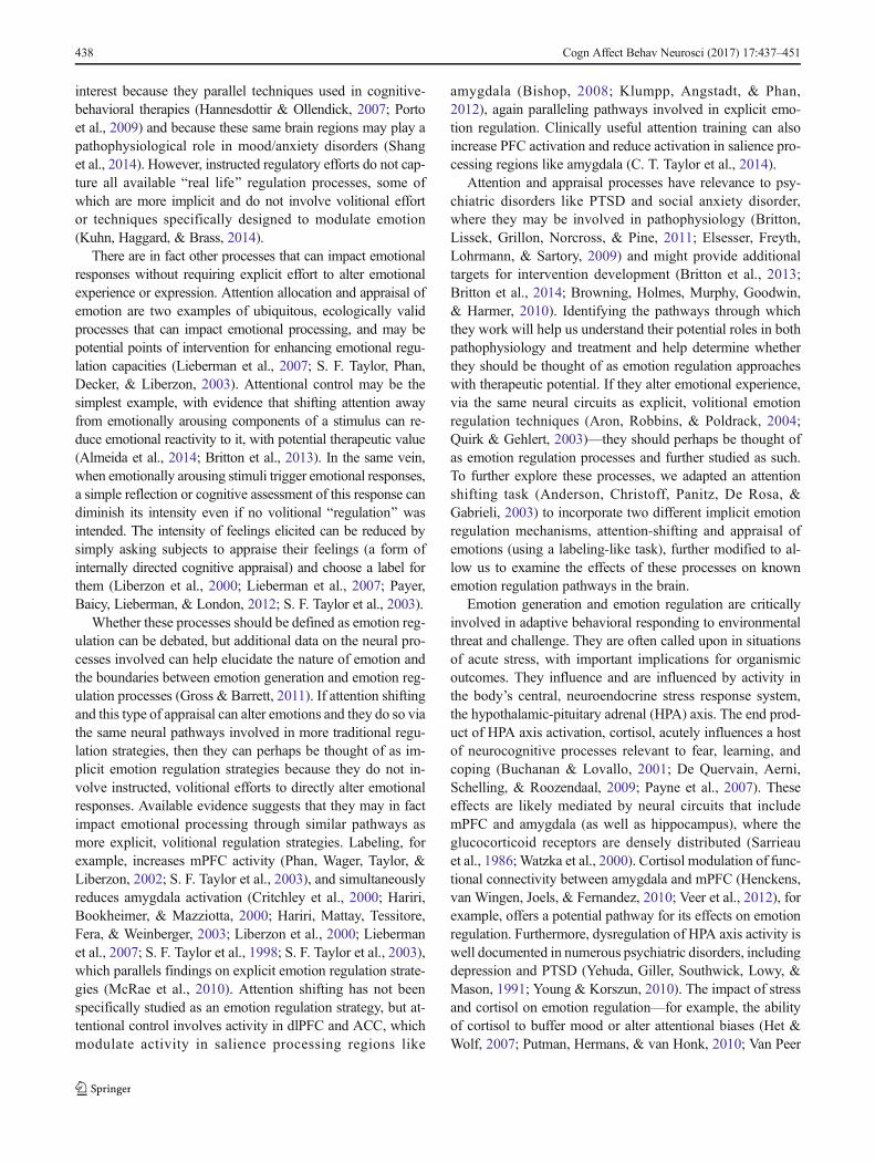
The Shifted-Attention Emotion Appraisal Task (SEAT) isillustrated in Fig. 1a. This builds on prior work that usedsimple face pictures to study emotion labeling as an emotionregulation procedure (Chen, Welsh, Liberzon, & Taylor,2010), allowing study of both attention shifting and this typeof appraisal in a single paradigm. Participants were shown ourcomposite images and asked to respond to three differentquestions regarding each image: (1) pay attention to the faceon the composite picture and determine if it is male or female(male/female condition); (2) pay attention to the scene on thecomposite picture, and determine if it is indoor or outdoor(indoor/outdoor condition); (3) pay attention to the face onthe composite picture, and determine if you like or dislikethe face (like/dislike condition). In all conditions the compos-ite pictures displayed fearful, angry, or neutral faces. Themale/female condition maintains attention on the emotionalstimuli without engaging appraisal and is a standard fMRIapproach to studying implicit emotional processing (Fusar-Poli et al., 2009). The other two conditions involve the sametype of implicit emotional processing (emotional facial ex-pressions are present and are processed), but additionally in-volve two types of nonintentional emotional regulation, name-ly (1) attention redirection (indoor/outdoor condition) and (2)appraisal (like/dislike condition). Each composite picture waspresented three times, once in each condition, with conditiontype in random order (180 trials total). Correct responses in themale/female and indoor/outdoor conditions involved accu-rately identifying the sex of the face (male/female) or thelocation of the scene (indoor/outdoor). Noncomposite pictures(face or place only) were presented in 40 trials in which par-ticipants were simply asked to determine whether it was a faceor place. A total of 220 trials were randomly ordered acrossfour runs with 55 trials per run. Trials comprised a centeredfixation crosshair for ~3–8 seconds, judgment cue for 750 ms+ 250 ms blank screen, and then composite pictures for1,500 ms. Prior to experimental trials, subjects completed apractice session with images not used in the experiment.
Cortisol administration and salivary cortisol analysis
HCT was administered as a single oral dose of 100 mg,120 minutes before start of neuroimaging. Peak levels of cor-tisol occur approximately 1.2 hours after HCT administrationand slowly decline over several hours (Derendorf et al., 1991).Saliva samples were collected at -110, -90, -70, -50, -30, -10,and +35 minutes relative to start of the task in the scanner.Samples were frozen at -80 °C and thawed before beingassayed in triplicate using commercially available Coat-a-Count radioimmunoassay kits from Diagnostic Products
Cogn Affect Behav Neurosci (2017) 17:437–451 439
Corporation (Los Angeles, CA). The intra- and interassay var-iabilities were less than 5% and 10%, respectively.
Cortisol assay results were analyzed to document a signif-icant rise in response to HCTadministration, using SPSS 17.0(Chicago, IL, U.S.A) and a three-way, mixed analysis of var-iance (ANOVA, p < .05 considered significant). Treatment(HCT, placebo) and participant sex (male, female) werebetween-subjects factors and time relative to administration(-110 -90, -70, -50, -30, -10, and +35 minutes) was thewithin-subjects factor. Cortisol level (in ug/dl) was the depen-dent variable. Bonferroni correction was applied for post hocpairwise comparisons.
Behavioral analysis
Behavioral data were analyzed using SPSS 17.0 (Chicago, IL,USA) with a mixed-effect general linear model (GLM).Treatment (HCT, placebo) and participant sex (male, female)
served as between-subjects factors while task conditions (in-door/outdoor, male/female, like/dislike) and pictured facialemotion (angry, fearful, neutral) served as within-subjects fac-tors. Reaction time (RT) was the primary dependent variablein this model. Accuracy was also analyzed as a continuousdependent variable, but since there was no Bcorrect^ responsein the like/dislike condition, the task condition factor in thismodel had only two levels (indoor/outdoor and male/female).In the like/dislike condition, we also analyzed the number oflikes and dislikes as a categorical dependent variable withtreatment and sex groups as between-subjects factors and fa-cial emotion as within-subjects factor.
MRI acquisition and preprocessing
All scanning was performed with blood-oxygen-level-dependent (BOLD) sensitive whole-brain fMRI on a 3.0Tesla GE Signa System (General Electric; Milwaukee, WI)
Fig. 1 Task schematic and brain activation patterns for main contrasts. Sample stimuli and instructions/groups are presented in panel a and brain activitymaps in panels b, c, and d. The left hemisphere is on the top in the axial slices, and on the left in the coronal slices (Color figure online)
440 Cogn Affect Behav Neurosci (2017) 17:437–451

using a standard radiofrequency coil. A total of 760 T2*-weighted reverse spiral gradient-recall echo volumes, withBOLD contrast (echo time = 30 ms, repetition time =2,000 ms, 64 × 64 matrix, flip angle = 90 degree, field of view= 22 cm, 40 contiguous 3-mm axial slices per volume), wereacquired during a single session. A high-resolution T1 scan(3D-SPGR; 256 × 160 matrix, field of view = 24 cm; slicethickness = 1.2 mm) was also acquired for anatomical locali-zation. TheMRI images were preprocessed and analyzed usingin-house batch mode of statistical parametric mapping (SPM8;Wellcome Trust Centre for Neuroimaging) provided by aMethods Core Team in the Department of Psychiatry at theUniversity of Michigan. Slice timing correction was performedfor functional volumes. Functional volumes were realigned tothe first volume in the experiment to correct for head motion,co-registered with the high-resolution sagittal images, anatom-ically normalized to the Montreal Neurological Institute (MNI)template brain, resampled to 3 × 3 × 3 mm voxels, andsmoothed with an 8 × 8 × 8-mm kernel.
MRI data analysis
The preprocessed MRI data were analyzed using the generallinear model framework in SPM8. There were 11 event regres-sors modeled: nine regressors for the composite pictures (threetypes of faces by three types of instruction) and two regressorsfor the noncomposite pictures (face only and place only). Theonsets and durations of events were convolved with a canon-ical hemodynamic response function (HRF) to create theevent regressors, in addition to covariates of six image realign-ment parameters to reduce movement induced artifacts. In thefirst-level analysis for each participant, the parameter esti-mates of event regressors were computed at each voxel.Appropriate linear contrasts were applied to the parameterestimates to produce contrast images and statistical parametricmaps (SPM t map). To evaluate activations associated withtask, treatment, and sex effects, we’ve constructed a mixedANOVA model to test the influence of the aforementionedvariables. We then performed post-hoc tests for three taskconditions—implicit emotion processing, emotion regulationby shifting attention, and emotion regulation by appraisal,respectively, by creating three contrasts ([male/female –face-only], [indoor/outdoor – male/female] and [like/dislike– male/female])—to characterize effects within regions fromthe main effect of task. Due to intrinsic limitation of the ana-lytic package including all four factors in a single ANOVAmodel was not possible, so to further examine the effect offacial emotions on brain activations, we constructed a separatemixed ANOVA model that included emotion, treatment andsex instead of task. The contrast images of interest in thesefirst-level models were used as subject-specific dependent var-iables in second-level random-effect models. Statisticalthreshold for whole-brain analysis were set at voxel-wise
FWE p < .05. In addition to whole brain analysis, we alsocreated a mask from activations seen in the whole brain anal-ysis of task main effects (voxel-wise FWE p < .05) for use ingroup analyses of HCT treatment and sex.
Specific regions of interests (ROIs) were also utilized, se-lected based on prior work (Sudheimer et al., 2013) in regardsto implicit emotion processing (male/female condition) andderived from the Anatomical Automatic Labeling software(Tzourio-Mazoyer et al, 2002). These ROIs included amyg-dala, subgenual anterior cingulate cortex, and ventral medialprefrontal cortex (all small volume corrected at voxel-wiseFWE p < .05). Beta values of surviving brain activations wereextracted and analyzed with mixed ANOVAwith significancethreshold set at p < .05, Bonferroni corrected.
Results
Behavioral results
For reaction time (RT), there was a significant main effect oftask, F(2, 72) = 7.528, p = .001, but no main effects of treat-ment, sex or facial emotion. The task effect was due to fasterRTs for the indoor/outdoor condition compared to the male/female condition, t(238) = -3.042, p = .002. There was also asignificant Task × Facial emotion interaction, F(4, 144) =13.788, p < .001—responses were faster for angry faces com-pared to neutral faces in the like/dislike condition (Mangry =1,029 ms;Mfearful = 1,084 ms;Mneutral = 1,155 ms; pangry–neutral< .02; pfearful–neutral ns). There was also a significant Treatment× Facial Emotion interaction, F(2, 72) = 3.626, p = .032—theHCT treated group responded faster to angry faces comparedto neutral faces (Mneutral = 1,111 ms;Mangry = 1,068 ms; pangry–neutral < .05) with no difference between emotions for the pla-cebo group (Mneutral = 1,071 ms; Mangry = 1,081 ms; pangry–neutral ns).
Overall accuracy in the male/female and indoor/outdoorconditions was greater than 80% (well above chance but be-low the 100% ceiling). The GLM here revealed main effectsof task, F(1, 36) = 137.03, p < .001, and emotion, F(2, 72) =29.29, p < .001, but no main effects of treatment or sex.Subjects were more accurate in the indoor/outdoor conditionthan the male/female condition (89.0% vs. 74.8%, p < .05).Subjects were less accurate for angry or fearful facial emotionscompared to neutral (Mangry = 79.8%; Mfearful = 79.6%;Mneutral = 86.3%; pangry–neutral < .025; pfearful–neutral < .025).This effect was primarily driven by reduced accuracy for an-gry and fearful faces in the male/female condition (significantTask × Facial Emotion interaction): F(2, 72) = 8.97, p < .001;Mangry = 71.8%;Mfearful = 71.0%;Mneutral = 81.6%. There wasalso a significant three-way interaction for Task × Treatment ×Sex, F(1, 36) = 11.72, p = .0015. In the male/female condition,HCT treated male subjects were significantly less accurate
Cogn Affect Behav Neurosci (2017) 17:437–451 441

than HCT treated female subjects, t(58) = -4.43, p < .001;Mmale = 66.0%, Mfemale = 80.1%, and displayed the numeri-cally worst accuracy seen in any cell in the study (seeSupplementary Table S1 for full accuracy and RT data).
The GLM on like and dislike judgements within thelike/dislike condition revealed only a main effect of facialemotion, F(2, 74) = 25.91, p < .001. Angry and fearful faceswere more disliked than neutral faces, F(1, 37) = 27.42, p <.001.
For salivary cortisol, there were main effects of treatment,F(1, 36) = 62.39, p < .0001, sex, F(1, 36) = 23.82, p < .0001,and time, F(6, 216) = 15.62, p < .0001. Cortisol levels rosedramatically over time in response to HCT, but not followingplacebo, and did so more in males than females (seeSupplementary Material, S2, for graphical display). At timeof first measurement, cortisol levels (-110 min) did not differbetween treatment groups, t(38) = 0.10, ns, or between sexgroups (males = 0.164 ug/dl; females = 0.117 ug/dl), t(38)=2.23, ns). They became significantly elevated over initialvalues 70 minutes after HCT administration (-50 min relativeto scanning), t(38) = 4.55, p = .00005. They remained signif-icantly higher with HCT than placebo during the fMRI session(3.17 vs. 0.080 ug/dl). Males had significantly higher cortisollevels than females during the scanner task, t(18) = 7.75, p =.0000003; 3.59 ± 0.27 vs. 1.22 ± 0.15 ug/dl. However, levelsduring the task were significantly higher than baseline for bothmales and females who received HCT, male: t(18) = -12.796,p < .00001; female: t(18) = -7.321, p < .00001. Task cortisollevels were not significantly elevated above baseline for eithersex after placebo, male: t(18) = 1.467, ns; female: t(18) =3.241, ns.
Functional MRI results
Main effects of task, treatment, sex, and emotion
There was a main effect of task in our first mixedANOVA model involving task, treatment and sex underwhole brain family-wise threshold at voxel-wise FWE p <.05. There was no main effect of treatment or sex on brainactivation in this model and the task by treatment interac-tion was not significant. We did find an interaction effectof treatment and sex that survived ROI analysis with amask derived from activations from all tasks at wholebrain family-wise threshold voxel-wise FWE p < .05. Inour second mixed ANOVA model examining the maineffects of emotions, treatment, and sex, we found no maineffect of emotion, and we saw sex by treatment interactionpresent in the first ANOVA model. To further investigatetask effects on brain activations, we performed post hoccomparisons and the peak activations are reported in theparagraphs below and Table 1.
Effects of implicit emotion processing: male/female–face-onlycontrast
The contrast for implicit emotion processing was constructedby comparing the male/female condition over the face-onlycondition (in which all facial expressions were neutral).Whole brain analysis with family-wise error (FWE) p < .05threshold revealed several significant clusters. Consistent withother implicit emotion activation studies (Fusar-Poli, 2009), asignificant activation peak was found in left amygdala ([-18, -1, -11]; Z = 4.90; k = 4; p < .001). Dorsal medial prefrontalcortex (dmPFC) and dorsal anterior cingulate cortex (dACC)were also consistently activated by emotional stimuli ([6, 14,43]; Z = 6.94; k = 552; p < .001). Activation was also seen inbilateral inferior frontal gyrus (IFG) and anterior insula (AI)clusters (left IFG/AI: [-33, 26, -2]; Z = 7.47; k = 477; p < .001;right IFG/AI: [39, 20, -5]; Z = 6.28; k = 300; p < .001).Bilateral occipital lobes were also activated (whole-brain re-sults are summarized in Table 1 and Fig. 1b).
Effects of shifting attention: indoor/outdoor–male/femalecontrast
In the indoor/outdoor condition (as compared to the implicitemotion processing–male/female condition), there was a maineffect of shif t ing at tent ion in the lef t and rightparahippocampal areas / fusiform gyrii ([-30, -43, -17]; Z >7.56; k = 339; p < .001; and [27, -46, -11]; Z = 6.90; k = 235; p< .001, respectively) with overlapping areas in theparahippocampal place area (PPC). Additionally, there weresignificant peaks in left and right calcarine/precuneus/lingualgyrus (left: [-15, -58, 13]; Z = 7.28; k = 198; p < .001; right:[18, -55, 16]; Z = 7.55; k = 189; p < .001). The bilateraloccipital lobes were also significantly activated duringshifting attention (left: [-39, -82, 31]; Z = 6.78; k = 238; p <.001; right: [45, -76, 25]; Z = 7.56; k = 163; p < .001). We sawno significant activations in emotion processing areas. In ad-dition, significant activations in the PCC and dlPFC were alsoobserved (PCC: [-3, -37, 40]; Z = 5.55; k = 107; p < .001;dlPFC: [33, 29, 40]; Z = 4.94; k = 27; p = .001; whole-brainresults are summarized in Table 1 and Fig. 1c).
Effects of appraisal modulation: like/dislike–male/femalecontrast
Modulation effects of appraisal (like/dislike) as comparedto the imp l i c i t emo t i on p roce s s i ng cond i t i on(male/female) were detected in several cortical areas.Regions in the frontal-parietal executive control networks,including dlPFC ([-36, 26, 37]; Z = 5.24; k = 20; p = .002)and parietal lobe ([-54, -55, 25]; Z = 7.04; k = 388; p <.001), were significantly activated. Furthermore, consis-tent with previous appraisal (Liberzon et al., 2000;
442 Cogn Affect Behav Neurosci (2017) 17:437–451

Mechias, Etkin, & Kalisch, 2010; Phan, Taylor, Welsh, &Ho, 2004) and explicit emotion regulation studies (McRaeet al., 2010; Silvers, Weber, Wager, & Ochsner, 2015)large areas in the dorsal and rostral mPFC were activated(dmPFC: [-9 38 46]; Z = 6.47; k = 340; p < .001; rmPFC:[-3, 62, 7]; Z = 5.21; k = 107; p < .001). Smaller clusterareas (k < 50) that were also significant include the leftIFG ([-54, 23, -5]; Z = 5.70; k = 43; p < .001) and leftPCC ([-9, -49, 28]; Z = 5.60; k = 31, p < .001), regionspreviously linked to volitional reappraisal (Goldin,McRae, Ramel , & Gross, 2008; Ochsner et al . ,2004;)whole-brain results are summarized in Table 1 andFig. 1d).
Cortisol effects
As noted, fMRI mixed ANOVA revealed no significant maineffect of treatment and no interaction between treatment andtask, suggesting that HCT had no impact on the analyses re-ported above. In support of this conclusion, direct comparisonof the HCT and placebo groups in parallel analyses across alltasks also revealed no significant brain activation differencesbetween them (at either a whole brain or clusterwise FWE p <.05 level). For further verification, we also repeated the mainanalyses on HCT and placebo groups separately. The brainactivation patterns seen in the combined analyses were simi-larly present in each group by itself (both visually and in
Table 1 Brain activation associated with each contrast
Region Side Z kE x y z
Implicit emotional processing–male/female–face-only contrast
Amygdala L 4.9 4 -18 -1 -11
dmPFC/dACC (BA6/8/9/32) L/R 6.94 552 6 14 43
Inferior frontal gyrus L 7.47 340 -33 26 2
R 6.71 374 51 23 22
Anterior insula L 7.47 123 -33 24 -6
R 6.28 102 37 24 -3
Cingulate gyrus (BA24) L/R 6.94 219 6 23 34
Fusiform L 8.39 66 -36 -73 -20
R 7.25 255 36 -64 -20
Thalamus L 4.7 12 -9 -16 5
R 4.86 14 9 -13 4
Occipital/lingual L 5.78 157 -30 -79 19
R 6.87 558 33 -91 4
Parietal lobe L 6.31 138 -24 -61 46
R 5.21 26 30 -57 46
Shifting attention: Indoor/outdoor–male/female contrast
Parahippocampus/fusiform L >8 216 -30 -43 -17
R 6.9 190 27 -46 -11
Occipital L 6.78 232 -39 -82 31
R 7.56 147 45 -76 25
Cuneus/precuneus/calcarine/lingual L 7.76 166 -15 -58 13
R 7.55 163 18 -55 16
Middle frontal gyrus R 4.94 27 33 29 40
PCC L/R 5.55 94 -3 -37 40
Appraisal: Like/dislike–male/female contrast
Inferior frontal gyrus (BA44/45/47) L 7.04 32 -54 23 -5
mPFC (BA10/8/6)/DLPFC (BA9) L 6.47 339 -9 38 46
Middle temporal/angular gyrus (BA39/40) L 7.04 297 -54 -55 25
PCC L/R 5.6 21 -9 -49 28
dmPFC (BA10) L/R 5.21 101 -3 62 7
Middle frontal gyrus (BA9) L 5.24 20 -36 26 37
Treatment × Sex interaction
dmPFC (BA10) L 4.42 4 -3 20 55
Cogn Affect Behav Neurosci (2017) 17:437–451 443

significance tests using a clusterwise threshold FWE p < .05;data available upon request).
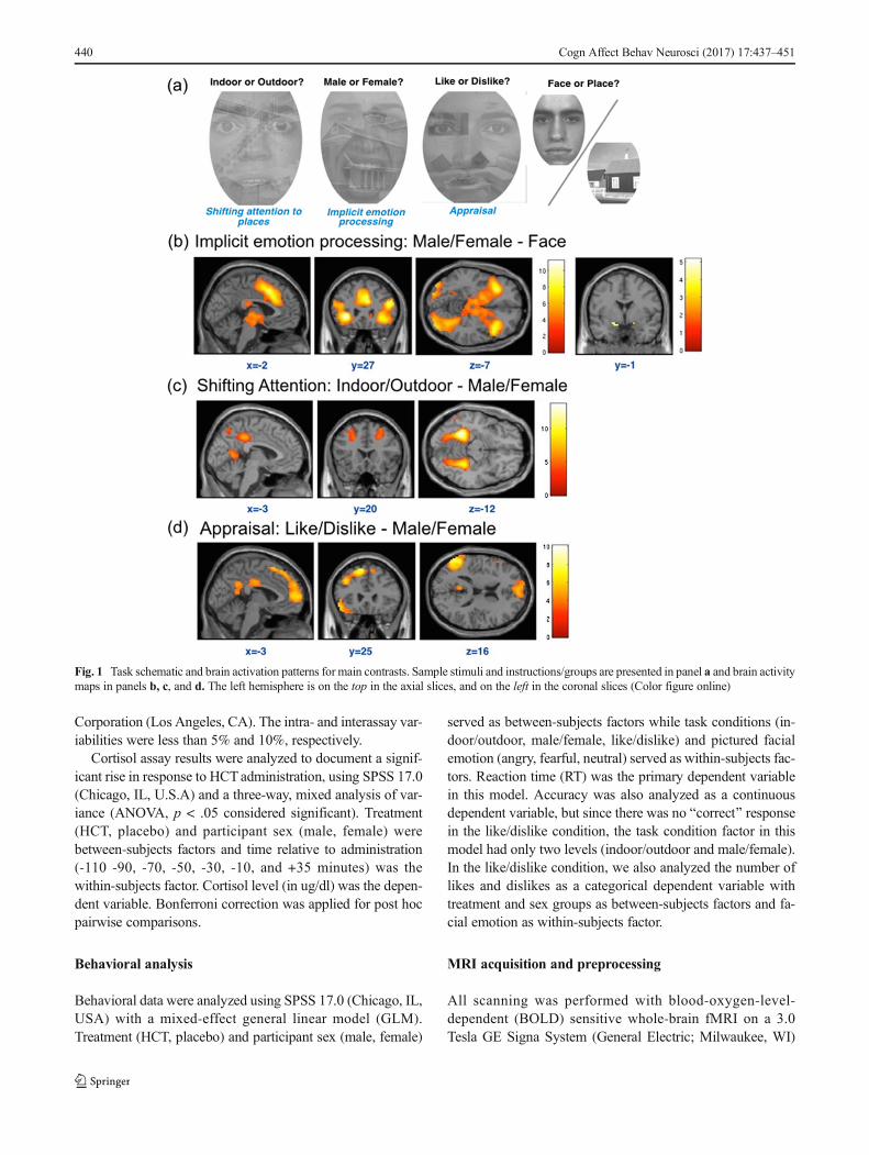
Despite the lack of any main effects of cortisol or sex (nobrain activation maps survived whole brain family-wise error(FWE) threshold p < .05 in implicit emotional activation, at-tention shifting, or appraisal conditions) on brain activation,cortisol effects did emerge in interaction with sex across thethree tasks. HCT uniformly reduced activation (relative toplacebo) in female subjects but increased activation (relativeto placebo) for male subjects in the dmPFC ([-3, 20, 55]; Z =4.42; k = 4; p < .001; Fig. 2). The differences in dmPFCactivation were significant in ROI analysis at FWE p < .05threshold using a mask derived from activations in all tasks(male/female, indoor/outdoor, and like/dislike; whole brainvoxel-wise FWE p < .05 threshold) from all subjects.
We also examined activations in a priori regions that wefound to be sensitive to cortisol administration in our previousstudy (Sudheimer et al., 2013), including amygdala, sgACC,and vmPFC, in the context of implicit emotion processing(male/female condition). The extracted betas (from the implic-it emotion task) were analyzed post hoc with a 2 × 2 ANOVA(Treatment × Sex) with Bonferroni corrections. In both thevmPFC and sgACC, there was a significant Treatment × Sexinteraction, vmPFC: F(1, 26) = 4.1501, p < .05; sgACC: F(1,36) = 4.5953, p < .05. Overall, we observed cortisol-inducedincreases in activations for the females but not for the males inall three regions, vmPFC: t(58) = -3.7476, p < .005; sgACC:t(58) = -2.9658, p < .005; amygdala: t(58) = -2.4351, p < .02(Fig. 3).
Discussion
Our primary goal was to examine the neural effects of at-tention shifting and appraisal of emotion, to determinewhether these cognitive processes effected responses toemotional stimuli via pathways parallel to those involvedin volitional, explicitly instructed emotion regulation tech-niques, such as reappraisal (Ochsner, Bunge, Gross, &Gabrieli, 2002; Gross & Barrett, 2011). If so, they mightbe usefully further explored as implicit emotion regulationstrategies. We were also interested in the effects of high dosecortisol on this circuitry, as a proxy for cognition–emotioninteractions under high stress conditions. We used a noveltask that allowed us to assess implicit emotional responses(male/female condition), attentional shift (indoor/outdoorcondition), and appraisal (like/dislike condition).Behaviorally, shifting attention to house scenes (indoor/out-door condition) increased accuracy and reduced reactiontime as compared to attending to emotional faces. Negativeemotional stimuli (angry and fearful faces) led to moreBdislike^ judgments and diminished performance accuracyas compared to neutral stimuli. As expected, implicit emotion
processing (attending to an emotional face, but to identify itssex) robustly activated amygdala, insula and mPFC regions.Attention shifting (attending to the building component ofthe compound images, rather than the faces) activated dlPFCand PCC, which are regions previously linked to emotionalregulation (Gross & Thompson, 2007). The attention shiftalso activated fusiform and parahippocampal gyri and bilat-eral occipital lobes. During appraisal of emotion (contrastingan emotion labeling condition to implicit emotional process-ing), large areas of dorsal to rostral mPFC were activated,consistent with prior work on emotion labeling as an emo-tion regulation strategy (Lieberman et al., 2007; Payer et al.,2012; Phan et al., 2004; S. F. Taylor et al., 2003). IFG, aregion implicated in volitional emotion regulation (Kimet al., 2013), and portions of PCC, implicated in both ap-praisal and volitional emotion regulation (Liberzon et al.,2000; Phan et al., 2004), were activated as well. Single doseexogenous cortisol administration had no overall effect, butinteracted with subject sex in modulating activation in thedmPFC area.
Fig. 2 Effects of hydrocortisone differed by subject sex. The top panelshows brain activations associated with the interaction of HCT and sexacross all three tasks (p < .001, unc. for display). The only significant peakwas in dmPFC (-3, 20, 55). HCT reduced activation in females, relative toplacebo, whereas it increased activation relative to placebo in males(Color figure online)
444 Cogn Affect Behav Neurosci (2017) 17:437–451
In the implicit emotion processing condition, subjects wereinstructed to identify gender to focus their attention on theemotional faces, without explicitly directing attention to theemotional content. That content was nevertheless implicitlyprocessed, as reflected in the increased Bdislike^ judgements,reduced gender identification accuracy, and lengthened reac-tion time for negative compared to neutral faces. We used thiswell-established implicit emotional processing condition(Breiter et al., 1996) as our comparator in order to avoid anyexplicit appraisal elements (e.g., naming emotions), in order toisolate appraisal processes (Critchley et al., 2000; Georgeet al., 1993; Habel et al., 2007; Lane, Fink, Chau, & Dolan,1997) in the appraisal of emotion condition. As seen by othersduring processing of emotional faces (Adolphs, 2002;LeDoux, 1995; Schneider et al., 1997; Schneider, Gur, Gur,&Muenz, 1994) and processing negative emotions more gen-erally (Etkin, Egner, & Kalisch, 2011; Maier et al., 2012),during implicit emotional processing our subjects activatedamygdala and insula, as well as dorsal medial prefrontal cor-tex (dmPFC) and the anterior portion of dorsal ACC(adACC).
Our task was designed to allow us to examine two commoncognitive processes of daily life—shifting attention and ap-praisal of emotion—both of which can potentially modulateemotional responding and perhaps serve as emotion regula-tion strategies. Shifting attention in this context does not meanignoring the stimuli altogether, but rather focusing volitionalattention on components without emotional salience. This isanalogous to a physician in an emergency room purposefullyfocusing attention on specific components of the injury, toassess and perhaps treat it, rather than allowing attention to
be captured by the patient’s distress and suffering. This likelymodulates emotional responding in a way that permits moreeffective action. We expected that engagement of regions inthe attention network would be required for this task, and thatthis would lead to decreased processing in emotion generatingregions, which would parallel findings from volitional emo-tion regulation strategies (McRae et al., 2010) and support theidea that attention shifting can function as an emotion regula-tion process. We did in fact detect the expected activations inattention network components, while no activations were seenin emotion processing regions, in contrast to what was ob-served in the implicit emotion condition. Attention networksare thought to be comprised of alerting, orientating andexecution/conflict monitoring components (Posner &Petersen, 1990). During our shifting attention condition, weobserved activation of lateral prefrontal cortex, the same re-gion implicated in Posner’s Borienting attention^ network(Koenigsberg et al., 2010). We also observed activation inPCC. Activation in this region has been linked to target detec-tion in a selective attention task (Shulman et al., 2010).Interestingly, PCC and precuneus activations have also beenreported during emotional distancing, a volitional emotionregulation strategy (Koenigsberg et al., 2010), but not in dis-traction studies (Kanske, Heissler, Schönfelder, Bongers, &Wessa, 2011; McRae et al., 2010). This suggests a potentiallysimilar role for PCC in regulating some aspects of attentionduring specific types of emotion regulation, like shifting at-tention and distancing. Future studies will be required to fur-ther clarify the precise role of PCC in these processes. Shiftingattention also activated fusiform gyrus and parahippocampalgyrus, with more overlapping areas in the parahippocampal
Fig. 3 Brain activations in a priori defined regions of interest, showing beta value extractions formales and females separatelywithin each ROI, from theimplicit emotion (male/female) task. Within each region, HCT produced increases in activation (or decreases in inactivation) that were not seen in males
Cogn Affect Behav Neurosci (2017) 17:437–451 445

place area (PPA; Epstein & Kanwisher, 1998). Since subjectswere instructed to shift attention to a place image (identifyingit as inside or outside), increased activation in theparahippocampal place area is entirely consistent with the taskand confirms that the manipulation worked, and subjects werefocusing on the place component of the picture, as instructed.In summary, the attention shift condition activated attentionrelated regions (dlPFC and PCC) and visual/space processingregions that were appropriate to the content of the stimuli towhich attention was shifted, and this was associated with di-minished activation in the emotion processing regions that hadbeen activated in the implicit emotional activation condition.
We were specifically interested in regulation by appraisalof emotion, because appraisal is both a very common mecha-nism that is engaged automatically while processing emotion-al stimuli (Critchley et al., 2000; S. F. Taylor et al., 2003), andalso can be used volitionally, as often practiced in cognitiveand cognitive–behavioral approaches (Beck, Emery, &Greenberg, 1985; Clark, 1986; Norton, Asmundson, Cox, &Norton, 2000). In previous studies, both our lab (S. F. Tayloret al., 2003) and others (Hariri et al., 2003) have demonstratedthat mPFC/ACC regions are involved in appraisal and thatengaging appraisal diminishes the emotional impact of nega-tive stimuli (S. F. Taylor et al., 2003). Medial prefrontal cortexactivations have been reported in the context of volitionalemotion regulation tasks, such as reappraisal (Kalisch,2009), and mPFC has been hypothesized to provide top-down control to emotion processing areas, such as the amyg-dala (Etkin et al., 2011). In contrast to reappraisal, our apprais-al task did not involve a purposeful effort to modulate emo-tional responses; it simply asked subjects to identify one oftheir emotional responses to the face presented (as like ordislike). However, the large dorsal to rostral mPFC activationsseen in our study overlap with the activations reported in stud-ies of reappraisal (Ochsner et al., 2002; Phan et al., 2004),suggesting that a process that does not involve instructed,volitional modulation of emotion activated regulatory regionssimilar to those activated by purposeful regulation strategies.Rostral mPFC regions have been also implicated in processingof self and self-relatedness (Chiao et al., 2009; Gilbert et al.,2007; Phan et al., 2004), so the large rostral mPFC activationswe saw may have also been partly elicited by the fact that inour appraisal task, subjects had to reflect on their own, internalemotional states. The dorsal lateral prefrontal cortex (dlPFC)activations that we saw also parallel the dlPFC activationsseen in studies using volitional emotion regulation strategiessuch as reappraisal (Kim et al., 2013; Ochsner et al., 2002),again supporting the hypothesis that both volitional and im-plicit emotion regulation processes use shared neural net-works to modulate emotional experience or expression.
We administered exogenous cortisol in a double blind fash-ion in order to examine effects of elevated stress hormone onappraisal and attention regulation. Cortisol has important
cognitive and emotional effects on brain with high relevanceto PTSD (Kaouane et al., 2012) and there is intriguing evi-dence of a potentially important role in PTSD treatment, likelymediated by these brain effects (Yehuda et al., 2015). Deeperunderstanding of its effects on all types of potential emotionregulation processes will be important in efforts to better un-derstand disorders with dysregulated HPA axis function anddetermine how to optimally use cortisol therapeutically.Behaviorally, cortisol administration affected both reactiontime and accuracy. It shortened reaction time to negative af-fective stimuli (angry faces) compared to neutral ones, whilereducing accuracy during implicit emotion processing(male/female condition) specifically in male subjects.However, cortisol had no significant main effects on brainactivation maps in fMRI analyses for any of the three tasks,and had no measurable impact in shaping the brain activationpatterns seen in attention shifting and appraisal of emotions.On the other hand, there were significant sex differences incortisol’s brain effects across the tasks. Cortisol administrationreduced activation in dorsal medial prefrontal cortex (dmPFC)in all three tasks in females, but it enhanced dmPFC activationin these tasks in males. Simultaneously, in females during theimplicit emotion task it increased activation in sgACC,vmPFC, and amygdala, emotion processing regions previous-ly shown to be sensitive to exogenous cortisol (Sudheimeret al., 2013). It did not have this effect in males.
Unfortunately, males and females also differed markedly insalivary cortisol levels achieved during the tasks. As a result,we cannot determine whether the neural differences betweenmales and females reflect a true sex difference in brain effectsof cortisol or are due to Bdose^ effects. They are of interest ineither case (see below), but follow-up work will be needed todetermine which explanation is correct. The sex differences inmeasured cortisol levels could reflect sex differences in corti-sol binding globulin, which can differ between males andfemales, partly due to the influence of estrogen, which ele-vates CBG, leading to increased cortisol binding and reducedlevels of free cortisol. Saliva measures reflect free cortisol,which is the active component most relevant to effects onbrain (Hellhammer, Wüst, & Kudielka, 2009). Free cortisolcan be reduced in females by menstrual cycle effects on estro-gen levels and by estrogen containing birth control pills(BCPs), neither of which were controlled here. Future studieswill need to measure CBG and carefully control for cycleeffects and BCP use in order to test for true sex differencesin brain effects of cortisol and distinguish them from Bdose^effects.
Glucocorticoids do have well-established dose-dependenteffects on brain, with a dose-response curve that is often de-scribed as an inverted U. Enhanced cognitive function in dif-ferent memory paradigms is often seen at Bmoderate^ cortisollevels, with less optimal function at very low levels and def-icits appearing at very high levels (Domes et al., 2005; Salehi,
446 Cogn Affect Behav Neurosci (2017) 17:437–451

Cordero, & Sandi, 2010; Schilling et al., 2013). We cannotdetermine whether the sexually dimorphic brain activationsseen here represent Bnegative^ effects of high cortisol in malesin this paradigm, but the cortisol dose-response literaturewould certainly predict differential brain activity with moder-ate versus high free cortisol levels. The levels seen during ourtask in females would be considered comparable to moderatestress levels, whereas the levels seen in the males are only seenunder conditions of high stress that perhaps includes physio-logical stress. The differential dose effects may be due tochanging ratios of occupancy of mineralocorticoid (MR) andglucocorticoid (GR) receptors, with high occupancy of GRreducing the MR/GR ratio and producing detrimental effects(Bohbot, Gupta, Banner, & Dahmani, 2011). Our male sub-jects who received HCT did display reduced accuracy in iden-tifying faces as male or female, relative to all other groups, andthis could reflect a detrimental effect of the uniquely highcortisol levels seen by their brains. If so, this would suggestthat the inverted U dose-response pattern for cortisol effectson memory, which has been the focus of most work in thisarea to date, may also apply to other cognitive functions. Thisaccuracy deficit, however, is unlikely to be related to the in-creased dmPFC activity seen in these male subjects. The in-creased activity associated with moderate cortisol elevationsseen in females in emotion processing regions (sgACC,vmPFC, and amygdala) could reflect an enhancedBappropriate^ response during the implicit emotion task inwhich it was seen (this enhancement was one of our a prioriexpectations). The absence of this effect in males may reflect aBdeficit^ due to their very high cortisol levels. However, this isclearly highly speculative, and more work is definitely neededto explore the interesting potential explanations for and impli-cations of these findings.
We also need to consider the possibility that the male–fe-male differences in cortisol effects on dmPFC, sgACC,vmPFC, and amygdala were not solely due to different corti-sol levels, but could also be influenced by sexually dimorphicbrain sensitivities to cortisol within the brain. Others havereported sex differences in dmPFC during emotion perception(Hofer et al., 2006), but stress or cortisol effects were not Binplay^ in that study. Amygdala and vmPFC are rich in gluco-corticoid receptors (Holsboer, 2000) and important in emotionprocessing and emotion regulation. If there are male–femaledifferences in the sensitivity of these regions to free cortisol,these will be important to identify, as they could shape sexu-ally dimorphic effects of cortisol on emotion regualtion strat-egies and capacities. Here, cortisol enhanced mPFC activationin men, perhaps supporting appraisal, while in women cortisolincreased activation in other cortical regions—such as sgACCand vmPFC—that are involved in mood regulation and ex-tinction retention. Though purely speculative at this point,there is clear potential relevance to well-known sex differencsin psychiatric disorders such as depression and PTSD, where
HPA axis dysregulation is also well established. More work isclearly needed.
Limitations
Several limitations must be acknowledged. We used emotion-al facial expressions as emotion generating stimuli. While thismethod has been widely used in fMRI studies (Fusar-Poliet al., 2009; Gur et al., 2002), it may have elicited less intenseemotional responses than use of other emotion generatingstimuli. Furthermore, in the implicit emotion condition, iden-tifying the gender of the face allows the emotional content tobe processed implicitly, but the identification task itself re-quires cognitive processing, and this cognitive processingcould reduce emotional activation, so full extent of potentialemotional activation may not have been seen even in thisBbaseline^ condition. The cognitive work of identificationcould also potentially interact with appraisal or attentionshifting strategies. We decided, however, that the benefits ofengaging our subjects in a specific task and maintaining theirattention outweighed the potential Blosses^ due to a weakeremotional activation signal or the potential for Bsubtractingout^ regions involved in both appraisal and genderrecognition.
It is also important to acknowledge that though we haveuse the terminology implicit emotion regulation to describewhat is happening with attention shifting and emotion labelingactivity, the concept of implicitness is complex and potentiallycontroversial. We do see brain changes suggesting that emo-tion regulation areas of the brain are Bin play^ with thesemanipulations, but we could also simply call this Buninstruc-ted^ emotion regulation. Our main goal in using the wordimplicit is to differentiate these strategies from the explicit,volitional efforts to alter emotional experience that have beenused in appraisal and related studies. We also note that mech-anisms other than implicit emotion regulation could be shap-ing the results seen. For example, in the attention shift task,subjects could have been aware of interference with efforts tofocus on the building created by the Bpull^ for attention fromthe embedded face. They may have resolved this interferenceby using Bexplicit^ regulation strategies. Creative future workwill be needed if we are to differentiate the explicit cognitivework of directing attention away from an emotionally evoca-tive cue from what we have called implicit emotional regula-tion. We suspect that they use similar neural pathways andboth can result in reduced emotional processing, so the differ-ences may be mainly semantic, but work on this is needed.
Results from the cortisol infusion must be considered pre-liminary. As noted above, because males and femalesachieved very different free cortisol levels in response to thesame dose of HCT, we cannot disentangle Bdose^ effects fromsex differences in brain sensitivities. Substantially larger stud-ies will be needed to fully dissect potential interactions
Cogn Affect Behav Neurosci (2017) 17:437–451 447

between sex, cortisol levels, and cortisol effects on brain dur-ing emotion regulation processes. The between-group designused also has some drawbacks. It is less sensitive than a withinsubject design, which reduces some variability; and individualdifferences in cortisol response can contribute additional var-iance. However, we wanted to avoid repetition and learningeffects that could pose substantive confounds (Wirth, Scherer,Hoks, & Abercrombie, 2011). Use of exogenous cortisol is asimplified downstream proxy for actual stress. Cortisol is aknown cognitive modulator, with established dysregulationin psychiatric disorders, so isolating its independent effectsis of interest, but future studies will need to examine the ef-fects of ecological stress, with its additional neural and psy-chosocial components, in these types of paradigms.Exogenous administration here was complicated by sex dif-ferences in levels achieved and high variability in levelsamong the males. We have seen this in other studies, and itneeds clarification in follow-up work. Future studies will alsoneed to examine impacts on other systems, such as the rewardsystem, that are also affected by stress and cortisol.
Conclusions
Emotion regulation pathways have been the focus of recentstudies, but most work has examined instructed, explicit, orBexogenous^ regulation techniques (Kuhn et al., 2014). Therehas been debate in the literature as to whether all emotionregulation strategies must involve active, volitional efforts tochange emotions (Gross & Barrett, 2011). Here, we demon-strate the utility of a task that allowed us to explore cognitiveprocesses that are not specifically targeting emotional process-es or circuitry. We used this task to test their ability to alteractivity in brain regions that are activated by more traditionalemotion regulation techniques—by shifting attention or ap-praising an internal emotional state, without attempting inten-tionally to alter emotional responses. Attention shifting acti-vated attention network brain areas (e.g., lPFC and PCC) andappropriate task-specific areas (e.g., parahippocampal placearea, reflecting successful allocation of attention to the placecomponent of the compound picture). Appraisal robustly ac-tivated mPFC as well as dlPFC and IFG, as also seen ininstructed reappraisal strategies. In both, regions activated byimplicit emotion processing were Bquiet.^ The data suggestthat both attention shifting and appraisal of emotions might bethought of as implicit emotion regulation strategies, actingthrough some of the same brain circuits involved in instructedemotion regulation. Further examination of such implicit emo-tion regulation processes appears warranted, with potentialrelevance to psychiatric disorders such as PTSD and socialanxiety disorder, where emotion regulation processes and at-tentional control have clinical salience.
The sexually dimorphic brain responses to cortisol are in-triguing despite the confound between Bdose^ and sex.
Understanding potential dose-dependent effects of cortisolon emotion regulation capacities is of clinical relevance topsychiatric disorders. It will be critical in efforts to determinethe true role of the HPA axis in these disorders and the truepotential utility of HPA axis interventions in treating them.Understanding sex differences in brain sensitivity to cortisol,if they exist, could help explain differences in emotion regu-lation strategies between men and women, and contribute toour understanding of sex differences in vulnerability to disor-ders like PTSD and depression. Disentangling Bdose^ and sexeffects, which may have both been in play in this study, isessential in future work.
Acknowledgements The research reported in this article was supportedby a grant from the National Institute of Mental Health (R24MH075999)to I. Liberzon.
Compliance with ethical standards
Conflict of interest None
Reference
Adolphs, R. (2002). Neural systems for recognizing emotion. CurrentOpinions in Neurobiology, 12(2), 169–177.
Almeida, O. P., MacLeod, C., Ford, A., Grafton, B., Hirani, V., Glance,D., & Holmes, E. (2014). Cognitive bias modification to preventdepression (COPE): Study protocol for a randomised controlledtrial. Trials, 15, 282.
Anderson, A. K., Christoff, K., Panitz, D., De Rosa, E., & Gabrieli, J. D.(2003). Neural correlates of the automatic processing of threat facialsignals. Journal of Neuroscience, 23(13), 5627–5633.
Aron, A. R., Robbins, T. W., & Poldrack, R. A. (2004). Inhibition and theright inferior frontal cortex. Trends in Cognitive Science, 8(4), 170–177.
Beck, A., Emery, G., & Greenberg, R. (1985). Anxiety disorders andphobias: A cognitive approach. New York, NY: Basic.
Berking, M., & Wupperman, P. (2012). Emotion regulation and mentalhealth: Recent findings, current challenges, and future directions.Current Opinion in Psychiatry, 25(2), 128–134.
Bishop, S. J. (2008). Neural mechanisms underlying selective atten-tion to threat. Annals of the New York Academy of Sciences,1129, 141–152.
Bohbot, V. D., Gupta, M., Banner, H., & Dahmani, L. (2011).Caudate nucleus-dependent response strategies in a virtual nav-igation task are associated with lower basal cortisol and im-paired episodic memory. Neurobiology of Learning andMemory, 96(2), 173–180.
Breiter, H. C., Etcoff, N. L., Whalen, P. J., Kennedy, W. A., Rauch, S. L.,Buckner, R. L.,…Rosen B. R. (1996). Response and habituation ofthe human amygdala during visual processing of facial expression.Neuron, 17, 875–887
Britton, J. C., Bar-Haim, Y., Clementi, M. A., Sankin, L. S., Chen, G.,Shechner, T.,…Pine, D. S. (2013). Training-associated changes andstability of attention bias in youth: Implications for Attention BiasModification Treatment for pediatric anxiety. DevelopmentalCognitive Neuroscience, 4, 52–64.
Britton, J. C., Lissek, S., Grillon, C., Norcross, M. A., & Pine, D. S.(2011). Development of anxiety: The role of threat appraisal andfear learning. Depression and Anxiety, 28(1), 5–17.
448 Cogn Affect Behav Neurosci (2017) 17:437–451

Britton, J. C., Suway, J. G., Clementi, M. A., Fox, N. A., Pine, D. S., &Bar-Haim, Y. (2014). Neural changes with attention bias modifica-tion for anxiety: A randomized trial. Social Cognitive and AffectiveNeuroscience, 10(7), 913–920.
Browning, M., Holmes, E. A., Murphy, S. E., Goodwin, G. M., &Harmer, C. J. (2010). Lateral prefrontal cortex mediates the cogni-tive modification of attentional bias. Biological Psychiatry, 67(10),919–925.
Buchanan, T. W., & Lovallo, W. R. (2001). Enhanced memory for emo-tional material following stress-level cortisol treatment in humans.Psychoneuroendocrinology, 26(3), 307–317.
Chen, A. C., Welsh, R. C., Liberzon, I., & Taylor, S. F. (2010). ‘Do I likethis person?’ A network analysis of midline cortex during a socialpreference task. NeuroImage, 51(2), 930–939.
Chiao, J. Y., Harada, T., Komeda, H., Li, Z., Mano, Y., Saito, D.,…Iidaka,T. (2009). Neural basis of individualistic and collectivistic views ofself. Human Brain Mapping, 30(November 2008), 2813–2820.
Clark, D. M. (1986). A cognitive approach to panic. Behaviour Researchand Therapy, 24(4), 461–470.
Critchley, H., Daly, E., Phillips, M., Brammer, M., Bullmore, E.,Williams, S.,…Murphy, D. (2000). Explicit and implicit neuralmechanisms for processing of social information from facial expres-sions: A functional magnetic resonance imaging study. HumanBrain Mapping, 9(2), 93–105.
De Quervain, D. J., Aerni, A., Schelling, G., & Roozendaal, B. (2009).Glucocorticoids and the regulation of memory in health and disease.Frontiers in Neuroendocrinology, 30(3), 358–370.
Derendorf, H., Mollmann, H., Barth, J., Mollmann, C., Tunn, S., &Krieg,M. (1991). Pharmacokinetics and oral bioavailability of hydrocorti-sone. Journal of Clinical Pharmacology, 31(5), 473–476.
Domes, G., Rothfischer, J., Reichwald, U., & Hautzinger, M. (2005).Inverted-U function between salivary cortisol and retrieval of verbalmemory after hydrocortisone treatment. Behaviour Neuroscience,119(2), 512–517.
Eisenberg, N. (2000). Emotion, regulation, and moral development.Annual Review of Psychology, 51, 665–697.
Ekman, P., & Friesen, W. V. (1976). Pictures of facial affect. Palo Alto,CA: Consulting Psychologists Press.
Elsesser, K., Freyth, C., Lohrmann, T., & Sartory, G. (2009).Dysfunctional cognitive appraisal and psychophysiological reactiv-ity in acute stress disorder. Journal of Anxiety Disorders, 23(7),979–985.
Epstein, R., &Kanwisher, N. (1998). A cortical representation of the localvisual environment. Nature, 392(6676), 598–601.
Etkin, A., Egner, T., & Kalisch, R. (2011). Emotional processing in an-terior cingulate and medial prefrontal cortex. Trends in CognitiveSciences, 15(2), 85–93.
Fusar-Poli, P., Placentino, A., Carletti, F., Landi, P., Allen, P., Surguladze,S.,…Politi, P. (2009). Functional atlas of emotional faces process-ing: A voxel-based meta-analysis of 105 functional magnetic reso-nance imaging studies. Journal of Psychiatry Neuroscience, 34(6),418–432.
George, M. S., Ketter, T. A., Gill, D. S., Haxby, J. V., Ungerleider, L. G.,Herscovitch, P., & Post, R. M. (1993). Brain regions involved in rec-ognizing facial emotion or identity: An oxygen-15 PET study. Journalof Neuropsychiatry and Clinical Neuroscience, 5(4), 384–394.
Gilbert, S. J.,Williamson, I. D. M., Dumontheil, I., Simons, J. S., Frith, C.D., & Burgess, P. W. (2007). Distinct regions of medial rostral pre-frontal cortex supporting social and nonsocial functions. SocialCognitive and Affective Neuroscience, 2, 217–226.
Goldin, P. R., McRae, K., Ramel, W., & Gross, J. J. (2008). The neuralbases of emotion regulation: Reappraisal and suppression of nega-tive emotion. Biological Psychiatry, 63(6), 577–586.
Gross, J. J. (1998). The emerging field of emotion regulation: An inte-grative review. Review of General Psychology, 2(3), 271.
Gross, J. J., & Barrett, L. F. (2011). Emotion Generation and Emotionregulation: One or two depends on your point of view. EmotionReview, 3, 8–16.
Gross, J. J., & Munoz, R. F. (1995). Emotion regulation and mental-health. Clinical Psychology-Science and Practice, 2(2), 151–164.
Gross, J. J., & Thompson, R. A. (2007). Emotion regulation: Conceptualfoundations. Handbook of Emotion Regulation, 3, 24.
Gur, R. C., Schroeder, L., Turner, T., McGrath, C., Chan, R.M., Turetsky,B. I.,…Gur, R. E. (2002). Brain activation during facial emotionprocessing. NeuroImage, 16(3, Pt. 1), 651–662.
Habel, U., Windischberger, C., Derntl, B., Robinson, S., Kryspin-Exner,I., Gur, R. C., & Moser, E. (2007). Amygdala activation and facialexpressions: Explicit emotion discrimination versus implicit emo-tion processing. Neuropsychologia, 45(10), 2369–2377.
Hannesdottir, D. K., & Ollendick, T. H. (2007). The role of emotionregulation in the treatment of child anxiety disorders. ClinicalChild and Family Psychology Review, 10(3), 275–293.
Hariri, A. R., Bookheimer, S. Y., & Mazziotta, J. C. (2000). Modulatingemotional responses: Effects of a neocortical network on the limbicsystem. Neuroreport, 11(1), 43–48.
Hariri, A. R., Mattay, V. S., Tessitore, A., Fera, F., & Weinberger, D. R.(2003). Neocortical modulation of the amygdala response to fearfulstimuli. Biological Psychiatry, 53(6), 494–501.
Hellhammer, D. H.,Wüst, S., & Kudielka, B.M. (2009). Salivary cortisolas a biomarker in stress research. Psychoneuroendocrinology, 34(2),163–171.
Henckens, M. J., van Wingen, G. A., Joels, M., & Fernandez, G. (2010).Time-dependent effects of corticosteroids on human amygdala pro-cessing. Journal of Neuroscience, 30(38), 12725–12732.
Het, S., & Wolf, O. T. (2007). Mood changes in response to psychosocialstress in healthy young women: Effects of pretreatment with corti-sol. Behavior Neuroscience, 121, 11–20.
Hofer, A., Siedentopf, C. M., Ischebeck, A., Rettenbacher, M. A., Verius,M.,…Fleischhacker, W. W. (2006). Gender differences in regionalcerebral activity during the perception of emotion: A functionalMRIstudy. NeuroImage, 32(2), 854–862.
Holsboer, F. (2000). The corticosteroid receptor hypothesis of depression.Neuropsychopharmacology, 23, 477–501.
Kalisch, R. (2009). The functional neuroanatomy of reappraisal: Timematters.Neuroscience& Biobehavioral Reviews, 33(8), 1215–1226.
Kanske, P., Heissler, J., Schönfelder, S., Bongers, A., & Wessa, M.(2011). How to regulate emotion? Neural networks for reappraisaland distraction. Cerebral Cortex, 21, 1379–1388.
Kaouane, N., Porte, Y., Vallée, M., Brayda-Bruno, L., Mons, N.,Calandreau, L.,…Desmedt, A. (2012). Glucocorticoids can inducePTSD-like memory impairments in mice. Science, 335(6075),1510–1513.
Kim, P., Evans, G. W., Angstadt, M., Ho, S. S., Sripada, C. S., Swain, J.E.,…Phan, K. L. (2013). Effects of childhood poverty and chronicstress on emotion regulatory brain function in adulthood.Proceedings of the National Academies of Science of the UnitedStates of America.
Klumpp, H., Angstadt, M., & Phan, K. L. (2012). Shifting the focus ofattention modulates amygdala and anterior cingulate cortex reactiv-ity to emotional faces. Neuroscience Letters, 514(2), 210–213.
Koenigsberg, H. W., Fan, J., Ochsner, K. N., Liu, X., Guise, K.,Pizzarello, S.,…Siever, L. J. (2010). Neural correlates of using dis-tancing to regulate emotional responses to social situations.Neuropsychologia, 48(6), 1813–1822.
Kompus, K., Hugdahl, K., Ohman, A., Marklund, P., & Nyberg, L.(2009). Distinct control networks for cognition and emotion in theprefrontal cortex. Neuroscience Letters, 467(2), 76–80.
Kuhn, S., Haggard, P., & Brass, M. (2014). Differences between endog-enous and exogenous emotion inhibition in the human brain. BrainStructure and Function, 219(3), 1129–1138.
Cogn Affect Behav Neurosci (2017) 17:437–451 449

Lane, R. D., Fink, G. R., Chau, P. M., & Dolan, R. J. (1997). Neuralactivation during selective attention to subjective emotional re-sponses. Neuroreport, 8(18), 3969–3972.
LeDoux, J. E. (1995). Emotion: Clues from the brain. Annual Review ofPsychology, 46(1), 209–235.
Liberzon, I., Taylor, S. F., Fig, L. M., Decker, L. R., Koeppe, R. A., &Minoshima, S. (2000). Limbic activation and psychophysiologicresponses to aversive visual stimuli: Interaction with cognitive task.Neuropsychopharmacology, 23(5), 508–516.
Lieberman, M. D., Eisenberger, N. I., Crockett, M. J., Tom, S.M., Pfeifer,J. H., & Way, B. M. (2007). Putting feelings into words: Affectlabeling disrupts amygdala activity in response to affective stimuli.Psychological Science, 18(5), 421–428.
Maier, S., Szalkowski, A., Kamphausen, S., Perlov, E., Feige, B.,Blechert, J.,…Tüscher, O. (2012). Clarifying the role of the rostraldmPFC/dACC in fear/anxiety: Learning, appraisal or expression?PLOS ONE, 7(11). doi:10.1371/journal.pone.0050120
McRae, K., Hughes, B., Chopra, S., Gabrieli, J. D. E., Gross, J. J., &Ochsner, K. N. (2010). The neural bases of distraction and reapprais-al. Journal of Cognitive Neuroscience, 22, 248–262.
Mechias, M. L., Etkin, A., & Kalisch, R. (2010). A meta-analysis ofinstructed fear studies: Implications for conscious appraisal of threat.NeuroImage, 49(2), 1760–1768.
Norton, P. J., Asmundson, G. J., Cox, B. J., & Norton, G. R. (2000).Future directions in anxiety disorders: Profiles and perspectives ofleading contributors. Journal of Anxiety Disorders, 14(1), 69–95.
Ochsner, K. N., Bunge, S. A., Gross, J. J., & Gabrieli, J. D. (2002).Rethinking feelings: An FMRI study of the cognitive regulation ofemotion. Journal of Cognitive Neuroscience, 14(8), 1215–1229.
Ochsner, K. N., & Gross, J. J. (2007). The neural architecture of emotionregulation. Handbook of Emotion Regulation, 1(1), 87–109.
Ochsner, K. N., Ray, R. D., Cooper, J. C., Robertson, E. R., Chopra, S.,Gabrieli, J. D., & Gross, J. J. (2004). For better or for worse: Neuralsystems supporting the cognitive down- and up-regulation of nega-tive emotion. NeuroImage, 23(2), 483–499.
Payer, D. E., Baicy, K., Lieberman, M. D., & London, E. D. (2012).Overlapping neural substrates between intentional and incidentaldown-regulation of negative emotions. Emotion, 12(2), 229–235.
Payne, J. D., Jackson, E. D., Hoscheidt, S., Ryan, L., Jacobs, W. J., &Nadel, L. (2007). Stress administered prior to encoding impairs neu-tral but enhances emotional long-term episodic memories. Learningand Memory, 14(12), 861–868.
Phan, K. L., Taylor, S. F., Welsh, R. C., & Ho, S.-H. (2004). Neuralcorrelates of individual ratings of emotional salience: A trial-related fMRI study. NeuroImage, 21, 768–780.
Phan, K. L., Wager, T., Taylor, S. F., & Liberzon, I. (2002). Functionalneuroanatomy of emotion: A meta-analysis of emotion activationstudies in PET and fMRI. NeuroImage, 16(2), 331–348.
Porto, P. R., Oliveira, L., Mari, J., Volchan, E., Figueira, I., & Ventura, P.(2009). Does cognitive behavioral therapy change the brain? A sys-tematic review of neuroimaging in anxiety disorders. Journal ofNeuropsychiatry and Clinical Neuroscience, 21(2), 114–125.
Posner, M. I., & Petersen, S. E. (1990). The attention system of the humanbrain. Annual Review of Neuroscience, 13, 25–42.
Putman, P., Hermans, E. J., & van Honk, J. (2010). Cortisol administra-tion acutely reduces threat-selective spatial attention in healthyyoung men. Physiological Behavior, 99, 294–300.
Quirk, G. J., & Gehlert, D. R. (2003). Inhibition of the amygdala: Key topathological states? Annals of the New York Academy of Sciences,985, 263–272.
Salehi, B., Cordero, M. I., & Sandi, C. (2010). Learning under stress: Theinverted-U-shape function revisited. Learning and Memory, 17(10),522–530.
Sarrieau, A., Dussaillant, M., Agid, F., Philibert, D., Agid, Y., & Rostene,W. (1986). Autoradiographic localization of glucocorticosteroid and
progesterone binding sites in the human post-mortem brain. Journalof Steroid Biochemistry, 25(5B), 717–721.
Schilling, T. M., Kölscha, M., Larra, M. F., Zecha, C. M., Blumenthal, T.D., Frings, C., & Schächinger, H. (2013). For whom the bell (curve)tolls: Cortisol rapidly affects memory retrieval by an inverted U-shaped dose–response relationship. Psychoneuroendocrinology,38(9), 1565–1572.
Schneider, F., Grodd, W., Weiss, U., Klose, U., Mayer, K. R., Nagele, T.,& Gur, R. C. (1997). Functional MRI reveals left amygdala activa-tion during emotion. Psychiatry Research, 76(2/3), 75–82.
Schneider, F., Gur, R. C., Gur, R. E., & Muenz, L. R. (1994).Standardizedmood inductionwith happy and sad facial expressions.Psychiatry Research, 51(1), 19–31.
Shang, J., Fu, Y., Ren, Z., Zhang, T., Du, M., Gong, Q.,…Zhang, W.(2014). The common traits of the ACC and PFC in anxiety disordersin the DSM-5: Meta-analysis of voxel-based morphometry studies.PLOS ONE, 9(3), e93432.
Shulman, G. L., Pope, D. L., Astafiev, S. V.,McAvoy,M. P., Snyder, A. Z., &Corbetta, M. (2010). Right hemisphere dominance during spatial selec-tive attention and target detection occurs outside the dorsal frontoparietalnetwork. Journal of Neuroscience, 30(10), 3640–3651.
Silvers, J. A., Weber, J., Wager, T. D., & Ochsner, K. N. (2015). Bad andworse: Neural systems underlying reappraisal of high- and low-intensity negative emotions. Social, Cognitive, & AffectiveNeuroscience, 10(2), 172–179.
Sripada, R. K., Marx, C. E., King, A. P., Rampton, J. C., Ho, S. S., &Liberzon, I. (2013). Allopregnanolone elevations following pregneno-lone administration are associated with enhanced activation of emotionregulation neurocircuits. Biological Psychiatry, 73(11), 1045–53.
Sudheimer, K. D., Abelson, J. L., Taylor, S. F., Martis, B., Welsh, R. C.,Warner, C.,…Liberzon, I. (2013). Exogenous glucocorticoids de-crease subgenual cingulate activity evoked by sadness.Neuropsychopharmacology, 38, 826–845.
Taylor, C. T., Aupperle, R. L., Flagan, T., Simmons, A. N., Amir, N.,Stein, M. B., & Paulus, M. P. (2014). Neural correlates of a com-puterized attention modification program in anxious subjects.Social, Cognitive, & Affective Neuroscience, 9(9), 1379–1387.
Taylor, S. F., Liberzon, I., Fig, L. M., Decker, L. R., Minoshima, S., &Koeppe, R. A. (1998). The effect of emotional content on visual recog-nition memory: A PET activation study. NeuroImage, 8(2), 188–197.
Taylor, S. F., Phan, K. L., Decker, L. R., & Liberzon, I. (2003). Subjectiverating of emotionally salient stimuli modulates neural activity.NeuroImage, 18, 650–659.
Tzourio-Mazoyer, N., Landeau, B., Papathanassiou, D., Crivello, F.,Etard, O., & Delcroix, N. (2002). Automated anatomical labelingof activations in SPM using a macroscopic anatomical parcellationof the MNI MRI single-subject brain. NeuroImage, 15, 273–289.
Van Peer, J.M., Roelofs, K., Rotteveel,M., van Dijk, J. G., Spinhoven, P.,& Ridderinkhof, R. (2007). The effects of cortisol administration onapproach–avoidance behavior: An event-related potential study.Biological Psychology, 76, 135–146.
Veer, I. M., Oei, N. Y. L., Spinhoven, P., van Buchem, M. A., Elzinga, B.M., & Rombouts, S. A. R. B. (2012). Endogenous cortisol is associ-ated with functional connectivity between the amygdala and medialprefrontal cortex. Psychoneuroendocrinology, 37(7), 1039–1047.
Watzka, M., Beyenburg, S., Blumcke, I., Elger, C. E., Bidlingmaier, F., &Stoffel-Wagner, B. (2000). Expression of mineralocorticoid and glu-cocorticoid receptor mRNA in the human hippocampus.Neuroscience Letters, 290(2), 121–124.
Wirth, M. M., Scherer, S. M., Hoks, R.M., & Abercrombie, H. C. (2011).The effect of cortisol on emotional responses depends on order ofcortisol and placebo administration in a within-subject design.Psychoneuroendocrinology, 36(7), 945–954.
Yehuda, R., Bierer, L. M., Pratchett, L. C., Lehrner, A., Koch, E. C., VanManen, J. A.,…Hildebrandt, T. (2015) Cortisol augmentation of apsychological treatment for warfighters with posttraumatic stress
450 Cogn Affect Behav Neurosci (2017) 17:437–451

disorder: Randomized trial showing improved treatment retentionand outcome. Psychoneuroendocrinology, 51, 589–597.
Yehuda, R., Giller, E. L., Southwick, S. M., Lowy, M. T., & Mason,J. W. (1991). Hypothalamic-pituitary-adrenal dysfunction in
posttraumatic stress disorder. Biological Psychiatry, 30(10),1031–1048.
Young, E., & Korszun, A. (2010). Sex, trauma, stress hormones anddepression.Molecular Psychiatry, 15(1), 23–28.
Cogn Affect Behav Neurosci (2017) 17:437–451 451