Nervous system - srmuniv.ac.in · do not develop from cells of neural tube ... • Crista...
75
Nervous system 1
Transcript of Nervous system - srmuniv.ac.in · do not develop from cells of neural tube ... • Crista...

Nervous system
1
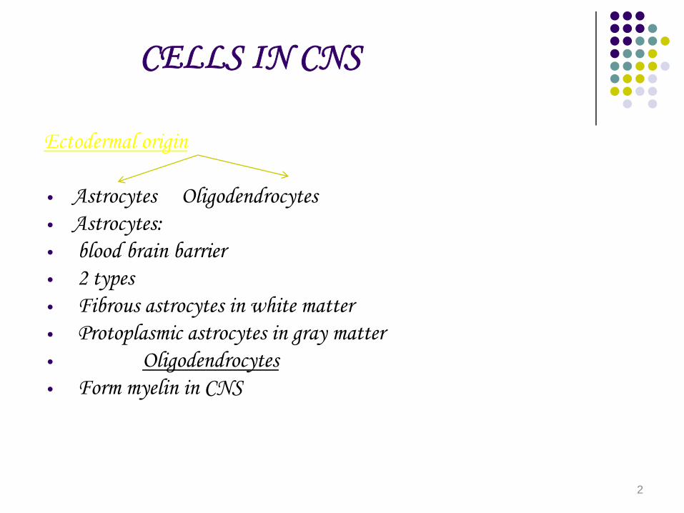
CELLS IN CNS
Ectodermal origin
• Astrocytes Oligodendrocytes• Astrocytes:• blood brain barrier• 2 types• Fibrous astrocytes in white matter• Protoplasmic astrocytes in gray matter• Oligodendrocytes• Form myelin in CNS
2
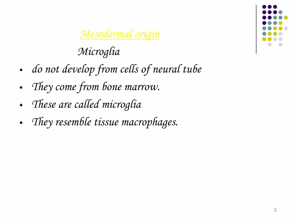
Mesodermal originMicroglia
• do not develop from cells of neural tube• They come from bone marrow.• These are called microglia• They resemble tissue macrophages.
3

NEURON
4
• Nerve has axon and dendrites and a cell body• Axon starts from axon hillock and ends in
terminal buttons which contains vesicles with neurotransmitters.
• Nodes of Ranvier: 1 micro metre constrictions, 1mm apart.
• Nerve surrounded by myelin sheath .• Myelin formed by schwann cells in PNS but in
CNS it is formed by Oligodendrogliocytes.• Transmisssion of transmitters by axoplasmic flow.
5

• Electric Potentials
• Non Propagative Propagative.• ALL OR NONE LAW: If the threshold is attained, no change
in action potential even if intensity of stimulus is increased.• Refractory Period
• Absolute Relative• From firing level to 1/3 from this pt. to start of • Repolarisation after depolarisation.
6
• Myelinated fibres conduct faster• SALTATORY CONDUCTION: Jumping from one node
to the other
• Depolarisation due to entry of Na ions• Repolarisation due to efflux of K ions• Action potentials starts in axon hillock due to increased
Na channels• Energy source is Na K ATP ase.
7
• In CNS neurons are surrounded by glial cells.
• 3 types of neuroglia• 1) Microglia - Scavenger cells• 2)Oligodendrocytes :- Forms myelin• 3)Astrocytes: 2 types, Forms blood brain
barrier• A) Fibrous• B) Protoplasmic
8
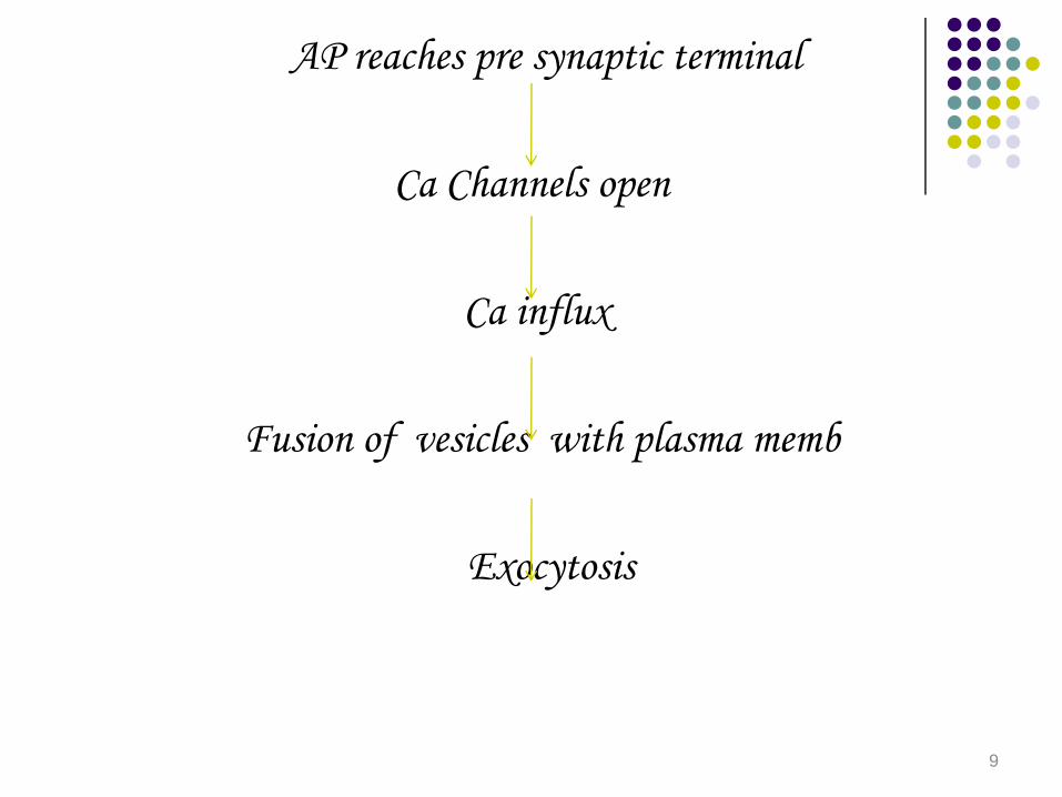
AP reaches pre synaptic terminal
Ca Channels open
Ca influx
Fusion of vesicles with plasma memb
Exocytosis
9
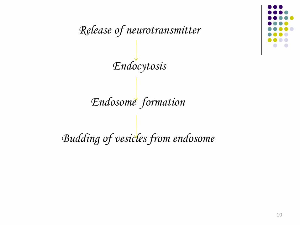
Release of neurotransmitter
Endocytosis
Endosome formation
Budding of vesicles from endosome
10
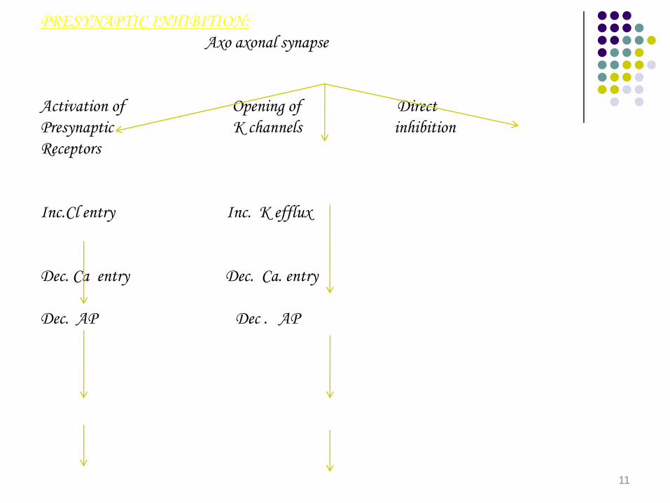
PRESYNAPTIC INHIBITION:Axo axonal synapse
Activation of Opening of DirectPresynaptic K channels inhibitionReceptors
Inc.Cl entry Inc. K efflux
Dec. Ca entry Dec. Ca. entry
Dec. AP Dec . AP
11

PRE SYNAPTIC FACILITATION:-Axo- axonal synapse
Release of serotonin
Inc. C AMP
Closes K channels
Prolongs AP
12

VENTRICLES
13

14
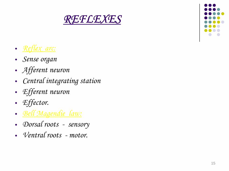
REFLEXES
• Reflex arc:• Sense organ• Afferent neuron• Central integrating station• Efferent neuron• Effector.• Bell Magendie law:• Dorsal roots - sensory• Ventral roots - motor.
15

REFLEXES
MonosynapticPolysynapticStretch ReflexStimulus : stretch of muscleSense organ : muscle spindle.Reflex :contraction of muscleEg: Knee jerk
Triceps jerk
16

REFLEX ARC
17

MUSCLE SPINDLE
• Intrafusal fibres• Extrafusal fibres• Intrafusal fibres-1) Nuclear chain fibre.• 2)Nuclear bag fibre.• Afferents - Ia• II• Efferent - Beta (both intrafusal & extrafusal)• - Gamma (only intrafusal)• Gamma efferent discharge-Jendrassik’s maneuver.
18

MUSCLE SPINDLE
19
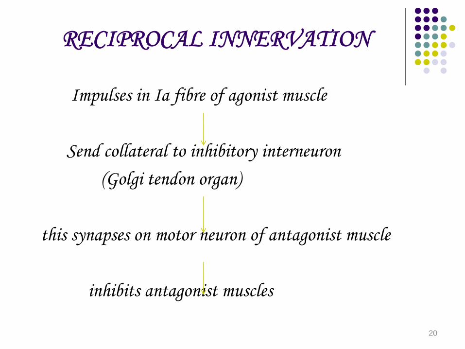
RECIPROCAL INNERVATION
Impulses in Ia fibre of agonist muscle
Send collateral to inhibitory interneuron(Golgi tendon organ)
this synapses on motor neuron of antagonist muscle
inhibits antagonist muscles
20

INVERSE STRETCH REFLEX
Also called as autogenic inhibitionIb afferent
Inhibitory interneuron (Golgi tendon organ)
Relaxation of muscle.This is due to overstretch of muscle.
21
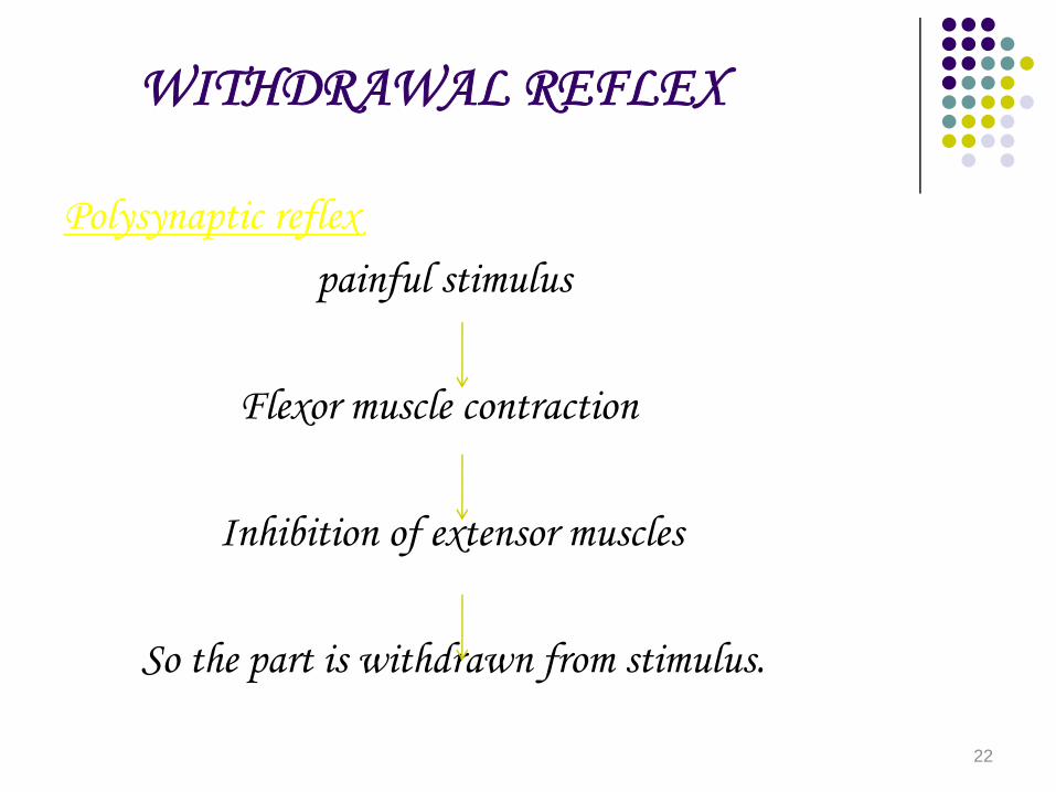
WITHDRAWAL REFLEX
Polysynaptic reflexpainful stimulus
Flexor muscle contraction
Inhibition of extensor muscles
So the part is withdrawn from stimulus.
22
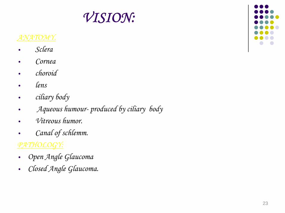
VISION:ANATOMY.• Sclera• Cornea• choroid• lens• ciliary body• Aqueous humour- produced by ciliary body• Vitreous humor.• Canal of schlemm.PATHOLOGY:• Open Angle Glaucoma• Closed Angle Glaucoma.
23
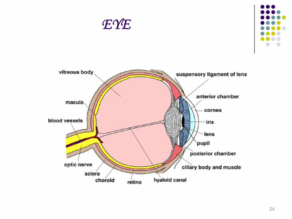
EYE
24
RETINA
• Retina has rods & cones and 4 types of neurons• 1) bipolar cells• 2)ganglion cells• 3)horizontal cells• 4)Amacrine cells
• Horizontal cells -connect rods & cones• Amacrine cells - connect ganglion cells.
25

LAYERS:• Layer of pigment epithelium• Layer of Rods & cones• Outer nuclear layer• Outer plexiform layer• Inner nuclear layer• Inner plexiform layer• Ganglion cell layer• Optic nerve fibres
26

PHOTO RECEPTOR MECHANISM
• Action potentials are generated only on ganglion cells• Local potentials in the rest of the cells• Normally in darkness:• Na K ATPase in inner segment• Pumps 3Na+ ions into outer segment
• Through open Na+ channels in outer segment
• Release of neurotransmitter.
27

When light falls on rods.Na+ channel in outer segment closes
Hyperpolarisation
No release of neurotransmitter
Generates action potentials in ganglion cells.
Activates cGMP phosphodiesterase
CGMP - 5’ GMPClosure of Na+ channels.
28
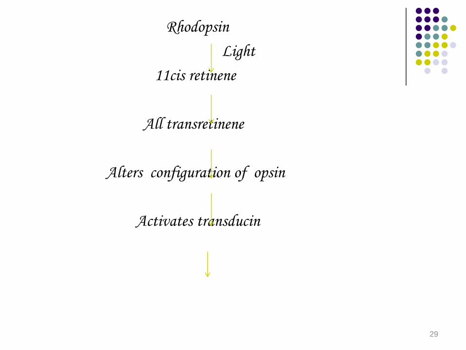
RhodopsinLight
11cis retinene
All transretinene
Alters configuration of opsin
Activates transducin
29
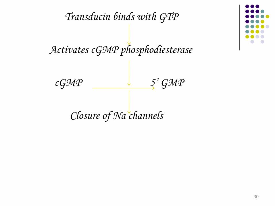
Transducin binds with GTP
Activates cGMP phosphodiesterase
cGMP 5’ GMP
Closure of Na channels
30
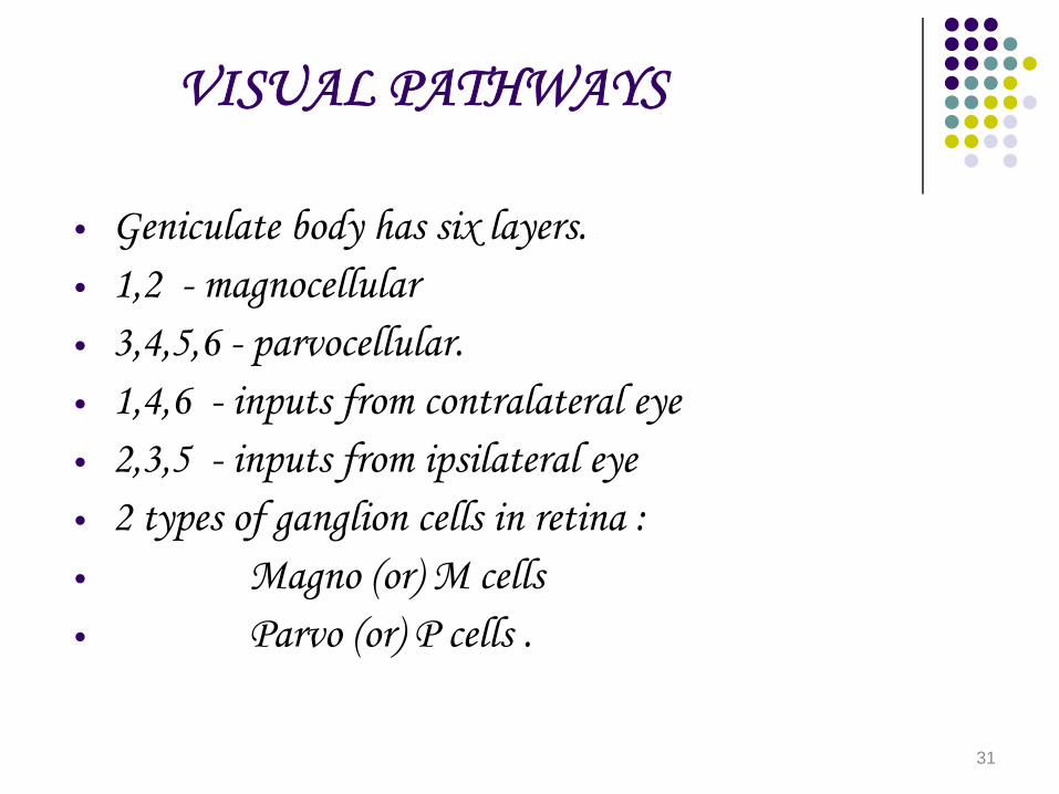
VISUAL PATHWAYS
• Geniculate body has six layers.• 1,2 - magnocellular• 3,4,5,6 - parvocellular.• 1,4,6 - inputs from contralateral eye• 2,3,5 - inputs from ipsilateral eye• 2 types of ganglion cells in retina :• Magno (or) M cells• Parvo (or) P cells .
31

M
Magnocellular laminas
SuperficialLayer 4c
Function:-Movement,Location,Spatial Organisation.
32
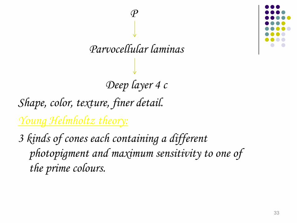
P
Parvocellular laminas
Deep layer 4 cShape, color, texture, finer detail.Young Helmholtz theory:3 kinds of cones each containing a different
photopigment and maximum sensitivity to one of the prime colours.
33
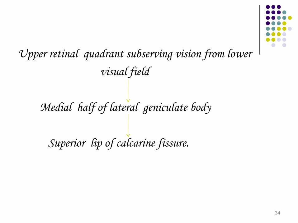
Upper retinal quadrant subserving vision from lowervisual field
Medial half of lateral geniculate body
Superior lip of calcarine fissure.
34

Lower retinal quadrant subserving vision fromupper visual field
Lateral half of lateral geniculate body
Inferior lip of calcarine fissure.
Fibres from lateral geniculate body that subserve macular vision
Posterior part of calcarine fissure.35
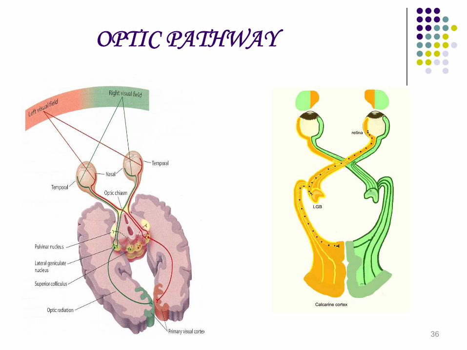
OPTIC PATHWAY
36
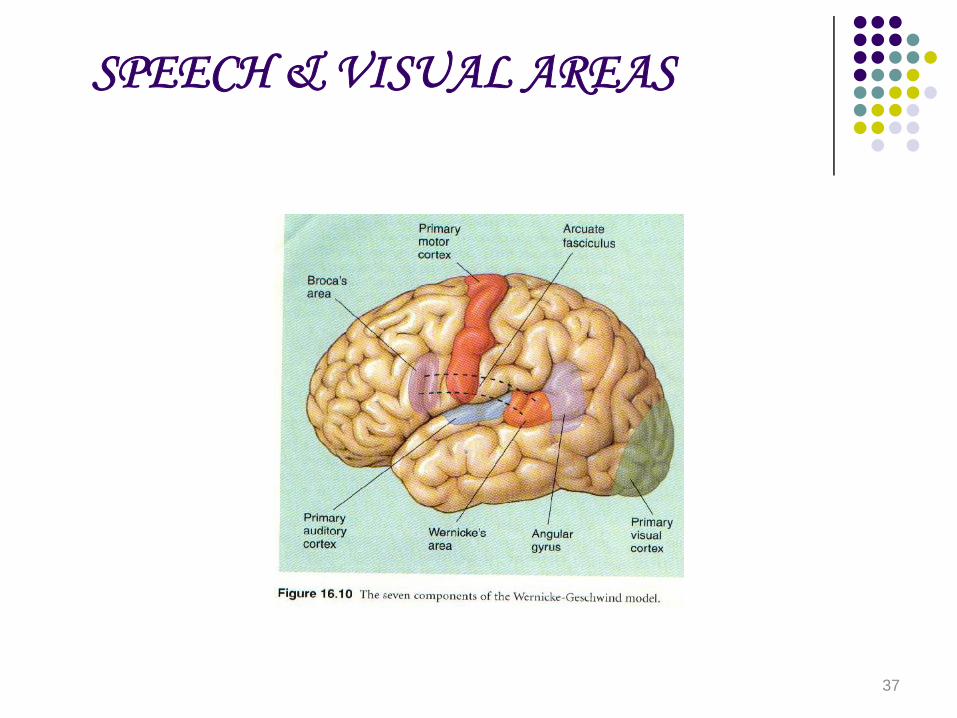
SPEECH & VISUAL AREAS
37
HEARING AND EQUILIBRIUM• Semicircular canals - Rotational acceleration.• Utricule – linear acceleration in • horizontal direction• Saccule – linear acceleration in vertical direction.• ANATOMY:• EXTERNAL & MIDDLE EAR.• External auditory meatus• Tympanic membrane• Eustachian tube• malleus, incus, stapes• foot plate of stapes-oval window.
38
INNER EAR (LABYRINTH)• Outer bony labyrinth• Inner Membranous labyrinth.• Bony labyrinth- perilymph• Membranous labyrinth-endolymph.• Cochlea• Basilar & Reissner’s membrane divide it into 3
chambers• Upper scala vestibuli- perilymph• lower scala tympani – perilymph• communicate through helicotrema• Scala vestibuli- ends in oval window• Scala tympani- ends in round window.
39
ORGAN OF CORTI
• extends from apex to base of cochlea.• Tunnel of corti formed by rods of corti.• Outer hair cells- pierce tectorial membrane• inner hair cells do not.• afferent neurons from inner hair cells mainly• efferent neurons reach outer hair cells mainly.
40
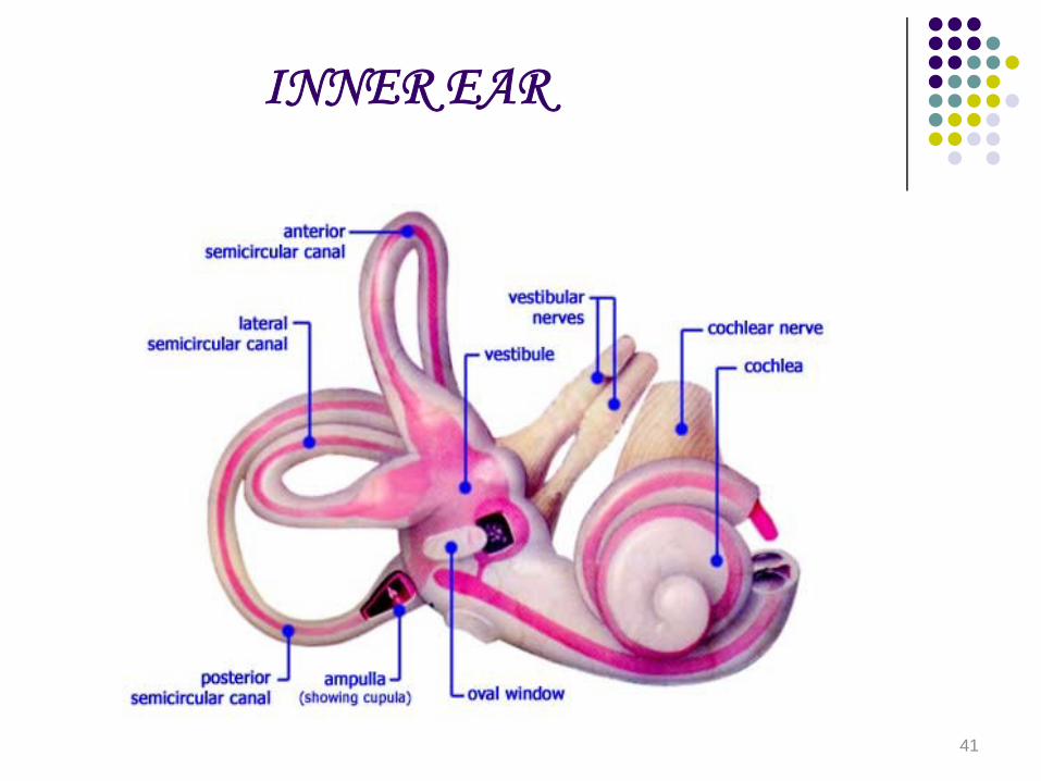
INNER EAR
41
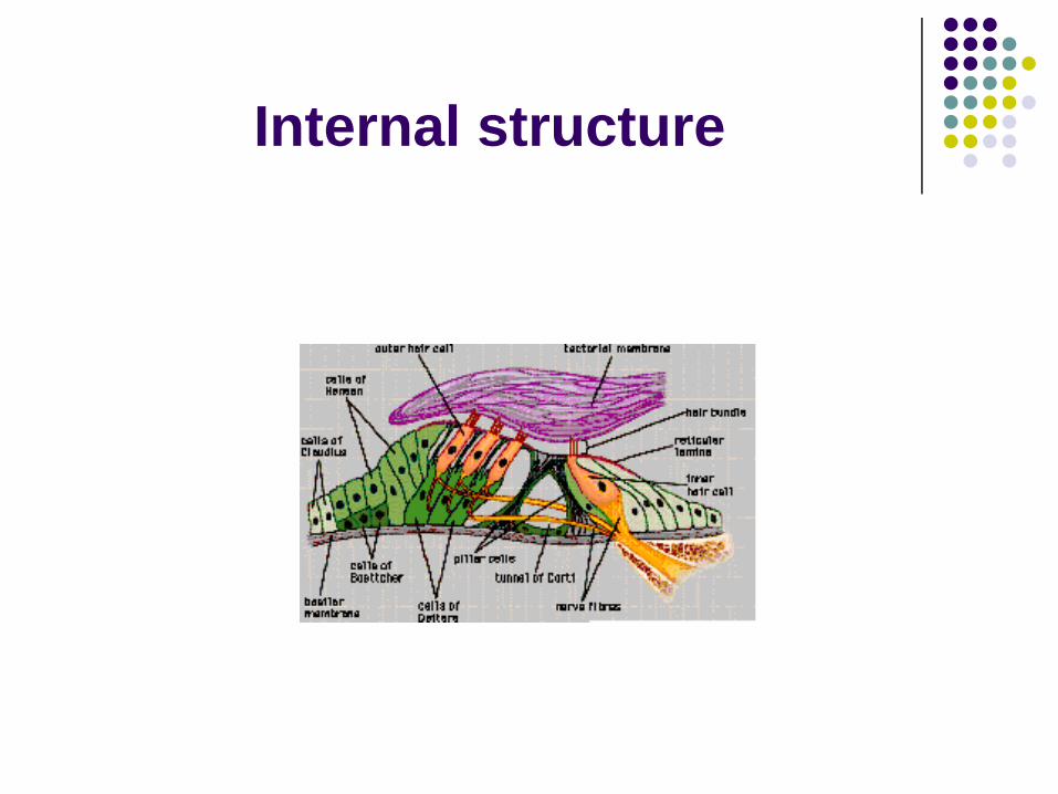
Internal structure

ORGAN OF CORTI
43
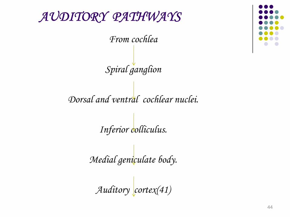
AUDITORY PATHWAYS
From cochlea
Spiral ganglion
Dorsal and ventral cochlear nuclei.
Inferior colliculus.
Medial geniculate body.
Auditory cortex(41)
44

SEMICIRCULAR CANALS,UTRICLE & SACCULE
• Semicircular canals contain crista ampullaris located in ampulla.
• Crista cmpullaris has sustentacular cells & hair cells closed by cupula.
• Utricle & saccule contain macula which has hair cells & sustentacular cells surrounded by otolithic membrane embedded with calcium carbonate crystals(otolith).
45
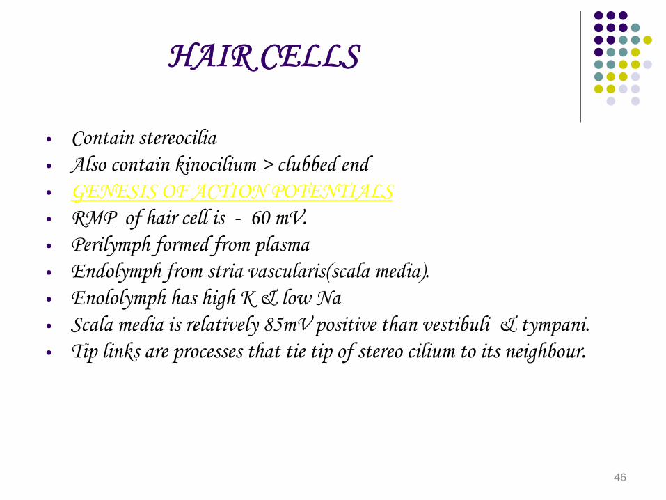
HAIR CELLS
• Contain stereocilia• Also contain kinocilium > clubbed end• GENESIS OF ACTION POTENTIALS• RMP of hair cell is - 60 mV.• Perilymph formed from plasma• Endolymph from stria vascularis(scala media).• Enololymph has high K & low Na• Scala media is relatively 85mV positive than vestibuli & tympani.• Tip links are processes that tie tip of stereo cilium to its neighbour.
46

Shorter stereocilia pushed to higher
Cation channel in tip links open
K & Ca entry
Depolarization
AP in neurons
K+ enters into sustentacular cells through tight junction.
Reaches strio vascularis
Secreted into endolymph.
47
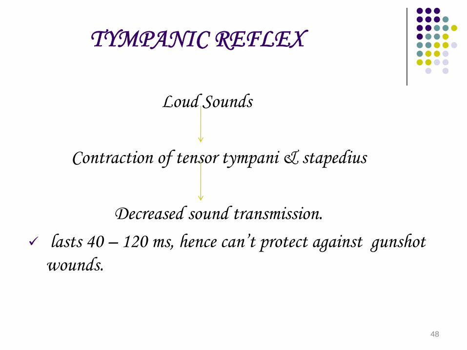
TYMPANIC REFLEX
Loud Sounds
Contraction of tensor tympani & stapedius
Decreased sound transmission.lasts 40 – 120 ms, hence can’t protect against gunshot wounds.
48

SMELL AND TASTE
• SMELL.• Olfactony receptors are located in olfactory mucous
membrane.• Axons of the receptors contact the dendrites of mitral
cells and tufted cells to form synapses called olfactory glomeruli.
• Granule cells have no axons and make synapses with dendrites of mitral and tufted cells.
49

TASTE
• 4 types buds ; made up of 4 types of cells• Type 1 & 2 cells : sustentacular cells• Type 3 cells : gustatory receptor cells• Type 3 cells have a microvillus which project into the taste
pore.• Fungiform papillae : tip of tongue• Filiform papillae : dorsum of tongue• Vallate papillae :‘V’ shaped on back of tongue.• Fungiform papillae : 5 taste buds• Vallate papillae : 100 taste buds.
50

TASTE PATHWAYS
Taste buds
Fibres via facial, glossopharyngeal, and vagus nerves.
Nucleus tractus solitarius
Ventral posteromedial nucleus of thalamus
Post – central gyrus.
51

TASTE RECEPTORS.
• Satty > ENaC• Sour > ENaC, HCN• Bitter > T2R family• Sweet > TIR 3 , gustducin.
52
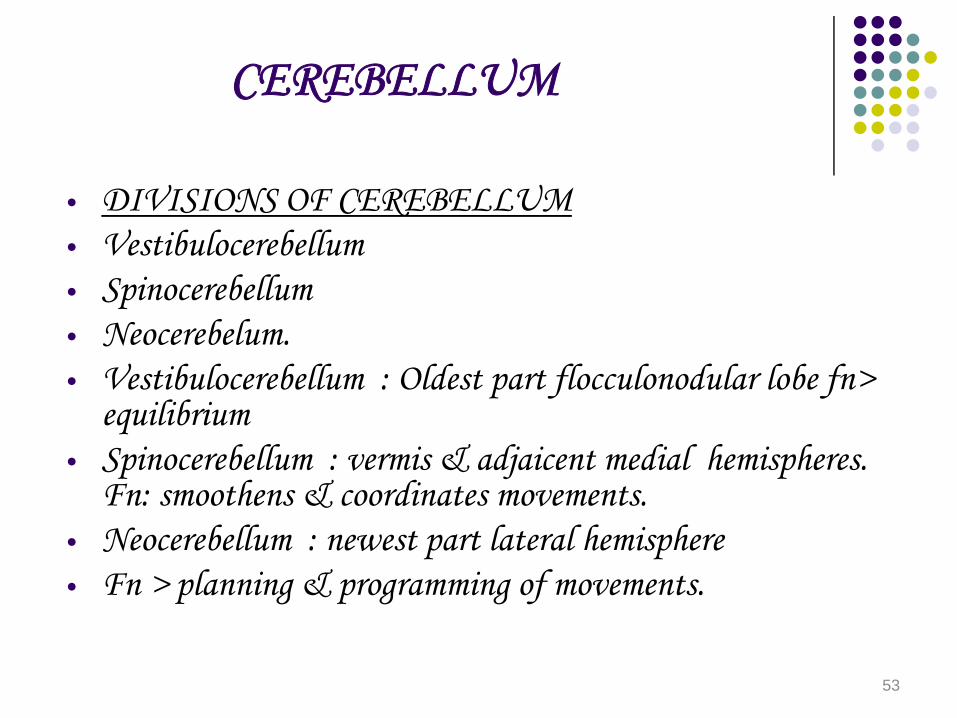
CEREBELLUM
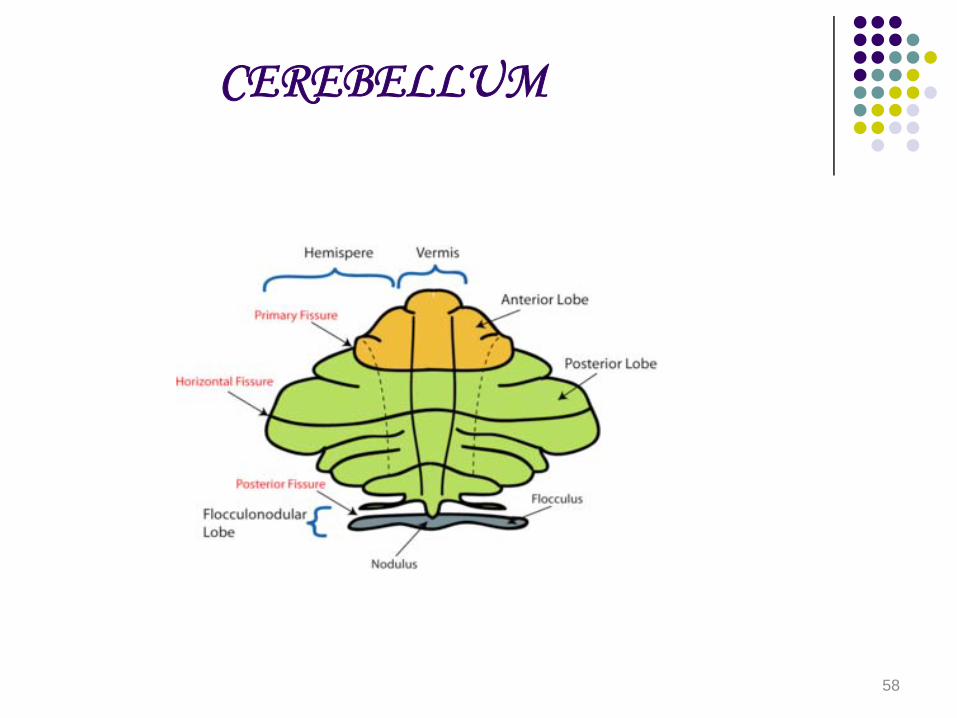
• DIVISIONS OF CEREBELLUM• Vestibulocerebellum• Spinocerebellum• Neocerebelum.• Vestibulocerebellum : Oldest part flocculonodular lobe fn>
equilibrium• Spinocerebellum : vermis & adjaicent medial hemispheres.
Fn: smoothens & coordinates movements.• Neocerebellum : newest part lateral hemisphere• Fn > planning & programming of movements.
53
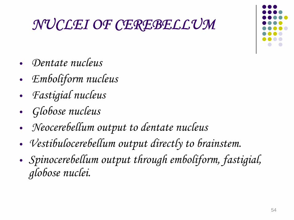
NUCLEI OF CEREBELLUM
• Dentate nucleus• Emboliform nucleus• Fastigial nucleus• Globose nucleus• Neocerebellum output to dentate nucleus• Vestibulocerebellum output directly to brainstem.• Spinocerebellum output through emboliform, fastigial,
globose nuclei.
54
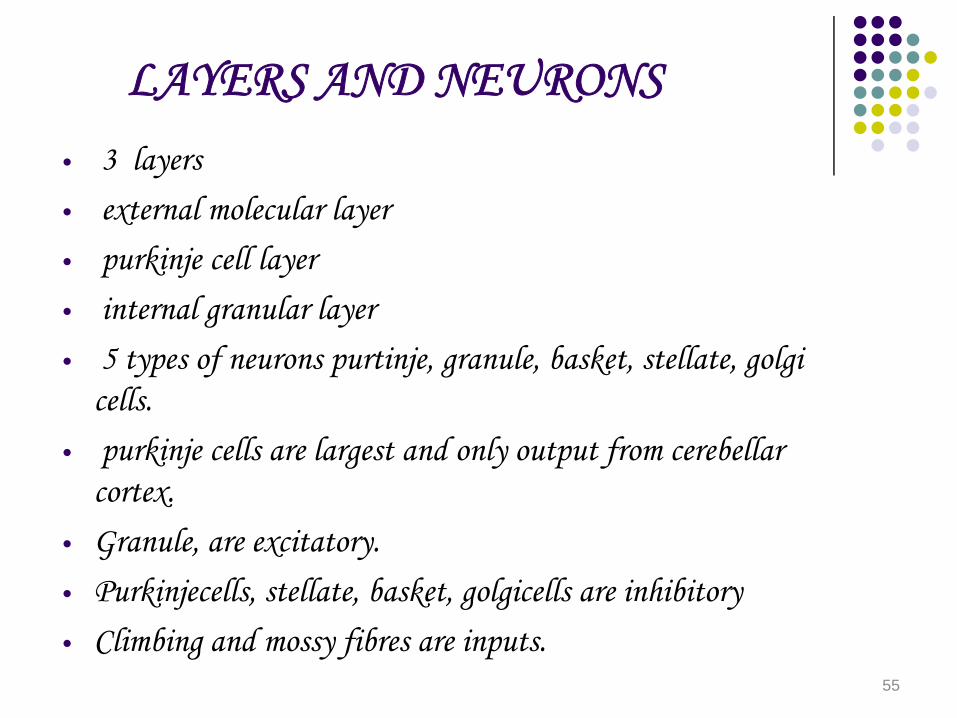
LAYERS AND NEURONS• 3 layers• external molecular layer• purkinje cell layer• internal granular layer• 5 types of neurons purtinje, granule, basket, stellate, golgi
cells.• purkinje cells are largest and only output from cerebellar
cortex.• Granule, are excitatory.• Purkinjecells, stellate, basket, golgicells are inhibitory• Climbing and mossy fibres are inputs.
55

FUNDAMENTAL CIRCUIT
• Climbing fiber has strong excitatory effect on purkinje cells
• Mossy fibres have weak excitatory effect on purkinje cells.
• Granule cells excite stellate and basket cells which inturn inhibit purkinje cells
• This is called feed forward inhibition• Purkinje cells inhibit deep cerebellar nuclei.• GABA secreted by stellate, basket, golgi, purkinje cells• Glutamate by granule cells
56

CEREBELLUM
57
CEREBELLUM
58
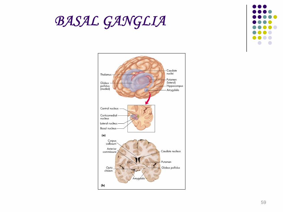
BASAL GANGLIA
59
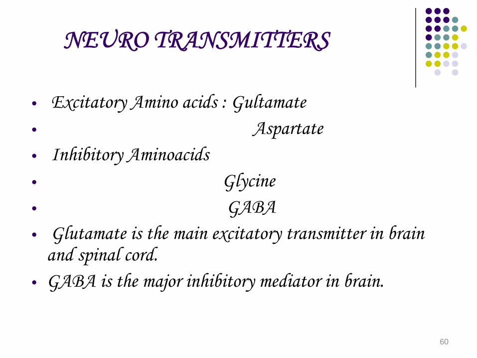
NEURO TRANSMITTERS
• Excitatory Amino acids : Gultamate • Aspartate• Inhibitory Aminoacids • Glycine• GABA• Glutamate is the main excitatory transmitter in brain
and spinal cord.• GABA is the major inhibitory mediator in brain.
60

HYPOTHALAMUS
• Anterior hypothalamus controls response to heat.• Posterior hypothalamus controls response to cold.• Lateral hypothalamus feeding centre.• Ventromedial nucleus of hypothalamus satiety centre.• Anterior hypothalamus : Osmoreceptor• Thirst• Sexual behavior• Circadian rhythm• Pituitary hormone regulation• defensive behavior.
61
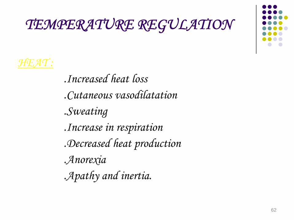
TEMPERATURE REGULATION
HEAT :.Increased heat loss.Cutaneous vasodilatation.Sweating.Increase in respiration.Decreased heat production.Anorexia.Apathy and inertia.
62

COLD
• Decreased heat loss• cutaneous vasoconstriction• Pilo erection• curling up• Increased heat production• shivering• Hunger• Increased voluntary activity• Increased secretion of norepinephrine/ epinephrine.
63
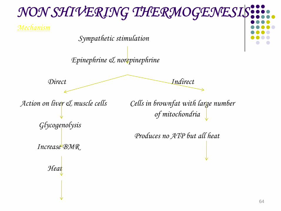
NON SHIVERING THERMOGENESISMechanism
Sympathetic stimulation
Epinephrine & norepinephrine
Direct Indirect
Action on liver & muscle cells Cells in brownfat with large numberof mitochondria
Glycogenolysis Produces no ATP but all heat
Increase BMR
Heat
64

CEREBRO SPINAL FLUID• CSF formation by choroid plexus around blood vessel and venticular
walls• Volume is 150 ml.• Rate of CSF formation is 550ml/day• Rate of CSF turnover is 3 times/day.• pH of CSF IS 7.34• CSF presssure is 50-180 mm Hg.• Ion with maximum CSF/plasma ratio is Magnesium.• Minimum CSF/plasma ratio is cholesterol and protein.• Equal CSF/plasma ratio is with osmolality• All negative ions are more in CSF.
65

CSF CONSTITUENTS
• Appearance clear and colourless.• proteins : 20 -40 mg/dl.• Glucose : 40-70 mg/dl.• Chlorides : 720-750 mg/dl.• CSF Sugar is about 2/3 of plasma sugar.
66

EMOTION
• Sensory aspect is in limbic system• expressive emotions develop in hypothalamus.• sensory aspect characterised by 3 parts• cognition is awareness of sensation and its cause.• Affect means development of feeling.• Conation is the desire to take action.• Expresion is motor part and consists of Autonomic
system.
67

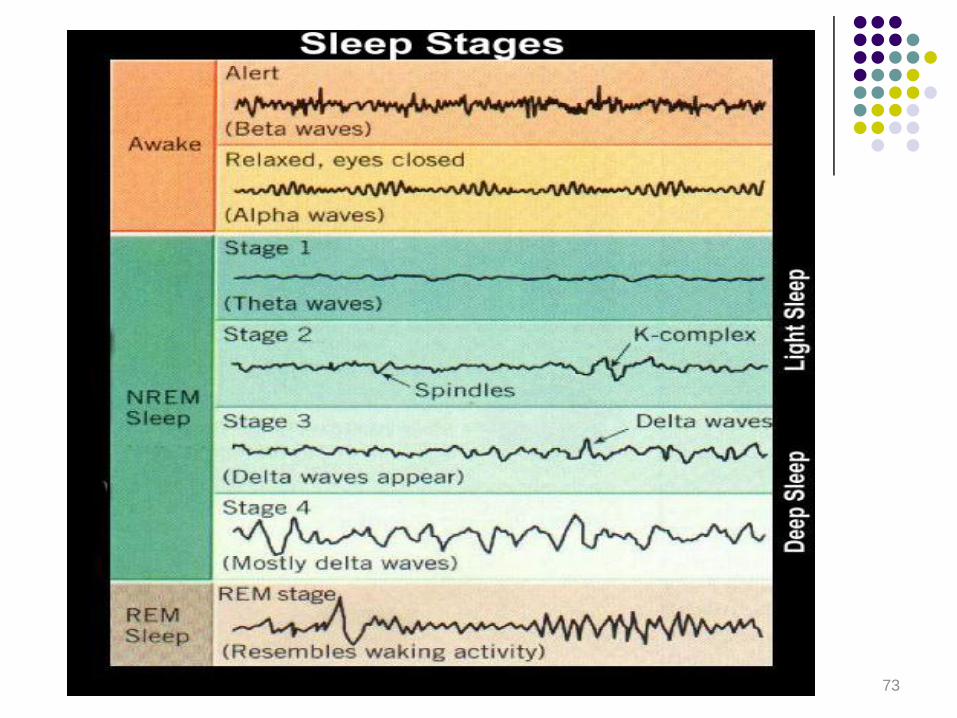
EEG.
• EEG consists of 4 waves.• These are alpha waves, beta waves, theta waves,
olelta waves.• Beta waves:• parietal & frontal region.• Frequency > 14 Hz.• Low amplitude.• seen in awake patients, at rest with eyes open,
children, drowsiness.
68
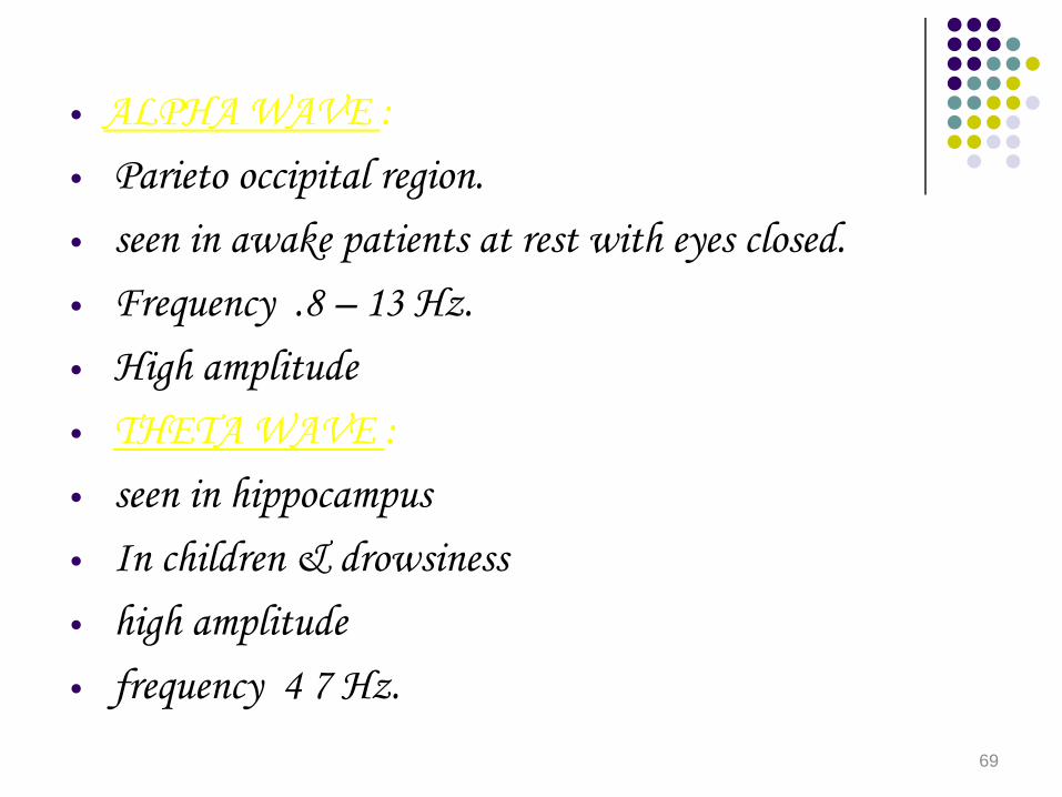
• ALPHA WAVE :• Parieto occipital region.• seen in awake patients at rest with eyes closed.• Frequency .8 – 13 Hz.• High amplitude• THETA WAVE :• seen in hippocampus• In children & drowsiness• high amplitude• frequency 4 7 Hz.
69

• DELTA WAVE : • seen in infant and n REM sleep• Large amplitude• Frequency 3 – 5 Hz.
70
NORMAL EEG
• Alpha wave reflects synchronized brain activity.• Alpha wave wax & wane & are suppressed completely
with eye opening or mental activity.• Alpha block is when eyes are opened alpha rhythm is
replaced by fast irregular low voltage activity.• Desynchronization is when alpha pattern is replaced by
another wave pattern during sensory stimulation or mental concentration. Also called alerting response.
71
SLEEP PATTERN
• 2 types of sleep.• n REM Sleep or slow wave sleep or orthodox sleep.• constitutes 70 – 80% of total sleep.• REM Sleep or paradoxical sleep.• constitutes 20-30% of total sleep.
72
73

STAGES OF nREM SLEEP
• Stage I : consists of theta waves.• Stage II : consists of sleep spindles and • K complexes.• Stage III : consists of delta waves which are• first to appear & then K-complexes.• Stage IV : consists of predominant delta • waves.
74
REM SLEEP
• Arousal is difficult.• Mixed frequency, low amplitude waves.• predominant beta activity. • Dreaming is seen.• Tone of neck is lost.• Penile tumescence.• Night mares.• Narcolepsy.
75


![Opposing Activities of LIT-1/NLK and DAF-6/Patched ... MANUSCRIPTS... · Schwann glial cell [3]. In the olfactory epithelium, sensory neurons are ensheathed by glia-like sustentacular](https://static.fdocuments.net/doc/165x107/609ddecb713c507e76044afb/opposing-activities-of-lit-1nlk-and-daf-6patched-manuscripts-schwann.jpg)