Nephrology Grand Rounds - NYU Langone Health Mansi.pdfLight-chain (AL) Amyloidosis Most-common type...
26
Nephrology Grand Rounds Mansi Mehta February 9 th , 2016
Transcript of Nephrology Grand Rounds - NYU Langone Health Mansi.pdfLight-chain (AL) Amyloidosis Most-common type...

Nephrology Grand Rounds Mansi Mehta
February 9th, 2016

Clinical Case CC: 75yo M from Ecuador presenting with progressive LE edema and AKI
HPI: Patient was recently diagnosed w/ Smoldering Myeloma after a hospitalization for syncope revealed uncontrolled HTN, proteinuria and labs concerning for MM - He was being followed in hematology clinic and then presented to the ED 2 months after diagnosis with abdominal distension and new LE edema

PMH: BPH and Hyperlipidemia
PSH: none
SH: no history Tobacco/EtOH/Illicit drug abuse; visiting from Ecuador
FH: father with heart disease; no h/o of malignancy or renal disease
Meds: tamsulosin and pravastatin
Allergies: KNDA
ROS: +LE edema, DOE, increased abdominal distention, decreased appetite

Physical Exam VS: Afebrile, BP: 115/75 P: 16 R: 64 O2sat: 96% on 2L NC
Gen: NAD, elderly Hispanic M, chronically-ill appearing
HEENT: no lymphadenopathy
CVS: +s1s2, RRR, +systolic murmur along R sternal border. No JVD
Resp: decreased BS at bases
Abdomen: moderately distended, NT, +ascites
Extremities: pitting edema of B/L LE up to mid-thighs

Labs
WBC
8.4 Sodium 142 Total Protein
5.8
H/H 15/45 Potassium 4.3 Albumin 2.0
Platelets 276 Chloride 110 AST 71
Bicab 23 ALT 33
Creatinine
41 TBili 3.1
Glucose 2.0 DBili 2.0
UA: 3+ protein, 2-5WBcs, 5-10RBCs UPr/Cr: 1189g/91g = 13g

HbA1C 6.1 HIV Negative
HBsAg Negative ANA Negative
HBsAb Negative C3 94 (75-140)
HCV Ab Negative C4 32 (10-34)
Lipid Panel
Total Cholesterol: 209 LDL: 139 HDL: 51 TG: 97

EKG: Normal Sinus Rhythm
Chest X-Ray: B/L pleural effusions and atelectasis.
ECHO: EF: 65%. Moderate concentric LV hypertrophy. Mitral inflow pattern c/w impaired relaxation and small pericardial effusion – may be consistent with an infiltrative process
Abdominal US: Moderate ascites. R kidney: 11.8cm L kidney: 11.5cm Increased bilateral cortical echogenicity. No hydronephrosis or calculi.

Work-Up SPEP: hypoalbuminemia w/ peak in beta-gamma region
UPEP: albumin and other serum proteins with 2 peaks in gamma region. Pattern compatible with monoclonal gammopathy.
UIFE: weak Bence Jones protein Lambda type
SIFE: Two IgA lambda bands identified IgA: 627 IgG: 450 IgM: 68
Serum Free Ig Light chains Kappa: 26.2 Lambda: 595 K/L: .04
B2 microglobulin: 4.3 (.8-2.2)

Work-up Cardiac MRI: Increase in LV and RV wall thickness. On
delayed contrast enhanced images there is diffuse rapid nulling of the entire myocardium as is typically seen in amyloidosis
Fat Pad biopsy: negative for amyloid

Differential Diagnosis Primary Amyloidosis
Light chain deposition disease
Myeloma Cast Nephropathy

Renal Biopsy

Primary Amyloidosis

Amyloidosis A family of disorders defined by the extracellular
deposition of protein fibrils with a characteristic B-pleated sheet conformation
To date, about 30 different amyloidogenic proteins have been identified
Described as “chameleon” proteins due to their ability to acquire more than one conformation - classified based on precursor proteins

Pathology Abnormal folding of an extracellular protein that is
normally soluble
In AL amyloid it is the result of either a proteolytic event or an AA sequence that makes a light chain thermodynamically unstable and prone to self aggregation
Aggregates form protofilaments that associate into amyloid fibrils
Serum amyloid P protein (SAP) interacts with the amyloid fibrils promoting fibril formation and aggreagation

Contiguous B-sheet polypetitde chains wind around one another to form an amyloid fibril with a distinct diameter of 7.5-10nm visible on EM
Ultrastructure of the fibril allows the intercalation of Congo red dye

Types of Systemic Amyloidosis

Diagnosis Abdominal fat biopsy
Sensitivity of Congo red staining of abdominal fat is approximately 80-90% and 65-75% in AL and AA amyloidosis
Renal Biopsy Likelihood of a missed
diagnosis is lower with a kidney biopsy than with biopsies of other tissues because amyloid fibrils are visible with EM

Light-chain (AL) Amyloidosis
Most-common type of systemic amyloidosis and most severe form affecting the kidney
Amyloid protein is an Ig light chain that is produced by a clonal population of plasma cells in the bone marrow
Clonal plasma cells express light chains of the lamba isotype more frequently than the kappa, with a ratio of 3:1
Incidence is 9 million per year with 10-15% occurring in association with MM
Prognosis of untreated AL amyloid is survival time of12 months and with treatment now exceeds 3 years.

Clinical Presentations Nephrotic Syndome
Restrictive cardiomyopathy
Hepatomegaly
Soft tissue involvement

Treatment High Dose Melphalan + ASCT
Dexamethasone + Melphalan
CyBorD

Randomized comparison of high dose melphalan + autologous hematopoietic stem cell transplant with standard dose melphalan + high dose dexamethasone
Multicenter, randomized, controlled trial including 100 patients
Primary outcome: overall survival

Results High-dose melphalan +
ASCT was not superior to the outcome with standard dose melphalan + dexamethasone
Treatment related mortality in the HDM group (24%) was higher (13%)
Jaccard A et al; NEJM 2007

Largest study of CyBor D for the initial treatment of AL amyloidosis
Series of 230 patients from 2 European referral centers
Overall hematologic response rate was 60%; cardiac at 17% and renal response rate at 25%
After a medium follow-up of 25monhs, estimate overall survival at 3years was 55%
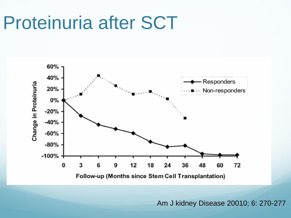
Proteinuria after SCT
Am J kidney Disease 20010; 6: 270-277

Survival based on renal response
Am J kidney Disease 20010; 6: 270-277

Transplant in AL Amyloidoisis Patient survival and graft survival analyzed in 21 renal
transplantation patients – 3 living and 18 deceased donor grafts
Medium estimated patient survival was 89 months
One and Five year survival rates were 95.2% and 71.4%
There were no graft failures as a result of recurrent amyloid
At five years there was scintigraphy evidence of recurrent amyloid in 6 functioning renal allografts accompanied in 5 patients with proteinuria
Pinney et Al; J of Clinical Oncology 2011