
Neoplastic diseases: Marek's disease, avian leukosis and - OIE
21
Rev. sci. tech. Off. int. cpiz., 2000,19 (2), 544-564 Neoplastic diseases: Marek's disease, avian leukosis and reticuloendotheliosis L.N. Payne & K. Venugopal Institute for Animal Health, Compton, Newbury, Berkshire RGZO 7NN, United Kingdom Summary The commercially important neoplastic diseases of poultry are Marek's disease, which is caused by a herpesvirus, and the avian leukoses and reticuloendotheliosis, which are caused by retroviruses. These diseases are responsible for economic loss due to both mortality and depressed performance. Marek's disease virus (MDV) and avian leukosis viruses (ALVs) are prevalent throughout the world, and new strains which arise in particular locations may spread across borders, thereby undermining national disease control measures. Reticuloendotheliosis virus (REV) is also present in many countries. Marek's disease virus is transmitted horizontally only, and international spread in hatching eggs and day-old chicks can be prevented by appropriate hygiene precautions. Transmission of ALV and REV occurs both horizontally and vertically (through the egg), and measures to prevent international spread are more demanding. Marek's disease is controlled by vaccination, whilst avian leukosis is controlled by virus eradication programmes, mainly at the primary breeding level. Similar virus control measures can be applied for reticuloendotheliosis if necessary. No strong evidence exists to suggest that these avian tumour viruses constitute a danger to public health. Keywords Avian leukosis - Control - Diagnosis - Epidemiology - International trade - Marek's disease - Poultry - Public health - Reticuloendotheliosis - Surveillance - Tumour viruses -Vaccination. Introduction Neoplastic diseases of poultry fall into two broad classes, namely: those with an infectious aetiology and those which are non-infectious. Those of the former category are of the greater economic importance because the viruses that cause these diseases are widely prevalent in commercial stock and the mesenchymal neoplasms that they cause affect relatively young birds. These infections and diseases can be enzootic and epizootic. Neoplasms of a non-infectious aetiology occur mostly in birds older than the usual lifespan of commercial birds. Such tumours are often of epithelial cell origin, with ovarian tumours being common in hens over two years of age. Even so, tumours of the magnum region of the oviduct, adenomas and adenocarcinomas, of non-infectious aetiology, can occur in commercial laying hens at the end of the first laying season. However, the non-infectious neoplasms are generally sporadic and not of great economic significance. Three main classes of virus cause neoplasms in poultry, as follows: a) Marek's disease virus (MDV), a herpesvirus b) avian leukosis virus (ALV), a retrovirus c) reticuloendotheliosis virus (REV), also a retrovirus. Another retrovirus, lymphoproliferative disease virus (4), has caused significant losses from lymphomas in turkeys in the United Kingdom and Israel, but now appears to be rare, and is not considered further in this paper. Domestic chickens are the poultry species most commonly affected by neoplasms, which may be caused by MDV, ALV or
Transcript of Neoplastic diseases: Marek's disease, avian leukosis and - OIE

Rev. sci. tech. Off. int. cpiz., 2000,19 (2), 544-564
Neoplastic diseases: Marek's disease, avian leukosis and reticuloendotheliosis
L.N. Payne & K. Venugopal
Institute for Animal Health, Compton, Newbury, Berkshire RGZO 7NN, United Kingdom
Summary The commerc ia l ly important neoplast ic d iseases of poul t ry are Marek's d isease, w h i c h is caused by a herpesvi rus, and the avian leukoses and ret icu loendothel ios is , w h i c h are caused by retrov i ruses. These diseases are responsible fo r economic loss due to both mortal i ty and depressed performance. Marek 's disease virus (MDV) and avian leukosis v i ruses (ALVs) are prevalent th roughout the w o r l d , and n e w strains w h i c h arise in par t icu lar locat ions may spread across borders, thereby undermin ing nat ional disease cont ro l measures. Ret iculoendothel ios is v i rus (REV) is also present in many countr ies. Marek's disease virus is t ransmi t ted horizontal ly only, and internat ional spread in hatching eggs and day-old ch icks can be prevented by appropr iate hygiene precautions. Transmission of ALV and REV occurs both horizontal ly and ver t ica l ly ( through the egg), and measures to prevent internat ional spread are more demanding. Marek's disease is contro l led by vacc ina t ion , wh i l s t avian leukosis is contro l led by virus erad icat ion programmes, mainly at the pr imary breeding level . Similar virus contro l measures can be appl ied for ret icu loendothel ios is if necessary. No strong ev idence exists to suggest tha t these avian tumour v i ruses const i tu te a danger to publ ic hea l th .
Keywords Avian leukosis - Control - Diagnosis - Epidemiology - International trade - Marek's disease - Poultry - Public health - Reticuloendotheliosis - Surveillance - Tumour viruses -Vaccinat ion.
Introduction Neoplastic diseases of poultry fall into two broad classes, namely: those with an infectious aetiology and those which are non-infectious. Those of the former category are of the greater economic importance because the viruses that cause these diseases are widely prevalent in commercial stock and the mesenchymal neoplasms that they cause affect relatively young birds. These infections and diseases can be enzootic and epizootic. Neoplasms of a non-infectious aetiology occur mostly in birds older than the usual lifespan of commercial birds. Such tumours are often of epithelial cell origin, with ovarian tumours being common in hens over two years of age. Even so, tumours of the magnum region of the oviduct, adenomas and adenocarcinomas, of non-infectious aetiology, can occur in commercial laying hens at the end of the first
laying season. However, the non-infectious neoplasms are generally sporadic and not of great economic significance.
Three main classes of virus cause neoplasms in poultry, as follows:
a) Marek's disease virus (MDV), a herpesvirus b) avian leukosis virus (ALV), a retrovirus c) reticuloendotheliosis virus (REV), also a retrovirus.
Another retrovirus, lymphoproliferative disease virus (4), has caused significant losses from lymphomas in turkeys in the United Kingdom and Israel, but now appears to be rare, and is not considered further in this paper.
Domestic chickens are the poultry species most commonly affected by neoplasms, which may be caused by MDV, ALV or
Rev. sci. tech. Off. int. Epiz., 19 (2| 545
REV. Turkeys and quail can suffer neoplasms caused by MDV and REV. Reticuloendotheliosis virus also causes neoplasms in geese and Muscovy ducks, and in game birds (pheasants and partridges). Ducks are not commonly affected by neoplasms, although hepatocellular carcinomas have been reported in the People's Republic of China, where mycotoxins in feed have been regarded as a likely aetiological factor, possibly with the involvement of duck hepatitis B virus, a member of the family Hepadnaviridae.
Economic importance Neoplastic diseases, and the viruses that cause them, are important in poultry for several reasons. The presence of the vims or the neoplasm causes economic loss from mortality and depressed performance. For Marek's disease (MD), additional costs arise from the development, production and use of vaccines for disease control, and for avian leukosis, from the implementation of virus eradication programmes, particularly by primary breeding companies. The viruses are prevalent throughout the world, but new strains arise periodically in particular locations (especially strains of MDV but also of ALV). If these spread between countries, national disease control measures can be undermined.
Before the introduction of vaccination of commercial flocks in 1971, MD was a major global disease of chickens. Vaccination dramatically reduced losses, but the disease remains one of significant economic importance, particularly because of the periodic appearance of new strains of MDV against which existing vaccines provide suboptimal protection. This has required the continued development of new vaccines and vaccination strategies (7). In 1984, the total world-wide economic loss caused by MD, including the cost of vaccination, was estimated at US$943 million (30). The International Animal Health Code of the Office International des Epizooties (OIE) places MD on List B, comprised of those diseases which have socio-economic and/or public health importance within countries and which are significant to international trade of animals and animal products (24).
Avian leukosis viruses are widespread but until recent years have not caused serious mortality nor been considered to be of major economic importance. Avian leukosis is placed on List C of the Food and Agriculture Organization Animal Health Yearbook 1995 (13) , designating diseases of socio-economic and/or public health importance at the local level. However, the world-wide spread of the newly emergent subgroup J strain of ALV (27, 43) and associated mortality during the 1990s has generated serious concern and warrants the inclusion of avian leukosis amongst diseases significant to international trade.
Infection by REV in commercial breeding flocks appears to be more prevalent than was once known. The infection is usually subclinical and without serious economic impact. Nevertheless, associated outbreaks of disease have been reported from some countries and the infection should be
regarded as at least a potential threat, and one that is significant in international trade.
In addition to direct effects on poultry, ALV and REV are potential contaminants of live virus vaccines produced in chicken cells for veterinary or human use, requiring use of vaccine substrate cells from specific-pathogen-free (SPF) poultry. If present as contaminants in poultry vaccines, these viruses present a disease hazard to poultry. No direct evidence exists to suggest that avian tumour viruses in poultry or poultry products present a disease hazard to humans, but any such possibility receives consideration from regulatory bodies ( 1 9 , 3 1 ) .
Avian tumour viruses in international trade The importation of poultry and poultry products into a country poses a risk of introducing infections, and veterinary authorities of importing countries therefore lay down procedures for minimising this risk. Exporting countries have procedures for providing certification of the health status of exported materials. Details vary between countries and the specific requirements must be obtained from the countries concerned. Within the European Union, rules exist for movement of animals and animal products between member countries and into member countries from third countries. Exports to third countries are mostly subject to bilateral trade agreements. Recommended procedures for regulating animal health aspects of international trade are provided by the OIE in the International Animal Health Code (24) and the Manual of Standards for Diagnostic Jests and Vaccines (23). The OIE is recognised by the World Trade Organization for the setting of international standards for animal health (40) .
Freedom from clinical disease and proper vaccination are sufficient to prevent the introduction of Marek's disease infection through hatching eggs or day-old chicks, since the disease is not vertically transmitted. In the case of ALV and REV, reliance on freedom from clinical disease in-imported birds or source flocks will not prevent introduction of infection, because of the occurrence of subclinical infection and vertical transmission. Veterinary Administrations of importing countries should therefore obtain from the exporting country more specific evidence of freedom from these infections if concerned about the introduction of ALV and REV (as discussed below in the sections entitled 'Avian leukosis' and 'Reticuloendotheliosis').
Marek's disease Diseases Marek's disease is a lymphoproliferative and neuropathic disease of domestic chickens, and less commonly, turkeys and quails, caused by a highly contagious, cell-associated, oncogenic herpesvirus (7). As a disease occurring world-wide, with increasing reports of vaccination failures and emergence

546 Rev. sci. tech Off. int. Epiz., 19 (2)
of more virulent pathotypes, MD poses a severe threat to the poultry industry. Developing strategies for the control of MD remains a significant challenge.
The pathogenesis of MD is complex. The infection is thought to be transmitted by the respiratory route from the inhalation of infected dust in poultry houses. Although the very early events in the disease are not yet clear, the pattern of events after infection with an oncogenic MDV in susceptible birds can be divided into the following stages:
a) early cytolytic infection b) latent infection c) late cytolytic infection with immunosuppression d) neoplastic transformation (6).
Marek's disease virus is a lymphotropic virus and targets lymphocytes, the principal cells of the immune system. B-lymphocytes, the cells of the antibody-forming arm of the immune system, are first targeted by the virus in a lytic infection. Following this, cytolytic infection occurs in the activated T-lymphocytes that are involved in cell-mediated immune responses. These early cytolytic events result in atrophic changes in the bursa of Fabricius and thymus, leading to severe debilitation of the immune system and marked immunosuppression. The cell-associated viraemia that develops during this period is believed to be the route by which the virus spreads throughout the body, including the feather follicle epithelium, the only site where a fully productive infection occurs which allows shedding of the virus into the environment. After the early cytolytic phase, the infection switches to a latent phase in the infected T cells, and the regressive changes in the lymphoid organs start resolving, largely restoring the architecture of these lymphoid organs. Following this, some of the latently infected T cells become targets for neoplastic transformation resulting in lymphomatous lesions in various visceral organs. Due to the complex nature of the pathogenesis with varying periods of latency, the incubation period of MD from the point of infection to the onset of clinical disease can vary from a few weeks to several months.
Generally, four different clinical forms of the disease are recognised in flocks infected with MDV, as follows:
a) the classical or neural form, where a large proportion of the birds show signs of paresis or paralysis involving the legs, wings and sometimes the neck. These cases, also referred to as 'fowl paralysis' or 'range paralysis', are usually seen in birds of two to twelve months of age;
b) the acute form, a more virulent form of the disease where lymphomatous lesions of various organs develop and high mortality occurs in the affected flocks. Birds as young as six weeks can be affected, with losses commonly occurring between three and six months. Involvement of the eyes and nerves as well as lymphomatous lesion of the skin is also
evident in some cases. Visceral and skin lesions due to MD are important causes of carcass condemnation in slaughterhouses;
c) transient paralysis, an uncommon condition in flocks infected with MDV, usually occurring between five and eighteen weeks of age. This is an encephalitic expression of infection characterised by sudden onset of paralytic symptoms that often only last for approximately 24 h to 48 h, although in some instances death can occur;
d) acute mortality syndrome, a form of the disease observed more recently, where the affected birds die with an early acute cytolytic disease well before the onset of lymphomas. The affected birds show characteristic atrophy of the bursa of Fabricius and thymus. This form of the disease is thought to be due to infection with highly virulent pathotypes of the virus.
Aetiology V i r u s s t r u c t u r e a n d r e p l i c a t i o n The isolation of cell-associated herpesvirus from MD tumours in the late 1960s (8) was an important historical landmark which led to an improved understanding of the disease and development of effective vaccines. Due to the lymphotropic nature of the virus, MDV was originally classified as a gammaherpesvirus together with Epstein-Barr virus, and the oncogenic herpesviruses of non-human primates, herpesvirus saimirí and herpesvirus ateles. However, on the basis of the genomic organisation, MDV is currently classified together with alphaherpesviruses such as the herpes simplex vims (HSV) in the family Herpesviridae. The deoxyribonucleic acid (DNA) of MDV is a linear double-stranded molecule of approximately 170 kilobases, consisting of a unique long region (UL) flanked by a set of inverted repeat (TR L and IRL) regions and a unique short region (US) flanked by another set of inverted repeat regions (IR S and TR S ) (34) . Thus, the genomic structure of MDV from left to right can be described as TRL-UL-IRL-IRS-US-TRS and is similar to that of other alphaherpesviruses. The viral genomes in the infected cells are maintained either as circular episomes or as integrated forms. The viral genome has the capacity to encode at least seventy proteins, sixty of which have counterparts in HSV, including structural proteins, metabolic enzymes and transactivating proteins such as VP 16 and ICP4 (34). However, at least ten MDV genes have no homologues in other herpesviruses. Similar to those of other herpesviruses, MDV genes also belong to three kinetic classes of immediate early, early and late genes, based on the requirements for viral protein synthesis and DNA replication. Compared to the extensive expression of genes, during the lytic infection, the transcription in latently infected and transformed cells has been largely restricted to the repeat regions of the MDV genome. Some of the important genes recognised within the repeat regions that could potentially be associated with transformation include the BamHI-H family transcripts, pp38, ICP4 sense and antisense ribonucleic acid (RNA) transcripts, and the meq gene.
fíev, sci. tech. Off. int. Epiz., 19 (2| 547
The main types of interaction that occur between the virus and the cell in MDV infection include the following:
a) a semiproductive infection similar to that which occurs in vivo in lymphoid organs and in vitro in cultured cells which results in the production of non-enveloped virions
b) a fully productive infection which occurs in the feather follicle epithelium with production of infectious enveloped virions
c) a non-productive infection which is commonly seen in tumours and lymphoblastoid cell lines where the viral genome is not expressed or is expressed to a limited extent only.
Marek's disease virus serotypes and pathotypes The first isolation of MDV in 1967 was followed by the description of a number of isolates from different parts of the world. Subsequently, these isolates where divided into three distinct serotypes, as follows:
a) serotype 1, which includes all the pathogenic or oncogenic strains of MDV in addition to the attenuated strains of these viruses
b) serotype 2, which includes naturally non-pathogenic strains of MDV
c) serotype 3, which includes the herpesvirus of turkeys (HVT), the non-oncogenic MDV-related virus, isolated from turkeys.
In addition to differences in biological properties, these serotypes can also be distinguished by serological methods using polyclonal and monoclonal antibodies, DNA and polypeptide analysis as well as by the polymerase chain reaction (PCR).
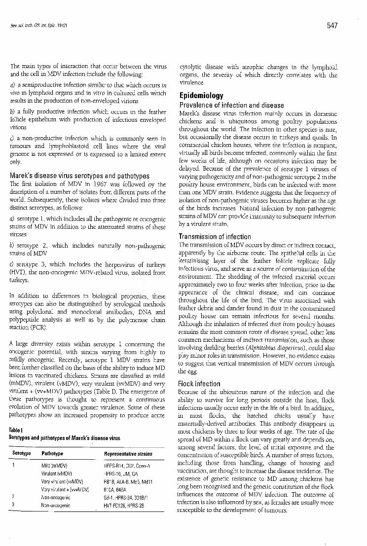
A large diversity exists within serotype 1 concerning the oncogenic potential, with strains varying from highly to mildly oncogenic. Recently, serotype 1 MDV strains have been further classified on the basis of the ability to induce MD lesions in vaccinated chickens. Strains are classified as mild (mMDV), virulent (vMDV), very virulent (vvMDV) and very virulent + (w+MDV) pathotypes (Table I). The emergence of these pathotypes is thought to represent a continuous evolution of MDV towards greater virulence. Some of these pathotypes show an increased propensity to produce acute
Table I Serotypes and pathotypes of Marek's disease virus
Serotype Pathotype Representative strains
Mild (mMDV) HPRS-B14, CU2, Conn-A Virulent (vMDV) HPRS-16, JM, GA Very virulent (wMDV) RB1B, ALA-8, Md5, Md11 Very virulent + (w+MDV) 61 OA,648A Non-oncogenic SB-1, HPRS-24.301B/1 Non-oncogenic HVT-FC126, HPRS-26
cytolytic disease with atrophic changes in the lymphoid organs, the severity of which directly correlates with the virulence.
Epidemiology Prevalence of in fect ion and disease Marek's disease virus infection mainly occurs in domestic chickens and is ubiquitous among poultry populations throughout the world. The infection in other species is rare, but occasionally the disease occurs in turkeys and quails. In commercial chicken houses, where the infection is rampant, virtually all birds become infected, commonly within the first few weeks of life, although on occasions infection may be delayed. Because of the prevalence of serotype 1 viruses of varying pathogenicity and of non-pathogenic serotype 2 in the poultry house environment, birds can be infected with more than one MDV strain. Evidence suggests that the frequency of isolation of non-pathogenic viruses becomes higher as the age of the birds increases. Natural infection by non-pathogenic strains of MDV can provide immunity to subsequent infecüon by a virulent strain.
Transmission of infect ion The transmission of MDV occurs by direct or indirect contact, apparently by the airborne route. The epithelial cells in the keratinising layer of the feather follicle replicate fully infectious virus, and serve as a source of contamination of the environment. The shedding of the infected material occurs approximately two to four weeks after infection, prior to the appearance of the clinical disease, and can continue throughout the life of the bird. The virus associated with feather debris and dander found in dust in the contaminated poultry house can remain infectious for several months. Although the inhalation of infected dust from poultry houses remains the most common route of disease spread, other less common mechanisms of indirect transmission, such as those involving darkling beetles (Alphitobius diaperinus), could also play minor roles in transmission. However, no evidence exists to suggest that vertical transmission of MDV occurs through the egg.
Flock infect ion Because of the ubiquitous nature of the infection and the ability to survive for long periods outside the host, flock infections usually occur early in the life of a bird. In addition, in most flocks, the hatched chicks usually have maternally-derived antibodies. This antibody disappears in most chickens by three to four weeks of age. The rate of the spread of MD within a flock can vary greatly and depends on, among several factors, the level of initial exposure and the concentration of susceptible birds. A number of stress factors, including those from handling, change of housing and vaccination, are thought to increase the disease incidence. The existence of genetic resistance to MD among chickens has long been recognised and the genetic constitution of the flock influences the outcome of MDV infection. The outcome of infection is also influenced by sex, as females are usually more susceptible to the development of tumours.

548 Rev. sci. tech. Off. int. Epiz., 19 (2|
Diagnostic methods Diagnostic procedures for avian tumour viruses include both pathological and virological methods. Pathological diagnosis identifies the nature of the tumour that is causing mortality, whereas virological diagnosis identifies viruses that are present in a bird or flock. As MDV, ALV and REV occur commonly, virological diagnosis does not necessarily establish the cause of the tumour. Nevertheless, histopathological identification of a tumour often determines the likely cause. However, in some cases, the presence of an infection in a flock may need to be established in the absence of tumour mortality.
Pathological diagnosis The main gross and histopathological features of the neoplasms under review are summarised in Table II. In general, while gross appearance can provide indications of the nature of the neoplasm, histopathological diagnosis is essential for accurate diagnosis. Histopathological diagnosis requires tumour material from birds which have died very recently and from several cases from an affected flock; this material should be placed in fixative. For the diagnosis of MD, the most useful set of tissues to collect include the liver, spleen, bursa of Fabricius, thymus, heart, proventriculus, kidney, gonads, nerves, skin and other gross tumour tissues.
Although clinical signs associated with MD can occur in chickens from four weeks of age, signs are most frequently seen between twelve and twenty-four weeks of age, and sometimes later. Significant diagnostic features of the classical and acute forms of MD are described below.
Classical form In the classical form of the disease, with mainly neural involvement, mortality rarely exceeds 10%-15%, occurring over a few weeks or many months. The most common clinical sign is partial or complete paralysis of the legs and wings. The characteristic pathological lesion is the enlargement of one or more of the peripheral nerves. The most commonly affected nerves that are easily seen on post-mortem examination are the brachial and sciatic plexus and nerve trunks, coeliac plexus, abdominal vagus and intercostal nerves. The affected nerves are grossly enlarged, and often two or three times the normal thickness. The normal cross-striated and glistening appearance of the nerves is lost, instead, the nerves have a greyish or yellowish appearance and are oedematous. Lymphomas are sometimes present in this form of the disease, most frequently as small, soft grey tumours in the ovary, kidney, heart, liver and other tissues.
Acute form In the acute form of the disease, where formation of lymphomas in the visceral organs usually occurs, the incidence of the disease is frequently between 10% and 30%, and in major outbreaks can reach 70%. Mortality can increase rapidly over a few weeks and then cease, or can continue at a steady or falling rate over several months. The typical lesion in this form of the disease is the widespread, diffuse lymphomatous involvement of visceral organs such as the liver, spleen, ovary, kidney, heart and proventriculus. Lymphomas are also occasionally seen in the skin around the feather follicles and in the skeletal muscles. Affected birds may also show involvement of the peripheral nerves similar to that seen in the classical form. The liver enlargement in younger
Table II Principal gross and microscopic features of importance in the differential diagnosis of the leukoses and Marek's disease lymphoma
Feature Lymphoid leukosis Erythroid leukosis Myeloid leukosis (myeloblastosis)
liver
Spleen
Bursa of Fabricius
Bone marrow
Blood
Cytology and histopathology
Other organs and tissues often grossly involved
Greatly enlarged; diffuse, miliary or nodular tumours; moderately firm Usually enlarged; diffuse, miliary or nodular tumours; soft Usually enlarged; nodular tumours Often tumorous; diffuse or focal
Occasionally lymphoblastic leukaemia
lymphoblasts; mainly extravascular infiltrations
Moderately enlarged; diffuse infiltration; cherry-red colour; soft Often enlarged; cherry-red; smooth; very soft
No changes
Semi-liquid; cherry-red
Kidneys, ovary
Erythroblastic leukaemia; immature erythrocytes; anaemia; thin buffy coat Erythroblasts; intravascular
Kidneys; may be haemorrhages in muscles
Myeloid leukosis (myelocytomatosis)
Marek's disease lymphoma
Greatly enlarged; diffuse infiltration; mottled; granular surface; firm Often enlarged; diffuse tumour; mottled; smooth; soft Sometimes tumorous
Diffuse, reddish-grey tumour infiltration
Myeloblastic leukaemia; thick buffy coat
Myeloblasts in intravascular and extravascular locations Kidneys, ovary
Often yellowish white nodular or diffuse tumours
Often nodular or diffuse tumours
No changes
Usually diffuse yellowish-grey tumour infiltration Myelocytic leukaemia; thick buffy coat
Myelocytes in intravascular and extravascular locations Kidneys, ovary, thymus, surface of bones (sternum, ribs, skull)
May be moderately to greatly enlarged; miliary or nodular tumours; firm Often atrophic; may be enlarged; usually diffuse tumours May be diffusely enlarged No changes
May be lymphocytosis or lymphocytic leukaemia
Pleomorphic, sometimes blastic, lymphoid cells in perivascular locations Nerves, kidneys, ovary, proventriculus, heart, muscle, skin, iris

Rev. sci. tech. Off. int. Epiz., 19 (2) 549
birds is usually moderate compared to that in adult birds where the liver is greatly enlarged and the gross appearance is very similar to that seen in lymphoid leukosis. Nerve lesions are less frequent in adult birds.
Features common to the acute and classical forms The peripheral nerves in both forms of the disease are affected by proliferative, inflammatory or minor infiltrative changes that are termed A-, B- and C-type lesions respectively. The A-type lesion consists of infiltration by proliferating lymphoblasts, large, medium and small lymphocytes, and macrophages, and appears to be neoplastic in nature. Nerves with B-type lesions show oedema and infiltration by small lymphocytes and plasma cells with Schwann cell proliferation; in this case the lesion appears to be inflammatory. The C-type lesion consists of mild scattering of small lymphocytes, often seen in birds that show no gross lesions or clinical signs; this is thought to be a regressive inflammatory lesion. Demyelination, which is frequently seen in nerves showing A- and B-type lesions is thought to be principally responsible for the paralytic symptoms.
Lymphomas seen in the visceral organs and other tissues are similar cytologically to the lymphoproliferations in the nerve A-type lesions. The lymphoid cells are usually of the mixed type, with a preponderance of small and medium lymphocytes. However, large lymphocytes and lymphoblasts may occasionally predominate, especially in adult birds. The polymorphic population of the lymphoid cells, as seen in impression smears or tissue sections of MD lymphomas, is an important feature in differentiating MD from lymphoid leukosis.
V i r o l o g i c a l d i a g n o s i s Isolation of Marek's disease virus Marek's disease virus infection in a flock can be detected by isolating the virus from the infected tissues. Materials, commonly used for the isolation of the virus are buffy coat cells from heparinised blood samples, or suspensions of lymphoma and spleen cells. As Marek's disease virus is highly cell-associated, the suspensions must contain viable cells. These cell suspensions are inoculated into monolayer cultures of chick kidney cells or duck embryo fibroblasts. Chicken embryo fibroblasts (CEF), although less sensitive for the primary isolation of serotype 1 MDV, can be used to isolate serotypes 2 and 3. Evidence of MDV replication in the culture can be seen as plaques which appear in three to four days. Less commonly, feather tips, from which cell-free MDV can be extracted, are also used for virus isolation.
Characterisation of Marek's disease virus serotypes Serotypes of MDV isolated in culture can be differentiated fairly accurately on the basis of the time of appearance, rate of development and morphology of the plaques. Plaques of HVT usually appear earlier and are larger than serotype 1 plaques, whereas serotype 2 plaques appear later, and are smaller than the serotype 1 plaques. The serotype specificity of the plaques can also be confirmed by using specific antibodies in
immunological tests. Recently, PCR tests have been developed which allow the differentiation of oncogenic strains of serotype 1 virus, and strains of serotypes 2 and 3 (3, 35) .
Detection of virus infection in tissues The methods commonly employed for the detection of virus in tissues include immunofluorescence and immunohistochemical methods using polyclonal and monoclonal antibodies, in situ hybridisation using MDV-specific nucleic acid probes, PCR and electron microscopy.
Serological tests The presence of antibodies to MDV in birds from approximately four weeks of age is an indication of infection. Antibodies detected in birds before this age are likely to represent maternally derived antibodies and are not considered evidence of active infection. Although no serological test has been prescribed for the detection of MDV-specific antibodies, the agar gel immunodiffusion (AGID) test is usually employed for this purpose. The antigen used in the test is either disrupted MDV-infected tissue culture cells, extract of the feather tips or skin containing feather tracts from infected chickens. The viruses, antigens and antisera are usually available from OIE Reference Laboratories for MD. A modification of the AGID test to detect MDV antigen in the feather tips by reactivity with MDV hyperimmune serum is also used. Other serological tests, such as the indirect immunofluorescence test, enzyme-linked immunosorbent assay (ELISA) and virus neutralisation (VN) have been described, but are used mostly for research purposes rather than for routine diagnosis.
Surveillance Marek's disease is a ubiquitous virus infection occurring in commercial poultry operations world-wide. Because the virus is able to survive for long periods both in the host and in the environment of the poultry house, the disease is unlikely to be completely eradicated. Control of the disease therefore depends essentially on the use of successful vaccination strategies. The vaccine strains are non-pathogenic viruses that establish a permanent infection in the vaccinated birds. The vaccines are capable of preventing lymphoma formation and clinical disease, but do not prevent superinfection by pathogenic MDV strains. Hence, surveillance for MD should verify that the birds do not suffer from clinical disease and are properly vaccinated. To achieve this, various tests mentioned under the section on diagnosis can be applied. In addition, importing countries should require the exporter to provide international animal health certificates (24) to ensure that imported chickens and day-old chicks are free from clinical disease and are vaccinated against MD and that the hatching eggs originate from a vaccinated source and have been shipped in clean unused packages. As MDV is not vertically transmitted, these measures are sufficient to prevent introduction of infection through day-old chicks or hatching eggs. However, vaccination and freedom from clinical disease

550 Rev. sci. tech. Off. int. Epiz.. 19(2)
would not exclude the possibility of introduction of pathogenic MDV isolates when importing older birds.
Marek's disease virus could persist in the skin (in feather follicles) of imported carcasses and portions, and in feathers, but is unlikely to be present in poultry meat and eggs. Live poultry should not be exposed to such potentially infected materials. Heat treatment (cooking) will destroy MDV.
Public health implications The identification of MDV and the widespread use of live vaccines against MD have raised some concerns that exposure to MDV from the environment or from consumption of poultry meat could be a cause of cancer in humans. However, a large body of evidence in both avian and human virology, serology, pathology and epidemiology strongly supports the conclusion that no aetiologic relationship exists between avian herpesviruses and human cancer (31). Recently, there has been speculation that MDV infection might be associated with multiple sclerosis (MS), principally based on serological findings. However, in detailed studies using sensitive methods such as PCR, no MDV-related sequences could be detected in the DNA of patients with MS, ruling out the involvement of MDV (16).
Prevention and control methods V a c c i n a t i o n The development of vaccines for the control of MD was a significant landmark both in avian medicine and basic cancer research, as this was the first example of a neoplastic disease controlled by the use of a vaccine. Currently and at least for the foreseeable future, vaccination represents the principal strategy for the prevention and control of MD, although other approaches, such as increasing the genetic resistance of birds and improving hygiene and biosecurity, should form valuable adjuncts for control programmes.
Live virus vaccines, used since 1970, remain the basis of disease control programmes. These are usually administered to day-old chicks at hatching to provide protection against the natural challenge the chicks are exposed to early in life from the infected poultry house environment. With the introduction of in ovo immunisation methods, an increasing number of birds are vaccinated by this route. Marek's disease vaccines are highly effective, often achieving over 9 0 % protection under commercial conditions. Vaccines available vary from country to country. In the United States of America (USA), strains of MDV belonging to all three serotypes have been licensed as vaccines (Table III).
In many countries, HVT continues to be widely used as a monovalent product because it is inexpensive, available as cell-free and cell-associated forms, and effective when the field exposure is not severe. The HVT and SB-1 strains comprised the first commercial bivalent vaccine based on the protective synergism demonstrated between serotypes 2 and 3 viruses. The CV1988 strain Rispens vaccines and modified versions are
Table III Strains of Marek's disease virus licensed to be used as vaccines in the United States of America
Vaccine strain Serotype
FC126 (HVT) 3 SB-1 2 301B/1 2 CVI988 clone C 1 CVI988/C/RB 1 CVI988 (Rispens) 1 R2/23 (Md 11/75) 1
widely used in many countries and appear to be effective against some of the w+MDV pathotypes.
Although MD vaccines have been successful in controlling major losses from the disease, threat of vaccine failure has continued to cause concern. The reasons for these possible failures include the following:
a) challenge with virulent viruses before the development of vaccinal immunity
b) interference with the development of immunity by the maternal antibodies
c) improper use of the vaccine
d) the use of a non-protective vaccine strain.
Vaccinating alternate generations with different types of vaccines can reduce the effects of interfering passive antibodies. Early exposure to MDV can be significantly prevented by improved hygiene and biosecurity measures.
Despite the success achieved by vaccines in controlling MD, the continuous evolution of MDV strains towards greater virulence leading to the emergence of vv and vv+ pathotypes of MDV is threatening to pose problems in the future (45). The development of more effective vaccines through recombinant DNA technology (33) , and the use of immunomodulatory approaches to enhance the response to vaccines should improve vaccination strategies in the future.
S e l e c t i o n f o r g e n e t i c r e s i s t a n c e Genetic resistance to MD is well documented, and susceptible and resistant lines can be developed by progeny testing, selection from survivors of MD challenge, or blood typing. Two distinct genetic loci that play a major role in controlling resistance have been identified. The best characterised association is the one between the chicken major histocompatibility complex (MHC) and resistance to MD, the most notable being the association with the B 2 1 allele. This association develops early in life and is accompanied by reduced numbers of infected T-cells. A second type of resistance associated with non-MHC genes is provided by the observation that RPL (Regional Poultry Laboratory) line 6 and 7 chickens, which are both homozygous for the same MHC

Rev. sci. tech. Off. int. Epiz., 19 (2) 551
allele, differ markedly in MD susceptibility. Mapping of genes associated with such resistance is in progress and evidence suggests that the NK region within chromosome 1 contains a resistance gene, which has been designated MDV1 (5). As aditional tools for selection for genetic resistance become available, the opportunities for genetic selection against MD will be extended.
Hygiene measures The use of vaccines should never be an excuse for poor management or lack of biosecurity measures. Removal of used litter and disinfection of buildings are important aspects of disease control, especially in view of the possibility of selection for pathogens with increased virulence. Furthermore, placing chicks in an environment heavily contaminated with virus, before solid immunity can be developed, can lead to vaccination breaks. Strict biosecurity is also necessary to prevent the introduction of new MDV strains onto a farm.
Avian leukosis Diseases The term leukosis' embraces several different leukaemia-like proliferative diseases of the haemopoietic system caused by ALV; the term leukosis is used because a leukaemic blood picture is not always present (10, 29) . The components of the haemopoietic system that may undergo neoplastic change include the lymphopoietic (lymphocytic) system, the erythropoietic (red cell) system, and the myelopoietic (myelocytic) system. Presenting signs of the leukoses are mostly non-specific (inappetence, weakness, emaciation, diarrhoea, pale wattles). The three forms of leukosis that arise are discussed below.
Lymphoid leukosis Until recently, lymphoid leukosis has been the commonest form of leukosis. It occurs in chickens from approximately four months of age. Although most commonly caused by ALV of subgroups A and B (Table IV), it should be noted that REV can induce a tumour that is indistinguishable grossly
Table IV Avian leukosis virus envelope subgroups in chickens
and microscopically (see the section entitled 'Reticuloendotheliosis'). Gross pathological changes are significant enlargement of the liver with miliary, diffuse or nodular tumour foci, often nodular tumours in the bursa of Fabricius, and tumorous enlargement of the spleen and other organs. Microscopically, the lesions consist of coalescing foci of extravascular uniformly immature lymphoid cells (lymphoblasts). The tumour cells are B-cells, expressing immunoglobulin M (IgM) and other B-cell markers, which originate in the bursa and metastasise to other organs.
Erythroid leukosis Erythroid leukosis (also termed erythroblastosis) is an uncommon, usually sporadic, tumour occurring principally in adult chickens, although occasionally observed in young birds (from five weeks old). The liver and spleen, and sometimes the kidneys, are moderately and diffusely enlarged and often of bright cherry-red colour. The bone marrow is bright red and liquid. Affected birds are often anaemic, with muscle haemorrhages and occasionally abdominal haemorrhage from a ruptured liver. The disease is an intravascular erythroblastic leukaemia. Microscopically, the liver shows intrasinusoidal accumulations of rather uniform, round, erythroblasts, the spleen shows accumulations of erythroblasts in the red pulp, and the bone marrow shows enlarged haemopoietic sinusoids filled with erythroblasts. An erythroblastic leukaemia is visible.
Myelo id leukosis Myeloid leukosis occurs in two, often overlapping forms termed myeloblastic myeloid leukosis (myeloblastosis) and myelocytic myeloid leukosis (myelocytomatosis). The disease has become particularly prevalent in broiler breeders and, less frequently, in broilers in many countries since 1996, and is caused by the recently recognised subgroup J ALV (27, 43) .
Before the advent of subgroup J ALV, myeloblastic myeloid leukosis was predominantly a sporadic disease of adult chickens. In this classic form, the liver and spleen are greatly enlarged and the liver frequently has a yellowish-grey granular ('Morocco leather') appearance. The bone marrow is firm and reddish-grey in colour. Microscopically, the liver shows intravascular, sinusoidal accumulations of immature myeloid
Envelope Prevalence Representative virus Oncogenicity Host virus subgroup strains (predominant neoplasm) receptor locus
A Common RAV-1, RPL-12 - Yes (II) tva B Uncommon RAV-2, MAV-2 Yes(LL) tvb C Rare RAV-7, RAV-49 Yes tvc D Rare RAV-50, CZAV Yes tvb E Common RAV-0, RAV-60 No tvb/tve* J Common HPRS-103, ADOI-Hd Yes (MI) Unknown
t f : lymphoid leukosis * The existence of two loci has not been confirmed M i : myeloid leukosis

552 Rev. sci. tech. Off. int. Epiz., 1912|
cells (myeloblasts and promyelocytes), and extravascular accumulations and perivascular cuffing, by these cells. The red pulp of the spleen is infiltrated by myeloid tumour cells, as are the extrasinusoidal myelopoietic areas in the bone marrow. A marked myeloid cell leukaemia is observed.
Before the appearance of subgroup J ALV, myelocytic myeloid leukosis occurred principally in young chickens. In this form, yellowish-white myelocytomas are present on the skeleton (head, ribs, vertebrae and inner sternum). Microscopically, the tumour is comprised of well-differentiated myelocytes with conspicuous cytoplasmic granules.
Myeloid leukosis caused by subgroup J ALV shows features of both the myeloblastic and myelocytic forms described above. The disease occurs primarily in adult broiler breeders, and occasionally in young breeders and broilers. Myelocytomas are present on the inner sternum, vertebrae, ribs, inner mandible, trachea and sometimes the eye. In addition, the liver, spleen, ovary and other organs are often enlarged by tumorous infiltration. Microscopically, the tumour cells are usually well-differentiated myelocytes, although tumours of more immature myeloid cells occur, sometimes in the same bird. The tumour is both extravascular and intravascular (leukaemia).
Other tumours A variety of solid tumours can be caused by ALV, including fibrosarcoma, chondroma, haemangioma, histiocytic sarcoma, mesothelioma, myxoma, nephroblastoma, osteoma, and the proliferative bone disorder, osteopetrosis, characterised by thickening of the long bones. These tumours can occur alone or accompanying the leukoses. Subgroup J ALV in particular causes a variety of such tumours in addition to myeloid leukosis, and also mixed myeloid and erythroid leukosis.
Aetiology Virus st ructure and repl icat ion Avian leukosis viruses are placed in the Alpharetrovirus genus of the viral family Retroviridae (26, 29) . In common with other members of this RNA virus family, ALVs are characterised by possession of the enzyme reverse transcriptase. This enzyme is necessary for the formation of a DNA provirus, which is integrated into the host genome during viral replication. Structurally, ALVs are simple viruses. From the 5 ' end to the 3 ' end of the genome, they possess three structural genes, gag/pro-pol-env, which encode, respectively, the proteins of the virion group-specific (gs) antigens and protease, the enzyme reverse transcriptase, and the envelope glycoproteins. These structural genes are flanked by genomic sequences concerned with the regulation of viral replication, which in the DNA provirus form the viral long terminal repeats (LTRs) which carry promoter and enhancer sequences. Some laboratory strains and field isolates of ALV also possess one (or rarely two) viral oncogene(s) inserted at
some site within the genome. Such viruses have acquired the oncogene by transduction of a cellular oncogene during oncogenesis. These viruses are usually genetically defective, with deletions within the genome, and require the presence of 'helper viruses' (co-infecting non-defective ALVs) to enable replication.
Avian leukosis v i rus envelope subgroups Avian leukosis viruses that occur in chickens can be classified into six subgroups (A, B, C, D, E and J ) , on the basis of differences in the viral envelopes which affect antigenicity, as determined by induction of virus neutralising antibodies, host range, and ability to interfere with infection by other ALVs of the same or differing subgroups (Table IV). The F, G, H and I subgroups of ALV are allocated to endogenous viruses found in ring-necked and golden pheasants, Hungarian partridge and Gambei's quail, respectively. Of the six ALV subgroups found in chickens, subgroup A is common, and subgroup B less common, both occur particularly in egg-type strains. Subgroups C and D are rare, subgroup E represents common infectious ev-type endogenous ALVs widely present in normal chickens, and the subgroup J is common in meat-type chickens. Viruses of subgroups A, B, C, D and j are oncogenic, causing mainly lymphoid leukosis (subgroups A and B) or myeloid leukosis (subgroup J ) . Subgroup E viruses are not oncogenic. Viruses within a subgroup usually cross-neutralise to varying extents, although antigenic variants of subgroup J ALV exist which do not cross-neutralise. A partial cross-neutralisation occurs between subgroup B and D viruses, but otherwise viruses in the different subgroups do not cross-neutralise.
Mechan isms of neoplast ic t ransformat ion Avian leukosis viruses induce neoplasms by one of two main types of mechanism, as follows:
a) viruses that do not carry a viral oncogene induce neoplasms by activation of a cellular proto-oncogene. This is possible when the virus becomes fortuitously integrated adjacent to a cellular proto-oncogene during viral replication. Thus, lymphoid leukosis is initiated by activation of the c-myc oncogene by the LTR promoter, a mechanism termed 'promoter insertion' or 'insertional mutagenesis'. Erythroid leukosis is caused by activation of the c-erbB oncogene. Initiation of neoplasms by this mechanism is slow, occurring after weeks or months. Such ALVs are termed 'slowly transforming' and the tumours are called 'slow onset tumours';
b) viruses that have a viral oncogene induce neoplasms by insertion of the oncogene into the genome of the target cell. Such ALVs are termed 'acutely transforming', and neoplastic cells are induced within a few days. Depending on the oncogene possessed by the virus, acutely transforming ALVs induce different types of neoplasm, for example: v-myc: myeloid leukosis (myelocytoma); v-myb: myeloid leukosis (myeloblastosis); v-erbB: erythroid leukosis; v-src: sarcoma.

•Rev.sci. tech. Off. int. Epiz., 19(2) 553
Avian leukosis virus pathotypes Strains of ALV often produce more than one type of neoplasm, although for each strain one particular neoplasm usually predominates. Strain of chicken, age at infection and dose of virus will influence the incidence and types of neoplasms induced.
Exogenous and endogenous avian leukosis viruses Avian leukosis viruses which are transmitted from bird to bird, either vertically (through the egg) or horizontally (by contact) as infectious virions, are termed 'exogenous' ALVs (Table V). All oncogenic ALVs fall into this category. However, some ALVs are integrated into the genome of normal birds as proviral sequences that are transmitted genetically as Mendelian genes, either as complete viral genomes able to code for infectious virus of subgroup E, or more usually, as incomplete (defective) genomes able to code for certain retroviral products (e.g. gs-antigen) only. These ALVs are termed 'endogenous' ALVs (Table V). The sites of integration of these endogenous viruses are termed 'ev loci'. Endogenous ALVs are apparently non-oncogenic although they may influence the response of the bird to infection by exogenous ALVs by inducing immunological tolerance or immunity. The ev21 locus linked to the sex-linked slow-feathering gene K, codes for an infectious subgroup E ALV, named EV21, which can cause immune tolerance to infection by exogenous ALV. Apart from the family of ev endogenous viruses closely related to exogenous ALV, other more distantly related endogenous families have been discovered in the normal genome, namely: EAV (endogenous avian virus), ART-CH (avian retrotransposon from chicken genome) and CR 1 (chicken repeat 1). The EAV family is of particular interest, since members designated EAV-HP (or ev/J) appear to be the source of the envelope gene of subgroup JALV.
Epidemiology Prevalence of infect ion and disease Exogenous ALVs are almost ubiquitous in commercial chickens on a world-wide basis, although many primary egg-type and meat-type breeding companies institute ALV
Table V Exogenous and endogenous avian retroviruses
Characteristics Exogenous avian Endogenous avian Characteristics retroviruses retrovirus families
Virus designation Subgroups A, B, C, D and Evloci (including J ALV subgroup E ALV)
EAV-0 EAV-HP (also termed ev/J) ART-CH CR1
Virus Vertical (by female) Genetic (by male and transmission Horizontal (by male and female)
female) Oncogenicity Yes No
ALV: avian leukosis virus
eradication schemes. Subgroup A viruses occur frequently and subgroup B viruses more rarely. Nevertheless, the incidence of neoplasms, mainly lymphoid leukosis, in infected flocks is usually low, in the order of l % - 2 % , although losses of up to 2 0 % can occur. Subgroup C and D viruses have not been widely recognised in the field. Endogenous subgroup E viral loci are present in virtually all stock, and may be expressed as exogenous subgroup E ALV in slow-feathering strains; however, subgroup E viral loci are not oncogenic. Subgroup J ALV is distributed globally in meat-type chickens; neoplastic losses, mainly from myeloid leukosis, vary up to a maximum of approximately 5 0 % .
In addition to losses from tumours, the presence of exogenous ALV infection can have an adverse effect on egg production, egg size, fertility, hatchability, growth rate and non-specific mortality (14).
Transmission of infect ion Two modes of natural transmission of exogenous ALV occur, namely: vertical and horizontal.
Vertical (congenital or egg) transmission In vertical transmission, ALV is passed from infected hens to their offspring. Although only a small minority of chicks are usually infected in this way, the route is important in transmitting the infection from one generation to the next, and in providing a source of contact infection to other chicks. Avian leukosis virus present in the magnum of the oviduct of the hen passes into the egg albumen and thence into the chick embryo during incubation. This route of congenital infection leads to strong associations between the presence of virus in vaginal swabs, egg albumen and embryos, which provide the basis for ALV eradication programmes in breeding stock, as discussed later. Infected males do not pass the infection to their progeny, even though semen may contain virus. However, males can infect hens venereally.
Horizontal transmission In horizontal transmission, ALV spreads from bird to bird by direct contact, or indirectly by exposure to virus in the environment. This mode is responsible for the high incidence of infection in flocks. Sources of virus from infected birds include faeces, saliva and desquamated skin. The period of survival of ALV outside the body is relatively short (a few hours), and consequently ALV is not highly contagious. Congenitally infected chicks are an important source of contact infection in the hatchery and during the brooding period; meconium and faeces from congenitally infected chicks contain high concentrations of ALV. Routes of infection are oculonasal, oral, respiratory and skin.
Flock infect ion The infective status of chickens in an ALV-infected flock can be categorised according to whether the chickens have cell-free viraemia (V+ or V - ) , serum antibodies (A+ or A - ) , and whether the chickens shed ALV to their cloaca or, for laying hens, egg albumen (S+ or S - ) . This information is

554 Rev. sci. tech. Off. int. Epiz., 19 (2)
important in ALV eradication programmes, and these categories allow several classes of birds to be identified, as described below.
Tolerant viraemic, antibody-negative, shedders (V+A-S+l These birds arise primarily as a result of vertical (congenital) infection from the dam, and they in turn produce V+A-S+ progeny. This class of birds is likely to constitute a minority of a flock. The birds have large amounts of virus, and consequently gs-antigen, in the cloacal or vaginal swabs, and for hens in the egg albumen, embryos and chick meconium. This class of bird may also arise following contact infection by ALV at a very young age (newly hatched chicks), as observed notably in meat-type chicks exposed to subgroup J ALV. Birds classed V+A—S+ are most prone to develop neoplasms.
Non-viraemic, antibody-positive, non-shedders (V-A+S-) and shedders (V-A+S+) Birds that acquire infection by contact after hatching are most likely to develop an immune infection. This class of birds is likely to constitute the majority of a flock. The birds are usually non-shedders which do not produce infected progeny, but some may be shedders and intermittently produce infected chicks. These birds are less likely than viraemic birds to develop neoplasms. Birds in the V - A + S -category are believed to harbour ALV somewhere in their tissues.
Non-viraemic, antibody-negative, non-shedders (V-A-S-) Birds of this class are either not yet infected or genetically resistant to infection.
Viraemic, antibody-positive birds (V+A+) A small class of birds with both viraemia and antibodies may be found. These are considered to be birds that are seroconverting from a transient viraemic state to an immune (V-A+) state. They may or may not be shedders.
Spread of infect ion Birds from infected flocks, and products, such as eggs and meat, could spread infection to other flocks and locations. Because ALV is vertically transmitted, infection may spread internationally in hatching eggs and day-old chicks, and spread in semen is possible. Importers need to guard against the introduction of ALV by requiring health certificates from the exporter relating to the specific disease and infection status of source flocks. Introduction of a new ALV into susceptible poultry populations could have serious consequences because of absence of immunity and of effective control measures in breeding and production populations other than at the primary breeding level.
Diagnostic methods Similar to MD, diagnostic procedures for avian leukosis include both pathological and virological methods.
Pathological diagnosis The main gross and histopathological features of the neoplasms under review are summarised in Table II. Although the gross appearance of the tumour can provide indications of the nature of a neoplasm, histopathological diagnosis is essential for accurate diagnosis of leukosis. For this, tumour material must be collected from birds which have died very recently and from several cases from an affected flock; this material must be placed in fixative. The most useful tissues to collect are liver, spleen, bursa of Fabricius, bone marrow, peripheral nerves and other grossly tumorous tissues.
The significant diagnostic features of the various leukoses are described below.
Lymphoid leukosis This occurs in birds from approximately four months of age. A useful diagnostic feature (although not always present) is gross nodular tumorous involvement of the bursa of Fabricius. Lymphoid leukosis is usually caused by ALV, but sometimes by REV. Microscopically, diagnostic features are the uniform large lymphocytes (lymphoblasts) that comprise the tumours, the presence of intrafollicular tumours in the bursa, and the tendency for the tumours in other tissues, such as the liver and spleen, to grow in an expansive nodular fashion. The tumour cells are B-cells, and with specialised immunocytochemical techniques, B-cell markers and IgM can be detected in the tumour cells. The tumour is extravascular and leukaemia is rare.
Erythroid leukosis This can occur in young birds, from approximately five weeks, and in adults. A smooth-appearing moderate enlargement of the liver and spleen, of cherry-red colour, occurs; haemorrhage from liver rupture may be present. Microscopically, the tumour is one of fairly uniform, round, erythroblasts. The tumour is intravascular, with accumulations of erythroblasts in the liver sinusoids and in the red pulp of the spleen. In the marrow, the erythropoietic sinuses are filled with erythroblasts.
Myeloid leukosis This occurs mainly in adult birds but sometimes in birds as young as five weeks. In the myeloblastosis form, enlarged liver, spleen and other organs can appear grossly similar to changes in lymphoid leukosis. In the myelocytomatosis form, these organs are often enlarged, but the frequent presence of cream-coloured myelocytomas on the skeleton, particularly on the inner aspect of the sternum, and sometimes on the skull, appear pathognomonic, and currently suggestive of infection by subgroup J ALV. Myelocytomas can also be seen in the oral cavity, trachea and eye. The myeloblastic form is comprised of immature myelocytes with few cytoplasmic granules. In the liver, tumour cells are present both extravascularly, around veins and arteries, and intravascularly, in the sinusoids. In the myelocytic form,

Rev. sci. tech. Off. int. Epiz., 19 (2) 555
tumour cells vary from well-differentiated myelocytes with conspicuous eosinophilic granules, to poorly differentiated myelocytes with few or no granules. The different forms sometimes occur in different areas within a bird, and in different birds in an affected flock. Areas of undifferentiated tumorous myelomonocytic stem cells can also occur. Both myeloblastic and myelocytic forms are often marked by leukaemia, and bone marrow becomes replaced by neoplastic myeloid cells.
Virological diagnosis In the following sections, the principles of the diagnostic methods are outlined. Details of performing the tests are available in technical manuals (12, 38) . Many of the methods used are technically demanding and need specialised laboratory facilities.
isolation of avian leukosis virus Avian leukosis virus may be isolated from materials from individual birds as a means of detecting the presence of the infection in the bird or flock. Materials commonly used for isolation include serum, buffy coat cells and tumour tissue, and for certain purposes, cloacal or vaginal swabs, egg albumen, embryos and meconium. Virus isolation is generally the ideal detection method (the so-called 'gold standard') since the technique can detect all ALVs and is the starting point for various other virus studies.
Material (normally cell-free) to be tested for exogenous ALV is inoculated into SPF C/E (susceptible to all ALV chicken subgroups except E) CEFs growing in tissue culture plates or wells of microtitre plates, and the cultures are incubated for seven days. Supernatants are collected and stored at - 7 0 ° C as virus isolates, and the CEFs are sonicated and tested for the presence of ALV p27 gs-antigen in an antigen ELISA test (available commercially). The C/E CEFs used for this assay will support growth of ALVs of the known exogenous subgroups A, B, C, D and J , but exclude subgroup E ALV of endogenous origin. The C/E CEFs derived from line 0 chickens (available commercially) are preferred since these do not possess gs-antigen of endogenous origin, thus allowing a clear distinction to be made between ALV-positive and ALV-negative test results.
If necessary, ALV of endogenous origin (subgroup E) can be identified by inoculating test material on cultures of C/E CEF and quail embryo fibroblasts of phenotype Q/BJ. Subgroup E ALV will grow in the Q/BJ cultures but be excluded from the C/E cultures.
Most avian leukosis viruses isolated from the field are slowly transforming viruses which lack a viral oncogene and induce neoplasms by promoter insertion activation of a cellular proto-oncogene, as discussed earlier. Occasionally, such ALVs may recombine genetically with a cellular oncogene to produce an acutely transforming ALV, which possesses a viral oncogene. Acutely transforming viruses may require
particular types of target cells, such as haemopoietic cells, for the oncogenic potential of the virus to be revealed. For example, acutely transforming subgroup J ALV occurs in the field in some myelocytomas. These can be identified by inoculating cell-free tumour extracts into monolayer cultures of chicken bone marrow cells or blood monocytes that within a few days undergo neoplastic transformation (28).
Characterisation of avian leukosis virus subgroups Characterisation of ALV isolates into envelope subgroups may be performed using several methods, as follows:
a) Viral interference assays, which test the ability of an isolate to interfere with (prevent) focus formation in C/E CEF cultures by Rous sarcoma virus (RSV) of known subgroup.
b) Virus neutralisation assays, in which an isolate is placed in a subgroup according to susceptibility to neutralisation by chicken antisera with known subgroup neutralising activity. The isolate, or the RSV pseudotype, is exposed to antiserum and then examined for growth, or focus formation, respectively, in C/E CEF cultures. An RSV pseudotype is an acutely transforming RSV which can be created by coating a replication defective RSV with the viral envelope of an ALV, and hence endowing the virus with the subgroup characteristics of the ALV, in this case of the isolate. Fluorescent antibody staining of infected cultures using antisera against different subgroups is also used. These methods are usually satisfactory, although in the case of subgroup J isolates, in which env gene mutations and consequent antigenic changes are frequent, subgroup J antisera may fail to neutralise new variants. However, mouse monoclonal antibodies have been developed which appear in fluorescent antibody tests to detect a wider range of subgroup J isolates.
c) Host range assays, in which an isolate, or the RSV pseudotype, is placed in a subgroup according to ability to grow in, or transform, respectively, CEFs of varying ALV subgroup susceptibility phenotypes. For example, a subgroup Ä ALV would be identified if the virus grew in C/E CEFs but did not grow in C/AE CEFs (resistant to subgroups A and E). This method relies on the availability of CEF phenotypes that exclude certain ALV subgroups. Naturally occurring CEFs that exclude subgroup J have never been detected, although workers in the USA have developed a C/J CEF line by J env gene transfection (17).
d) Polymerase chain reaction assays specific for various ALV subgroups have been developed (15, 36 , 37) . These tests are sensitive, rapid, and may be used to detect ALV proviral sequences in tumour material or cultured CEFs. By use of a reverse transcription step (RT-PCR), the tests can detect viral RNA (e.g. infectious virus). However, a problem with PCR methods is that the primer pairs used may not detect all variant viruses, necessitating continuing redesign of primers.

556 Rev. sci. tech. Off. int. Epiz., 19 (2)
Characterisation o f avian leukosis virus pathotypes The oncogenic spectrum o f an ALV strain may be determined by inoculation of the ALV either intravenously i n t o
eleven-day-old chick embryos or intravenously o r intra-abdominally into one-day-old chicks. Inoculation i n t o
embryos provides the most sensitive expression o f oncogenic potential. After hatching, inoculated chickens may need t o be kept for n i n e months t o a low for tumour expression. The chickens used need t o be SPF, susceptible t o infection by the envelope subgroup of the ALV being studied a n d also susceptible to induction of particular neoplasms. Suitable chicken strains are available in research, laboratories which specialise in avian tumour viruses.
Detection of avian leukosis virus pZ7 group-specific-antigen Awiam leukosis viruses o f all subgroups share a common p27 g s - a n t i g e n , which may be detected by an antigen ELISA test. This test is commercially available, relatively cheap, rapid (requires one day), and can be used o n a large scale f o r detecting ALV in vaginal or cloacal swabs o f chickens that shed ALW and in albumen This test
is particularly used in ALV eradication p r o g r a m m e s , a s
discussed later, and for detecting ALV in cultured CEFs, as (described earlier. The main disadvantage of the tes t for certain applications is the lack o f subgroup specificity. The test does matt discriminate between different exogenous ALVs (which may not be of concern),, and also detects (usually at a lower lewel gs antigen of endogenous ALV origin. In eradication schemes this can cause difficulties in bird selection, since the USUAL requirement i s to identify and remove birds shedding exogenous ALV, but to keep birds which may have endogenous ALV only. Careful selection o f the cut-off point between 'negative' and "positive* r e s u l t s is required. This is judged p a r t l y from experience a n d through the use of other tests to discriminate between e x o g e n o u s and endogenous viruses. Similar difficulties arise if the p27 ELISA is used in attempts to identify chicks o r older birds infected with exogenous ALV, by testing meconium or serum, since gs-antigen of endogenous origin may be detected. Similarly, other tests are required to discriminate between exogenous or e n d o g e n o u s ALV.
Detection of avian leukosis virus antibodies Detection of antibodies against ALV is used in flock surveillance to detect presence or absence of infection by exogenous ALV, and to identify particular classes of birds in epidemiological studies and ALV eradication programmes. Virus neutralisation and antibody ELISA tests are used, as follows:
a) Virus neutralisation tests. Antibody is detected by ability to neutralise infectivity of ALVs of known subgroup, or of the RSV pseudotypes. Although virus and antibody within subgroups usually cross-neutralise, antibodies against variant viruses may fail to neutralise a representative subgroup virus. This has been observed particularly with subgroup J , due to virus mutation, as discussed earlier, and is a limitation to the
use of VN tests. These tests are slow (requiring seven to ten days) and are technically demanding.
b) Antibody ELISA tests. Viral antigens may be used in ELISA tests to detect subgroup-specific ALV antibodies. Tests for antibodies to subgroups A, B and J are commercially available. The subgroup J antibody ELISAs use env antigen produced in a baculovirus system and appear to detect antibodies to all variant viruses studied. The antibody ELISA tests are rapid (requiring one day), specific, and suitable for large scale testing.
Surveillance In recent years, awareness of ALV by poultry producers has been raised by the global spread of subgroup J virus and the serious flock losses often associated with the infection. Concerns about ALV infections in flocks have encouraged producers to use the commercially available ELISA kits for detecting ALV gs-antigen and subgroup A, B and J antibodies in order to monitor for the importation of the infection or to test for prevalence.
Although these tests can be valuable, difficulties are associated with the interpretation of the results. This applies particularly to use of the gs-antigen ELISA, because of the inability to differentiate between antigen of exogenous and endogenous ALV origin.
Examples of the possible uses of these tests, and the limitations of the results, are as follows:
a) Testing of serum from adult flocks for gs-antigen as an indication of infection by exogenous ALV. Although exogenous ALV can raise gs-antigen levels in serum, expression of endogenous ALV commonly has the same effect, so that results cannot be interpreted. This test is of little value if used in isolation.
b) Testing of serum from adult flocks for antibodies to ALV. Sera should be tested for antibodies to A, B and J . Positive results indicate the presence of infection in the flock. Individual birds without antibodies in an infected flock may be viraemic shedders of ALV (V+A-S+).
c) Testing of chick meconium for gs-antigen as an indication of infection by exogenous ALV. High gs-antigen levels are likely to be caused by exogenous virus, but endogenous virus can also cause positive readings. Confirmation of the presence of exogenous ALV is necessary by other methods.
d) Testing of serum of day-old chicks for antibodies to ALV. Sera should be tested for antibodies to A, B and J . Positive results indicate the presence of the infection in the female parents. Some chicks may be congenitally infected with exogenous ALV of the same subgroup and will spread the infection to other chicks.
Recommended procedures Primary breeding companies of layer-type and meat-type stock have made significant progress in eliminating ALV of

Rev. sci. tech. Off. int. Epiz., 19 (2) 557
subgroups A, B and J in the elite breeding lines. These companies should therefore increasingly be able to provide breeding stock free from these infections. Importing customers should request information about the ALV status of flocks, hatching eggs and chicks. In addition, importing countries may carry out checks on imported materials if deemed necessary.
The recommended surveillance procedures for imported stock are described below.
Hatching eggs Fifty albumen samples, collected with separate needles through the eggshell should be tested by the gs-antigen ELISA. Positive samples should be tested by RT-PCR using primers designed to detect exogenous ALV. Primers specific for each exogenous subgroup (specifically, A, B and J ) should be used to identify the subgroup.
Day-old chicks Fifty meconium samples (or cloacal swabs), should be tested by gs-antigen ELISA. Positive samples should then be tested by RT-PCR as for albumen samples above. Serum samples can be tested for antibodies to ALV of subgroups A, B, and J using antibody ELISA kits (commercially available). The presence of antibody would indicate presence of infection in the dam parent flock and therefore the possibility of vertically transmitted ALV in the chicks.
Chickens in rear The chickens should be reared in quarantine and tested for evidence of ALV infection. At four weeks, fifty serum samples should be tested for ALV viraemia, and at eight weeks, fifty serum samples should be tested by ALV antibody ELISA for subgroups A, B and J .
Semen
If semen is imported, it should be tested for exogenous ALV.
Other products Avian leukosis virus could persist in imported poultry carcasses, meat, eggs and feathers; live poultry should therefore not be exposed to such materials. Heat treatment (cooking) will destroy ALV.
Specific-pathogen-free flocks and vaccine production Exogenous ALVs constitute the most common vertically transmitted pathogens. It is therefore essential that SPF chicken flocks developed for the production of embryos, and avian substrate cells used for production of veterinary (including avian) and human vaccines, are free from infection by exogenous ALV and other avian pathogens (22, 41) . Vaccine seed virus, substrate cells, and final vaccine should also be tested for freedom from exogenous ALVs, including viruses of subgroups A, B and J , and other infectious agents. Live virus vaccines produced in avian cell substrates that are not derived from SPF flocks are highly likely to be
contaminated with exogenous ALV, and outbreaks of neoplasia in poultry flocks have been caused by use of contaminated poultry vaccines.
Public health implications No firm evidence exists to suggest that ALVs present a health risk to humans, although the possibility of this has not been definitely excluded (19) . Certain strains of RSV, particularly of subgroup D, are known to infect and transform mammalian cells in vitro, including human cells, and can induce sarcomas and other tumours in mammals in vivo, including primates. However, despite exposure of humans to ALV from live poultry, poultry meat and eggs, no conclusive epidemiological or other evidence has been adduced of any risk of cancer. In addition, exposure of humans to exogenous ALV present at one time (although not currently) as a contaminant of live vaccines such as yellow fever and measles was not associated with a recognised hazard. Antibodies to ALV have been detected in poultry workers and others but these may result from antigenic exposure rather than ALV replication in humans (20). The potential for ALV replication in mammalian cells is believed to be slight.
Recently, the origin of low levels of reverse transcriptase activity found in live human vaccines (measles, mumps and yellow fever) grown in SPF avian cells has been investigated (32, 42 , 44) . These studies revealed the presence of RNA of endogenous ALV and EAV retrovirus origin in the vaccines, but no evidence of infectious virus or of ALV or EAV sequences in vaccine recipients. The use of these vaccines was therefore considered not to be harmful.
Prevention and control methods No specific treatments or vaccines are available for control of the leukoses. The current approach is the eradication of exogenous ALV from elite egg-type and meat-type breeding stock by primary breeding companies, to produce infection-free commercial grandparent or parent breeding progeny, together with hygiene measures aimed at preventing reinfection or limiting spread of infection if commercial flocks subsequently do become infected. Breeding companies are making good progress in eradicating ALV from elite lines. Less is known about the infective status of multiplication and commercial generations and this is likely to vary depending on the efforts made by individual breeders and producers. Avian leukosis virus of subgroups A and B can be present in egg-type or meat-type stock, usually without causing serious losses from leukosis. In the case of subgroup J ALV, infection present in meat-type stock is much more likely to be accompanied by significant leukosis mortality.
Prior to the institution of ALV eradication programmes by breeding companies, which began in layers in the early 1980s and in meat-type stock in the early 1990s, genetic selection was employed by some companies to breed strains with improved liveability and reduced susceptibility to leukosis.

558 Rev. sci. tech. Off. int. Epiz., 19 (2)
These approaches are still used but have become less important since the advent of ALV eradication.
The principles and procedures of these control methods are discussed in the following sections.
Eradicat ion of exogenous avian leukosis v i rus Eradication of exogenous ALV from a flock depends on breaking the vertical transmission of virus from dam to progeny and prevention of reinfection of the progeny. These procedures depend on the identification and elimination for breeding purposes of hens that shed ALV to the egg albumen and hence to the embryos and chicks. These hens, which belong to the infective classes V+A-S+ and V-A+S+, are usually identified by using ELISA to test cloacal or vaginal swabs, or albumen from eggs, for the presence of high levels of ALV gs-antigen. Although infected roosters are believed not to pass ALV to progeny, these birds are also tested for shedding of ALV by ELISA tests for gs-antigen in the cloacal swabs, in order to reduce the prevalence of ALV and also the risk of venereal infection of hens.
A programme of testing elite pedigree birds, currently in use for eradication of subgroup J ALV in meat-type birds, involves the following procedures:
a) when birds are twenty weeks old, cloacal swabs from both sexes are tested for gs-antigen by a commercial ELISA kit,'and positive birds are rejected;
b) at twenty-two weeks, serum samples from both sexes are tested for viraemia by virus isolation, and positive birds are rejected. Viruses isolated may be tested by PCR to confirm subgroup specificity;
c) at twenty-three weeks or over, egg albumen from the first two eggs from all hens is tested for gs-antigen, and positive hens are rejected;
d) at twenty-six weeks or over, meconium samples from the first chicks of all dams are tested for gs-antigen, and positive dam families and dams are rejected;
e) at approximately forty weeks, when replacements are to be taken, repeat samples of albumen and meconium are tested for gs-antigen, and positive dam families and dams are rejected.
These five test procedures are aimed at ensuring that all shedding hens and roosters are progressively detected, allowing for any false negative results and for late shedders to appear.
Hatched chicks are reared in isolation in small groups and tested for viraemia and ALV antibodies from approximately eight weeks of age to verify freedom from infection.
Various factors are considered critical to achieving ALV eradication. These include the following:
a) accuracy of sample identification, the test procedure, identification of birds, eggs, meconium samples and chicks, and confirmation of removal of positive reactors;
b) in the hatchery, segregation of infected birds, hatching infected birds on separate days or in separate machines, accuracy of pedigreeing chicks, processing of clean chicks before those that are possibly infected, changes of vaccination needles between batches of chicks (to minimise possible transfer of ALV on the needle), and minimisation of stress;
c) on the rearing farm, brooding and rearing of potentially infected lines separate from clean lines, minimisation of group sizes (to limit spread), delaying vaccination challenge (to reduce stress), prevention of bird migration errors, changing vaccination needles, and minimising stress.
In situations where some lines are infected with ALV and others not, the more serious consequences occur when the female parent line is infected, rather than the male parent line, since vertical transmission of ALV to the next generation occurs from the female line and not from the male line.
Primary breeding companies could not feasibly perform on multiplication flocks the same exhaustive testing programme applied to elite flocks because of the scale that would be required. However, primary breeding companies are likely to continue to apply some of the testing procedures (for example gs-antigen testing of cloacal swabs at approximately twenty weeks, with eradication of reactors, and also surveillance tests) to great-grandparent multiplication stock to minimise any residual infection levels, and to apply surveillance tests to monitor progress.
Select ion for genetic resistance Two principal types of genetic resistance to leukosis have been recognised, namely: genetic resistance to ALV infection and genetic resistance to development of leukotic tumours (9, 25) . Although examples of the latter type of resistance have been observed, for example resistance to lymphoid and myeloid leukosis, little is known about the mechanisms involved or the mode of genetic inheritance. As a consequence, this type of resistance has not specifically been exploited by poultry breeders. In contrast, much more is known about the occurrence and control of genetic resistance to infection, and poultry breeders have used this to develop resistant stock.
Resistance to infection depends on the lack of specific ALV receptors on the cell membrane, which interact with viral envelope glycoprotein and allow infection to occur. The presence or absence of these receptors is under simple genetic control. Three autosomal loci, tva, tvb and tvc independently control susceptibility to infection by ALV of subgroups A, B and D, and C, respectively, with dominant susceptibility

Rev. sci. tech. Off. int. Epiz., 19 (2) 559
genes encoding the presence of virus receptors and recessive resistance genes encoding the absence of these receptors. The genes for susceptibility to the three subgroups are as, b5 and c 5, and the resistance genes, ar, br and c r , respectively. The tvb receptor has also been reported to control infection by exogenous subgroup E ALV. Genetic resistance in chickens to infection by subgroup J ALV has not been recognised, all chicken strains being susceptible. In recent years, progress has been made in understanding the nature of the receptors (47) . The receptor for subgroup A ALV is related to the low-density lipoprotein receptor and that for subgroups B and D is a member of the tumour necrosis factor family. Thus, ALVs appear to have taken advantage of receptors for other molecules to gain entrance to the cell.
Poultry breeders can artificially select for the presence of the resistance genes, ar and V, at the tva and tvb loci, and thus develop strains of chickens resistant to infection by the common A and B subgroup ALVs. Genotypes of unknown parents may be determined in progeny tests in which the unknown parents, for example dams of genotype a5a5, asar or ar ar, are mated to a recessive tester line rooster of known genotype arar. The progeny of each mating type are, respectively, all susceptible (ascr), 1:1 susceptible (asar):resistant (arar), and all resistant (ar ar). Thus, on the basis of these phenotype segregation patterns, the unknown genotype can be determined and dams of the desired genotype, for example arar, can be selected for breeding. Progeny are phenotyped for susceptibility or resistance by inoculating a standard dose (100 pock-forming units) of RSV of the appropriate subgroup, e.g. A, onto the chorioallantoic membrane of eleven-day-old embryos, then counting the number of tumour pocks that develop after a further eight days of incubation.
Vaccinat ion No vaccines are available against ALV infection. Relatively little research has been performed on vaccine development because vaccination is unlikely to be effective against a vertically transmitted virus which induces immunological tolerance. However, the propensity of subgroup J ALV to induce tolerant rather than immune infections following early contact infection has stimulated interest in developing vaccines against subgroup J ALV to protect chicks against early exposure.
Hygiene measures Good hygiene and biosecurity are very important adjuncts to disease control, particularly for leukosis for which no vaccines exist and where commercial flocks free from infection are at risk from re-infection. Good general farm management procedures include isolation of premises, all-in/all-out management, cleaning and disinfection of premises between crops, use of new litter, safe disposal of old litter, and site security. Hatchery hygiene is of equal importance. Adoption of the recommended procedures will help to prevent the introduction of ALV (and other infections) and limit contact
spread to other stock. The efficacy of these procedures is helped by the fact that ALV is a fragile virus outside the bird, with a half-life of only a few hours at room temperature, and is susceptible to all common disinfectants. Sources of detailed information about hygiene measures in poultry management should be consulted (48).
Reticuloendotheliosis Diseases Reticuloendotheliosis virus causes a number of disease syndromes in poultry and game birds (2, 4 6 ) , and subclinical infections are not uncommon.
Runting disease syndrome Non-defective REV can induce in chickens and ducks a variety of non-neoplastic lesions that are collectively designated as the runting disease syndrome. The lesions include runting, bursal and thymic atrophy, enlarged peripheral nerves, abnormal feather development, proventriculitis, enteritis, anaemia, and liver and spleen necrosis. Cellular and humoral immunosuppression also occurs. The abnormal feathering, in which the barbules of wing feathers are adhered to the feather shaft, -is termed 'nakanuke' in Japanese, and has been observed in chicken flocks vaccinated with REV-contaminated vaccines. The peripheral nerve lesions occasionally reported are histologically similar to those induced by MDV.
Chronic lymphoid neoplasms Non-defective REV can induce two types of lymphoid neoplasms in chickens. Bursal lymphomas may occur after a long latent period involving the bursa and other organs that are indistinguishable from lymphoid leukosis induced by ALV. The tumour is of B-cell origin, arising in the bursa, and is caused similarly by REV proviral insertional activation of the cellular myc oncogene. Non-bursal lymphomas of T-cell origin have also been induced experimentally. These have latent periods as short as six weeks, and involve the thymus, liver, heart and spleen.
Chronic lymphomas also occur following natural REV infection in ducks, geese, pheasants, quail and turkeys. Whether these lymphomas originate from B - or T-cells has not been determined. Histologically, the lymphomas appear more similar to T-cell tumours.
Acu te ret iculum cel l neoplasia (ret iculoendothel iosis) The genetically defective T strain of REV carries the v-rel oncogene, probably derived from the cellular oncogene, c-rel, and requires a non-defective REV as a helper virus for replication. The T strain was originally isolated from a turkey with leukotic lesions. The T strain REV is highly oncogenic, inducing a widespread proliferation of primitive mesenchymal or reticuloendothelial cells and death within one to three weeks when injected into young chicks. More

560 Rev. sci. tech. Off. int. Epiz., 19 (2|
than one type of cell is probably targeted, including both immature B- and T-cells. The disease induced by T strain REV is experimental and a naturally occurring counterpart has not been recognised.
Aetiology Reticuloendotheliosis virus belongs to a genus of the family Retroviridae. Non-defective REV has gag, pol and env genes, but is more closely related to mammalian retroviruses than to ALV. No serological relationship exists with ALV. Reticuloendotheliosis virus isolates fall into a single serotype, with three antigenic subtypes. As mentioned above, the T strain carries a viral oncogene, v-rel. Reticuloendotheliosis virus strains are exogenous viruses and endogenous counterparts are not recognised.
Epidemiology Prevalence of infect ion and disease Little is known about the epidemiology of REV infection in naturally infected chicken or turkey flocks. Infection has been reported in Australia, Israel, Japan and the USA. In the USA, REV infection is detected in a significant proportion of commercial layer, broiler, and turkey flocks, usually with no disease or deleterious effect on production. However, lymphomatous disease is occasionally observed in turkeys, chickens and other species, and hence REV infection is potentially dangerous.
Transmission of infect ion Reticuloendotheliosis virus is' transmitted both horizontally by contact with infected chickens and turkeys, and vertically from tolerantly infected chicken and turkey dams. Vertical transmission can also occur from infected male chickens and turkeys. No evidence exists to suggest genetic transmission. Reticuloendotheliosis virus can be detected in faeces and cloacal swabs. The REV gs-antigen, and sometimes infectious virus, can be detected in albumen of eggs from tolerantly infected hens. Evidence suggests that mosquitoes can transmit REV.
Spread of infect ion As with ALV, birds from infected flocks, and the products of these birds, such as eggs and meat, could spread infection to other flocks and locations. Reticuloendotheliosis virus could be spread nationally and internationally in hatching eggs, day-old chicks and semen. The safeguards required are therefore similar to those for ALV.
Diagnostic methods Pathological diagnosis Pathological diagnosis of REV-induced lymphomas and other lesions presents difficulties. In chickens, REV can induce a lymphoma indistinguishable grossly and microscopically from lymphoid leukosis. Similarly, nerve lesions would be difficult to distinguish from MD neuropathy. Non-B lymphomas in chickens have not been recognised as occurring naturally. In turkeys, REV-induced lymphomas are likely to resemble Marek's disease lymphomas. In this
situation, virological diagnosis will contribute to the diagnosis. Infections with REV in chicken flocks do not usually seem to result in significant tumour mortality. However, in investigating any tumour mortality problems in chickens and turkeys, testing for evidence of REV, in addition to ALV and MDV, would be prudent. If REV infection is found to be present, this cause of tumours must be considered. When lymphomas are found in ducks, geese, pheasants and quail, REV is a likely cause.
The diverse and varied signs of the runting disease syndrome can be seen in other infectious diseases. Tests for REV infection in affected flocks should be performed. The abnormal feathering, nakanuke, occasionally observed in REV infection, appears to be pathognomonic, but requires virological confirmation.
Virological diagnosis Only the principles of the diagnostic methods used are outlined here. Details of performing the tests are available in technical manuals (12).
Isolation and characterisation of reticuloendotheliosis virus Source materials are as for ALV. Reticuloendotheliosis virus strains may be grown in duck and chick embryo fibroblasts, and several other avian fibroblast cultures and cell lines. Resistant avian cell phenotypes are not recognised. Presence of viral growth in cell cultures may be recognised by cytopathic foci with some REV strains, and by staining by fluorescent antibodies of cell foci under agar overlays. An REV gs-antigen ELISA may also be used to detect virus growth, and PCR assays have been developed. Envelope subgroups do not occur, but three minor subtypes have been recognised by VN tests and reaction with monoclonal antibodies.
Detection of reticuloendotheliosis virus p30 group-specific-antigen As with ALV, an antigen ELISA specific for the p30 gs-antigen has been described. This is considered suitable for use on test materials such as egg albumen in REV epidemiological studies, as for ALV.
Detection of reticuloendotheliosis virus antibodies Virus neutralisation tests, agar gel precipitin tests, and antibody ELISA tests (which are commercially available) may be used for flock surveillance and for identifying classes of infected birds.
Surveillance Recommended surveillance procedures are similar to those for ALV. Companies providing hatching eggs or chicks can be asked for information about the REV infection status of flocks and if desired, checks can be conducted on imported poultry. Recommended procedures and tests are described below.

Rev. sci. tech. Off. int. Epiz., 19 (2) 561
Hatching eggs Fifty albumen samples should be tested by the REV gs-antigen
ELISA (not available commercially). Positive samples should
then be tested by RT-PCR using REV-specific primers.
Day-old ch icks Testing of meconium samples or cloacal swabs may be
possible using the REV gs-antigen ELISA. Serum samples can
be tested for REV antibodies by the REV antibody ELISA
(commercially available). This would indicate infection in the
dam parent flock and therefore the possibility of vertically
transmitted REV in the chicks.
Chickens in rear The chickens should be reared in quarantine. Fifty serum
samples should be tested for REV at four weeks, and for REV
antibodies at eight weeks by antibody ELISA.
Semen
Imported semen should be tested for REV.
Other products Reticuloendotheliosis virus could persist in imported poultry
carcasses, meat, eggs and feathers, and live poultry should not
be exposed to such materials. Heat treatment (cooking) will
destroy REV.
Specific-pathogen-free flocks and vaccine production As for ALV, SPF flocks, vaccine seed virus, substrate cells, and
final vaccine, must be free from REV. Polymerase chain
reaction assays are of value for such screening (1 , 39) . In the
past, use of MD vaccines and fowl pox vaccines contaminated
by REV has led to serious clinical disease problems in
vaccinated flocks ( 1 1 , 1 8 ) .
Public health implications No direct evidence associates REV with a human health
hazard. However, this question has received attention
especially because of the relationship of REV to mammalian
retroviruses and because REV is known to infect and replicate
in cultured mammalian cells. Recent studies on the infectivity
of REV for cultured human cells have provided evidence that
REV vector virus can infect, and plasmid REV DNA can
transfect, cultured human cells, with integration and low-level
replication (21) . Antibodies to REV have been detected in sera
from poultry workers and control subjects, but whether these
resulted from infection, antigen exposure, or cross-reaction
with other antigens, is not known (20). Although no strong
evidence exists of disease risk to humans, such a possibility
needs further consideration.
Prevention and control methods Because the nature of REV infections in chickens and turkeys
is usually sporadic and subclinical, control procedures have
generally not been considered necessary commercially.
However, freedom from infection in breeding flocks that
produce progeny for export is likely to be required by
importing countries. Eradication of REV could probably be
achieved by prevention of vertical transmission, by detecting
shedding dams and sires for REV gs-antigen by ELISA, and
rearing progeny in isolation, using methods similar to those
for control of ALV infections. Successful eradication using this
approach has been reported in Israel. No vaccines are used
against REV infection and no genetic resistance to infection
has been recognised.
Maladies néoplasiques : maladie de Marek, leucose aviaire et réticulo-endothéliose
L.N. Payne & K. Venugopal
Résumé Les maladies néoplas iques ayant une inc idence sur le commerce sont la maladie de Marek , due à un herpèsv i rus, ainsi que les leucoses aviaires et la ré t icu lo-endothé l iose, provoquées par des rétrov i rus. Ces maladies entraînent d ' importantes pertes économiques dues à la morta l i té et à la baisse des per formances. Le virus de la maladie de Marek et ceux de la leucose aviaire ont une prévalence m o n d i a l e ; les nouvel les souches qui appara issent dans une région déterminée peuvent f ranch i r les f ron t iè res et compromet t re les mesures prophy lact iques mises en œuvre au plan nat ional . Le virus de la ré t icu lo-endothé l iose est éga lement présent dans de nombreux pays. La t ransmiss ion du virus de la maladie de M a r e k est exc lus ivement hor izontale et des précaut ions d'hygiène appropr iées permet tent d'évi ter la d isséminat ion

562 Rev. sci. tech. Off. int. Epiz., 19(2]
in ternat ionale du virus par les œufs à couver et les poussins d'un jour. La t ransmiss ion des virus des leucoses aviaires et de la ré t icu lo-endothé l iose est à la fo is horizontale et ver t icale (par les œufs), et il est plus dif f ic i le d'éviter une disséminat ion d'un pays à l 'autre. La prophylaxie de la maladie de Marek repose sur la vacc inat ion alors que celle de la leucose aviaire passe par des programmes d 'éradicat ion de l ' infect ion v i ra le , no tamment chez les jeunes reproducteurs . Des mesures simi laires de lutte contre le v i rus peuvent être appl iquées à la rét icu lo-endothél iose si nécessai re. Il n'existe aucune preuve formel le du danger de ces virus néoplasiques aviaires pour la santé publ ique.
Mots-clés Commerce international - Diagnostic - Épidémiologie - Leucose aviaire - Maladie de Marek - Prophylaxie - Réticulo-endothéliose - Santé publique - Surveillance -Vaccinat ion-Virus néoplasiques-Volai l les.
Enfermedades neoplásicas: enfermedad de Marek, leucosis aviary reticuloendoteliosis aviar
L.N. Payne & K. Venugopal
Resumen
Las enfermedades aviares neoplás icas impor tantes desde un punto de vista comerc ia l son la enfermedad de Marek , provocada por un herpesvi rus, y las leucosis y ret iculoendotei ios is aviares, causadas por retrovirus. Estas enfermedades son responsables de pérdidas económicas debidas tanto a la morta l idad de animales como a su menor rendimiento. Dado que el v i rus de la enfermedad de Marek y los virus de la leucosis aviar son prevalentes en todo el mundo, las nuevas cepas que surgen en una zona concre ta pueden propagarse a t ravés de las f ron teras , debi l i tando así los disposi t ivos nac iona les de lucha contra la enfermedad. El virus de la ret icu loendotei ios is está presente también en numerosos países. El virus de la enfermedad de Marek se t ransmi te sólo horizontalmente, de modo que la adopción de medidas de h ig iene adecuadas permi te prevenir su di fusión in ternacional a t ravés de huevos para incubación o de polluelos de un día. Las leucosis y la ret icu loendote i ios is , en cambio, se t ransmi ten tanto horizontal como ver t ica lmente (a t ravés del huevo), lo que compl ica las medidas necesar ias para prevenir su propagac ión in ternacional . La en fermedad de M a r e k se contro la por vacunac ión , mientras que la lucha contra la leucos is aviar exige programas de er rad icac ión de l v i rus , pr inc ipa lmente en el eslabón in ic ia l de la cadena, esto es, las insta lac iones de cr ia. Simi lares medidas de cont ro l pueden apl icarse en caso necesar io a la ret icu loendote l ios is . No hay ninguna prueba f ided igna que permita suponer que estos v i rus tumora les aviares const i tuyen un pel igro para la sa lud públ ica.
Pa labras c l a v e
Aves de corral - Comercio internacional - Control - Diagnóstico - Enfermedad de Marek -Epidemiologia - Leucosis aviar - Reticuloendotelìosis - Salud pública - Vacunación -Vigilancia - Virus tumorales.

Rev. sci. tech. Off. int. Epiz., 19 (2) 563
References
1. Aly M.M., Smith E.J. & Fadly A.M. (1993). - Detection of reticuloendotheliosis virus infection using the polymerase chain reaction. Avian Pathol, 22, 543-554.
2. Bagust T.J. (1993). - Reticuloendotheliosis vims. In Virus infections of birds (J.B. McFerran & M.S. McNulty, eds). Elsevier Science Publishers, B.V., Amsterdam, 437-454.
3. Becker Y., Asher Y., Tabor E., Davidson I., Malkinson M. & Weisman Y. (1992). - Polymerase chain reaction for differentiation between pathogenic and non-pathogenic serotype 1 Marek's disease (MDV) and vaccine viruses of MDV-serotypes 2 and 3.J. virol. Meth., 40 (3), 307-322.
4. Biggs P.M. (1997). - Lymphoproliferative disease of turkeys. In Diseases of poultry, 10th Ed. (B.W. Calnek with HJ. Barnes, C.W. Beard, L.R. McDougald & Y.M. Saif, eds). Iowa State University Press, Ames, 485-489.
5. Bumstead N. (1998). - Genomic mapping of resistance to Marek's disease. Avian Pathol., 27 (Suppl. 1), 78-81.
6. Calnek B.W. (1986). - Marek's disease - a model for herpesvirus oncology. CRC crit. Rev. Microbiol, 12 (4), 293-320.
7. Calnek B.W. & Witter R.L. (1997). - Marek's disease. In Diseases of poultry, 10th Ed. (B.W. Calnek with H.J. Barnes, C.W. Beard, L.R. McDougald & Y.M. Saif, eds). Iowa State University Press, Ames, 369-413.
8. Churchill A.E. & Biggs P.M. (1967). - Agent of Marek's disease in tissue culture. Nature, 215 (100), 528-530.
9. Crittenden L.B. (1975). - Two levels of genetic resistance to lymphoid leukosis. Avian Dis., 19 (2), 281-292.
10. De Boer G.F. (ed.) (1987). - Avian leukosis. Martinus Nijhoff Publishing, Boston, 292 pp.
11. Fadly A.M., Witter R.L, Smith E.J., Silva R.F., Reed W.M., Hoerr F.J. & Putnam M.R. (1996). - An outbreak of lymphomas in commercial broiler breeder chickens vaccinated with a fowlpox vaccine contaminated with reticuloendotheliosis virus. Avian Pathol, 25 (1), 35-47.
12. Fadly A.M. & Witter R.L. (1998). - Oncornaviruses: leukosis/sarcoma and reticuloendotheliosis. In A laboratory manual for the isolation and identification of avian pathogens, 4th Ed. (J.R., Glisson, D.J. Jackwood, J.E. Pearson, W.M. Reed & D.E. Swayne, eds). American Association of Avian Pathologists, Kennett Square, Pennsylvania, 185-196.
13. Food and Agriculture Organization (FAO) (1997). - Animal health yearbook 1995, FAO, Rome, 280 pp.
14. Gavora J.S. (1986). - Influence of avian leukosis virus infection on production and mortality and the role of genetic selection in the control of lymphoid leukosis. In Avian leukosis (G.F. de Boer, ed.). Martinus Nijhoff Publishing, Boston, 241-260.
15. Hauptli D., Bruckner L. & Ottiger H.P. (1997). - Use of reverse transcriptase polymerase chain reaction for detection of vaccine contamination by avian leukosis virus. J . virol. Meth., 66 (1), 71-81.
16. Hennig H., Wessel K., Sondermeijer P., Kirchner H. & Wandinger K.P. (1998). - Lack of evidence for Marek's disease vims genomic sequences in leukocyte DNA from multiple sclerosis patients in Germany. Neurosci. Lett, 250 (2), 138-140.
17. Hunt H.D., Lee L.F., Foster D., Silva R.F. & Fadly A.M. (1999). - A genetically engineered cell line resistant to subgroup J avian leukosis virus infection (C/J). Virology, 264 (1), 205-210.
18. Jackson C.A., Dunn S.E., Smith D.I., Gilchrist P.T. & MacQueen P.A. (1977). - Proventriculitis, 'nakanuke' and reticuloendotheliosis in chickens following vaccination with herpesvirus of turkeys. Aust. vet.J., 53 (9), 457-459.
19. Johnson E.S. (1994). - Poultry oncogenic retroviruses and humans. Cancer Detect. Prev., 18, 9-30.
20. Johnson E.S., Overby L. & Philpot R. (1995). - Detection of antibodies to avian leukosis/sarcoma viruses and reticuloendotheliosis viruses in humans by western blot assay. Cancer Detect. Prev., 19, 472-486.
21. Johnson E.S. & Griswold CM. (1996). - Oncogenic retroviruses of cattle, chickens and turkeys: potential infectivity and oncogenicity for humans. Med. Hypoth., 46, 354-356.
22. Ministry of Agriculture, Fisheries and Food (MAFF) (1990). - Guidelines for the production and control of avian viras vaccines. MAL74. MAFF, London, 82 pp.
23. Office International des Epizooties (OIE) (1996). - Manual of standards for diagnostic tests and vaccines, 3rd Ed. OIE, Paris, 723 pp.
24. Office International des Epizooties (OIE) (1999). -International animal health code: mammals, birds and bees, 8th Ed. OIE, Paris, 468 pp.
25. Payne L.N. (1985). - Genetics of cell receptors for avian retroviruses. In Poultry genetics and breeding (W.G. Hill, J.M. Manson & D. Hewitt, eds). British Poultry Science, Edinburgh, 1-16.
26. Payne L.N. (1992). - Biology of avian retroviruses. In The Retroviridae, Vol. I (J.A. Levy, ed.). Plenum Press, New York, 299-404.
27. Payne L.N. (1998). - HPRS-103: a retrovirus strikes back. The emergence of subgroup J avian leukosis virus. Avian Pathol, 27 (Suppl. 1), 36-45.
28. Payne L.N. Gillespie A.M. & Howes K. (1993), - Recovery of acutely transforming viruses from myeloid leukosis induced by the HPRS-103 strain of avian leukosis virus. Avian Dis., 37 (2), 438-450,

564 Rev. sci. tech. Off. int. Epiz., 19 (2)
29. Payne L.N. & Fadly A.M. (1997). - Leukosis/sarcoma group. In Diseases of poultry, 10th ed. (B.W. Calnek with H.J. Barnes, C.W. Beard, L.R. McDougald & Y.M. Saif, eds). Iowa State University Press, Ames, 416-466.
30. Purchase H.G. (1985). - Clinical disease and its economic impact. In Marek's disease (L.N. Payne, ed.). Martinus Nijhoff Publishing, Boston, 17-24.
31. Purchase H.G. & Witter R.L. (1986). - Public health concerns from human exposure to oncogenic avian herpesviruses. J. Am. vet. med. Assoc., 189, 1430-1436.
32. Robertson J.S., Nicolson C., Riley A.-M., Bentley M., Dunn G., Corcoran T., Schild G.C. & Minor P. (1997). -Assessing the significance of reverse transcriptase activity in chick cell-derived vaccines. Biologicals, 25 (4), 403-414.
33. Ross L.J.N. (1998). - Recombinant vaccines against Marek's disease. Avian Pathol., 27 (Suppl. 1), 65-73.
34. Ross N.L. (1999). - T-cell transformation by Marek's disease virus. Trends Microbiol., 7 (1), 22-29.
35. Silva R.F. (1992). - Differentiation of pathogenic and non-pathogenic serotype 1 Mareks disease viruses (MDVs) by the polymerase chain reaction amplification of the tandem direct repeats within the MDV genome. Avian Dis., 36 (3), 521-528.
36. Smith E.J., Williams S.M. & Fadly A.M. (1998). - Detection of avian leukosis virus subgroup j using the polymerase chain reaction. Avian Dis., 4 2 (2), 375-380.
37. Smith L.M., Brown S.R., Howes K., McLeod S., Arshad S.S., Barron G.S., Venugopal K., McKay J.C. & Payne L.N. (1998). — Development and application of polymerase chain reaction (PCR) tests for the detection of subgroup J avian leukosis virus. Virus Res., 54 (1), 87-98.
38. Spencer J.L. (1986). — Laboratory diagnostic procedures for detecting avian leukosis virus infections. In Avian leukosis (G.F. de Boer, ed.). Martinus Nijhoff Publishing, Boston, 213-240..
39. Takagi M., Ishikawa K., Nagai H., Sasaki T., Gotoh K. & Koyama H. (1996). — Detection of contamination of vaccines with the reticuloendotheliosis virus by loseise transcriptase polymerase chain reaction (RI-PCR), Virus Res., 4 0 (2), 113-121.
40. Thiermann A.B. (1997). - The relationship between the World Trade Organisation and the Office International des Epizooties. In Contamination of animal products: prevention and risks for animal health (P. Sutmoller, ed.). Rev. sci. tech. Off. int. Epiz., 16 (1), 13-16.
41. Thornton D.H. (1985). - Quality control and standardization of vaccines. In Marek's disease (L.N. Payne, ed.). Martinus Nijhoff Publishing, Boston, 267-291.
42. Tsang S.X., Switzer W.M., Shanmugam V., Johnson J.A., Goldsmith C , Wright A., Fadly A., Thea D., Jaffe H., Folks T.M. & Heneine W. (1999). - Evidence of avian leukosis virus subgroup E and endogenous avian vims in measles and mumps vaccines derived from chicken cells: investigation of transmission to vaccine recipients. J . Virol., 73 (3), 5843-5851.
43. Venugopal K. (1999). - Avian leukosis vims subgroup J: a rapidly evolving group of oncogenic retroviruses. Res. vet. Sci., 67 (2), 113-119.
44. Weissmahr R.N., Schupbach J . & Boni J . (1997). - Reverse transcriptase activity in chicken embryo fibroblast culture supernatants is associated with particles containing endogenous avian retrovirus EAV-0 RNA. J . Virol., 71 (4), 3005-3012.
45. Witter R.L. (1997). - Avian tumour viruses: persistent and evolving pathogens. Acta vet. hung., 45 (3), 251-266.
46. Witter R.L. (1997). - Reticuloendotheliosis. In Diseases of poultry, 10th Ed. (B.W. Calnek with H.J. Barnes, C.W. Beard, L.R. McDougald & Y.M. Saif, eds). Iowa State University Press, Ames, 467-484.
47. Young J.A.T. (1998). - Avian leukosis vims-receptor interactions. Avian Pathol., 27 (Suppl. 1), 21-25.
48. Zander D.V., Bermudez A.J. & Mallinson E.T. (1997). -Principles of disease prevention: diagnosis and control. In Diseases of poultry, 10th Ed. (B.W. Calnek with H.J. Barnes, C.W. Beard, L.R. McDougald & Y.M. Saif, eds). Iowa State University Press, Ames, 3-45.


![Renal Neoplastic Response to Leukosis Virus Strains BAI A ... · [CANCER RESEARCH 36, 339-353, February 1976] INTRODUCTION Previous reports described the induction of avian renal](https://static.fdocuments.net/doc/165x107/5e7dbde299c3446a2d60d505/renal-neoplastic-response-to-leukosis-virus-strains-bai-a-cancer-research-36.jpg)