Neonatal Jaundice Li weizhong. Introduction Neonatal Jaundice is known as the visible clinical...
40
Neonatal Jaundice Li weizhong
-
Upload
christopher-gilbert -
Category
Documents
-
view
219 -
download
0
Transcript of Neonatal Jaundice Li weizhong. Introduction Neonatal Jaundice is known as the visible clinical...

Neonatal Jaundice
Li weizhong

Introduction
Neonatal Jaundice is known as the visible clinical manifestation of dying skin and sclera yellow during the neonatal period, resulting from deposition of bilirubin in the neonatal bodies.
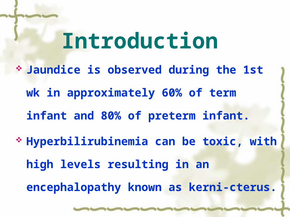
Introduction Jaundice is observed during the 1st wk in
approximately 60% of term infant and 80% of
preterm infant.
Hyperbilirubinemia can be toxic, with high
levels resulting in an encephalopathy known
as kerni-cterus.
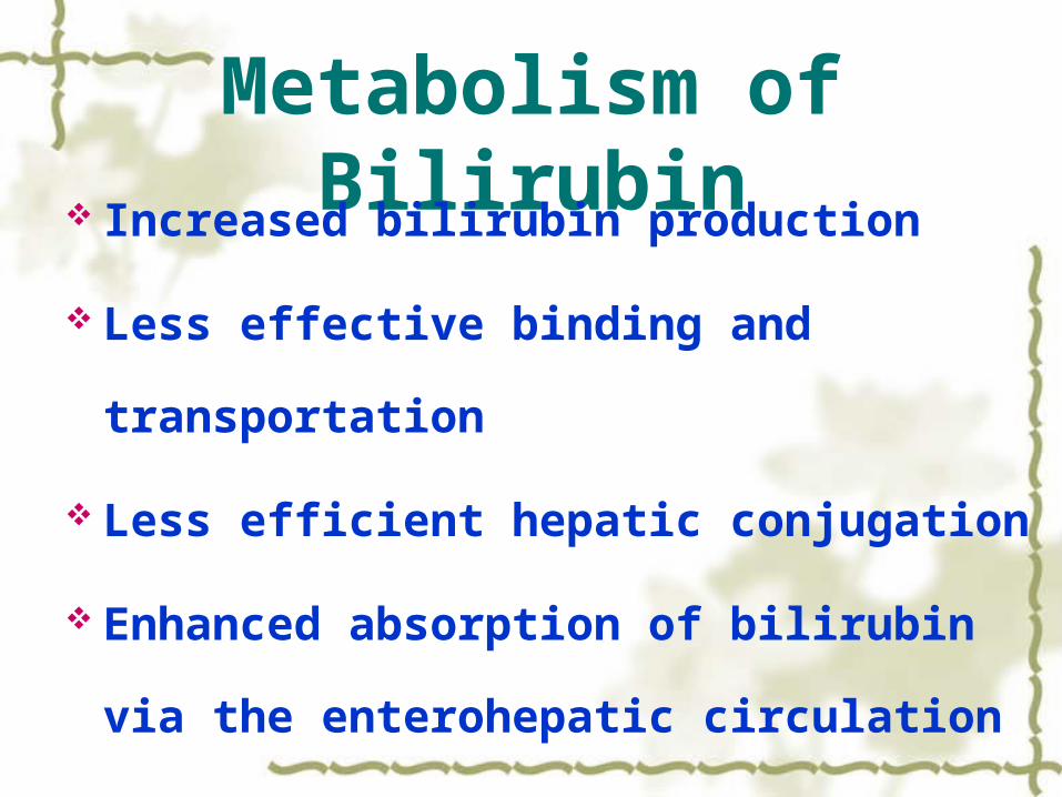
Metabolism of Bilirubin Increased bilirubin production
Less effective binding and transportation
Less efficient hepatic conjugation
Enhanced absorption of bilirubin via the
enterohepatic circulation
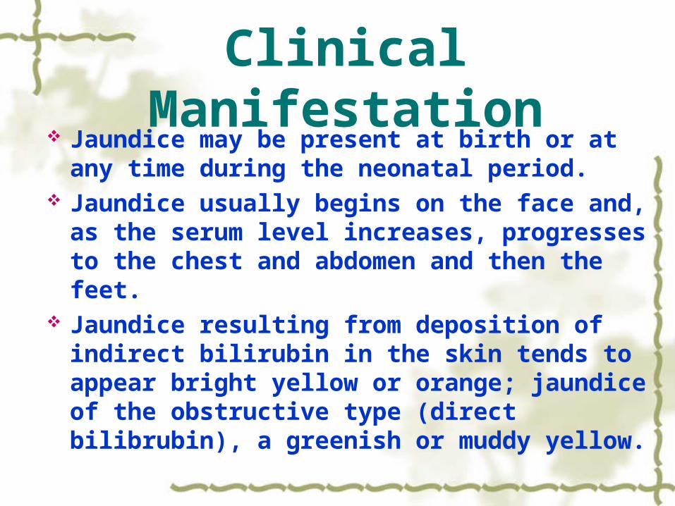
Clinical Manifestation Jaundice may be present at birth or at any
time during the neonatal period. Jaundice usually begins on the face and, as
the serum level increases, progresses to the chest and abdomen and then the feet.
Jaundice resulting from deposition of indirect bilirubin in the skin tends to appear bright yellow or orange; jaundice of the obstructive type (direct bilibrubin), a greenish or muddy yellow.

Methods of Diagnosis A complete diagnostic evaluation
Determination of direct and indicrect bilirubin fractions
Determination of hemoglobin Reticulocyte count Blood type Coombs’ test Examination of the peripheral blood smear

Classifications
Direct-reacting hyperbilirubinemia
Hepatitis
Cholestasis
Inborn errors of metabolism
Sepsis

Classifications Indirect-reacting hyperbilirubinemia
Hemolysis
Reticulocytosis
Evidences of red blood cell destruction
A positive Coomb’s test
Blood group incompatibility
Positive results of specific examination

ClassificationsDirect and indirect- reactin hyperbilirubinemia
Hepatitis
Sepsis
Liver damage complicated by Hemolysis
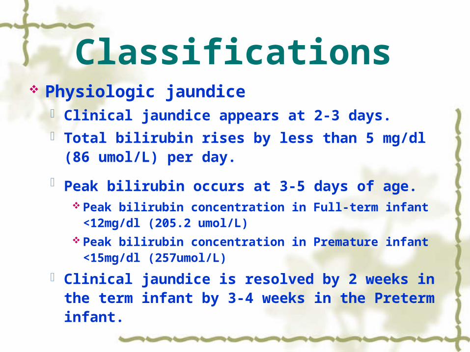
Classifications Physiologic jaundice
Clinical jaundice appears at 2-3 days. Total bilirubin rises by less than 5 mg/dl (86
umol/L) per day.
Peak bilirubin occurs at 3-5 days of age. Peak bilirubin concentration in Full-term infant <12mg/dl
(205.2 umol/L) Peak bilirubin concentration in Premature infant
<15mg/dl (257umol/L)
Clinical jaundice is resolved by 2 weeks in the term infant by 3-4 weeks in the Preterm infant.
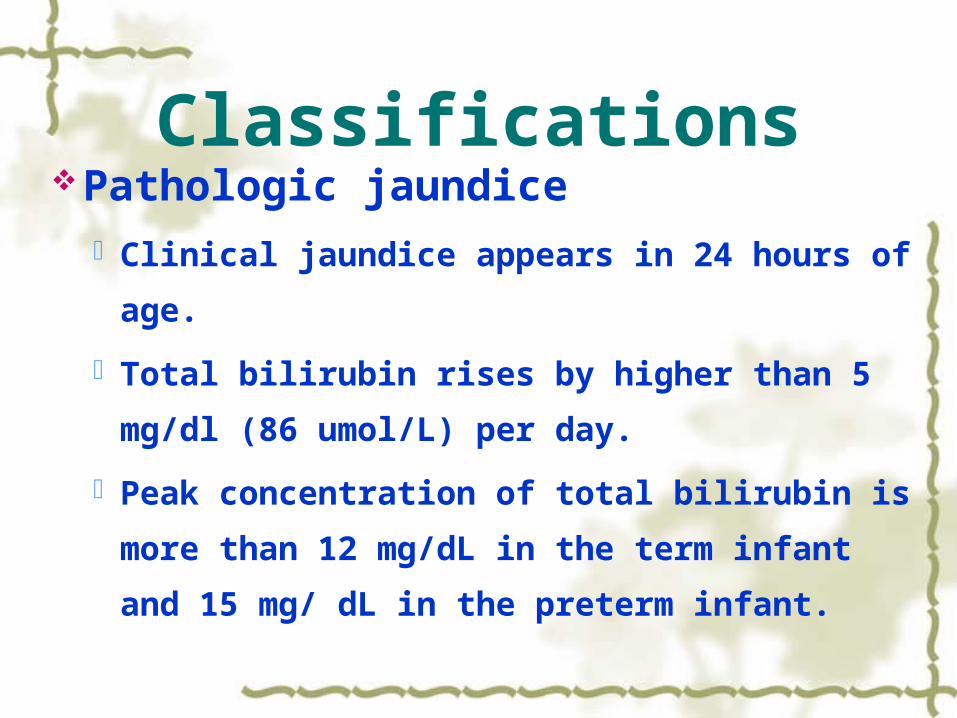
ClassificationsPathologic jaundice
Clinical jaundice appears in 24 hours of age.
Total bilirubin rises by higher than 5 mg/dl
(86 umol/L) per day.
Peak concentration of total bilirubin is more
than 12 mg/dL in the term infant and 15 mg/
dL in the preterm infant.

ClassificationsPathologic jaundice
Clinical jaundice is not resolved in 2
weeks in the term infant and in 4 weeks in
the Preterm infant.
Clinical jaundice appears again after it has
been resolved.
Direct bilirubin concentration is more than
1.5 mg/dL (26umol/L).

Causes of Pathologic Jaundice
Infective jaundice
Neonatal hepatitis
TORCH infection
Neonatal sepsis

Causes of Pathologic Jaundice
Jaundice associated without infection
Hemolytic disease of the newborn
ABO incompatibility
Rh incompatibility
Biliary atresia
Jaundice associated with breast- feeding
Causes of Pathologic Jaundice
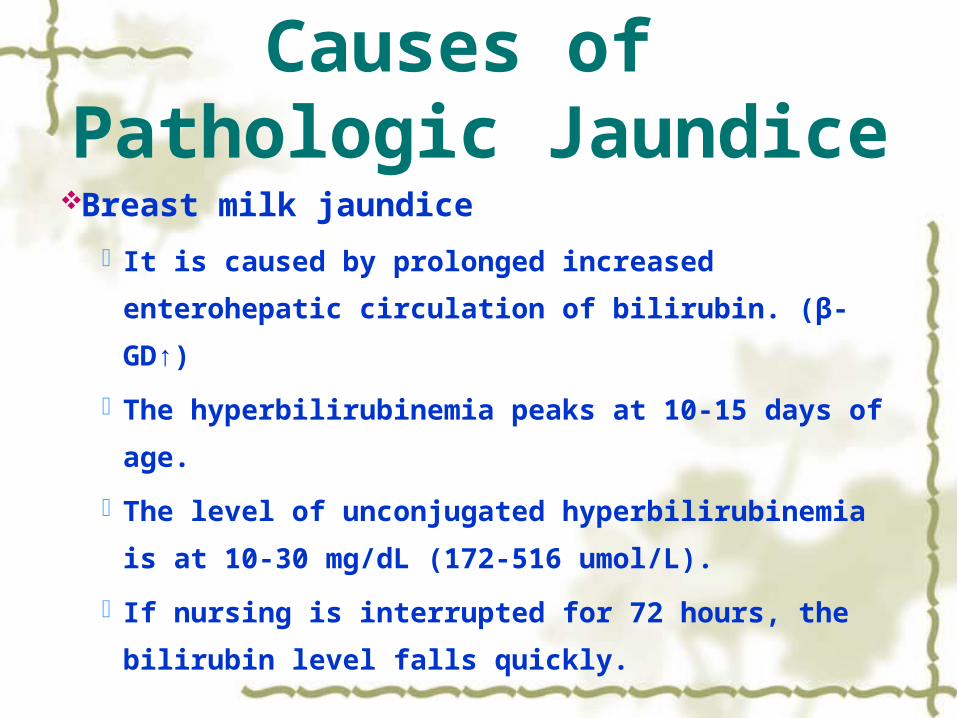
Breast milk jaundice
It is caused by prolonged increased enterohepatic
circulation of bilirubin. (β-GD↑)
The hyperbilirubinemia peaks at 10-15 days of
age.
The level of unconjugated hyperbilirubinemia is at
10-30 mg/dL (172-516 umol/L).
If nursing is interrupted for 72 hours, the bilirubin
level falls quickly.

Causes of Pathologic Jaundice
Genetic disease Congenital deficiencies of the enzymes
glucose-6-phosphate dehydrogenase (G-6-PD)
Thalassemia Cystic fibrosis
Drug Vitamin k Novobiocin
Hemolytic Disease of
the Newborn
Li weizhong

Introduction Hemolytic disease of the newborn
It is an isoimmunity hemolysis associated with ABO or Rh incompatibility.
It results from transplacental passage of maternal antiboddy active against RBC antigens of the infant, leading to an increased rate of RBC destruction.
It is an important cause of anemia and jaundice in newborn infant.

Etiology and Pathogenesis
ABO hemolytic disease
ABO incompatibility
Type O mothers
Type A or B fetuses
Presence of IgG anti-A or Anti-B antibodies in
type O mother
Frequently occurring during the first pregnancy
without prior sensitization
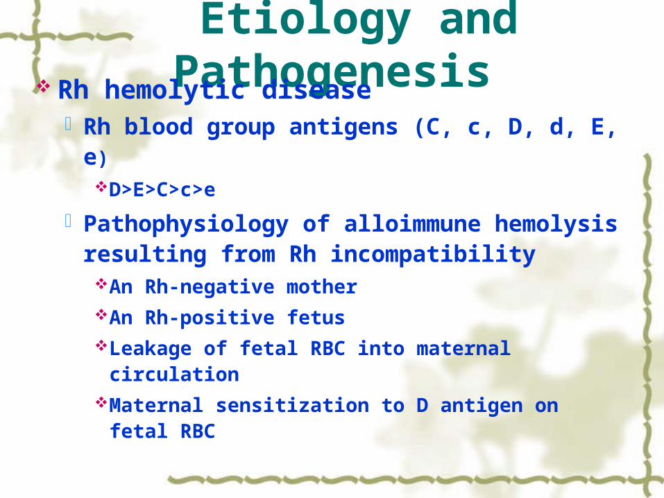
Etiology and Pathogenesis Rh hemolytic disease
Rh blood group antigens (C, c, D, d, E, e)D>E>C>c>e
Pathophysiology of alloimmune hemolysis resulting from Rh incompatibilityAn Rh-negative motherAn Rh-positive fetusLeakage of fetal RBC into maternal circulationMaternal sensitization to D antigen on fetal RBC

Etiology and PathogenesisProduction and transplacental passage
of maternal anti-D antibodies into fetal
circulation
Attachment of maternal antibodies to
Rh-positive fetal RBC
Destruction of antibody-coated fetal
RBC

Etiology and Pathogenesis Rh hemolytic disease was rare during the first
pregnancy involving an Rh-positive fetus.
Once sensitization has occurred, re-exposure
to Rh D RBC in subsequent pregnancies leads
to an anamnestic response, with an increase
in the maternal anti-Rh D antibody titer.
The likelihood of an infant being affected
increased significantly with each subsequent
pregnancy.

Etiology and Pathogenesis Significant hemolysis occurring in the
first pregnancy indicates prior maternal exposure to Rh-positive RBC.Fetal bleeding associated with a previous
spontaneous or therapeutic abortionEctopic pregnancyA variety of different prenatal proceduresTransfusion of some other blood product
containing Rh D RBC in an Rh-negative mother

Clinical Manifestations Jaundice
Anemia
Hydrops
Massive enlargement of the liver and
spleen
Bilirubin encephalopathy (Kernicterus)
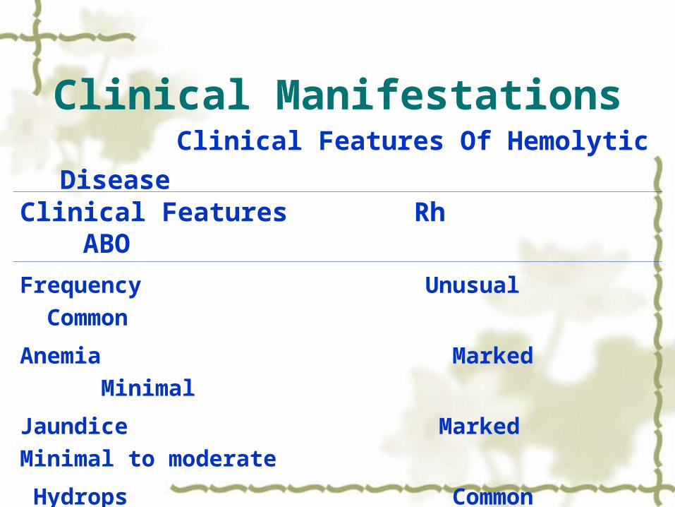
Clinical Manifestations Clinical Features Of Hemolytic Disease
Clinical Features Rh ABO
Frequency Unusual Common
Anemia Marked Minimal
Jaundice Marked Minimal to moderate
Hydrops Common Rare
Hepatosplenomegaly Marked Minimal
Kernicterus Common Rare

Laboratory Diagnosis Laboratory Features Of Hemolytic Disease Laboratory Features Rh ABO
blood type of Mother Rh negative O
blood type of Infant Rh positive A or B
Anemia Marked Minimal
Direct Commb’s test Positive Negative
Indirect Commb’s test Positive Usually positive
Hyperbilirubinemia marked Variable
RBC morphology Nucleated RBC Spherocytes

Diagnosis
The definitive diagnosis requires
demonstration of blood group
incompatibility and of corresponding
antibody bound to the infant’s RBC.

Diagnosis Antenatal Diagnosis
History Expectant parents’ blood types Maternal titer of IgG antibodies to D or E
(>1:32)
At 12 ~ 16 wkAt 28 ~ 32 wkAt 36 wk
Fetal Rh and ABO status Fetal jaundice level
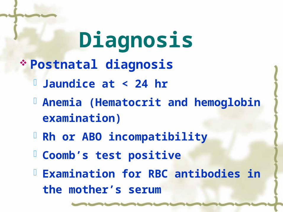
Diagnosis Postnatal diagnosis
Jaundice at < 24 hr
Anemia (Hematocrit and hemoglobin
examination)
Rh or ABO incompatibility
Coomb’s test positive
Examination for RBC antibodies in the
mother’s serum

Differential Diagnosis
Congenital nephrosis
Neonatal anemia
Physiological jaundice
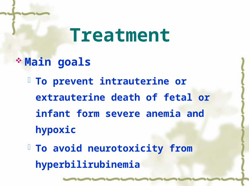
Treatment Main goals
To prevent intrauterine or extrauterine
death of fetal or infant form severe anemia
and hypoxic
To avoid neurotoxicity from
hyperbilirubinemia

Treatment Treatment of the unborn infant
Utero transfusion Indication
Hydrops Anemia (Hematocrit<30%)
Method Packed RBC matching with the mother’s
serum Umbilical vein transfusion

Treatment Delivery in advance
Indication
Pulmonary maturity
Fetal distress
Maternal titer of Rh antibodies > 1:32
35 ~ 37 wk of gestation

Treatment Treatment of the liveborn infant
Immediate resuscitation and supportive therapyTemperature stabilizationCorrection of acidosis: 1-2mEq/kg of sodium
bicarbonateA small transfusion compatible packed RBCVolume expansion for hypotensionProvision of assisted ventilation for respiratory
failure
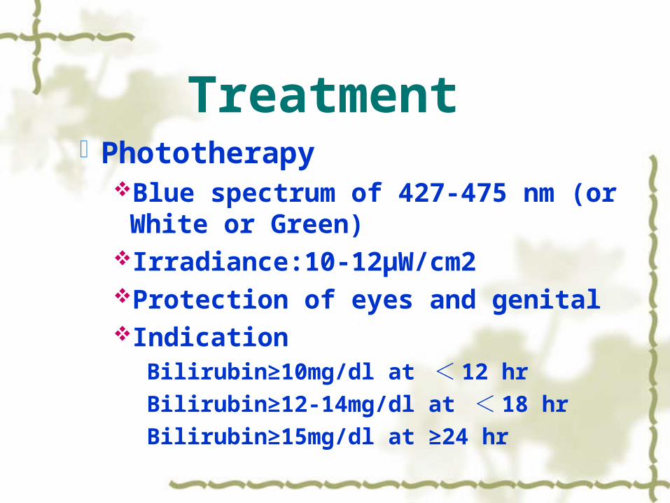
Treatment Phototherapy
Blue spectrum of 427-475 nm (or White or Green)
Irradiance:10-12μW/cm2Protection of eyes and genital Indication
Bilirubin≥10mg/dl at < 12 hr
Bilirubin≥12-14mg/dl at < 18 hr
Bilirubin≥15mg/dl at ≥24 hr

Treatment Side effect of phototherapy
Diarrhea
Dehydration
Riboflavin destruction
Hypocalcemia
Bronze-baby syndrome

Treatment Exchange transfusion
Indication Hemoglobin < 120g/L Hydrops, hepatosplenomegaly and heart failure Bilirubin in the 1st 12 of life>0.75mg/dl/hr Bilirubin concentration>20mg/dl Factors supporting early exchange transfusion:
Previous kernicterus in a sibling, reticulocyte counts greater than 15%, asphyxia of neonate and premature infant

Treatment Blood volume of exchange transfusion
Double-volume exchange transfusion :150-
180ml/kg
Blood choose of Rh incompatibility Rh in accordance with mother
ABO in accordance with neonate
Blood choose of ABO incompatibility Plasm of AB type
RBC of O type

Treatment Drug treatment
Intravenous immune globulin
(IVIG)
Human albumin
Protoporphyrins : Sn-PP; Zn-PP
Glucocorticoids: Dexamethasone
Inducer of liver enzyme: Luminal

Prevention Intramuscular injection of 300ug of human
anti-D globulin to an Rh-negative mother
Within 72 hr of delivery of an ectopic pregnancy
Abdominal trauma in pregnancy
Amniocentesis
Chorionic villus biopsy
Abortion