Neonatal Hyperbilirubinemia & Phototherapy · jaundice Identify the ... Neonatal age Physiologic...
17
1 Neonatal Hyperbilirubinemia & Phototherapy Instructor: Sarah Starling Outline: Janet Mendis, MSN, RNC-NIC, CNS Summer Morgan RNC-NIC, CPNP UC San Diego Health System Objectives Describe the mechanism of physiolgic jaundice Identify the mechanisms predisposing the infant to physiological jaundice Discuss the use of phototherapy for hyperbilirubinemia Apply assessment criteria foe the infant who is jaundiced Identify risk factors for polycythemia Identify the treatment and nursing interventions for polycythemia What is Jaundice? Yellowish discoloration of skin and sclera of newborns due to bilirubin Also known as jaundice of the newborn…
Transcript of Neonatal Hyperbilirubinemia & Phototherapy · jaundice Identify the ... Neonatal age Physiologic...
1
Neonatal Hyperbilirubinemia &
Phototherapy
Instructor: Sarah Starling Outline: Janet Mendis, MSN, RNC-NIC, CNS
Summer Morgan RNC-NIC, CPNPUC San Diego Health System
Objectives
Describe the mechanism of physiolgicjaundice
Identify the mechanisms predisposing theinfant to physiological jaundice
Discuss the use of phototherapy forhyperbilirubinemia
Apply assessment criteria foe the infant whois jaundiced
Identify risk factors for polycythemia Identify the treatment and nursing
interventions for polycythemia
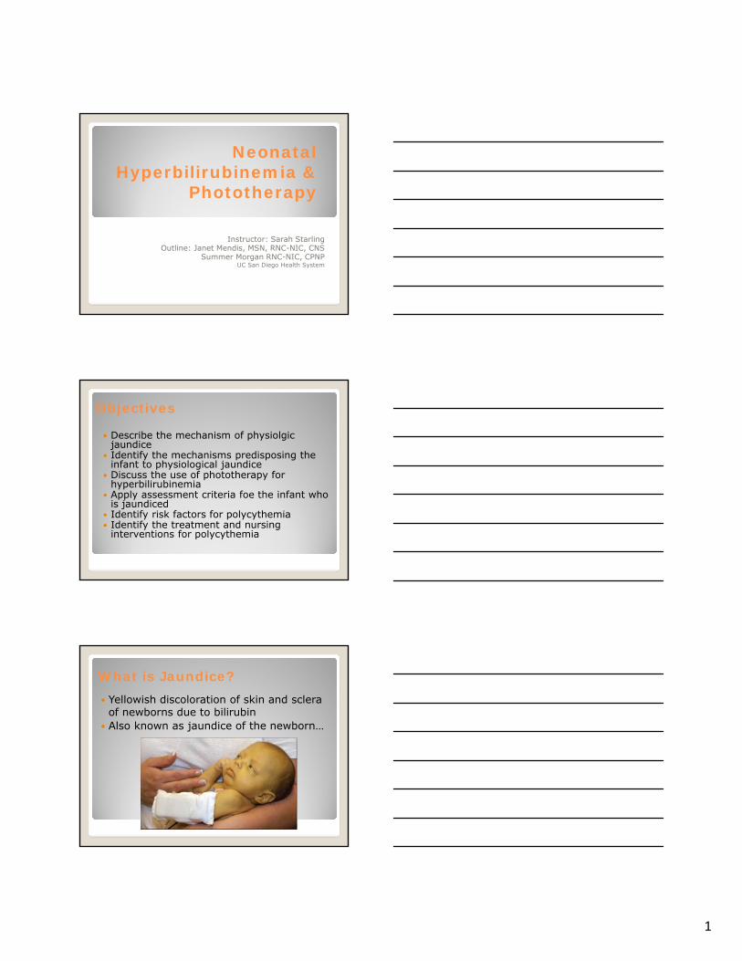
What is Jaundice? Yellowish discoloration of skin and sclera
of newborns due to bilirubin Also known as jaundice of the newborn…
2
Definitions:
Hyperbilirubinemia- an elevated totalserum bilirubin (TSB) level◦ Abnormal values vary by age, days of life,current illness and conditions
Jaundice- the yellowish coloration of theskin and sclera caused by presence ofbilirubin in elevated concentrations
Definitions:
Conjugated- Direct SerumHyperbilirubinemia
Unconjugated- Indirect Hyperbilirubinemia Kernicterus- Irreversible, chronic sequelae
of bilirubin toxicity
Bilirubin:
By-product of the breakdown of red bloodcells
Presents as a higher than normal level ofbilirubin in the blood for the gestationalage and chronicle age in hours
A breakdown of 1 gram of hemoglobin=35mg of bilirubin
3
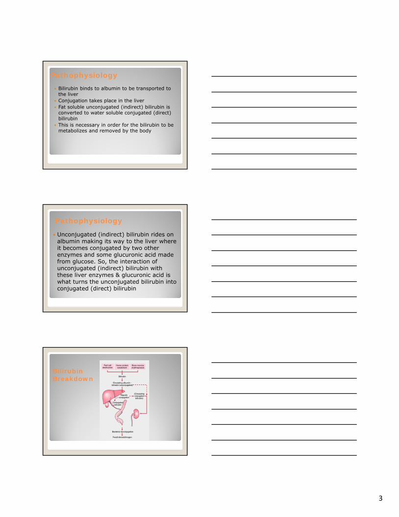
Pathophysiology Bilirubin binds to albumin to be transported to
the liver Conjugation takes place in the liver Fat soluble unconjugated (indirect) bilirubin is
converted to water soluble conjugated (direct)bilirubin
This is necessary in order for the bilirubin to bemetabolizes and removed by the body
Pathophysiology
Unconjugated (indirect) bilirubin rides onalbumin making its way to the liver whereit becomes conjugated by two otherenzymes and some glucuronic acid madefrom glucose. So, the interaction ofunconjugated (indirect) bilirubin withthese liver enzymes & glucuronic acid iswhat turns the unconjugated bilirubin intoconjugated (direct) bilirubin
Bilirubin Breakdown
4
Pathophysiology In order for bilirubin to clear from the body it must
be:
Conjugated in liver, excreted in bile, and eliminatedvia urine and stool
Most common reason that neonates need medicalattention:
“Physiologic jaundice”-a normal phenomenon during transition, butdifferent from “Pathological jaundice”
Becomes concerning when levels continue to riseUnconjugated bilirubin is NEUROTOXIC
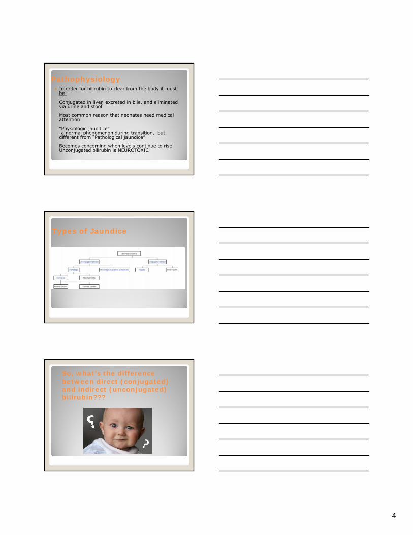
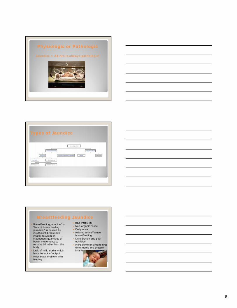
Types of Jaundice
So, what’s the difference between direct (conjugated) and indirect (unconjugated) bilirubin???
5
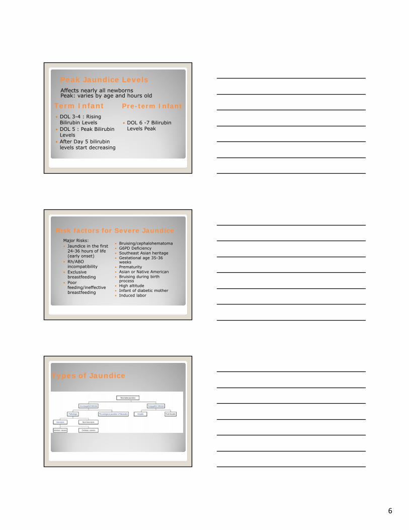
This form of bilirubin does not dissolve in water (it is insoluble). Indirect bilirubin travels through the bloodstream to the liver, where it is changed into a soluble form (conjugated or direct)
Direct bilirubin dissolves in water (it is soluble) and is made by the liver from indirect bilirubin
Unconjugated(Indirect bilirubin)
Conjugated(Direct Bilirubin)
Unconjugated (Indirect Bilirubin)• Elevated levels of bilirubin are caused by
any of the following:imbalance in production, transport, uptake, conjugation, excretion, and reabsorption • This is most concerning due to risk for
encephalopathy/kernicterus if not treatedrapidly
Conjugated(Direct Bilirubin)• Considered elevated when:
• Level > 2.0 mg/dL (severe > 5.0mg/dL)
• Level > 15% of total serum bilirubin
• Risk factors:• Low gestational age• Early and/or prolonged exposure to
TPN• Lack of enteral feeding• Sepsis
6
Term Infant DOL 3-4 : Rising
Bilirubin Levels DOL 5 : Peak Bilirubin
Levels After Day 5 bilirubin
levels start decreasing
Pre-term Infant
DOL 6 -7 BilirubinLevels Peak
Peak Jaundice LevelsAffects nearly all newbornsPeak: varies by age and hours old
Risk factors for Severe JaundiceMajor Risks: Jaundice in the first
24-36 hours of life(early onset)
Rh/ABOincompatibility
Exclusivebreastfeeding
Poorfeeding/ineffectivebreastfeeding
Bruising/cephalohematoma G6PD Deficiency Southeast Asian heritage Gestational age 35-36
weeks Prematurity Asian or Native American Bruising during birth
process High altitude Infant of diabetic mother Induced labor
Types of Jaundice
7
Pathological Jaundice Definition: -Increase of bilirubin >0.2 mg/dL/hr -Jaundice in the first 24-36 hours of age -Persists> 7days
Underlying Causes: -Increased bilirubin production -Decreased bilirubin excretion -Combo of both
Physiological Jaundice Most infants develop visible jaundice due to elevation of
unconjugated bilirubin concentration during their first week This pattern of Hyperbilirubinemia has been classified into
two functionally distinct periods
Phase one◦ Term infants - jaundice lasts for about 10 days with a rapid rise of
serum bilirubin ◦ Preterm infants - jaundice lasts for about two weeks, with a rapid
rise of serum bilirubin Phase two◦ Bilirubin levels decline for two weeks, eventually mimicking adult
values◦ Preterm infants - phase two can last more than one month◦ Exclusively breastfed infants - phase two can last more than one
month
Physiological Jaundice Causes Low enzyme activity◦ Glucuronosyltransferase which normally converts
unconjugated bilirubin to conjugated bilirubin that can be excreted into the gastrointestinal tract Before birth, this enzyme is actively down-regulated,
since bilirubin needs to remain unconjugated in order to cross the placenta to avoid being accumulated in the fetus
After birth, it takes some time for this enzyme to gain function
Shorter life span of fetal red blood cells Relatively low conversion of bilirubin to
urobilinogen by the intestinal flora◦ Results in relatively high absorption of bilirubin back into
the circulation
8
Physiologic or Pathologic
Jaundice < 24 hrs is always pathologic!
Types of Jaundice
Breastfeeding Jaundice KEY POINTS Non-organic cause Early onset Related to ineffective
breastfeeding Dehydration and poor
nutrition More common among first
time moms and preterminfants
Breastfeeding jaundice" or"lack of breastfeedingjaundice," is caused byinsufficient breast milkintake, resulting ininadequate quantities ofbowel movements toremove bilirubin from thebody.
Lack of milk intake whichleads to lack of output
Mechanical Problem withfeeding
9
Where as ‘breast milk’jaundice is a biochemicaloccurrence and the higherbilirubin possibly acts asan antioxidant. Breastmilk jaundice occurs laterin the newborn period,with the bilirubin levelusually peaking in thesixth to 14th days of life.This late-onset jaundicemay develop in up to onethird of healthy breastfedinfants
KEY POINTS Non-hemolytic jaundice Late onset Begins after day 3-5 of
life Increased bilirubin levels
peak between 5-10mg/dLat about 2 weeks of life
May persist for months Develops in about 2-4%
of breastfed newborns Runs in families
Breast Milk Jaundice
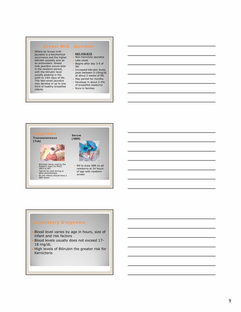
DiagnosisTranscutaneous(Tcb)
Serum(SBR)
Bilimeter being used by thePediatric team on FMCC (MD’s & NP)
Cannot be used during or after phototherapy
At risk infants should have a SBR drawn
RN to draw SBR on all newborns at 24 hours of age with newbornscreen
Blood level varies by age in hours, size ofinfant and risk factors
Blood levels usually does not exceed 17-18 mg/dL
High levels of Bilirubin the greater risk forKernicteris
Laboratory Diagnosis
10
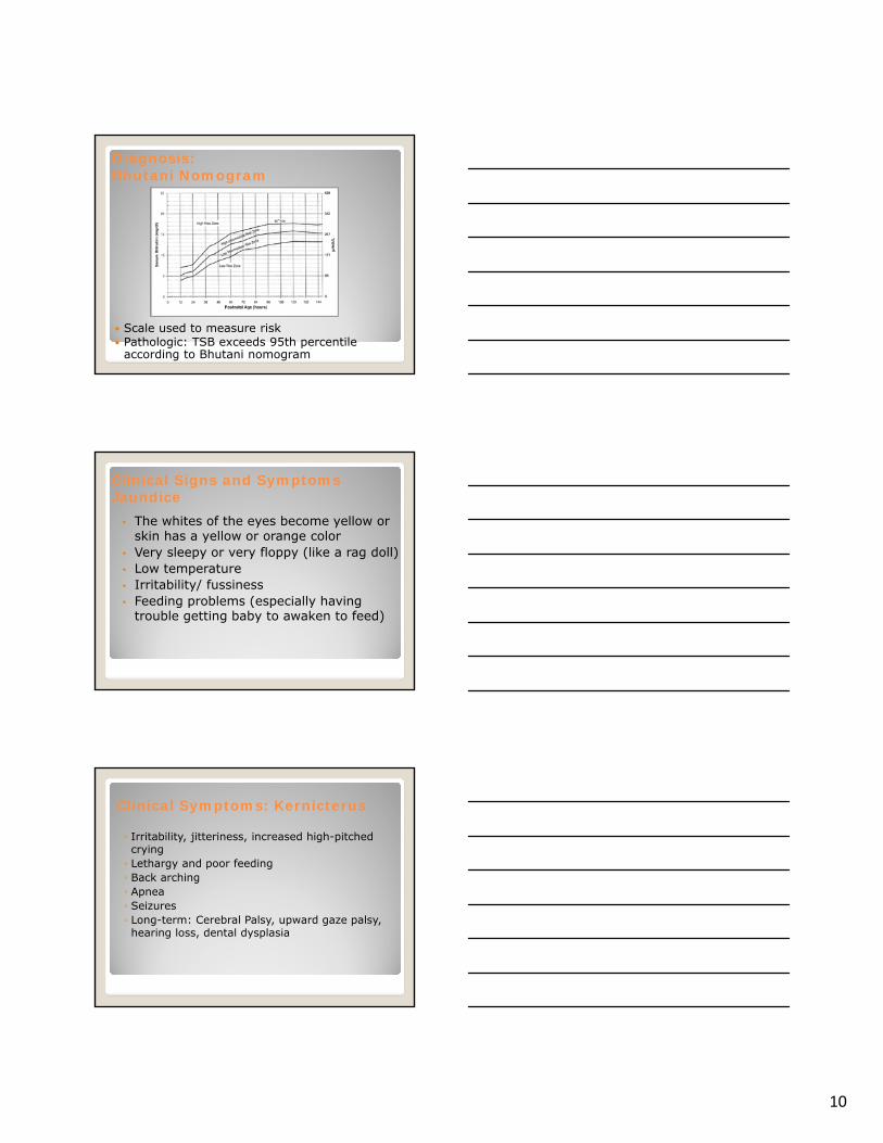
Diagnosis:Bhutani Nomogram
Scale used to measure risk Pathologic: TSB exceeds 95th percentile
according to Bhutani nomogram
Clinical Signs and Symptoms Jaundice
• The whites of the eyes become yellow orskin has a yellow or orange color
• Very sleepy or very floppy (like a rag doll)• Low temperature• Irritability/ fussiness• Feeding problems (especially having
trouble getting baby to awaken to feed)
Clinical Symptoms: Kernicterus
◦ Irritability, jitteriness, increased high-pitchedcrying◦ Lethargy and poor feeding◦ Back arching◦ Apnea◦ Seizures◦ Long-term: Cerebral Palsy, upward gaze palsy,hearing loss, dental dysplasia
11
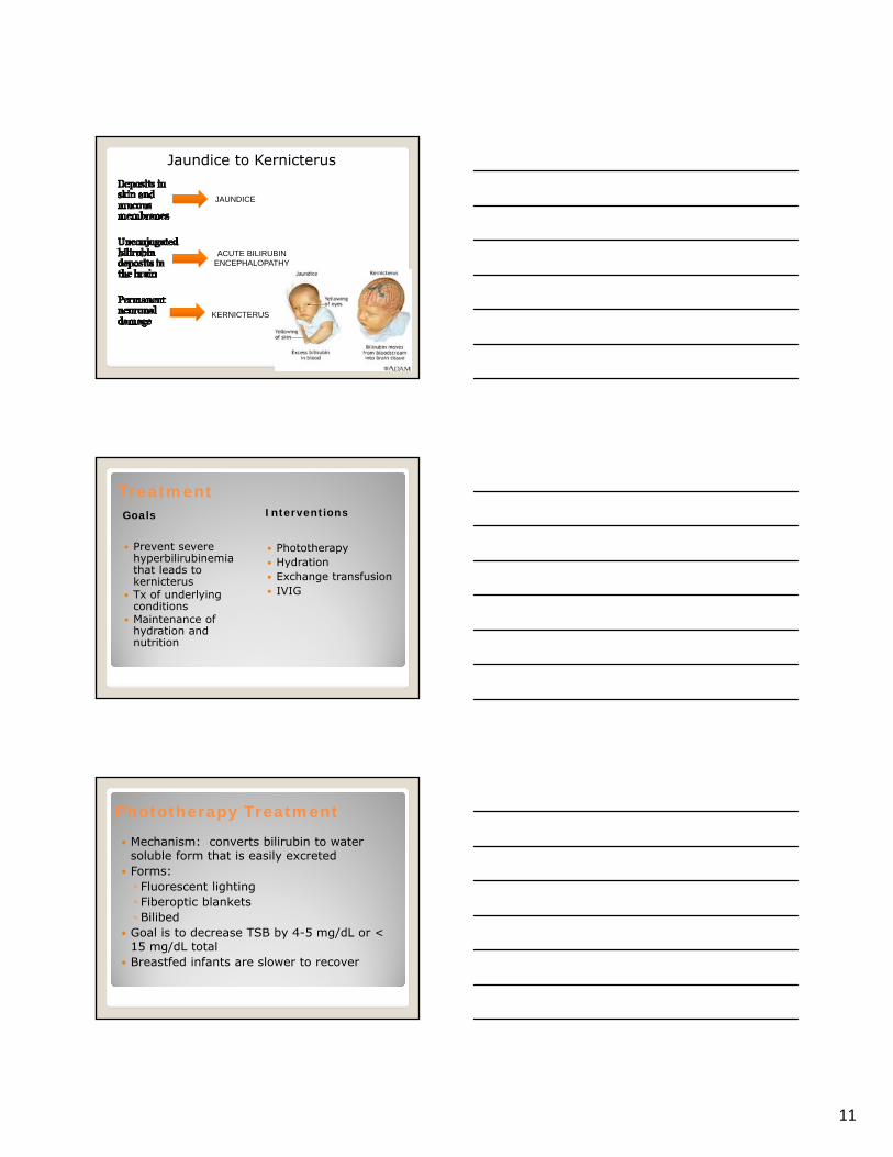
JAUNDICE
ACUTE BILIRUBIN ENCEPHALOPATHY
KERNICTERUS
Jaundice to Kernicterus
TreatmentGoals Interventions
Prevent severehyperbilirubinemiathat leads tokernicterus
Tx of underlyingconditions
Maintenance ofhydration andnutrition
Phototherapy Hydration Exchange transfusion IVIG
Phototherapy Treatment
Mechanism: converts bilirubin to watersoluble form that is easily excreted
Forms:◦ Fluorescent lighting◦ Fiberoptic blankets◦ Bilibed
Goal is to decrease TSB by 4-5 mg/dL or <15 mg/dL total
Breastfed infants are slower to recover

12
Phototherapy
Effectiveness: Important factors- Spectrum, irradiance, distance, surface area.
Phototherapy Treatment Primary treatment is Phototherapy Helps to breakdown bilirubin so that it can
clear through the stool and urine Does not treat the underlying cause
Treatment:Prevent Kernicterus:• Observe baby for jaundice• Evaluate each baby’s risk factors for
jaundice• Thorough Handoff report & provider
communication• Educate families• Bilicheck before discharge:• At UCSD, all babies receive a serum
bilirubin draw at the time of newbornscreen. Results are plotted on the HourSpecific Bilirubin Nomogram
13
Treatment:Prevent Kernicterus:
• Damage fromKernicterus isIrreversible
• Prevention is Key!!!!!!!
Nursing Care: Know the device you are working with distance
between device and baby How do you check irradiance and what is the output
range for the device hours of use Eye shields if necessary Monitor for hydration status/knowing what stools
should look like Monitor time out for feeding and holding
Thermoregulation What to do with a fussy baby in phototherapy
Nursing Assessment
• Assessment should be done q-shift, moreoften for high risk infants
• Evaluate risk factors• Check Bilirubin levels on all infants before
discharge• Plot on Bhutani Curve nomogram and
assess risk zone and any changes
14
• Promote and support successful breastfeeding• Perform a systematic assessment before
discharge for the risk of severehyperbilirubinemia
• Provide early and focused follow-up based onassessment
• When indicated, treat newborns withphototherapy or exchange transfusion to preventthe development of severe hyperbilirubinemiaand, possibly, bibirubin encephalopathy(kernicterus).
• (Pediatrics 2004; 114:297-316)
APA Practice Guideline (2004): Hyperbilirubinemia
• Early discharges (before 48hrs of life)• Make sure adequate follow-up will be possible• All babies should be seen 24-48 hours post
discharge by HCP for follow-up and assessment• Lactation support for breastfeeding families is
important• Educate families on what to look for, how to get
help and what risk factors their child has
Education, Education, Education!
Discharge Precautions
Definitions
Polycythemia is increased total RBC mass◦ Central venous hematocrit > 65% Above 65% blood viscosity rises exponentially
Polycythemic hyperviscosity is increasedviscosity of the blood resulting fromincreased numbers of RBCs◦ Not all polycythemic infants have symptoms ofhyperviscosity
15
Conditions that alter incidence
Altitude: increased RBC mass Neonatal age◦ Physiologic increase in hematocrit due to fluidshifts away from intravascular compartmentwith maximum at 2-4 hours of age
Obstetric factors: delayed cord clampingor “stripping” of the umbilical cord
High-risk delivery, especially if precipitous
Perinatal processes
Enhanced fetal erythropoiesis usuallyrelated to fetal hypoxia◦ Placental insufficiency Maternal hypertension, abruption, post-dates,IUGR, maternal smoking
◦ Endocrine disorders: due to increased oxygenconsumption IDM (>40% incidence), congenital thyrotoxicosis,CAH, Beckwith-Wiedemann syndrome (hyperinsulinism)
Hypertransfusion
Delayed cord clamping Placental vessels contain 1/3 of the fetal blood
volume, half of which will be returned within 1 minute
Gravity: positioning below the placentawill increase placental transfusion
Meds: oxytocin can increasecontractions and thus transfusion Decreased in c-section b/c no contractions
Twin-twin transfusion Maternal-fetal transfusion Intrapartum asphyxia
Enhances net umbilical flow toward the infant, while acidosis increases capillary leak leading to reduced plasma volume
16
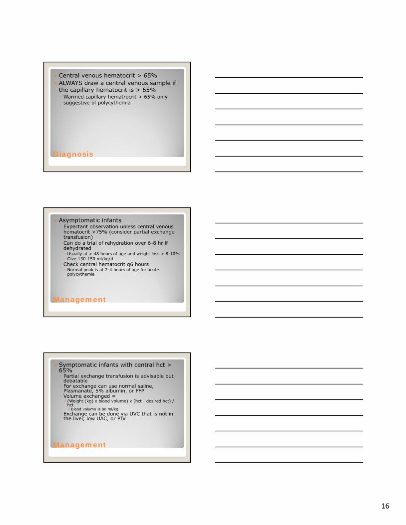
Diagnosis
Central venous hematocrit > 65% ALWAYS draw a central venous sample if
the capillary hematocrit is > 65%◦ Warmed capillary hematrocrit > 65% onlysuggestive of polycythemia
Management
Asymptomatic infants◦ Expectant observation unless central venoushematocrit >75% (consider partial exchangetransfusion)◦ Can do a trial of rehydration over 6-8 hr ifdehydrated Usually at > 48 hours of age and weight loss > 8-10% Give 130-150 ml/kg/d
◦ Check central hematocrit q6 hours Normal peak is at 2-4 hours of age for acute
polycythemia
Management
Symptomatic infants with central hct >65%◦ Partial exchange transfusion is advisable butdebatable◦ For exchange can use normal saline,Plasmanate, 5% albumin, or FFP◦ Volume exchanged = (Weight (kg) x blood volume) x (hct - desired hct) /
hct◦ Blood volume is 80 ml/kg
◦ Exchange can be done via UVC that is not inthe liver, low UAC, or PIV
17
Summary Consider the risk factors, particularly prematurity
and hemolysis Follow up is key! Consider how well baby is feeding, parents’
ability to return to the doctors office. The higher the number of risk factors, the lower
the level at which to intervene Sometimes, you will be surprised. We can’t
always prevent Hyperbilirubinemia, but weshould always prevent kernicterus
References:• American Academy of Pediatrics, Subcommittee on Hyperbilirubinemia.
American Academy of Pediatrics, Steering Committee on Quality Improvement and Management. Classification of
recommendations for clinical practice guidelines. Pediatrics. 2004;114:874-877
• Fanaroff and Martin chapters on hyperbilirubinemiaGartner LM, Herschel M. Jaundice and breastfeeding. Pediatr Clin
North Am. 2001;48:389-399• Johnson LH, Bhutani VK, Brown AK. System-based approach to
management of neonatal jaundice and prevention of kernicterus. J Pediatr. 2002;140:396-403Lange: “Neonatology: Management, Procedures, On-Call Problems, Diseases and Drugs”
• Keren R et al. Visual assessment of jaundice in term and late preterm infants. Arch Dis Child Fetal Neonatal Ed. 2009 Sep;94(5):F317-22. Epub 2009 Mar 22.
• Mishra S et al. Transcutaneous bilirubinometry reduces the need forblood sampling in neonates with visible jaundice. Acta Paediatr.
2009 Dec;98(12):1916-9. Epub 2009 Oct 7. • Management of hyperbilirubinemia in the newborn infant 35 or more
weeks of gestation. Pediatrics. 2004;114:297-316
All images found on google image.