Patología Respiratoria neonatal Síndrome de distress respiratorio neonatal.


Neonatal Emergencies

NoticeMedicine is an ever-changing science. As new research and clinical experience broaden our knowledge, changes in treatment and drug therapy are required. The authors and the publisher of this work have checked with sources believed to be reliable in their efforts to provide information that is complete and gener-ally in accord with the standards accepted at the time of publication. However, in view of the possibility of human error changes in medical sciences, neither the editors nor the publisher nor any other party who has been involved in the preparation or publication of this work warrants that the information contained herein is in every respect accurate or complete, and they disclaim all responsibil-ity for any errors or omissions or for the results obtained from use of the informa-tion contained in this work. Readers are encouraged to confi rm the information contained herein with other sources. For example and in particular, readers are advised to check the product information sheet included in the package of each drug they plan to administer to be certain that the information contained in this work is accurate and that changes have not been made in the recommended dose or in the contraindications for administration. This recommendation is of particular importance in connection with new or infrequently used drugs.

Neonatal Emergencies
Richard M. Cantor, MD, FAAP, FACEPAssociate Professor of Emergency Medicine
Pediatric Emergency DepartmentState University of New York—Upstate Medical University
Syracuse, New York
P. David Sadowitz, MDAssociate Professor of Emergency Medicine
Pediatric Emergency DepartmentState University of New York—Upstate Medical University
Syracuse, New York
New York Chicago San Francisco Lisbon LondonMadrid Mexico City Milan New Delhi San Juan
Seoul Singapore Sydney Toronto
Medical

Copyright © 2010 by The McGraw-Hill Companies, Inc. All rights reserved. Except as permitted under the United States CopyrightAct of 1976, no part of this publication may be reproduced or distributed in any form or by any means, or stored in a database orretrieval system, without the prior written permission of the publisher.
ISBN: 978-0-07-171398-6
MHID: 0-07-171398-0
The material in this eBook also appears in the print version of this title: ISBN: 978-0-07-147020-9, MHID: 0-07-147020-4.
All trademarks are trademarks of their respective owners. Rather than put a trademark symbol after every occurrence of a trademarked name, we use names in an editorial fashion only, and to the benefit of the trademark owner, with no intention ofinfringement of the trademark. Where such designations appear in this book, they have been printed with initial caps.
McGraw-Hill eBooks are available at special quantity discounts to use as premiums and sales promotions, or for use in corporatetraining programs. To contact a representative please e-mail us at [email protected].
TERMS OF USE
This is a copyrighted work and The McGraw-Hill Companies, Inc. (“McGraw-Hill”) and its licensors reserve all rights in and to thework. Use of this work is subject to these terms. Except as permitted under the Copyright Act of 1976 and the right to store andretrieve one copy of the work, you may not decompile, disassemble, reverse engineer, reproduce, modify, create derivative worksbased upon, transmit, distribute, disseminate, sell, publish or sublicense the work or any part of it without McGraw-Hill’s prior con-sent. You may use the work for your own noncommercial and personal use; any other use of the work is strictly prohibited. Your rightto use the work may be terminated if you fail to comply with these terms.
THE WORK IS PROVIDED “AS IS.” McGRAW-HILL AND ITS LICENSORS MAKE NO GUARANTEES OR WARRANTIESAS TO THE ACCURACY, ADEQUACY OR COMPLETENESS OF OR RESULTS TO BE OBTAINED FROM USING THEWORK, INCLUDING ANY INFORMATION THAT CAN BE ACCESSED THROUGH THE WORK VIA HYPERLINK OR OTH-ERWISE, AND EXPRESSLY DISCLAIM ANY WARRANTY, EXPRESS OR IMPLIED, INCLUDING BUT NOT LIMITED TOIMPLIED WARRANTIES OF MERCHANTABILITY OR FITNESS FOR A PARTICULAR PURPOSE. McGraw-Hill and itslicensors do not warrant or guarantee that the functions contained in the work will meet your requirements or that its operation willbe uninterrupted or error free. Neither McGraw-Hill nor its licensors shall be liable to you or anyone else for any inaccuracy, erroror omission, regardless of cause, in the work or for any damages resulting therefrom. McGraw-Hill has no responsibility for the con-tent of any information accessed through the work. Under no circumstances shall McGraw-Hill and/or its licensors be liable for anyindirect, incidental, special, punitive, consequential or similar damages that result from the use of or inability to use the work, evenif any of them has been advised of the possibility of such damages. This limitation of liability shall apply to any claim or cause what-soever whether such claim or cause arises in contract, tort or otherwise.

v
Contents
Contributors. . . . . . . . . . . . . . . . . . . . . . . . . . . . .. . . . . . . . . . . . . . . . . . . . . . . . . . . . . . . . . vii
Preface . . . . . . . . . . . . . . . . . . . . . . . . . . . . .. . . . . . . . . . . . . . . . . . . . . . . . . . . . . . . . . . . . ix
Acknowledgments . . . . . . . . . . . . . . . . . . . . . . . . . . . . .. . . . . . . . . . . . . . . . . . . . . . . . . . . . . xi
Chapter 1. HEENT Emergencies of the Infant . . . . . . . . . . . . . . . . . . . . . . . . . . . . . . . . . . 1 Deborah J. Mann, MD
Chapter 2. Neurologic Emergencies . . . . . . . . . . . . . . . . . . . . . . . . . . . . . . . . . . . . . . . . . 25 Linnea Wittick, MD
Chapter 3. Respiratory Emergencies . . . . . . . . . . . . . . . . . . . . . . . . . . . . . . . . . . . . . . . . . 57 Jennifer Mackey, MD
Chapter 4. Cardiac Emergencies . . . . . . . . . . . . . . . . . . . . . . . . . . . . . . . . . . . . . . . . . . . . 73 Jahn Avarello, MD
Chapter 5. Gastrointestinal Emergencies . . . . . . . . . . . . . . . . . . . . . . . . . . . . . . . . . . . . . . 101 Derek Cooney, MD Richard M. Cantor, MD, FAAP/FACEP
Chapter 6. Neonatal Genitourinary Emergencies . . . . . . . . . . . . . . . . . . . . . . . . . . . . . . . . 129 Brian Stout, MD
Chapter 7. Orthopedic Emergencies in the Neonate . . . . . . . . . . . . . . . . . . . . . . . . . . . . . 145 P. David Sadowitz, MD Lisa Keough, MD Norma Cooney, MD
Chapter 8. Dermatologic Disorders in the First 30 Days of Life . . . . . . . . . . . . . . . . . . . . . 157 James D’Agostino, MD

vi CONTENTS
Chapter 9. Neonatal Infections . . . . . . . . . . . . . . . . . . . . . . . . . . . . . . . . . . . . . . . . . . . . . 179 P. David Sadowitz, MD LaLainia Secreti, MD Jeff Lapoint, DO
Chapter 10. Hematologic Emergencies in the Neonate . . . . . . . . . . . . . . . . . . . . . . . . . . . . . 193 P. David Sadowitz, MD Trisha Tavares, MD
Chapter 11. Selected Topics in Neonatal Pharmacology . . . . . . . . . . . . . . . . . . . . . . . . . . . . 217 Jeanna Marraffa, PharmD Jamie Nelsen, PharmD
Index . . . . . . . . . . . . . . . . . . . . . . . . . . . . . . . . . . . . . . . . . . . . . . . . . . . . . . . . . . . . . . . . . 235

vii
Contributors
Jahn Avarello, MDDirector, Pediatric Emergency MedicineHuntington HospitalAttending, Pediatric Emergency MedicineNorth Shore University HospitalManhasset, New York
Richard M. Cantor, MD, FAAP, FACEPAssociate Professor of Emergency MedicinePediatric Emergency DepartmentState University of New York-Upstate
Medical UniversitySyracuse, New York
Derek Cooney, MDAssistant ProfessorDepartment of Emergency MedicineState University of New York-Upstate
Medical UniversitySyracuse, New York
Norma Cooney, MDAssistant ProfessorDepartment of Emergence MedicineState University of New York-Upstate
Medical UniversitySyracuse, New York
James D’Agostino, MDAssistant Professor of Emergency Medicine
and Pediatrics State University of New York-Upstate Medical University Syracuse, New York
Lisa Keough, MDAssistant ProfessorDepartment of Emergency
MedicineState University of New York-Upstate
Medical UniversitySyracuse, New York
Jeff Lapoint, DOResident PhysicianDepartment of Emergency
MedicineState University of New York-Upstate
Medical UniversitySyracuse, New York
Jennifer Mackey, MD, FAAPAssistant ProfessorDepartment of Emergency Medicine and
PediatricsState University of New York-Upstate
Medical UniversitySyracuse, New York
Deborah J. Mann, MDAssistant ProfessorDepartment of Emergency
MedicineState University of New York-Upstate
Medical UniversitySyracuse, New York

viii NEONATAL EMERGENCIES
Jeanna Marraff a, PharmDAssistant ProfessorDepartments of Emergency Medicine and
MedicineSection of Clinical PharmacologyState University of New York Upstate
Medical UniversitySyracuse, New York
Jamie L. Nelsen, PharmD, DABATAssistant ProfessorDepartment of Emergency MedicineState University of New York-Upstate
Medical UniversitySyracuse, New York
P. David Sadowitz, MDAssociate Professor of Emergency
MedicinePediatric Emergency DepartmentState University of New York-Upstate
Medical UniversitySyracuse, New York
LaLainia Secreti, MDAssistant ProfessorDepartment of Emergency MedicineState University of New York-Upstate
Medical UniversitySyracuse, New York
Brian Stout, MDAssistant ProfessorDepartment of Emergency MedicineState University of New York-Upstate
Medical UniversitySyracuse, New York
Trisha Tavares, MDAssistant ProfessorDepartment of PediatricsState University of New York-Upstate
Medical UniversitySyracuse, New York
Linnea Wittick, MDFellow in Pediatric Emergency MedicineDepartment of Emergency MedicineState University of New York-Upstate
Medical UniversitySyracuse, New York

ix
The delivery of emergency care to infants and children remains both a challenge and a privilege. It can be one of the most humbling yet rewarding experiences for the emergency health care provider. This text was developed to assist our colleagues in the evaluation and treatment of children of a young age. The gen-esis of this text arose from both clinical expe-rience and an obvious need within the practice of emergency medicine for a greater emphasis to be placed on these high risk infants. At such young developmental and chronological ages, these patients present with a miriad of undif-ferentiated complaints. Their histories may be short but the complexity of their problems
may indeed be quite complex. The goal of this text is to guide the provider in a systematic approach to any and all problems within this fragile population.
The text is divided into sections based on organ systems. There will be much cross-over within each section, only highlighting the commonalty of complaint that can result from a multitude of disparate medical problems. We are hopeful that our readers fi nd it to be a useful tool in addressing the needs of the very young infant.
Richard M. Cantor, MD, FAAP/FACEPP. David Sadowitz, MD
Preface

This page intentionally left blank

xi
To my valued friend and colleague Dr. Sadowitz, who has always served as a wonderful role model for excellence in the delivery of pedi-atric care.
To my mentors Drs. Oski, Tunnessen, and Stock-man, who have empowered me with the work ethic I practice today.
To my patients who have provided me with the blessed coverage of caring for them.
To my wife Nina, and my children Gillian and Liza, who energize, love, and support me every moment of everyday.
Richard M. Cantor
To my friend and colleague Dr. Cantor, whose wisdom, knowledge and wit have made this project a great learning experience.
To those who have taught me by their exam-ple and experience, my gratitude for their wis-dom and patience.
To students and practitioner of emergency medicine; it is my hope that the material in this book will be a valuable tool in the quest to provide excellent care to children in a busy ER setting.
To my wife Cheryl and my children Amy, Ben, Jared, Emily, Elizabeth, Ryan, Jordan, Mitchell, and Madeline for their constant love and support that have encouraged me in this endeavor.
To my God whose unfailing love and grace is the foundation of my life.
P. David Sadowitz
Acknowledgments

This page intentionally left blank

1
INJURIES ASSOCIATED WITH �
THE BIRTHING PROCESS 1
BRUISING OF THE INFANT �
HEAD & FACE; CHILD ABUSE 3
RASHES OF THE NEWBORN �
SCALP & FACE 4
MALFORMATIONS OF �
THE SKULL 6
OPHTHALMIC PROBLEMS 10 �
ABSENT RED REFLEX: �
LEUKOCORIA 14
PERSISTENT TEARING: �
EPIPHORA 17
NASAL PROBLEMS 18 �
ORAL PROBLEMS 20 �
INJURIES ASSOCIATED �WITH THE BIRTHING PROCESS
The head of the newborn should be inspected for the presence of scalp protuberances, lac-erations, abrasions, and abnormal hair pat-terns. The fontanelles are normally soft and fl at, and should be palpated with the infant in the sitting position. Cranial sutures should also be palpated and should be open with up to several millimeters of distance between them. Passage through the birth canal may cause cranial sutures to overlap resulting in a tempo-rary skull deformity called molding. Molding typically resolves in 2-3 days after delivery. Failure to resolve may indicate craniosynosto-sis, whereas widely split sutures may indicate
increased intracranial pressure and hydroceph-alus. The trauma of vaginal or assisted delivery may cause scalp swelling such as caput succe-daneum or bleeding, which causes cephalo-hematomas and subgaleal hemorrhages. Child abuse must be suspected in all cases of head or facial trauma in infants.
CAPUT SUCCEDANEUM
Caput succedaneum is an area of edema over the presenting part of the head. It is common after vaginal delivery and may give the new-born’s head a cone-shaped appearance. The edema is due to pressure exerted by the cervix and vaginal walls upon the presenting part of the infant’s head during the birthing process.
CHAPTER 1
HEENT Emergencies of the Infant
Deborah J. Mann, MD

2 NEONATAL EMERGENCIES
This swelling may or may not cross suture lines and resolves in the fi rst few days of life. No treatment is necessary.
CEPHALOHEMATOMA
Cephalohematoma is a collection of blood under the periosteum (Figure 1–1). It is a com-mon complication of childbirth and is present in 1-2% of newborns.1 On palpation, cepha-lohematomas are fl uctuant but do not cross suture lines. The edges of a cephalohematoma become more distinct over the fi rst few days of life as opposed to caput succedaneum, which resolves in the fi rst few days of life. As the hema-toma resolves and the breakdown of red blood cells occurs, the risk of hyperbilirubinemia
increases. Cephalohematomas resolve over a period of weeks to months and require no treatment.
The risk of cephalohematomas increases with the use of forceps and in vacuum-assisted deliveries. If a cephalohematoma crosses a suture line then suspect an underlying skull fracture and rule out child abuse. If a skull fracture is suspected or there are neurologic symptoms, a CT of the head is indicated.
SUBGALEAL HEMATOMA
Subgaleal hematoma results from trauma to the scalp with subsequent bleeding into the potential space between the skull periosteum and the scalp galea aponeurosis (Figure 1–1).
B
A
Figure 1–1. Cephalohematoma versus subgaleal hematoma.

CHAPTER 1 HEENT EMERGENCIES OF THE INFANT 3
Because this space has no containing mem-branes or boundaries, the subgaleal hematoma may extend from the orbital ridges to the nape of the neck. This vast space can easily accom-modate up to half of a neonate’s blood volume and allow life-threatening hemorrhage. Once bleeding begins, it can be diffi cult to control because of potential coagulopathy. Because of this, physicians must maintain a high index of suspicion and treat aggressively to prevent mortality.
Early signs of subgaleal hemorrhage include pallor, hypotonia, tachycardia, tachyp-nea, and increasing head circumference.2 Late signs include anemia, a fl uctuant and boggy scalp, and hyperbilirubinemia.3
The diagnosis is generally a clinical one and should be suspected in any infant or child with a boggy fl uctuant scalp. The swelling may obscure the fontanelle and cross suture lines, which distinguishes subgaleal hemorrhage from cephalohematoma. Periorbital or periau-ricular ecchymosis may be present. In the new-born, the swelling often develops insidiously over 8-72 hours of age. Subgaleal hematomas occur in up to 45 per 10,000 vacuum-assisted deliveries.4 After 72 hours, the presence of a subgaleal hematoma in an infant or child is indicative of trauma and again, child abuse must be excluded.
Treatment is aimed at controlling hemor-rhage and coagulopathy if present with trans-fusion of packed red blood cells and fresh frozen plasma. Pressure-wrapping of the head should be considered in consultation with neurosurgery because it may cause increased intracranial pressure, decreased cerebral per-fusion, and even herniation.
BRUISING OF THE �INFANT HEAD & FACE; CHILD ABUSE
Child abuse is a problem that cannot be ignored. An estimated 1 million suspected
abuse cases are reported in the United States each year.5 Head injuries are the primary cause of child abuse-related fatalities, which means that all physicians must consider child abuse when evaluating any infant with head or facial trauma (Figure 1–2). This is particularly true in nonambulatory children, as less than 1% of nonambulatory children sustain accidental cutaneous injuries.
The head is the most common site for nonaccidental bruising.6 Other patterns of bruising that are consistent with abuse include bruises to the face and ears, bruises that are not over bony prominences, multiple and clus-tered bruises, bruises of uniform shape, or pat-terned bruises (bruises that mirror the form of the striking object).7-15 However, fatal nonacci-dental head injury and nonaccidental fractures may occur in the absence of bruising.
Bruises must never be interpreted in iso-lation, but must always be assessed in the
Figure 1–2. Facial bruising suggestive of nonaccidental trauma. Source: From Strange GR, Schafermeyer RW, Ahrens WR, et al. Pediatric Emergency Medicine, 3rd ed. New York, NY: McGraw-Hill, 2009.

4 NEONATAL EMERGENCIES
context of the patient’s medical and social history, developmental stage, the explanation given for the bruises, and a full clinical exam-ination. It is the primary responsibility of the physician to report any suspected abuse.
In all cases of unexplained or suspicious bruising, a full skin examination and head-to-toe assessment for other injuries must be performed. The examination should include inspection of the fundi for retinal hemorrhages (Figure 1–3), inspection of the mouth for injuries, and an age-appropriate genital examination. Diagnostic tests should include a head com-puted tomography (CT) scan in all infants with suspected head trauma, even in the absence of bruising or hematomas. Fatal nonaccidental head injury may occur without bruising.16
RASHES OF THE NEWBORN �SCALP & FACE
CRADLE CAP (SEBORRHEA)
Cradle cap is seborrheic dermatitis that occurs in up to 50% of all infants. It is generally seen
in infants less than 3 months of age and is rare after 12 months. It is characterized by a nonpru-ritic, yellowish, patchy, greasy, scaly, crusty, skin rash of the scalp (Figure 1–4). When the fl ex-ural folds and intertriginous areas are involved, erythema is predominant rather than scale. It occurs where the concentrations of sebaceous oil glands are heaviest, and is therefore fre-quently prominent around the ears, eyebrows, eyes, and nose. When the face and body are involved it is known as seborrheic dermatitis.
The condition is benign, self-limiting, and does not cause discomfort in the infant. Treatment includes washing with mild baby shampoo and gently combing away the scale. If seborrheic dermatitis is persistent, a 2% keto-conazole shampoo is generally an effective treatment. In resistant cases, 1% hydrocortisone lotion may be used topically up to 3 times a day. This should be distinguished from atopic der-matitis, which is typically pruritic, occurs after 3 months of age, and relapses after treatment.
ACNE NEONATORUM
Acne neonatorum occurs in up to 20% of new-borns and presents as closed comedomes on
Figure 1–4. Cradle cap (seborrheic dermatitis).
Figure 1–3. Retinal hemorrhages in nonaccidental trauma. Source: From Knoop KJ, Stack LB, Storrow AB. Atlas of Emergency Medicine, 2nd ed. New York, NY: McGraw-Hill, 2005.

CHAPTER 1 HEENT EMERGENCIES OF THE INFANT 5
the forehead, nose, and cheeks (Figure 1–5). Neonatal acne is thought to be the result of maternal or infant androgens that stimulate sebaceous glands. The acne is self-limiting, usually resolves within 4 months, and does not require treatment. However, if the acne is extensive or persistent for more than 4 months, then treatment with topical 2.5% benzoic per-oxide lotion may be considered.17 When neo-natal acne is severe and unrelenting, look for signs of hyperandrogenism. Investigate for adrenal cortical hyperplasia, virilizing tumors, and endocrinopathies.18
MILIA
Milia, commonly known as milk spots, are very common and occur in up to 50% of new-born infants.19 These small 1- to 2-mm, pearly white papules occur on the face and are caused by the retention of keratin within the dermis (Figure 1–6). While most milia are seen on the nose and cheeks, they may be present on the upper trunk, limbs, penis, and mucous mem-branes. No treatment is necessary because
they typically resolve spontaneously within the fi rst month of life.
MILIARIA
Miliaria is caused by the partial closure of eccrine sweat glands. It may affect up to 40% of infants and is usually seen in the fi rst month of life.20 Several clinical subtypes exist, but miliaria crystallina and miliaria rubra are most common.
Miliaria crystallina is due to superfi cial eccrine duct closure with the subsequent devel-opment of 1- to 2-mm vesicles that have no surrounding erythema. They occur in greatest concentration on the head, neck, and trunk. Vesicle ruptures are followed by desquamation over hours to days.
Miliaria rubra, commonly known as heat rash, is due to deeper obstruction of eccrine sweat glands.21 Small erythematous pap-ules and vesicles develop over covered areas
Figure 1–5. Acne neonatorum. Source: From Wolff K, Goldsmith LA, Katz SI, et al. Dermatology in General Medicine, 7th ed. New York, NY: McGraw-Hill, 2008.
Figure 1–6. Milia. Source: From Wolff K, Goldsmith LA, Katz SI, et al. Dermatology in General Medicine, 7th ed. New York, NY: McGraw-Hill, 2008.

6 NEONATAL EMERGENCIES
of skin. Treatment includes avoidance of overheating, removal of excess clothing, and cool baths.
MALFORMATIONS OF �THE SKULL
CRANIOSYNOSTOSIS
Skull deformities in the newborn are not uncommon, but they still pose a diagnos-tic and therapeutic challenge. The challenge is distinguishing benign conditions, such as positional skull fl attening, from the more seri-ous condition of craniosynostosis.
The newborn skull is composed of seven bones separated by connective tissue, sutures, and fontanelles (Figure 1–7). This arrange-ment allows the transient distortion of the
skull during the birthing process and permits the rapid growth of the brain. Fontanelle and suture closure occur in a predictable pattern (Tables 1–1 & 1–2). Craniosynostosis is the pre-mature fusion of one or more cranial sutures and may result in an abnormal head shape.
In primary craniosynostosis, the skull compensates for the expanding brain with growth at nonossifi ed sutures. Premature fusion of a cranial suture prevents growth of the skull perpendicular to the affected suture. As the brain increases in size, it forces com-pensatory growth parallel to the fused suture. The resultant skull deformity is thus depen-dent upon the particular suture or sutures affected. Multiple sutures that fuse while the brain is still growing pose an increased risk of elevated intracranial pressure.
In secondary craniosynostosis, the brain fails to grow and the sutures fuse in a manner that causes microcephaly. Intracranial pressure is usually normal and surgical intervention is rarely needed.
The underlying cause of craniosynostosis is unclear. However, craniosynostosis involving a single suture is often sporadic and occurs as an isolated defect. In contrast, craniosynostosis
Anterior fontanelle
Posterior fontanelle
Lambdoidal suture
Sagittalsuture
Coronalsuture
Metopicsuture
Figure 1–7. Infantile fontanelles.
TABLE 1–1. � AGE OF FONTANELLE CLOSURE
Fontanelle Closure
Posterior 2 monthsAnterior lateral 3 monthsPosterior lateral 1 yearAnterior 2 years
TABLE 1–2. � AGE OF SUTURE CLOSURE
Suture Age Closure Begins
Metopic 2 monthsSagittal 22 monthsCoronal 24 monthsLambdoid 26 months

CHAPTER 1 HEENT EMERGENCIES OF THE INFANT 7
involving multiple sutures is often part of a larger syndrome with additional abnormalities. Common syndromes are Crouzon and Apert syndromes.
Craniosynostosis is often present at birth, but the skull deformity may not be appar-ent until after the fi rst few months of life. Diagnosis is dependent primarily upon physi-cal examination. Radiographic studies includ-ing plain radiography of the skull and CT of the head are used to characterize the struc-tural abnormalities. CT is better at identify-ing sutures than plain fi lms and can be used to evaluate the extent of fusion. Despite the advantages of CT, a specifi c diagnosis may be diffi cult when abnormalities overlap with mul-tiple syndromes. Molecular diagnosis is avail-able for Apert and Crouzon syndromes.
Diagnosis is important because compli-cations of craniosynostosis include increased intracranial pressure and inhibition of brain growth with associated impairment in cogni-tive and neurodevelopment function.
Lambdoid synostosis must be differenti-ated from positional skull fl attening (also called deformational plagiocephaly, occipital plagio-cephaly, posterior plagiocephaly, and plagio-cephaly without synostosis). The incidence of positional skull fl attening has increased, in part because of campaigns that promote supine sleeping positions to prevent sudden infant death syndrome.22,23 The incidence of the more common positional skull fl attening is 1 in 300 live births versus the rarer lamb-doid synostosis, which affects 3 in 100,000 live births.24,25 Risk factors for positional skull fl at-tening include limited head rotation, supine sleeping position, and decreased activity levels.
Infants with a typical rounded head at birth may be deformed at a few weeks or months of age. Positional skull fl attening is best diagnosed by examining the infant’s head from the top vertex view. The position of the ear is the most reliable indicator in distinguish-ing positional skull fl attening from lambdoid
synostosis. In positional skull fl attening, the ipsilateral ear is displaced away or anteriorly from the fl attened area.26 In contrast, in lamb-doid synostosis, the ipsilateral ear is displaced posteriorly toward the fused suture or fl at-tened area of the skull.27
If positional skull fl attening is recognized, the parents should be instructed to alternate the infant’s sleep positions on the right and left occiput and to limit seating (eg, baby carriers, strollers) that maintains the head in a supine position. Parents should also be encouraged to give the infant supervised “tummy time” each day. All infants should follow up with their pri-mary care doctor.
If craniosynostosis or hydrocephalus is suspected, a careful history and examination should be done to exclude signs and symp-toms of an elevated intracranial pressure. Signs and symptoms of increased intracranial pressure specifi c to the neonate and young infants include bulging fontanelle, widened cranial sutures, prominent scalp veins, poor head control, and upward gaze palsy (“setting sun” eyes). General symptoms of increased intracranial pressure are papilladema, vom-iting, and lethargy. In all cases of suspected increased intracranial pressure, a head CT should be ordered to evaluate for suture fusion and hydrocephalus. All infants with suspected elevated intracranial pressure should be seen emergently by neurosurgery.
HYDROCEPHALUS
Hydrocephalus is a disorder in which the cere-bral ventricular system contains an excessive amount of cerebral spinal fl uid (CSF) and is dilated by the increased intracranial pres-sure. The prevalence of congenital and infan-tile hydrocephalus is estimated at 0.48-0.81 per 1000 live births.28 The excess of CSF is attributed to an imbalance in its production and absorption. CSF is produced by the cho-roid plexus of the lateral and 4th ventricles. It

8 NEONATAL EMERGENCIES
circulates through the ventricular system and is reabsorbed into the systemic venous cir-culation. There are a multitude of causes of hydrocephalus, but preterm infants with intra-ventricular hemorrhage (IVH) are at particu-lar risk. Thirty-fi ve percent of preterm infants with IVH develop hydrocephalus.29 Regardless of the cause, symptoms are similar and are caused by increases in intracranial pressure. The acuity of symptoms is related to the rapid-ity of increases in the intracranial pressure.
Anatomic or functional obstruction of the CSF fl ow is the most common cause of hydrocephalus. Dilation of the ventricular sys-tem ensues proximal to the obstruction and eventually the subarachnoid space over the hemispheres is obliterated (Figure 1–8). The vascular system is then compressed causing venous pressures within the dural sinus to rise. Eventually, the ependymal lining of the ventricles is disrupted and CSF moves directly
into brain tissue, causing interstitial edema of the periventricular white matter.
In infants, as CSF accumulates, the cranial sutures spread and the skull expands. Skull expansion allows the intracranial pressure to be spread over a greater surface area, which prevents acute increases in intracranial pres-sure. This chronic hydrocephalus typically results in a substantial enlargement of the head. Marked enlargement of the head does not occur with acute increases in CSF or after fusion of cranial sutures, which result in signif-icantly increased intracranial pressure.
The signs and symptoms of hydrocepha-lus derive from increased intracranial pressure. Neonates and infants may present with bulging fontanelles, widened cranial sutures, frontal bossing (an abnormal skull contour in which the forehead becomes prominent), prominent scalp veins, poor head control, and upward gaze palsy. Examination may also reveal spas-ticity in the extremities, especially in the legs. Excessive head growth may be noted on serial measurements of head circumference noted on growth charts.
In cases of rapid increases in intracranial pressure or delayed diagnosis of hydroceph-alus, the infant may present in extremis as the brain stem is affected. These infants will appear ill and are often unresponsive with dilated pupils, papilladema, respiratory fail-ure, posturing, hypertension, and bradycar-dia. Emergent neurosurgical consultation and intervention is needed.
Diagnosis of hydrocephalus may be made by antenatal ultrasonography, CT of the head, or erial head measurements plotted on growth charts and confi rmed with ultrasound.
Survival in untreated hydrocephalus is very poor. Approximately 50% of affected children die before the age of 3 years and few survive until adulthood.28 The prevalence of children with hydrocephalus is rising because of the advent of intracranial shunting leading to improved survival. Intracranial shunts were developed to divert excess accumulation of CSF and avert the
Figure 1–8. Hydrocephalus. Source: From Strange GR, Schafermeyer RW, Ahrens WE, et al. Pediatric Emergency Medicine, 3rd ed. New York, NY: McGraw-Hill, 2009.

CHAPTER 1 HEENT EMERGENCIES OF THE INFANT 9
development of hydrocephalus. Treatment with a surgical shunt does not cure hydrocephalus, but treats the symptoms and stops progression of neurologic deterioration. These shunts are composed of proximal tubing with a one-way valve that is placed in the ventricle, plus a distal tube that drains fl uid to an extracranial site, most often the peritoneal cavity. This confi guration is commonly known as a ventriclulperitoneal (VP) shunt (Figure 1–9). Other common extracranial drainage sites include the right atrium, pleural cavity, gallbladder, urinary bladder, ureter, stom-ach, fallopian tube, bone marrow, mastoid, and thoracic duct.
Intracranial shunts are life saving but are prone to malfunction and failure, accounting for many pediatric visits to the emergency department. Mechanical failure of intracranial shunts including infection is 40% in the fi rst year after placement.28 The majority of mechan-ical malfunctions in the fi rst year are due to obstruction of the ventricular catheter,28 which
is believed to occur because the shunt over drains and substantially reduces the size of the ventricles. This decrease in ventricular size causes the ends of the catheter to lie against the ependyma and choroid plexus, blocking the holes at the end of the catheter. Fracture of the tubing, overdrainage, and migration are less common causes of mechanical failure.
The clinical presentation of mechani-cal intracranial shunt failure is varied and is dependent on the rate of rise of the intracra-nial pressure, the child’s age, the location of the catheter’s distal tip, as well as timing of the shunt placement and other comorbid con-ditions. The progression of shunt malfunction may be insidious and the symptoms are often vague and nonspecifi c. Parents or caregivers of children with shunts that have had a pre-vious malfunction are often adept at identify-ing subsequent episodes of shunt malfunction. This experience makes them useful resources for the treating physicians when the symptoms are vague. As always, the physician needs to screen for signs and symptoms of increased intracranial pressure.
Shunt infection is a common complica-tion and occurs in up to 10% of shunts and at a slightly higher rate in newborns. Most shunt infections occur within 6 months of shunt placement.30 Infecting organisms are usually part of the patient’s own skin fl ora and include, most commonly, Staphylococcus epidermidis.28 Less frequently seen pathogens include S aureus, enteric bacteria, diphthe-roids, and Streptococcus species.31
Shunt infections should be suspected in any child with persistent fever. However, the clinical presentation for shunt infection is highly variable and often occurs in the absence of fever. Irritability and meningeal signs may be present. Check the surgical site for signs of infection such as erythema, edema, and purulent drainage. If shunt infec-tion is suspected then neurosurgery should be consulted. Defi nitive diagnosis requires analysis of the CSF. Tapping of the shunt
Figure 1–9. Diagram of a ventriculo-peritoneal shunt.
Ventriculoperitoneal Shunt Placement
Enlarged left ventricle
Entry intocranium
Valve (behind ear)
Underneath skin
Extra tubing inperitoneal cavityfor growth

10 NEONATAL EMERGENCIES
should be done by or with consultation of a neurosurgeon. In the presence of shunt infec-tion, operative removal of the shunt and the placement of a temporary external ventricu-lar drain are required. Appropriate antibiotic therapy should be started in consultation with a neurosurgeon.
If shunt malfunction with infection are suspected, then a CT scan of the head and a shunt series (a series of radiographs cover-ing the entire course of the shunt tubing) is recommended. Neurosurgery should be con-sulted in all cases of intracranial shunt mal-function with infection.
OPHTHALMIC PROBLEMS �
A good eye examination in the infant is depen-dent on patient cooperation. Infants and youn-ger children are best examined in the upright position, in the comfort of their parent’s arms. Examination of the newborn infant’s eyes may be particularly diffi cult because the eyelids are often edematous after delivery. Most infants will open their eyes spontaneously if held upright in a room with low ambient lighting.
The eye examination should note the positioning and spacing of the eyes as well as the appearance of the sclera and conjunc-tiva and the condition of the eyelids. The pres-ence of eye discharge or excessive tearing may indicate a pathologic condition. Pupillary size and reactivity should be evaluated. The pres-ence of the red refl ex must be documented. Extraocular movements should be symmetri-cal and can be elicited by holding the child in a vertical position and gently rocking them from side to side. The tracking of objects or a penlight is age dependent and should be expected at 3 to 4 months of age.
The scleras are normally white, but sub-conjunctival hemorrhages are common with trauma to the head and face that can occur during delivery. The sclera may have a light blue coloration in premature infants, but a
deep blue sclera should prompt consideration of osteogenesis imperfecta.
The conjunctiva should be inspected for hemorrhage, infl ammation, or purulent dis-charge. Silver nitrate administration for pre-vention of ophthalmia neonatorum due to gonococcal infection frequently causes chemi-cal conjunctivitis. In all cases of conjunctivitis, a bacterial cause should be excluded.
The cornea in most newborns is approx-imately 10 mm in diameter.32 An enlarged cornea greater than 12 mm may suggest glaucoma. The cornea should be clear and transparent. All patients that present with a “red eye” need a fl uoroscein examination to exclude corneal abrasion, corneal ulcer, or herpes keratitis.
Pupils should be round and reactive to light. Pupillary reaction is seen consistently after 32 weeks of gestational age. A red refl ex should be present when eyes are examined using an oph-thalmoscope (Figure 1–10). Ophthalmoscopic examination should begin at a distance of a few feet with the beam of light projected on the upper face, and then the distance is reduced to focus the beam onto to each fundus. The lens setting of the ophthalmoscope should be zero. If visualization of the red refl ex is diffi cult, the otoscope may be used. First remove the mag-nifying glass from the examiner’s line of vision,
Figure 1–10. The red refl ex.

CHAPTER 1 HEENT EMERGENCIES OF THE INFANT 11
then look through the otoscopic aperture and aim the beam of light at the fundus. Evaluate for the red refl ex. Absence of the red refl ex indicates abnormalities of the lens (congenital cataract), retina (retinoblastoma), or vitreous.
RED EYE
Ophthalmia Neonatorum
Conjunctivitis in infants less than 4 weeks old is called ophthalmia neonatorum and might be aseptic or septic. Aseptic conjunctivitis is becoming less common and is often due to silver nitrate solution administered for the prophylaxis of bacterial conjunctivitis. The most common cause of septic conjunctivitis is Chlamydia trachomatis. Other causes of septic conjunctivitis are Neisseria gonorrhea, Staphylococcus aureus, Streptococcus pneu-moniae, S viridans, Staph epidermidis, and herpes simplex virus (HSV). The incidence of septic neonatal conjunctivitis in the United States ranges from 1-2%. Common features of septic conjunctivitis include erythema of the conjunctiva and eyelids with purulent dis-charge. Although the clinical presentations of neonatal conjunctivitis vary with etiology, there is signifi cant overlap making physical examination alone an unreliable diagnostic tool. A Gram stain and culture of the conjunc-tival exudate and a culture of the conjunctival epithelium should be done in all cases.
Chemical Conjunctivitis
At one time, aseptic neonatal conjunctivitis was most often chemical conjunctivitis caused by the administration of silver nitrate solu-tion for prophylaxis of infectious conjunctivi-tis. This is becoming less common as the use of erythromycin ointment has replaced silver nitrate solution in the prophylaxis of bacterial conjunctivitis. Silver nitrate is typically admin-istered on the fi rst day of life. The presenta-tion of chemical conjunctivitis is one of mild,
transient conjunctival erythema and tearing that spontaneously resolve in 2 to 4 days. No treatment is needed.
Chlamydial Conjunctivitis
Chlamydia trachomatis is the most common infectious cause of ophthalmia neonatorum in the United States with an incidence of 6.2 per 1000 live births. C trachomatis is transmitted to newborns via exposure to an infected moth-er’s genital fl ora during vaginal delivery. There are case reports of transmission of Chlamydia infection after cesarean section with and with-out ruptured membranes. The risk of acquired neonatal chlamydial conjunctivitis in infants born to infected mothers is between 20% and 50%.33-35 None of the current prophylactic regi-mens to prevent ophthalmia neonatorum are effective in preventing chlamydial conjunctivitis or extraocular infection such as pneumonia.36
The typical incubation period for chlamyd-ial conjunctivitis is 5 to 14 days after delivery. Presentation prior to 5 days is rare.37 Clinically, the infant may have a range of symptoms from mild scleral hyperemia with a watery eye dis-charge that becomes mucopurulent, to eyelid swelling with chemosis and pseudomembrane formation (Figure 1–11).
Figure 1–11. Chlamydial conjuctivitis. Source: From Shah BR, Lucchesi M. Atlas of Pediatric Emergency Medicine. New York, NY: McGraw-Hill, 2006.

12 NEONATAL EMERGENCIES
Blindness is much rarer than in gonococ-cal conjunctivitis and much slower to develop. Blindness is not caused by corneal damage as in gonococcal disease, but as a result of eye-lid scarring and pannus formation. The pan-nus is a membrane of granulation tissue that develops if a patient is left untreated for more than 2 weeks.38 With prompt treatment healing occurs without complications.
Chlamydia should be suspected in any infant less than 1 month old with conjunc-tivitis. The “gold standard” for the diagnosis of C trachomatis is culture of a sample taken from the everted eyelid.39 Samples for cul-ture must include conjunctival epithelial cells because C trachomatis is an obligate intracel-lular organism. Exudates are not adequate for the testing of C trachomatis. Additional test-ing should include Gram stain and culture to exclude Neisseria gonorrhea. Also consider nucleic acid amplifi cation tests (NAAT); how-ever, although NAATs have high sensitiv-ity and specifi city in the diagnosis of genital infections in women, there is insuffi cient data in neonatal C trachomatis infections to replace isolation cultures as the “gold standard.”33
Erythromycin (50 mg/kg per day PO in 4 divided doses) for 14 days is the treatment of choice for C trachomatis conjunctivitis and pneumonia, as recommended by the American Academy of Pediatrics Committee on Infectious Disease and the Centers for Disease Control.39,40 Treatment failure after a course of erythromy-cin occurs in up to 20% of cases of chlamyd-ial conjunctivitis. Infants should receive close follow up, and may require a second course of erythromycin (50 mg/kg per day PO in 4 divided doses for 14 days) should the infection fail to resolve with the fi rst course of therapy.
Treatment for chlamydial conjunctivitis should not be started without a positive diag-nostic test. The administration of oral erythro-mycin and azithromycin has been associated with infantile hypertrophic pyloric stenosis. This risk appears greatest when the medica-tions are given within the fi rst 2 weeks of life.
Alternative therapies are not well studied and the American Academy of Pediatrics and the Centers for Disease Control continue to rec-ommend oral erythromycin as fi rst-line ther-apy for chlamydial infections. When starting oral erythromycin therapy in the newborn, the parents should be counseled regarding the potential risk of infantile hypertrophic pylo-ric stenosis (IHPS) and the infant should be closely monitored for signs of obstruction.
Gonococcal Conjunctivitis
Gonococcal conjunctivitis tends to be more severe than the other forms of ophthalmia neonatorum and has the greatest potential for harm to the newborn. Before the advent of routine newborn prophylaxis of ophthalmia neonatorum with silver nitrate ophthalmic solution, gonococcal conjunctivitis was the leading cause of blindness in the United States. Gonococcal infections in pregnant women in developing countries are estimated at less than 1% and the risk of perinatal transmission occurs in 30% to 50% of cases.41,42
The eye is the most frequent site of gon-ococcal infection in the newborn and symp-toms typically arise at 2 to 5 days after birth. The infection is typically bilateral and severe (Figure 1–12). Clinical features include pro-found lid edema, chemosis, and copious and purulent discharge. Corneal ulcers may occur and rapidly progress to corneal perforation if treatment is delayed.
The diagnosis of gonococcal conjunctivi-tis is suspected in the newborn who devel-ops conjunctivitis after the fi rst day of life or who seems to have chemical conjunctivitis that is severe and persistent. In these cases a Gram stain of the exudate should be done and examined for Gram-negative intracellular diplococci. In addition, cultures of the exudate on a modifi ed Thayer-Martin medium should be done. If Gram-negative diplococci are noted on the Gram stains, additional cultures of the anus and oropharynx should be done.
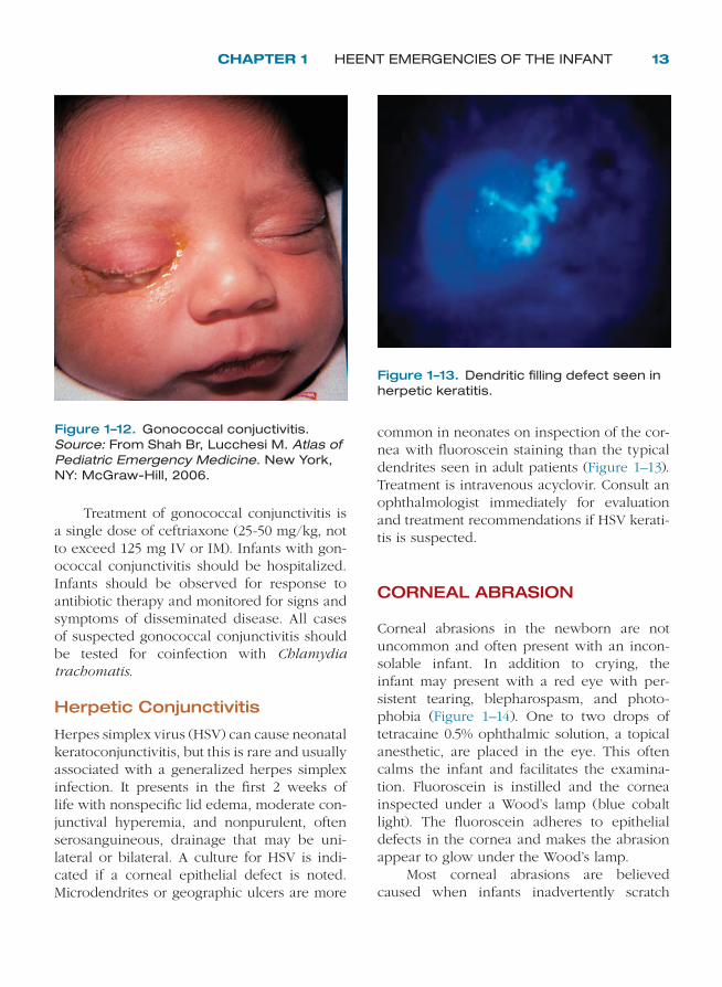
CHAPTER 1 HEENT EMERGENCIES OF THE INFANT 13
Treatment of gonococcal conjunctivitis is a single dose of ceftriaxone (25-50 mg/kg, not to exceed 125 mg IV or IM). Infants with gon-ococcal conjunctivitis should be hospitalized. Infants should be observed for response to antibiotic therapy and monitored for signs and symptoms of disseminated disease. All cases of suspected gonococcal conjunctivitis should be tested for coinfection with Chlamydia trachomatis.
Herpetic Conjunctivitis
Herpes simplex virus (HSV) can cause neonatal keratoconjunctivitis, but this is rare and usually associated with a generalized herpes simplex infection. It presents in the fi rst 2 weeks of life with nonspecifi c lid edema, moderate con-junctival hyperemia, and nonpurulent, often serosanguineous, drainage that may be uni-lateral or bilateral. A culture for HSV is indi-cated if a corneal epithelial defect is noted. Microdendrites or geographic ulcers are more
common in neonates on inspection of the cor-nea with fl uoroscein staining than the typical dendrites seen in adult patients (Figure 1–13). Treatment is intravenous acyclovir. Consult an ophthalmologist immediately for evaluation and treatment recommendations if HSV kerati-tis is suspected.
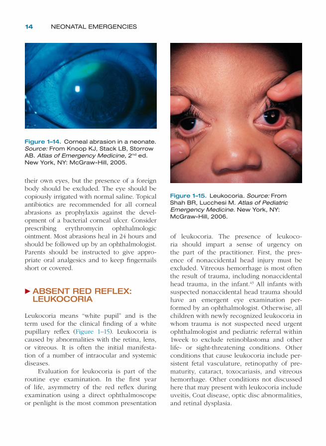
CORNEAL ABRASION
Corneal abrasions in the newborn are not uncommon and often present with an incon-solable infant. In addition to crying, the infant may present with a red eye with per-sistent tearing, blepharospasm, and photo-phobia (Figure 1–14). One to two drops of tetracaine 0.5% ophthalmic solution, a topical anesthetic, are placed in the eye. This often calms the infant and facilitates the examina-tion. Fluoroscein is instilled and the cornea inspected under a Wood’s lamp (blue cobalt light). The fl uoroscein adheres to epithelial defects in the cornea and makes the abrasion appear to glow under the Wood’s lamp.
Most corneal abrasions are believed caused when infants inadvertently scratch
Figure 1–12. Gonococcal conjuctivitis. Source: From Shah Br, Lucchesi M. Atlas of Pediatric Emergency Medicine. New York, NY: McGraw-Hill, 2006.
Figure 1–13. Dendritic fi lling defect seen in herpetic keratitis.
14 NEONATAL EMERGENCIES
their own eyes, but the presence of a foreign body should be excluded. The eye should be copiously irrigated with normal saline. Topical antibiotics are recommended for all corneal abrasions as prophylaxis against the devel-opment of a bacterial corneal ulcer. Consider prescribing erythromycin ophthalmologic ointment. Most abrasions heal in 24 hours and should be followed up by an ophthalmologist. Parents should be instructed to give appro-priate oral analgesics and to keep fi ngernails short or covered.
ABSENT RED REFLEX: �LEUKOCORIA
Leukocoria means “white pupil” and is the term used for the clinical fi nding of a white pupillary refl ex (Figure 1–15). Leukocoria is caused by abnormalities with the retina, lens, or vitreous. It is often the initial manifesta-tion of a number of intraocular and systemic diseases.
Evaluation for leukocoria is part of the routine eye examination. In the fi rst year of life, asymmetry of the red refl ex during examination using a direct ophthalmoscope or penlight is the most common presentation
of leukocoria. The presence of leukoco-ria should impart a sense of urgency on the part of the practitioner. First, the pres-ence of nonaccidental head injury must be excluded. Vitreous hemorrhage is most often the result of trauma, including nonaccidental head trauma, in the infant.43 All infants with suspected nonaccidental head trauma should have an emergent eye examination per-formed by an ophthalmologist. Otherwise, all children with newly recognized leukocoria in whom trauma is not suspected need urgent ophthalmologist and pediatric referral within 1week to exclude retinoblastoma and other life- or sight-threatening conditions. Other conditions that cause leukocoria include per-sistent fetal vasculature, retinopathy of pre-maturity, cataract, toxocariasis, and vitreous hemorrhage. Other conditions not discussed here that may present with leukocoria include uveitis, Coat disease, optic disc abnormalities, and retinal dysplasia.
Figure 1–14. Corneal abrasion in a neonate. Source: From Knoop KJ, Stack LB, Storrow AB. Atlas of Emergency Medicine, 2nd ed. New York, NY: McGraw-Hill, 2005.
Figure 1–15. Leukocoria. Source: From Shah BR, Lucchesi M. Atlas of Pediatric Emergency Medicine. New York, NY: McGraw-Hill, 2006.

CHAPTER 1 HEENT EMERGENCIES OF THE INFANT 15
RETINOBLASTOMA
Retinoblastoma is the most common intraocu-lar tumor of childhood and exists in sporadic and heritable forms. Approximately 1 in 15,000 live births are affected with retinoblastoma and the annual incidence is 11 per 106 chil-dren under the age of 4 years.44,45 This means an estimated 200-500 new cases of retinoblas-toma occur in the United States every year. The majority of cases are diagnosed in chil-dren less than 2 years of age.46
Approximately 25% of retinoblastoma cases are bilateral, which is always inherited and typically presents in the fi rst year of life. However, 95% of these patients will have no previous family history of retinoblastoma. Unilateral disease is usually sporadic and diag-nosed after the fi rst year of life.47,48
If left untreated retinoblastoma will grow to fi ll the eye and destroy the globe. Metastasis usually begins after 6 months, and death occurs within a few years. The most common route of metastasis is direct extension to the central nervous system (CNS) via the optic nerve or the choroid to the orbit. However, tumor cells may disperse through the subarachnoid space to the contralateral optic nerve or through the CSF to the CNS. Hematogenous spread to the lung, bone, and brain occurs. Lymphatic dis-semination of tumor cells into the conjunc-tivae, eyelids, and extraocular tissues occurs as well.
The most common clinical presentation of retinoblastoma is leukocoria (Figure 1–16). Strabismus is the second most common clin-ical fi nding associated with retinoblastoma.49 All children with either leukocoria or stra-bismus, or both, should be evaluated by an ophthalmologist. However, other clinical signs may herald the disease and include: decreased vision, ocular infl ammation, vitreous hemor-rhage, hyphema, orbital cellulitis, proptosis, glaucoma, eye pain, and fever. A family his-tory of retinoblastoma should include ques-tions about the possible occurrence of other
eye tumors, eye loss, and cancers, especially osteogenic sarcoma, which has a strong asso-ciation with retinoblastoma.
The diagnosis of retinoblastoma is made based upon the clinical examination, and the presence of intratumoral calcifi cation on CT of the orbit or ocular ultrasonograhy.
CATARACT
A cataract is an opacifi cation of the lens. Congenital cataracts are present at birth or in early infancy.50 The incidence of congeni-tal cataracts in the United States is 1.2 to 6.0 cases per 10,000 live births. If undetected and untreated, a cataract may cause partial or total blindness in an infant. Most congenital cata-racts are associated with intrauterine infec-tions, rubella being the most common cause. Other intrauterine infections associated with cataracts include rubeola, chicken pox, toxo-plasmosis, herpes simplex virus, herpes zoster, poliomyelitis, infl uenza A, Epstein-Barr virus, syphilis, and cytomegalovirus. Unilateral cata-racts are usually sporadic events; they account for approximately one-third of congenital cat-aracts and are associated with ocular abnor-malities, intrauterine infection, and trauma. Bilateral cataracts are often inherited; they are indicators of a number of systemic, genetic, and metabolic disorders and require a full work-up. Metabolic and systemic diseases are
Figure 1–16. White pupil in a neonate with retinoblastoma.

16 NEONATAL EMERGENCIES
found in as many as 60% of bilateral cataracts patients. Cataracts may also occur as a result of high-dose, long-term corticosteroid therapy.50
An irregular or asymmetric red refl ex is the most common clinical fi nding indicative of a congenital cataract. This fi nding should prompt urgent ophthalmologic and pediatric follow-up, the goal being to prevent visual loss due to deprivation amblyopia. Cataract surgery is the treatment of choice and is most effective in preventing visual loss if preformed prior to 17 weeks of age.
PERSISTENT FETAL VASCULATURE
Persistent fetal vasculature (PFV) is caused by the failure of the embryonic primary vit-reous and hyaloid vasculature to involute dur-ing gestation. In addition to leukocoria, the involved eye is often mildly micro-ophthalmic with a shallow anterior chamber and promi-nent vessels on the iris. Infants with PFV may develop glaucoma, cataracts, intraocular hem-orrhage, or retinal detachment.51,52 Refer all patients with leukocoria or suspected PFV to an ophthalmologist.
RETINOPATHY OF PREMATURITY
Retinopathy of prematurity (ROP) is a disease that affects the immature vasculature of the retina in premature infants. The neovascular-ization of the retina may be aggressive and progress to retinal detachment and blindness. All babies that weigh less than 1500 g at birth or are younger than 32 weeks gestational age at birth are at risk of developing ROP. The inci-dence of ROP has increased as smaller and younger infants have survived. The factors that play a role in the pathogenesis of ROP are still not well understood, but risk factors have been identifi ed. They include assisted
ventilation for more than 1 week, surfactant therapy, intraventricular hemorrhage, bron-chopulmonary dysplasia, sepsis, elevated arte-rial oxygen tension, and large volumes of blood transfusions.53,54 ROP presents with leukocoria when retinal detachment has occurred and an emergent ophthalmologic consult is recom-mended. Patients with ROP are at increased risk for strabismus, glaucoma, and cataracts.
TOXOCARIASIS
Toxocariasis, also known as visceral larval migrans, is most commonly found in children 1 to 5 years of age. Common complaints are poor vision and strabismus. Ocular changes may be the only manifestation of the disease caused by the dog ascarid (Toxocara canis) or cat ascarid (T catis). Frequently there is no antecedent history of symptomatic visceral larval migrans. The infection often causes uveitis, which is the presence of infl amma-tory cells and debris in the vitreous and may result in the development of a secondary cataract. Both these changes produce leuko-coria. Additionally, a whitish subretinal granu-loma or large infl ammatory mass (nematode endophthalmitis) may develop and be seen on funduscopic examination. These fi ndings may be confused with retinoblastoma. All patients with leukocoria should be referred urgently to an ophthalmologist.
VITREOUS HEMORRHAGE
Vitreous hemorrhage causes leukocoria when there is extensive organization of the blood to form a clot prior to its degradation. The most common cause of vitreous hemorrhage in chil-dren is trauma, including nonaccidental head trauma. Vitreous hemorrhage should prompt a careful history, physical examination, and work-up to exclude shaken baby syndrome. Vitreous hemorrhage is also associated with a

CHAPTER 1 HEENT EMERGENCIES OF THE INFANT 17
number of other conditions: retinopathy of pre-maturity, persistent hyperplastic primary vitre-ous, leukemia, and other blood dyscrasias.
PERSISTENT TEARING: �EPIPHORA
Tears keep the eyes moist and clear of debris. The tear fi lm contributes to corneal clarity and the transmission of a focused image to the retina. Tears are produced by the lacrimal glands and drain through the lacrimal drain-age system (Figure 1–17). The punctum is the opening on the medial surface of each eye-lid and serves as the entrance to the canalic-ulus, which drains tears into the lacrimal sac. Tears collect in the lacrimal sac and drain into the nasolacrimal duct, which empties into the nose via the inferior meatus.
The valve of Hasner located at the dis-tal end of the nasolacrimal duct is a mucosal fl ap that prevents air from tracking into the lacrimal duct system when the nose is blown.
Nasolacrimal duct obstruction is the most com-mon cause of persistent tearing, infection, and eye discharge in children. The differential for epiphora includes dacryostenosis, dacrocystitis, and glaucoma, all of which are discussed here. Additional causes of persistent tearing include corneal abrasion, conjunctivitis, and eyelid abnormalities such as trichiasis (ingrown eye-lashes) and entropion (inversion of the eyelid).
DACRYOSTENOSIS: NASOLACRIMAL DUCT OBSTRUCTION
Dacryostenosis is the most common cause of persistent tearing in children and occurs in up to 20% of newborn infants.55 Six percent of children will have epiphora due to nasolacri-mal duct obstruction in the fi rst year of life.56 Blockage can occur at any point along the lacrimal drainage system, but most frequently occurs at the membrane of Hasner.
Infants with nasolacrimal duct obstruc-tion present with a history of persistent or intermittent tearing without blepharospasm or photophobia. On examination no nasal drain-age is noted, despite excessive tearing. There may be crusting or matting of the eyelashes in the absence of conjunctivitis.
First line treatment of nasolacrimal duct obstruction is lacrimal duct massage. To per-form lacrimal duct massage, moderate pressure is applied over the lacrimal sac in a downward direction. This massaging motion forces tears from the lacrimal sac into the nasolacrimal duct and increases the hydrostatic pressure enough to open the valve of Hasner, which relieves the obstruction. Parents should prac-tice on themselves and then perform this on the child at least 3 times a day. Parents should be instructed to keep their fi ngernails short and to wash their hands before massaging the infant’s nasolacrimal sac.
Nasolacrimal duct obstruction resolves spontaneously in 90% of infants by 6 months.56 Figure 1–17. The lacrimal duct apparatus.
Tearsproduced Blockage

18 NEONATAL EMERGENCIES
If nasolacrimal duct obstruction fails to resolve spontaneously by 12 months of age, then prob-ing of the lacrimal duct by an ophthalmologist is recommended.
DACRYOCYSTITIS
Acute dacryocystitis is an ophthalmologic emergency and a complication of nasolacrimal duct obstruction. Mucopurulent drainage from the puncta occurs when bacteria grows in tears retained in the lacrimal sac. This infection is most frequently caused by alpha-hemolytic streptococci, Staphylococcus epidermidis, and S aureus. On examination the lacrimal sac may be erythematous and swollen with increased warmth and tenderness on palpation.
Acute dacryocystitis requires admission for intravenous antibiotics and consultation with an ophthalmologist. Complications of acute dacryocystitis include preseptal celluli-tis, orbital cellulitis, sepsis, and meningitis.
GLAUCOMA
Congenital glaucoma is present at birth, but may not be recognized until infancy or early childhood. It is a rare condition that occurs in 1 in 10,000 live births.57 It is characterized by improper development of the eye’s aqueous outfl ow system. Impaired drainage of aque-ous fl uid from the anterior chamber leads to increased intraocular pressures, which causes damage to the optic nerve and blindness. As the intraocular pressure increases, peripheral vision is lost, followed by the progressive loss of central visual, and, eventually, complete blindness. Surgical intervention is required for defi nitive treatment.
The typical triad of symptoms for infantile glaucoma includes epiphora (chronic or intermit-tent tearing), photophobia, and blepharospasm. All symptoms are results of increased intraoc-ular pressure, which causes globe distension
and ocular enlargement known as an “ox eye” or buphthalmos. Distension of the cornea sec-ondary to elevated intraocular pressure causes corneal edema, which is seen as a cloudiness or haziness of the cornea on inspection. The corneal edema causes tremendous glare, which leads to photophobia. The photophobia causes tearing and blepharospasm. Increases in cor-neal size secondary to increases in intraocu-lar pressure are not seen in other conditions with epiphora. The normal corneal diameter in infants is 10 mm, increasing to 12 mm by 2 years of age. A horizontal corneal diameter greater than 12 mm, or asymmetry in corneal diameters, suggests glaucoma.58,59 All infants and children with suspected glaucoma need an urgent ophthalmologic consultation.
The goal of therapy in glaucoma is to pre-serve sight. Treatment of infantile glaucoma is surgical because of the rapidity of ocular dam-age and loss of sight. Medications are most often used postoperatively.
NASAL PROBLEMS �
The external nose is a pyramid-shaped struc-ture composed of bony and cartilaginous structures. The nasal septum divides the two nostrils. The superior, middle, and inferior turbinates make up the lateral nasal walls. The turbinates are erectile structures made of mucosa and spongy bone covered by mucous membrane. The nasal turbinates swell and contract in response to changes in tempera-ture, crying, allergen exposure, and illness.
These structures are best examined with the child in the sitting position. The child’s head is tilted back while the examiner sits directly opposite the patient. The examiner holds the otoscope, with an ear speculum attached, in their dominant hand. Simultaneously the examiner uses their nondominant hand to sta-bilize the patient’s head by resting the ulnar aspect of the hand against the forehead and using the thumb to elevate the tip of the nose.

CHAPTER 1 HEENT EMERGENCIES OF THE INFANT 19
The normal nasal mucosa is pink and moist. The vestibules should be patent and visible to the levels of the middle turbinates. The sep-tum should be in the midline.
The nasopharynx is located posterior to the nasal cavity and is superior to the soft pal-ate and oropharynx. The paired choanae form the anterior border of the nasopharynx and are divided by the nasal septum. Airfl ow through the nose begins at the nostrils as the negative pressure of inspiration draws air back through the nasal passages to the choanae and then to the larynx, trachea, and into the bronchi.
Infants are obligatory nasal breathers from birth to 6 weeks and thereafter prefer to breathe through their noses until 6 months of age.60-63 The characteristic upturned nose of infancy and their relatively large tongue allow the infant to breathe and swallow simul-taneously while breastfeeding. The posterior portion of the tongue exerts upward pressure on the soft palate during feeding that forms a seal that temporarily blocks the oral airway. This blockage of the oral airway combined with nasal breathing during feeding ensures swallowing without aspiration. This dynamic process has been described as the “veloglossal sphincter.”64 This process makes mouth breath-ing more cumbersome than nasal breathing for infants. For these reasons occlusion of the infant’s nose is serious and can prove fatal.
CHOANAL ATRESIA
Choanal atresia is the most common congen-ital anomaly of the nose. Choanal atresia is caused by the persistence of the bucconasal membrane or bony septum in the posterior nares and occurs in approximately 1 in 7000 births. Girls are affected more frequently. Most cases are unilateral.65
Bilateral choanal atresia is a life-threaten-ing emergency that typically presents shortly after birth. These infants typically have symp-toms of severe upper airway obstruction and
cyclical cyanosis. As the infant struggles inef-fectively to breathe through the nose, the infant becomes cyanotic and then begins to cry, which allows the child to breathe through the mouth and resolves the cyanosis. When the infant stops crying or attempts to feed, the cya-nosis recurs. Bilateral choanal atresia requires the insertion of an oral airway to keep the infant’s mouth open and the oral airway pat-ent, allowing the infant to breath. If the oral airway fails to alleviate respiratory distress and prevent recurrent cyanosis, then endotracheal intubation is necessary. Surgical correction of the obstruction is required.
Unilateral choanal atresia may go unde-tected in the newborn nursery and not become apparent until the infant develops an upper respiratory infection (URI). The swelling of the nasal mucosa and associated secretions of the URI block the normally patent nare and symptoms mimicking those of bilateral choanal atresia occur. These infants have stri-dor, labored breath sounds, and cyanosis that worsens during feeding and improves during crying. Unilateral choanal atresia may also present with chronic unilateral rhinitis.66
In infants with suspected choanal atresia, a size 5-8 French catheter should be passed from the nose into the oropharynx.64,65 The catheter should be passed a distance of at least 3 to 3.5 cm from the alar rim. If the catheter cannot be passed, then choanal atresia is suspected. An obstruction due to mucosal swelling and turbinate hypertrophy will allow the catheter to pass into the pharynx, and the obstruction is determined to be functional, not mechanical.
The diagnosis of choanal atresia is con-fi rmed by CT scan with intranasal contrast that shows narrowing of the posterior nasal cav-ity at the level of the pterygoid plate. For best results it is recommended that nasal secretions be suctioned and a topical vasoconstrictor be applied to nasal mucosa prior to the CT scan.
Infants that have respiratory distress or diffi culty feeding should be admitted to the hospital. An oral airway must be established

20 NEONATAL EMERGENCIES
and gavage feeding may be needed. Defi nitive treatment is surgical and requires otolaryngol-ogy consult. Up to 60% of infants with choanal atresia have other associated anomalies, includ-ing anomalies of heart and eyes that warrant cardiology and ophthalmology consults.66
ORAL PROBLEMS �
The exam of the newborn infant’s mouth should include inspection and palpation. Examination of the mouth begins with visual inspection of the lips for their overall shape, color, and for anatomic defects.
Sucking pads are areas of thickened epithelium on the lip mucosa. These may be present at birth and cause is unknown. They resolve spontaneously and require no treatment. The oral mucosa should be moist. The lips and oral cavity should be free of ulcerations. Ulcerations are associated with herpetic stomatitis, aphthous ulcers, met-abolic disorders, and drug reactions. Small white shiny masses called epithelial pearls are common on the gingiva. Epithelial pearls often occur in clusters. White bumps seen in the midline at the junction of the hard and soft palate are Epstein pearls. Both epi-thelial pearls and Epstein pearls are normal fi ndings in the infant. The palatine tonsils are generally not visible until the infant is 6 to 9 months of age. Palpation is impor-tant as some cleft palate abnormalities may not be seen, but are palpable. A cleft uvula should raise the suspicion of a palate defect. With palpation of the mouth, the normal and awake newborn will usually suck the exam-iner’s fi nger. The examiner’s fi nger will be drawn into the infant’s mouth as the tongue moves back and forth against the palate. A small, cyst-like mass, called a ranula, may be felt on the fl oor of the mouth. These masses are benign and are caused by the obstruc-tion of salivary glands. Natal teeth, if pre-sent, should be checked for looseness. Loose
natal teeth pose a potential aspiration risk and should be extracted.
THRUSH: OROPHARYNGEAL CANDIDIASIS
Thrush is the most common infection of the oral cavity in healthy newborns. It is caused by an overgrowth of the fungus Candida albi-cans, which is part of the normal fl ora of the gastrointestinal and genitourinary tracts in humans. Candida albicans typically causes disease when the balance of normal fl ora is disrupted by the use of antibiotics, or there is a compromised immune system due to disease or the use of steroids. The former instance includes antibiotics administered directly to the infant, to the mother during delivery, or to a breastfeeding mother.
Symptoms typically develop in the fi rst few weeks of life. Initially the parent may notice a white fi lm in the mouth that looks like milk or formula that will not go away. The infant may be fussy or have diffi culty feeding because of pain. The infant may pass the infection to the mother’s nipples during breastfeeding. The mother may notice reddened tender nipples and unusual pain while nursing or between feedings.
On examination white plaques may be noted on the buccal mucosa, palate, tongue, or the oropharynx (Figure 1–18). These plaques do not scrape off with a tongue blade. In addi-tion, pinpoint areas of bleeding are seen under-neath the plaques when they are scraped.
The diagnosis is usually a clinical one and is confi rmed when plaque scrapings viewed with a KOH preparation reveal budding yeasts, with or without pseudohyphae. Treatment con-sists of topical antifungals such as miconazole gel, which has a superior cure rate and lower rate of recurrence than nystatin suspension.67 The breastfeeding mother with symptoms of candidiasis of the nipples should apply the same topical antifungals to the nipples and

CHAPTER 1 HEENT EMERGENCIES OF THE INFANT 21
areolas after nursing. Breastfeeding should not be interrupted. If symptoms in the breastfeed-ing woman or infant are persistent, consider oral fl uconazole.
NATAL TEETH
Natal teeth are relatively rare and occur in approximately 1 in 3,000 births. The majority of natal teeth occur as isolated events, but may run in families or be associated with some syndromes. Natal teeth are typically seen on the lower gum located where the future lower central incisors will be (Figure 1–19). These teeth are usually poorly formed with a weak root structure, which frequently makes the teeth wobbly and therefore an aspiration risk. To prevent aspiration, loose natal teeth should be extracted.68
ORAL INJURIES
Orofacial injuries in the nonambulatory infant are often the hallmark of abuse.69 A careful and thorough oral examination is necessary in any infant with orofacial injuries in whom
child abuse is suspected. Some experts believe the mouth and oral cavity may be a focus for physical abuse because of its signifi cance in communication and eating.70 Oral injuries most commonly feature bruising or lacera-tion of the lips.14 Oral injuries may be infl icted with instruments, such as eating utensils and pacifi ers that are forced into the mouth, or perhaps with bottles during forced feeding. This mechanism can cause bruising or lacer-ation of the lips; it may also tear the frenu-lum; bruise the gingiva or alveolar mucosa; lacerate, bruise or contuse the tongue; bruise the soft palate and uvula; and puncture the posterior oropharynx. The infant with perfo-ration of the posterior pharynx may present with subcutaneous emphysema, fever, drool-ing, and respiratory distress. Gagging the infant may cause bruising at the corners of the mouth. Smothering the infant may tear the frenulum of the upper lip and be associated with facial petechiae.
All injuries to the head, face, and mouth in nonambulatory infants must be distinguished from abuse. The reported mechanism of injury must be consistent with physical fi ndings, and it must fi t with the developmental capabilities of the injured infant. The infant must have a full head-to-toe examination to exclude other unexplained injuries. Multiple injuries, injuries in different stages of healing, or inconsistent history all make abuse more likely. All cases
Figure 1–18. Oral candida.
Figure 1–19. Natal teeth.

22 NEONATAL EMERGENCIES
of suspected child abuse must be reported to local or state child protective services, which is mandatory in all 50 U.S. states. Consider admission to the hospital for any infant with suspected physical abuse.
REFERENCES
1. Southgate WM, Pittard WB. Classifi cation and physical examination of the newborn infant. In: Klaus MH, Fanaroff AA, eds. Care of the High-Risk Neonate. 5th ed. Philadelphia, PA: WB Saunders; 2001:100.
2. Plauche WC. Subgaleal hematoma. A com-plication of instrumental delivery. JAMA. 1980;244:1597-1598.
3. Benaron DA. Subgaleal hematoma causing hypovolemic shock during delivery after failed vacuum extraction: a case report. J Perinatol. 1993;13:228-231.
4. American College of Obstetricians and Gynecologists, Operative vaginal delivery. ACOG Practice Bulletin no 17. Washington, DC: American College of Obstetricians and Gynecologists; 2000.
5. U.S. Department of Health and Human Services National Center on Child Abuse and Neglect. The Third National Incidence Study of Child Abuse and Neglect (NIS-3). Washington, DC: U.S. Government Printing Offi ce; 1996.
6. Atwal GS, Rutty GN, Carter N, Green MA. Bruising in non-accidental head injured chil-dren; a retrospective study of the prevalence, and distribution and pathological associations in 24 cases. Forensic Sci Int. 1998;96(2):215-230.
7. Feldman KW. Patterned abusive bruises of buttocks and the pinnae. Paediatrics. 1992;90(4):633-636.
8. Brinkman B, Püschel K, Mätzsch T. Forensic dermatological aspects of the battered child syndrome. Akteuelle Dermatologie. 1979;5(6):-217-232.
9. De Silva S, Oates RK. Child homicide the extreme of child abuse. Med J Aust. 1993;158(5)300-301.
10. Dunstan FD, Guildea ZE, Kontos K, Kemp AM, Sibert JR. A scoring system for bruise patterns: a tool for identifying abuse. Arch Dis Child. 2002;86(5):330-333.
11. Ellerstein NS. The cutaneous manifestations of child abuse and neglect. Am J Dis Child. 1979;133:906-909.
12. Johnson CF, Showers J. Injury variables in child abuse. Child Abuse Negl. 1985;9:207-215.
13. Leavitt EB, Pincus RL, Bukachevsky R. Otolaryngologic manifestations of child abuse. Arch Otolaryngol Head Neck Surg. 1992;118(6):629-631.
14. Naidoo S. A profi le of the oro-facial injuries in child physical abuse at a children’s hospital. Child Abuse Negl. 2000;24(4):521-534.
15. Sussman SJ. Skin manifestations of the battered-child syndrome. J Pediatr. 1968;72(1)99-101.
16. Saternus KS, Kernbach-Wighton G, Oehmichen M. The shaking trauma in infants—kinetic chains. Forensic Sci Int. 2000;109:203.
17. Van Praag MC, Van Rooij RW, Folkers E, et al. Diagnosis and treatment of pustular disorders in the neonate. Pediatr Dermatol. 1997;14(2):131-143.
18. Katsambas AD, Katoulis AC, Stavropoulos P. Acne neonatorum: a study of 22 cases. Int J Dermatol. 1999;38(2):128-130.
19. Paller A, Mancini AJ, Hurwitz S. Clinical Pediatric Dermatology: A Textbook of Skin Disorders of Childhood and Adolescence. 3rd ed. Philadelphia, PA: Elsevier–Saunders; 2006:737.
20. Feng E, Janniger CK. Miliaria. Cutis. 1995; 55(4):213-216.
21. Schachner L, Press S. Vesicular, bullous, and pustular disorders in infancy and childhood. Pediatr Clin North Am. 1983;30(4):609-629.
22. Turk AE, McCarthy JG, Thorne CH, Wisoff JH. The “back to sleep campaign” and deforma-tional plagiocephaly: is there a cause for con-cern?. J Craniofac Surg. 1996;7:12.
23. Hutchison BL, Hutchison LA, Thompson JM, Mitchell EA. Plagiocephaly and brachyceph-aly in the fi rst two years of life: a prospective cohort study. Pediatrics. 2004;114:970.
24. Benson ML, Oliverio PJ, Yue NC, Zinreich SJ. Primary craniosynostosis: imaging features. AJR Am J Roentgenol. 1996;166:697-703.
25. Ghali GE, Sinn DP, Tantipasawasin S. Man-agement of nonsyndromic craniosynostosis. Atlas Oral Maxillofac Surg Clin North Am. 2002;10:1-41.
26. Kelly KM, Littlefi eld TR, Pomatto JK, Ripley CE, Beals SP, Joganic EF. Importance of early

CHAPTER 1 HEENT EMERGENCIES OF THE INFANT 23
recognition and treatment of deformational plagiocephaly with orthotic cranioplasty. Cleft Palate Craniofac J. 1999;36:127.
27. Mulliken JB, Vander Woude DL, Hansen M, LaBrie RA, Scott RM. Analysis of posterior pla-giocephaly: deformational versus synostotic. Plast Reconstr Surg. 1999;103:371-380.
28. Chumas P, Tyagi A, Livingston J. Hydro-cephalus—what’s new?. Arch Dis Child Fetal Neonatal Ed. 2001;85:F149.
29. Volpe JJ. Intracranial hemorrhage: Germinal matrix-intraventricular hemorrhage. In: Volpe JJ, ed. Neurology of the Newborn. 4th ed. Philadelphia, PA: WB Saunders; 2001:428.
30. Forward KR, Fewer D, Stiver HG. Cerebralspinal fl uid shunt infections. J Neurosurg. 1983;59:389.
31. Shapiro S, Boaz J, Kleiman M, Kalsbeck J. Origin of organisms infecting ventricular shunts. Neurosurgery. 1988;22:868.
32. Gupta BK, Hamming NA, Miller MT. The eye. In: Fanaroff AA, Martin RJ, eds. Neonatal–Perinatal Medicine: Diseases of the Fetus and Infant. 7th ed. St. Louis, MO: Mosby; 2002:1568.
33. American Academy of Pediatrics. Chlamydial trachomatis. In: Pickering LK, ed. Red Book: 2006 Report of the Committee on Infectious Diseases. 27th ed. Elk Grove Village, IL, Author; 2006:252.
34. Schachter J, Grossman M, Sweet RL, Holt J, Jordan C, Bishop E. Prospective study of peri-natal transmission of Chlamydia trachomatis. JAMA. 1986;255:3374-3377.
35. Frommell GT, Rothenberg R, Wang S, McIntosh K. Chlamydial infection of mothers and their infants. J Pediatr. 1979;95:28.
36. Chen JY. Prophylaxis of ophthalmia neonato-rum: comparison of silver nitrate, tetracycline, erythromycin and no prophylaxis. Pediatr Infect Dis J. 1992;11:1026.
37. Darville T. Chlamydia. In: Remington JS, Klein JO, Wilson CB, Baker CJ, eds. Infectious Diseases of the Fetus and the Newborn. 6th ed. Philadelphia, PA: Elsevier Saunders; 2006:384.
38. Mordhorst CH, Dawson C. Sequelae of neona-tal inclusion conjunctivitis and associated dis-ease in parents. Am J Ophthalmol. 1971;71:861.
39. Johnson RE, Newhall WJ, Papp JR, et al. Screening tests to detect Chlamydia trachoma-tis and Neisseria gonorrhoeae infections, 2002. MMWR Recomm Rep. 2002;51:1.
40. Workowski KA, Berman SM. Sexually transmit-ted diseases treatment guidelines, 2006. MMWR Recomm Rep. 2006;55:1.
41. Laga M, Meheus A, Piot P. Epidemiology and control of gonococcal ophthalmia neonatorum. Bull World Health Organ. 1989;67:471.
42. Alexander ER. Gonorrhea in the newborn. Ann NY Acad Sci. 1988;549:180.
43. Schloff S, Mullaney PB, Armstrong DC, Simantirakis E. Retinal fi ndings in children with intracranial hemorrhage. Ophthalmology. 2002;109:1472.
44. Devesa SS. The incidence of retinoblastoma. Am J Ophthalmol. 1975;80:263.
45. Tamboli A, Podgor MJ, Horm JW. The inci-dence of retinoblastoma in the United States: 1974 through 1985. Arch Ophthalmol. 1990;108:128.
46. Rubenfeld M, Abamson DH, Ellsworth RM, Kitchin FD. Unilateral vs bilateral retinoblas-toma. Correlations between age at diagnosis and stage of ocular disease. Ophthalmology. 1986;93:1016.
47. Young JL, Smith MA, Roffers SD, et al. Retinoblastoma. In: Ries LA, Smith MA, Gurney JG, et al., eds. Cancer Incidence and Survival Among Young Children and Adolescents: United States SEER Program, 1975-1995. Bethesda, MD: National Cancer Institute; 1999:73.
48. Montegi T. Lymphocyte chromosome survey in 42 patients with retinoblastoma: effort to detect 13q14 deletion mosaicism. Hum Genet. 1981;58:168.
49. Abramson DH, Frank CM, Susman M, et al. Presenting signs of retinoblastoma. J Pediatr. 1998;132:505.
50. Green M. The eyes. Pediatric Diagnosis: Interpretation of Symptoms and Signs in Children and Adolescents. 6th ed. Philadelphia, PA: W.B. Saunders Company; 1998:15-36.
51. Hadad R, Font RL, Reeser F. Persistent hyper-plastic primary vitreous. A clinicopathologic study of 62 cases and review of the literature. Surv Ophthamol. 1978;23:123.
52. Cheng KP, Hiles DA, Biglan AW. The differ-ential diagnosis of leukocoria. Pediatr Ann. 1990;19:376.
53. Seiberth V, Linderkamp O. Risk factors in reti-nopathy of prematurity. A multivariate statisti-cal analysis. Ophthalmologica. 2000;214:131.

24 NEONATAL EMERGENCIES
54. Flynn JT, Bancalari E, Snyder ES, et al. A cohort study of transcutaneous oxygen tension and the incidence and severity of retinopathy of prematurity. N Engl J Med. 1992;326:1050.
55. Peterson RA, Robb RM. The natural course of congenital obstruction of the nasolacri-mal duct. J Pediatr Ophthalmol Strabismus. 1978;15:246.
56. MacEwen CJ, Young JD. Epiphora during the fi rst year of life. Eye. 1991;5 (Pt 5):596.
57. deLuise VP, Anderson DR. Primary infan-tile glaucoma (congenital glaucoma). Surv Ophthalmol. 1983;28:1.
58. Chew E, Morin JD. Glaucoma in children. Pediatr Clin North Am. 1983;30:1043.
59. Seidman DJ, Nelson LB, Calhoun JH, et al. Signs and symptoms in the presentation of primary infantile glaucoma. Pediatrics. 1986;77:399.
60. Moss ML. The veloepoglotttic sphincter and obli-gate nose breathing in the neonate. J Pediatric. 1970;67:330-331.
61. Swift PGF, Emory JL. Clinical observations on response to nasal occlusion in infancy. Arch Dis Child. 1973;48:947-951.
62. Nathan CA, Seid AB. Neonatal rhinitis. Int J Pediatr Otorhinolaryngol. 1997;39:59-65.
63. Moss ML. The veloepoglotttic sphincter and obligate nose breathing in the neonate. J Pediatric. 1970;67:330-331.
64. Myer CM III, Cotton RT. Nasal obstruction in the pediatric patient. Pediatrics. 1983;72:766.
65. Szeremeta W, Parikh TD, Widelitz JS. Congenital nasal malformtaions. Otolaryngolo Clin North Am. 2007;40:97.
66. Ferdman RM, Linzer JF, Sr. The Runny Nose in the Emergency Department: Rhinitis and Sinusitis. Clin Pediatr Emerg Med. 2007;8(2):123-130.
67. Hoppe JE. Treatment of oropharyngeal candidi-asis and candidal diaper dermatitis in neonates and infants: review and reappraisal. Pediatr Infect Dis J. 1997;16:885-894.
68. Diley DC, Siegal MA, Budnick S. Diagnosing and treating common oral pathologies. Pediatr Clin North Am. 1991;38:1227-1264.
69. Kellogg N. Oral and dental aspects of child abuse and neglect. Pediatrics. 2005;116:1565.
70. Vadiakas G, Roberts MW, Dilley DC. Child abuse and neglect: ethical issues for dentistry. J Mass Dent Soc. 1991;40:13-15.

25
NORMAL NEONATAL �
BEHAVIOR 25
A WORD ABOUT �
THE FORMER NICU PATIENT 26
THE NEUROLOGIC �
HISTORY 27
THE NEUROLOGIC �
EXAMINATION 28
THE CRYING INFANT 32 �
THE HYPOTONIC NEONATE 36 �
THE JITTERY NEONATE 43 �
NEONATAL SEIZURES 47 �
NORMAL NEONATAL �BEHAVIOR
To many observers, a neonate may appear to just sleep all day with occasional breaks for eating, but in actuality the normal neonate spends the day learning to make sense of a set of novel stimuli. At birth, the newborn is thrust into a new environment and must learn to survive in this new world. It must learn to respond appropriately to a bombardment of new stimuli, develop a sleep-wake cycle, regu-late temperature control, determine who these new strange people are, and manage to grab a bite to eat every once in a while.
Neonates exist in several behavioral states—sleep, drowsy, alert, active, fussy, and crying—which largely dictate their posture and behavior. During their alert state, they lie in a fl exed position with little motor control and with purposeless hand opening and clos-ing.1 They can turn their head from side to
side without being able to lift it against gravity. They progress over their fi rst month to assume a slightly more relaxed posture with the ability to hold their head in a tonic neck position. By 4 to 6 weeks of age, they can hold their chin up when in the prone position. As they age, they gain slightly better control of their suck-ing mechanisms as well.
Neonates must constantly learn to pro-cess new stimuli as they explore their new world. They learn to habituate to the famil-iar and only respond to a new stimulus, as seen when they will turn their head to a novel sound such as their mother entering a room. They recognize and prefer patterns in colors, consonants, contour, and intensity.2 By 4 to 6 weeks of age, they consistently fi xate on objects and follow objects both horizontally and vertically with their gaze. When neonates become overstimulated, they often yawn, look away, or begin to suck on their hands or lips. This response to stimuli will often affect
CHAPTER 2
Neurologic EmergenciesLinnea Wittick, MD

26 NEONATAL EMERGENCIES
their overall muscle tone and spontaneous movement.
Neonates also become more social over the fi rst month of life. They are born with a visual preference for faces and show recogni-tion memory by preferentially turning to their own mother’s face. A newborn’s focal length is about 8 to 12 inches, the perfect distance to gaze at their mother while being held for feedings. They move from simply having an involuntary smile to occasional social smiles by the end of the neonatal period.
The sleep-wake cycle also changes, to the relief of every parent, throughout the neonatal period. Initially, the newborn sleeps for equal amounts of time throughout the day and night. In the fi rst week, the infant sleeps for approx-imately 16.5 hours each day. It will wake approximately every 2 to 3 hours for feedings. As the infant’s neurologic system matures, it learns to consolidate periods of sleep so that by the end of the fi rst month, the infant sleeps for 15.5 hours, still with an equal distribution between day and night. By two months of age, most babies will wake 2 to 3 times dur-ing the night to feed, with some infants sleep-ing 6 hours at a stretch. By 3 months of age, the infant sleeps approximately 15 hours a day with most of the sleeping during the nighttime hours.
A WORD ABOUT THE �FORMER NICU PATIENT
With the increasing number of premature deliveries, a rudimentary understanding of the basic developmental and neurologic con-sequences NICU infants face becomes impor-tant. Approximately 9% of all newborns require intensive care. Because some of these newborns require a prolonged NICU stay, they will likely not be seen in the emergency department (ED) during the neonatal period. However, given their complicated history and increased likelihood of having long-term medical complications, an
understanding of common outcomes will be helpful to the ED physician.
Most premature infants are discharged near what should have been their term delivery date. In order to be discharged home, a prema-ture infant must nipple all of its feeds, demon-strate steady growth, have no recurrent apneas or bradycardias, and maintain its temperature. Many may have some slight hypotonia. In gen-eral, premature infants, barring any underly-ing neurologic pathology, will function at the developmental level of their gestational age and not their chronologic age. They generally catch up to other children their own chronologic age by the time they reach 2 years of age. Parents may need reassurance that it is normal for their infant to lag behind for a time. However, very premature infants with a complicated NICU course are more likely to suffer signifi cant brain injury and progress to developmental delay, cerebral palsy, and metal retardation. Five to 10% of all premature infants <1500 g develop a major motor defi ciency, whereas 25% to 50% have developmental or visual diffi culties.3
One common complication of prematurity causing neurological sequelae that should be understood when evaluating an infant is intra-ventricular hemorrhage (IVH). With improved care the relative number of infants sustaining these insults is decreasing, and the very fact that more infants survive extreme prematurity means that more infants with IVH survive as well. Eighty percent of infants born between 23 and 34 weeks of gestation will sustain an IVH with the incidence increasing as the birth weight decreases.4 The fragile subepenydmal germi-nal matrix bleeds easily until about 36 weeks of gestation when it becomes fully developed. Eighty to 90% of these bleeds occur by day 3 of life, especially after events that quickly change cerebral blood fl ow such as asphyxia, RDS, pneumothorax, and rapid volume expansion. Symptoms of an acute bleed range from the completely asymptomatic infant to an abrupt onset of apnea, cyanosis, lethargy, changes in the neurologic examination, abnormal gaze,

CHAPTER 2 NEUROLOGIC EMERGENCIES 27
seizure, and acidosis. Diagnosis is usually made by ultrasound of the head through the anterior fontanelle at days 4 to 7 of life. Findings are usu-ally graded to aid in diagnosis and prognosis. Grade I bleeds occur in 35% of IVH and involve only the germinal matrix or fi ll less than 10% of the ventricle. Only 20% of these infants develop a complication such as cerebral palsy or signif-icant mental retardation. Grade II hemorrhages bleed through the ependyma and fi ll 10% to 50% of the ventricle, affecting approximately 40% of infants with IVH. Grade III hemorrhages fi ll over 50% of the ventricle and display a tran-sient hydrocephalus. Of infants with grade II or grade III hemorrhages, 41% will suffer signifi -cant morbidity. Grade IV hemorrhage extends beyond the ventricle and affects the periventric-ular white matter. These infants face a poorer prognosis, with 86% developing CNS delay.4 The ultrasound is repeated again in 6 weeks to follow the development of hydrocephalus and extent of bleeding and infarct. If these infants progress to having hydrocephalus, neurosurgi-cal intervention will be needed. These infants will also need close developmental care.4,5
THE NEUROLOGIC �HISTORY
The neonatal neurologic examination may seem intimidating. Unlike an older child or adult, the physician cannot ask the patient to perform certain tasks, and the patient cannot respond to direct questioning. However, with a systematic approach and just a few tricks and modifi cations, the physician can still perform a successful neonatal neurologic examination.
As with any physician encounter, a neona-tal examination should begin with a thorough history. In addition to questions concerning pre-senting signs and symptoms, several aspects of the history can infl uence the neurologic diag-nosis. Historical features of the pregnancy and delivery are vital (Table 2–1). Did the mother notice normal fetal movements or was there a
decrease in fetal movements that could signify a neuromuscular disorder? Did she notice parox-ysmal, rhythmic movements possibly indicating fetal seizures? What medications did the mother take during her pregnancy that may have affected fetal development? What medications is she taking now that could affect the infant if she is breastfeeding? Was the baby full term and were there any complications at the delivery? APGAR scores of less than 6 at 1 and 5 minutes may be indicative of perinatal asphyxia, which could now affect the baby’s neurologic status. Birth weight is an important feature. A small for gestational age (SGA) infant (ie, weight less than the 10th percentile at birth), can signify a problem with fetal development. Large for gestational age (LGA) infants also present with their own set of complications from risks of hypoglycemia (infant of a diabetic mother) to complications arising from birth trauma. Both SGA and LGA infants have increased risk of morbidity and mortality compared to infants
TABLE 2–1. � SUGGESTED COMPONENTS OF THE NEONATAL HISTORY
Historical Issues
Predictive
Possibilities
Was there a decrease in fetal movement during gestation?
Neuromuscular disorder
Were there paroxysmal, rhythmic movements noted during gestation?
Seizure disorder
What medications were taken prenatally?
Adverse fetal development
If breastfeeding, what medications are being taken presently?
Adverse neurologic effects
Were there low APGAR scores?
Perinatal asphyxia
Is there a family history of mental retardation, seizures, early strokes, or consanguinity?
Neurologic problems

28 NEONATAL EMERGENCIES
born at normal weight. Any problems the baby encountered in the nursery or NICU should be reviewed because the fi rst days of life pro-vide clues to the present neurologic status. The baby’s normal behavior—feeding, sleep, crying, and alertness patterns—should be discussed to uncover any changes at time of presentation. A family history of mental retardation, learning disabilities, seizures, early strokes, or consan-guinity should be obtained.
THE NEUROLOGIC �EXAMINATION
The physical examination should begin with a general assessment of the baby. Perform the examination in a calm manner with the baby feeling safe and comfortable. Keeping the baby in the mother’s lap for much of the examina-tion helps keep the baby quiet and responsive. Offering a bottle or a gloved fi nger to suck on can provide valuable information as well as facilitate the examination. At this point, note the general sense of the baby’s overall neuro-logic status. The baby should be alert or arouse easily, depending on the normal feeding and sleeping pattern.2 A lethargic or irritable baby should raise suspicions of a pathologic pro-cess. The alert baby displays spontaneous eye movements and moves in response to stimulus. When crying, a neonate should easily console. The physician should note if the infant cries appropriately to noxious stimuli and whether the cry is vigorous or weak and high pitched. The patient’s vital signs should also be noted as these can have a relationship to the patient’s neurologic fi ndings.
EXAMINATION OF THE HEAD AND CRANIAL NERVES
Head Size
The head examination should begin with a measurement of the head circumference. The
occipital-frontal head circumference is mea-sured just above the ears and eyebrows, around the widest part of the head. The nor-mal neonate’s head measures about 35 cm at birth and increases about 1 cm per week.6 The head circumference refl ects the intracranial volume and, therefore, brain development.2 1 Microcephaly, where head circumference is less than 2 SD below the 3rd percentile for age and gender, refl ects abnormal brain growth and developmental delay. This may be due to many causes such as Down syndrome, TORCH infec-tions, and fetal alcohol syndrome (Table 2–2). Macrocephaly, a head circumference greater than 2 SD above the 97th percentile, may refl ect an intracranial process such as hydrocepha-lus, hemorrhage, or storage diseases. The rate of growth often is more useful than a single measurement taken in time. Previous measure-ments from the primary physician may help determine whether an abnormal head size is indeed a pathologic process. If previous mea-surements are unobtainable and the physician fi nds an abnormal head size in the context of
TABLE 2–2. � CAUSES OF DEVIATION IN HEAD CIRCUMFERENCE
Microcephaly Macrocephaly
GeneticTrisomy 21, 13,18Cru du ChatCornelia de Lange
HydrocephalusDandy-Walker
malformationChiari malformation
Congenital infectionCMVRubellaToxoplasmosis
CNS infectionMeningitis
TumorFamilialSyndromes
Glutaric academia type I
NF type I
CNS infectionMeningitisEncephalitis
Drug Exposure Fetal alcohol
syndromeRadiation
HIECraniosynostosis

CHAPTER 2 NEUROLOGIC EMERGENCIES 29
an otherwise normal neurologic examination, the infant should be referred to the primary physician for growth monitoring and further testing.
Fontanelles
There are several features of the infant skull that differ from an older child’s examination. One of the most obvious is the presence of fontanelles (see Figure 1–7). Often, parents will report that their baby’s “soft spot” has sunken during an illness, so familiarity with a normal fontanelle is important. Although 6 fontanelles actually exist, most physicians generally note only 2. The anterior fontanelle lies at the junction of the coronal and the sagittal sutures and measures 0.6-3.6 cm at birth. It normally closes by about 9-18 months of age. A large anterior fontanelle can be associated with Down syndrome, hypo-thyroidism, achondroplasia, and increased intra-cranial pressure (ICP). A small fontanelle can be secondary to abnormal brain development, congenital infections, fetal alcohol syndrome, and some genetic syndromes. The posterior fontanelle lies at the meeting of the lambdoidal and sagittal sutures and measures 0.5-1 cm. It closes anywhere between birth and 4 months of age. Fontanelles should feel fl at and soft and be assessed with the infant quiet and upright. Lying supine may cause the fontanelles to falsely pro-trude. A depressed fontanelle can signify states of dehydration. A bulging and tense fontanelle may be a sign of increased intracranial pressure. Also, auscultate over the anterior fontanelle; the presence of a bruit may indicate an underlying arterial-venous malformation.
Head Shape
Many normal variations of the shape of the neonate’s head exist. Positional plagiocephaly, caused by preferential positioning over time, can cause a misshapen head with the ear of the affected side pushed forward and the cranium appearing fl at on the preferred side. Usually,
mild plagiocephaly can be treated by advising the parents to alternate sides the infant lies on. More severe cases will need referral to a neu-rosurgeon for treatment as the neonate grows older. Molding occurs during a vaginal deliv-ery when the bones of the cranium overlap to facilitate passage through the birth canal. The bones can be felt through the skin as they over-lap at the suture lines. Parents may need reas-surance that this will resolve in 1 to 2 days.
Caput secundum is generalized subcuta-neous edema of the scalp that arises during a vaginal delivery. It is often an asymmet-ric bogginess that will cross over the suture lines. It normally resolves in 7 to 10 days. Cephalohematomas, subperiosteal hemor-rhages caused by birth trauma, appear simi-lar to caput secundum. As these are contained within the periosteum, they will not cross over the suture lines and are often unilateral. This makes it easy to differentiate cephalohemato-mas from caput secundum on examination. Due to the breakdown of the hemoglobin within, extensive or bilateral cephalohematomas may facilitate the development of jaundice.
Cephalohematomas (see Figure 1–1) nor-mally resolve within 4 to 6 weeks. Calcifi cations felt as hard ridges along the skull may remain behind. These should not be confused with craniosynostosis, a pathologic condition of premature fusing of the sutures. This occurs most commonly along the sagittal suture fol-lowed by the coronal suture. A heaped up ridging along the suture line may be palpated along with an asymmetry of the infant’s head. Normally, the sutures expand to allow for rapid brain growth. In craniosynostosis, the brain cannot grow against the closed suture, so growth increases in a direction parallel to the fused suture.2 On examination, normal, open sutures are absent and the head displays asymmetry. Cranial radiographs or computed tomograohy (CT) scans and neurosurgic con-sult are indicated; surgical correction at a later date may be indicated to allow for proper brain development.

30 NEONATAL EMERGENCIES
Cranial Nerves
Examination of the cranial nerves may be accomplished with simple maneuvers and observations (Table 2–3).2 The neonate’s ability to fi x on and follow an object provides a gross assessment of visual patency. At 1 month, a neo-nate’s eyesight is 20/150, causes a focal length of 8 to 12 inches, the perfect visual acuity to focus on the mother’s face while feeding.
Eye movements and pupillary response should be assessed with the newborn bun-dled, comfortable, and in a darkened room or held vertically to facilitate eye opening.
The pupil should be examined for a red refl ex. The appearance of a gray or asymmet-rically colored pupil can signify congenital cataracts or retinoblastoma (see Figure 1–16) and necessitates an immediate referral to an ophthalmologist. Subconjuctival hemorrhages do not always indicate nonaccidental trauma, and may arise from birth trauma, coughing, or sneezing. These should resolve in 1 to 2 weeks.1 A funduscopic examination may demonstrate retinal hemorrhages, which may be seen in 38% of normal vaginal deliveries.2 However, if present after 2 weeks of age or encountered in another setting, they may be considered signs of nonaccidental head trauma.
Eye movements can be observed while the infant fi xes on and follows either a bright object or its mother’s face. Jerky eye move-ments are a normal fi nding during the fi rst few months of life as well as disconjugate gaze at rest or with eye movements.2 Facial asymme-try, especially while crying, may show a defect in CN VII. A rough assessment of hearing may be performed by clapping near the infant and observing for refl ex blinking.
MOTOR & SENSORY EXAMINATION
The motor and sensory examination occurs mostly through observation of the infant dur-ing the entire examination. The neonate usu-ally rests with its limbs in fl exed positions.1 It will move all of its extremities symmetrically. Tone can be assessed by passively moving the limbs and feeling opposing fl exion.2 A neonate will demonstrate some amount of head control. When pulled to a sitting position by its arms, its head should not lag far behind. Tone can also be noted by holding the infant over the exam-ination table by its trunk. The infant should be able to bear most of its weight with its feet on the table, and should not slide through the examiner’s hands. Muscle bulk offers another tool to assess the baby’s tone. A hypotonic baby will show less muscle bulk and possibly a single palmar crease, seen most commonly in Down syndrome but also in other syndromes with decreased tone in utero. The baby should withdraw to touch or pinprick. Normal reac-tions to touch should reassure the physician. Response to noxious stimuli, such as an IV insertion or a cold room or touch can also give clues to the neonate’s sensory status.
REFLEXES
The neonate displays unique refl exes not seen during other stages of life1,6 (Table 2–4). These
TABLE 2–3. � CRANIAL NERVE EXAMINATION
Maneuver Performed Cranial Nerve
Pupillary reaction to light II, IIIEOM while fi xing on object III, IV, VICorneal refl ex VWithdrawal to pinprick VFacial expression/asymmetry VIIBlinking to loud noise VIIISuck V, VII, XIISwallow IX, X, XIISCM strength, contractures XITongue fasciculations XII
Source: From Refs. 1, 2, 6.

CHAPTER 2 NEUROLOGIC EMERGENCIES 31
develop at approximately 28 to 32 weeks of ges-tation and provide a convenient way to assess the patient’s tone and strength (Figure 2–1). For example, an asymmetric Morro refl ex
may indicate a brachial plexus injury or frac-tured clavicle. The inability to perform the step refl ex may indicate decreased tone. Deep tendon refl exes, while often overlooked in
TABLE 2–4. � PRIMITIVE REFLEXES
Refl ex Stimulus Response
Crawl Place infant on abdomen Flexes legs under and makes crawling motions
Morro Pull arms away from supine infant and suddenly release
Arms abduct and extend, hands open, then arms fl ex in and hands close
Rooting Stroke cheek or corner of mouth Head turns to stimulus, mouth opensPalmer/plantar grasp Place fi nger along palm or sole
of footGrasps fi nger or curls toes around
fi ngerTonic neck (fencing) Turn head to one side while lying
supineFacial arm extension, occipital arm
fl exionStepping Hold upright with sole of feet
resting on fl at surfaceMakes alternating stepping movements
Swimmer’s Hold infant prone while supporting under the chest, stroke adjacent to the spine
Flexion of pelvis toward the stimulus
Protective Place cloth over face Arches and turns head side to side, brings hands to face
Source: From Refs. 1, 2, 6.
Figure 2–1. Common neonatal refl exes.
Tonicneckreflex
Graspreflex
Stepreflex Crawl reflex

32 NEONATAL EMERGENCIES
the neonatal examination, can easily be elic-ited.2 The pectoralis, bicep, brachioradialis, and knee refl ex are all obtainable at this age. The overall fl exor tone of the infant usually suppresses the triceps refl ex and may possibly make the bicep refl ex diffi cult to elicit. Attempt the refl ex with the joint in various angles of fl exion to provide the optimal tendon stretch to produce a response. Often, a knee refl ex will elicit a cross adductor response, whereas a knee refl ex causes the hip adductor on both the ipsilateral and contralateral sides to con-tract. This occurs normally in the fi rst month of life in contrast to older children and adults. The Babinski refl ex also differs from that of an adult. During the fi rst few weeks of life, an up-going great toe is common.6 Eliciting ankle clonus should also not cause alarm as 5 to 10 beats of clonus in a neonate is acceptable in the context of a normal neurologic exam-ination. A grading system usually is diffi cult to assign to a neonate; therefore, as long as the refl exes are symmetrical throughout all extremities, the examination should be con-sidered normal.
THE CRYING INFANT �
Few things become more frustrating to both parents and physicians than a crying baby. Often, parents present with an apparently oth-erwise healthy baby with no other complaints than “he won’t stop crying!” Anxiety, stress, and depression can lead the caregiver to bring the child for an evaluation.7 Poole et al. found that in one ED, unexplained crying in an oth-erwise well infant compromised 1 in 400 infant visits.8 In the UK alone, $108 million per year is spent to evaluate crying in the fi rst 12 weeks of life.7
Babies cry with the purpose of commu-nicating their needs for attention, thirst, and hunger, or because of discomfort and pain.7,8 The internet offers books, classes, and infor-mational videos promising parents the ability
to interpret their baby’s every cry. Studies show that parents often successfully interpret their baby’s needs according to their cry. Yet there exists some amount of “paroxysmal fuss-ing” in the majority of normal babies not used to communicating with caretakers. All babies display a regular cyclic crying period without environmental factors or stimuli. One of the most well-known studies of infant crying was done by Braselton in 1962 and described a crying pattern in 80 healthy infants.9 He states that at 2 weeks, most babies cry for an aver-age of 1-3/4 hours per day. This increases to a peak of 2-3/4hours at 6 weeks of age and then decreases to an average of 1 hour per day by 3 months as infants became more devel-opmentally advanced and fi nd other ways to communicate and soothe themselves. He also notes a time dependency to the infants’ cry-ing, with the majority of time spent crying focused in the afternoon and evening. Often, the parents just need reassurance that their baby displays normal infant behavior. When parents appear especially stressed by their crying infant, techniques for dealing with their crying baby should be discussed.10
However, infants presenting to the ED with a complaint of excessive crying that falls outside of the range of normal crying need a complete evaluation to rule out pathologic causes of crying.8 Five to 60% of crying infants possess a diagnosable illness as a reason for their crying. Confounding the picture, studies show that parental perceptions of crying do not correlate with the seriousness of the illness and vary widely among mothers. Therefore, the physician should seriously investigate the cause behind a complaint of excessive crying to rule out a potentially serious illness in this vulnerable population.
DIFFERENTIAL DIAGNOSIS
The differential diagnosis of the crying infant is extensive7 (Table 2–5). A broad history and

CHAPTER 2 NEUROLOGIC EMERGENCIES 33
physical examination begins the process of ruling out serous illness. One study found that the history alone directly led to the diagno-sis in 20% of cases,8 demonstrating the need for a comprehensive history. The physician needs to fi rst rule out any possibility of a life-threatening infection such as sepsis, meningi-tis, encephalitis, or urinary tract infection. Any history of fever should prompt the physician to begin a full sepsis work-up. Also inquire about any possible causes leading to electrolyte dis-turbances such as normal feeding behavior and any recent deviation from normal feeding,
because electrolyte abnormalities or hypogly-cemia are easily treated. Otherwise, a broad, detailed history about onset of symptoms, deviation from normal behavior, pregnancy and delivery history, medications, and previ-ous medical history should be done.
PHYSICAL EXAMINATION
The physical examination may lead directly to or contribute to the diagnosis in 53% of infants.8 As with any patient, the examination should
TABLE 2–5. � PATHOLOGIC CAUSES OF CRYING
Infection
MeningitisEncephalitisSepsisUTIOsteomyelitisSeptic arthritisPneumoniaAcute otitis mediaGinivostomatitisRetropharyngeal CellulitisOmphalitisFever
CV- SVT
MyocarditisCongenital heart defectCongestive heart failure
GI
Malrotation with midgut volvulusIntussseceptionRefl uxConstipationAnal fi ssureAcute gastroenteritisFormula intoleranceColic
Toxins
Drug withdrawalMedication exposure in breast milkIngestion
Dermatologic
EczemaDiaper dermatitisCellulitisAbscessHair tourniquetImmunizationsInsect bites
Trauma
Nonaccidental traumaFractureCorneal abrasion
GU
Incarcerated inguinal herniaTesticular torsionHair tourniquetHydroceleBalantitisComplication of circumcision
Neurologic
Increased ICPHydrocephalus
Metabolic
Electrolyte abnormalityHypoglycemiaInborn errors of metabolism
Source: From Refs. 7, 8.

34 NEONATAL EMERGENCIES
be systematic and complete with a focus on specifi c areas. Life-threatening causes of cry-ing such as infection, abuse, intussusceptions, intoxication, and metabolic diseases should be ruled-out quickly.
General Appearance
First, the general appearance of the baby may direct the physician in the work-up. Completely undress the neonate to expose any fi ndings that might be hidden beneath clothing. Findings of increased crying with movement may point to meningitis or a musculoskeletal injury. An alert and fussy baby offers more reassurance than a fussy baby that is neither alert nor respon-sive to its surroundings. The vital signs can also give a general overview of the patient. An elevated temperature can point to an infec-tious etiology and point the work-up in that direction. Tachycardia may be secondary to supraventricular tachycardia (SVT), fever, dis-comfort, or dehydration. Tachypnea can be secondary to infection, neurologic, cardiac, or metabolic disorders.
Head
The head needs a thorough examination. The fontanelle and sutures may show signs of ele-vated intracranial pressure secondary to infec-tion, congenital hydrocephalus, or trauma.7 A funduscopic eye examination may reveal ret-inal hemorrhages (see Figure 1–3) suggestive of nonaccidental trauma. Excessive tearing can indicate glaucoma, a foreign body, or infec-tion. The eyelids should be everted to inspect for foreign bodies. A fl uorescein examination under a Wood’s lamp can rule out corneal abrasion (Figure 2–2). Often, the application of tetracaine before the examination will calm the baby, signifying an abrasion and leading to its diagnosis and treatment. The ears should be examined for acute otitis media. Contusions around the ears may suggest trauma. Any signs of trauma merit a head CT looking for subdural hematomas. The mouth and hypopharynx
should be completely examined looking for burns, thrush, or stomatitis, which can be treated appropriately.
Chest & Abdominal Examination
A thorough lung and cardiac examination may reveal also a cause behind the crying. A high suspicion for SVT, congenital heart disease, and myocarditis may lead to a correct diagno-sis of increased crying and fussiness.
The abdominal examination may reveal many different causes of crying. A full abdo-men with easily palpated stool is signifi cant for constipation when the history corresponds with an infant with hard, round stools. In a chron-ically constipated infant, consider a work-up and surgical consultation for Hirschsprung dis-ease. A soft abdomen with the history of vomit-ing or diarrhea may suggest gastroenteritis and adequate hydration status should be assessed. A rectal examination may reveal anal fi ssures. Blood at the rectum suggests intussusception. A patent anus should be documented. Genitalia should be assessed for incarcerated hernias (Figure 2–3), hair tourniquets (Figure 2–4), and complications from a circumcision. Diaper rashes should be appropriately treated.
Figure 2–2. Corneal abrasion in a crying neonate. Source: From Knoop KJ, Stack LB, Storrow AB: Atlas of Emergency Medicine, 2nd ed. New York, NY: McGraw-Hill, 2002.

CHAPTER 2 NEUROLOGIC EMERGENCIES 35
Musculoskeletal Examination
A complete musculoskeletal examination may discover the etiology of crying. The long bones should be palpated for possible fractures or osteomyelitis. Any tenderness deserves a radiograph. Most fractures in the neonatal period are suspicious for abuse, and if found, a complete skeletal survey to search for other fractures must be done. However, a fractured clavicle found in the neonatal period may be due to shoulder dystocia after a diffi cult deliv-ery. Red, swollen, or tender joints should lead the physician to look for septic joints. Hair tourniquets are an easily missed diagnosis. The physician should carefully examine each digit
for a hair or string wrapped around a digit and cutting off circulation. The skin should also be inspected for possible rashes, bruises, abra-sions, or abscesses.
DIAGNOSTIC TESTS
Generally, the physician should not order screening laboratory or radiographic tests without a particular diagnosis in mind. A uri-nalysis and urine culture may be the only cost-effective screening tests.8 Otherwise, tests should be chosen based on clinical suspicion of a particular diagnosis.
MANAGEMENT
Often, the history and physical examination will not direct the physician to a specifi c diag-nosis. In fact, 40% of neonates worked-up for excessive crying will not yield a diagnosis at their initial presentation.8 If the infant has an unrevealing history, a normal physical exam-ination, and has reproducibly ceased crying during the initial assessment, the child may return home with prompt follow-up. On the other hand, if the infant continues to cry and cannot be consoled, further studies should be considered,8 such as barium enema for intus-susceptions, CT of the head to rule out trauma or increased intracranial pressure, blood tests and spinal tap to evaluate for infections, or a metabolic work-up including amino and organic acids, ammonia, lactate, pH, and urine-reducing substances. If no diagnosis is found for a continually fussy infant, they should be admitted for further work-up and observation.
The physician may entertain a diagnosis of colic only after ruling out all other seri-ous illness. The defi nition of colic generally is accepted as recurrent, paroxysmal spells of excessive crying and fussiness lasting more than 3 hours a day for more than 3 days a
Figure 2–3. Inguinal hernia in a crying neonate.
Figure 2–4. Hair tourniquet in a crying neonate.

36 NEONATAL EMERGENCIES
week for over 3 weeks. It usually presents in the 2nd or 3rd week of life and lasts until the infant is 3 to 4 months old.8 These infants are otherwise well. Often, parents will present within the fi rst few days of crying before a def-inite pattern of crying becomes established. As discussed above, a thorough history and phys-ical examination should fi rst rule out a serious illness before giving a diagnosis of colic.
THE HYPOTONIC NEONATE �
Neonatal hypotonia, informally known as “fl oppy baby syndrome,” occurs not uncom-monly in the neonatal period. Decreased tone from a neurologic disorder usually becomes noticeable directly after birth or during early infancy. Hypotonia can be defi ned as a sub-jective decrease of resistance to passive range of motion, usually found in the limbs, trunk, and facial muscles (Figure 2–5). This differs from weakness, which is defi ned as a decrease in the power generated by the musculature. A neonate may or may not display weakness
associated with decreased tone. The presence or absence of weakness may even help lead to the underlying cause of hypotonia. The physi-cian must fi nd a quick diagnosis for the hypo-tonic neonate as there exist several medical conditions where a rapid diagnosis and treat-ment will affect the prognosis. A thorough history and physical exam should easily point to nonneurologic diseases requiring immedi-ate attention such as sepsis, hypothyroidism, hypoglycemia, malnutrition, malrotation, toxin exposure, and congenital heart disease. If sus-picious for one of these diseases, the appropri-ate laboratory evaluation and treatment should be quickly initiated.
DIFFERENTIAL DIAGNOSIS
When evaluating neonatal hypotonia, it often helps to approach the differential diagnosis in a systematic manner. Most diseases fall into two categories—those disorders affecting the upper motor neuron, or central disorders, and those disorders affecting the lower motor neuron, or
Figure 2–5. Examples of decreased neonatal tone.
Hypotonia(Decreased
Muscle Tone)

CHAPTER 2 NEUROLOGIC EMERGENCIES 37
peripheral disorders (Table 2–6).11-13 Peripheral disorders can further be divided into those affecting each section of the neuromuscular unit. Delineating the disorder into one of these broad categories will help the physician arise at a correct diagnosis and tailor the work-up to avoid costly and timely diagnostic tests.
Central disorders generally affect the upper motor neuron—the brain and spi-nal cord. Approximately 66% of patients with hypotonia present with a central defect.11 They usually display decreased strength with active motion in addition to hypotonia. In addition to multiple other symptoms, these patients more commonly present with seizures than do peripheral disorders. They often experience an altered level of consciousness. Many of these disorders are associated with spasticity and increased refl exes, but usually present in the neonatal period with hypotonia and weakness.
Several nonneurologic disorders result-ing in central hypotonia exist and should quickly be ruled out.11,14 As mentioned earlier, sepsis, encephalitis, GI disturbances, and car-diac failure also may present as lethargy and hypotonia. A toxic ingestion due to medical errors, exposure in breast milk, or intentional
administration also may present with lethargy. Several metabolic disorders such as phenylket-inuria, Zellweger syndrome, and galactos-semia also present with hypotonia. Metabolic disorders usually present within the fi rst 12 to 24 hours of life after enough toxic metabolites build up in the neonate’s system, or when the infant becomes stressed by a febrile illness. A strong family predisposition suggests the diagnosis. Electrolytes, blood gas, lactate, and ammonia levels can be sent for a quick screen-ing in the acutely ill child suspected of having a metabolic disorder. These all require prompt treatment because delayed treatment can have dire outcomes.
Direct Brain Insults
Direct insults to the brain often cause a central hypotonia.15,14 Hypoxic ischemic encephalopa-thy from birth trauma or prolonged perinatal hypoxia can lead to decreased tone. Intracranial hemorrhage secondary to premature deliv-ery, neonatal stroke, or nonaccidental trauma also often progress to hypotonia. In addition to injuries, defects in brain structure may lead to decreased tone. Lissincephaly, which literally
TABLE 2–6. � DIFFERENTIAL DIAGNOSIS OF HYPOTONIA
Systemic Central Peripheral
Infection CardiacMetabolicHypoxia
Hypoxic ischemic encephalopathyChromosome disordersSyndromesPrader-WilliSpinal cord injury
Anterior horn cellsSpinal muscular atrophy IPeripheral nerve
Peripheral neuropathiesGuillian-Barré syndrome
Neuromuscular junctionMyasthenia syndromesInfantile botulism
MusclePompe diseaseCongenital myopathiesCongenital myotonic dystrophiesCongenital muscular dystrophies
Metabolic disorders
Source: From Refs. 11-14, 16.

38 NEONATAL EMERGENCIES
means smooth brain, arises from defective neuronal migration causing absent or partially formed gyri and sulci in the cerebral cortex. These children display abnormal facies, fail-ure to thrive, seizures, and mental retardation. Holoprosencephaly is a disorder of the forebrain or prosencephalon that divides it into two sep-arate cerebral hemispheres and causes hypo-tonia with dysmorphic features of the midline face, seizures, and mental retardation. A head CT or magnetic resonance imaging (MRI) will diagnose causes of hypotonia due to changes in brain structure. Treatment consists of sup-portive care and developmental assistance.
Chromosomal Disorders
Several chromosomal disorders may cause central hypotonia.11,14,16 Chromosomal disor-ders generally display characteristic dysmor-phic features that aid in their diagnosis.11 While many of these disorders will be suspected on clinical grounds based on a characteristic dysmorphic appearance, referral to a geneti-cist for karyotyping provides both a diagnosis and allows for discussions about future family planning. A few common diseases will be pre-sented here, but many others exist.
Down syndrome or trisomy 21 is the most common chromosomal disorder and genetic cause of mental retardation. It results from a trisomy of the distal end of the long arm of chromosome 21. Found in 1 in 600 to 700 live births,17 in addition to hypotonia, these infants display some variability of the well-known dysmorphisms of brachiocephaly, small ears, up slanted palpebral fi ssures, epicanthal folds, a fl at midface, a single transverse crease, small nipples, and a central posterior hair whorl. Almost 40% of these patients have a con-genital heart defect, most commonly an AV canal defect or a VSD. They also frequently have pulmonary hypertension, obstructive GI lesions, hypothyroidism, and increased suscep-tibility to infection. Trisomy 18, Edwards syn-drome, and trisomy 13 also display hypotonia,
however, few of these infants survive beyond infancy.
Prader-Willi syndrome (PWS)11,17 is another common chromosomal syndrome presenting with hypotonia. This arises in 1 of 12,000 to 15,000 live births from the paternal deletion of several genes on Ch15q. These patients present with hypotonia, feeding diffi culties secondary to poor suck and swallow ability, and failure to thrive. This diagnosis may escape the phy-sician in the hypotonic neonate as most physi-cians associate hyperphagia and obesity with PWS. These well-known symptoms actually do not develop until approximately 2 years of age, however. In addition, these patients dis-play diamond-shaped eyes, a prominent nasal bridge, small hands and feet, lightly colored skin compared to their family, hypogonads, and developmental delay.
Spinal cord injuries may also cause cen-tral hypotonia.14 These may occur when the fetus is delivered in the breech position with a hyperextended neck or during cervical presentations with the aggressive use of for-ceps. These complications are extremely rare occurring in 1 of 29,000 deliveries and usu-ally present in the delivery room with severe respiratory distress. In addition, these patients will have hypotonia with asymmetry between the upper and lower extremities, depending on the location of the lesion. They may have bladder dysfunction, decreased rectal tone, and vasomotor instability. Most likely the diag-nosis will be made soon after delivery with an MRI of the spinal cord. These patients may present to the emergency department if the newborn is intubated immediately in the deliv-ery room for respiratory distress and assumed that all symptoms of hypotonia are attributed to hypoxic-ischemic encephalopathy (HIE). These patients may develop increased deep tendon refl exes and spasticity as a later pro-gression of the injury.
Disorders of the peripheral nervous system present differently from disorders of the cen-tral nervous system.15 In addition to hypotonia,

CHAPTER 2 NEUROLOGIC EMERGENCIES 39
these babies display generalized weakness. They usually appear alert and interactive. However, 28% of neonates with peripheral nerve disorders also sustain some degree of hypoxia secondary to birth complications, con-founding the clear distinction between a cen-tral and a peripheral disease.11 These disorders can be further grouped into disorders affecting each section of the motor-neuron unit: anterior horn cells, the peripheral nerve, the neuromus-cular junction, and the muscle itself.
One example of a disorder affecting the anterior horn cell is spinal muscular atro-phy (SMA) type I, or Werdnig-Hoffmann syndrome.14,16 This autosomal recessive defect affects the SMN gene on chromosome 5. It usually presents in the fi rst 6 months of life with 1/3 presenting in the neonatal period with weakness, hypotonia, and decreased refl exes. Often, the mother recalls a decrease in fetal movement, and the infant presents in a breech presentation. The disease generally progresses gradually, with symptoms often fi rst noted after an exacerbation by a febrile illness. These infants have proximal, progres-sive weakness of the voluntary muscles with decreased movement of the extremities and the inability to lift the head. It weakens the bul-bar muscles leading to poor suck and swallow mechanisms and poor secretion control. They feed poorly, display tongue fasciculations, and drool excessively. The disease weakens the respiratory muscles, and the child will display “paradoxical respirations” where the abdo-men rises and the chest wall falls with each respiration. This leads to a bell-shaped chest wall, and respiratory weakness predisposes these infants to respiratory failure and pneu-monia, the leading cause of death. Although these infants fail to meet developmental mile-stones, they are bright, inquisitive, and have alert facies. Without supportive care, 80% of these children will die by 8 months of age and all will die by 2 years of age. Other types of SMA syndrome with better prognoses exist, but these present after the neonatal period.
The diagnosis is suspected on clinical grounds and supported by an EMG and muscle biopsy. More recently, a genetic test has become avail-able; it is a useful tool for making an accurate diagnosis and allows for genetic counseling when considering future pregnancies.
Disorders of the neuromuscular junction present with hypotonia, generalized weak-ness, ptosis, facial diplegia, feeding issues, and respiratory symptoms.14,17 One common disorder is transient neonatal myasthenia gra-vis. This condition affects approximately 10% of children born to mothers with myasthenia gravis. It occurs when the mother’s acetylcho-line receptor antibody crosses over the pla-centa and binds to the neonate’s acetylcholine receptors. These patients present with facial palsies, ptosis, decreased suck and swallow, a weak cry, and generalized weakness and hypotonia. They may encounter respiratory diffi culties, especially during a concurrent ill-ness. Although a strong suspicion in a child of a mother with myasthenia gravis may lead to the diagnosis, improvement in symptoms after administration of edrophonium or neo-stigmine provides confi rmation. The symp-toms normally resolve in 6 weeks once the infant’s acetylcholine receptors regenerate into the synaptic membranes and the maternal antibody is no longer present. Until that time, cholinesterase inhibitors such as neostigmine or pyridostigme allow for better feeding and symptom control.
Infantile botulism also affects the neuro-muscular junction.17-20 It normally presents in infants between 6 weeks and 12 months of age but has been reported in an infant as young as 6 days old. Infantile botulism results from ingestion of spores from the gram- positive anaerobe Clostridium botulinum, which can be found in soil, contaminated foods such as home canning products, dust, and honey. Parents are instructed not to feed their chil-dren honey until they reach 1 year of age. This bacterium produces 7 different neurotoxins, 2 of which cause the symptoms described in

40 NEONATAL EMERGENCIES
infantile botulism. The neurotoxin binds irre-versibly to cholinergic synapses, preventing the release of acetylcholine, and thus prevent-ing effective nerve conduction. When toxin binds to over 70% of the presynaptic receptors, voluntary muscle movement and autonomic function begin to fail. When over 90% of the receptors are bound, diaphragmatic function begins to fail, placing the infant in danger of respiratory arrest.
Infantile botulism presents more commonly in the United States than elsewhere in the world, with 75 to 100 cases diagnosed each year.18 These infants fi rst present with only complaints of constipation. They progress to a descending fl accid paralysis and slowly pro-gressive hypotonia with poor feeding, a head lag, and a weak cry. It may affect the cranial nerves with ptosis, poor extraocular move-ments, a poor suck, swallow, and gag, and arefl exic and unreactive pupils. These patients remain alert throughout the illness. They pro-gress to poor respiratory effort and failure sec-ondary to weakness of the accessory muscles and the diaphragm. Laboratory testing for the toxin in a stool sample is diagnostic. An EMG also helps confi rm the diagnosis. Affected infants require close monitoring and support-ive care with a low threshold for mechanical ventilation. The hypotonia progresses over 2 to 10 days with a nadir at 1-2 weeks. Nutritional support often is needed, as these patients have diffi culty feeding. Without treatment, full recovery will take approximately 4 to 6 weeks. There does exist a human-derived botulinum immunoglobulin, BabyBIG, shown to signifi -cantly decrease the length of hospital stay and supportive measures needed.18,20
Disorders affecting the peripheral nerve locally present with weakness and wasting in the distally involved muscle group.13,15 Both hereditary and acquired disorders exist. One example is Guillain-Barré syndrome. This infl ammatory demyelinating disorder occasion-ally occurs in neonates and sometimes even as congenital disease. Patients with a peripheral
neuropathy are arefl exic in addition to hypo-tonic. Other examples of peripheral neu-ropathies are Dejerine-Sottas syndrome and congenital hypomyelination syndrome. The diagnosis and treatment of all of these patients should be performed under the guidance of a pediatric neurologist.
Disorders affecting tone at the level of the muscle12,14-17 also occur and include congeni-tal muscular dystrophies, which are autosomal recessive genetic disorders presenting at birth with weakness and joint contractures. They are caused by a defect in the muscle structure leading to progressive weakness and wasting secondary to degenerative and fi brotic muscle changes. Congenital myopathies, on the other hand, are nonprogressive disorders, also pre-senting in neonates. These infants demonstrate hypotonia, proximal weakness, fl abby muscles, and facial features such as a high, arched pal-ate and the inability to fully close the mouth. Congenital myotonic dystrophies present with distal muscle group weakness, global develop-mental delay, and failure to thrive. This disor-der actually arises from the muscles’ inability to relax after contraction and symptoms worsen after long periods of rest. Twenty-fi ve percent of these infants die from respiratory distress. These patients actually regain some muscle strength as they grow older and will survive if aggressive respiratory support is instituted.
Pompe disease, also known as acid malt-ase defi ciency, is another example of a dis-ease affecting muscle.12-14,17 This autosomal recessive glycogen storage disease results in glycogen deposition in cardiac and skeletal muscle, liver, and brain tissue in about 1 of 138,000 live births. Initial symptoms normally appear at 1.6 months, but may present sooner, with feeding diffi culty, failure to thrive, or car-diac failure. In addition, these patients display weakness, macroglossia, tongue fasciculations, decreased facial tone, wide-open eyes, poor oral secretion control, and ankle clonus. They may display an elevated respiratory rate, nasal fl aring, and increased accessory muscle use

CHAPTER 2 NEUROLOGIC EMERGENCIES 41
due to weakness. Their muscles often feel rub-bery on palpation, and they often have hepa-tosplenomegaly. Due to glycogen deposits in the cardiac muscle, they develop cardiomegaly and heart failure, and a murmur or a gallop may be heard on auscultation. Without mechanical support, these patients typically progress rap-idly to death from cardiac or respiratory failure in their fi rst year. Diagnosis was traditionally reached by fi nding muscular glycogen depos-its in a biopsy sample, but genetic testing now provides more defi nite answers. Recently, an experimental enzyme replacement therapy was developed that may improve long-term functionality for these patients.
Benign congenital hypotonia is a concept fraught with controversy.11,12,16,17 First described when fewer diagnoses of the neuromuscu-lar unit were known, these patients display decreased tone from birth but have normal strength, refl exes, development, and, usually, intelligence. Often multiple family members will display similar symptoms without formal diagnosis after laboratory testing, imaging, and EMG studies. Often, the symptoms will resolve over time, but these patients will often develop joint laxity and hypermobility leading to easy joint dislocations as adults. Strictly a diagnosis of exclusion, a full negative work-up must be completed before the diagnosis is considered. As more genetic tests become available and more disorders become known, these patients may one day fi nd an actual diagnosis.
HISTORY & PHYSICAL EXAMINATION
Discovering a diagnosis begins with a thorough history and physical examination.11 Recent ill-ness, fever, or acute onset of hypotonia leans toward nonneurological etiologies of hypotonia that require immediate diagnosis and treatment. However, these may also cause an exacerba-tion of a previously unknown or underlying disease causing hypotonia. After excluding
patients needing immediate treatment for emer-gent causes of hypotonia or stabilizing those with chronic decreased tone, categorizing patients into either central or peripheral dis-ease will guide further testing and treatment.14 One study showed that after the history alone, 50% of patients easily fall into one of these two categories.12
The physician should determine the tim-ing of onset of hypotonia. Hypotonia begin-ning in the fi rst 12 to 24 hours of life strongly suggests an inborn error of metabolism. Other diseases present with progressive hypotonia, which may not be as noticeable in the young baby. The patient’s level of consciousness, pos-ture at rest and during activity, a progression or change in tone, and abnormal ocular move-ments should be pursued. A feeding history can give the physician a means of tracking the progression and severity of disease. A detailed prenatal history including maternal health and drug exposure should be known. Often moth-ers will notice decreased fetal movements in utero. A short umbilical chord or breech presentation often results from decreased fetal movements. A traumatic delivery or low APGAR score can point to a hypoxic brain injury now causing hypotonia. Up to one-third of all infants with hypotonia have a history of requiring resuscitation at birth.16
A history of early seizures, abnormal eye movements, fi xed staring, and frequent apneas signify a recent brain injury. A detailed fam-ily history also is important. A detailed ped-igree may aid in the diagnosis of a genetic disorder previously unknown to run in the family. Elevated parental age can predispose to certain chromosomal disorders as can con-sanguinity. Any affected siblings may direct further work-up. Sometimes, previously undi-agnosed older siblings will be given a diagno-sis when a new sibling arrives with similar and more pronounced symptoms.
A complete physical examination will also direct the physician in the work-up and diagnosis of neonatal hypotonia. Most patients

42 NEONATAL EMERGENCIES
with hypotonia will demonstrate full abduction and external rotation of their legs with fl accid extension of the arms while at rest. When the physician pulls the neonate to a sitting posi-tion by the neonate’s arms, the neonate will demonstrate signifi cant head lag. When hold-ing the neonate’s trunk upright between the physician’s hands, the hypotonic baby will not support its own weight by its legs and easily slips through the physician’s hands.
Central lesions of the upper motor neu-ron will produce a generally fl accid baby with mostly axial hypotonia but preserved strength during active motion. They may have either normal or increased refl exes and their level of consciousness may be depressed to the extent of being obtunded. Facial dysmorphisms char-acteristic of a particular syndrome are more often associated with central etiologies of hypotonia.
Peripheral or lower motor neuron defects display decreased tone along with often pro-found weakness. Decreased antigravitational movement will be observed. Different symp-toms will manifest depending on what part of the neuromuscular unit is affected.11,15 Neonates with defects of the motor neuron will be dif-fusely weak with sparing of the diaphragm, eyes and sphincters.
If the defect lies in the nerve, the distal muscle groups will be involved especially the intrinsic muscles of the hands and feet, lead-ing to decreased palmar and solar creases. The neuromuscular junction defects will present with bulbar and oculomotor defects so affected patients will have ptosis, decreased extraocu-lar movements, and poor suck and swallow. Finally, defects at the muscle itself will present with proximal weakness, fl accid diplegia, and diminished refl exes. Many diseases also have specifi c fi ndings as discussed above; there-fore, a thorough and complete examination is important in correctly identifying the disease. Remember that many patients may present with a mixed picture as a peripheral defect may predispose the baby to a diffi cult delivery
and perinatal hypoxia, creating an overlying central defect.
DIAGNOSTIC TESTS
The fi rst priority when these patients pre-sent to the emergency department is to rule out life-threatening causes of decreased tone. These usually involve nonneurologic causes such as sepsis, hypoglycemia, metabolic dis-turbances, cardiac defects, trauma, or toxic exposures. A full sepsis work-up should begin if highly suspicious. Check a bedside glucose level and electrolytes as these abnormalities are easily treated. An EKG or chest radiograph may assist in diagnosing a congenital cardiac defect as a cause of hypotonia and lethargy. If trauma is suspected, quickly obtain a head CT. If the infant is suffi ciently lethargic, a dose of Narcan (naloxone) 0.1 mg/kg given IV or IM will both diagnose and treat a toxic exposure to narcotics.
MANAGEMENT
If a neurologic cause of decreased tone is sus-pected, the fi rst concern becomes supportive care. Carefully assess the patient’s respiratory status. Respiratory rate, depth of inspiration, nasal fl aring, and accessory muscle use require continuous monitoring. These patients usu-ally have a poor respiratory effort at baseline, which any overlying illness can signifi cantly worsen. Take the necessary measures to stabi-lize and maintain the airway and have a low threshold for admitting the patient with any signs of respiratory distress. Also evaluate the patient’s ability to feed. If the patient is unable to protect its airway due to poor suck and swallowing mechanisms or cannot consume adequate calories for growth, the patient will need to be admitted for further interventions.
Besides immediate initial evaluation and interventions, the ED physician is unlikely

CHAPTER 2 NEUROLOGIC EMERGENCIES 43
to be involved in the complete work-up of a patient with neonatal hypotonia. Usually, fur-ther testing continues in the inpatient setting once the initial stabilization of the patient is completed and an underlying reason for the acute onset of illness needs to be found, or as an outpatient by a specialist or the patient’s primary care doctor in a patient deemed stable enough to go home. However, the ED physi-cian should initialize the work-up in the correct direction to avoid the necessity of expensive and painful laboratory procedures. A CT or MRI of the head may easily identify a hyp-oxic etiology or brain abnormality. Otherwise, guided by the history and physical examina-tion, the physician should choose a testing modality appropriate for the most likely diag-nosis, as discussed above.
THE JITTERY NEONATE �
Jitteriness in a neonate is often a normal fi nd-ing. It can be defi ned as involuntary, rhyth-mic tremors of equal amplitude around a fi xed axis and is one of the most common involun-tary movements of the neonate.21,22 Two-thirds of normal babies demonstrate tremors after the Moro refl ex in the fi rst week of life. Up to 44% of healthy term newborns display jit-teriness with 23% having mild symptoms, 8% with moderate tremors, and 13% with severe jitteriness.21,23 Behavioral states infl uence level of jitteriness, with increased movement in babies that are crying or stressed.23 A nor-mal, nonpathological tremor responds to out-side stimulus. For example, as an extremity is held still, jitteriness decreases.2 Also, a startle response to a loud noise often is accompanied by jitteriness. It normally begins to decrease during the second week of life.23 The average age of complete resolution is 7.2 months of age, and 81% of neonates will no longer dis-play tremors by their 9th month.21
However, jitteriness occasionally has a pathologic cause. Generally, tremors occurring
while the neonate is in a calm, alert state or dur-ing sleep signify a pathologic cause. Tremors beginning after the 3rd day of life also deserve further evaluation. Jitteriness should not be confused with seizures.
Jitteriness, as opposed to seizures, always ceases with a stimulus, such as holding an affected limb. Also, infants with benign trem-ors do not display gaze abnormalities or auto-nomic changes.2,23,24 They remain alert and interactive throughout the abnormal move-ments (Table 2–7).
DIFFERENTIAL DIAGNOSIS
Metabolic Causes
Hypoglycemia commonly causes jitteri ness.21,22 In the fetus, glucose stores are laid down in the liver during the 3rd trimester. Newborns undergoing acute stress such as sepsis or hyp-oxia quickly utilize these stores and become hypoglycemic. Premature or intrauterine growth rate restricted (IUGR) infants will not have built up as large a store in utero and may deplete these stores more quickly in times of stress. Any infant with feeding intolerance can also become hypoglycemic. Gastroenteritis, with frequent vomiting and diarrhea, poses a signifi cant threat. Parents should be ques-tioned on how they are mixing the infant’s formula, as a diluted mixture can lead to hypoglycemia and other electrolyte abnormal-ities. If the infant is tolerating feeds well and
TABLE 2–7. � DIFFERENTIATING JITTERINESS FROM SEIZURES
Jitteriness Seizures
Frequency Fast SlowContinues with
stimulusNo Yes
Automatisms No YesAbnormal eye
movementsNo Yes

44 NEONATAL EMERGENCIES
shows no signs of infection, inborn errors of metabolism should be considered as a cause of hypoglycemia. If an inborn error of metab-olism is suspected, the appropriate work-up should begin.
Hypocalcaemia also causes jitteriness in neonates.21,22,24 Decreased calcium causes neu-romuscular irritability leading to jitteriness, tremors, overall irritability, and tetany. They may display systemic symptoms such as poor feeding, vomiting, and lethargy. In extreme cases, neuromuscular irritability can lead to laryngospasm or seizures, therefore appro-priate evaluation and prompt treatment are important.
All neonates are susceptible to decreased calcium because of the normal process of building their calcium stores in utero. Maternal calcium is actively transported across the pla-centa by a pump regulated by maternal para-thyroid hormone. Most of the fetal calcium stores are laid down during the 3rd trimes-ter, making the last few months of gestation especially important for the neonate’s calcium stores. The fetal calcium concentration actually rises above the mother’s, making the calcium level of the newborn relatively high. This level rapidly falls after delivery with the disappear-ance of the placental calcium transport and then normalizes over the next 2 weeks.
Early neonatal hypocalcemia occurs dur-ing the fi rst 2 to 3 days of life during this early transition to normalized calcium lev-els.25 This transient exaggeration of the nor-mal decrease in calcium reaches its nadir in about 24 hours. Several factors increase the risk of early hypocalcemia. Premature, IUGR, and low birth weight infants and infants with perinatal asphyxia have impaired opportuni-ties to acquire adequate maternal calcium and, therefore, may exhibit an exaggerated decline in calcium levels. Infants of diabetic mothers also have an increased risk of hypocalcaemia the fi rst 24 to 72 hours of life due to a decrease in maternal parathyroid hormone secretion leading to decreased transplacental calcium
transport. Twenty to 50% of babies born to diabetic mothers will experience hypocalce-mia.26 Other maternal factors, such as anticon-vulsant medications, toxemia of pregnancy, and poor vitamin D status can lead to early hypocalcemia. Usually, early neonatal hypo-calcemia resolves in several weeks. After the calcium level is acutely normalized, the infant may receive an oral supplement to maintain normal calcium levels.
Late neonatal hypocalcemia presents toward the end of the 1st week of life due to a variety of causes.25,27 In the past, an increased phosphate load contained in infant formulas caused the suppression of parathy-roid hormone release leading to hypocalce-mia. Formula companies have since decreased the amount of phosphate in their formulas. DiGeorge syndrome, also known as velocar-dofacial syndrome, arises from the dysgene-sis of the 3rd and 4rth pharyngeal pouches during embryogenesis. This results in absent parathyroid glands, cleft palate, cardiac defects, dysmorphic facies, thymus malfor-mation, and renal defects. This arises from a microdeletion of CH22q11.2 in 1 in 4000 newborns. Sixty percent of these infants will present with hypocalcemia secondary to an absent parathyroid gland, often as the present-ing symptom. Maternal hyperparathyroidism causes an increase in calcium transfer across the placenta. This leads to suppression of the neonate’s secretion of parathyroid hormone, which continues after delivery and presents as hypocalcaemia in the 3rd week of life. Congenital hypoparathyroidism also leads to hypocalcemia. These patients have decreased calcium, increased phosphate, and low para-thyroid hormone levels. This may easily be confused with pseudohypoparathyroidism, which is caused by a lack of response by tar-get organs to parathyroid hormone. It presents like hypoparathyroidism with decreased cal-cium levels and increased phosphate levels, but the parathyroid hormone level is normal or elevated because the parathyroid gland

CHAPTER 2 NEUROLOGIC EMERGENCIES 45
continues to secrete hormone in response to low calcium levels.
Drug Exposures
Cocaine is the most common in utero drug exposure that can cause increased jitteri-ness in the newborn. Fifty to 59% of babies exposed to cocaine in utero display jitter-iness in contrast to the 44% of unexposed infants.23,28,29 In 1998, 2.8% of pregnant moth-ers used illicit drugs during their pregnancy with one-tenth of them using cocaine. Cocaine crosses the placenta and causes an increase in catecholamine levels in the developing fetus.29 Elevated catecholamines alone are known to cause jitteriness.30 Cocaine also causes vaso-oclusive changes contributing to fetal hypoxia and possible brain injury.29-31 This leads to dys-regulation of autonomic control and jitteriness. Maternal cocaine use increases the likelihood of spontaneous abortion or prematurity sec-ondary to placental abruption from cocaine-induced constriction of the uterine arteries. They are more likely to be low birth weight and IUGR infants, with an increased risk for congenital brain malformations, intracranial hemorrhages, intestinal complications such as necrotizing enterocholitis, limb malformations, infections, and microcephaly.6,29,30 They gener-ally display poor feeding, sleep disturbances, diffi culty with arousal modulation, and hyper-tonia in addition to a course tremor. Diagnosis is made by a positive urine toxicology screen from either the mother or the baby. Treatment is simply supportive care and developmental assistance in addition to treatment for other conditions caused by the maternal cocaine abuse.
Marijuana exposure in utero also causes jitteriness.21,23 These babies show an increased startle response and a decreased visual response to stimulation in addition to the underlying tremor. This increased response may not appear directly after birth because of the effects of general anesthesia used during
delivery, therefore a new tremor may begin only after discharge home. Babies exposed to alcohol or tobacco in utero will not display an increased tremor.23 As marijuana is not thought to cause long-term effects on the newborn, these mothers may continue to breastfeed.6
Selective serotonin reuptake inhibitors (SSRIs) are another increasingly more common in utero drug exposure causing increased jit-teriness. Depression affects 10-20% of preg-nant mothers,32 with 35% of these patients using medication during their pregnancy.32,33 An increasing number of pregnant women are taking SSRIs for their favorable safety pro-fi le during pregnancy. These medicines work by inhibiting the reuptake of serotonin at the presynaptic junction. They cross the placenta and cause an increase in the serotonin levels in the fetus possibly causing jitteriness. They also have impaired behavioral state regula-tion, spend increased time in REM sleep, have increased autonomic dysregulation, and dis-play increased motor activity. Further studies are being done on the safety profi le of SSRIs during pregnancy as these new fi nding become clear. In a jittery baby whose mother was using SSRIs during her pregnancy, and who have a normal glucose and a negative urine toxicology screen, it may be assumed that the medications caused the jitteriness and only supportive care and education is needed. At this time, SSRIs are considered safe in breastfeeding.
Drug withdrawal also causes jitteriness in neonates.21,34 Neonatal abstinence syndrome may occur after in utero exposure to opi-ates or benzodiazepine. These infants display tremors, hypertonia, increased high-pitched crying, irritability, yawning, sneezing, stuffi -ness, vomiting, diarrhea, poor sucking ability, and even seizures. Symptoms usually occur within hours of delivery. Many mothers are enrolled in a methadone program during their pregnancy. While helping mothers to wean off of their opiate addiction, this drug still leads to withdrawal symptoms in 60% to 80% of their babies.23,34 As methadone has a longer half-life,

46 NEONATAL EMERGENCIES
symptoms will not appear until 24 to 48 hours after delivery. Infants exposed to narcotics will develop symptoms within 4 days of birth in 96% of cases.6 If the drug was taken closer to delivery, symptoms will be delayed. Most hos-pitals use a neonatal abstinence scoring system to monitor severity of symptoms and guide treatment. Patients with mild symptoms, and therefore a lower score, need only support-ive care in a low stimulus environment. The infants should be swaddled well and offered frequent, small, high calorie feedings. If symp-toms progress, these infants need pharmaco-logic treatment to allow for adequate growth as the babies ability to feed declines and calo-ric need increases with increased tremors. Traditionally, these infants received paregoric, a mixture of alcohol, opium, benzoic acid, cam-phor, and glycerin. Morphine and methadone have become increasingly popular alterna-tives. Phenobarbital may be used with infants exposed to either multiple drugs or strictly benzodiazepines. The choice of medication often differs according to the practices of the treating institution. Generally, infants born to mothers known to use narcotics are monitored after until symptoms resolve or they require treatment. Once starting a medication, a wean-ing schedule can begin and the patients may be discharged home with a reliable caretaker. Symptoms may persist for 4 to 6 months with a mean treatment time of 17 days to several weeks.34 Infants may breastfeed while their mother is on a methadone weaning schedule. If a neonate receiving treatment presents to the ED with increasing symptoms, the dose of the medication may need to be increased in consultation with the prescribing physician.6,34
HISTORY & PHYSICAL EXAMINATION
The differential diagnosis of pathologic jit-teriness in a neonate focuses on a few broad categories making fi nding a possible cause
relatively simple. A few aspects of the history will provide clues as to the necessity of a full work-up. A description of the actual tremor, including the baby’s level of alertness, whether it occurs while the infant is awake or asleep, or if the tremor changes with stimulation, helps determine if this is a pathologic cause of jitteriness, as previously discussed. Any changes in temperature or possible signs of sepsis should alert the physician to an infec-tions etiology. A birth history of prematurity, perinatal asphyxia, intracranial hemorrhage, IUGR, or maternal diabetes may signify an underlying disorder.22 Any medications used by the mother, both during the pregnancy and currently being during mother breastfeeding, should be discussed. A complete feeding his-tory will also provide important information.
The physical examination will also help determine if the jitteriness deserves further evaluation. As mentioned, jitteriness in a cry-ing or startled baby is reassuring. Jitteriness while the baby is asleep or alert and quiet demands further evaluation. Normal eye movements with the infant focused and track-ing appropriately also offers reassurance. The sucking-stimulus test may help to differentiate those with tremors from an underlying etiol-ogy.22 A gloved fi nger placed in the infant’s mouth should instantly cause the tremor to stop in patients with a nonpathologic cause of jitteriness. The tremor should resume after withdrawal of the fi nger from the patient’s mouth. In patients in whom the tremor con-tinues while the infant sucks on the fi nger, an underlying cause of jitteriness should be inves-tigated.31 A complete neurologic exam should also be completed.
MANAGEMENT
The management in the ED of the jittery neonate begins by ruling out severe, life- threatening illness. With any deviance of temperature from normal—either too high or

CHAPTER 2 NEUROLOGIC EMERGENCIES 47
too low—or any other signs of infection, a full sepsis work-up needs to begin. Next, a bedside glucose level should be obtained. If the glucose falls between 40 and 60 mg/dL in an alert and active infant, give an oral chal-lenge of either formula or breast milk. The glucose should be checked again within 30 min for normalization. If the glucose does not improve to a level over 60, the baby cannot tolerate feeds, or initially falls below 40 mg/dL, an IV dose of .5 g/kg dextrose should be given. An infant generally cannot tolerate con-centrated glucose formulations without cen-tral access as their small veins easily sclerose. Therefore, 4-5 cc/kg of 10% glucose solution should be given to meet the correct dosage. Again, the glucose should be rechecked in 30 min for resolution. A maintenance dose of 5-7 mg/kg/min should be given to maintain a constant glucose level. If the infant does not display signs of formula intolerance, vomit-ing, or dehydration as the source of hypogly-cemia, an inborn error of metabolism should be suspected and the appropriate work-up begun.
If hypoglycemia is not found to be the cause of neonatal jitteriness and the history and physical examination do not suggest another cause, test the electrolytes, specifi cally looking for hypocalcemia. Ionized calcium should be tested in addition to a serum calcium level, as this may be falsely decreased second-ary to hypoalbuminemia or changes in acid-base status. Symptomatic infants should be treated with 10% calcium gluconate, 100 mg/kg infused over 5 to 10 min. Calcium chloride 20 mg/kg may also be given. Use extreme cau-tion in giving these in a peripheral IV as they can cause tissue necrosis after extravasation. They should never be infused through a scalp vein. If the calcium levels do not improve, give magnesium sulfate 25-50 mg/kg.25 If the infant needs IV treatment for symptom con-trol, they should be admitted for a complete work-up for hypocalcemia and referred to an endocrinologist.
If the history suggests an in utero drug exposure or drug withdrawal, the safety of the patient returning home needs evaluation. If the severity of the jitteriness prevents the infant from taking in enough calories for ade-quate growth, the patient needs to be admitted for growth monitoring and possibly medical treatment. The social situation of the infant should also be assessed. If it is deemed unsafe for the infant to return home, or the mother feels overwhelmed by an irritable baby, a short admission may be in order. However, as dis-cussed, most infants will simply display nor-mal, physiologic jitteriness. After ruling out all pathologic causes, reassure the parents that their infant displays normal behavior. As these babies have been shown to be more visu-ally inattentive, and therefore less interactive and diffi cult to console, parental education becomes important to prevent frustration and facilitate bonding. Prompt follow-up with their primary physician should offer further reas-surance and education and ensure appropriate resolution of symptoms.
NEONATAL SEIZURES �
Seizure is the most common neurologic emer-gency presenting to the ED during the neona-tal period.2 Of term newborns, 0.2% to 1.4 % present with seizures and 10% to 20% of pre-mature babies experience a seizure.35-37 Unlike a fi rst seizure in older children, a new-onset seizure during the neonatal period is almost always associated with signifi cant illness or underlying brain pathology, and, there-fore, always deserves prompt work-up and intervention.35-37
Given that neonatal seizures have an underlying etiology, they must quickly be rec-ognized and treated. Overall mortality of neo-nates with a seizure is as high as 15% in term infants with 2/3 developing sequelae such as mental retardation, cerebral palsy, and epi-lepsy. Prognosis ultimately depends upon

48 NEONATAL EMERGENCIES
the underlying etiology. Metabolic, subarach-noid hemorrhage, hypocalcemia, and familial disorders generally have a better prognosis, whereas patients with HIE, CNS malforma-tions, and massive intracranial bleeds tend to fare poorly.35,36 Ongoing or prolonged sei-zures lead to synaptic reorganization, cell death, a decrease in plasticity, and a predis-position to further seizures.37 These infants become predisposed to cognitive, behavioral, and epileptic complications with 5.3 times the possibility of having mental retardation and 50 to 70 times the risk of developing cerebral palsy.36
The infant brain’s structure and develop-ment makes it more susceptible to seizures not common in older children.2,37,38 At delivery, the neonatal brain is still in the process of achiev-ing cortical organization. The neonatal brain has relatively increased excitatory synapses and is still in the process of developing many inhibitory structures, such as the substan-tia nigra, making the seizure threshold much lower. The infant’s brainstem and dienceph-alon are relatively more developed than the cortical structures, leading to distinctly unique seizure types.
HISTORY & PHYSICAL EXAMINATION
The neonate rarely presents with a general-ized tonic-clonic seizure due to several dif-ferences in brain structure and development of the infant’s brain when compared to the older child2,35,38 (Table 2–8). The most com-mon neonatal seizure is a clonic seizure. These are slow, rhythmic jerking movements of the facial, extremity, and axial musculature. It occasionally migrates but, due to the imma-ture, disorganized brain, always migrates in a non-Jacksonian manner.2 Tonic seizures consist of sustained posturing of either a sin-gle limb or, less often, of both of the upper and lower extremities. These occur much less
frequently than they do in older children. Myoclonic seizures present as rapid jerks or twitches, mostly of the fl exor muscle groups. When occurring during sleep or as isolated episodes upon waking, they are considered a benign occurrence. Lastly, infants may pre-sent with subtle seizures. These may include oral-buccal-lingual movements, ocular move-ments, autonomic dysfunction, or stereotyp-ical movements.38 Apnea may also rarely be attributable to a seizure, but only in the pres-ence of other seizure movements. Many of these movements may occur in a normal neo-nate and should only be considered seizures if the movement does not respond to stimu-lus or involves an abnormal eye movement or autonomic symptoms.
DIFFERENTIAL DIAGNOSIS
Perinatal Asphyxia & Hypoxic-Ischemic Encephalopathy (HIE)
Perinatal asphyxia leading to HIE is the most common cause of neonatal seizures,35 accounting for 2/3 of neonatal seizures. Most present within the fi rst 24 hours of life with
TABLE 2–8. � SEIZURE TYPES
Clonic Most common Slow, rhythmic jerking Always non-Jacksonian
Tonic Sustained posturing Involves either single limb or
generalized
Myoclonic Rapid jerking Flexor muscles
Subtle Oral-buccal-lingual (lip smacking, chewing)
Ocular movements (eye deviation, sustained eye opening)
Autonomic dysfunction Stereotypical movements
(pedaling, swimming, stepping)

CHAPTER 2 NEUROLOGIC EMERGENCIES 49
60% occurring in the fi rst 12 hours2 making this an unusual cause for seizures seen in the ED. However, the anoxic event may occur at any time during gestation, and if the hypoxic insult occurred early in gestation, the infant may remain asymptomatic until a later time.35 Risk factors for HIE include prolonged labor, fetal bradycardia, late decelerations, meco-nium staining, an APGAR score less than 6 at 1 and 5 minutes, and neonatal resuscitation. During the fi rst few days of life, newborns with HIE will have acute encephalopathy with decreased level of consciousness, poor tone, diffi culty feeding, apneas, and seizures. They often require ventilator support and close mon-itoring as the hypoxic event also can lead to multi-organ dysfunction. These infants slowly recover function over the next few days and weeks, but 15% to 30% will have long-term CNS dysfunction such as mental retardation, and motor and developmental delay. They are 2 to 5 times more likely to develop seizures than normal infants. These seizures will present as a variety of types, but commonly are focal sec-ondary to focal areas of injury. Often, these are diffi cult to control with medication with overt status possible.35 The seizures often are severe and occur frequently. Hypoxic ischemic encephalopathy in the setting of status epilep-ticus has a much worse prognosis that hypoxic ischemia alone.37 A CT of the head may show areas of infarct or cerebral atrophy if the insult occurred early in utero.
Infections
Five to 10% of neonatal seizures occur second-ary to intracranial infections or sepsis.2,35,37,38 Seizures caused by bacterial infections usu-ally occur toward the end of the 1st week of life. Nonbacterial TORCH infections may also present with seizures. Toxoplasmosis and con-genital rubella will present around the 3rd day of life along with other related symptoms. Coxsackie and cytomegalovirus (CMV) also cause seizures.2 Herpes simplex virus (HSV)
usually presents after the 1st week with either seizures or encephalopathy. Have a strong sus-picion for HSV in any neonate with seizure.39 For any neonate presenting with a possible infection, a spinal tap needs to be done and appropriate antibiotics started. Acyclovir should be added if symptoms such as a vesicular rash, maternal history for HSV (although most cases of congenital HSV infection occur during the mothers primary, asymptomatic infection), or an increased spinal fl uid WBC count without any fi ndings on gram stain exist. A plasma clearance rate for HSV may be tested.
Electrolyte Abnormalities
Many electrolyte abnormalities cause neonatal seizures.2,35-38 These occur after the fi rst day of life after maternal regulation of electrolytes ceases. Abnormalities of sodium levels may cause seizures. Hyponatremia may result from water intoxication. A complete feeding history needs to be taken. Formula or breast milk will provide adequate free water, and a neonate should never receive supplemental fl uids, even in states of dehydration. Specifi cally ask about teas, juices, or water that the infant receives. Specifi cally ask how the formula is mixed, possibly having the parents demonstrate how they prepare the formula. Other disorders such as syndrome of inappropriate antidiuretic hor-mone secretion (SIADH), CF, profuse diarrhea, and malabsorption may cause derangements in sodium levels. Adrenal or pituitary disorders such as congenital adrenal hypoplasia can also cause changes in sodium. Hypoglycemia can cause seizures. Infants who are LGA, SGA, or born to diabetic mothers have a higher like-lihood of hypoglycemia. These infants may present with jitteriness, hypotonia, and apnea, in addition to seizures. Infants with gastroen-teritis or other causes of feeding intolerance can also be hypoglycemic. Inborn errors of metabolism or endocrine disorders are also rarely found to be causes of persistent hypo-glycemia. A bedside glucose level quickly

50 NEONATAL EMERGENCIES
offers both diagnosis and treatment for the seizure. Hypocalcemia should also be consid-ered as a cause of seizures. These infants usu-ally present with a focal seizure (Volpe) and often have accompanying jitteriness and neu-romuscular irritability. Both hypoglycemia and hypocalcemia are more completely discussed in the section on neonatal jitteriness.
Metabolic Disorders
Inborn errors of metabolism are a set of rare causes of metabolic derangements leading to neurological sequelae and seizures.2,35-38 Over 400 inborn errors of metabolism exist with more being discovered.40 These autosomal recessive, single gene defects lead to decreased or absent activity of a particular enzyme caus-ing either accumulation of a toxic metabolite or a decrease in an important metabolic end product.41 Most of these patients are well at birth and present within the fi rst 2 to 3 days as the toxic metabolites have a chance to build within the infant’s system or with a sudden deterioration secondary to a stressor such as sepsis or gastroenteritis. These patients will display early signs of encephalopathy such as poor feeding, irritability, vomiting, poor weight gain, hypotonia, tachypnea, or tachycardia. Some parents may have noticed a strange odor such as sweet smelling urine (maple syrup urine disease) or sweaty feet (isovoleric acade-mia). Some disorders will have characteristic dysmorphic features.
As the illness progresses, these neonates display persistent hypoglycemia, acidosis, dehydration, hypoperfusion, apnea, lethargy, arrhythmias, liver dysfunction, or seizures. A family history of affected siblings, consanguin-ity, developmental delay, mental retardation, or unexplained sudden perinatal death should alert the physician to underlying inborn errors of metabolism. Often, these babies are imme-diately assumed to be septic, and a diagno-sis of inborn errors of metabolism fails to be considered.
The fi rst priority with inborn errors of metabolism is to treat the seizures, as dis-cussed at the end of this section. Quick labora-tory evaluation can lead to both the diagnosis of the disorder and provide the means to treat the seizure. Nonspecifi c laboratory exams will point toward particular inborn errors of metabolism. Often, these infants present with persistent, recurrent hypoglycemia from a defect in carbohydrate metabolism or fatty acid oxidation within the fi rst 48 to 72 hours of life.40 Others present with metabolic acidosis from organic acidemias or pyruvate oxidation defects. All states perform newborn screening on all babies but each state tests for a different number of disorders. However, many infants present before the results become available, and many more inborn errors of metabolism exist than are tested for. Glucose, electrolytes, ammonia, lactate, liver function tests, and serum amino acid levels along with urine glu-cose and ketones should be sent in the initial evaluation for inborn errors of metabolism. These infants will likely need admission for further treatment and extensive workup.40,41
Intracranial Hemorrhage
Intracranial hemorrhage also causes seizures in neonates.2,35,37,38 This occurs more frequently in premature infants in the form of intraven-tricular hemorrhage, but is possible for a vari-ety of reasons in the term neonate. Infants with intracranial hemorrhage usually present with lethargy, altered mental status, vomiting, and seizures. They may be unresponsive and apneic with no history of any inciting event. Although rare, birth trauma can cause paren-chymal bleeding and occasionally intraventric-ular hemorrhage. A more common etiology is nonaccidental trauma. A direct blow to the head or shaken baby syndrome can cause sub-arachnoid hemorrhage, subdural hemorrhage, and multiple skull fractures. Often, the infant presents with no visible signs of trauma, so any intracranial hemorrhage should raise suspicion

CHAPTER 2 NEUROLOGIC EMERGENCIES 51
for abuse, and a skeletal survey and referral to ophthalmology to document retinal hemor-rhages should quickly proceed. Hemorrhagic disease of the newborn can also cause intra-cranial bleeding. This occurs after home births when vitamin K is not administered after deliv-ery. These infants present with mucosal bleed-ing from the nose, GI tract, and circumcision site, in addition to intracranial hemorrhage.
Congenital Brain Malformations
Congenital brain malformations also pre-sent with neonatal seizures.2,35,37,38 These may occur any time after birth. The seizure type is unpredictable with a variable presentation. These patients may have dysmorphic features or microcephaly according to their underlying disorder. Several disorders, such as tuberous sclerosis or neurofi bromatosis, will have cuta-neous fi ndings. A CT of the head is a use-ful screening tool in the ED looking for gross malformations. Likely, the neonate will need an MRI in the future to look for more subtle fi ndings.
Pyridoxine Defi ciency
Pyridoxine defi ciency is a rare cause of neo-natal seizures, occurring in only 1 of 400,000 to 750,000 infants,2,35,36,42,43 but is important to remember because it is easily treatable. An autosomal recessive defect in pyridoxine metabolism leads to decreased GABA synthe-sis, an important inhibitory neurotransmitter. The mother may report episodic, recurrent stac-cato movements during the pregnancy, often thought to be hiccups. Usually within hours of delivery, the infant presents with a variety of different seizure types. These infants may be encephalopathic with temperature instability, vomiting, respiratory distress, and metabolic acidosis. They appear agitated and hyperexcit-able. Traditional seizure medications will have no effect. Any neonate continuing to seize
after receiving a complete course of antisei-zure medication deserves a trial of pyridoxine. Diagnosis is traditionally made when seizure activity on EEG monitoring stops after receiv-ing an IV dose of pyridoxine. However, many patients require several weeks of oral pyri-doxine before complete resolution. Pyridoxine can cause respiratory and neurologic depres-sion, so close monitoring for several days is required.
Benign Idiopathic Neonatal Convulsions
Several seizure or seizure-like conditions with no long-term sequelae or developmen-tal issues exist.2 Benign idiopathic neonatal convulsions,2,38 or the “fi fth day fi ts,” present as focal clonic or occasionally tonic self- limited seizures each lasting 1 to 3 min. Eighty to 90% of these will occur on the 4th through 6th day and may recur over the next 24 to 48 hours. Between seizures, the infant has a normal neurologic examination. The diagno-sis can only be made in term infants with an uneventful antenatal history, normal APGARs, no family history of seizure disorders, and after a full work-up yields no other etiologies. These infants subsequently develop normally with no increased incidence of seizures or developmental delay.
Benign familial neonatal seizures2,38 are a similar disorder presenting during the fi rst week of life with 10 to 20 seizures per day with nor-mal interictal periods (Volpe). Again, all diag-nostic studies must remain negative. This is an autosomal dominant disorder with variable penetrance. The seizures generally cease by 3 to 6 months of age with an 11% to 16% chance of these patients developing a seizure disor-der later in life but otherwise normal devel-opment. Benign neonatal sleep myoclonus2,44 occurs in the fi rst week of life with either bilat-eral or unilateral myoclonus of both the upper and lower extremities occurring only during active sleep. It persists for approximately 2

52 NEONATAL EMERGENCIES
months with no sequelae. Hyperkerkplexia2 or “stiff-man syndrome” is an exaggerated star-tle response. These infants have an abnormal response to both auditory and visual stimuli with a sustained tonic spasm. These patients may progress to apnea, bradycardia, and even death during an episode. This disorder will respond to benzodiazepines, and all patients will outgrow it by 2 years of age.
DIAGNOSTIC TESTS
Laboratory studies should cast a broad net in order to exclude all possible causes of neona-tal seizures.45 Complete a sepsis workup with a CBC, urinalysis and urine culture, and spinal tap and begin appropriate antibiotics as men-ingitis has signifi cant morbidity and mortality if not diagnosed and treated promptly. In addi-tion to culture of the spinal fl uid, also perform studies for HSV, as this may present as seizure in the 1st months of life. Electrolytes, if not already obtained, need to be checked along with the acid-base status of the patient. A urine toxicology screen may be useful if there are fi ndings of concern on history or physical examination. Send serum amino acids, lactate, and ammonia for the diagnosis of an inborn error of metabolism.40 Neonates with seizure need a screening CT of the head to discover hemorrhage, intracranial calcifi cations sec-ondary to TORCH infection, a late infarct, or gross brain abnormalities.38 An MRI may be more useful in diagnosing more subtle brain abnormalities, but as this study is often diffi -cult to obtain in a timely manner and requires sedation, evaluation may begin with a MRI obtained after the patient is admitted for fur-ther evaluation.38
MANAGEMENT
Treatment should focus on the acute, rapid treatment of the seizure and then identifi cation
of the underlying etiology2,35,37 (Table 2–9). The fi rst priority when treating a seizing neo-nate is to secure adequate ventilation and perfusion. While evaluating and securing the airway, obtain IV or intraosseous access and check a quick bedside glucose level. Blood should also immediately be sent for electro-lytes—specifi cally sodium, calcium, and mag-nesium. If the infant is hypoglycemic, give a quick bolus of 2-4 mL/kg of a 10% glucose solution to deliver .5 g/kg. This should be followed by 5-7 mg/kg/min of maintenance glucose after the initial correction. Remember that an infant’s small veins are easily sclero-sed, so use nothing with a higher concentra-tion of glucose than 10% through a peripheral IV. Treat other electrolyte abnormalities as they become known.
The fi rst line of therapy for a neonatal seizure is phenobarbital.36,37 An IV loading dose of 20 mg/kg given at a rate of 1-2 mg/min should raise the serum levels to within the therapeutic range of 20-40 mg/L. The dose may be repeated up to 40 mg/kg if no response. If IV access is diffi cult to obtain, this drug may be given IM. This drug may cause apnea and hypotension, so ensure close cardiovascular monitoring with resusci-tation equipment available. Some controversy
TABLE 2–9. � TREATMENT OF NEONATAL SEIZURES
ABCs: Ensure airway, IV access, cardiac monitoring
If hypoglycemic: Glucose 10% 4 mL/kg IV, then appropriate maintenance fl uids
If hypocalcemia: Calcium gluconate 10% 4 mL/kg IV
Phenobarbital: 20 mg/kg IV given over 1-2 mg/kg/min, may repeat up to 40 mg/kg
Phenytoin: 20 mg/kg IV given over 1 mg/g/minLorazepam: .05-.1 mg/kg IV, may be repeated
up to .15 mg/kgPyridoxine: 100 mg IV, may be repeated up to
total 500 mg

CHAPTER 2 NEUROLOGIC EMERGENCIES 53
surrounds this medication as only 1/3 to 1/2 of infants will actually respond, with a better response in neonates not in status epilepti-cus and with mild to moderate background EEG abnormalities.37 Babies responding to phenobarbital generally have a better progno-sis than babies that require further therapy.37 Phenobarbital may also offer a false reassur-ance as it causes an electrical dissociation so that the seizure will appear to stop when, in reality, the infant is only sedated and has underlying EEG abnormalities.
The next line of therapy if phenobarbital fails is phenytoin.36,37 This is given IV as a 15 to 20 mg/kg dose over 1-2 mg/min. It may not be given IM as it causes tissue necrosis. As a fi rst-line agent, approximately 45% of neo-nates respond.46 However, its side effect pro-fi le of hypotension and cardiac arrhythmias secondary to the propylene glycol diluents make this the second-line therapy in neonates. Fosphenytoin at a dose of 30 mg/kg IV or IM is becoming more popular in the treatment of neonatal seizures due to its better safety profi le.
Benzodiazepines, while the mainstay of treatment in older children and adults, are the third line of therapy in neonates.36,37 If the infant continues to seize, give lorazepam .05-.1 mg/kg IV over 2 to 5 min. This may be repeated up to a total dose of .15 mg/kg. Diazepam has also been used at .25 mg/kg
over 2 to 5 minutes. This may be an excel-lent choice either in the fi eld by EMS or when an IV cannot be promptly obtained as rec-tal dosing at .5 mg/kg offers seizure control. Remembered that all of these medications can cause respiratory depression and the infant needs close monitoring. For seizures refrac-tory to the above treatments with no other underlying cause, give pyridoxine. Administer an initial loading dose of 100 mg IV and repeat up to a total of 500 mg. Traditionally, only one dose was attempted in refractory seizures and the diagnosis made if the infant acutely responded with cessation of seizures with EEG monitoring. However, some infants will not respond to the fi rst dose and require up to several weeks of oral treatment with 5 mg/kg/day of oral pyridoxine.
Once the acute seizure desists, a dil-igent work-up is necessary as neonatal sei-zures are rarely idiopathic. Often the timing of the seizure can give clues as to the eti-ology (Table 2–10).35 The history should cover pregnancy, including maternal infec-tions, substance abuse, and medications, labor and delivery, and the initial hospi-tal course. Current issues such as feeding, including what the infant eats and how it is prepared, sleep patterns, level of alertness, and activity, should be assessed. Family his-tory of seizures should be covered. Any signs of infection should immediately prompt a
TABLE 2–10. � TIMING OF PRESENTATION
Days 1 and 2 Days 2 to 4 Day 4 to 6 Months
Infection Infection InfectionIntracranial hemorrhage Hypoglycemia HypocalcaemiaHypoxia Inborn errors of metabolism HyponatremiaHypoglycemia Drug withdrawal HypernatremiaHypocalcaemia Cerebral malformation Inborn errors of metabolismCerebral malformations Trauma Cerebral malformationTrauma Neonatal epilepsy syndromes Neonatal epilepsy syndromesPyridoxine defi ciency
Source: From Ref. 35.

54 NEONATAL EMERGENCIES
suspicion for sepsis. Any history of neuromus-cular instability should be noted. The mother may have noticed a strange smell or taste to the child. The physical examination, in addi-tion to a complete neurologic examination, should include looking for any signs of brain malformations such as dysmorphic features. A bruit auscultated through the anterior fon-tanelle represents an arterial-venous malfor-mation. Any signs of trauma in a neonate are abnormal and need further investigation. Cutaneous fi ndings such as café au lait spots, herpetic vesicles, or ash leaf spots may also suggest a particular syndrome.
REFERENCES
1. Lowe MC, Jr, Woolridge DP. The normal new-born exam, or is it? Emerg Med Clin North Am. 2007;25:921-946, v.
2. Volpe JJ. Neurology of the Newborn. 3rd ed. Philadelphia, PA: W.B. Saunders Company; 1995.
3. Hamrick SE, Miller SP, Leonard C, et al. Trends in severe brain injury and neurodevelopmen-tal outcome in premature newborn infants: the role of cystic periventricular leukomalacia. J Pediatr. 2004;145:593-599.
4. Futagi Y, Toribe Y, Ogawa K, Suzuki Y. Neurodevelopmental outcome in children with intraventricular hemorrhage. Pediatr Neurol. 2006;34:219-224.
5. Bassan H, Limperopoulos C, Visconti K, et al. Neurodevelopmental outcome in survivors of periventricular hemorrhagic infarction. Pediatrics. 2007;120:785-792.
6. Thureen PJ, ed. Assessment and Care of the Well Newborn. 1st ed. W.B. Saunders Company; 1999.
7. Herman M, Le A. The crying infant. Emerg Med Clin North Am. 2007;25:1137-1159, vii.
8. Poole SR. The infant with acute, unexplained, excessive crying. Pediatrics. 1991;88:450-455.
9. Brazelton TB. Crying in infancy. Pediatrics. 1962;29:579-588.
10. Reijneveld SA, van der Wal MF, Brugman E, Sing RA, Verloove-Vanhorick SP. Infant crying and abuse. Lancet. 2004;364:1340-1342.
11. Richer LP, Shevell MI, Miller SP. Diagnostic pro-fi le of neonatal hypotonia: an 11-year study. Pediatr Neurol. 2001;25:32-37.
12. Paro-Panjan D, Neubauer D. Congenital hypo-tonia: Is there an algorithm? J Child Neurol. 2004;19:439-442.
13. Howell RR, Byrne B, Darras BT, Kishnani P, Nicolino M, van der Ploeg A. Diagnostic chal-lenges for pompe disease: an under-recognized cause of fl oppy baby syndrome. Genet Med. 2006;8:289-296.
14. Stiefel L. Hypotonia in infants. Pediatr Rev. 1996;17:104-105.
15. Crawford TO. Clinical evaluation of the fl oppy infant. Pediatr Ann. 1992;21:348-354.
16. Birdi K, Prasad AN, Prasad C, Chodirker B, Chudley AE. The fl oppy infant: retrospec-tive analysis of clinical experience (1990-2000) in a tertiary care facility. J Child Neurol. 2005;20:803-808.
17. Prasad AN, Prasad C. The fl oppy infant: con-tribution of genetic and metabolic disorders. Brain Dev. 2003;25:457-476.
18. Clemmens MR, Bell L. Infant botulism present-ing with poor feeding and lethargy: a review of 4 cases. Pediatr Emerg Care. 2007;23:492-494.
19. Cox N, Hinkle R. Infant botulism. Am Fam Physician. 2002;65:1388-1392.
20. Committee on Infectious Diseases, AAP. Red Book: 2006 Report of the Committee on Infectious Diseases. 27th ed. USA: American Academy of Pediatrics; 2006.
21. Uddin MK, Rodnitzky RL. Tremor in children. Semin Pediatr Neurol. 2003;10:26-34.
22. Armentrout DC, Caple J. The jittery newborn. J Pediatr Health Care. 2001;15:147-149.
23. Parker S, Zuckerman B, Bauchner H, Frank D, Vinci R, Cabral H. Jitteriness in full-term neo-nates: Prevalence and correlates. Pediatrics. 1990;85:17-23.
24. Rosman NP, Donnelly JH, Braun MA. The jit-tery newborn and infant: a review. J Dev Behav Pediatr. 1984;5:263-273.
25. Haynes S. A neonate with persistent twitching. patient report. Clin Pediatr (Phila). 2007;46:458-459.
26. Donati-Genet PC, Ramelli GP, Bianchetti MG. A newborn infant of a diabetic mother with refractory hypocalcaemic convulsions. Eur J Pediatr. 2004;163:759-760.

CHAPTER 2 NEUROLOGIC EMERGENCIES 55
27. Manzar S. Transient pseudohypoparathy-roidism and neonatal seizure. J Trop Pediatr. 2001;47:113-114.
28. Ebrahim SH, Gfroerer J. Pregnancy-related sub-stance use in the united states during 1996-1998. Obstet Gynecol. 2003;101:374-379.
29. Singer LT, Arendt R, Minnes S, Farkas K, Salvator A. Neurobehavioral outcomes of cocaine- exposed infants. Neurotoxicol Teratol. 2000;22:653-666.
30. Bauer CR, Langer JC, Shankaran S, et al. Acute neonatal effects of cocaine exposure dur-ing pregnancy. Arch Pediatr Adolesc Med. 2005;159:824-834.
31. Sims M, Artal R, Quach H, Wu PY. Neonatal jitteriness of unknown origin and circulating catecholamines. J Perinat Med. 1986;14:123-126.
32. Santos RP, Pergolizzi JJ. Transient neonatal jitteriness due to maternal use of sertraline (zoloft). J Perinatol. 2004;24:392-394.
33. Zeskind PS, Stephens LE. Maternal selective serotonin reuptake inhibitor use during preg-nancy and newborn neurobehavior. Pediatrics. 2004;113:368-375.
34. Ebner N, Rohrmeister K, Winklbaur B, et al. Management of neonatal abstinence syndrome in neonates born to opioid maintained women. Drug Alcohol Depend. 2007;87:131-138.
35. Hill A. Neonatal seizures. Pediatr Rev. 2000; 21:117-121; quiz 121.
36. Wirrell EC. Neonatal seizures: To treat or not to treat? Semin Pediatr Neurol. 2005;12:97-105.
37. Rennie JM, Boylan GB. Neonatal seizures and their treatment. Curr Opin Neurol. 2003;16:177-181.
38. Mizrah EM. Neonatal seizures and neonatal epi-leptic syndromes. Neurol Clin. 2001;19:427-463.
39. Young Infants Clinical Signs Study Group. Clinical signs that predict severe illness in chil-dren under age 2 months: a multicentre study. Lancet. 2008;371:135-142.
40. Colletti JE, Homme JL, Woodridge DP. Unsuspected neonatal killers in emer-gency medicine. Emerg Med Clin North Am. 2004;22:929-960.
41. Burton BK. Inborn errors of metabolism in infancy: A guide to diagnosis. Pediatrics. 1998;102:E69.
42. Gospe SM, Jr. Pyridoxine-dependent seizures: new genetic and biochemical clues to help with diagnosis and treatment. Curr Opin Neurol. 2006;19:148-153.
43. Gospe SM, Jr. Current perspectives on pyridoxine-dependent seizures. J Pediatr. 1998;132:919-923.
44. Paro-Panjan D, Neubauer D. Benign neonatal sleep myoclonus: experience from the study of 38 infants. Eur J Paediatr Neurol. 2008;12:14-18.
45. Scarfone RJ, Pond K, Thompson K, Fall I. Utility of laboratory testing for infants with seizures. Pediatr Emerg Care. 2000;16:309-312.
46. Painter MJ, Scher MS, Stein AD, et al. Phenobarbital compared with phenytoin for the treatment of neonatal seizures. N Engl J Med. 1999;341:485-489.

This page intentionally left blank

57
NORMAL LUNG �
DEVELOPMENT 57
NORMAL NEWBORN �
BREATHING 58
ABNORMAL BREATHING 58 �
APNEA 59 �
TACHYPNEA 61 �
WHEEZING 61 �
BRONCHIOLITIS 61 �
BRONCHOPULMONARY �
DYSPLASIA 63
NEONATAL PNEUMONIA 65 �
PERTUSSIS 66 �
CONGENITAL ANOMALIES �
OF THE LUNG 68
STRIDOR 68 �
LARYNGOMALACIA/ �
TRACHEOMALACIA 69
SUMMARY 70 �
NORMAL LUNG �DEVELOPMENT
In utero, the fetal lungs are fi lled with fl uid and gas exchange occurs via the placenta. Fetal lung fl uid and fetal breathing movements are two important factors in the development of healthy newborn lungs.1 There are 5 stages of lung devel-opment; embryonic, pseudoglandular, canalic-ular, saccular, and alveolar.1 The alveolar stage continues up to 2 years of age where the alveoli are continuing to septate and multiply.2 Lung development continues into young adulthood with enlargement of the terminal bronchioles and alveoli. If there is an arrest in develop-ment in any one of the stages of development, congenital pulmonary diseases may result. For
example, the failure of normal division of air-way structures at the pseudoglandular stage may lead to pulmonary hypoplasia or seques-tration, cystic adenomatoid malformation, and, crucially, failure of the pleuro-peritoneal mem-brane to close, which may result in congeni-tal diaphragmatic hernia.2 Preterm infants born during the time of surfactant production (the canalicular and saccular stages) have surfactant defi ciency and chronic lung disease of prema-turity. Surfactant decreases the surface tension of the alveoli and prevents alveolar collapse.
TRANSITION AT DELIVERY
In the term newborn, hormonal infl uences and chest wall squeeze during labor expel
Chapter 3
Respiratory EmergenciesJennifer Mackey, MD

58 NEONATAL EMERGENCIES
fl uid from the lungs. Lack of chest compres-sion during caesarian delivery predisposes the infant to the development of transitional delay and transient tachypnea. The fi rst breaths open the alveoli, which should not collapse with adequate surfactant levels. The stretch on the type II pneumocytes stimulates surfactant production as well and pulmonary blood fl ow is increased. The combination of drying of the lung fl uid and decreased pul-monary vascular resistance results in a well-oxygenated baby.
NORMAL NEWBORN �BREATHING
The normal neonatal respiratory rate is between 40 and 50 breaths per minute. Tachypnea would therefore be a respiratory rate greater than 60 breaths per minute. Many parents will be concerned about sneezing and nasal congestion, both of which are normal in newborns. It is typical for newborns to fre-quently sneeze, even without a respiratory infection. Due to their small nasal passages, newborns frequently sound congested. When evaluating a newborn with congestion, as long as the newborn does not have cyanotic epi-sodes, poor feeding, or tachypnea, the conges-tion is benign and can be treated with saline nose drops and nasal suction.
Parents are frequently distressed by irreg-ular breathing in their newborns and present with a chief complaint of “funny breathing.” A good history and physical examination will help to decipher whether this breathing may be periodic breathing. Periodic breathing, due to a newborn’s immature respiratory center, is a normal irregular breathing pattern. It typi-cally involves faster breathing that is shallow and almost like panting followed by a slowing of the respiratory rate. Periodic breathing can also be associated with cessation of respiration but is not worrisome unless there is cyanosis, pallor, or decreased tone. A complete history
and observation will ensure that the neonate does not have true apnea.
ABNORMAL BREATHING �
SIGNS OF RESPIRATORY DISTRESS
There are many clinical signs of respiratory distress in the neonate; tachypnea, grunting, wheezing, retractions, stridor, cough, and cya-nosis are the most common. These signs may or may not indicate a respiratory etiology. For example, a neonate with tachypnea may have bronchiolitis, pneumonia, or exacerbation of bronchopulmonary dysplasia. Another neonate with tachypnea may be in shock and compen-sating for the associated acidosis. A wheez-ing neonate may have bronchiolitis or heart failure associated with pulmonary edema. It is, therefore, imperative that a complete his-tory and physical examination be performed on all neonates with respiratory distress to aid in the correct diagnosis. Associated symptoms of respiratory distress include poor feeding, dehydration, and lethargy.
An initial approach to any neonate in respiratory distress must always begin with an evaluation of airway, breathing, and circula-tion. The neonate should be placed on a mon-itor to evaluate the cardiac rate and rhythm, respiratory rate, oxygenation, and blood pres-sure. Oxygen should be supplied as needed. Cyanosis may or may not be associated with respiratory distress. If oxygen fails to improve the hypoxemia, one must consider a cardiac eti-ology of the respiratory distress. The hyperoxia test can be helpful in this delineation as well as a cardiac examination, chest radiograph, and ECG. All neonates in respiratory distress should have a chest radiograph. When evalu-ating a neonatal chest radiograph, the thymus is expected to appear as a triangular- shaped mass in the right upper lung fi eld. This is called a “sail sign” (Figure 3–1).

CHAPTER 3 RESPIRATORY EMERGENCIES 59
APNEA �
The defi nition of apnea is the cessation of air-fl ow for 20 seconds or longer. This defi nition can be altered to include cessation of airfl ow of any length of time associated with cyano-sis. Apnea must be differentiated from normal newborn breathing patterns including periodic breathing. There are three types of apnea: cen-tral, obstructive, and mixed.
1. Central: No airfl ow and no respiratory effort (no chest wall movement)
2. Obstructive: No airfl ow and respiratory effort (chest wall movement)
3. Mixed: Components of both central and obstructive
Apnea in the neonate should always be con-sidered abnormal, and an etiology should be determined. The most common cause of apnea
in the neonatal period is apnea of prematurity, which frequently persists beyond term gestation in infants delivered at 24 to 28 weeks of gesta-tional age.3 It is not uncommon to evaluate pre-mature newborns in the emergency department (ED) whose parents report having the alarm sound on their home monitor, alerting parents of a potential problem. In that clinical scenario as well as in any evaluation of an apneic child, a complete history is crucial to be sure that the neonate was truly apneic. The parent should be able to recount what the child looked like during the alarm to exclude an inappropriate alarm. For example, if the newborn was pink during the alarm, this was likely a false alarm. Parents may be unable to accurately estimate the amount of time elapsed during breathing cessation. The presence of central cyanosis regardless of the time involved is true apnea.
CLINICAL MANIFESTATIONS
Many neonates in the fi rst few days of life may have acrocyanosis, which is a benign fi nding. Acrocyanosis is a peripheral, as opposed to central, cyanosis that includes the hands and feet of the newborn and is caused by periph-eral vasoconstriction and vascular instabil-ity. Central cyanosis refers to blue or purple discoloration of the tongue and lips, which indicates hypoxemia. Hypoxemia needs to be corrected promptly with supplemental oxy-gen and or assisted ventilation to prevent the development of bradycardia.
The differential diagnosis in the apneic neonate is vast; a thorough history can be helpful in determining the etiology. Fever or hypothermia may suggest an infectious etiol-ogy. Lip smacking, eye rolling, increased tone, or shaking of one or more extremity may suggest seizure as the etiology. For a com-plete list of causes of apnea in the neonate see Table 3–1. On physical examination close attention should be given toward the cardio-pulmonary and neurologic systems.
Figure 3–1. Normal neonatal chest x-ray demonstrating the thymic shadow (“the sail sign”).

60 NEONATAL EMERGENCIES
DIAGNOSTIC TESTING
After a complete history and physical exami-nation, and if the parent clearly has witnessed apnea or the clinican suspects apnea from the history, the following diagnostic tests may be helpful in determining a diagnosis: bed-side dextrostick; basic electrolytes; complete blood count as hypoglycemia, electrolyte dis-turbances and anemia can cause apnea; and
chest x-ray and ECG to evaluate for possible pneumonia, congenital pulmonary lesions, heart size, and rhythm disturbances.
Apnea can be a sole presentation of seri-ous bacterial illness in the neonate; therefore, unless there is another clear reason for the apnea (eg, subdural hemorrhage in an abused infant, respiratory syncytial virus [RSV] bron-chiolitis), a complete sepsis evaluation must be performed. A complete sepsis evaluation includes a complete blood count and differen-tial, blood culture, urinalysis, urine Gram stain, urine culture, and spinal fl uid studies and cul-ture. Other studies to consider on a case-by-case basis would include a nasal wash for RSV and infl uenza especially between November and March, CT scan of the brain (suspected abuse, abnormal neurologic exam, or pos-sible herpes encephalitis), and EEG. Further imaging such as an MRI of the brain may be needed during hospital admission if there is concern of seizure.
MANAGEMENT
Many patients seen in the ED will have apnea by history, and therefore continuous monitor-ing of the heart rate, respiratory rate, oxim-etry, and blood pressure are essential. If a neonate becomes apneic while in the ED, gentle stimulation may resolve the event. If stimulation does not induce spontaneous breathing then oxygen and bag mask venti-lation should be used as needed. Intubation may be required for patients with recurrent apneic episodes, or one prolonged episode at discretion of the treating physician. Prompt correction of hypoxemia is essential as pro-longed hypoxemia will cause bradycardia. For patients who develop bradycardia with a heart rate <60 beats per minute, cardio-pulmonary resuscitation should be initiated. Neonates with apnea should be admitted to the hospital for observation and other diag-nostics tests that may be indicated.
TABLE 3–1 � CAUSES OF NEONATAL APNEA
Respiratory
Viral illnessesRespiratory syncytial virusAdenovirusInfl uenzaParainfl uenzaEnteroviridaeRhinovirus
PertussisPneumoniaCongenital pulmonary lesions
Congenital diaphragmatic herniaCystic adenomatoid malformation
Cardiac
ArrhythmiasCongenital cyanotic heart defects
Patent ductus arteriosus
Neurologic
HydrocephalousMeningitis/encephalitisSeizure
Intracranial injury (abuse)Subdural/subarachnoid bleedCerebral edema
Gastrointestinal
Gastroesophageal refl uxAspiration
Hematologic
Anemia
Metabolic
HyponatremiaHypoglycemia

CHAPTER 3 RESPIRATORY EMERGENCIES 61
TACHYPNEA �
A respiratory rate greater than 60 breaths per minute in a neonate is considered tachypnea, or fast breathing. Tachypnea can be caused by respiratory and nonrespiratory etiolo-gies. Nonrespiratory etiologies of tachypnea broadly include metabolic, cardiac, neurologic, or infectious causes. Always consider sepsis as a cause of tachypnea. Tachypnea is a sign of respiratory distress, and may or may not be associated with cyanosis. Retractions, or use of accessory muscles of respiration, may be associated with tachypnea. The respiratory causes of tachypnea can be parenchymal or nonparenchymal. In the evaluation of a tac-hypneic neonate, adventitious breath sounds are diagnostically helpful.
The tachypneic, wheezing neonate may have bronchiolitis; if the infant was prema-ture and has a history of bronchopulmonary dysplasia with the same presentation, the epi-sode may be an exacerbation of the existing chronic respiratory disease. Tachypnea and wheezing may allude to a cardiac diagnosis; palpate for an enlarged liver and listen for a murmur to exclude heart failure and myocar-ditis. For a complete list of causes of neonatal tachypnea, see Table 3–2. All neonates with
tachypnea should have a chest radiograph to assist in diagnosis.
WHEEZING �
Wheezing is a musical noise heard on expira-tion that indicates a lower airway obstruction. A lower airway obstruction can be caused by bronchoconstriction, edema, or airway com-pression. Keep in mind that if the obstruction is severe and air entry is minimal, one may not hear wheezing; a prolonged expiratory phase may be heard instead. Neonates have very compliant chest walls due to their car-tilaginous rib cage and, therefore, the use of accessory muscles can be dramatic on exam-ination. Retractions of the supraclavicular, suprasternal, intercostals, subcostal, and sub-xiphoid areas all indicate increased work of breathing. Head bobbing can be seen when neonates use accessory muscles in the neck. Wheezing is associated with tachypnea. The most common causes of wheezing in the neo-nate are bronchiolitis and bronchopulmonary dysplasia in those that were premature.
BRONCHIOLITIS �
EPIDEMIOLOGY
Bronchilolitis is a lower respiratory tract infec-tion, most commonly caused by RSV that occurs mostly commonly between December and March in epidemic numbers.4 Neonates are par-ticularly susceptible to respiratory distress and apnea with RSV as well as other bronchiolitis-causing viruses. RSV infection leads to more than 90,000 hospitalizations annually.5 Transmission occurs by direct or close contact with respira-tory secretions.6 The incubation period of the disease is 4-6 days, and viral shedding may take place for as long as 4 weeks.6 RSV is the most common but not the only cause of bron-chiolitis. Other bronchiolitis-causing viruses
TABLE 3–2. � RESPIRATORY CAUSES OF NEONATAL TACHYPNEA
Parenchymal Nonparenchymal
Bronchopulmonary dysplasia
Pleural effusionPneumothorax
BronchiolitisPneumoniaPulmonary edemaPulmonary hypoplasiaCongenital diaphragmatic
herniaCongenital cystic
adenomatoid malformation

62 NEONATAL EMERGENCIES
include: infl uenza, parainfl uenza, human meta-pneumovirus, and adenovirus, as well as many other respiratory viruses.
CLINICAL MANIFESTATIONS
Neonates with bronchiolitis may present with rhinorrhea, cough, tachypnea, wheezing, crackles, and retractions to different degrees. Lethargy, irritability, poor feeding, and apnea may also be presenting signs of bronchiolitis.6 Premature infants and those with congenital heart disease are the most vulnerable to severe disease. The pathophysiology of bronchiolitis can explain the wheezing and increased work of breathing. The virus infects the bronchioles creating edema and secretions in these lower airways, which decreases the airway diameter and hence causes wheezing. Some neonates may have a degree of bronchospasm, although the majority do not. Bronchiolitis may or may not be associated with fever.
When assessing a patient with bronchioli-tis, the most important aspects of the examina-tion are the infant’s work of breathing, aeration, oxygen saturation, and hydration status. The differential diagnosis includes myocarditis, car-diac failure, pneumonia, and sepsis. On phys-ical examination hepatomegaly and a murmur may be indicative of a cardiac etiology.
DIAGNOSTIC STUDIES
Virologic tests for RSV demonstrate a high pre-dictive value.6 Many institutions have the capa-bility to perform a rapid antigen detection test for RSV and infl uenza. Nasal secretions can also be collected for viral culture. Determining the viral etiology of bronchiolitis will not change the patient management, although it may be necessary for admission and patient cohorting, as well as isolation.
Current evidence does not support the use of routine radiography in children with bronchiolitis.5 A chest x-ray would typically
show hyperinfl ation and atelectasis due to mucous plugging (Figure 3–2). Two studies suggest that the presence of consolidation and atelectasis on a chest radiograph is associated with increased risk for severe disease.5,7,8 A chest radiograph should be performed at the physician’s discretion and may be potentially helpful if the diagnosis is unclear.
Many neonates with bronchiolitis have fever. Neonates with fever typically have com-plete sepsis evaluations including a complete blood count, blood culture, urine studies, and lumbar puncture. A study by Titus and Wright demonstrated a low risk of serious bacterial infection in infants less than 8 weeks of age with documented RSV in comparison to a control group of non-RSV infants. It has been suggested that examination of the urine in RSV-positive patients be performed as they found a clinically relevant rate of concomitant urinary tract infec-tion with RSV.9 The author does recommend a complete sepsis evaluation in the toxic-appear-ing neonate with RSV bronchiolitis.
MANAGEMENT
The treatment of patients with bronchiolitis is supportive. Oxygen should be administered
Figure 3–2. Typical chest x-ray seen in brochiolitis.

CHAPTER 3 RESPIRATORY EMERGENCIES 63
as needed. Intravenous hydration should be initiated for the clinically dehydrated or poor-feeding infant. The patient should be placed on respiratory droplet and contact precautions to prevent nosocomial spread. A designated patient stethoscope should be used to decrease spread by fomites. Neonates are obligatory “nasal breathers” and typically have copious amounts of nasal discharge with this infection. Many times nasal suction with a fl exible cath-eter can relieve some of the increased work of breathing, and is recommended. There have been many randomized controlled studies (RCTs) of bronchodilator use in bronchiolitis that do not demonstrate consistent benefi t. A Cochrane review found 8 RCTs involving 394 children.10-17 The studies involved both beta and alpha agonists. Overall, results of the meta-analysis indicated that approximately 1 in 4 patients treated with bronchodilators might have a transient improvement.5
It is the author’s opinion that a trial of a nebulized beta agonist accompanied by a pre- and post-administration chest examination for signs of improvement in respiratory effort, wheezing, and oxygen saturation is appro-priate. Bronchodilators should continue as needed if an improvement is noted clinically. For severe disease, inhaled racemic epineph-rine may be helpful due to the alpha effects on airway edema. The use of steroids has not been shown to be useful in the treatment of bronchiolitis. Chest physiotherapy can be per-formed to assist in aerating areas of atelecta-sis and expulsion of mucoid material. Hospital admission is recommended for all neonates with bronchiolitis as they have an increased risk of apnea and severe disease.
BRONCHOPULMONARY �DYSPLASIA
EPIDEMIOLOGY
Bronchopulmonary dysplasia (BPD), also known as chronic lung disease (CLD), is not
exclusive to but is most common in prema-ture infants born less than 30 weeks of ges-tation. BPD is a clinical diagnosis, defi ned by oxygen dependence for a specifi c period of time after birth and accompanied by char-acteristic radiographic fi ndings that corre-spond to anatomic abnormalities.18 In 1967 Northway fi rst described BPD as developing in premature infants exposed to mechanical ventilation and oxygen supplementation.19 BPD is not a diagnosis to be made in the ED setting. The diagnosis will be typically made shortly after birth in the neonatal intensive care unit. It is important to review BPD in this setting as one may see patients present-ing to the ED with an exacerbation of this disease. Accepted defi nitions of BPD are an oxygen requirement at 28 postnatal days20,21 or an oxygen requirement at 36 weeks post-menstrual age.22,23
Approximately 60,000 infants under 1500 g (1.5% of all newborns) are born in the United States each year,24 and bronchopulmonary dysplasia develops in about 20% of them.25 BPD is a multifactorial illness. Other risk fac-tors besides low birth weight and prematurity include a patent ductus arteriosus, postnatal sepsis, antenatal maternal infection (eg, cho-rioamnionitis), and maternal or neonatal colo-nization with Ureaplasma histolyticum.18
CLINICAL MANIFESTATIONS
The diagnosis of BPD, as mentioned above, is clinical and diagnosed typically in the neona-tal intensive care unit. When a patient presents to the ED, the parents will likely be aware of the diagnosis. The patient may be on oxygen at home or use inhaled steroids or broncho-dilators routinely. If the history is unclear a review of the neonatal admission record can be helpful.
The neonate with an exacerbation of BPD will present with one or more of the follow-ing; tachypnea, retractions, wheezing, and crackles. These clinical exacerbations can

64 NEONATAL EMERGENCIES
occur in association with pulmonary edema, superimposed infection, or right heart failure.18 Patients with severe BPD may have pulmonary hypertension, which may lead to right-sided heart failure. A thorough history including the presence or absence of fever, cold symptoms, cough, cyanosis, or increased oxygen require-ment may help to determine the etiology of the exacerbation.
A chest radiograph will show varying amounts of bilateral haziness representing atelectasis and potentially pulmonary edema (Figure 3–3). In severe BPD, there may be hyperinfl ation of the lung fi elds. A comparison chest radiograph if available can be helpful as a baseline as which to compare.
DIAGNOSTIC STUDIES
BPD is not a diagnosis made in the ED, although the etiology of the exacerbation will be. A viral etiology should be considered as a possible exacerbating factor; therefore, depending on the time of year, a nasopharyngeal wash for RSV, infl uenza, and general viral culture should be done as clinically relevant. Upper respira-tory tract infections may also cause increased lower airway brochospasm and exacerbation.
If the exacerbation is associated with fever, a chest radiograph could be helpful in evaluat-ing for pneumonia. Other fi ndings on chest radiograph that can be diagnostically helpful are heart size (seen in right-heart failure), or evidence of pulmonary edema. An ECG can be helpful in the acute exacerbation if pulmo-nary hypertension is suspected.
MANAGEMENT
The treatment is largely supportive during an exacerbation of BPD. Oxygen should be administered to maintain oxygen saturations ≥92%. A nasal cannula or oxyhood may be used at the physician’s discretion. Intravenous hydration should only be used as needed with-out over hydration, as subsets of patients with BPD have baseline pulmonary edema. Many patients with BPD may be maintained on bronchodilators. Established BPD is associated with increases in airway resistance, decreased dynamic compliance, and wheezing.26 The use of bronchodilators or beta agonists should be used in an acute exacerbation of BPD. A pre- and postexamination will be helpful in deter-mining the clinical response. Given the lack of reliable evidence to support (or refute) bron-chodilator use for the prevention and treatment of BPD, it may be reasonable to restrict bron-chodilator therapy to infants with clinical signs of bronchial obstruction or increased work of breathing for improving lung function.26
Many neonates with bronchopulmonary dysplasia may be currently taking routine inhaled steroids. Continuation of the inhaled steroids during an exacerbation may decrease infl ammatory changes in the lungs and reduce lower airway obstruction. The use of oral or intravenous corticosteroids such as predniso-lone in a short 5-day course may be helpful in the treatment of an exacerbation of BPD. Diuretics may be helpful if the patient clin-ically or radiographically has evidence of pulmonary edema. If the patient is currently
Figure 3–3. Chest x-ray in a 2 month old with brochopulmonary dysplasia (BPD).

CHAPTER 3 RESPIRATORY EMERGENCIES 65
taking a diuretic for BPD or an addition is made during an exacerbation, examination of electrolytes particularly sodium, potassium, and calcium is prudent.
NEONATAL PNEUMONIA �
EPIDEMIOLOGY
Pneumonia, as defi ned as infl ammation and infection of the lung, is a unique disease in the neonatal population. Pneumonia contrib-utes to between 750,000 and 1.2 million neo-natal deaths annually, accounting for 10% of global child mortality.27 The greatest risk of death from pneumonia in childhood is in the neonatal period.28 Neonatal pneumonia can be acquired early or late. Early-onset neonatal pneumonia, presenting within the fi rst week of life, is acquired by aspiration of infected amni-otic fl uid at birth. Risk factors for early-onset pneumonia include maternal fever, evidence of chorioamnionitis, positive Group B strep-tococcal status in the mother and a history of sexually transmitted diseases. Late-onset pneu-monia in the neonate can be nosocomial from infected individuals or hospital equipment. Neonates may develop nosocomial pneumo-nia, particularly if they required prolonged intubation.29
CLINICAL MANIFESTATIONS
Neonates with pneumonia may present with signs of systemic illness including hyper- or hypothermia, jaundice, hepatosplenomeg-aly, lethargy, irritability, anorexia, vomiting, or abdominal distension.30 Respiratory signs may appear at the onset or later in the illness. Respiratory signs include tachypnea (heart rate >60), cough, use of accessory muscles of respiration (suprasternal, supraclavicular, intercostal, or subcostal retractions), nasal fl ar-ing, crackles on auscultation, cyanosis, and
apnea. In severe illness neonates may present in respiratory failure or shock.
In viral pneumonias associated with bronchiolitis (eg, RSV), the patient may present with wheezing or with apnea alone. A compre-hensive list of causes of neonatal pneumonia can be found in Table 3–3.
DIAGNOSTIC TESTING
A chest radiograph should be obtained in any neonate with suspected pneumonia. The chest radiograph fi ndings may be varied depend-ing on the organism. Potentially one may see increased localized densities and air broncho-grams, peribronchial cuffi ng, effusions, and/or pneumatoceles. Pleural effusions, abscess cavities, and pneumatoceles are frequent in infants with staphylococcal infections, but may occur in pneumonia caused by Group A Streptococcus, Escherichia coli, or Klebsiella pneumoniae.30 The offending organism may be isolated from the blood; therefore, it is rec-ommended to obtain a complete blood count and blood culture. A urine culture and lumbar
TABLE 3–3. � CAUSES OF NEONATAL PNEUMONIA
Bacterial Viral
Group B streptococcus RSVListeria monocyto-
genesHuman meta-
pneumovirusEscherichia coli Infl uenzaKlebsiella Parainfl uenzaProteus Herpes simplexStaphylococcus aureus CytomegalovirusPseudomonasGroup A streptococcusNeisseria meningitidis
Human immuno-defi ciency virus
Atypical Fungal
Chlamydia trachomatis Candida albicansMycoplasmaMycobacterium spp.

66 NEONATAL EMERGENCIES
puncture should also be performed on any neonate with a suspicion of bacterial pneumo-nia, as hematogenous spread of the organism is not uncommon.
On physical examination, if any skin lesions are noted, particularly pustules worri-some for Staphylococcus or Streptococcus, they should be cultured. Vesicular lesions should be examined and fl uid sent for herpes simplex polymerase chain reaction (PCR). Chlamydia pneumonia may be associated with conjunc-tivitis; therefore, if signs of conjunctivitis are present, the eyes should be swabbed keep-ing in mind that Chlamydia is an intracellu-lar organism and that swab should involve the conjunctiva itself as well as the discharge.
In winter months consider RSV and infl u-enza as possibilities. A nasopharyngeal swab can be sent for rapid antigen detection or viral culture. Pertussis should be suspected if there is a family history of chronic cough particularly in adolescents and adults, or if there is apnea, cyanosis, or the classic paroxysmal cough.
MANAGEMENT
Management is largely supportive with oxygen and intravenous fl uid as needed. One should anticipate the possibility of respiratory failure and be prepared to secure a defi nitive airway. If the patient’s hemodynamic status permits, a complete blood count with differential, blood culture, urinalysis, Gram stain, urine culture and spinal fl uid studies should be obtained prior to broad antibiotic coverage. Antibiotic coverage should be determined by the local resistance patterns. Ampicillin and a third- generation cephalosporin are the typical agents used initially until organism identifi cation can be made. Consider the addition of acyclovir to cover herpes simplex. All neonates with pneumonia should be admitted to the hospi-tal for close observation, supportive care, and intravenous antibiotic therapy. Intensive care admission should be considered.
PERTUSSIS �
EPIDEMIOLOGY
Pertussis is a life-threatening illness in neo-nates. Despite global immunization practices, there is a waning of immunity that occurs in adolescence and adulthood that results in an increased rate of exposure to young infants. Pertussis is a disease in which infants are at the greatest risk for death or serious compli-cations.31 A history of a chronic cough in an adult or adolescent in the home should raise suspicion of the disease.
Since the early 1980s, reported cases of pertussis have increased, with cyclical peaks occurring every 3 to 4 years.32 In the years 2004 and 2005, pertussis was at an all time high with 25,827 and 25,616 cases, respectively, on a national level.33 There was a decrease in 2006 to 15,632 cases nationally.33 This decrease in incidence likely represents the cyclical nature of the disease. Pertussis is caused by Bordetella pertussis, a gram-negative rod. The bacterium infects the lower respiratory epithe-lium causing edema and cellular debris. The transmission of the disease is by respiratory droplets.
CLINICAL MANIFESTATIONS
The classic presentation in neonates includes chronic cough in paroxysms that may include cyanosis, diffi culty feeding due to choking or gagging, and apnea. Posttussive emesis is also frequently seen. The cough is episodic with multiple coughs in series ending with an inspiration. Neonates do not typically generate enough force to create an inspiratory “whoop”; therefore, its absence should not eliminate the diagnosis. Examination of the pharynx with a tongue depressor may stimulate the cough, which can be helpful in diagnosis.
Affected neonates are typically afebrile. In a retrospective study by Mackey et al to

CHAPTER 3 RESPIRATORY EMERGENCIES 67
predict pertussis in a pediatric emergency population, there were 4 variables that were highly predictive of pertussis: age less than 2 months, history of cough or choking asso-ciated with cyanosis, cough in the ED, and rhonchi on auscultation.34 If all 4 of these predictors are present, pertussis should be high on the differential diagnosis and the patient should placed on droplet precautions. Additional clinical fi ndings seen in pertussis may include facial and truncal petechiae and subconjunctival hemorrhages due to the force-ful cough. Complications of pertussis among infants include pneumonia (22%), seizures (2%), encephalopathy (<0.5%), and death.35 On the basis of cases reported to local and state health departments (1990-1999), the case fatal-ity rate was approximately 1% in infants youn-ger than 2 months of age.35
There are typically three phases of the dis-ease; catarrhal, paroxysmal, and convalescent.
Catharral
The initial 1 to 2 weeks of the illness con-sists of “cold-like” symptoms of rhinorrhea and nasal congestion. There may be a mild cough that progresses.
Paroxysmal
In the next 2 to 6 weeks, the cough is quite dis-tinctive and consists of coughing in episodes where there are multiple coughs in sequence with an inspiration at the end of the paroxysm. While coughing the neonate may become cya-notic, dusky, or very red. There also may be choking and gagging involved and posttussive emesis.
Convalescent
Over the next several weeks the cough will start to subside with less frequent paroxysms of cough. Pertussis literally translates to “100 day cough”; therefore, parents can expect a long course.
DIAGNOSTIC STUDIES
There are no rapid diagnostic studies for per-tussis; therefore, one needs a high clinical suspicion for the disease. The initial diagno-sis may be presumed by history and charac-ter of the cough. There are studies that may support the diagnosis of pertussis including a predominant lymphocytosis on a complete blood count and the classic chest x-ray fi nding of a “shaggy right heart border” (Figure 3–4). The existing diagnostic studies available are culture, direct fl uorescent antibody, and PCR. The pertussis PCR has a high sensitivity and specifi city for the disease (93.5% and 97.1%, respectively) and is widely available.36 A naso-pharyngeal swab in charcoal agar is sent for the PCR sample.
MANAGEMENT
Supportive management is the mainstay of treatment. Many patients require oxygen and intravenous fl uid therapy. The patient should be placed on droplet precautions as soon as a diagnosis of pertussis is entertained to prevent
Figure 3–4. The shaggy right heart border seen in pertussis.

68 NEONATAL EMERGENCIES
nosocomial spread of the disease. Because of the severity of this disease in the neonatal period, it is recommended that all neonates with pertussis or presumed pertussis should be hospitalized; intensive care may be necessary.
Antimicrobial agents are recommended for the patient and any close contacts including all family members living with the patient. The antimicrobial agent will decrease the transmis-sibility of the disease but does not generally decrease the course of the illness. Azithromycin is currently the drug of choice, and an appro-priate dose per age should be given.
CONGENITAL ANOMALIES �OF THE LUNG
Neonates may present after discharge from the nursery with congenital pulmonic anomalies. Typically they will present with respiratory
distress (tachypnea or grunting). The anoma-lies be realized on a chest radiograph but may also be discovered as incidental fi ndings. The lesions are not exclusive to but include con-genital cystic adenomatoid malformation, con-genital lobar emphysema, bronchogenic cysts, and pulmonary hypoplasia due to a congenital diaphragmatic hernia. Table 3–4 reviews these lesions (Figure 3–5). All of these anomalies require a surgical consultation.
STRIDOR �
Stridor is a high-pitched respiratory noise caused by an obstruction in the upper airway.
TABLE 3–4. � CONGENITAL ANOMALIES OF THE LUNG
Congenital cystic adenomatoid malformation:• Hamartoma consisting of cystic and
adenomatous lesions• Incidence: 1 in 25,000-35,000
pregnancies37
Congenital lobar emphysema:• Hyperinfl ated pulmonary lobe(s)38,39
• Incidence: 1 in 20,000-30,00040
Bronchogenic cysts:• Abnormal budding of the tracheal
diverticulum• Potentially may become infected
Congenital diaphragmatic hernia:• Failure of diaphragmatic closure resulting
in migration of abdominal contents into the chest, causing pulmonary hypoplasia due to compression
• Incidence: 1 in 5,000 live births41
The management of these lesions is supportive. Patients with congenital diaphragmatic hernias may benefi t from a nasogastric tube to decompress the bowel located in the chest.
Figure 3–5. Chest x-ray demonstrating a diaphragmatic hernia. Source: From Brunicardi FC, Andersen DK, Billiar TR. Schwartz’s Principles and Practice of Surgery, 8th ed. New York, NY: McGraw-Hill; 2005.

CHAPTER 3 RESPIRATORY EMERGENCIES 69
The obstruction causes a turbulence of air-fl ow resulting in a sound that can be heard on inspiration or expiration. The upper air-way can be divided into two regions: the extrathoracic and intrathoracic areas. The extrathoracic airway refers to the airway from the nasopharynx to the thoracic inlet. The extrathoracic area is further divided into the supraglottic and subglottic areas. The supra-glottic area refers to the anatomic areas from the nasopharynx to the false vocal cords. On inspiration, a negative intraluminal pressure is established that causes collapse of the airway, thus causing stridor. The supraglottic airway
does not have much cartilaginous support and is more susceptible to collapse. The subglottic area includes the vocal cords and the trachea up to point where the trachea meets the tho-racic inlet. This portion of the upper airway has more cartilaginous support and therefore can be more rigid; the stridor may be heard on inspiration and expiration when located in this area. The intrathoracic airway refers to the distal trachea located within the thorax and the main stem bronchi. Obstruction in this area may be heard as expiratory stridor due to the increase in intrathoracic pressure during expiration.
In neonates with stridor, the clinician must consider congenital anomalies of the upper airway as highly suspect. In a study by Zoumalan et al where 202 patients less than 1 year of age were examined for the etiology of stridor, 170 (84%) of patients had a diagno-sis of a congenital anomaly.42 Of the congenital causes of stridor, laryngomalacia is the most common and accounts for 75%.43 The second most common cause is vocal cord paralysis.43,44 The most common cause of congenital expi-ratory stridor is tracheomalacia.43 For a com-plete list of etiologies of neonatal stridor, see Table 3–5.
LARYNGOMALACIA/ �TRACHEOMALACIA
EPIDEMIOLOGY
Laryngomalcia is the most common cause of congenital stridor. In the Zoumalan study of causes of stridor, of all of the laryngeal causes, laryngomalacia accounted for 94% of the diag-noses. Laryngomalacia results from the col-lapse of supraglottic structures (such as the arytenoids cartilages and epiglottis) during inspiration.45 This effect results in inspiratory stridor. Tracheomalacia also refers to a “fl oppy” airway but in the location of the intrathoracic trachea.
TABLE 3–5. � ETIOLOGIES OF NEONATAL STRIDOR
Extrathoracic Intrathoracic
SupraglotticNasal deformitiesCraniofacial
anomaliesTumors:
PapillomasHemangiomas
Vascular ringsMediastinal tumors:
LymphomaTeratomaGranuloma
Cystic hygromaRhabdomyosarcomaLymphadenopathy
SubglotticLaryngomalacia/
tracheomalaciaLaryngeal websLaryngeal cystsVocal cord paralysisSubglottic stenosis
CongenitalAcquired
CroupBacterial tracheitisEpiglottitis
Other causes
Gastroesophageal refl uxAngioedemaHypocalcemiaHydrocephalousCompression of the recurrent laryngeal nerve

70 NEONATAL EMERGENCIES
CLINICAL MANIFESTATIONS
A neonate with laryngomalacia typically pres-ents within the fi rst few days or weeks of life with intermittent stridor. Typically the stridor worsens when the infant is supine and agitated, and improves in the prone position. The neo-nate may demonstrate supraclavicular, intercos-tal, and sternal retractions. Placing the infant in the prone position may diminish the stridor because gravity pulls laryngeal structures ante-riorly, improving the patency of the glottis.45 Upper respiratory tract infections and gastro-esophageal refl ux may worsen the stridor. In severe cases neonates may have failure to thrive due to the increase in metabolic requirements. Lastly, the intermittent stridor does not gener-ally cause a life-threatening obstruction.
Tracheomalacia is similar to laryngomala-cia in that it is caused by a weakness of the air-way cartilage, specifi cally of the trachea in the intrathoracic area. On expiration the increase in intrathoracic pressure collapses the trachea and creates a high-pitched noise on expiration, which creates a wheeze. This wheeze may be diffi cult to differentiate clinically from other causes of neonatal wheezing. The wheeze heard in tracheomalacia often is central, low-pitched, and homophonous.45 Placing an infant with tracheomalacia in a prone position may also alleviate the symptoms.
DIAGNOSTIC STUDIES
The diagnosis of laryngomalacia and trache-omalacia are typically made by history and physical examination. If there is clinical con-cern regarding a mediastinal mass or any external airway compression, a chest radio-graph may be helpful. Patients with the typ-ical presentation of laryngomalacia where the stridor is intermittent and improves with posi-tioning and there are no growth concerns can be followed by the primary care physician. If the clinical picture is atypical or if the stridor
is accompanied by poor feeding, failure to gain weight, or episodes of cyanosis, apnea, or hoarseness, the child requires further eval-uation by an otolaryngologist.46
MANAGEMENT
Ensure appropriate oxygenation and ventila-tion in all patients with laryngomalacia and tracheomalacia. Supine positioning should be attempted if there is respiratory distress. If the diagnosis of laryngomalacia is certain from the history and if the neonate is acutely worsen-ing and appears to have an upper respiratory tract infection, a trial of racemic epinephrine may help to decrease the infl ammation and alleviate some of the work of breathing. Beta-agonists have not been shown to alleviate the wheezing in tracheomalacia.
SUMMARY �
Respiratory complaints in the neonatal period can present in many different ways. Signs of increased respiratory effort include tachypnea, cough, retractions, wheezing, and stridor. All patients should be initially approached in the same manner with examination of the airway, breathing, and circulation. When an interven-tion is needed, initiation should be prompt to avoid persistent hypoxemia and respiratory fatigue. Hydration status should be considered as many neonates in respiratory distress may have poor feeding due to respiratory rate or effort.
Keep in mind that all respiratory com-plaints may not indicate a respiratory etiol-ogy. A chest radiograph should be examined for any patient with respiratory distress, which may aid in diagnosis. A low threshold for hos-pital admission is necessary as is the case in many respiratory illnesses. It is the neonate who may be at highest risk for complications such as RSV and pertussis.

CHAPTER 3 RESPIRATORY EMERGENCIES 71
REFERENCES
1. Kotecha S. Lung growth for beginners. Paediatr Respir Rev. 2000;1:308-313.
2. Joshi S, Kotecha S. Lung growth and develop-ment. Early Hum Dev. 2007;83:789-794.
3. Eichenwald EC, Aina A, Stark AR. Apnea fre-quently persists beyond term gestation in infants delivered at 24 to 28 weeks. Pediatrics. 1997;100:354-359.
4. Mullins JA, Lamonte AC, Bresee JS. Substantial variability in community respiratory syncy-tial virus season timing. Pediatr Infect Dis J. 2003;22:857-862.
5. Lieberthal AS, Baucher H, Hall CB, et al. Diagnosis and management of bronchiolitis. Subcommittee on diagnosis and management of bronchiolitis. Pediatrics. 2006;118:1774-1793.
6. American Academy of Pediatrics. Respiratory syncytial virus. In Pickering LK, Baker CJ, Long SS, McMillan JA, eds. Red Book: 2006 Report of the Committee of Infectious Diseases. 27th ed., Elk Grove Village, IL: AAP, 560-563.
7. Wang EE, Law BJ, Stephens D. Pediatric Investigators Collaborative Network on Infections in Canada (PICNIC) prospective study of risk factors and outcomes in patients hospitalized with respiratory syncytial viral lower respiratory tract infection. J Pediatr. 1995;126:212-219.
8. Shaw KN, Bell LM, Sherman NH. Outpatient assessment of infants with bronchiolitis. Am J Dis Child. 1991;145:151-155.
9. Titus MO, Wright SW. Prevalence of serious bacterial infections in febrile infants with respi-ratory syncytial virus infection. Pediatrics. 2003;112:282-284.
10. Kellner JD, Ohlsson A, Gadomski AM, Wang EE. Bronchodilators for bronchiolitis. Cochrane Database Syst Rev. 2000;(2):CD001266.
11. Lowell DI, Lister G, Von Koss H, McCarthy P. Wheezing in infants: the response to epineph-rine. Pediatrics. 1987;79:939-945.
12. Alario AJ, Lewander WJ, Dennehy P, Seifer R, Mansell AL. The effi cacy of nebulized metapro-terenol in wheezing infants and young chil-dren. Am J Dis Child. 1992;146:412-418.
13. Henry RL, Milner AD, Stokes GM. Ineffectiveness of ipratropium bromide in acute bronchiolitis. Arch Dis Child. 1983;58:925-926.
14. Klassen TP, Rowe PC, Sutcliffe T, Ropp LJ, McDowell IW, Li MM. Randomized trial of salbutamol in acute bronchiolitis. J Pediatr. 1991;118:807-811.
15. Lines DR, Kattampallil JS, Liston P. Effi cacy of nebulized salbutamol in bronchiolitis. Pediatr Rev Commun. 1990;5:121-129.
16. Mallol J, Barrueo L, Giradi G, et al. Use of nebu-lized bronchodilators in infants under 1 year of age: analysis of four forms of therapy. Pediatr Pulmonol. 1987;3:298-303.
17. Tal A, Bavilski C, Yohai D, Bearman JE, Gorodischer R, Moses SW. Dexamethasone and salbutamol in the treatment of acute wheezing infants. Pediatrics. 1983;71:13-18.
18. Vaucher YE. Bronchopulmonary dysplasia: an enduring challenge. Pediatr Rev. 2002;23:349-358.
19. Baraldi E, Filippone M. Chronic lung dis-ease after premature birth. N Engl J Med. 2007;357:1946-1955.
20. Kraybill EN, Runyan DK, Bose CL, Khan JH. Risk factors for chronic lung disease in infants with birth weights of 751 to 1000 grams. J Pediatr. 1989;115:115.
21. Sinkin RA, Cox C, Phelps DL. Predicting risk for bronchopulmonary dysplasia: selection criteria for clinical trials. Pediatrics. 1990;86:728.
22. Shennean At, Dunn MS, Ohlsson A, Lennox K. Abnormal pulmonary outcomes in premature infants: prediction from oxygen requirement in the neonatal period. Pediatrics. 1988;82:527.
23. Marshall DD, Kotelchuck M, Young TE, et al. Risk factors for chronic lung disease in the surfactant era: a North Carolina population—based study of very low birth weight infants. Pediatrics. 1999;104:1345.
24. Martin JA, Hamilton BE, Sutton PD, Ventura SJ, Menacker F, Munson ML. Births: Final Data for 2003. Hyattsville, MD: National Center for Health Statistics, Centers for Disease Control and Prevention, 2005.
25. Lemons JA, Bauer CR, Oh W, et al. Very low birth weight outcomes of the National Institute of Child health and human development neo-natal research network, January 1995 through December 1996. Pediatrics. 2001;107(1):e1.
26. Pantalitschka T, Poets CF. Inhaled drugs for the prevention and treatment of bronchopulmonary dysplasia. Pediatr Pulmonol. 2006;41:703-708.

72 NEONATAL EMERGENCIES
27. The Child Health Research Project. Reducing Perinatal and Neonatal Mortality: Report of a Meeting. Vol 3. Baltimore, MD: Author. 1999;6-12.
28. Duke T. Neonatal pneumonia in developing countries. Arch Dis Child Fetal Neonatal Ed. 2005;90:F211-F219.
29. Gaston B. Pneumonia. Pediatr Rev. 2002;23:132-140.
30. Klein JO, Barnett ED. Neonatal pneumonia. Semin Pediatr Infect Dis. 1998;9:212-216.
31. Colletti JE, Homme JL, Woodbridge DP. Unsuspected neonatal killers in emer-gency medicine. Emerg Med Clin North Am. 2004;22:721-728.
32. Karras DJ. Update on emerging infections: news from the Centers for Disease Control and Prevention. Ann Emerg Med. 2002;40:115-119.
33. Centers for Disease Control and Prevention (CDC). Summary of notifi able diseases, United States, 2008. MMWR Morb Mortal Wkly Rep. 2008;55:1-94.
34. Mackey JE, Wojcik S, Long R, Callahan JM, Grant WD. Predicting pertussis in a pediat-ric emergency department population. Clin Pediatr. 2007;46:437-440.
35. American Academy of Pediatrics. Respiratory syncytial virus. In Pickering LK, Baker CJ, Long SS, McMillan JA, eds. Red Book: 2006 Report of the Committee on Infectious Diseases. 27th ed., Elk Grove Village, IL: AAP, 498.
36. Loeffelholz MJ, Thompson CJ, Long KS, et al. Comparison of PCR, culture, and direct fl uorescent-antibody testing for detection
of Bordetella pertussis. J Clin Microbiol. 1999;37:2872-2876.
37. Laberge JM, Flageole H, Pugash D, et al. Outcome of the prenatally diagnosed congenital cystic adenomatoid lung malformation: a Canadian experience. Fetal Diagn Ther. 2001;16:178.
38. Kravitz RM. Congenital malformations of the lung. Pediatr Clin North Am. 1994;41:453.
39. Stanton M, Davenport M. Management of congenital lung lesions. Early Hum Dev. 2006;82:289.
40. Thakral CL, Maji DC, Sajwani MJ. Congenital lobar emphysema: experience with 21 cases. Pediatr Surg Int. 2001;17:88.
41. Behrman RE, Kliegman RM, Jenson MD. Nelson Textbook of Pediatrics. 16th ed. Philadelphia, PA: W.B. Saunders; 2000, p. 1231.
42. Zoumalan R, Maddalozzo J, Holinger L. Etiology of stridor in infants. Annals of Otology, Rhinology & Laryngology. 2007;116(5):329-334.
43. Baren JM, Rothrock SG, Brennan JA, Brown L. Pediatric Emergency Medicine. Philadelphia, PA: W.B. Saunders; 2008, p. 314.
44. De Jong Al, Kuppersmith RB, Sulek M, Friedman EM. Vocal cord paralysis in infants and children. Otolaryngol Clin North Am. 2000;33:131-149.
45. Vincencio AG, Parikh S. Laryngomalacia and Tracheomalacia: Common dynamic airway lesions. Pediatr Rev. 2006;27:e33-e35.
46. Adam HM. Comment on Vincencio AG, Parikh S. Laryngomalacia and Tracheomalacia: Common dynamic airway lesions. Pediatr Rev. 2006;27:e35.

73
EPIDEMIOLOGY 73 �
PATHOPHYSIOLOGY OF �
NEONATAL CIRCULATION 73
DUCTAL DEPENDENT �
LESIONS AND PVR 75
TET (HYPERCYANOTIC) �
SPELLS 84
PROFOUND SHOCK 85 �
CONGESTIVE HEART FAILURE 87 �
CLASSIC ARRHYTHMIAS 93 �
This chapter is meant to be a guide to evaluat-ing the neonate who is thought to have cardiac disease. Although management of the differ-ent presentations is discussed, all providers who feel they are dealing with neonatal car-diac disease process should consult a pediatric cardiologist and intensivist as soon as possible to ensure the best possible outcome for the patient.
EPIDEMIOLOGY �
The overall incidence of congenital heart dis-ease in the United States is approximately 8 per 1000 live births.1 The incidence of severe cases requiring expert cardiology care is approxi-mately 2.5 to 3 per 1000 live births with mod-erately severe forms accounting for another 3 per 1000 live births. Although the majority of children born with congenital heart dis-ease are diagnosed either in utero or during their newborn hospital stay, some will present
after the newborn period with initial signs and symptoms ranging from subtle (mild conges-tive heart failure and/or cyanosis) to profound shock. Presentations after the newborn period have become more prevalent given the recent trend of earlier discharge after delivery. In many cases, it is up to the emergency provider to be the fi rst to make a diagnosis of heart disease in the neonate. Parents may have one or many of an assortment of chief complaints, and the patients may have varying signs and symptoms. It is often a diffi cult task to identify the subtle signs and symptoms of cardiac dis-ease; therefore it is imperative that providers know the various presentations of the neonate with cardiac disease.
PATHOPHYSIOLOGY OF �NEONATAL CIRCULATION
In order to conceptualize neonatal congenital cardiac disease, it is essential to understand the
chapter 4
Cardiac EmergenciesJahn Avarello, MD

74 NEONATAL EMERGENCIES
normal pathophysiologic circulatory changes that take place after delivery (Figure 4–1).2-4 In utero, fetal blood is oxygenated by the pla-centa and bypasses the lungs due to the high pulmonary vascular resistance (PVR) of the fetus. Blood enters the placenta via the umbil-ical arteries, is oxygenated, and returns to the fetus via the umbilical vein. From the umbil-ical vein about half of the blood enters the ductus venosis and bypasses the liver. The oxygenated blood then enters the right atrium (RA) via the inferior vena cava (IVC), where approximately 33% is shunted into the left atrium (LA) through the foramen ovale while the rest mixes with deoxygenated blood from the superior vena cava (SVC) and enters the right ventricle (RV). From the LA, the oxygen-ated blood enters the left ventricle (LV), where
it is ejected into the aorta and is preferentially distributed to the fetal coronary, cerebral, and upper torso circulations. The deoxygenated blood that has entered the RV from the IVC and SVC enters the pulmonary artery (PA) but because of the elevated PVR, approximately 90% of the blood is shunted through the duc-tus arteriosus and enters into the aorta mixing with oxygenated blood from the LV. So, in all, there are 3 major normal anatomic shunts of the fetal circulation: the ductus venosis, the foramen ovale, and the ductus arteriosus.
With the delivery of the fetus comes the generation of increased negative pressure as the baby takes his or her fi rst breath. The more negative intrathoracic pressure expands the lungs and augments the elimination of fl uid from the lungs. Under the infl uence of
Figure 4–1. Circulation in fetus, newborn, and adult. Source: From Ganong WF. Review of Medical Physiology, 22nd ed. New York, NY: McGraw-Hill; 2005.
Placenta
Body
L heart
Lungs
R heart
Body
L heart
Lungs
R heartR heart
Body
L heart
Lungs
R heart
FETUS
DADA
FO
NEWBORN ADULT

CHAPTER 4 CARDIAC EMERGENCIES 75
multiple factors, the pulmonary vasculature dilates, therefore leading to a reduction in the PVR and an increase in pulmonary blood fl ow (PBF), resulting in a rise in the PaO
2 (arterial
partial pressure of oxygen). The increased PaO
2 assists in the closure of the ductus veno-
sis, ductus arteriosus, and the umbilical ves-sels. Blood fl ow to the lungs increases by 4- to 10-fold shortly after birth and subsequently increases venous return to the left atrium, increasing the left atrial pressure and thus encouraging the closure of the foramen ovale. This closure prevents clinically signifi cant right-to-left shunting. As the ductus arteriosus functionally closes over the fi rst 24 hours, the pulmonary and systemic circulations are effec-tively separated leading to a reduction in mean pulmonary artery pressures. The ductus arte-riosus usually does not reach complete ana-tomic closure until 2 to 3 weeks after birth.
At delivery, fetal circulation transitions from one dependent upon placental oxy-genation and shunts both at the intra- and extracardiac level, to one dependent upon oxygenation in the lungs devoid of the need for shunting. Unless the fetus undergoes a perinatal traumatic event or has an underlying anatomic or physiologic abnormality, the tran-sition from fetal to newborn circulation should occur smoothly and without the need for med-ical intervention.
DUCTAL DEPENDENT �LESIONS AND PVR
Some congenital heart defects (CHD) are dependent upon the ductus arteriosus remain-ing patent to: 1) sustain systemic blood fl ow (ie, hypoplastic left heart syndrome, critical aortic stenosis, and interrupted aortic arch); 2) ensure adequate PBF (ie, pulmonary atresia, critical pulmonary stenosis, and tricuspid atre-sia); or 3) ensure adequate mixing between the systemic and pulmonary circulations (ie, transposition of the great arteries). Depending
upon the degree of the lesion, the open ductus may be the only source of fl ow between the pulmonary and systemic circulations. Patients with ductal dependant lesions usually present in the fi rst 3 weeks of life as the ductus arte-riosus closes. That being said, it is imperative to remember that in the face of the acutely decompensating infant with cyanosis and/or shock in the fi rst few weeks of life, there is a potential for the ductus arteriosus to be reopened with the use of prostaglandin E
1
(PGE1).
As discussed above, a series of events leads to a rapid drop in the PVR after birth (Figure 4–2). This reduction of the PVR along with the rise in systemic arterial pressure and functional closure of the ductus arteriosus allows for an increase in PBF. Because the PVR drops more than PBF increases, the mean pulmonary artery pressure falls rapidly after birth as well.
As the PVR continues to drop over the fi rst few weeks to months after birth, lesions
PULMONARYARTERIAL
MEAN PRESSUREmmHg
PULMONARYBLOOD FLOW
mL/Kg/min
PULMONARYVASCULAR
RESISTANCEmmHg/mL/min/Kg
WEEKS–7 –5 –3 –1 1 3
0.20.40.60.81.01.21.41.61.8
4080
120160
102030405060
5 7
BIRTH
Figure 4–2. Changes in total pulmonary arterial pressure, pulmonary blood fl ow, and pulmonary vascular resistance (PVR) in the prenatal period. Source: From Rudolph’s Pediatrics, 21st ed. New York, NY: McGraw-Hill, 2003, p. 1752.

76 NEONATAL EMERGENCIES
with left-to-right shunting (ie, VSD, AV canal, and PDA) will have an increasing amount of blood entering the pulmonary circulatory system, often causing the child to present with signs and symptoms of congestive heart failure.
GENERAL ASSESSMENT
History
During the newborn period, the presentation of CHDs is often acute and life threatening, and therefore does not initially allow time for an in depth history. There is a higher inci-dence of CHDs in the presence of certain maternal medical conditions, infections, and medications taken throughout pregnancy (ie, maternal diabetes, systemic lupus erythema-tosis, and drug use such as lithium). There may be a family history of CHD as well as fi ndings on prenatal ultrasounds suggestive of CHD. The child may have developed par-ticular symptoms of which a caretaker would be unaware unless specifi cally questioned. A summary of a focused history can be found in Table 4–1.
Examination
The initial evaluation starts with the physi-cian’s fi rst glance of the newborn. Often it can be determined whether a patient has a life-threatening emergency based on an obser-vational fi rst impression even before a physical examination. The pediatric assessment trian-gle (PAT) is an tool to help establish the sever-ity of illness or injury and to determine the urgency of intervention.5 The PAT is based on the 3 categories of observations and can help rapidly answer some pertinent questions.
Appearance: Does the baby have good tone or is he/she limp and listless? Does the child appear to be alert and responding appro-priately to the environment? Does he or she appear consolable, or irritable and inconsol-able? If there is a cry, is it weak or vigorous?
Work of Breathing: Is the baby breathing at a normal rate; if not, does breathing appear too slow or too fast? Are there any abnor-mal airway sounds such as stridor, wheezing, or grunting? Are there any retractions, nasal faring, or head bobbing?
Circulation to the Skin: Is there any pallor, mottling or cyanosis?
After the general observational assess-ment has been made, a brief, focused exam-ination needs to be done. First, ensure that the baby is breathing and maintaining his/her own airway. Count the respiratory rate and auscultate for abnormal breath sounds and a heartbeat. Palpate the pulses to assess the heart rate and quality, and note any discrep-ancy between the brachial pulses as well as weak femoral pulses, which may indicate an aortic arch abnormality. Look for mottling or cyanosis and assess the capillary refi ll. Palpate the precordium for hyperactivity and/or thrills while simultaneously looking for any surgi-cal scars suggestive of cardiothoracic surgery.
TABLE 4–1. � FOCUSED HISTORY IN THE NEONATE WITH SUSPECTED CHD
Prenatal Maternal medicationsMaternal infectionsMaternal medical conditionsUltrasound fi ndings
Perinatal Risk factors for infections or persistent pulmonary hypertension
Gestational ageDelivery (route and course)
Postnatal Feeding difficultiesBreathing difficultiesExcess weight gainNICU stay and why

CHAPTER 4 CARDIAC EMERGENCIES 77
Auscultate the head, chest, and abdomen to listen for any abnormal sounds such as bruits, murmurs, or gallops. Palpate the abdomen to assess for hepatomegaly. Finally, ensure that the child is attached to a cardiorespiratory monitoring system and has blood pressures evaluated in all limbs with oxygen saturations determined in at least the right arm and a lower limb to evaluate the pre- and postduc-tal circulation.
COMMON PRESENTATIONS OF CONGENITAL HEART DISEASE
The age at which a child presents with signs and symptoms of a CHD is dependent upon the severity of the anatomic defect, patency of any shunts, and the PVR (Table 4–2). Children
with CHD will present with cyanosis, conges-tive heart failure (CHF), or shock, or with an asymptomatic heart murmur.6
Cyanosis seen in the child with CHD is sec-ondary to the shunting of deoxygenated blood from the right to the left side of the heart. The shunting may take place through an atrial and/or ventricular septal defect, as well as though a patent ductus arteriosus. Depending on the lesion, there may be cyanosis with increased PBF or with decreased PBF. Common etiolo-gies of cyanotic CHD include:
1. Tetralogy of Fallot (TOF)2. Total anomalous pulmonary venous
return (TAPVR)3. Transposition of the great arteries (TGA)4. Tricuspid atresia5. Truncus arteriosus6. Pulmonary atresia or stenosis7. Ebstein anomaly
Neonates presenting with cyanosis must fi rst be categorized into having either central cyanosis or peripheral cyanosis (acrocyanosis). Acrocyanosis is a bluish discoloration of the hands, feet, and sometimes the circumoral area that is commonly seen in the newborn period. It is secondary to peripheral vasoconstriction rather than arterial oxygen desaturation, and is most evident when the child is exposed to cold temperatures. Although mostly a benign fi nding, acrocyanosis can be accentuated if the child is in distress from a general illness or has poor perfusion from a shock state. Central cyanosis is a bluish discoloration involving the lips, tongue, skin, nailbeds, and mucous membranes that always refl ects a pathologic cause. The presentation of cyanosis can be secondary to cardiac, respiratory, or hemoglo-bin disorders.
Differentiation between the cyanosis from a cardiac disorder vs that from respiratory or hemoglobin disorders can be quickly assessed by utilizing the hyperoxia test (oxygen chal-lenge test).7 The test is done by fi rst obtaining
TABLE 4–2. � AGE OF PRESENTATION OF COMMON CAUSES OF CHF
Diagnosis
% of
Patients
0-6 days
D-transposition of the great arteriesHypoplastic left heartTetralogy of FallotCoarctation of the aortaVentricular septal defectOthers
1914873
49
7-13 days
Coarctation of the aortaVentricular septal defectHypoplastic left heartD-transposition of the great arteriesTetralogy of FallotOthers
1614877
48
14-28 days
Ventricular septal defectCoarctation of the aortaTetralogy of FallotD-transposition of the great arteriesPatent ductus arteriosusOthers
161277
53

78 NEONATAL EMERGENCIES
a preductal (right radial) blood gas while the child is breathing room air (FiO
2 = 0.21) and
then repeating the test after the child has inspired 100% oxygen (FiO
2 = 1) for a period
of 10 to 15 minutes. With pulmonary disease, PaO
2 usually rises to >150 mm Hg. When there
is signifi cant right-to-left shunting, the arterial PaO
2 usually does not exceed 150 mm Hg, and
the rise is usually not more than 10 to 30 mm Hg. It is possible for the PaO
2 to be as low as
50 mm Hg yet have a pulse oximetry reading in the 90s; therefore, the use of pulse oxim-etry, while being a reasonable screening test, should not be relied upon for fi nal interpreta-tion. In the scenario of methemoglobinemia, the patient appears cyanotic and the pulse oxi-metry routinely registers in the mid 80s both before and after the patient receives 100% oxy-gen (Table 4–3).
It is possible to have a cyanotic congeni-tal heart defect in the face of anemia and not actually be able to clinically detect cyanosis. In order to calculate the hemoglobin oxy-gen saturation, divide the saturated hemoglo-bin (g/dL) by the total hemoglobin (g/dL). It takes 3 to 5 g/dL of desaturated hemoglobin to detect cyanosis. In normal newborn with, let’s say 18 g/dL of hemoglobin, it would take an oxygen saturation of 86% (18 g/dL–3 g/dL of desaturated hemoglobin divided by 18 g/dL) to detect cyanosis. If the same newborn were anemic with a hemoglobin of 9 g/dL, it would take an oxygen saturation of 67% (9 g/dL–3 g/dL of desaturated hemoglobin divided by 9 g/dL) to detect cyanosis. It can be seen, then, that in an anemic patient with a cardiac lesion causing a hemoglobin oxygen saturation in the 70s, it is possible to be acyanotic.
TABLE 4–3. � EXAMPLES OF HYPEROXIA TEST RESULTS (OXYGEN CHALLENGE TEST)
FiO2 = 0.21
PaO2 (% saturation)
FiO2 = 1.00
PaO2 (% saturation) PaCO
2
NormalPulmonary diseaseNeurologic diseaseMethemoglobinemiaCardiac disease
Separate circulationa
Restricted PBF b
Complete mixing without restricted PBFc
Persistent pulmonary hypertension
PFO (no R-to-L shunt)PFO (R-to-L shunt)
70 (95)50 (85)50 (85)70 (85)
<40 (<75)<40 (<75)
50 (85)
Preductal
70 (95)<40 (<75)
Postductal
<40 (<75)<40 (<75)
>200 (100)>150 (100)>150 (100)>200 (85)
<50 (<85)<50 (<85)
<150 (<100)
VariableVariable
35505035
353535
35-5035-50
aD-transposition of the great arteries with intact ventricular septumbTricuspid atresia with pulmonary stenosis or atresia; pulmonary atresia or critical pulmonary stenosis with intact ventricular septum; or tetralogy of FallotcTruncus, total anomalous pulmonary venous return, single ventricle, hypoplastic left heart, D-TGA with ventricular septal defect, tricuspid atresia without pulmonary stenosis or atresia.
Abbreviations: PBF, pulmonary blood fl ow; PFO, patent foramen ovale.
Source: From the Harriet Lane Handbook, 7th ed., 2005, p. 194.

CHAPTER 4 CARDIAC EMERGENCIES 79
Differential Diagnosis
Decreased PBFPulmonary atresia and severe pulmonary ste-nosis can be isolated intracardiac anomalies or concur with other anomalies such as a VSD (Figure 4–3). When seen with a VSD and a normal tricuspid valve, pulmonary atresia may be considered a severe form of TOF. Both pul-monary atresia and severe pulmonary stenosis are dependent upon both fl ow across the sep-tum and the PDA for systemic fl ow; therefore, they usually present with cyanosis fairly early in the neonatal period.
With pulmonary atresia, the murmur heard is usually the machine-like murmur of the PDA and possibly a soft, systolic blowing murmur heard at the lower left and right sternal borders representing tricuspid insuffi ciency. The mur-mur of pulmonary stenosis is dependent upon the severity of the lesion; often, a loud ejection murmur is heard best at the left-upper ster-nal border and is sometimes associated with a thrill. The pulmonic component of the second heart sound may be delayed with severe pul-monary stenosis and absent with pulmonary atresia. Left ventricular hypertrophy and a mild
left QRS axis are usually seen with pulmonary atresia. Pulmonary stenosis is usually associ-ated with right ventricular hypertrophy, right atrial enlargement, and a mild left QRS axis as well. Chest x-ray can show varying cardiomeg-aly with decreased pulmonary vascularity.
Ebstein anomaly is an abnormality of the tricuspid leafl et(s) and secondary right ven-tricular defects (Figure 4–4). It is characterized by varying degrees of inferior displacement of the proximal attachments of the septal and posterior leafl ets of the tricuspid valve from the atrioventricular ring, and is commonly associated with other cardiac defects.8 There has been an association with fi rst trimester use of lithium; overall, Ebstein anomaly repre-sents <1% of all congenital heart disease. The more severe forms have insuffi cient tricuspid valves, reduced right ventricular cavity size, and right ventricular outfl ow tract obstruc-tion resulting in severe cyanosis, which is a common presentation in the neonatal period. Although the rate has decreased from a num-ber of years ago, neonates presenting with cyanosis still have a high mortality rate.9
RV Wall Thickened(Hypertrophy)
Pulmonary Stenosis(Narrow Valve)
AO
LA
PA
RA
RV
LV
Figure 4–3. Pulmonary stenosis.
Figure 4–4. Ebstein anomaly.
Small RVApical Displacementof Tricuspid Valve
SmallASD/PDF(Common)
Dilated RA

80 NEONATAL EMERGENCIES
Ebstein anomaly has a fairly typical mur-mur, chest x-ray, and ECG. On auscultation, a systolic murmur that increases in intensity with inspiration can be heard and may be associ-ated with a mid-diastolic murmur. A triple or quadruple gallop associated with widely split fi rst and second heart sounds are character-istic. Chest x-ray shows an enormous cardio-megaly (“wall to wall heart”) with diminished pulmonary vascularity (Figure 4–5).10 The clas-sical ECG fi ndings are right bundle branch block, large P waves, right axis deviation, and PR prolongation. A Wolff-Parkinson-White pat-tern may be present and will have a left bun-dle branch pattern. Some patients may present with a re-entrant SVT because of the accessory pathways that are commonly associated with the lesion.
Tricuspid atresia (TA) represents approx-imately 1% to 2% of all congenital heart dis-ease in the fi rst year of life, and results from agenesis of the tricuspid orifi ce without direct right AV communication (Figure 4–6). Pulmonary blood fl ow is dependent on intra-atrial communication and an associated VSD and/or PDA. Without a VSD, pulmonary atre-sia and a hypoplastic right ventricle will likely
be present. Approximately 30% of cases are associated with transposition of the great ves-sels and a large VSD (often presenting with a CHF picture), but overall there is normal artery anatomy more than half the time.
Unless the VSD and right ventricular out-fl ow tract are widely patent, these neonates often present with severe cyanosis as the PDA closes. The severity of the cyanosis is indirectly related to the amount of pulmonary blood fl ow. The quiet precordium distinguishes tricuspid atresia from other forms of cyanotic congenital heart defects with the exception of hypoplas-tic right heart syndrome. Auscultation usually reveals a single S2, a machine-like murmur indicating the presence of a PDA, and a sys-tolic murmur indicative of a VSD. The systolic murmur is best heard at the left lower ster-nal border and can range from soft to harsh depending upon the size of the VSD. Neonates with TA and a large VSD may have long and harsh pansystolic murmurs. Without transposi-tion of the great vessels, chest x-ray will likely show a normal-sized heart with decreases in pulmonary vascularity. With transposition there will likely be mild cardiomegaly with increased pulmonary vascularity. An ECG may
Figure 4–6. Tricuspid atresia.
Tricuspid Atresia
Oxygen-rich bloodOxygen-poor bloodMixed blood
AO = aortaPA = pulmonary arteryLA = left atriumRA = right atriumLV = left ventricleRV = right ventricle
UnderdevelopedRight Ventricle
ClosedTricuspidValve
OpeningBetweenAtria
AO
LAPA
RARV
LV
Figure 4–5. Chest x-ray in Ebstein anomaly. Source: From Chen MY, Pope TL, Ott DJ, et al. Basic Radiology. New York, NY: McGraw-Hill; 2004.

CHAPTER 4 CARDIAC EMERGENCIES 81
show left ventricular hypertrophy, right atrial hypertrophy with prominent P waves in limb lead II, left QRS axis deviation, and a short P-R interval.
Tetralogy of Fallot (TOF) represents approximately 10% of all congenital heart disease and is one of the most common forms of CHDs in the neonatal period.11 It is defi ned as having 4 anatomic abnormalities: 1) VSD, 2) right ventricular outfl ow obstruc-tion, 3) overriding aorta, and 4) right ven-tricular hypertrophy (Figure 4–7). Clinical presentations vary depending on the numer-ous anatomic variations that exist. The sever-ity of right ventricular outfl ow obstruction is directly related to the degree of cyanosis seen. Because of the large VSD associated with TOF, the pressure in the right ventricle is nearly equal to that of the left ventricle. Neonates may have an acute hypoxic episode (“hypercyanotic spell” or “tet spell”) with any action that results in a reduction of sys-temic vascular resistance, therefore leading to a right-to-left shunting of deoxygenated blood. Once the right-to-left shunting begins, a viscous cycle of increased systemic venous return and increased right-to-left shunting
follows, resulting in hypoxia, hypercapnea, and acidosis.12 Following is a list of factors that may precipitate a tet spell:
1. Crying2. Straining (ie, defecation)3. Feeding4. Fever5. Hypovolemia (ie, gastroenteritis)
Most neonates who present after the new-born period with a sudden onset of cyano-sis, irritability, and deep rapid respirations (hyperpnea) will likely have one of the above events in their immediate history. The hyper-pnea during the event helps distinguish a tet spell from other disease processes that pro-duce tachypnea (ie, respiratory tract infec-tions). Their physical examination includes a prominent right ventricular impulse on pre-cordial examination. There may be a single S2, an ejection click, and loud systolic ejec-tion murmur. The chest x-ray classically shows a boot-shaped heart with normal heart size and normal to decreased pulmonary vascular markings (Figure 4–8). The ECG is likely to show right ventricular hypertrophy and right axis deviation.
Figure 4–8. Boot-shaped heart in tetralogy of Fallot.
Figure 4–7. Tetralogy of Fallot.
Tetrology of Fallot
Aorta
PulmonaryArtery
AortaShifted toRight
OpeningBetweenVentriclesNarrowing
(Stenosis)of thePulmonaryArtery
Thickening (Hypertrophy) of theWall of the Right Ventricle
Oxygen-rich bloodOxygen-poor bloodMixed blood

82 NEONATAL EMERGENCIES
Increased PBFTotal anomalous pulmonary venous return (TAPVR) is characterized by pulmonary venous drainage into the right atrium or the systemic veins that ultimately drain into the right side of the heart. Although TAPVR represents a small percentage of patients with CHDs, close to half of patients will develop symptoms during the fi rst month of life.13 There are 4 different subtypes:
1. Supracardiac (50%)2. Cardiac (20%)3. Infracardiac and subdiaphagmatic (20%)4. Mixed (10%)
The common denominators of the 4 sub-types of lesions are an increase in systemic venous return, increased pulmonary blood fl ow, and a right-to-left shunt.
A right-to-left shunt at the atrial level (PFO, ASD) is always seen with this diagno-sis and approximately one-third of patients have other associated defects such as a PDA, VSD, TGA, heterotaxy syndrome, and aortic arch anomalies (Figure 4–9). Neonates who have an obstruction of the pulmonary venous return will almost always present shortly after birth with severe cyanosis and respiratory distress requiring emergent surgical interven-tion. Obstruction is most commonly seen in the infracardiac and subdiaphragmatic sub-type as the vessels make their way through the diaphragm to the portal system. The more common nonobstructive pattern may not be present until the PVR starts to drop and blood fl ow increases to the lungs causing signs and symptoms of CHF. Right-to-left shunting will increase, causing a noticeable cyanosis
Most neonates with a nonobstructed TAPVR will present with signs of CHF and mild-to-moderate cyanosis.14 Heart sounds may be normal early in the neonatal period but later on a gallop rhythm with a soft ejec-tion murmur (left sternal border) and a midi-astolic rumble (lower left sternal border and
apex) may be heard. The chest x-ray may show cardiomegaly with pulmonary vascular engorgement. The classic “snowman sign” on chest x-ray is not usually seen in the neona-tal period. TAPVR that has pulmonary venous obstruction can have a very mean appear-ing chest x-ray that can be misinterpreted as diffuse interstitial pneumonia. The ECG may show RAD, RAH, and RVH.
d-Transposition of the great arteries (d-TGA) is the most common of the many vari-eties of aortic malposition. It is also the most common cause of cyanotic heart disease in the neonatal period with an incidence of 3 to 4 /10,000 live births. In d-TGA, the aorta orig-inates from the right ventricle and the main pulmonary artery from the left ventricle, giving the neonate 2 separate circulations that run in parallel instead of in series (Figure 4–10). One could conceptualize that if the blood return-ing from the systemic circulation was put directly back into the systemic circulation, and the blood returning from the lungs was put directly back into the lungs, that this would be incompatible with life unless there was a congenital anomaly connecting the 2 parallel
Figure 4–9. Total anomalous pulmonary venous return.
Total Anomalous Pulmonary Venous Return
All Four PulmonaryVeins Connectto SuperiorVena Cava
SuperiorVena Cava
Aortic Valve
Mitral ValveOpeningBetween Atria
TricuspidValve
Pulmonary Valve
Pulmonary Vein
InferiorVena Cava
Oxygen-rich bloodOxygen-poor bloodMixed blood
AO = aortaPA = pulmonary arteryLA = left atriumRA = right atriumLV = left ventricleRV = right ventricle
AO
LAPA
RA
RV
LV

CHAPTER 4 CARDIAC EMERGENCIES 83
systems. With VSD occurring in only 20% to 40% of patients with d-TGA, and ASD being a rare coexistence, most newborns rely on the ductus arteriosus and/or the foramen ovale to remain patent. Unfortunately, the ductus usu-ally closes fairly soon in these patients, and the increase in pulmonary vascular return occur-ring as a result of the anomaly cause the fora-men to close soon after birth. For this reason a majority of these patients become severely cyanotic and critically ill within hours after birth and require an emergent atrial septos-tomy. Those who have a connection between the 2 systems (ie, VSD, ASD) will likely present within the fi rst few weeks of life with cyanosis and signs and symptoms of CHF. The sever-ity of cyanosis can vary with the size of the shunt.
The auscultory examination may only reveal a grade 2 to 3/6 ejection murmur, heard best at the middle of the left sternal border, and a loud single S2. If there is a VSD, a loud, harsh systolic murmur may be heard. The chest x-ray can vary from almost normal to severely abnormal. Neonates with larger VSDs are more
likely to have cardiomegaly and increased pul-monary vascular markings. The characteristic “egg on a string” fi nding on the chest x-ray is diagnostic if present, and represents a narrow superior mediastinum, small thymic shadow, and an egg-shaped heart. The ECG may show RVH and RAD, which are normal fi ndings in the fi rst week of life.
Truncus arteriosus is characterized by the supply of systemic, pulmonary, and coronary arteries from a single arterial trunk that originates from the ventricular chambers (Figure 4–11). The truncus has a single valve with between 2 and 5 leafl ets and, except in rare cases, is asso-ciated with a VSD. The lesion occurs in approx-imately 1 per 10,000 live births.
The truncal root supplies both oxygenated and deoxygenated blood to the systemic and pulmonary circulations at systemic pressures, with the fl ow to either of those circulations depending upon the resistance of each system. The presentation of a neonate with truncus arteriosus is dependent upon the PBF. In most cases, PBF is increased and patients have mild cyanosis with signs and symptoms of CHF. If
Figure 4–10. Transposition of the great arteries.
Transposition of Great Arteries
Oxygen-rich bloodOxygen-poor bloodMixed blood
AO = aortaPA = pulmonary arteryLA = left atriumRA = right atriumLV = left ventricleRV = right ventricle
OpeningBetweenAtria
Vessels Switched
Vessel Connecting Aortaand Pulmonary Artery
AO
LAPA
RA
RV
LV
Figure 4–11. Truncus arteriosus.
Truncus Arteriosus
Oxygen-rich bloodOxygen-poor bloodMixed blood
AO = aortaPA = pulmonary arteryLA = left atriumRA = right atriumLV = left ventricleRV = right ventricle
OpeningBetweenVentricles
Continued Aorta andPulmonary Artery
AO
LAPA
RA
RV
LV

84 NEONATAL EMERGENCIES
the PVR is elevated or if there is a restriction at the level of the pulmonary arteries, the patient will have more pronounced cyanosis and less of a CHF picture.
On clinical examination, the precordium may be hyperdynamic with a fairly typical loud, pansystolic murmur that is often heard at the lower-left sternal border, radiating to the entire precordium. A blowing, diastolic high-pitched murmur or rumble may be heard along the left sternal border. The second heart sound is usually single and loud. With 2 to 6 leafl ets on the single valve of the truncal root, 1 or many ejection clicks may be heard as well. The ECG may show biventricular hyper-trophy. The chest x-ray usually shows cardio-megaly with increased pulmonary vascular marking but is dependent on the amount of PBF. Thirty to 50% of patients may have a right aortic arch.
Management
The initial focus on any sick neonate is to evaluate and manage the ABCs. Oxygen (non-rebreather at 10-15 L/min) should be placed on the patient immediately and the airway controlled accordingly. The patient should be hooked up to a cardiopulmonary monitor, vital signs obtained with pre- and postductal pulse oximetry measurements as well as upper (right) and lower extremity blood pressures. An ECG and chest x-ray must be done but should not interfere with the initial patient stabilization. IV access should to be obtained as soon as possible (unless the patient is a known TET with a hypercyanotic spell [see the next sec-tion], then calming may be the fi rst approach) with blood drawn for labs (CBC, electrolytes, bedside glucose, blood culture) and urine sent for analysis and culture. If an ABG is going to be done as part of the hyperoxia test, it should be obtained from the right arm (pre-ductal) with the patient breathing room air. For patients who seem lethargic and/or have respi-ratory distress, consider elective intubation as
neonates tire quickly and rapidly progress to respiratory failure.
Sepsis can present as cyanosis, so once blood and urine are obtained, broad-spectrum antibiotics should be given as soon as possi-ble. It is not necessary to evaluate the cerebro-spinal fl uid prior to treatment with antibiotics, but an attempt should be made once the child is stabilized and if the diagnosis of CHD has not been confi rmed.
As stated above, patients with cyanotic heart disease will either have increased PBF or decreased PBF associated with their lesion with a PDA being the means of blood fl ow between the systemic and pulmonary circula-tions.6 If the immediate confi rmatory diagno-sis of CHD is not available (echocardiography) but there is suspicion of CHD, infusion of PGE1 should be initiated at 0.05 to 0.1 μg/kg to maintain ductal patency. For those with increased PBF (ie, d-TGA), the ductus allows oxygenated blood to fl ow from the pulmonary arteries into the pulmonary circulation. In those with decreased PBF (ie, pulmonary ste-nosis), opening the ductus provides increased blood fl ow to the lungs with a subsequent increase in return of oxygenated blood to the systemic circulation. Apnea as a side effect of PGE1 is of most concern (Table 4–4) and elec-tive intubation should be strongly considered, especially if the patient is going to be trans-ported to another facility.
If the patient becomes unstable after PGE1 infusion is initiated, there may be a rare lesion that has obstructed blood fl ow out of
TABLE 4–4. � PGE1 SIDE EFFECTS
ApneaHypotensionFeverTachycardiaDiarrheaSeizuresPulmonary congestionHypoglycemia

CHAPTER 4 CARDIAC EMERGENCIES 85
the pulmonary veins (ie, obstructed TAPVR) or left atrium (ie, hypoplastic left heart syndrome, severe mitral stenosis) and the PGE1 infusion should be immediately discontinued. In these instances, echocardiography and interven-tional catheterization or surgery arrangements must be made immediately.
A pediatric cardiologist should be imme-diately consulted, as there are certain lesions that may be rapidly stabilized with interven-tional catheterization and others that may need emergent surgery. For instance, D-TGA with restrictive atrial shunting can be tempo-rarily stabilized with balloon atrial septostomy while critical valvar pulmonary stenosis may be amenable to balloon dilation of the pulmo-nary valve.
TET (HYPERCYANOTIC) �SPELLS
In any neonate presenting with a hypercyan-otic spell in which the diagnosis is unknown the initial aim should be to focus on the ABCs then initiate prostaglandin E
1 (PGE1) therapy,
as there may be a ductal dependant lesion. If there is a known diagnosis or high suspi-cion of TOF, then treatment should be targeted on breaking the hypercyanotic viscous cycle. Overall, the goal is to increase SVR and reduce the amount of right-to-left shunting across the VSD. Some neonates will respond to calming and being placed in the knees-to-chest posi-tion. Neonates who do not respond to posi-tioning should immediately have an IV placed for administration of IV fl uids (10-20 mg/kg of normal saline) and medications. A bolus of IV fl uids will maximize preload and may be somewhat protective of the potential hypoten-sion seen with medications used during the spell. Morphine may be given IM, SC, or IV (0.1-0.2 mg/kg/dose). Morphine is thought to suppress the respiratory center responsible for the hyperpnea portion of the viscous cycle, although caution should be taken, as it has the
potential to further decrease SVR. IV sodium bicarbonate (1 mEq/kg IV) may lessen the aci-dosis, potentially reducing the stimulation of the respiratory center. Ketamine can be given either IV or IM (1-2 mg/kg/dose) to provide sedation and potentially increase SVR. If none of the previous maneuvers break the cycle, then consideration should be given to start of IV phenylephrine (5-20 μm/kg/dose via IV bolus) for its alpha agonistic effects in elevat-ing SVR. Lastly, paralysis and intubation should be considered if none of the previous medical interventions have shown effect. Paralysis may be obtained with the use of IV vecuronium (0.1-0.2 mg/kg/dose IV) or rocuronium (0.6-1 mg/kg/dose IV). Make certain to provide seda-tion if paralysis and intubation is going to be performed.
PROFOUND SHOCK �
Neonates who present with signs of poor sys-temic circulation need to be rapidly evalu-ated and categorized as having CHD or other diagnosis such as sepsis or metabolic disease, as these can have very similar presentations. Neonates with congenital obstructive left heart syndromes or left ventricular outfl ow tract obstructive disorders will usually present in the fi rst few weeks of life as their ductal dependant systemic circulation becomes com-promised with the closure of the PDA. As the PDA closes and systemic circulation becomes compromised, shock will surely ensue; how-ever, in the interim, they can present with signs and symptoms that crossover with left-to-right shunting physiology.
DIFFERENTIAL DIAGNOSIS
Hypoplastic Left Heart Syndrome
Hypoplastic left heart syndrome (HLHS) is the most common cause of neonatal death sec-ondary to CHD.15 It is characterized by the

86 NEONATAL EMERGENCIES
underdevelopment of the left side of the heart. The lesions include atresia or stenosis of the mitral and aortic valves, left atrial and ventricu-lar hypoplasia, and hypoplasia of the ascending aorta. The right side of the heart must support both systemic and pulmonary circulations through a patent ductus arteriosus resulting in marked hypertrophy and dilation. The driving force of fl ow through the PDA is a combination of high PVR and low SVR pre- and antenatally. At birth the SVR dramatically increases and the PVR starts to decline ultimately resulting in a compromise of systemic fl ow across the PDA.
Those newborns that did not get diag-nosed in utero will usually present in the fi rst days to weeks of life with signs of CHF and profound shock (Table 4–5). Neonates pre-senting with HLHS often have a grayish pallor and poor peripheral pulses. Precordial exami-nation may reveal a prominent right ventricu-lar impulse. On auscultation there will usually be a single second heart sound (P2) heard best at the upper-left sternal border. Although not prominent, a soft midsystolic murmur, midi-astolic rumble, and systolic ejection click may be heard. ECG will usually show RVH, LVH, and decreased left-sided forces. Cardiomegaly and pulmonary congestion may be prominent on chest x-ray, especially after the fi rst few days of life.
Critical Aortic Stenosis
Neonates with critical aortic stenosis have a defect that is either valvar, supravalar, or sub-valvar.15 Their age of presentation depends
on the degree of stenosis. Many cases that present in the neonatal period have coexist-ing anomalies such as mitral valve anomalies, aortic coarctation, and hypoplastic left ventri-cle. Neonates with critical aortic stenosis are dependent on right-to-left shunting and will present with profound shock if that shunt is compromised (ie, PDA closure). Prior to PDA closure they may present with CHF. Depending on the left ventricular output, a systolic thrill as well as a systolic murmur may be heard but are absent with greatly reduced cardiac output. When present, the murmur is best heard at the mid-left sternal border. There is often an early systolic ejection click. ECG will likely show right axis deviation in the neonatal period and chest x-ray usually reveals marked cardiomeg-aly and pulmonary venous congestion.
Coarctation of the Aorta
Coarctation of the aorta is a narrowing of the upper thoracic aorta caused by posterior infolding or indentation opposite the insertion of the ductus arteriosus and occurs in approx-imately 4 per 10,000 live births. As the aorta narrows, systemic fl ow becomes dependant on a PDA (Figure 4–12). Depending on the severity of the coarctation and the degree of
TABLE 4–5. � FINDINGS IN NEONATES WITH CARDIOGENIC SHOCK
TachycardiaHypotensionDiaphoresisPoor perfusion (dusky, mottled)OliguriaAcidosis
Figure 4–12. Coarctation of the aorta.
Coarctation of Aorta
Oxygen-rich blood
Oxygen-poor blood
AO = aortaPA = pulmonary arteryLA = left atriumRA = right atriumLV = left ventricleRV = right ventricle
Narrowed Aorta
AO
LAPA
RA
RV
LV

CHAPTER 4 CARDIAC EMERGENCIES 87
ductal closure presentation can vary from pro-found shock to slowly progressing CHF. If the ductus closes completely, the patient has no means of systemic circulation and will present with profound shock.
Although there is no specifi c murmur asso-ciated with aortic coarctation, murmurs from associated lesions (ie, VSD) may be heard. If the PDA has not yet closed then a machine-like murmur may be heard. ECG may show right axis deviation and right ventricular hypertro-phy. Chest x-ray may show marked cardiomeg-aly and pulmonary venous congestion.
Interrupted Aortic Arch
Complete interrupted aortic arch is character-ized by a disconnect of the transverse aortic arch and the descending thoracic aorta. Blood fl ow to the descending aorta is supplied by a patent ducts arteriosus. Most cases have an associated ventricular septal defect.
The clinical presentation and fi ndings are very similar to that of an aortic coarctation and such are determined by ductal patency. The coexistence of a VSD may add a murmur to the clinical examination.
With a disconnection of the upper and lower body systemic circulation, as seen in severe aortic coarctation and interrupted aortic arch, differential cyanosis may be appreciated. Differential cyanosis is when the upper part of the body is pink and the lower is cyanotic (or vice versa). Some patients will have this fi nding on examination of skin color, while in others it will only be detected by pulse oxim-etry. To detect it, oxygen saturation must be measured from both a preductal site (prefera-bly the right arm) and a post-ductal site (lower extremity).
MANAGEMENT
When managing the neonate who pres-ents with profound shock it is imperative to
ensure a stable airway, as they can quickly tire and become apneic. Immediately place the patient on oxygen and cardiopulmonary monitoring, and be sure to have a bag-valve-mask at bedside with an appropriately sized mask and oxygen already fl owing. Vital signs should include pre- (right arm) and post- (lower extremity) ductal pulse oximetry and blood pressures. Two IV/IO access points need to be immediately obtained. When con-cerned about cardiogenic shock only 10 mL/kg of crystalloid should initially be given, as a larger bolus may lead to fl uid overload. An ECG and portable chest x-ray should be done as soon as possible; labs to be done initially should include a bedside glucose, CBC, BMP, blood culture, and an ABG. The hyperoxia test will assist in the diagnosis but may not be ideal when in the midst of stabilizing a neonate in shock.
The mainstay of treating a neonate who presents with shock and is thought to have a ductal dependant lesion is to immediately start an infusion of PGE1 (0.05-0.1 μ/kg) to re-open/maintain the ductus. As men-tioned above, apnea is a major side effect of PGE1 infusion and elective intubation should be strongly considered. Although it would be ideal to have a defi nite diagnosis of a ductal-dependent lesion prior to staring the infusion, it is not necessary as starting the PGE1 infusion as soon as possible could be a life-saving intervention. Because it is often diffi cult if not impossible to immedi-ately rule out septic shock, antibiotics should be given as soon as possible, with plans for a lumbar puncture as soon as the patient is stabilized.
A pediatric cardiology consult and echo-cardiogram should be obtained as soon as possible and, if available, a pediatric intensivist should be notifi ed and included in the patient stabilization. Should the shock state progress to a rhythm disturbance, then the resuscitation should continue following the PALS guidelines for rhythm disturbances.

88 NEONATAL EMERGENCIES
CONGESTIVE HEART �FAILURE
CHF is a result of excessive PBF and/or insuf-fi cient systemic blood fl ow (ie, myocardi-tis). The presentation of infants with left-to -ight shunting physiology is relatively depen-dent upon the PVR and the size of the shunt (the larger the size of the shunt means the less resistance to fl ow across it). As the PVR drops over the fi rst weeks to months of life, the increase in left-to-right shunting causes excessive PBF leading to signs and symptoms of CHF.
Within the spectrum of CHF, it is impor-tant to include other etiologies such as those secondary to acquired heart disease. Diagnoses to consider are myocarditis, endocrine and metabolic disorders, endocarditis, and dys-rhythmias. Common causes of CHF from left to right shunting are16,17:
1. Ventricular septal defect2. Atrioventricular septal defect3. Patent ductus arteriosus4. Large atrial septal defect
With the presentation of CHF often being subtle, patients can be mistaken for having a viral respiratory infection such as bronchiolitis
(Table 4–6). Even if a child tests positive for a virus such as respiratory syncytial virus (RSV), it is important to remember that such a clinical picture can exacerbate the symptoms of CHF, adding to the confusion. Infants with CHF can present with various combinations of signs and symptoms.
DIFFERENTIAL DIAGNOSIS
Ventricular Septal Defect
The most common of all congenital heart defects, ventricular septal defects (VSDs) occur in approximately 3.6 per 1000 live births (Table 4–7).18 The anomaly may be isolated or associated with other heart defects. The defects vary in size and as such determine the timing and overall clinical presentation. Small defects in the muscular septum account for the majority of lesions and usually spontaneously close. To the contrary, larger lesions may con-sist of nearly complete absence of the inter-ventricular wall creating a common ventricle that can lead to CHF and pulmonary hyper-tension (Figure 4–13).
Neonates with larger VSDs may present with signs and symptoms of CHF and vary-ing cyanosis. A thrill may be appreciated on
TABLE 4–6. � PRESENTATION OF INFANTS WITH CHF
History Physical Exam Chest X-ray
Sweating (especially with feeds)Difficulty feedingFailure to thrive
TachypneaTachycardiaMurmurRespiratory distressWheeze or ralesPeripheral edemaCyanosis (usually responds to oxygen)HepatomegalyGallop rhythmShock
CardiomegalyPulmonary venous
congestionHyperinfl ation
Source: Modifi ed from Fuhrman: Pediatric Critical Care. St. Louis, MO: Mosby/Elsevier, p. 401.

89
TABLE 4–7. � PRESENTATIONS OF SPECIFIC CHDS
Presentation
Findings
Exam ECG Chest X-ray
Cyanotic
↓PBFPulmonary atresia (intact VSD)Severe pulmonary stenosisEbstein anomaly
Tricuspid atresiaTetralogy of Fallot
↑PBFTAPVR (unobstructed)D-TGATruncus arteriosus
No P2, soft blowing M (TI), machine-like M (PDA)Prolonged P2, ejection click, machine-like M (PDA)Triple or quadruple gallop; systemic and
mid-diastolic MSingle S2, machine like-M (PDA), systolic MSingle S2, ejection click, loud systolic ejection M
Soft-ejection M, mid-diastolic RVSD-related ejection M if anyPansystolic M, diastolic R; 1 or many ejection clicks
Mild left QRS axis, LVHMild left QRS axis, RVH, RAHRBBB, large P waves, right axis
deviation and PR prolongationLVH, RAH, left QRS, short P-RRVH,RAD
RAD, RAH, RVHRAD, RVHRVH, LVH
Heart size varies, ↓PVMCM, ↓PVMEnormous CM
Normal to mild CM; usually ↓Boot-shaped heart
↑PVM, cardiomegaly“Egg on a string”Typically cardiomagaly and
↑PVM (varies depending upon PBF)
Profound Shock
Hypoplastic left heartCritical aortic stenosisCoarctation of the aortaInterrupted aortic arch
Midsystolic M and midiastolic Ra
Systolic murmurb
From other defectsVSD related
RVH, LVHRADRAD, RVHRAD,RVH
CM, PCCM,PCCM,PCCM,PC
CHF
VSDAV septal defect
PDALarge ASDs
Grade 2-5/6 holosystolic M with thrillHolosytolic M, thrill, mid-diastolic rumble
Machine-like MSystolic ejection M
RVH, LVH, RAHLAD, LVH, RBBB, prolonged
PR intervalLVH, RVHRAD, RVH
CM, ↑PVM
aNot prominent. bIf present. Abbreviations: CHD, congenital heart disease; CM, cardiomegaly; PC, pulmonary venous congestion; RAD, right axis deviation; RVH, right ventricular hypertrophy; RAH, right atrial hypertrophy; M, murmur; LAD, left axis deviation; PVM, pulmonary vascular markings; M, murmur; RBBB, right bundle branch block; VSD, ventricular septal defect; LVH, left ventricular hypertrophy; PDA, patent ductus arteriosus; TI, tricuspid insufficiency.

90 NEONATAL EMERGENCIES
precordial examination and a systolic murmur of varying degrees will likely be heard. The murmur of a VSD is usually a grade 2 to 5/6 holosystolic murmur, heard best at the left-lower sternal border and may radiate through-out the precordium. The ECG of larger VSDs may show LVH, RVH, and LAH. Chest x-ray may show cardiomegaly and increased pulmo-nary vascular markings.
Atrioventricular Septal Defect
Atrioventricular septal defects are also known as complete AV canal defects and endocardial cushion defects. They account for approxi-mately 3% to 5% of all congenital heart disease and are found in approximately 15% to 20% of newborns with Down syndrome. The defect involves the following:
1. Atrial septal defect2. Ventricular septal defect3. Atrioventricular valve defects
The defect has 3 subtypes: complete, incomplete (or partial), and transitional
(Figure 4–14). The difference between a com-plete and incomplete defect is that the ven-tricular septum is intact in the incomplete subtype. In the transition type, the leafl ets of the common AV valve are stuck to the ven-tricular septum. The common ground for all of the subtypes is a left-to-right shunt at vary-ing degrees with subsequent volume overload. Neonates can present with CHF and cyanosis, with the more complex defects presenting ear-lier in life.
The precordial examination will likely reveal a hyperactive heart with an associated thrill. Auscultory examination can vary with the most consistent murmur being th at of a VSD. A midiastolic rumble may also be heard. ECG may show left axis deviation, P-R inter-val prolongation, RBBB, and LVH. Chest x-ray will likely show marked cardiomegaly with the pulmonary vasculature dependent on the level of shunting and the amount of AV valve regurgitation.
Figure 4–13. Ventricular septal defect.
Ventricular Septal Defect
Opening Between Ventricles
AO
LA
RA
RV
LV
Oxygen-rich bloodOxygen-poor bloodMixed blood
AO = aortaPA = pulmonary arteryLA = left atriumRA = right atriumLV = left ventricleRV = right ventricle
Figure 4–14. Atrioventricular septal defect.
Atrioventricular Canal Defect
Oxygen-rich bloodOxygen-poor bloodMixed blood
AO = aortaPA = pulmonary arteryLA = left atriumRA = right atriumLV = left ventricleRV = right ventricle
AO
LAPA
RARV
LV
PulmonaryValve
OpeningBetweenVentricles
TricuspidValve
UndevelopedLeaflet
OpeningBetweenAtria
Aortic Valve
Mitral Valve
Undeveloped Leaflet

CHAPTER 4 CARDIAC EMERGENCIES 91
Patent Ductus Arteriosus
The ductus arteriosus may remain patent as an isolated lesion that is often idiopathic or as a means of maintaining communication between the pulmonary and systemic circu-lation in a CHD that would otherwise sepa-rate the 2 systems (ie, d-TGA) (Figure 4–15).18 In the former, the higher systemic pressure directs ductal fl ow toward the pulmonary circulation (unless there is underlying pul-monary hypertension) and, depending on the amount of blood entering the pulmo-nary artery, can lead to signs and symptoms of CHF in the neonatal period. That being said, the vast majority of neonates with a PDA will be free of CHF symptoms and diagnosed after an asymptomatic murmur is heard on examination.
As stated above, when the PDA is large, the neonate may present with signs and symp-toms of CHF. Assuming a large PDA, precor-dial examination may reveal a thrill that is best felt in the left second intercostals space. A
machine-like (or rolling-thunder) murmur that reaches maximum intensity at the end of sys-tole is classic. The pulse pressure may be wide with associated bounding pulses. Depending upon the amount of blood returning to the left atrium, a mid-diastolic may be heard due to increased blood fl ow across the mitral valve. ECG may show RVH and LVH with larger PDAs, but otherwise is normal with smaller lesions. Chest x-ray will show cardiomegaly and increased pulmonary vascular markings with larger lesions.
Large ASD
An ASD is a rare cause of CHF in any neo-nate unless there are other defects or if the defect is large; even then, the presentation is usually after the fi rst month of life. With large ASDs accompanied by signifi cant left-to-right shunting, oxygenated blood enters the pulmo-nary circulation with increasing overload as the PVR drops (Figure 4–16). This overload of the pulmonary circulation leads to signs and symptoms of CHF.
Figure 4–16. Atrial septal defect.
Atrial Septal Defect
AO
LA
PA
RA
RV
LV
Oxygen-rich bloodOxygen-poor bloodMixed blood
Aortic Valve
Mitral ValveOpeningBetweenAtria
TricuspidValve
Pulmonary Valve
AO = aortaPA = pulmonary arteryLA = left atriumRA = right atriumLV = left ventricleRV = right ventricle
Figure 4–15. Patent ductus arteriosus.
Patent Ductus Arteriosus
Oxygen-rich bloodOxygen-poor bloodMixed blood
AO = aortaPA = pulmonary arteryLA = left atriumRA = right atriumLV = left ventricleRV = right ventricle
Vessel ConnectingAorta andPulmonary Artery
AO
LAPA
RA
RV
LV

92 NEONATAL EMERGENCIES
If a neonate does present with CHF because of a signifi cant ASD-related left-to-right shunt, they may have a widely split S2 and a systolic murmur secondary to an increased amount of blood through the right ventricu-lar outfl ow tract and pulmonary valve. ECG may show right axis deviation/right ventricu-lar hypertrophy and possibly and a mild right ventricular conduction delay. Chest x-ray may show cardiomegaly and increased pulmonary vascular markings.
MANAGEMENT
The initial focus is on evaluating and stabi-lizing the ABCs followed by the correction of any metabolic supply and demand mis-matches.19 Neonates may have CHF second-ary to volume overload, excess afterload, decreased contractility, rhythm abnormalities, or any combination of the aforementioned, and should be treated accordingly. If there is decreased cardiac contractility and subse-quent volume overload, then the focus will be to remove excess fl uid (ie, furosemide) and increase cardiac contractility (ie, dopamine and dobutamine).20 On the other hand, if the failure is secondary to excess afterload, then the focus is to decrease the afterload with a vasodilating agent such as nitroprusside or milrinone.
With the initial ABCs, include O2 sup-
port via a nonrebreather and early IV access. Elevation of the head to 30 to 45 degrees is optimal. Vital signs should include pre- (right extremity) and postductal pulse oximetry as well as upper (right) and lower extremity blood pressures. If the patient is having sig-nifi cant respiratory distress or failure, then a defi nitive airway should be placed.
Tertiary evaluation should include a chest x-ray and ECG, as well as blood sent for CBC, electrolytes, blood culture, and brain natri-uretic peptide (BNP).21 The chest x-ray will help determine heart size and whether or not there
is any pulmonary edema. ECG may augment the diagnosis by revealing ventricular hyper-trophy, ST changes, and/or T wave abnor-malities. The CBC and electrolytes will aid in evaluating for anemia, acidosis, and any elec-trolyte imbalances that may need correction to maximize cardiac contractility. Evaluation of the BNP has been a well-known marker of CHF in adults and now appears to be a helpful indicator when there are clinical signs of heart failure in children. An important point when evaluating a BNP level in the neonate is that a level of 232 pg/mL is normal at birth and decreases to approximately 48 pg/mL by the end of the fi rst week, and to less than 33 pg/mL by 2 weeks of age. Although the mainstay of diagnosis is echocardiography and should be done as soon as possible, it is not always immediately obtainable and the use of clini-cal judgment with the available ancillary data must be relied upon to make the initial diag-nosis of CHF.
When treating a neonate with clinically signifi cant CHF, diuretics and inotropes are the usual drugs of choice.20 In patients with volume overload, the goal is to remove excess fl uid from the circulation to decrease preload. A loop diuretic such as furosemide can be given at a dose of 0.5-1 mg/kg IV. When con-tractility is an issue, there are a few inotropic agents that may be used. Dopamine (5-10 μg/kg/min) can be given to increase contractility but will not have a major effect on reducing the afterload. Caution needs to be taken as higher doses (>10-15 mcg/kg/min) have known vasoconstrictor and chronotropic properties. Neonates do not have large catecholamine stores; therefore, because its pharmacologic effects are secondary to indirect stimulation of the b
1 receptor by release of norepineph-
rine, dopamine may have decreasing effi cacy over time in this age group. When reduction of afterload as well as inotropy is the desired effect, dobutamine and milrinone are good agents. Dobutamine directly stimulates b
1 and
b2 receptors leading to increased contractility

CHAPTER 4 CARDIAC EMERGENCIES 93
and a reduction in SVR. It is given at a dose of 2.5-20 μg/kg/min and is titrated to effect. Milrinone is a phosphodiesterase inhibitor that acts by increasing intracellular cyclic adenosine monophosphate (cAMP). It can be considered an “ino-dilator” in that it produces positive inotropy and causes systemic and pulmonary vasodilation. It is dosed via an initial loading dose of 50 μg/kg over 15 minutes followed by a maintenance dose of 0.35-75 μg/kg/min.
If there is a sudden cardiovascular col-lapse, then the neonate should be carefully re-evaluated and further investigation into other cardiovascular and medical causes should be done. It is unlikely that the sudden collapse would be due to an isolated left-to-right shunt (ie, VSD, PDA, AV canal, large ASD).
Asymptomatic Murmur
It is far more likely that a pediatric provider will encounter a neonate with an asymptom-atic heart murmur rather than with signs and symptoms of a structural defect. That being said, it is pertinent to know the qualities of a physiologic murmur in order to weed out the pathologic entities. Overall, heart murmurs in the fi rst 6 months of life are more likely to refl ect a structural defect than an innocent process, with exception being in the new-born period where it has been reported that 60% of healthy newborns have an audible murmur.22,23
Generally speaking, innocent murmurs are always heard in systole (usually early in systole), have a short duration, a low inten-sity (grade 1 or 2), and a vibrating (or musical) quality. There will never be a diastolic com-ponent (except for a venous hum) and there are no extra or absent heart sounds. The most common innocent murmur heard in the neo-natal period is the physiologic peripheral pul-monic stenosis murmur (PPPS) that is usually a grade I-II/IV soft ejection murmur best heard at the left-upper sternal border. It is character-istically heard at the bilateral axillae and the
back. The murmur is caused by the relatively small size of the pulmonary artery branches compared to the large main pulmonary artery, as well as the sharp angle created as they branch from the main pulmonary artery. Most murmurs related to PPPS should disappear by 6 months as the vessels grow in diameter.
In contrast to an innocent murmur, path-ologic murmurs are usually longer and louder than innocent murmurs and often obscure heart sounds. Heart sounds may be abnormally split, excessive (ie, S4), or absent. They are usually grade 3 or higher and may be associated with thrills and/or extra sounds such as clicks. All pansystolic and diastolic murmurs, with the exception of a venous hum, are pathologic.
If a pathologic murmur is detected, fur-ther testing should be done (ie, ECG, chest x-ray, CBC, BMP) and a pediatric cardiologic consultation should be arranged. For neonates with asymptomatic murmurs that are thought to be ductal dependent, immediate cardiac consultation should be obtained.
CLASSIC ARRHYTHMIAS �
The heart rate in the fi rst month of life ranges between 90 and 180 bpm depending upon whether the patient is awake, sleeping, or crying. There are many variations of normal arrhythmias, the most common being sinus arrhythmia.24 It can be very pronounced and sound very abnormal on auscultation and is the autonomic refl ex associated with respira-tion. Pauses will come with the patient’s inspi-rations. In sinus arrhythmia, one of the keys is to note that the P-wave morphology and the P/QRS relationship is constant even though the distance between the two vary. Patients can have tachycardias or bradycardias from sec-ondary causes that are not indicative of car-diac disease. The most common scenario that any pediatric practitioner will see is the infant with sinus tachycardia secondary to fever or crying.

94 NEONATAL EMERGENCIES
Premature atrial contractions (PAC) are extremely common in the fi rst weeks of life and are almost always benign. Sometimes the premature atrial contractions are blocked and sometimes they are conducted. They have narrow complex QRS waves associated with them. Sometimes the physician may see a newborn with atrial bigeminy (every other atrial beet blocked) and the baby may appear bradycardic, but this is usually a benign pro-cess and should break with time. In general, PACs, either blocked or conducted, are very common in the newborn period. Premature beats can come from the junction (AV node), which is a narrow complex, but there is no P wave in front of them. They are relatively rare but usually benign.
Premature ventricular contractions (PVCs) come from the ventricle and they cause a wider complex QRS. PVCs are not uncommon in the newborn period and in fact have been found in up to 18% healthy neonates. Polymorphic and frequent PVCs are more likely to be sig-nifi cant. That being said, neonates with PVCs found on an ECG should undergo a full cardiac
evaluation to ensure the absence of other underlying electrophysiologic and congenital abnormalities.
The neonate with a dysrhythmia can pre-sent with any of the following:
1. Irritability2. Diffi culty feeding3. Sweating4. Vomiting5. Respiratory distress6. Shock
The approach to any arrhythmia should be systematic. First, an effort should be made to obtain a 12-lead ECG. Determine whether the rhythm is to fast or to slow; then evalu-ate the QRS complex to establish if it is wide or narrow and if every P wave has a QRS that follows it, and if every QRS complex has a preceding P wave. If the QRS complex is narrow and is not associated with P waves, then the physician is most likely dealing with a form of supraventricular tachycardia (SVT) (Figure 4–17). Rhythms originating in the
Figure 4–17. Supraventricular tachycardia. Source: From McPhee SJ, Papadakis MA: Current Medical Diagnosis and Treatment, 48th ed. New York, NY: McGraw-Hill; 2009.
III aVF
aVL
aVR
II
I

CHAPTER 4 CARDIAC EMERGENCIES 95
ventricle are usually wide complexes, bizarre in confi guration and randomly associated with P waves.
BRADYARRHYTHMIAS
The most common reason that a neonate will be bradycardic is sinus bradycardia sec-ondary to respiratory failure and subsequent hypoxia. This is crucial to recognize because rapid correction of the hypoxia will likely correct the sinus bradycardia and avoid car-diac arrest.
First degree is a prolonged PR inter-val; second degree has 2 types, Mobitz I (Wenckebach) and Mobitz II. Mobitz I has pro-gressive PR prolongation followed by a non-conductive beat (Figure 4–18). First degree and Mobitz I are usually benign. Mobitz II has intermittent loss of atrioventricular (AV)
conduction without preceding PR prolonga-tion and commonly progresses to complete block (third degree) but is uncommon in the neonatal period. Thirddegree heart block or complete heart block is a form of AV dissoci-ation. It is sometimes diffi cult to differentiate Mobitz type I and complete heart block. One way to differentiate is that the R-R interval is constant in complete heart block, whereas in second degree Mobitz I the R-R interval var-ies. The most common causes are maternal collagen vascular disease such as maternal lupus, congenitally corrected L-transposition of the great vessels, and myocarditis. Cardiac surgery for such pathologies as VSD, trans-position, subaortic stenosis, and endocar-dial cushion defects can cause third-degree heart block as well. Symptoms can be sub-tle and include poor feeding, shortness of breath, and signs and symptoms of CHF. If the patient is symptomatic or if the rate is
Figure 4–18. Types of heart blocks. Source: From Ganong WF. Review of Medical Physiology, 22nd ed. New York, NY: McGraw-Hill; 2005.
PR = 0.16 sNormal complex
PR = 0.38 sFirst-degree heart block
Second-degree heart block(2:1 heart block)
Complete heart block. Atrial rate, 107; ventricular rate, 43
Second-degree heart block(Wenckebach phenomenon)
aVFaVF
aVF
Two V leads in leftbundle branch block
V6
V5

96 NEONATAL EMERGENCIES
less than 60 bpm in the infant then pacing is needed.
TACHYARRHYTHMIAS
As stated above, most neonates who are tachy-cardic will have a sinus tachycardia secondary to crying or an existing fever. Although it is possible for a neonate with sinus tachycardia to have a rate in the lower 200s, extreme cau-tion must be taken, as supraventricular tachy-cardia must fi rst be ruled out.
Abnormal tachycardias consist of SVT, junctional tachycardia, and ventricular tachy-cardia. Atrial fl utter and atrial fi brillation are included in SVT—SVT simply refers to any tachyarrhythmia that has its origin above the level of the ventricles.
The majority of SVTs in children are recip-rocating (reentry) AV tachycardias (RAVT) rather than a single foci that is rapidly fi ring. Most cases are narrow-complex tachycardias with absent P waves and heart rates greater than 220 bpm. Unstable patients need cardio-version with 0.5-1 j/kg and 2 j/kg if the fi rst attempt is unsuccessful. The treatment of the stable patient is IV adenosine 0.1-0.3 mg/kg; because adenosine has less than a 10-sec half-life, it needs to be pushed as a rapid infusion with a rapid 5-10 cc bolus of normal saline after administration. Adenosine blocks the AV node, hence terminating AV reciprocating
tachycardias. Vagal maneuvers such as ice to the face or rectal stimulation may work but should not slow down the process of pre-paring for IV adenosine or cardioversion. Digoxin does have some utility in the patient with RAVT but is not recommended for the patient with Wolff-Parkinson-White (WPW) syndrome and therefore should not be used unless in conjunction with expert pediatric cardiac consultation. Verapamil should not be used for the treatment of SVTs in the fi rst year of life, particularly in the fi rst month of life. Calcium channel blockers are potent neg-ative inotropes that can further decompensate the ventricular function in a neonate who has already sustained prolonged tachycardia and may already have ventricular compromise. There have been reports of neonates treated with a calcium channel blocker that had sud-den death.
Twenty-fi ve to 50% of infants with SVT have WPW; 80% have a normal heart and 20% an abnormal heart (eg, L-TGA, Ebstein anomaly, and hypertrophic cardiomyopathy). In WPW there is a normal AV node as well as another accessory pathway for electricity to move between the atria and the ventricles. The ECG in a pre-exited heart will have a delta wave when the patient is in normal sinus rhythm (Figure 4–19). The diagnosis of WPW should be heavily considered when the ECG (while in sinus rhythm) has a short PR interval and an up slurring of the QRS (delta wave).
Figure 4–19. ECG in Wolff-Parkinson-White syndrome. Source: From Hay WW, Levin MJ, Sondheimer JM, Deterding RR. Current Diagnosis and Treatment, 19th ed. New York, NY: McGraw-Hill; 2009.
Delta
I
II
Delta No delta No deltaDelta
Spontaneous Intermittent Preexcitation
Delta Delta

CHAPTER 4 CARDIAC EMERGENCIES 97
Atrial fi brillation and atrial fl utter are rare events in the neonatal period and when pre-sent are usually secondary to a congenital heart defect or post intra-atrial surgery. Post surgical sick sinus syndrome is the usual culprit and can lead to a multitude of other arrhythmias including:
1. Profound sinus bradycardia 2. Sinoatrial exit block 3. Sinus arrest with junctional escape 4. Paroxysmal atrial tachycardia
Ventricular dysrhythmias are far less com-mon than SVT in the neonatal period. There is a wide QRS complex and the heart rate is usu-ally between 150 and 200 bpm. Causes include but are not limited to the following:
1. Electrolyte imbalances 2. Metabolic disturbances 3. Systemic infections 4. Sympathomimetic agents 5. Cardiac tumors 6. Drug toxins 7. Congenital cardiac disease 8. Acquired cardiac disease 9. Post cardiac surgery10. Prolonged Q-T syndrome
Topical anesthetics, such as dibucaine and others that may be used for teething, can
cause deadly ventricular tachycardias with a very small ingestion. These infants may also be hypoxic and cyanotic secondary to the oxidant effects of the anesthetic inducing methemo-globinemia. Aggressive treatment with sodium bicarbonate and an antidysrhythmic such as lidocaine should be given in this scenario.
Accelerated idioventricular rhythm is the most common ventricular tachycardia in the neonate. It is caused by an autonomic mech-anism that arises from the right ventricular outfl ow tract. The ventricular rate is the same or faster than the atrial rate (usually within 12-20 beats) and there is often AV dissocia-tion during the tachycardia. There may be an association with anesthetics given the higher prevalence in neonates who were delivered by cesarean section. The patients are usu-ally asymptomatic and the dysrhythmia usu-ally resolves spontaneously over weeks to months.
Regardless of the origin of the ventricu-lar tachycardia, the unstable patient should be treated following PALS guidelines. In neonates with wide complex tachycardias who have pulses but poor perfusion, synchronized car-dioversion should be attempted (under seda-tion if possible) followed by the administration of antidysrhythmics if needed. If the patient is pulseless (with a shockable rhythm), then immediate defi brillation should be attempted (Figures 4–20 & 4–21).

98 NEONATAL EMERGENCIES
Figure 4–20. Treatment of bradycardia. Source: Pediatric Advanced Life Support Textbook, American Heart Association, 2009.
BradycardiaWith a Pulse
Causing Cardiorespiratory Compromise
Support ABCs as needed Give oxygenAttach monitor/defibrillator
Bradycardia still causing cardiorespiratory compromise?
Support ABCs; give oxygen if neededObserveConsider expert consultation
Perform CPR if despite oxygenation and ventilation HR <60 bpm with poorperfusion
Persistent symptomatic bradycardia?
Give epinepnephrine IV/IO: 0.01 mg/kg (1:10,000: 0.1 ml/kg) Endotracheal tube: 0.1 mg/kg (1:1000: 0.1mg/kg)
Repeat every 3-5 min.If increased vagal tone orprimary AV block: Giveatropine, fist dose 0.02 mg/kg, may repeat. (Minimum dose: 0.1 mg; maximum total dose for child: 1 mg.)Consider cardiac pacing.
Reminders:o During CPR, push hard and fast (100/min).o Ensure full chest recoil.o Minimize interruptions in chest compressions.o Support ABCs.o Secure airway.
Search for and treat possible contributing factors:Hypoglycemia ToxinsHypoxia Tamponade, cardiaH+ ion (acidosis) Tension pneumothoraxHyper-hypokalemia Thrombosis (cardiac/pulmonary) Trauma (hypovolemia)Hypoglycemia
No
No
Yes
Yes

Figure 4–21. Treatment of tachycardia. Source: Pediatric Advanced Life Support Textbook, American Heart Association, 2009.
Evaluate QRS duration.
Possible Ventricular Tachycardia
Evaluate rhythm with 12-lead ECG or monitor.
Probable Sinus Tachycardia History consistent with known cause Variable A-Az: constant PR Infants: Rate usually <220 bpm Children: Rate usually <180 bpm
Search for cause and treat.
Probable Supraventricular Tachycardia Vague or nonspecific history P waves absent/abnormal HR not variable History of abrupt rate changes Infants: Rate usually >220 bpm Children: Rate usually >180 bpm
If IV access readily available: Give adenosine 0.1 mg/kg (maximum first dose 6 mg) by rapid bolus. May double first dose and give once (maximum second dose 12 mg).
orSynchronized cardioversion: 0.5to 1 J/kg: if not effective, increase to 2 J/kg. Sedate if possible but do notdelay cardioversion.
Considervagalmaneuvers(no delays).
Expert Consultation Advised Amiodarone: 5 mg/kg IV over20-60 min
orProcainamide: 15 mg/kg IVover 30-50 minutes
Do not routinely administer amiodarone and procainamide together.
Synchronized cardioversion:0.5 to 1 J/kg; if not effective, increase to 2 J/kg. Sedate if possible but do not delay cardioversion.
May attempt adenosine if it does not delay electrical cardioversion.
During evaluation:Secure and verify airway and vascular access when possible. Consider expert consultation. Prepare for cardioversion.
Treat possible contributing factors:Hypoglycemia Toxins Hypoxia Tamponade, cardia H+ ion (acidosis) Tension pneumothorax Hyper-hypokalemia Thrombosis (cardiac/pulmonary) Trauma (hypovolemia) Hypoglycemia
Tachycardia With Pulses and Poor Perfusion Assesses and support ABCs Give oxygen Attach monitor/defibrillator

100 NEONATAL EMERGENCIES
REFERENCES
1. Hoffman J, Kaplan D. The incidence of congenital heart disease. J Am Coll Cardio. 2002;39:1890-900.
2. Friedman A, Fahey J. The transition from fetal to neonatal circulation: Normal responses and implications for infants with heart disease. Semin Perinatol. 1993;17:106-121.
3. Oishu P, Hoffman J, Fuhrman P, Fineman J. Regional circulation. In: Pediatric Critical Care. 3rd ed. Philadelphia: Mosby; 2006:225-250.
4. Rudolph M. Fetal circulation and cardiovas-cular adjustments after birth. In: Rudolph’s Pediatrics. 21st ed. New York: McGraw Hill Medical; 2003.
5. Dieckmann R, Pediatric Assessment. In: APLS The Pediatric Emergency Medicine Resource. 4th ed. Sudbury: MA, Jones and Bartlett, 2007.
6. Brown K. The infant with undiagnosed cardiac disease in the emergency department. Clin Ped Emerg Med. 2005;6:200-206.
7. Frommelt M, Frommelt P. Cyanosis. In: Practical Strategies in Pediatric Diagnosis and Therapy. 2nd ed. Philadelphia: Elsevier; 2004:160-163.
8. Paranon S. Acar P. Ebstein’s anomaly of the tricuspid valve: from fetus to adult: congenital heart disease. Heart. 2008;94(2):237-243.
9. Celermajer DS, Bull C, Till JA, et al. Ebstein’s anomaly: presentation and outcome from fetus to adult. J Am Coll Cardiol. 1994;23(1):170-176.
10. Yetman A, Freedom R, McCrindle B. Outcome in cyanotic neonates with Ebstein’s anomaly. Am J Cardiol. 1998;81:749-754.
11. Yee L. Cardiac emergencies in the fi rst year of life. Emerg Med Clin N Am. 2007;25:981-1008.
12. Teitel D. Right-to-left shunts. In: Rudolph’s Pediatrics. 21st ed. New York: McGraw-Hill Medical; 2003:1814-1832.
13. Nurkalem Z. et al. Total anomalous pulmo-nary venous return in the fourth decade. Int J Cardiol. 2006;113:124-126.
14. Marino B. Diagnosis and management of the newborn with suspected congenital heart dis-ease. Clin Perinatol. 2001;28(1):91-136.
15. Rudolph M. Obstructive congenital cardiac lesions. In: Rudolph’s Pediatrics. 21st ed. New York: McGraw Hill Medical; 2003:1800-1812.
16. Rudolph M. Left to Right shunts. In: Rudolph’s Pediatrics. 21st ed. New York: McGraw Hill Medical; 2003.
17. Bernstein D. Acyanotic congenital heart dis-ease: The left-to-right shunt lesions. In: Nelsons Textbook of Pediatrics. 18th ed. Philadelphia: Saunders; 2003.
18. Inaba A. Congenital heart disease. In: Pediatric Emergency Medicine. 1st ed. Philadelphia: Saunders; 2008:277-287.
19. Wessel D, Laussen P. Cardiac intesive care. In: Pediatric Critical Care. 3rd ed. Philadelphia: Mosby; 2006:419-460.
20. Caligaro I, Burman C. Pharmacologic consid-erations in the neonate with congenital heart disease. Clin Perinatol. 2001;28(1):209-222.
21. Westlind A. et al. Clinical signs of heart failure are associated with increased levels of natri-uretic peptide types B and A in children with congenital heart defects or cardiomyopathy. Acta Paediatr. 2004;93:340-345.
22. Braudo M, Rowe RD. Auscultation of the heart-early neonatal period. Am J Dis Child. 1961;101:575-586.
23. Frommelt MA. Differential diagnosis and approach to a heart murmur in term infants. Pediatr Clin N Am. 2004;51:1023-1032.
24. Tanel RE, Rhodes LA. Fetal and neonatal arrhythmias. Clin Perinatol. 2001;28:187-197.

101
GENERAL CONCEPTS 101 �
CLASSIC COMPLAINTS 101 �
VOMITING 103 �
DIARRHEA 115 �
CONSTIPATION 118 �
JAUNDICE 123 �
GI BLEEDING 125 �
GENERAL CONCEPTS �
Feeding patterns vary greatly during the fi rst few weeks of life. Many parents are overly concerned regarding the adequacy of their nutritional efforts. The concept of “food equals love” cannot be overstated because, to the new parent, adequate weight gain and regu-lar intake are often the only measures of their “success” as a parent. That being said, ade-quate weight gain often rules out most patho-logic problems in the young infant.
Variation in times between feedings is the rule in the fi rst few weeks during the establish-ment of a self-regulated pattern. By the end of fi rst month, more than 90% of infants establish a suitable and reasonably regular schedule.
Most normal newborns lose 5% to 10% of their birth weight over the fi rst week of life. Thereafter, as a general rule, a weight gain of 20 to 30 g per day is considered acceptable. Utilization of standard growth charts will provide guidance in assessing the “problem” feeder.
There are many forms of infant formulas available on the market today. They differ in many aspects, ie, calories, forms of protein, fats, and carbohydrates. These are reviewed in Table 5–1.
CLASSIC COMPLAINTS �
It is common for children to present to the emergency department (ED) for evaluation of gastrointestinal (GI) symptoms during the neonatal period. Feeding diffi culties, vomit-ing, diarrhea, constipation, and jaundice are all relatively routine concerns, and each of these are covered in the following. Due to the anxiety that many parents and caregiv-ers experience when their child is suffering from one of these symptoms, it is important to take a detailed history, observe the child and presenting adult(s), perform a thorough examination, and be attentive to the present-ing concerns. In addition to details relating
chapter 5
Gastrointestinal EmergenciesDerek Cooney, MD
Richard M. Cantor, MD, FAAP/FACEP

102 NEONATAL EMERGENCIES
TABLE 5–1. � AVAILABLE INFANT FORMULAS AND COMPOSITION
Brand Name
Calories
per Ounce
Protein
Type Fat Type
Carbohydrate
Type
Clinical
Indication(s)
Milk-Based Formulas
Enfamil Low Irona
Enfamil with Irona
Enfamil Premature Lipila
Carnation Good Startb
Carnation Follow Upb
Similacc
Similac with Ironc
20 kcal/oz Milk Palm, soy, coconut, safflower and sunfl ower oils
Lactose Standard formulas
Soy-Based Formulas
Isomilc
Enfamil Prosobeea
Carnation Allsoyb
20 kcal/oz Soy Soy oil, palm olein, coconut, sunfl ower and safflower oils
Sucrose, corn syrup solids
Infants with galactosemia or for parents that are strict vegetarians
Formulas for Preterm Infants
Enfamil EnfaCare Lipila
Enfamil Prematurea
Similac Special Carec
22-24 kcal/oz
Milk MCT Lactose Premature infants; low birth weight
Predigested Protein Formulas
Pregestimila
Nutramigena
Alimentumc
20 kcal/oz Hydrolyzed casein
MCT Sucrose, corn syrup solids
Infants with digestive problems; cystic fi brosis; cow’s milk allergy; fat malabsorption issues
Others
Similac PM 60/40c 20 kcal/oz Whey protein and sodium Caseinate
High oleic safflower, soy, and coconut oils
Lactose Infants with inborn errors of metabolism; infants with impaired renal function
BCAD 1 & 2c 20 kcal/oz Amino acids Soy oil Corn syrup solids, sucrose, and modifi ed corn starch
Inborn errors of metabolism
(continued)

CHAPTER 5 GASTROINTESTINAL EMERGENCIES 103
to the symptomology, other historical details should be gathered. It is important to know basic information about the child’s living conditions, feeding schedule, and urine and stool output. Prenatal health of the mother and known complications (preeclampsia, infections, drug use, etc), birth history (pre-maturity, anoxia, etc), maternal medications during the prenatal and perinatal period, health of siblings, and any congenital condi-tions of the mother or father should also be reviewed.
VOMITING �
Neonatal vomiting is a common complaint. Many times the child is well and experienc-ing benign episodes of spitting up or refl ux. However, it is important to consider that even fi rst-time parents and caregivers may not be overreacting to a minor problem, such as over-feeding. Although seemingly uncommon, the ED practitioner should always consider possi-ble serious causes of vomiting during the eval-uation of a neonate.
Historical features of onset, force of eme-sis, color of vomitus, frequency of episodes, and associated symptoms may help narrow the differential if gathered and considered in the initial phase of evaluation. The association
of pain with the episode(s) of vomiting may be an important clue that the cause is pathologic and not a benign.1 Close attention should be paid to gastric distension, peritonitis, colonic prominence and signs of volume status. When considering serious life-threatening causes of vomiting in the neonate, consultation with a pediatric surgeon and intensive care specialist should be implemented early in the evaluation of the patient.
NONEMERGENT CAUSES
Spitting Up
It is true that many of the neonates and infants presenting for emergent evaluation of vomit-ing are having episodes of benign regurgi-tation or spitting up. Spitting up breast milk or formula is common and overfeeding is the primary cause. After a detailed history and physical examination to rule out other causes, parental reassurance and education are all that are needed. It is also wise to take the time to answer any other questions or concerns the parents or caregivers may have, as this may save them unnecessary visits to the ED in the future. Appropriate follow-up with a primary doctor should also be ensured. In this case, it is not appropriate to switch formula type or place the child on non-nutritive substitutes
TABLE 5–1. � (CONTINUED)
Brand Name
Calories
per Ounce
Protein
Type Fat Type
Carbohydrate
Type
Clinical
Indication(s)
GAc 20 kcal/oz Amino acids
Palm olein oil, coconut oil, soy oil, sunfl ower oil
Corn syrup solids, sucrose, modifi ed corn starch, maltodextrin
Inborn errors of metabolism
Abbreviation: MCT, medium-chain triglycerides.aMead Johnson Nutritionals.bNestle.cAbbott Laboratories.

104 NEONATAL EMERGENCIES
like an electrolyte solution. Simple spitting up from overfeeding should not be confused with gastroesophageal refl ux, as this is a different and more complicated entity. Formula allergy can be considered, but changes in formula are usually not indicated on ED evaluation unless signifi cant signs are present. See the section on Formula Intolerance and Cow’s Milk Sensitivity later in this chapter.
Gastroesophageal Refl ux
Passive regurgitation is common in neonates secondary to the fact that the infant esophagus is relatively short. Coupled with this anatomic disadvantage, infants may also have elements of overfeeding and poor postprandial position-ing. Approximately one-half of neonates will exhibit refl ux after feeding and this pattern is considered normal.2 Efforts to educate parents and caregivers about feeding schedules and proper positioning will benefi t infant and par-ents alike.3 Parental reassurance can be diffi -cult at times, but is crucial. This is especially true for fi rst-time parents in danger of devel-oping a blunted or abnormal bond with their infant. Follow-up with primary care is impor-tant. Most cases of refl ux are benign; however, symptoms of pathologic refl ux, or gastroesoph-ageal refl ux disease (GERD), should be rec-ognized and the child referred for additional evaluation.4 It is sometimes helpful to observe the child feeding in the ED to help differenti-ate refl ux from more serious diagnoses.
Pathologic GERD is more common in chil-dren with prematurity and congenital anoma-lies of the oropharynx, GI tract, chest, lungs, and central nervous system.5 Failure to thrive, feeding resistance or dysphagia, opisthotonic posturing, irritability, apnea or respiratory distress, and hematemesis are all signs asso-ciated with pathologic refl ux and should trig-ger a specifi c work-up for GERD.6 There may also be a strong association between refl ux and cow milk allergy in up to 50% of infants less that 1 year of age.7 The evolution of the
symptomatology consistent with this condi-tion may be subtle and primary care follow-up is recommended for all children with refl ux. Admission may be required if apnea, respira-tory symptoms, or signifi cant GI bleeding is noted. If the child appears ill, other causes of vomiting should also be considered.
EMERGENT CAUSES
Necrotizing Enterocolitis
Necrotizing enterocolitis (NEC) is a devastating disease of the newborn that presents in both premature and full-term neonates. NEC rep-resents the most common neonatal GI emer-gency and is the most common cause of GI perforation in this age group. Mortality for this disorder is 10% to 50%.8 Low birth weight and prematurity are risk factors; approximately 90% of cases are in premature neonates. The exact cause of NEC is unknown, but is thought to be related to bowel wall injury or hypoxia that leads to infl ammation and colonization of the bowl wall by bacterial fl ora. Some variation in day of onset is possible and seems to be related to gestational age and birth weight. Premature neonates present later than term infants, and this equates to a longer period of vulnerability to the development of this disorder.
Epidemiology
The rate of development of NEC is approxi-mately 0.7 per 1000 births of full-term neonates.9 Age of presentation is typically 0 to 10 days of life (term neonates day 3 to 4 and premature neonates day 10). Some cases of late presen-tation include development of NEC as late as 6 months. A number of risk factors have been described including neonatal respiratory dis-tress, maternal preeclampsia, congenital heart disease, hypothyroidism, maternal cocaine abuse, hypotension, and acidosis (Table 5–2). Reports of epidemics in NICU populations may indicate that a specifi c predisposing pathogen

CHAPTER 5 GASTROINTESTINAL EMERGENCIES 105
also exists. Because most neonates with this condition have other indications for continued hospitalization, it is uncommon to see NEC present to the ED. However, as it becomes more standard practice to plan early discharges for premature neonates, ED presentation may become more common.
History
History may be diffi cult to obtain from parents of children less than 10 days of age, even when the child has been hospitalized for a short period prior to returning to the ED. One of the earliest symptoms is abdominal distention and should be addressed when commented on by parents of a neonate presenting to the ED. It is important to note that, although common, there may not be a history of abnormal feed-ing or vomiting. Diarrhea may also be present. Other important features of the clinical history may include irritability, fever, lethargy, and bloody or blood streaked stool.
Clinical Manifestations
Physical examination in the early presentation may be limited to abdominal distension. The child may present with fever, irritability, lethargy, and peritoneal signs. As the course progresses the child will begin to show clinical signs of shock, including hypotension, tachycardia, and tachypnea. Coffee ground or bilious emesis and
guiac positive or grossly bloody stool may also develop at the time of evaluation. Shock indi-cates late presentation and denotes increased likelihood of intestinal perforation.
Diagnostic Studies
Abdominal radiographs are the diagnostic modality of most importance. Pneumatosis intestinalis, portal vein air, and peritoneal air may all be seen on plain radiographs (Figure 5–1). These are all worrisome signs that should prompt the diagnosis of NEC in this age group. Other radiographic signs that may be present earlier in the course include fi ndings of bowel wall thickening and dilated loops of bowel. Ultrasound evaluation can also reveal portal gas and ascites.10 Laboratory evaluation is of less diagnostic value, but may show evi-dence of acidosis and end organ failure denot-ing septic shock. Blood cultures and baseline labs should be obtained. Blood gas may also be of some value in evaluating disease course.
TABLE 5–2. � RISK FACTORS FOR NEC
• Premature birth/low birth weight• Neonatal respiratory distress—hypoxia,
perinatal asphyxia, cyanosis• Maternal preeclampsia• Congenital heart disease—hypoplastic left
heart, coartation of the aorta, patent ductus arteriosis
• Hypothyroidism• Maternal cocaine abuse• Hypotension• Acidosis
Figure 5–1. An infant with pneumatosis intestinalis in the left lower quadrant Source: From Brunicardi CF, Andersen DK, Billiar TR, et al. Schwartz’ Principles and Practice of Surgery, 8th ed. New York, NY: McGraw-Hill; 2005.

106 NEONATAL EMERGENCIES
Management
If the diagnosis is confi rmed radiographically, immediate pediatric surgical consultation is required. Fluid resuscitation and broad spec-trum antibiotics with coverage for enteric bac-teria should be initiated. A nasogastric tube may be placed to decompress the stomach. Because of the severity of this condition, atten-tion should be paid to the patient’s response to resuscitation and plans for PICU placement should be initiated. Rapid transport to an appropriate center after initial stabilization is necessary if surgical coverage for neonates is not available. If the diagnosis is in question, admission with serial examinations and radio-graphs at a center with appropriate surgical coverage is indicated.
Pyloric Stenosis
Hypertrophic pyloric stenosis (PS) is a fairly common cause of nonbilious vomiting in infants between 2 and 8 weeks of age and occurs in 2 to 4 of 1000 births. The diagnosis should be considered in a previously healthy child who develops nonbilious vomiting after the second week and who feeds vigorously after each episode of vomiting. Hypertrophy of the pyloris is thought to occur due to abnor-mal development of the myenteric plexus of the pyloris. This leads to a lack of response to vasoactive neurotransmitters and a hypertro-phy of the pyloris. The resulting gastric outlet obstruction can lead to signifi cant dehydra-tion and metabolic derangements and requires prompt evaluation for surgical correction.
Epidemiology
There is a 2- to 5-fold higher incidence of PS in males versus females.9 Firstborn males seem to have a higher rate of developing this condition. Caucasian infants are more likely to present with PS and there appears to be a genetic com-ponent that is familial in some cases. Preterm infants may present 1 to 2 weeks later than term infants and PS may also present at birth.
History
Infants typically present with a history of wors-ening refl ux that gradually changes to projec-tile vomiting. It is not unusual for an infant to receive several evaluations for refl ux prior to the diagnosis. The child is typically a vigorous feeder and will initially attempt to feed after episodes of emesis. If the infant develops gas-tritis there may be a history of coffee-ground emesis. A history of loose stool may lead to the misdiagnosis of gastroenteritis. Therefore, the diagnosis must be considered in all infants between 2 to 8 weeks who present with signif-icant nonbilious vomiting.
Clinical Manifestations
Classically a palpable “olive” in the epigastrium has been considered the physical exam fi nd-ing pathognomic of this disorder. This fi nding is fairly diffi cult to appreciate on the average child in the ED. Palpation of the hypertro-phic pyloris requires a relaxed infant with an empty stomach. This may be appreciated by the astute practitioner after emesis or iatro-genic decompression of the stomach while the infant is sucking on a pacifi er or on dextrose solution. Peristaltic waves in the left upper quadrant may also be noted. Other examina-tion fi ndings of note are signs of dehydration and irritability.
Diagnostic Studies
Standard plain radiographs of the abdomen may show a distended stomach with or with-out peristaltic waves—the so called “caterpil-lar sign.” Typically, gastric outlet obstruction can be deduced, however, this is not specifi c for PS. Evaluation of the infant with suspected PS should include ultrasound of the pyloris by a trained technician or specially qualifi ed physician. Ultrasound interpretations are sub-ject to institutional variation and can be rela-tive to patient age; however, muscle thickness >3-4 mm, pyloric channel length >14-18 mm, pyloric thickness >10 mm, and a ratio of mus-cle thickness to pyloric thickness of >0.27

CHAPTER 5 GASTROINTESTINAL EMERGENCIES 107
are all indicators of a positive test for PS.11,12 An upper GI series may be obtained if the diagnosis is in question following ultrasound (Figures 5–2 to 5–4). This contrast study is considered positive when a delay of gastric emptying is noted and one of several signs is present. The “shoulder sign” describes the mass impression of the hypertrophic pyloris imposed upon the antrum. A thin linear con-trast line passing through the narrowed pylo-ris creates the “string sign.” A double contrast line or “double-track” line may also represent PS. Fluoroscopic evaluation has the potential advantage of being used as a functional study, and may also help make the diagnosis of gas-troesophageal refl ux as an alternative diag-nosis when PS is not present. Unfortunately, fl uoroscopy delivers more radiation than a standard radiograph and consideration should be paid to the potential risk to the infant. This test may not be needed if the ultrasound is conclusive.
The classic metabolic fi nding in an infant with PS is metabolic alkylosis with hypo-kalemia and hypochloridemia secondary to prolonged vomiting and loss of stomach
acid. These fi ndings may be absent if the infant presents early in the course of disease. Laboratory evaluation should include baseline serum studies including CBC, electrolytes, and BUN/creatinine, to evaluate renal func-tion. The child may be jaundiced and liver function tests should be obtained to evaluate for an elevation in unconjugated bilirubin.
Management
Initial fl uid resuscitation with normal saline (NS) should be followed by D5 0.45 NS at 1.5 times maintainance to gradually correct the metabolic alkylosis. Potassium replacement with 10 to 20 mEq/L should be given if nec-essary, but only after urine output has been established. Placement of a nasogastric tube is not required, and may cause an exacerba-tion of metabolic alkylosis secondary to con-tinued loss of stomach acid. Pediatric surgical consultation should be obtained promptly on identifi cation of the gastric outlet obstruction. If necessary, transfer to an appropriate center should occur after initial stabilization.
Figure 5–2. The “string sign” in pyloric stenosis.
Figure 5–3. The “double track sign” in pyloric stenosis.

108 NEONATAL EMERGENCIES
Malrotation/Midgut Volvulus
Malrotation refers to failure of the embryonic gut to rotate 270 degrees around the superior mesenteric vascular axis, resulting in a lack of the usual anatomic arrangement and stability of the small intestine. The distal duodenum does not arrive in the left abdomen and therefore lacks the stability usually provided by the liga-ment of Treitz. The intestine is then predisposed to twisting around its single mesenteric attach-ment and obstructs at the distal duodenum or proximal jejunum. This obstruction (or volvu-lus) may also be associated with vascular com-promise secondary to twisting of the superior mesenteric artery and vein.13 Ladd bands (ie, fi brous attachments arising from the right peri-toneal gutter) result from this abnormal devel-opment and may lead to intestinal obstructions. Midgut volvulus can cause ischemia and a loss
of the entire midgut in a matter of hours and, therefore, represents a surgical emergency.
Epidemiology
Eighty percent of cases of malrotation present during the neonatal period, most within the fi rst 7 to 10 days. There is a 2:1 male to female ratio in incidence.
History
These neonates present with bilious vomiting in 80% to 100% of cases. Bilious vomiting in the neonate should be presumed to be sec-ondary to obstruction, as up to 40% of cases will require surgical intervention. This per-centage is lower in the fi rst 72 hours; however, intestinal obstruction should still be consid-ered. In children with midgut volvulus, bil-ious vomiting may be followed by abdominal
Figure 5–4. Ultrasound fi ndings in pyloric stenosis.

CHAPTER 5 GASTROINTESTINAL EMERGENCIES 109
distension, irritability, and blood in the child’s stool. Bowel necrosis may occur within 2 to 4 hours and shock can ensue quickly.
Clinical Manifestations
Initially the child may appear healthy despite the presence of vomiting. Repeat physical examination is important and may reveal progressive abdominal distension, peritoneal signs, guaiac positive stool, or gross blood and signs of shock as the intestine becomes necrotic.
Diagnostic Studies
Radiographic evaluation of the neonate with bilious emesis is essential. Unfortunately non-contrasted abdominal fi lms may be unremark-able in 20% of cases. Intestinal obstruction and gastric distension are suggestive of midgut vol-vulus; however, an upper GI is considered the gold standard test and is required to rule out this
life-threatening diagnosis (Figures 5–5 & 5–6). Obtaining the upper GI study may also lead to an alternate diagnosis, such as gastroesopha-geal refl ux, pyloric stenosis, hiatal hernia, gas-tric volvulus, or small bowel atresia. On upper GI of a neonate with volvulus, contrast does not cross over onto the left side of the abdomen and a “corkscrew” sign will be present, represent-ing the twisting of the jejunum.14 Ultrasound is available in some centers as a screening test. Evaluation of the orientation of the superior mesenteric artery and vein may lead to the diag-nosis without need for fl uoroscopy and asso-ciated radiation.15 If ultrasound is negative, an upper GI should still be performed to rule out volvulus. CT may also identify malrotation.16
Baseline labs should be obtained, includ-ing CBC, electrolytes, renal and liver function tests and type and screen. Venous blood gas and coagulation studies should be obtained if the infant appears ill.
Figure 5–5. Radiographs demonstrating a dilated stomach and duodenal bulb highly suggestive of malrotation with volvulus.
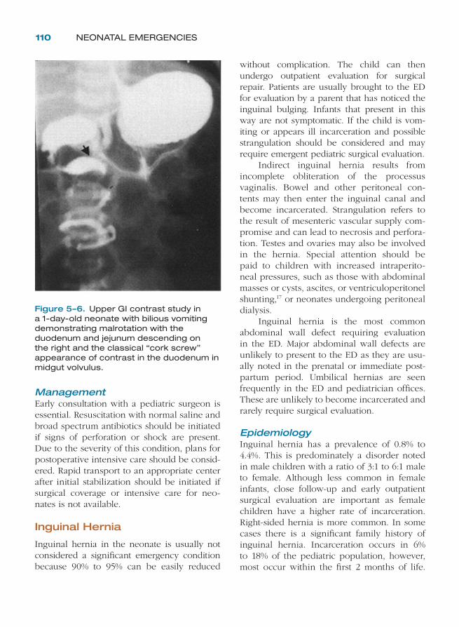
110 NEONATAL EMERGENCIES
Management
Early consultation with a pediatric surgeon is essential. Resuscitation with normal saline and broad spectrum antibiotics should be initiated if signs of perforation or shock are present. Due to the severity of this condition, plans for postoperative intensive care should be consid-ered. Rapid transport to an appropriate center after initial stabilization should be initiated if surgical coverage or intensive care for neo-nates is not available.
Inguinal Hernia
Inguinal hernia in the neonate is usually not considered a signifi cant emergency condition because 90% to 95% can be easily reduced
without complication. The child can then undergo outpatient evaluation for surgical repair. Patients are usually brought to the ED for evaluation by a parent that has noticed the inguinal bulging. Infants that present in this way are not symptomatic. If the child is vom-iting or appears ill incarceration and possible strangulation should be considered and may require emergent pediatric surgical evaluation.
Indirect inguinal hernia results from incomplete obliteration of the processus vaginalis. Bowel and other peritoneal con-tents may then enter the inguinal canal and become incarcerated. Strangulation refers to the result of mesenteric vascular supply com-promise and can lead to necrosis and perfora-tion. Testes and ovaries may also be involved in the hernia. Special attention should be paid to children with increased intraperito-neal pressures, such as those with abdominal masses or cysts, ascites, or ventriculoperitonel shunting,17 or neonates undergoing peritoneal dialysis.
Inguinal hernia is the most common abdominal wall defect requiring evaluation in the ED. Major abdominal wall defects are unlikely to present to the ED as they are usu-ally noted in the prenatal or immediate post-partum period. Umbilical hernias are seen frequently in the ED and pediatrician offi ces. These are unlikely to become incarcerated and rarely require surgical evaluation.
Epidemiology
Inguinal hernia has a prevalence of 0.8% to 4.4%. This is predominately a disorder noted in male children with a ratio of 3:1 to 6:1 male to female. Although less common in female infants, close follow-up and early outpatient surgical evaluation are important as female children have a higher rate of incarceration. Right-sided hernia is more common. In some cases there is a signifi cant family history of inguinal hernia. Incarceration occurs in 6% to 18% of the pediatric population, however, most occur within the fi rst 2 months of life.
Figure 5–6. Upper GI contrast study in a 1-day-old neonate with bilious vomiting demonstrating malrotation with the duodenum and jejunum descending on the right and the classical “cork screw” appearance of contrast in the duodenum in midgut volvulus.

CHAPTER 5 GASTROINTESTINAL EMERGENCIES 111
Associated congenital anomalies should also be considered and connective tissue disorders have also been described as risk factors for developing inguinal hernia (Table 5–3).
History
Details of time of onset, location of bulge or swelling, variations during sleep verses wake-fulness, and recent increased size or fi rmness are all usual aspects of the inguinal hernia his-tory. In addition to parental description of the groin bulging or scrotal swelling, other histori-cal features are important and should be gath-ered by the examining clinician. Symptoms related to bowel obstruction and ischemia should be reviewed with the parents to ensure there is no clinical suspicion of incarceration. Irritability may be the fi rst and only histori-cal feature; however, as time passes, the infant may suffer vomiting, abdominal distension, and obstipation. Lethargy and bloody stools are ominous signs of strangulation and possi-ble necrosis and perforation.
Clinical Manifestations
Although the examination of a neonate for presence of indirect inguinal hernia is not diffi cult, attention should be paid to several points. First, male infants should be carefully examined for presence of both testes in the scrotum, as an undescended teste may be mis-taken for a hernia. The hernia bulge should be in the expected location and should not
appear fl uctuant (Figure 5–7). Tumors and abscesses can sometimes be mistaken for her-nia as well. If the hernia bulge is noted to be intensely tender or discoloration is noted, care should be taken not to attempt reduction. Similarly, necrotic bowel from strangulation should not be reduced in the ED. An indirect inguinal hernia that cannot be reduced in the usual fashion should be evaluated for possible incarceration. Examination fi ndings of abdom-inal distension, vomiting, blood per rectum, and signs of shock should all be considered consistent with incarceration with strangula-tion until proven otherwise.
Diagnostic Studies
Radiographic evaluation with plain x-ray will reveal air-fl uid levels when partial or complete intestinal obstruction is present (Figure 5–8). Small bowel dilation is also a prominent fea-ture of obstruction from incarcerated hernia; however, midgut volvulus should also be con-sidered when small bowel obstruction is iden-tifi ed in the neonate. Free air may be present if strangulation has led to perforation. Ultrasound may be used to differentiate hernia bulge from
TABLE 5–3. � ASSOCIATED RISK FACTORS FOR INGUINAL HERNIA
PrematurityUndescended testeAscitesVP shuntPeritoneal dialysisIntraabdominal massAbdominal wall
defects
Ehlers-Danlos syndrome
Hunter-Hurler syndrome
Marfan syndromeMucopolysaccharidosisFamily historyHypospadiaTesticular feminization
Inguinal hernia
Figure 5–7. Drawing of an inguinal hernia.

112 NEONATAL EMERGENCIES
abscess and tumor mass. Evaluation for hydro-cele and testicular pathology is also possible during ultrasound evaluation. Baseline blood labs are of little value unless the infant appears ill or strangulation is suspected.
Management
In cases of uncomplicated indirect inguinal her-nia, reduction in the ED can lead to discharge with parental instructions and close follow-up for surgical evaluation and repair. If the hernia cannot be easily reduced without signifi cant discomfort to the infant, then analgesics and possible sedation should be given to facilitate relaxation of the abdomen. If the hernia is ini-tially thought to be incarcerated but can even-tually be reduced in the ED, the child should be seen by surgery and considered for admis-sion, as recurrence and complications are of a greater concern in these patients.
Before the initial attempt at bimanual reduction, the child should be placed in a Trendelenburg position and comforted in a way to encourage relaxation. If the hernia
reduces then the child is unlikely to incur complications as an outpatient referral to sur-gery and can be discharged. If the hernia sac fails to reduce, bimanual reduction using gentle, steady compression of the distal sack should be initiated in an effort to evacuate the contents of the bowel from the herniated portion. Gentle proximal support should be employed to guide the sac back through the external inguinal ring as the distal hand is used to apply slightly more pressure in sup-port of reducing the sac. If reduction does not occur within 5 minutes of applying pressure, efforts should be suspended and sedation may be required. A cold pack may also be applied to the groin. Appropriate procedure sedation should take place only after discussion with the parent and consent is obtained. Bimanual reduction should be attempted again after the child falls asleep.
If the neonate is found to have an incar-cerated inguinal hernia, pediatric surgi-cal evaluation is necessary. In cases where signs of obstruction, strangulation, or shock are present, immediate surgical consulta-tion is required. In the presence of strangu-lation with potential necrosis of the bowel, bimanual hernia reduction should not be per-formed in the ED, but rather by the surgeon. Intravenous resuscitation with normal saline and broad spectrum antibiotics, along with other supportive measures, should be initi-ated in anticipation of surgical treatment and appropriate intensive care unit placement. A nasogastric tube can be placed to allevi-ate gastric distension in patients with bowel obstruction.
Small Bowel Atresias
Small bowel atresia affects as many as 1 in 1500 to 4000 births.18 There appears to be a higher rate in children of low birth weight. Duodenal atresia is commonly associated with chromosomal abnormality,19,20,21 whereas jejunal and ileal are general not. Small bowel
Figure 5–8. Inguinal hernia with accompanying bowel obstruction.

CHAPTER 5 GASTROINTESTINAL EMERGENCIES 113
atresia can also be found in conjunction with atresias of the stomach and colon in some fam-ilies of French-Canadian descent. When atresia is severe the child will often be diagnosed by prenatal ultrasound along with maternal poly-hydramnios or with small bowel obstruction in the immediate postpartum period. Due to the variability in luminal narrowing, presenta-tions may be more subtle and therefore diag-nosis may be delayed.
Duodenal Atresia
Abnormal embryonic GI development can lead to duodenal webs, stenosis, and atresia. This condition may present at any time in the neo-natal period, or even later. Variability in the degree of narrowing of the duodenum makes the clinical presentation of this entity more var-iable. Although narrowing may be secondary to annular pancreas, Ladd bands, and volvulus, duodenal atresia should also be considered in severe cases of proximal small bowel obstruc-tion. The etiology of this condition is not clear; however, the prevailing theory appears to be a lack of complete recanalization of the duode-num during early development.22 Severe cases are likely to be detected in the prenatal and immediate postpartum period, and are there-fore less likely to present primarily to the ED. Because of this fact, midgut volvulus must be the diagnosis of primary consideration in the neonate with bilious (or bile streaked) vomit-ing in the ED.
Epidemiology
Duodenal obstruction has in incidence of about 1 in 10,000 live births.23 Many times this condition is associated with other abnor-malities and is seen with some frequency in children with trisomy 21 (Down syndrome) and Feingold syndrome.24 Cardiac and renal anomalies are also fairly common. A number of other GI malformations are associated with this condition and should also be considered (Table 5–4).25
History
Vomiting is typically bilious or bile streaked in nature. This is due to the fact that 85% of cases are associated with obstruction distal to the ampulla of Vater. The other 15% may not have bile in their emesis and may appear to vomit clear fl uid or a recent feeding. If the narrow-ing is of a lesser degree, the infant may have a delayed diagnosis. Historical features may then also include constipation and a failure to gain weight.
Clinical Manifestations
Abdominal distension and vomiting are usu-ally noted at the time of evaluation. The infant may also present with tachycardia signs of dehydration. When comparison to birth weight is made, the child will likely not have made appropriate weight gain.26
Diagnostic Studies
Plain radiography of the abdomen will usually reveal a distended stomach and proximal small bowel (Figure 5–9). This is the classic “double bubble” sign. The absence of gas beyond the proximal small bowel is very suggestive of duo-denal obstruction, but does not exclude malro-tation as the cause. Therefore, even when the “double bubble” sign is noted in the absence of distal bowel gas, an upper GI and/or ultrasound should be performed to exclude the diagnosis of midgut volvulus. In cases where clinical fea-tures are less severe, it may also be appropriate to obtain a CT to further delineate the duodenal
TABLE 5–4. � GI ABNORMALITIES ASSOCIATED WITH DUODENAL ATRESIA
Annular pancreas (most common)
MalrotationBiliary atresiaPreduodenal
portal veinCholedochal cyst
Anomalous bile duct communication
Pancreatic duct atresiaEsophageal atresiaImperforate anus

114 NEONATAL EMERGENCIES
pathology.27 CT scanning should only be done in consultation with a pediatric surgeon and is not considered a standard diagnostic modality for the diagnosis of duodenal atresia.
Management
Supportive measures, including resuscitation with normal saline, should be initiated in response to signs of dehydration. If a prox-imal small bowel obstruction is identifi ed, a nasogastric tube for decompression may be indicated. Pediatric surgical consultation and admission are necessary as operative manage-ment is almost always indicated.
Jejunal Atresia
Jejunal atresia is also a cause of bilious vomiting in the neonate. Presentation is fairly variable, however, and infants may present with nonbil-ious emesis. Jejunal atresia, in contrast to duo-denal atresia, is not commonly associated with chromosomal abnormalities or with congenital
malformations in cardiac or renal systems. However, there may be a familial component.28
Epidemiology
Jejunal atresia is more common than duode-nal atresia and may present in as many as 1 in 1500 neonates. Maternal polyhydramnios, prematurity, and low birth weight have all be associated with this condition.
History & Clinical Manifestations
The infant will have some history of feeding diffi culty. Vomiting may or may not be bilious in nature. In some cases the neonate will fail to pass meconium. Parents who bring their newborn to the ED may report abdominal dis-tension and constipation in addition to vomit-ing. Severe atresia usually results in signifi cant bilious vomiting.
Diagnostic Studies & Management
Abdominal plain fi lms will reveal multiple loops of distended small bowel and air fl uid levels. Management is similar to that of duo-denal atresia and should involve pediatric sur-gical evaluation and consideration of other causes of small bowel obstruction.29
Other GI Causes
There are a number of other GI disorders of the neonate that may cause vomiting as the pri-mary presenting symptom. These conditions are almost always diagnosed in the immedi-ate postpartum period and the neonate is not likely to present to the ED unless the mother had minimal (or no) prenatal care and an out-of-hospital delivery. It is worthwhile to mention several of them here as out-of-hospital delivery may be common with respect to geographic, socioeconomic, and cultural considerations.
Tracheoesophageal fi stula and esophageal atresia usually present with the fi rst feeding and is rarely missed, later to present to the ED. The neonate will attempt to feed, but will
Figure 5–9. The “double bubble” sign in duodenal atresia. Source: From Brunicardi CF, Andersen DK, Billiar TR, et al. Schwartz’s Principles and Practice of Surgery, 8th ed. New York, NY: McGraw-Hill; 2005.

CHAPTER 5 GASTROINTESTINAL EMERGENCIES 115
experience spitting, coughing, choking, and sometimes cyanosis. Inability to pass a naso-gastric tube is usually considered diagnostic in this situation. Upper GI is not recommended due to the risk of aspiration. Immediate pedi-atric surgical consultation is needed.30 Pyloric atresia differs from pyloric stenosis in that the defect is structural rather than functional and will therefore present at or shortly after birth. A single gas bubble, representing the distended stomach, will be present with a complete absence of intestinal gas. Gastric perforation may occur. Nasogastric tube and resuscitation are initiated while awaiting pedi-atric surgical consultation. Intussusception is extremely uncommon in neonates and infants under 3 months. Presentation is usually between 3 months and 6 years and abdominal mass, rectal bleeding, vomiting and intermit-tent abdominal pain are classically described.9 Air or contrast enema is typically used to diag-nose the condition, however, ultrasound may also be employed.11,12 Immediate surgical con-sultation is required. Infants may sometimes present with only lethargy.
Extragastrointestinal Causes
When evaluating the neonate presenting with vomiting it is important to consider causes out-side of the alimentary tract. A number of impor-tant conditions involving other systems of the body may be overlooked due to a readiness to evaluate only the GI causes of vomiting. The emergency physician must consider conditions related to increases in intracranial pressure, metabolic derangements,31,32,33 and infections.
Proper attention to these causes of vomiting will avoid unnecessary delay in diagnosis and potential mismanagement (Table 5–5).
DIARRHEA �
Despite the fact that diarrhea in the neonate is usually caused by a self-limited process it can be a life-threatening condition. Aggressive management is sometimes needed to avoid dehydration with its potential complications. Diarrhea is caused by a lack of normal func-tion of the bowel mucosa and is usually caused by enteritis of some type. Infl ammation of the bowel wall can be caused by atopy, infection, and even ischemia.34 Other causes of diarrhea include congenital atrophy of the villi and short gut syndrome (following loss of intestine to some other condition).35,36 A complete history and physical examination is important. Basic information that must be reviewed includes amount of oral intake, urinary output, number and character of stools (whether they are bloody or nonbloody, and their color and consistency), change in behavior, irritability, and fever or other signs of infection. Exposure to sick con-tacts (especially those with diarrheal illness), presence of other children in the home, and child care history should also be reviewed.
NONEMERGENT CAUSES
Normal Stool Changes
Neonates typically have loose stool after the passage of meconium is complete. During
TABLE 5–5. � VOMITING FROM CAUSES OTHER THAN THE GI SYSTEM
Neurologic Metabolic Infections Other
• Intracranial mass• Intracranial bleeding• Hydrocephalus• Closed-head trauma
• Inborn errors of metabolism
• Hypothyroidism• Congenital adrenal
hyperplasia
• Meningitis• Sepsis• Urinary tract
infection• Otitis media
• Narcotic withdrawal

116 NEONATAL EMERGENCIES
the fi rst 5 days the character of the stool will change from the thick dark meconium to a more liquid greenish-brown. The stool is commonly yellow in color and seedy in con-sistency by the fi fth day of life. Normal healthy infants may have a stool after every feeding. This is especially common in breastfed neo-nates. First-time parents and caregivers may be alarmed by this initial change in stool con-sistency and may present for evaluation of diarrhea. If the child appears healthy and is gaining weight parental assurance and educa-tion may be all that is required.
Overfeeding
Neonates and infants that are fed excessively may present with very frequent stooling. This is commonly a problem when the infant is fed in response to every cry or other sign of dis-comfort. A careful feeding history may lead to this conclusion in an otherwise normal healthy infant. Education concerning comforting and coping techniques is essential especially for fi rst-time parents and caregivers. These chil-dren may also suffer from excessive spitting up, refl ux, and colic.
EMERGENT CAUSES
Infectious Enteritis
Pathogens causing injury to the proximal small intestine lead to infl ammation and dysfunction of the mucosa, producing watery diarrhea and sometimes vomiting usually associated with viral pathogens. Bacterial pathogens usually affect the colon and blood may also be pre-sent in the stool. Parasitic infections causing diarrhea are rare in the neonate.
Epidemiology & History
Rotavirus is the most common viral pathogen causing diarrhea and is most commonly seen in the winter. Rotavirus has been known to be
associated with electrolyte abnormalities (includ-ing hypocalcemia), seizures, development of nec-rotizing enterocolitis, and increased frequency of both apnea and bradycardia. Dehydration can be signifi cant and IV rehydration was the standard therapy for many years. Despite the usual history of vomiting, recent literature has shown that oral rehydration therapy is appropri-ate in most cases of mild to moderate dehydra-tion. Rotavirus can be found in asymptomatic carriers and outbreaks in newborn nurseries are not uncommon. Adenovirus, enteroviruses, and Norwalk agent, among other viral pathogens, also cause a similar presentation.
Escherichia coli has a number of forms and may have a variable presentation depen-dent of the strain of pathogen responsible for the infection. Enteroinvasive and enterohem-orrhagic strains of E. coli present with bloody diarrhea, whereas other strains typically pre-sent with green watery diarrhea without blood or mucus. As with viral pathogens, the primary concern is with regard to dehydration and elec-trolyte disturbances. Other pathogens respon-sible for bloody diarrhea include Shigella, Campylobacter, Yersinia, and Aeromonas.
Clinical Manifestations
A thorough physical examination is warranted to determine if there are any other sources of infection. Temperature and pulse are impor-tant indicators of infection and dehydration. A rectal examination is important for obtaining a stool sample but also for checking for anal fi s-sures as possible etiology of bloody diarrhea. The patient should be weighed unclothed for an accurate weight. Close attention to respirations, detection of sunken fontanel, skin elasticity, whether patient makes tears, mucous mem-branes, urinary output, and capillary refi ll will help determine the degree of dehydration.
Diagnostic Studies
In cases of signifi cant dehydration it may be helpful to obtain labs to evaluate electrolytes and renal function. Bloody diarrhea may

CHAPTER 5 GASTROINTESTINAL EMERGENCIES 117
prompt the clinician to consider a diagnostic evaluation for GI bleeding. Sending stool cul-tures and smear may be helpful in the over-all management of the neonate, but does not affect the initial management of the patient. Results of stool cultures may lead to antibiotic therapy in select cases.
Management
Any neonate with diarrhea and any evidence of dehydration deserves admission for further evaluation and care. Mild dehydration (3% to 5%) should be treated with oral rehydration therapy if possible. If dehydration is moderate (6% to 9%) or severe (>10%), then generally IV fl uid rehydration is needed. Antimotility agents such as loperamide, opium, and diphenoxy-late should not be used. In addition, antibiot-ics should not be given unless culture results yield a specifi c bacterial agent, and the patho-gen and the patient’s clinical condition corre-spond to warrant antimicrobial therapy.
Noninvasive strains of E. coli can be treated with oral neomycin or colistin sul-fate. Neomycin therapy may eradicate the bacteria and shorten the course of illness. An absorbable antibiotic, such as ampicillin or trimethoprim-sulfamethoxazole, may be indicated for enteroinvasive bacteria causing diarrhea. Salmonella infection in the neonate can be treated with ampicillin, amoxicillin, bactrim, cefotaxime, or ceftriaxone. In cases of neonates with confi rmed bacterial enteritis, it may be appropriate to obtain a consultation with a pediatric infectious disease specialist.
Formula Intolerance/Cow’s Milk Sensitivity
Sensitivity to protein in formula derived from cow’s milk and/or soy can cause signifi cant dif-fi culty for the neonate. Immunologic reaction to these proteins causes an infl ammation of the small and large bowel and can therefore pre-sent as diarrhea and/or vomiting. Symptoms are typically evident within a few days to weeks
after initial ingestion of the cow’s milk protein. Diagnostic testing to determine sensitivity may not be necessary if symptoms cease after with-drawing the cow’s milk or soy based formula.
Epidemiology
Around 3% of infants will have a sensitivity to cow’s milk protein.7 Soy protein sensitivity is less common and is found in about 1%. Some infants will be sensitive to both.37 Most chil-dren will overcome this sensitivity by age 3.
History
Children usually present with signifi cant diar-rhea and vomiting that usually occurs within 2 hours of ingestion. Infants presenting with this disorder may have been previously hospi-talized for a septic workup due to a previous presentation with lethargy.
Clinical Manifestations
In addition to vigorous vomiting and diarrhea, infants commonly present with heme positive (or grossly bloody) stool and may also have a tender abdomen. Irritability and lethargy may also be present. The child may even present in apparent shock. Avoidance of the offending protein leads to resolution of symptoms.
Diagnostic Studies
Stool studies and baseline CBC, electrolytes, and renal function should be obtained. Ill-appearing neonates should be considered septic until proven otherwise, and a full septic workup may be indicated in relation to this important differential. Fecal leukocytes with eosinophils are usually seen. Serum studies may reveal an increase in polymorphonuclear leukocyte count. If deemed appropriate by an allergy/immunology specialist, a protein challenge may be performed. This should not be done in the ED or while the infant is still symptomatic.
Management
Supportive care and avoidance of the offend-ing food protein(s) is the mainstay of care

118 NEONATAL EMERGENCIES
for these patients. Immediate care includes resuscitation and possible administration of steroids. Admission should be considered in any neonate with signifi cant diarrhea and/or GI bleeding. Consultation with an allergy/immunology specialist is indicated. Follow-up at regular intervals is appropriate. Infants with confi rmed, or suspected, food protein sensi-tivity should be placed on hydrolyzed formula or amino acid-based formula. Switching from cow’s milk formula to soy-based formula may not be effective, as some infants may be sen-sitive to both.
Other Causes
Systemic infection, autoimmune enteropathy, congenital microvillus atrophy, and congeni-tal nutrient malabsorption are all rare causes of neonatal diarrhea. The clinician should also consider causes of partial bowel obstruc-tion or chronic constipation that may lead to diarrhea. Unfortunately, diarrhea in the infant is also a presentation of Munchausen syn-drome by proxy and this entity must also be considered.
CONSTIPATION �
A lack of signifi cant bowel output is a com-mon concern for parents and caregivers. After passage of meconium, it is common for neo-nates to have small amounts of stool 1 to 2 times daily. This can be especially true when children are breastfed. Children presenting for constipation within the fi rst 5 days should have a review of their diaper count and con-sideration for pathologic causes of inadequate bowel output. During the fi rst 2 days of life, the neonate is expected to pass meconium 1 or more times per day. On days 3 to 5 the bowel output should consist of 2 or more brownish-green stools followed by a change to a more yellow color after 5 days. A neonate with less than 2 stools per day should be eval-uated for dehydration and potential nutritional
defi ciency. If there are any signs of abdominal distension or vomiting other pathologic causes should be investigated.
NONEMERGENT CAUSES
Some parents and caregivers have been instructed to expect a stool after each feed-ing or may have had a previous child with a similar pattern of bowel output. Although this is common for neonates and infants, it is not absolutely necessary for successful weight gain. As long as the child appears healthy and has passed meconium and at least 2 to 4 stools per day for the fi rst week, there may be no need for concern.
Breastfed neonates usually have more bowel output than formula-fed children. In contrast to the 2-6 stools expected in the formula-fed neonate, it is common for breast-fed neonates to have a stool following every feeding. A low number of stools in this group may be of more concern because underfeed-ing can occur and the neonate may become dehydrated because their intake is not as eas-ily quantifi ed. If there is any question as to the nutritional status of the child, weight gain should be closely monitored by their primary physician. Dehydrated neonates, or those with jaundice or inadequate bowel output, should be admitted for further evaluation and care. In some cases of lactation failure the neonate may develop hypernatremic dehydration, a serious condition requiring aggressive resuscitation with isotonic fl uids, followed by correction of water losses and electrolyte abnormalities.
EMERGENT CAUSES
Hirschsprung Disease
Hirschsprung disease (HD) is a congeni-tal absence of ganglion cells in part or all of the colon, and rarely the small bowel, that leads to an inability for the effected region

CHAPTER 5 GASTROINTESTINAL EMERGENCIES 119
to relax. This effectively causes a functional colonic obstruction to develop. Due to the fact that migration of embryonic ganglion cells is cephalocaudal, the aganglionic region will be without gaps and will involve the distal colon. HD (also known as congenital megacolon) is an important cause of constipation, as it can lead to signifi cant complications in the neona-tal period and requires surgical consultation. Presentation varies and in some cases the dis-order may go unrecognized until later child-hood when the diagnosis is made as the result of evaluation for chronic constipation.
Epidemiology
HD is not uncommon and is found in 1 in 5000 live births.38 The development of this dis-ease is thought to be multifactorial. There is a family history in 10% of cases.39 Male infants are more commonly affected with a 4:1 male to female ratio. Infants are usually the prod-uct of an uneventful pregnancy with a normal delivery history and are usually of adequate birth weight. HD is more common in chil-dren with trisomy 21 (Down syndrome) and Waardenburg syndrome. The rectosigmoid colon is involved in 75% of cases. However, the entire colon can be effected in up to 10% of patients. In severe cases (<1%), the entire intestine may be involved.
History
Early diagnosis can be attributed to care-ful attention to bowel history. Ninety to 95% of cases will have a failure to pass meco-nium within the fi rst 24 to 48 hours of life. The infant is usually otherwise healthy and appears normal. Postpartum discharge within 24-48 hours is now common and the diag-nosis can therefore be missed. Parents may report a lack of stool over a number of days and may have even reported this at the time of newborn follow-up, only to be told that the child appears normal. In some cases, the infant may manifest only chronic constipation and the diagnosis will be delayed into infancy
and even childhood or adulthood. A history of delayed passage of meconium or retention of the meconium plug requiring evacuation should lead to an evaluation for aganglionic colon. Some infants will present with a history of constipation with infrequent, voluminous, and sometimes explosive bowel movements. Parents may also comment on a foul odor to the stool.
Neonates with more severe obstructive pathology will present earlier with additional symptoms of obstipation, abdominal disten-sion, and vomiting. Vomiting may be bilious or even feculent in nature. Patients who develop megacolon can manifest Hirschsprung entero-colitis and present with a history of fever, leth-argy and obtundation.
Clinical Manifestations
Depending on the severity of the condition and the time of presentation the child will frequently appear well. Some abdominal distension may be present with hyperactive bowel sounds. Rectal examination often reveals a vacant ampulla. The examiner may witness a rapid evacuation of gas and stool after completion of the rec-tal examination. Infants with enterocolitis may present with fever, lethargy, tachycardia, and signs of shock. Peritoneal signs should be con-sidered consistent with perforation.
Diagnostic Studies
Initial evaluation should include a plain abdominal radiograph. Large bowel obstruc-tion is expected to be seen (Figure 5–10). In addition, this fi lm should be closely evaluated for the presence of free air. Barium enema reveals a “saw-tooth” pattern of contrast in the aganglionic colon with a proximal area of signifi cant dilation. Barium enema has been reported with various levels of sensitivity and a negative result does not rule out the diag-nosis in all cases. If clinical suspicion is sig-nifi cant, a surgical consultation for anorectal manometry and to evaluate for rectal biopsy should be obtained.40

120 NEONATAL EMERGENCIES
Management
Supportive measures and pediatric surgi-cal consultation are the mainstay of therapy. Infants presenting with sepsis and/or perfora-tion will require fl uid resuscitation and broad spectrum antibiotics. These patients should be initially stabilized and transferred to a center with pediatric surgical services and appropri-ate intensive care if such resources are not available at the presenting facility.
Meconium Ileus
Meconium can create an intestinal obstruction in neonates defi cient in pancreatic enzymes. The blockage is usually located at the terminal ileum and, as such, causes a clinical picture consistent with distal small bowel obstruction. Ten to 15% of infants with cystic fi brosis will develop meconium ileus. When meconium fails to pass within 24 to 48 hours in a full-term infant, other conditions should also be considered (Table 5–6).
Epidemiology
Meconium ileus is found in 1 in 2800 live births. Essentially a complication of cystic fi brosis, 90% to 95% of neonates with meco-nium ileus have a positive sweat chloride
test. Other GI manifestations of cystic fi brosis include constipation, distal intestinal obstruc-tive syndrome, aquired megacolon, rectal pro-lapse, and pancreatits.
History
Meconium ileus is usually diagnosed prior to postpartum discharge when the child does not pass meconium in the fi rst 24 to 48 hours of life. Bilious vomiting and abdomi-nal distension usually follow. The child may return to the ED after having had treatment to relieve the obstruction, as infants with cystic fi brosis are prone to recurrence of intestinal obstruction.
Clinical Manifestations
The abdomen will appear distended and tender. Loops of distended bowel may be pal-pable. In cases of associated volvulus or meco-nium peritonitis (secondary to perforation) the infant may be tachycardic and exhibit signs of shock.
Diagnostic Studies
Despite the fact that meconium ileus rep-resents a functional distal small bowel obstruction, plain abdominal radiographs are expected to show specifi c characteristic signs (Figure 5–11).41 The meconium is usu-ally visualized in the right lower quadrant and is described as having a “soap bubble” or “ground glass” appearance as it is mixed with gas.42 Multiple distended loops of small bowel are notable; however, the expected air fl uid
TABLE 5–6. � FAILURE TO PASS MECONIUM
Muconium ileusMuconium plug
syndromeHirschsprung
diseaseAnorectal stenosis/
lack of patency
Small left colon syndromeHypogangliosisNeuronal intestinal
dysplasiaMegacystis-microcolon-
intestinal hypoperi stalsis syndrome
Figure 5–10. Plain radiographs demon-strating air-fl uid levels in the intestine and no gas in the rectum, suggestive for Hirschsprung disease.

CHAPTER 5 GASTROINTESTINAL EMERGENCIES 121
levels of a typical small bowel obstruction are absent. When contrast enema is obtained, the colon appears narrow. This is known as “microcolon” and is because the colon has not been used. Free air and the presence of cal-cifi cations are consistent with perforation and should be cause for emergent surgical consul-tation. Baseline labs and a sweat chloride test should also be obtained.
Management
Fortunately, response to therapy is usually good. Enemas are largely effective, and con-trast medium may serve a therapeutic and diagnostic role in this regard. If obstruction cannot be alleviated after multiple enemas, or
if signs of perforation develop, surgical explo-ration is necessary.43 After relief from obstruc-tion, recurrence should be prevented by proper treatment of the underlying cause. Intestinal malabsorption is typically treated with pancre-atic enzyme preparation in order to prevent further GI complications.
Meconium Plug Syndrome
Meconium plug syndrome (MPS) refers to colonic obstruction by meconium. Despite the fact that MPS is seemingly not related to cystic fi brosis or Hirschsprung disease, both of these more serious conditions should be considered at the time of presentation. Mostly a benign cause of colonic obstruction, MPS is usually present in children with a predisposition to poor bowel motility.
Epidemiology
MPS has an incidence of 1 in 500 to 1000 neonates.44 Neonates of low birth weight or immaturity are at risk for this condition. Other associated risks are maternal diabetes and hypothyroidism. An iatrogenic cause of MPS is the administration of magnesium sulfate to the mother in the peripartum period.
History
Symptoms of obstruction may present several days after birth. Bilious vomiting, abdominal distension, and failure to pass meconium may be reported by the parents.
Clinical Manifestations
The abdomen is usually not tender on exam-ination. Rectal examination may result in dis-lodgment of the meconium plug, relieving the colonic obstruction and confi rming the diagnosis.
Diagnostic Studies
Plain radiographic evaluation of the abdomen will reveal signs of distal small bowel or large bowel obstruction.
Figure 5–11. A 3 day old with multiple dilated loops of gut and a supine pneumoperitoneum. Perforation of a meconium ileus was found at surgery.

122 NEONATAL EMERGENCIES
Management
Water enema will usually dislodge the meco-nium plug and serve as diagnostic and thera-peutic. Close follow-up is important, as 10% to 15% of patients thought to have this condition will ultimately be diagnosed as actually having Hirschsprung disease.
Anorectal Malformations
Failure of the completion of the usual embry-onic development of the urologic structures and rectum lead to a variety of anorectal mal-formations. Anal stenosis and anal atresia (low type) may be overlooked at the time of delivery and subsequently present to the ED. It is common for anorectal malformations to be detected during the newborn examination and therefore diagnosis is usually made prior to discharge from hospital.45
Anal atresia refers to the lack of the usual anus as an opening to the outside of the body. Developmentally speaking, this has a high and low type and leads to the development of different fi stulas to allow for the passage of stool from the body. Anal stenosis represents around 20% of anorectal malformations and is essentially a very tight anus. This may be strictly a small opening, or may be secondary to the presence of anal web.
Epidemiology
One in 5000 infants will be born with an anorectal malformation.10 About half of these children will also have one or more associated conditions. These may include duodenal atresia, esophageal atresia, tra-cheoesophageal fi stula, cloacal extrophy in females, cardiac and urologic malformations, vertebral and upper limb abnormalities, and Down syndrome. Males are slightly more likely to present with anorectal malforma-tions, but are much more likely to present with high type anal atresia when compared with females.
History
If the child has been discharged home after passing meconium, the child may present with a history of thin, stringy, or ribbon-like stools that are passed with apparent diffi culty. If an associated urologic malformation is pre-sent, the child may present with fever or other signs of urinary tract infection. Parents may report cloudy or dark urine. Female children may have a history of stool coming from the vagina.
Clinical Manifestations
Anal atresia that presents as a classic imper-forate anus is not likely to be missed at the time of the newborn examination shortly after delivery. If a child is the product of an out-of-hospital birth, however, the examiner may fi nd just that. The presence of an apparent anus is more likely in children who were born in- hospital and initially missed. A close inspection of the perineum may reveal a tight-appearing anus that lies slightly anterior to the expected location of the anus. This may represent the perineal fi stula in males and the anocutaneous or rectofourchette fi stula in females.10
Diagnostic Studies & Management
Diagnostic studies are usually not indicated. Once the abnormality is discovered or sus-pected, pediatric surgical consultation should be obtained. Children suffering from urologic abnormalities should be tested for renal func-tion and urinary tract infection.
Other Causes
Other, rarer causes of apparent constipa-tion exist but are unlikely to present to the ED. Colonic atresia and colonic stenosis are commonly associated with other abnormal-ities of the GI system, abdominal wall, and urologic structures. When presenting as an isolated malformation, clinical presentation will include constipation (or failure to pass

CHAPTER 5 GASTROINTESTINAL EMERGENCIES 123
meconium), abdominal distension, and bil-ious vomiting. Plain radiographs will show an obstructive picture. The diagnosis is con-fi rmed by contrast enema that reveals a blind end to a microcolon. Duplications of the GI system may occur anywhere along the ali-mentary tract and may contain heterotopic tissues, such as gastric mucosa or pancreatic tissue. The duplications may be tubular, cys-tic, or fusiform. Presentation is variable, creat-ing some diagnostic diffi culty. However, these abnormalities are often identifi ed in utero by prenatal ultrasound.
JAUNDICE �
Sixty percent of normal newborns and 80% of preterm infants become clinically jaundiced during the fi rst week of life.46,47 Normal neo-nates produce bilirubin at around 2 to 3 times the rate an average adult. Details concerning the initial time of presentation and duration of the jaundice are key to determining the etiol-ogy. Neonates are rarely jaundiced at the time of delivery and presentation within the fi rst 24 hours is considered pathologic until proven otherwise.
Indirect or unconjugated, bilirubin is pre-dominately present in the body as a breakdown product of hemoglobin, but also represents a byproduct of precursor cell breakdown and, in some cases, ineffi cient hemoglobin synthe-sis. Neonates have larger red cells with greater hematocrit and a shorter life span of around 90 days. Direct bilirubin refers to the conju-gated form that is normally excreted into the bowel and transported out of the body with feces. This mechanism can be derailed by obstruction of the biliary system or of the small or large bowel.
Bilirubin encephalopathy or kernicterus is a serious neurologic condition related to the deposition of unconjugated bilirubin in the brain. This lipid soluble form of bilirubin
may cross the blood brain barrier and results in disruption of brain cell metabolism, espe-cially in the basal ganglia.48 For the devel-opment of this condition, levels greater than 20 mg/dL are usually required in healthy term newborns. Severe infection, hypoxia, hemo-lysis, and specifi c drug exposures may lead to neurologic damage in patients with levels below 20 mg/dL. Preterm neonates are also at increased risk. Presentation or jaundice with fever, bulging fontonel, lethargy, irritability, poor feeding, tremor, seizure, or other neuro-logic signs should raise immediate concern for bilirubin encephalopathy.
Nonemergent Causes
Physiologic jaundice typically becomes appar-ent on days 2 or 3 and peaks between the days 2 or 4 of like. Resolution should be seen by the day 7. Breast milk jaundice typically presents after the day 7 of life, peaks around week 2 to 3, and resolves from week 3 to 10.
Physiologic Jaundice
Physiologic jaundice is the result of increased bilirubin production from the breakdown of fetal red blood cells in addition to the limitation of the conjugation of bilirubin by the immature neonatal liver. In more detail, the increased bilirubin load on liver cells may be secondary to an increased erythrocyte volume, decreased erythrocyte survival, increased early-labeled bilirubin, or an increased enterohepatic circu-lation of bilirubin. A decreased hepatic uptake of bilirubin from plasma may occur. The most common reason is secondary to a decrease in ligandin, which is the bilirubin-binding protein in liver cells. Phenobarbital has been shown to increase the ligandin levels. A decrease in bili-rubin conjugation is caused by uridine diphos-phoglucuronosyl transferase. The purpose of this enzyme is to convert insoluble unconju-gated bilirubin to bilirubin glucuronide, the water-soluble conjugated bilirubin, allowing

124 NEONATAL EMERGENCIES
for excretion. And lastly, defective bilirubin excretion impairs bilirubin excretion.
Breast Milk Jaundice
Breast milk jaundice is associated with increased enterohepatic circulation of bili-rubin. Decreased caloric intake may lead to decreased stool production. Breastfed infants produce stool that contains less bilirubin com-pared with formula-fed infants. Unconjugated bilirubin is associated with unabsorbed fat in the intestine. In the breastfed infant there is an increase in intestinal fat absorption, thereby increasing the amount of unconjugated biliru-bin. Moreover, breast milk slows the formation of urobilin in the intestine, which increases the reabsorption of bilirubin. An increased activity of beta-glucuronidase in breast milk produces unconjugated bilirubin, which can be reabsorbed in the gut. And lastly, mutations of the Igene (Gilbert sydrome) prolong breast milk jaundice.49
Epidemiology
Elevations in bilirubin may be related to famil-ial, ethnic, and other genetic predisposing fac-tors. Hispanics tend to have a higher incidence of elevated total serum bilirubin (TSB) levels. Black infants, on the other hand, tend to have lower TSB. Siblings of children with a his-tory of elevated TSB also tend to have higher TSBs. Also of note, physiologic jaundice may be more prominent in infants of Asian and Greek descent. Asian and Native American infants may have a higher rate of breast milk jaundice.
History
The complex differential causes of jaundice in the neonate make it imperative to obtain a detailed history including family, pregnancy and delivery, and postnatal history. Physiologic jaundice, and breast milk jaundice to a lesser degree, should be a diagnosis of exclusion.
Family history of Gilbert syndrome, hemo-lytic disorders, maternal illness, delayed cord-clamping, birth trauma, breastfeeding history, loss of stool color, and parental nutrition are some of the important details of the history sometimes necessary to determine the cause of jaundice. A pharmacologic history is also important as some drugs alter the bilirubin-albumin binding affi nity. Anticonvulsants, diuretics, antibiotics, and sedatives may all be causative agents.
Physiologic jaundice usually becomes noticeable on the day 3 of life in term infants and by day 5 in preterm children. Breast milk jaundice presents later, around days 7 to 14. The child may have a history of diffi culty ini-tiating feeding or the mother may note a slow onset of milk production. Both types can be more prominent in neonates with a history of inadequate oral intake or dehydration.
Other Causes of Jaundice in the Young Infant
There are many additional causes of hyper-bilirubinemia in the young infant.50 These are broken down by types of hyperbilirubinemia and presented in Table 5–7.
TABLE 5–7. � CAUSES OF HYPERBILIRUBINEMIA BY BILIRUBIN SUBGROUP
Unconjugated
Predominance
Conjugated
Predominance
Physiologic jaundiceBreast milk jaundiceBirth trauma
(cephalohematoma)Polycythemia (delayed
cord clamping)Hemolysis (ABO
incompatability RBC defects)
Intestinal obstructionHypothyroidism
Urinary tract infectionCongenital viral
infection (CMV)Biliary atresiaDubin-Johnson
syndromeRotor syndrome

CHAPTER 5 GASTROINTESTINAL EMERGENCIES 125
Diagnostic Studies & Management
In general, almost all causes of conjugated hyperbilirubinemia mandate consultation and probable admission. In contrast, most causes of unconjugated hyperbilirubinemia are self limited and benign (with the excep-tion of blood group incompatibilities).51 In this regard, diagnostic studies should include a CBC with peripheral smear (to look for hemo-lysis), bilirubin fractionization, blood typing, and Coombs testing. Some general charac-teristics of laboratory fi ndings are listed in Table 5–8.
Most jaundiced infants may be safely discharged from the ED as long as adequate follow-up is arranged. General guidelines for phototherapy are provided in Table 5–9.
GI BLEEDING �
Gastrointestinal bleeding in the neonate requires investigation due to the fact that it may herald serious life-threatening condi-tions that require emergent surgical or medi-cal intervention. This being stated, a common cause for upper and lower GI blood is sim-ply the swallowing of maternal blood at the time of birth or even from cracked nipples of the breastfeeding mother. Historical details and a careful physical examination are crucial when evaluating the neonate with apparent GI bleeding. In cases of signifi cant GI bleed-ing, diagnostic evaluation with baseline labs, radiographs, and appropriate consultation are required to ensure timely diagnosis and treatment.
Upper GI Bleeding
Hematemesis within the fi rst several days of life may represent swallowing of maternal blood during delivery. The neonate will usu-ally appear well otherwise, and an Apt test would be expected to be negative. If the Apt test is positive, this benign process is ruled out and causes of GI bleeding should be con-sidered. More serious causes of upper GI bleeding in the neonate include milk pro-tein sensitivity, esophagitis, stress gastritis, or ulcer and vascular malformations.52 Neonates
TABLE 5–8. � ETIOLOGY OF HYPERBILIRUBINEMIA BY LABORATORY PROFILE
Coombs
Testing Anemia
Most Likely
Diagnosis
Positive Present Blood group incompatability
Negative Present RBC defectNegative Absent Breast milk
jaundice
TABLE 5–9. � MANAGEMENT OF HYPERBILIRUBINEMIA IN THE HEALTHY TERM INFANT WITH NO RISK FACTORS
25-48 h 49-72 h >72 h (3 days)
Consider phototherapy 12-15mg/dL 15-18 mg/dL 18-20 mg/dLPhototherapy >15 mg/dL >18 mg/dL >20 mg/dLConsider exchange transfusion 19-22 mg/dL 22-24 mg/dL 24-25 mg/dLExchange transfusion >22 mg/dL >24 md/dL >25 mg/dL
Note: (1) Infants with jaundice in the fi rst 24 h are not considered healthy infants. (2) TSB of 25 mg/dL or higher is a medical emergency and the child should be admitted immediately.
Source: Adapted from 1994 AAP Practice Guideline.

126 NEONATAL EMERGENCIES
born in the out-of-hospital setting may pre-sent with hematemesis as a presenting sign of hemorrhagic disease of the newborn sec-ondary to vitamin K defi ciency. Ill neonates may present with disseminated intravascular coagulation (DIC) or liver failure and may be septic. Surgical emergencies such as necro-tizing enterocolitis and midgut volvulus must also be considered.38
Lower GI Bleeding
Anal fi ssure is the most common cause of rec-tal bleeding in the neonate. If identifi ed in the otherwise healthy-appearing infant, no other diagnostic evaluation may be needed. Other causes to be considered are allergic enterocoli-tis, infectious enterocolitis, necrotizing entero-colitis, midgut volvulus, and Hirschsprung disease.53 Vitamin K defi ciency, DIC, and liver failure may also present as lower GI bleeding.
Diagnostic Studies
Neonates with signifi cant GI bleeding in the fi rst several days of life should have the blood tested for source (maternal vs neonate). This can be done with the Apt test. If negative, and the child appears well, observation only may be appropriate. If the Apt test is positive or cannot be completed in a timely manner, additional diagnostic evaluation is appropriate. All ill neonates with GI bleeding should have further diagnostics.54 This evaluation should include CBC, coagulation tests, liver function tests, BUN and creatinine, and plain fi lms for obstruction. Other diagnostic testing and con-sultation should be guided by the differential diagnosis established by patient history and examination.
Management
If the child is not septic, no surgical emer-gency can be identifi ed, and coagulation testing is normal, admission to the pediatrics service or gastroenterology unit is still appro-priate in cases where Apt testing is positive
and no rectal fi ssure or other benign expla-nation can be identifi ed. The neonate may require endoscopy and both gastroenterology and pediatric surgery may be required at the institution where the child is to be admitted.
REFERENCES
1. Reyna TM, Reyna PA. Gastroduodenal disor-ders associated with emesis in infants. Semin Pediatr Surg. 1995;4(3):190-197.
2. Hyman PE, Milla PJ, Benninga MA, Davidson GP, Fleisher DF, Taminiau J. Childhood functional gastrointestinal disorders: neonate/ toddler. Gastroenterology. 2006;130(5):1519-1526.
3. Gremse DA. Gastroesophageal refl ux: life-threatening disease or laundry problem? Clin Pediatr (Phila). 2002;41(6):369-372.
4. Naik-Mathuria B, Olutoye OO. Foregut abnor-malities. Surg Clin North Am. 2006;86(2):261-284, viii.
5. Poets CF. Gastroesophageal refl ux: a critical review of its role in preterm infants. Pediatrics. 2004;113(2):e128-e132.
6. Rasquin-Weber A, Hyman PE, Cucchiara S, et al. Childhood functional gastrointestinal dis-orders. Gut. 1999;45(Suppl 2):II60-II68.
7. Salvatore S, Vandenplas Y. Gastroesophageal refl ux and cow milk allergy: is there a link? Pediatrics. 2002;110(5):972-984.
8. Hostetler MA, Schulman M. Necrotizing entero-colitis presenting in the Emergency Department: case report and review of differential consid-erations for vomiting in the neonate. J Emerg Med. 2001;21(2):165-170.
9. Louie JP. Essential diagnosis of abdominal emergencies in the fi rst year of life. Emerg Med Clin North Am. 2007;25(4):1009-1040, vi.
10. Rao P. Neonatal gastrointestinal imaging. Eur J Radiol. 2006;60(2):171-186.
11. Maclennan AC. Investigation in vomiting children. Semin Pediatr Surg. 2003;12(4):220-228.
12. Vasavada P. Ultrasound evaluation of acute abdominal emergencies in infants and children. Radiol Clin North Am. 2004;42(2):445-456.
13. Green P, Swischuk LE, Hernandez JA. Delayed presentation of malrotation and midgut

CHAPTER 5 GASTROINTESTINAL EMERGENCIES 127
volvulus: imaging fi ndings. Emerg Radiol. 2007; 14(6):379-382.
14. Hajivassiliou CA. Intestinal obstruction in neo-natal/pediatric surgery. Semin Pediatr Surg. 2003;12(4):241-253.
15. Blumer SL, Zucconi WB, Cohen HL, Scriven RJ, Lee TK. The vomiting neonate: a review of the ACR appropriateness criteria and ultrasound’s role in the workup of such patients. Ultrasound Q. 2004;20(3):79-89.
16. Zissin R, Osadchy A, Gayer G, Shapiro-Feinberg M. Pictorial CT of duodenal pathology. Br J Radiol. 2002;75(889):78-84.
17. Duong M, Dinoulos JG, Gupta A, et al. Index of suspicion. Pediatr Rev. 2005;26(1):23-33.
18. Prasad TR, Bajpai M. Intestinal atresia. Indian J Pediatr. 2000;67(9):671-678.
19. Al-Salem AH, Qaissaruddin S, Karthikeya Varma K. Pyloric atresia associated with intestinal atre-sia. J Pediatr Surg. 1997;32(8):1262-1263.
20. Celli J, van Bokhoven H, Brunner HG. Feingold syndrome: clinical review and genetic map-ping. Am J Med Genet A. 2003;122(4):294-300.
21. Pameijer CR, Hubbard AM, Coleman B, Flake AW. Combined pure esophageal atresia, duo-denal atresia, biliary atresia, and pancreatic ductal atresia: prenatal diagnostic features and review of the literature. J Pediatr Surg. 2000;35(5):745-747.
22. Berrocal T, Torres I, Gutiérrez J, Prieto C, del Hoyo ML, Lamas M. Congenital anomalies of the upper gastrointestinal tract. Radiographics. 1999;19(4):855-872.
23. Escobar MA, Ladd AP, Grosfeld JL, et al. Duodenal atresia and stenosis: long-term follow-up over 30 years. J Pediatr Surg. 2004;39(6):867-871; dis-cussion 867-871.
24. Sajja SB, Middlesworth W, Niazi M, Schein M, Gerst PH. Duodenal atresia associated with proximal jejunal perforations: a case report and review of the literature. J Pediatr Surg. 2003;38(9):1396-1398.
25. Kelly DA, Davenport M. Current management of biliary atresia. Arch Dis Child. 2007;92(12):1132-1135.
26. Nixon HH. Duodenal atresia. Br J Hosp Med. 1989;41(2):134, 138, 140.
27. Jayaraman MV, Mayo-Smith WW, Movson JS, Dupuy DE, Wallach MT. CT of the duo-
denum: an overlooked segment gets its due. Radiographics. 2001;21 Spec No:S147-S160.
28. Shorter NA, Georges A, Perenyi A, Garrow E. A proposed classifi cation system for familial intestinal atresia and its relevance to the under-standing of the etiology of jejunoileal atresia. J Pediatr Surg. 2006;41(11):1822-1825.
29. Kays DW. Surgical conditions of the neonatal intestinal tract. Clin Perinatol. 1996;23(2): 353-375.
30. Stark Z, Patel N, Clarnette T, Moody A. Triad of tracheoesophageal fi stula-esophageal atresia, pulmonary hypoplasia, and duodenal atresia. J Pediatr Surg. 2007;42(6):1146-1148.
31. Burton BK. Inborn errors of metabolism in infancy: a guide to diagnosis. Pediatrics. 1998;102(6):E69.
32. Cappellini MD, Fiorelli G. Glucose-6-phosphate dehydrogenase defi ciency. Lancet. 2008; 371(9606):64-74.
33. Kwon KT, Tsai VW. Metabolic emergencies. Emerg Med Clin North Am. 2007;25(4):1041-1060, vi.
34. Armon K, Stephenson T, MacFaul R, Eccleston P, Werneke U. An evidence and consensus based guideline for acute diarrhoea manage-ment. Arch Dis Child. 2001;85(2):132-142.
35. Galea MH, Holliday H, Carachi R, Kapila L. Short-bowel syndrome: a collective. J Pediatr Surg. 1992;27(5):592-596.
36. Sigalet DL. Short bowel syndrome in infants and children: an overview. Semin Pediatr Surg. 2001;10(2):49-55.
37. Sicherer SH. Food protein-induced enteroco-litis syndrome: clinical perspectives. J Pediatr Gastroenterol Nutr. 2000;30(Suppl):S45-S49.
38. Kessmann J. Hirschsprung’s disease: diag-nosis and management. Am Fam Physician. 2006;74(8):1319-1322.
39. Pearl RH, Irish MS, Caty MG, Glick PL. The approach to common abdominal diagnoses in infants and children. Part II. Pediatr Clin North Am. 1998;45(6):1287-1326, vii.
40. Lewis NA, Levitt MA, Zallen GS, et al. Diagnosing Hirschsprung’s disease: increasing the odds of a positive rectal biopsy result. J Pediatr Surg. 2003;38(3):412-416; discussion 412-416.
41. McAlister WH, Kronemer KA. Emergency gas-trointestinal radiology of the newborn. Radiol Clin North Am. 1996;34(4):819-844.

128 NEONATAL EMERGENCIES
42. Gupta AK, Guglani B. Imaging of congenital anomalies of the gastrointestinal tract. Indian J Pediatr. 2005;72(5):403-414.
43. Rescorla FJ, Grosfeld JL. Contemporary man-agement of meconium ileus. World J Surg. 1993;17(3):318-325.
44. Loening-Baucke V, Kimura K. Failure to pass meconium: diagnosing neonatal intestinal obstruction. Am Fam Physician. 1999;60(7): 2043-2050.
45. Peña A, Hong A. Advances in the manage-ment of anorectal malformations. Am J Surg. 2000;180(5):370-376.
46. Maisels MJ, McDonagh AF. Phototherapy for neo-natal jaundice. N Engl J Med. 2008;358(9):920-928.
47. Maisels MJ, Watchko JF. Treatment of jaundice in low birthweight infants. Arch Dis Child Fetal Neonatal Ed. 2003;88(6):F459-F463.
48. Wennberg RP, Ahlfors CE, Bhutani VK, Johnson LH, Shapiro SM. Toward under-standing kernicterus: a challenge to improve
the management of jaundiced newborns. Pediatrics. 2006;117(2):474-485. Erratum in: Pediatrics. 2006;117(4):1467.
49. Gartner LM, Herschel M. Jaundice and breast-feeding. Pediatr Clin North Am. 2001;48(2):389-399.
50. Ip S, Chung M, Kulig J, et al. An evidence-based review of important issues concern-ing neonatal hyperbilirubinemia. Pediatrics. 2004;114(1):e130-e153.
51. Dennery PA, Seidman DS, Stevenson DK. Neonatal hyperbilirubinemia. N Engl J Med. 2001;344(8):581-590.
52. Chawla S, Seth D, Mahajan P, Kamat D. Upper gastrointestinal bleeding in children. Clin Pediatr (Phila). 2007;46(1):16-21.
53. Leung AK, Wong AL. Lower gastrointesti-nal bleeding in children. Pediatr Emerg Care. 2002;18(4):319-323.
54. Raine PA. Investigation of rectal bleeding. Arch Dis Child. 1991;66(3):279-280.

129
RENAL ABNORMALITIES 129 �
COLLECTING SYSTEM & �
BLADDER ANOMALIES 132
PENILE & URETHRAL �
ABNORMALITIES 137
URINARY TRACT INFECTION 139 �
SCROTAL ABNORMALITIES 140 �
AMBIGUOUS GENITALIA 142 �
GYNECOLOGIC �
ABNORMALITIES 142
Congenital genitourinary abnormalities often present during the neonatal period. As such, a fi rm understanding of these problems is necessary for any emergency physician. This chapter serves to elucidate the most common of these conditions and includes a discus-sion of renal abnormalities, collecting system abnormalities, bladder abnormalities, penile and urethral abnormalities, scrotal abnormal-ities, various gynecologic abnormalities, and urinary tract infections.
RENAL ABNORMALITIES �
Renal abnormalities invariably include some form of renal dysgenesis due to abnormal devel-opment of the kidney in utero. These abnormali-ties are often identifi ed prenatally, however some patients will present in the neonatal period.
Embryologically, the kidney originates from the ureteral bud and the metanephric
blastema. During week 5 of gestation, the ure-teral bud develops from the Wolffi an duct and penetrates the metanephric blastema. This ureteral bud undergoes multiple divisions and by week 20 of gestation forms the entire col-lecting system including the ureters, renal pel-vis, calyces, papillary ducts, and the collecting tubules. Nephron differentiation begins during week 7 of gestation and over 30% of nephrons are present by week 20 of gestation.1 At any point during renal development dysgenesis can occur. Dysgenesis refers to multiple forms of abnormal renal development including apla-sia, dysplasia, hypoplasia, and cystic disease.
UNILATERAL RENAL AGENESIS
Epidemiology
Unilateral renal agenesis is a relatively com-mon condition with an incidence of 1 in 450
chapter 6
Neonatal Genitourinary Emergencies
Brian Stout, MD

130 NEONATAL EMERGENCIES
to 1000 live births and is found with increased frequency in the neonate with a single umbil-ical artery (Figure 6–1).1
Clinical Presentation
This condition is often found while evaluating a neonate for urinary tract symptomatology. True renal agenesis is defi ned by no ureter or hemitrigone, as well as no kidney on the same side. Often, the contralateral kidney under-goes compensatory hypertrophy that can be palpated during the neonatal abdominal examination. Fifteen percent of patients with unilateral renal agenesis have vesicoureteral refl ux.1 Males have an absent ipsilateral vas deferens while females can have Mullerian duct abnormalities.
Diagnostic Testing
Renal ultrasound as well as renal function test-ing and urinalysis should be obtained.
Management
Referral to a pediatric nephrologist is appropri-ate and any issues with renal function should be addressed in an inpatient admission.
RENAL DYSPLASIA
Pathophysiology
Renal dysplasia refers to persistence of prim-itive ducts in the kidney derived from abnor-mal metanephric differentiation. This entity can affect all or a portion of the kidney. The pathophysiology leading to this disease is due to ureteral bud development in an abnormal location that causes inappropriate penetration and induction of the metanephric blastema.1
Clinical Presentation
Renal dysplasia can also be associated with posterior urethral valves and an absent portion of the ureter. If cysts are present, the disease process is termed cystic dysplasia. If the entire kidney is affected with many cysts, the dis-ease is termed a multicystic dysplastic kidney. Multicystic kidney disease is the most common cause of an abdominal mass in a neonate and is present in approximately 1 of 2000 live births.1 These kidneys often have little or no function. Contralateral vesicoureteral refl ux is present in 15% of patients. Five to 10% of patients will have contralateral hydronephrosis.1
Diagnostic Testing
Ultrasound of the kidney will demonstrate a kidney replaced by cysts of various sizes with no communication. No renal parenchyma is present (Figure 6–2).
Management
Management of the patients in the emergency department (ED) would include an ultrasound, renal function profi le, urinalysis, and consultation
Figure 6–1. Renal agenesis.

CHAPTER 6 NEONATAL GENITOURINARY EMERGENCIES 131
with a pediatric nephrologist. A confi rming renal scan should be performed along with a VCUG looking for refl ux. These can be accomplished on an outpatient basis if no abnormalities are present on the renal panel. Later complications include rennin-mediated hypertension and Wilms tumor. These patients require annual follow-up with a pediatric nephrologist.
RENAL HYPOPLASIA
Pathophysiology
Renal hypoplasia refers to a kidney that is structurally normal but small in size. The kid-ney is small due to a less than normal num-ber of nephrons and calyces. Like many renal conditions identifi ed in the neonatal period, this entity is often found while evaluating a patient for other urinary problems. Because of the reduced number of nephrons and caly-ces, these kidneys usually do not have normal function. If renal hypoplasia is present bilater-ally, chronic renal failure can ensue.
Clinical Presentation
Symptoms include polyuria and polydipsia.
Management
If found on ultrasound, these patients should undergo a renal function profi le, urinalysis, and pediatric nephrology consultation. Any abnormalities in renal function should be addressed with an inpatient admission.
Abnormalities in shape and position of the kidneys are a relatively common fi nding in neonates and may be found in the evaluation of a neonate in the ED. In normal develop-ment, the kidneys ascend from the pelvis to a position behind the ribs. If this process is incomplete, renal ectopic or nonrotation can develop, leading to an abnormal position or shape of the kidney.
RENAL ECTOPIA & FUSION ABNORMALITIES
Pathophysiology
Renal ectopia refers to an abnormal location of the kidney with an incidence estimated at 1 of 900 live births.1 Pelvic, iliac, thoracic, or contralateral placement of the kidney has been described. Ninety percent of patients with a contralateral kidney have fusion of 2 kidneys.1
Figure 6–2. Multicystic kidneys.

132 NEONATAL EMERGENCIES
Fusion abnormalities, leading to an abnor-mality in shape, are even more common than ectopia alone. Horseshoe kidney is the most common fusion abnormality, occurring in 1 of 500 live births.1
Clinical Presentation & Diagnostic Testing
The large, fused kidney is often palpable in the neonatal patient and can be diagnosed on ultrasound. The risk of Wilms tumor is fourfold in patients with a horseshoe kidney.1 There is an increased incidence of multicystic dysplastic kidney usually affecting one side of the kidney. Crossed-fused ectopia is another common fusion abnormality involving the fusion of the parenchyma of the 2 kidneys. Most commonly, the left kidney crosses the midline to fuse with the lower pole of the right kidney.
Management
A renal function panel should assess renal function initially and any abnormalities should be addressed with an inpatient admission. If all results are normal, further workup can be arranged as an outpatient.
COLLECTING SYSTEM & �BLADDER ANOMALIES
The renal collecting system is made up of caly-ces, ureters, bladder, and urethra. Problems can develop in any of these areas that lead to specifi c disease processes (Figure 6–3).
VESICOURETERAL REFLUX
Epidemiology & Pathophysiology
Vesicoureteral refl ux (VUR) is one of the most common collecting system problems found in
neonatal patients and is defi ned by the retro-grade fl ow of urine from the bladder to the ureter or renal pelvis. This entity occurs as the result of an abnormal attachment of the ureter to the bladder. Normally the attach-ment is oblique, forming a fl ap-like valve (Figure 6–4).
If this tunnel into the bladder is short or absent, refl ux develops (Figure 6–5). This is considered a birth defect and is found in approximately 1% of children.2 VUR is an important issue due to the increased risk of recurrent urinary tract infection in patients
Ureter
Bladder
Directionof urine
flow
Muscle wallof bladder
Ureter openinginto bladder
Figure 6–4. Normal ureter entering bladder.
Figure 6–3. Normal renal architecture.
Interlobular Vein
Renal VeinRenal Artery
Renal Hilus
Ureter
Collecting Duct
Single Nephron(much enlargedto show relativeposition)
Interlobular ArteryRenalPyramids
Renal Pelvis
Renal CapsuleMedulla (also knownas “Renal Medulla”and “KidneyMedulla”)
Kidney Cortex(also known as “Renal Cortex”)

CHAPTER 6 NEONATAL GENITOURINARY EMERGENCIES 133
with this abnormality. Prior to the wide-spread knowledge regarding this entity, VUR accounted for 15% to 20% of end stage renal disease in children and young adults.2
Grading of vesicoureteral refl ux has been established on a scale from I to V; grade V represents the most severe form. VUR can be primary or secondary depending upon the cause.
Duplication anomalies are one of the most common causes for vesicoureteral refl ux and occur in approximately 1 of 125 children.2 In this condition, 2 ureters drain 1 kidney. At least 1 of the ureters is in an abnor-mal position on the bladder. Refl ux occurs
in approximately 50% of patients with dupli-cation anomalies.2 If the ureter drains out-side the bladder, it is considered an ectopic ureter. If the ureter drains into the bladder neck, vesicoureteral refl ux is almost univer-sally present. Duplication anomalies are also associated with ureteral diverticulae and ureterocele.
Neuropathic bladder and posterior ure-thral valves are also associated with vesi-coureteral refl ux with an occurrence rate of 25% and 50%, respectively.2 Fifteen percent of patients with vesicoureteral refl ux also have renal agenesis, multicystic dysplastic kidney, or ureteropelvic junction obstruction.2
As stated previously there is a genetic link to VUR. Approximately 35% of siblings have refl ux that is often asymptomatic.2 In these siblings, 12% will have evidence of renal scar-ring.2 Fifty percent of children of mothers with refl ux will also have refl ux.2
Clinical Presentation
From a clinical standpoint, many patients with refl ux will be asymptomatic and the disease will be found in the evaluation for a urinary tract infection (UTI). Typically, these patients are diagnosed by a VCUG and ultrasound. Any neonate found to have a UTI should be admitted for antibiotic treatment but should undergo ultrasound and voiding cystoure-throgram, usually done during the inpatient admission.
Management
Treatment usually involves medical manage-ment because most refl ux tends to improve over time. Typically, antibiotic prophylaxis with Bactrim or nitrofurantoin is used. Surgical therapy is resolved for more severe cases that are unlikely to resolve.
The prognosis is good for most patients, in that refl ux tends to improve over time except in the most severe cases. Refl ux without infec-tion does not lead to renal scarring.
Figure 6–5. Vesicoureteral refl ux. Source: From Brunicardi FC, Andersen DK, Billiar TF. Schwartz’s Principles of Surgery, 8th ed. New York, NY: McGraw-Hill; 2005.

134 NEONATAL EMERGENCIES
URINARY TRACT OBSTRUCTION
Pathophysiology
Obstruction to urine fl ow can occur anywhere along the urinary tract from the urethral mea-tus to the calyces. These obstructions can be due to a variety of causes, including congenital causes, trauma, neoplasia, calculi, infl amma-tion, or surgery. Early fetal obstruction leads to renal dysplasia, whereas late fetal obstruction causes dilatation of the collecting system and renal parenchymal damage.
Clinical Presentation
Clinically, neonatal patients with urinary obstruction will present with abdominal pain or fl ank pain secondary to the collecting sys-tem dilatation and hydronephrosis. This can be diffi cult to identify in the neonatal patient and the initial presentation may be only irrita-bility. Also, failure to thrive may be a signifi -cant component in their history.
Diagnosis can prove to be diffi cult in these patients as many of will be asymptom-atic. If a palpable abdominal mass is present, this typically indicates a dysplastic kidney. If the bladder is palpable in a male patient, this may indicate an intravesicular obstruction. A patent urachus is also indicative of a urinary obstruction. Abdominal ascites is a common fi nding in patients with posterior urethral valves. UTI may also be present.
Diagnostic Testing
Numerous imaging modalities may be required to evaluate neonates with presumed urinary obstruction; these include renal ultrasound, VCUG, and radioisotope studies to evaluate renal function.
Differential Diagnosis & Management
Hydrocalycosis refers to dilatation of the calyx secondary to infundibular stenosis. This entity
can result from a developmental or infectious process. It is often discovered during evalua-tion for pain or urinary tract infection.
Ureteral pelvic junction (UPJ) obstruc-tion is the most common form of obstruc-tion found in childhood and can present in the neonatal period.3 Typically, this is an intrinsic form of stenosis. UPJ obstruction can present in a number of ways including fetal hydronephrosis found on ultrasound; palpable abdominal mass found on a neo-nate or infant; abdominal, fl ank, or back pain; febrile urinary tract infection; or hema-turia after minimal trauma. Sixty percent of patients have obstruction on the left side and 10% will have bilateral obstruction.3 There is a male to female preponderance of 2 to 1.3 Renal function may be impaired secondary to elevated pressures. In these cases, surgery is required for defi nitive treatment. Primary evaluation of UPJ obstruction should include a renal function panel, renal ultrasound, a CT scan of the abdomen and pelvis in some cases (Figure 6–6), and VCUG. Inpatient admission is usually indicated in the neona-tal patient.
Midureteral obstruction is a rare con-genital condition that can be identifi ed in the
Figure 6–6. Ureteral pelvic junction obstruction.

CHAPTER 6 NEONATAL GENITOURINARY EMERGENCIES 135
neonatal patient. Similar to other obstructive lesions, this can present as a palpable abdom-inal mass due to hydronephrosis of the ipsilat-eral kidney. Often, this is due to a retrocaval ureter. Treatment universally involves surgery and, as such, pediatric urology should be involved immediately upon diagnosis.
Ectopic ureter defi nes a condition where a ureter is abnormally placed in the bladder wall or drains outside of the bladder. This entity is 3 times more common in females than males.3 In females, 35% of patients have a ureter entering the bladder neck, 35% have a ureter entering the urethrovaginal sep-tum, and 25% have a ureter entering the vagina.3 Other locations include the cervix, uterus, and Gartner duct. Urinary inconti-nence occurs with ectopic placement of the ureter except when the ureter is implanted in the bladder neck.
In males, the ectopic ureter is implanted in a variety of locations. Forty-seven percent of patients have a ureter entering in the pos-terior urethra, 33% have a ureter entering in the seminal vesicle, 10% have an entrance in the prostatic utricle, 5% in the ejaculatory duct, and 5% in the vas deferens.3 Unlike females, males do not have any incontinence, though the abnormality can be associated with a UTI or epididymitis.
Diagnosis is usually established by an ultrasound obtained for a palpable mass found on physical examination. A hydronephrotic kidney is invariably present on the side of the ectopic ureter. A VCUG needs to be obtained along with a urologic consult.
Surgical repair may involve ureteral reim-plantation or nephrectomy.
Ureterocele is defi ned as a cystic dilata-tion of the terminal ureter. These lesions are obstructive due to a pinpoint orifi ce. Similar to an ectopic ureter, there is a female prepon-derance over males. These lesions are almost always associated with ureteral duplication.
The fi rst type of ureterocele is the sim-ple ureterocele. The simple ureterocele is a
simple cystic dilatation of the ureter with a normal insertion location on the blad-der. There is no duplication associated with the simple ureterocele. Often, this lesion is found during an evaluation of a patient for a UTI. Treatment involves drainage though the lesion itself and may cause refl ux, in which case surgical reimplantation is required. In small lesions, no treatment may be required at all.
In more complicated cases of uretero-cele involving ureteral duplication and abnormal insertion, treatment almost univer-sally involves surgery. Typically, this involves excision of the affected pole of the kidney and the ectopic ureter. If the ureterocele is small, drainage alone is sometimes success-ful. In a septic infant, immediate urologic consultation is required with drainage of the ureterocele.
Megaureter defi nes a ureter that is dilated through its entire length. There are numerous causes including many nonobstructive causes. Diagnosis is established through an ultrasound or VCUG, during or after an evaluation for a UTI (Figure 6–7).
Typical symptoms include abdominal pain and hematuria. Nonobstructive management is conservative while obstructive lesions usually require surgery.
Prune-belly syndrome, also known as Eagle-Barrett or triad syndrome, is a rare con-dition occurring in approximately 1 of 40,000 live births and causes massive dilatation of the ureter and upper tracts of the collecting sys-tem.3 Ninety-fi ve percent of affected patients are males.3
The clinical fi ndings in prune-belly syn-drome typically include a large, distended bladder and can include a patent urachus or urachal diverticulum. Vesicoureteral refl ux is typically present. The prostatic urethra is dilated while the prostate itself is hypoplas-tic. The kidneys are usually dysplastic and in males, posterior urethral valves may be present.

136 NEONATAL EMERGENCIES
All patients with this entity should receive prophylactic antibiotics. If no obstruction is present, this is all the treatment required at the time. If obstruction is present, vesicostomy is required. Thirty percent of patients will go on to have end-stage renal disease.3
Bladder neck obstruction is usually a sec-ondary form of collecting system obstruction caused by an ectopic ureterocele, bladder cal-culi, or tumor of the prostate. Typical symptoms include diffi culty voiding, urinary retention, and urinary tract infection. Bladder distension is universal, often with overfl ow incontinence.
Posterior urethral valves are the most com-mon cause of severe renal and urologic disease. This entity only affects males and has an inci-dence of 1 in 8000 live births.3 This congenital abnormality with the presence of valves in the
urethra obstructs the fl ow of urine, leading to dilatation of the prostatic urethra to bladder muscle hypertrophy. Vesicoureteral refl ux will develop in 50% of patients if untreated.3 Renal changes ranging from hydronephrosis to dys-plasia can occur. Without effective treatment, 30% of patients will go on to end stage renal disease or chronic renal insuffi ciency.3
The diagnosis can often be established prenatally, however some patients will be missed. The diagnosis should be suspected in any male neonate with a palpable, distended bladder or a weak urine stream. Initial workup should include a renal ultrasound, and, if pos-terior urethral valves are suspected, a voiding cystourethrogram should be obtained. A renal function panel should be obtained to evaluate renal function. Pediatric urologic consultation should be sought immediately.
Treatment involving antibiotic prophy-laxis is required in all patients and defi nitive treatment is accomplished by valve ablation. Postoperative incontinence is common and some patients will also have polyuria lasting for weeks. Some patients may have residual hydronephrosis of the kidneys.
Urethral atresia is an obstructive condi-tion that is often diagnosed in utero and will present in the fi rst days of life due to absent urine production. Due to obstruction, the bladder distends, ultimately causing bilateral hydroureteronephrosis. Emergent urinary diversion using a vesicostomy is required.
Urethral hypoplasia is an obstructive con-dition in which the urethral lumen is small. This is a rare condition that, similar to urethral atresia, causes a distended bladder and bilateral hydronephrosis. Renal dysplasia can develop causing end stage renal disease. Treatment involves a vesicostomy, followed by urethral reconstruction, dilatation, or diversion.
Urethral stricture is a condition in which a portion of the urethra is signifi cantly nar-rowed causing an obstruction. This condition is much more common in males than females. The most common cause for this condition is
Figure 6–7. Megaureter.

CHAPTER 6 NEONATAL GENITOURINARY EMERGENCIES 137
urethral trauma. Symptoms develop gradually, as does the lesion, and includes bladder insta-bility, hematuria, and dysuria. Diagnosis is accomplished by IV urography or retrograde urethrography and ultrasound (Figure 6–8). Treatment is accomplished by dilatation or urethroplasty.
Anterior urethral valves are another rare obstructive condition. This can be associated with a diverticulum in the penile urethra. This expands and can lead to a palpable mass on the ventral surface of the penis. Other phys-ical examination fi ndings are consistent with those for posterior urethral valves. The urinary stream is typically weak. Diagnosis is estab-lished by VCUG and treatment involves exci-sion of the valves and diverticula.
Bladder abnormalities can also cause uri-nary obstruction. These conditions include bladder diverticula, urachal abnormalities including urachal cyst, and patent urachus.
Neurogenic bladder usually results con-genitally from spinal abnormalities, such as spina bifi da. Symptoms include urinary incon-tinence, UTI, and upper tract deterioration. Ten to 15% of patients have hydronephrosis and 25% have vesicoureteral refl ux.3
Diagnosis involves ultrasound, postvoid bladder residuals, and VCUG.
PENILE & URETHRAL �ABNORMALITIES
HYPOSPADIAS
Epidemiology & Pathophysiology
Hypospadias is a common, congenital con-dition present in 1 of 250 live births where the urethral opening is abnormally located on the ventral surface of the penis.4 In most infants, hypospadias is an isolated abnormal-ity. Associated abnormalities include incom-plete development of the prepuce, chordee, absent foreskin ventrally, and inguinal her-nia. A bifi d scrotum can also be present. Ten percent of patients have undescended testes.5 The etiology of this congenital malforma-tion is unclear. There is an increased inci-dence of hypospadias in infants where the mother received antiandrogenic or estrogenic compounds.
Classifi cation of the condition is based upon the location of the urethral opening. Sixty percent of patients will have a distal hypospadias, 25% subcoronal or midpenile, and 15% proximal.5 Less common locations include glanular, coronal, subcoronal, mid-penile, penoscrotal, scrotal, and perineal (Figure 6–9).
Clinical Presentation
The abnormal location of the urethral opening leads to several complications including defor-mity of the urinary stream, sexual dysfunction secondary to curvature of the penis, infertility, and meatal stenosis.
Management
Surgical treatment represents the defi nitive therapy and is usually performed at 6 to 12 months of age. Circumcision should never be performed in the infant with hypospadias until the repair is complete.
Figure 6–8. Urethral stricture.

138 NEONATAL EMERGENCIES
CHORDEE
Chordee refers to a ventral curvature of the penis due to tight bands in the penis. Usually there is incomplete development of the fore-skin; in the neonate there is a dorsal hood similar to that seen in hypospadias. If this con-dition is identifi ed in the evaluation of a neo-nate, it is important to refer the patient to a pediatric urologist for surgical repair
PHIMOSIS
Phimosis is a penile condition in which there is an inability to retract the prepuce. It is important to note that at birth there is physiologic phimo-sis that normally resolves by year 3 of life.
COMPLICATIONS OF CIRCUMCISION
Circumcision is the most common elective sur-gical procedure performed on newborn boys
in the United States and 0.2% to 3% of boys who underwent circumcision require a subse-quent operative procedure.5
Clinical Presentation
Many neonates are brought to the ED following this procedure with concerns regarding bleed-ing or the appearance of the circumcision site. After circumcision, the penis normally dem-onstrates an erythematous glans with a white healing eschar. This appearance often may cause concern, particularly to new parents. After a thorough physical examination, the concerns of the parents should be addressed and reassurance given. The most common complications of a circumcision include bleed-ing, wound infection, meatal stenosis, second-ary phimosis, removal of insuffi cient foreskin, and penile adhesions.
Management
For bleeding complications postcircumcision, direct pressure is always the fi rst line of treat-ment and usually the only treatment needed. Topical treatments include topical thrombin as well as silver nitrate cauterization. If bleeding continues despite these local treatments, uro-logic consultation should be obtained. Rare, serious complications include sepsis, amputa-tion of the distal part of the glans, removal of an excessive amount of foreskin, and develop-ment of a urethrocutaneous fi stula.
PENILE TORSION
Penile torsion is a condition that may pre-sent to the ED due to parental concern about the appearance of the penis. The condition involves a rotation deformity of the shaft of the penis, usually counterclockwise. This con-dition is invariably cosmetic in nature and the parents can be reassured and referred to pedi-atric urology for outpatient treatment.
Glanular
Subcoronal
Midshaft
Distal Penile
Proximal Penile
Penoscrotal
Scrotal
Perineal
Types of Hypospadias
Figure 6–9. Hypospadias.

CHAPTER 6 NEONATAL GENITOURINARY EMERGENCIES 139
INCONSPICUOUS PENIS
Another penile issue that may present to the ED is the inconspicuous penis. This essen-tially refers to a penis that is small, at least in appearance. There are a variety of different subtypes of inconspicuous penis. A webbed penis refers to a penis that has the scrotum extending onto the anterior ventral penis. The penis itself is not small but appears to be small due to concealment of the penile shaft by the scrotum. If circumcision is performed, the penis can retract into the scrotum and second-ary phimosis can develop.
A second type of inconspicuous penis is the concealed penis. In this condition, the penis is hidden by suprapubic fat. The cause can be congenital or iatrogenic after circum-cision. Again, it is important to note that the penis itself is not small in this case.
A third type of inconspicuous penis is the trapped penis, which is acquired after cir-cumcision. In this case, the penis is literally embedded in the suprapubic fat. UTIs and urinary retention can develop and patients presenting with this condition should be eval-uated for these complications. Again, the penis itself is normal size.
The fourth type of inconspicuous penis is the micropenis, which is a true small penis. Micropenis is defi ned by a penile size that is 2.5 standard deviations below the mean, which equates to penile length less than 1.9 cm in the stretched position.5 The etiology of micrope-nis is usually related to a congenital hormone problem in the infant. These patients need no emergent treatment but referrals to a pediat-ric urologist and an endocrinologist should be made.
MEATAL STENOSIS
Meatal stenosis refers to stenosis of the ure-thral meatus. This is most commonly seen after circumcision and it is due to severe
infl ammation postcircumcision. Typically, due to the pinpoint orifi ce, parents will describe a forceful, fi ne stream of urine possibly with a dorsal defl ection. Referral to a pediatric urol-ogist should be made for defi nitive treatment by meatoplasty.
URINARY TRACT �INFECTION
EPIDEMIOLOGY & PATHOPHYSIOLOGY
Urinary tract infection can be a serious and potentially life-threatening problem in neo-nates and must be considered in any neonate presenting with a fever. The infection can be present in the urinary bladder or the kidney itself. The overall prevalence of a febrile infant having a UTI is approximately 5%; however, the prevalence of UTI in the neonatal popu-lation is most likely higher.4 Congenital abnor-malities including vesicoureteral refl ux and posterior urethral valves place the affected infant at increased risk for infection.
CLINICAL PRESENTATION
Clinically, the neonate with a UTI can present with a variety of symptoms and severity. Some neonates may appear quite well with minimal symptoms such as vomiting and poor feed-ing while others can appear septic with high fevers.
DIAGNOSTIC TESTING
A thorough history and physical examination should be performed along with a urinalysis, urine Gram stain, and urine culture if a UTI is suspected. It is important to obtain a ster-ile specimen by catheterization to avoid skin contamination commonly seen when bagged

140 NEONATAL EMERGENCIES
specimens are collected. The gold standard for diagnosis is the urine culture; however, most culture results will not be available for at least 24 hours. As such, the diagnosis is based upon the urinalysis and/or Gram stain. A white blood cell count in the urine of 5 or greater per high power fi eld would be consistent with a UTI. However, given the age of the patient, any abnormality on the urinalysis indicating infection, such as a positive nitrite or leuko-cyte esterase, should be given strong weight and prompt treatment. Also, bacteremia from the UTI can be a problem for patients in this age range. In a study looking at the utility of blood cultures in febrile children with UTIs, Pitetti and Choi found that the prevalence of bacteremia was 22.7% in children younger than 2 months in this setting.6 Given this fact, a complete blood count and blood cultures and lumbar puncture should be obtained in febrile neonates who may have UTIs.
MANAGEMENT
Initial treatment of UTI in neonates involves broad-spectrum antibiotics, usually ampicil-lin and cefotaxime or gentamycin. Admission is required until the 48-hour culture results return. A renal ultrasound is usually indi-cated and a VCUG should be obtained once the infection has resolved. E. coli is the most common pathogen responsible for UTI in the neonate.
SCROTAL ABNORMALITIES �
UNDESCENDED TESTES
Epidemiology & Pathophysiology
Scrotal abnormalities encompass a wide vari-ety of conditions that include abnormalities of the scrotum itself as well as abnormalities involving the scrotal contents. In many of these
conditions, the primary cause of the observed defects is due to an abnormality in the descent of the testes to their intrascrotal location.
The testes originate in the retroperitoneal area and, during gestation, descend to the level of the internal inguinal ring. They do not complete their descent until shortly before or soon after birth. If an abnormality develops where both or one of the testes does not com-plete its descent, it is referred to as an unde-scended testis.
Clinical Presentation
An undescended testis is a common disorder with an incidence that is approximately 30% in premature boys compared to 3% in term boys.4 If the testis is going to descend on its own, it usually does so by about 3 months of age. If there is no descent by 6 months of age, the testis likely will not complete its descent. Bilateral cryptorchidism occurs in about 10% to 20% of patients.7 Undescended testes are usu-ally located in the inguinal canal though they can be ectopic in location as well, including intra-abdominal locations. In 15% of patients, the missing testis will be nonpalpable and in those, only 50% will actually have a testis pre-sent.7 If both testes are nonpalpable, strong consideration should be given to the diagnosis of a virilized female with congenital adrenal hyperplasia.
Problems associated with this condition include infertility, malignancy, hernia, torsion of the undescended testis, and psychological issues later in life. Fertility is often a signifi -cant concern of parents and it is important to note that 85% of boys with a unilateral unde-scended testis are fertile and that 50% to 65% of boys with bilateral undescended testes are fertile after repair.7
Diagnosis
This diagnosis is made after a thorough phys-ical examination. If the testis is palpated in the inguinal canal of the neonate and cannot

CHAPTER 6 NEONATAL GENITOURINARY EMERGENCIES 141
be milked down into the scrotum, the parents should be reassured and the patient monitored by the pediatrician for descent of the testis. If no testis can be palpated, the patient should undergo an abdominal ultrasound to look for the undescended testis.. This does not need to be done on an emergent basis but should be discussed with the parents; the patient should be referred to a pediatric urologist for further evaluation and management.
Management
Treatment is accomplished through orchio-pexy at age 9 to 15 months. If the testis is not found on ultrasound, laparoscopy is usu-ally performed due to the risk of malignancy developing in an undescended testis.
ENLARGED SCROTUM & SCROTAL SWELLING
Scrotal swelling is a common complaint in many neonates presenting to the ED. There are a variety of conditions that can be the cause of scrotal swelling. It is important on history to note if the swelling is acute or chronic and whether it is painful or painless.
Diagnosis & Management
Hydrocele is one of the most common causes of scrotal swelling in the neonate. It occurs in approximately 1% to 2% of males.7 The hydrocele may communicate with the abdominal cavity or may be noncommunicating. Communicating hydroceles will typically be small in the morn-ing and will enlarge by the evening. They will disappear with compression on physical exam-ination. Noncommunicating hydroceles will not vary in size. They are typically smooth and not tender on physical examination. Most non-communicating hydroceles will disappear by age 1 year. If they do not or are large they will require surgical intervention, usually after 2 years of age.8
Neonatal testicular tumors are rare. Typically, a mass can be palpated off the tes-ticle or the testicle itself may be enlarged. Most of these tumors will not transillumi-nate as they are solid masses. Gonadal stro-mal tumors are the most common; yolk sac tumors and mature teratomas are next most common.4 If a mass is palpated, an ultrasound should be obtained to delineate the mass. Tumor markers such as alpha-fetoprotein and beta-human chorionic gonadotropin along with screening labs such as a complete blood count, comprehensive metabolic profi le, and urinalysis should be obtained. Finally, imme-diate urologic consultation in conjunction with pediatric oncology consultation should be obtained to determine the need for surgi-cal intervention.
Neonatal or extravaginal torsion may present as acute scrotal swelling in early life. Testicular torsion presents as the acute unilat-eral swelling of the scrotum usually with an ipsilateral swollen, and possibly tender scrotum. The scrotum itself may be red and infl amed. Typically, the mass does not transilluminate. When diagnosing this condition, a urinalysis should be obtained as well as an ultrasound to examine the mass and blood fl ow to the testi-cle. Urologic consultation should be obtained although salvage of the testicle is unlikely as often, the testicle has undergone torsion during its descent from the abdominal cavity in utero. Surgery for this condition is usually performed for fi xation of the contralateral testicle.
Scrotal hematoma is another neonatal con-dition that may present as acute scrotal swell-ing with a scrotal mass. This lesion can present secondary to a tumor or torsion. Typically, this mass will be solid and will not transilluminate. Similar to torsion, an ultrasound to determine if the testicle is intact and urinalysis should be obtained, in addition to a coagulation pro-fi le and complete blood count. If a tumor is suspected or found, tumor markers should be obtained as well. Immediate urologic consul-tation should be obtained as well.

142 NEONATAL EMERGENCIES
Inguinal hernias can present as a scrotal mass in the neonatal period. Typically, parents will notice a bulge in the inguinal canal or scro-tum that may or may not remit. This is usu-ally the inciting event for presentation to the ED. If the hernia is incarcerated, feeding issues may be present along with vomiting. On phys-ical examination, a palpable bulge may be felt in the scrotum extending up to and into the inguinal canal. The scrotum may be swollen and infl amed. The mass may or may not transil-luminate. If the mass is compressible, reduction can be attempted with traction and gentle but fi rm pressure trying to push the hernia through the inguinal ring. If it is reducible, surgery should be consulted for possible surgical cor-rection. If vomiting has been present, a basic metabolic profi le along with fi ngerstick blood sugar should be obtained early on. If the her-nia is not reducible, an ultrasound should be obtained along with immediate surgical consul-tation because most likely the hernia is incar-cerated and will require surgical intervention.
AMBIGUOUS GENITALIA �
CLINICAL PRESENTATION
Most patients with ambiguous genitalia have been thoroughly evaluated prior to any pre-sentation to the ED. However, surgical therapy for these patients may not be performed until later in life. For this reason, emergency med-icine physicians must be familiar with these patients and their comorbid conditions.
The most common physical examina-tion fi ndings in these patients include clitoral enlargement with palpable labial/scrotal masses or microphallus with varying degrees of hypo-spadias and clitoromegaly with labial fusion.
DIAGNOSIS & MANAGEMENT
Salt wasting may be a comorbid condition when the cause of ambiguous genitalia is
21-hydroxylase defi ciency. In the most severe cases, these patients have a defi ciency of both aldosterone and cortisol ultimately leading to an adrenal crisis. Unless patients are diagnosed prenatally or soon after birth and treatment is initiated, they will present in crisis, appear-ing septic, at about 2 weeks of age. Signs and symptoms typically include weight loss, anorexia, vomiting, dehydration, weakness, hypotension, hypoglycemia, hyponatremia, and hypokalemia. An appropriate history should be obtained with careful attention to fever, vomiting, diarrhea, and urine output. A careful physical examination should be per-formed as well. Appropriate laboratory evalua-tion includes a full septic workup with a basic metabolic profi le and fi ngerstick blood sugar. Treatment in the ED involves IV fl uids, antibi-otics, and corticosteroids.
Inguinal hernia is also associated with ambiguous genitalia. Diagnosis and treatment is identical to that discussed earlier.
GYNECOLOGIC �ABNORMALITIES
Gynecologic abnormalities in the neonate are relatively uncommon but emergency medi-cine physicians should be familiar with the most common conditions that could present to the ED.
MASTITIS
Clinical Presentation
Mastitis typically presents as an infl amed, tender, and swollen breast bud. Due to mater-nal estrogen, often the breasts of a neonate (male or female) may be swollen and draining so-called “witch’s milk.” Breast bud tissue may be palpable. To distinguish this normal condi-tion from mastitis, in mastitis the nipple and surrounding tissue will be red, swollen, and tender. Often, discharge of purulent material

CHAPTER 6 NEONATAL GENITOURINARY EMERGENCIES 143
from the nipple may be present. Staphylococcal and streptococcal bacteria are common causes. Fever may be present as well.
Diagnostic Testing
Diagnosis is based upon the clinical presenta-tion; however, given the likelihood of a bac-terial cause and the age of the patient, a full septic workup should be performed including urinalysis, urine Gram stain, urine culture, complete blood count and differential, blood culture, and lumbar puncture.
Management
Treatment should be initiated with broad-spectrum antibiotics such as ampicillin and cefotaxime or gentamycin once blood cultures have been obtained. If drainage is present, it should be cultured as well and if there is any concern for an abscess, immediate surgical consultation should be obtained.
IMPERFORATE HYMEN
Clinical Presentation
Imperforate hymen is another condition that may present in a neonate. Typically, the female neonate will present with a vaginal and pos-sibly a palpable suprapubic mass. Depending upon the size and time course, obstructive symptoms may be present due to hydroneph-rosis and renal failure. On physical examina-tion, a large vaginal mass that is grayish-white to blue in color is typically present. The ure-thral meatus should appear normal.
Diagnostic Testing
An ultrasound should be obtained as it can determine if the vagina is fl uid-fi lled or if there is a solid mass present. If urinary obstruc-tion is being considered, a renal ultrasound should be obtained as well. Immediate uro-logic consultation should be obtained, plus
a urinalysis, urine Gram stain, urine culture, and basic metabolic profi le to assess renal function.
GYNECOLOGIC MASSES
Gynecologic masses are exceedingly rare in the neonatal population. Typically, these present as vaginal masses that protrude out between the labia. A Gartner duct cyst is one of the most likely masses to be encountered. This typically presents as a smooth, translucent mass pro-truding out of the vaginal introitus. The ure-thral meatus is usually normal. These masses often drain spontaneously, though some might require surgical intervention.8
The other mass most likely to be encountered in the neonatal female patient is sarcoma botryoides, which is a rhabdomyo-sarcoma of the vagina. This mass resembles a “cluster of grapes” protruding from the vagi-nal introitus. Similar to the Gartner duct cyst, the urethral meatus is usually normal. Basic screening labs should be obtained on any patient with a vaginal mass and urologic or gynecologic consultation should be obtained immediately.
REFERENCES
1. Elder JS. Congenital anomalies and dysgenesis of the kidneys. In: Kliegman RM, et al., eds. Nelson Textbook of Pediatrics. 18th ed. Philadelphia, PA: Saunders Elsevier; 2007:2221-2223.
2. Elder JS. Vesicoureteral refl ux. In: Kliegman RM, et al., eds. Nelson Textbook of Pediatrics. 18th ed. Philadelphia, PA: Saunders Elsevier; 2007:2228-2234.
3. Elder JS. Obstruction of the urinary tract. In: Kliegman RM, et al., eds. Nelson Textbook of Pediatrics. 18th ed. Philadelphia, PA: Saunders Elsevier; 2007: 2234-2243.
4. Mesrobian HO, Balcom AH, Durkee CT. Urologic problems of the neonate. Pediatr Clin North Am. 2004;(51):1051-1062.

144 NEONATAL EMERGENCIES
5. Elder JS. Anomalies of the penis and urethra. In: Kliegman RM, et al., eds. Nelson Textbook of Pediatrics. 18th ed. Philadelphia, PA: Saunders Elsevier; 2007:2253-2260.
6. Pitetti RD, et al. Utility of blood cultures in febrile children with UTI. Am J Emerg Med. 2002;(20):271-274.
7. Elder JS. Disorders and anomalies of the scrotal contents. In: Kliegman RM, et al., eds. Nelson Textbook of Pediatrics. 18th ed. Philadelphia, PA: Saunders Elsevier; 2007:2260-2265.
8. Leslie JA, Cain MP. Pediatric urologic emer-gencies and urgencies. Pediatr Clin North Am. 2006;(53):513-527.

145
NONACCIDENTAL TRAUMA 145 �
FRACTURES 146 �
BRACHIAL PLEXUS INJURIES 147 �
MUSCULAR TORTICOLLIS 149 �
DEVELOPMENTAL HIP �
DYSPLASIA 150
SEPTIC ARTHRITIS AND �
OSTEOMYELITIS 153
Despite improvements in obstetrical care, birth injuries represent a signifi cant cause of neona-tal morbidity. Many of these entities may not be readily apparent at birth and thus are “dis-covered” during visits to the primary care phy-sician and emergency medicine physician. This chapter reviews and summarizes these entities. Congenital orthopedic problems, the neonatal septic hip and osteomyelitis, and nonacciden-tal trauma (NAT) will also be discussed.
NONACCIDENTAL TRAUMA �
Unrecognized fractures, intracranial bleeding, and internal organ injury are entities to con-sider in the irritable or crying infant. It is imper-ative to get a complete history including the time of the injury if known and the mechanism of reported injury. In addition, it is important
to document the reliability of the historian, ie, is the history believable and is the person tell-ing the story consistent in the details. Fractures represent the second most common presenta-tion of NAT. Eighty percent of fractures occur-ring in infants less than 1 year of age have been attributed to abuse.1 The entire body should be carefully examined and palpated looking for deformities, crepitus, tenderness, and decreased range of motion. If a fracture is suspected then radiologic images should be obtained and, in most cases, an infant skeletal survey if there is any suspicion of NAT. Fractures with a high specifi city for NAT include fractures present in the absence of trauma, metaphyseal fractures, midshaft extremity fractures, posterior rib frac-tures, spinous process fractures, and sternal fractures.2,3 A CBC with differential and plate-let count, PT-PTT-TT, bone scan, dilated retinal examination, and CT-MRI of the brain should
chapter 7
Orthopedic Emergencies in the Neonate
P. David Sadowitz, MD
Lisa Keough, MD
Norma Cooney, MD

146 NEONATAL EMERGENCIES
be obtained as a part of the workup if NAT is suspected. Social work and child protective services should be consulted in the setting of suspected NAT.
FRACTURES �
CLAVICLE FRACTURE
Pathophysiology & Epidemiology
The most common neonatal fracture is a fractured clavicle that occurs during the birthing process and is present in 1% to 3% of newborns.4,5 This is particularly the case during diffi cult delivery of the shoulder in vertex presentations and the extended arms in breech presentations.
Clinical Presentation
The majority of fractures are found shortly after birth during the newborn’s examination. However, 40% of fractures are not discovered until the infant is home.6 Parents may notice that the infant is not moving one arm or a deformity or tenderness in the clavicle area.
Diagnosis
The clinician can usually diagnose a clavicle fracture clinically or with radiologic imaging (Figure 7–1).
During the clinical examination the cli-nician may notice a deformity in the clavicle area with crepitus and/or pain to palpation. In addition, an absent Moro refl ex may be observed on the affected side.
Management
Most clavicle fractures have an excellent prog-nosis and will heal without treatment.4 If the infant appears to be uncomfortable, the affected arm may be immobilized against the abdomen to help decrease movement and thus decrease pain. Acetaminophen may also be used to decrease the infant’s discomfort.
LONG BONE FACTURES
Pathophysiology & Epidemiology
Long bone fractures represent an uncommon birth injury. Humeral fractures occur in 0.05 of 1000 live births and femoral fractures are found in 0.13 of 1000.7,8 Low birth weight, breech presentation, and caesarian section are the most common settings for these injuries.9
Clinical Presentation
The initial clinical presentations include decreased movement and swelling of the affected extremity, pain with movement of the extremity, and crepitance at the fracture site. On occasion, these fractures are not detected on the initial physical examination.
Diagnosis
The diagnosis of an extremity fracture is con-fi rmed by radiographic studies. Ultrasound examination may be useful for the diagnosis of fractures occurring at the epiphyseal region, as this portion of the bone is not ossifi ed at birth.10
Management
Splinting and immobilization are the most common treatment options. Proximal femur fractures may require a Pavlik harness or spica cast. Long-term sequelae from long bone frac-tures are rarely seen.8
Figure 7–1. Clavicular fracture.

CHAPTER 7 ORTHOPEDIC EMERGENCIES IN THE NEONATE 147
BRACHIAL PLEXUS �INJURIES
PATHOPHYSIOLOGY & EPIDEMIOLOGY
Brachial plexus injuries occur in 0.1% to 0.2% of live births.11 The signifi cant risk fac-tors for this injury include fetal malpresenta-tion, forceps delivery, shoulder dystocia, and macrosomia.11 Brachial plexus injuries are characterized by decreased mobility of the affected extremity and are generally discov-ered shortly after birth but may not be discov-ered until after the child has been discharged from the hospital. Bilateral injuries are seen in 4% of cases. When parents discover this entity
at home they usually seek immediate evalua-tion to determine the cause.
Brachial plexus injuries are primarily caused by trauma during delivery and are the result of a stretching injury from lateral fl ex-ion of the head with slow prolonged traction or shoulder dystocia deliveries. There are 4 types of associated neuronal injury. Type 1 injuries are characterized by a temporary conduction block (neuropraxia). Type 2 injuries occur when the axon is severed but the surrounding neurologic elements are intact (axonotmesis). In type 3 injuries there is complete postganglionic nerve disruption (neurotmesis). Type 4 brachial plexus injury is characterized by an avulsion of the brachial plexus with complete preganglionic separation from the spinal cord (Figure 7–2).
Roots
C6
C7
T1
T2
C5
Brachial Plexus
1) Dorsal scapular2) Long Thoracic3) To subclavius4) Suprascapular5) Medical pectoral6) Medial cutaneous nerve of arm
7) Medial cutaneous nerve of forearm8) Upper subscapular9) Lower subscapular
10) Lateral pectoral12) Ulnar
13) Median14) Radial15) Axillary16) Musculocutaneous
Trunks
DivisionsCords
BranchesSuperior
Middle
Inferior LateralPosterior
Medial
1
2
4
3
11
1615
14
13
12
76
5
89 10
Figure 7–2. Normal brachial plexus.

148 NEONATAL EMERGENCIES
CLINICAL PRESENTATION
The child with a brachial plexus injury typi-cally presents with a limp arm held against the trunk. Deep tendon refl exes may be absent. If there is involvement of the entire arm, a care-ful examination for Horner syndrome (ptosis, meiosis, anhydrosis) should be performed. Intrinsic hand weakness suggests a stellate ganglion injury. Any impairment of respiratory status should suggest the possibility of phrenic nerve injury. Erb palsy is the most common type of brachial plexus injury (90% of cases) in the neonatal period.12 This injury involves the C5 and C6 nerve roots with resultant inabil-ity to abduct the shoulder, externally rotate the arm, and supinate the forearm. The arm hangs limply at the shoulder and is adducted and internally rotated, the elbow is extended and forearm pronated (waiter’s tip position) (Figure 7–3).
The hand and wrist are spared and there is a normal grip. The bicep refl ex is absent. The Moro refl ex will be asymmetric. Rarely, the nerve fi bers supplying the diaphragm are involved and there may be a paralysis of the diaphragm on the ipsilateral side. Klumpke paralysis (1% of brachial plexus injuries) involves C8-T1 nerves.13 Affected infants will have weakness of the hand muscles and impaired fl exion of the wrist and fi ngers. If the sympathetic fi bers from T1 are involved, the infant may have an ipsilateral ptosis and meio-sis (Horner syndrome). This type of neurologic injury is much less common than Erb palsy and more likely to result in permanent impair-ment. Ten percent of infants with brachial plexus injury have disruption of the entire plexus, producing a totally fl accid extremity with absent refl exes.
DIAGNOSIS
There are multiple studies that can be used to delineate the source and extent of injury.
Initially shoulder fi lms are obtained to rule out bony injury to the clavicle and shoulder. A chest x-ray is often obtained in any situa-tion where phrenic nerve injury and hemidia-phragm paralysis is suspected. High resolution MRI of the cervical spine may delineate com-plete avulsion of the nerve root and is the best imaging study for diagnosing and evaluating nerve. The differential diagnosis of brachial plexus injury includes fractured clavicle, frac-tured humerus, congenital malformation, spi-nal cord lesions, and brain lesions.
MANAGEMENT
In general treatment consists of immobiliz-ing the arm across the abdomen for the fi rst
Figure 7–3. Erb palsy.

CHAPTER 7 ORTHOPEDIC EMERGENCIES IN THE NEONATE 149
week to decrease pain. After the fi rst week it is best to start physical therapy that should involve passive range of motion at the shoul-der, elbow, wrist, and hand. Ultimately, active range of motion and strengthening exercises are important to minimize bony deformi-ties and contractures.14 Surgical intervention remains controversial and there is a lack of consensus regarding to the timing of surgi-cal intervention.15 The majority of neonates with brachial plexus injuries will recover completely; however, 3% to 10% will have permanent impairment of arm and hand movement.16,17 If no improvement is seen in the week 1 of life then complete recovery is unlikely. Rare causes of brachia plexus injury include neoplasms and hemangiomas that may cause compression injury to the brachial plexus.
MUSCULAR TORTICOLLIS �
PATHOPHYSIOLOGY & EPIDEMIOLOGY
Congenital muscular torticollis is character-ized by a palpable mass or tightness in the sternomastoid muscle with the head tilted toward the abnormal sternocleidomastoid muscle and the chin rotated away from the abnormal muscle. This entity is present in 0.4% to 2% of live births.18 The cause of this developmental abnormality remains unclear. Many investigators believe that intrauterine trauma occurs, causing muscle fi ber tears in the sternomastoid muscle with forma-tion of a hematoma and subsequent fi bro-sis in the muscle. A second theory is that fetal positioning may cause muscle infl am-mation and edema leading to compartment syndrome, muscle necrosis, and fi brosis. Histologic examination of the sternomastoid region reveals fi brosis and muscle atrophy. In support of these theories is the observa-tion of an increased incidence of torticollis
in infants delivered with forceps or in breech deliveries.
CLINICAL PRESENTATION
There are 3 types of congenital torticollis described: postural torticollis (torticollis with no evidence of a muscle mass or tightness), muscular torticollis (muscle tightness with no mass), and sternomastoid tumor (mass pre-sent in the muscle). The diagnosis of torticol-lis is typically made between days 7 and 28 of age. The characteristic clinical presentation is a child with a palpable mass or tight ster-nomastoid muscle with the head tilted toward the lesion and the chin turned away from the lesion.19 Children with congenital torticollis often have associated musculoskeletal abnor-malities including hip dysplasia (7%), C1-C2 subluxation, metatarsus adductus, talipes equi-novarus, and plagiocephaly.19-21
DIAGNOSIS
Cervical spine abnormalities involving the muscles, nerves, or bone can cause torticollis. Craniosynostosis occurring in unilateral fash-ion can lead to plagiocephaly and torticollis.22,23 Klippel-Feil syndrome is a disorder character-ized by congenital fusion of cervical vertebrae with associated torticollis. Additional abnormal-ities include Sprengel deformity (elevation of the scapula), scoliosis, congenital heart disease, renal anomalies, and deafness.24 Pterygium colli is a congenital condition marked by a web of skin extending from the acromial process to the mastoid, limiting neck motion.25 This entity is seen in Turner syndrome, Noonan syndrome and trisomy 18. Brachial plexus palsy is also associated with torticollis.
The diagnosis of congenital muscular torticollis is usually made on careful phys-ical examination with palpation of a mass within the sternocleidomastoid muscle and

150 NEONATAL EMERGENCIES
the characteristic head tilt toward the abnor-mal muscle with the chin turned away from the lesion. Cervical spine fi lms should be obtained to rule out congenital cervical spine abnormalities. Ultrasound has been used in some centers as a means to confi rm the diagnosis.
MANAGEMENT
Congenital muscular torticollis may resolve spontaneously; however, in many cases sig-nifi cant craniofacial deformity will develop without treatment. The principle therapy for this lesion is frequent passive and active stretching of the abnormal muscle. All infants with a range of motion limited by 10 degrees or less had an excellent outcome with stretch-ing of the abnormal sternomastoid muscle. More than 90% of infants with torticollis were effectively treated with manual stretching of the sternomastoid muscle if the initial passive motion was limited no more than 10 degrees. In contrast, infants at 6 months of age with persistent head tilt and a restriction of range of motion of 15% or greater will require surgery.26,27 If the lesion remains uncorrected, the patient can develop plagiocephaly and permanent head tilt. Parents can enhance movement and stretching of the affected muscle by positioning the child to rotate the chin toward the affected muscle during feed-ing. Toys can be positioned so that the child must rotate the chin toward the affected mus-cle in order for the child to visualize the toy. Parents are taught to gently rotate the chin to touch the shoulder of the affected muscle and to tilt the head away from the affected mus-cle until the ear touches the shoulder of the unaffected side.
Surgical intervention may be required if there is signifi cant limitation of motion, pla-giocephaly, or facial asymmetry after 6 to 12 months of conservative therapy.28
DEVELOPMENTAL HIP �DYSPLASIA
The term developmental dysplasia of the hip (DDH) has replaced the term congenital hip dislocation to cover the spectrum of abnormal-ities that can occur in the development of the shape and position of the femoral head with respect to the acetabulum.29,30 Specifi c entities are outlined in Table 7–1.
EPIDEMIOLOGY
Many infants have ligamentous laxity in conjunction with immature development of the acetabulum in the fi rst few weeks of life that resolves over the fi rst 1 to 2 months of life with no need for intervention.31 True dislocation of the hip is present in 1% to 2% of live births, is 4 times more common in females, and may be bilateral in 20% of cases, but may not be detected on the
TABLE 7–1. � DEVELOPMENTAL DYSPLASIA OF THE HIP: SPECIFIC ENTITIES
Dislocation The femoral head is completely outside the acetabulum
Subluxation The femoral head is partially outside the acetabulum
Dislocatable At rest the hip is within the acetabulum but can be dislocated during physical exam maneuvers
Subluxatable The hip is within the acetabulum at rest and can be partially dislocated during physical exam maneuvers
Reducible The hip is dislocated at reset but can be reduced with manipulation
Dysplasia Abnormal developmental of the acetabulum or femoral head

CHAPTER 7 ORTHOPEDIC EMERGENCIES IN THE NEONATE 151
initial newborn examination.31-33 Hip dys-plasia occurs more commonly in fi rst-born infants and in breech presentations. The eti-ology of hip dysplasia is multifactorial and is believed to result from abnormal positioning in the uterus in conjunction with ligamen-tous laxity.33,34 This entity is often seen in association with torticollis and metatarsus adductus (Figure 7–4).35,36
FACTORS
Conditions that limit fetal mobility pose an increased risk of DDH and include oligohy-dramnios, congenital abnormalities (torticol-lis, plagiocephaly), multiple gestations, and breech presentation. In breech presentations, 12% of female infants and 2.6% of male infants have DDH.33
PATHOPHYSIOLOGY
The etiology of hip dysplasia is multifacto-rial and is believed to result from a combi-nation of ligamentous laxity and abnormal pressure on the developing femoral head.34 The resultant pressure on the femoral head
directs the head out the acetabulum, pre-venting the normal growth and deepening of the acetabular socket and development of the labrum.
CLINICAL PRESENTATION & DIAGNOSIS
The diagnosis of DDH is made by careful physical examination, the Ortalani test, and the Barlow maneuver (Figure 7–5).
RISK
The Ortolani test demonstrates a click with abduction of the hip and medial pressure on he thigh in the supine infant. Barlow maneu-ver is performed by pushing the femur poste-riorly with the hip fl exed and the knee fl exed 90 degrees. The test is positive if the hip dis-locates. The Galeazzi sign may be present in the affected infant. The knee on the affected side will appear lower when the hips and knees are fl exed at 90 degrees. The diagno-sis is made by the classic physical examina-tion fi ndings. X-rays in the fi rst few months of life may demonstrate abnormalities of the femoral head and acetabulum but cannot be used to establish the diagnosis in all cases of DDH. Ultrasound can be very useful in diag-nosis and can aid in reduction of the true hip dislocation.
MANAGEMENT
Orthopedic consultation should be obtained. Generally a Pavlik harness is used to keep the femoral head positioned correctly. Open, surgical reduction is reserved for patients who do not respond to the above noted measure.
Figure 7–4. Developmental dysplasia of the hip.

152 NEONATAL EMERGENCIES
Figure 7–5. The Ortalani test (A–C) and the Barlow maneuver (D–F).
A B
C
D
FE

CHAPTER 7 ORTHOPEDIC EMERGENCIES IN THE NEONATE 153
SEPTIC ARTHRITIS AND �OSTEOMYELITIS
EPIDEMIOLOGY & PATHOPHYSIOLOGY
Septic arthritis and osteomyelitis are uncom-mon diseases in the neonate. The failure to rapidly diagnose and treat these entities can lead to complete destruction of the articu-lar cartilage, destruction of the growth plate, and dislocation of the affected joint causing permanent disability. In most cases there is no one precipitating event that causes sep-tic arthritis. Septic arthritis and osteomye-litis have been reported following a variety of interventions in the neonate including heel punctures, umbilical artery catheteri-zation, urinary catheterization, and femoral vein puncture.37 Hematologic dissemination of a bacterial infection to the bone or joint represents the most common source of bac-teria causing bone and joint infections in the neonate.
The pathogenesis of osteomyelitis differs in the neonate in comparison to the older child and adult. During the neonatal period there are blood vessels that cross the growth plate, linking vessels in the metaphysis to ves-sels in the epiphysis. The blood fl ow in these transphyseal vessels is sluggish at best and may predispose to sequestration of bacteria, which can then rapidly proliferate within the epiphysis and rupture into the joint space.38 As the result of an immature immune sys-tem, neonates may present with multiple foci of infection (multifocal osteomyelitis) along with contiguous joint infection. The most common sites of septic arthritis in the neo-nate are the hip and shoulder, which may be secondary to their anatomic structure. The synovial membrane in the shoulder and hip inserts into the epiphysis, which is located within the joint allowing for a natural bridge for infection.38
MICROBIOLOGY
In a review of 92 cases, the pathogens included Staphylococcus species (62%), Candida species (17%), enteric gram-negative species (13%), and Streptococcus species and Haemophilus infl u-enzae (4%).39,40
CLINICAL PRESENTATION
The initial presentation of the infant with sep-tic arthritis or osteomyelitis may be a diffi cult challenge as the typical signs of infection such as fever, redness, or swelling may be absent. The initial fi ndings may be fever and irritabil-ity that are common to many neonatal infec-tions. A careful physical examination is needed to document pain or irritability on movement of the affected extremity or joint. There may be decreased movement of the affected arm or leg. A neonate with the classic presentation for a septic hip will maintain the affected leg with hip fl exion and external rotation of the leg. Internal and external rotation may be limited. On examination, the hip will be in a position of fl exion and external rotation. In addition, there may be localized redness and swelling over the affected area as well as tenderness to the sacro-iliac joint. As noted earlier it is imperative to recall that erythema and swelling are late fi nd-ings; the absence of these fi ndings does not exclude the diagnosis of a septic joint. Given the often subtle fi ndings on physical examina-tion, a detailed history and physical examina-tion in conjunction with appropriate laboratory studies and imaging are needed to make an accurate diagnosis of a septic joint.
DIAGNOSIS
Laboratory Studies
Laboratory studies that are useful to establish the diagnosis of osteomyelitis and septic joints

154 NEONATAL EMERGENCIES
include include a complete blood count, eryth-rocyte sedimentation rate (ESR), C-reactive protein, and aspiration of the affected bone or synovial fl uid for analysis to include Gram stain, culture, cell count, and glucose. The peripheral white blood cell count in a neo-nate with a septic joint is typically >12,000/μL. The ESR is also usually greater than 40 mm/h and the C-reactive protein is also typically ele-vated. A minimal amount of synovial fl uid is required for analysis—only a few drops are needed for Gram stain and culture. A WBC count in the synovial fl uid >50,0000/μL (range 50,000-200,000/μL) with 75% neutrophils is very suggestive of septic arthritis.41 The effu-sion in infected joints typically exhibits a low glucose. Appropriate cultures of the affected join or bone are crucial in appropriately treat-ing septic arthritis and osteomyelitis especially in this era where the incidence of methicillin resistant staphylococcal infection is increased.
Radiographic Studies
Early diagnosis of septic arthritis is essential to avoid debilitating sequelae. Radiographic imag-ing early in course of the disease may show evidence of soft tissue swelling or joint capsu-lar distension (widening of the joint space or even subluxation). Ultrasound examination of
the affected joint is very useful in determining the presence and extent of joint effusion and in guiding the aspiration of fl uid from the joint (Figure 7–6).
Skeletal scintigraphy using technetium-la-beled isotopes (bone scan) is another modal-ity of evaluation for osteomyelitis. The MRI represents the gold standard for skeletal and muscle imaging and to evaluate for the pres-ence of fl uid in the joint space (Figure 7–7).42 However the MRI is limited by the need for sedation in order to obtain the scan.
MANAGEMENT
Once septic arthritis is suspected, immediate orthopedic consultation is required for surgi-cal drainage and washout of the hip. Initiation of antibiotics in a timely manner is exceed-ingly important. It is ideal to obtain cultures of the joint aspirate prior to antibiotic therapy so that bacteria-specifi c antibiotics may be used based on culture results; however, if there is any delay in expected time to surgical drain-age, kefzol 20 mg/kg IV q12 in the fi rst 7 days of life, then 20 mg/kg IV q8h after day 7, zosyn 75 mg/kg IV q6h, or Cefotaxime 50 mg/kg/IV every 8 hours are acceptable antibiotic choices pending culture results. Vancomycin
Figure 7–7. MRI demonstrating hip effusion.Figure 7–6. Joint effusion on ultrasound.

CHAPTER 7 ORTHOPEDIC EMERGENCIES IN THE NEONATE 155
15 mg/kg IV every 8 hours should be given if methicillin resistant staphylococcal infection is suspected.
COMPLICATIONS
There are many complications of septic arthri-tis. Specifi c to the hip, complications include aseptic necrosis of the femoral head. The injury to the femoral head can lead to failure of acetabular development and dislocation of the femoral head.
REFERENCES
1. Pierce MH, Bertocci G. Fractures resulting from infl icted trauma: assessing injury and his-tory compatibility. Clin Pediatr Emerg Med. 2006;7:143.
2. Leventhal JM, Thomas SA, Rosenfi eld NS, et al. Distinguishing child abuse from unintentional injuries. AMJ Dis Child. 1993;147(1):87.
3. Kleinman PK. Diagnostic Imaging of Child Abuse. 2nd ed. St. Louis, MO: Mosby; 1998:9.
4. Hsu TY, Hung FC, Lu YJ, et al. Neonatal clavi-cle fracture: clinical analysis of incidence, pre-disposing factors, diagnosis and outcome. Am J Perinatol. 2002;19(1):17.
5. Many A, Brenner SH, Yaron Y, et al. Prospective study of incidence and predisposing factors for clavicle fracture in the newborn. Acta Obstet Gynecol Scand. 1996;75(4):378.
6. Joseph PR, Rosenfeld W. Clavicle fractures in neonates. Am J Dis Child. 1990;144(2):165.
7. Erkaya S, Tuncer RA, Kutlar I, et al. Outcome of 1040 consecutive breech deliveries: clinical experience of a maternity hospital in Turkey. Int J Gynaecol. 1997;59(2):115.
8. Morris S, Cassidy N, Stephens M, et al. Birth-associated femoral fractures: incidence and outcome. J Pediatr Orthop. 2002;22(1):22.
9. Nadas S, Gudenchet F, Capasso P, et al. Predisposing factors in obstetrical fractures. Skeletal Radio. 1993;22(3):195.
10. Fisher NA, Newman B, Lloyd J, Mimouni. Ultrasonographic evaluation of birth injury to the shoulder. J Perinatol. 1995;22(3):398.
11. Perlow JH, Wigton T, Hart J, et al. Birth trauma. A fi ve year review of incidence and associated perinatal factors. J Reprod Med. 1996;41(10):754.
12. Eng GD, Binder H, Getson P, O’Donall R. Obstetrical brachial plexus injury (OBPI) out-come with conservative management. Muscle Nerve. 1996;19(7):884.
13. al-Qattan MM, Clarke HM, Curtis CG. Klumpke’s birth palsy. Does it really exist? J Hand Surg Br. 1995;20(1):19.
14. Shenaq SM, Berzin E, Lee T, et al. Brachial plexus birth injuries and current management. Clin Plast Surg. 1998;25(4):527.
15. McNeely PD, Drake JM. A systematic review of brachial plexus surgery for birth-related brachial plexus injury. Pediatr Neurosurg. 2003;38(2):57.
16. Greenwald AG, Schute PC, Shiveley JL. Brachial plexus birth palsy: a 10 year report on the incidence and prognosis. J Pediatr Orthop. 1984;4(6):689.
17. Michelow BJ, Clarke HM, Curtis CG, et al. The natural history of brachial plexus palsy. Plast Reconstr Surg. 1995;93(4):675.
18. Cheng JC, Au AW. Infantile torticollis: a review of 624 cases. J Pediatr Orthop. 1994;14:802.
19. Tunnessen, WW. Torticollis. In: Tunnessen WW, Roberts KB, eds. Signs and Symptoms in Pediatrics. 3rd ed. Philadelphia: Lippincott, Williams, & Wilkins; 1999:353.
20. Hollier L, Kim J, Grayson BH, et al. Con-genital muscular torticollis and the associated craniofacial changes. Plast Reconstr Surg. 2000;105:827.
21. Cheng JC, Kun A, Chen TM, et al. The clinical presentation and outcome of treatment of con-genital muscular torticollis in infants—study of 1086 cases. J Pediatr Surg. 2000;35:1091.
22. Slate RK, Posnick JC, Armstrong DC, et al. Cervical spine subluxation associated with congenital muscular torticollis and craniofacial asymmetry. Plast Reconstr Surg. 1993;91:1187.
23. Raco A, Raimondi AJ, De Ponte FS, et al. Congenital torticollis in association with Craniosynostosis. Childs Nerv Syst. 1999;15:163.
24. Staheli LT. Spin and neck. In: Staheli LT, ed. Fundamentals of Pediatric Orthopedics. 2nd ed. Philadelphia: Lippincott Raven; 1998:73.
25. Green M. The neck. In: Green M, ed. Pediatric Diagnosis: Interpretation of Symptoms and

156 NEONATAL EMERGENCIES
Signs in Children and Adolescents. 6th ed. Philadelphia: WB Saunders; 1998:63.
26. Cheng JC, Wong MW, Tang SP, et al. Clinical determinants of the outcome of manuel stretch-ing in the treatment of congenital muscular tor-ticollis in infants. A prospectice study of eight hundred and twenty-one cases. J Bone Joint Surg Am. 2001;83-A:679.
27. Emery C. The determinants of treatment duration for congenital muscular torticollis: results of con-servative management with long-term followup in 85 cases. Arch Phys Med Rehabil. 1987;68:222.
28. Stassen LF, Kerawala CJ. New surgical tech-nique for the correction of congenital muscular torticollis (wry neck). Br J Oral Maxillofac Surg. 2000;38:142.
29. American Academy of Orthopaedic Surgeons Advisory Statement. “CDH” should be “DDH.” Park Ridge, IL: American Academy of Orthopaedic Surgeons; 1991.
30. Ilfi eld FW, Westin GW, Makin M. Missed or developmental dislocation of the hip. Clin Orthop. 1986;203:276.
31. von Rosen S. Diagnosis and treatment of con-genital dislocation of the hip joint in the new-born. J Bone Joint Surg Br. 1962;44B:284.
32. BIalik V, Bialik GM, Blazer S, et al. Develop-mental dysplasia of the hip: a new approach to incidence. Pediatrics. 1999;103:93.
33. Clinical practice guideline: early detection of developmental dysplasia of the hip. Committee on Quality Improvement, Subcommittee on
Developmental Dysplasia of the Hip. American Academy of Pediatrics. Pediatrics. 2000;105:896.
34. Massie WK, Howorth MB. Congenital disloca-tion of the hip. III. Pathogenesis. J Bone Joint Surg Am. 1951;33:190.
35. Tien YC, Su JY, Lin GT, et al. Ultrasonographic study of the coexistence of muscular torticol-lis and dysplasia of the hip. J Pediatr Orthop. 2001;21:343.
36. Kumar SJ, Macewen GD. The incidence of hip dysplasia with metatarsus adductus. Clin Orthop. 1982;2(3):234.
37. Knudson CJ, Hoffman EB. Neonatal osteomye-litis. J Bone Joint Surg. 1990;72(6):846.
38. Ogden JA. Pediatric osteomyelitis and septic arthritis: the pathology of neonatal disease. Yale J Biol Med. 1979;52:543.
39. Deshpande SS, Taral N, Modi N, Singrakhia M. Changing epidemiology of neonatal sep-tic arthritis. J Orthop Surg (Hong Kong). 200412(1):10.
40. Dan M. Septic arthritis in young infants: clinical and microbiological correlation and therapeutic implications. Rev Infect Dis. 1984;6:147.
41. Swan A, Amer H, Dieppe P. The value of synovial fl uid assays in the diagnosis of joint disease: a literature review. Ann Rheum Dis. 2002;61:493.
42. Morrison WB, Schweitzer ME, Bock GW, et al. Diagnosis of osteomyelitis: Utility of fat-suppressed-contrast-enchanced MR imaging. Radiology. 1993;189:251.

157
SKIN ANATOMY 157 � EXAMINATION 157 �
Skin lesions occurring in the neonatal period often prompt the parents to present the infant for evaluation. Thorough knowledge of neo-natal skin physiology and benign vs patho-logic conditions are parental expectations of professionals providing neonatal care. This chapter assists those professionals, providing an understanding of the common, benign, pathologic, worrisome, and rare dermatologic disorders that can present in the fi rst 30 days of life.
SKIN ANATOMY �
Skin is divided into 3 layers: epidermis, der-mis, and the subcutaneous tissue. The skin is thicker on the dorsal and extensor surfaces with the exception of the palms and soles. The thickness of the epidermis (stratifi ed squa-mous epithelium) ranges from 0.05 mm on the eyelids to 1.5 mm on the palms and soles. The dermis (papillary outer layer and reticular lower layer) varies in thickness from 0.3 mm on the eyelids to 3.0 mm on the back.
The skin of the newborn differs from the adult skin in several ways. Although the
epidermal thickness is similar, the newborn skin is less hairy, has less sweat and sebaceous gland secretions, has decreased collagen and elastic fi bers in the dermis, a thinner der-mis with fewer intercellular attachments, and fewer melanosomes. The preterm (less than 32 weeks of gestation) infant’s epidermis and der-mis are thinner and the differences detailed above are signifi cantly magnifi ed compared to a full-term newborn. The signifi cance of these differences is that the newborn has increased transepidermal water loss, increased perme-ability to topical agents, and increased ten-dency for the skin to blister. With the decreased collagen and elastic fi bers the newborn skin is less elastic. The newborn is less equipped to tolerate thermal stress or excess sunlight and is more likely to develop blisters or erosions in response to heat, chemical irritants, mechani-cal trauma, and infl ammatory skin conditions.
EXAMINATION �
The newborn infant should have a thorough physical examination, including the skin, within 24 hours of birth to identify anomalies,
chapter 8
Dermatologic Disorders in the First 30 Days of Life
James D’Agostino, MD

158 NEONATAL EMERGENCIES
birth injuries, jaundice, or cardiopulmonary disorders.1 The normal newborn’s skin varies from light to dark pink. A ruddy, plethoric neo-nate may have polycythemia. A polycythemic neonate may appear cyanotic despite normal oxygenation due to the high content of unsatu-rated hemoglobin. Pallor may indicate anemia in the neonate. The hands, feet, and perioral areas may reveal acrocyanosis, which is nor-mal due to vasoconstriction. Dark, musky col-oration of the tongue and mucous membranes, however, suggests cyanosis from hypoxemia.
The skin should be inspected for abnor-malities that may suggest an underlying sys-temic disorder. Yellow appearance of the skin often suggests jaundice and is best seen in natural light. Jaundice that occurs within the fi rst 24 hours of birth is always pathologic and requires an extensive investigation. Prolonged exposure to intrauterine meconium may cause the newborn skin to appear green. If there are grunting respirations, meconium aspiration is a possibility. Look for abnormal pigmentation, congenital nevi, macular vascular stains (par-ticularly in the fi rst branch of the trigeminal nerve or forehead region), hemangiomas, and other unusual lesions and document these in the patient’s record.
BIRTHMARKS
Birthmarks or nevi are circumscribed malfor-mations of the skin that may be predominantly epidermal, adnexal, melanocytic, or vascular, or a compound overgrowth of these tissues. Nevi can be divided into 3 groups: pigmented, vascular, and those resulting from abnormal development.
Pigmented Birthmarks
Congenital nevi (Figure 8–1) are present at birth and may vary from a few millimeters to several centimeters. They may contain hair and if so it is usually course. Most are fl at at
birth but become thicker during childhood. Congenital melanocytic nevi occur in up to 0.2% to 2.1% of infants at birth.2 The risk of developing melanoma in very large nevi is signifi cant.3 Congenital melanocytic nevi are thought to arise from disrupted migration of melanocyte precursors in the neural crest. Colors range from brown to black. Melanoma developed in 0.5% to 0.7% of patients with mostly large lesions.2,4 The mean age at diagno-sis for melanoma was 15.5 years.2 Management of these lesions can be challenging for the family and physician. Large, thick lesions should be removed as soon as possible.5 There is a great risk of melanoma in patients with nevi covering greater than 5% of body surface area.5 The risk for malignant degeneration for smaller congenital nevi is unknown. A der-matologist should check all congenital nevi or moles. If the congenital nevus is not surgically removed, it should be examined on a regular basis. Unfortunately, nevi invariably change as the child grows, making evaluation challeng-ing. Because of the possibility of malignant degeneration of congenital nevi, some experts recommend that all congenital nevi be consid-ered for prophylactic excision.6 Nevertheless, any nevus that changes color, shape, or thick-ness warrants further evaluation to rule out
Figure 8–1. Congenital nevus. Source: Wolff K, Johnson RA, Suurmond D. Fitzpatrick’s Color Atlas and Synopsis of Clinical Dermatology, 5th ed. New York, NY: McGraw-Hill; 2005.

CHAPTER 8 DERMATOLOGIC DISORDERS 159
melanoma. Patients should be followed reg-ularly, even after the congenital melanocytic nevus has been removed, because of recur-rence at the original site and because one-third of melanomas arise in different sites from the original nevus.2,7
Café-au-lait spots (Figure 8–2) are pale, brown macular lesions that may be present at birth and vary in size from 0.5 to 20 cm; they are estimated to be present in 10% to 20% of normal children and increase in number and size with age. Six or more spots greater than 1.5 cm in diameter are presumptive evidence for neurofi bromatosis in children over age 5 years. Café-au-lait macules have smooth, regular bor-ders, the shape of which has been compared to the coast of California and are present in 90% to 100% of patients with neurofi bromatosis. Similar look-a-like lesions having irregular bor-ders are seen in polyostotic fi brous dysplasia (Albright syndrome), the shape of which has been compared to the coast of Maine.8
Dermal melanosis or “Mongolian spots” (Figure 8–3) is another type of birthmark. These fl at, bluish-gray or brown lesions arise
when the melanocytes are trapped deep in the skin. These lesions most commonly arise on the lower back and buttocks. They are easily mistaken for bruises. These lesions should be well documented on the patient’s record because their appearance at later vis-its in some settings may raise the suspicion of child abuse. These lesions are more com-mon in African American, Native American, Asian, and Hispanic populations.9 Most lesions fade by 2 years of life and do not need treatment.10
Vascular Birthmarks
Congenital vascular lesions are easily grouped into 2 major categories: hemangiomas and vas-cular malformations. Hemangiomas, a collec-tion of dilated arterial vessels in the dermis surrounded by masses of proliferating endo-thelial cells, are present at birth 40% of the
Figure 8–2. Café-au-lait spot. Source: From Wolff K, Goldsmith LA, Katz SI, et al. Fitzpatrick’s Dermatology in General Medicine, 7th ed. New York, NY: McGraw-Hill; 2008.
Figure 8–3. Mongolian spot.

160 NEONATAL EMERGENCIES
time and the remainder appear during the fi rst year of life. This is in contrast to vascular mal-formations, 99% of which are present at birth. Hemangiomas occur in 1.1% to 2.6% of new-borns11 and are present in 10% of infants by the age of 1 year.
HemangiomasAt birth hemangiomas may be unapparent or marked by a pale patch of skin. Congenital and infantile hemangiomas are referred to as strawberry hemangiomas. The lesions begin as nodular masses or fl at, telangiectatic mac-ules that may be mistaken for bruises. Look-a-like lesions are other forms of nodules (ie, melanoma). Perform the “pressure test” on the suspected nodule: After pressing fi rmly on the nodule for 30 sec, near total involution is char-acteristic of hemangiomas. The proliferating endothelial cells are responsible for the unique growth characteristics of hemangiomas as the lesions age. The strawberry hemangiomas, therefore, grow rapidly in the fi rst few months of life. They are bright red lesions with well-
defi ned borders forming nodular, protuberant, and compressible masses. Fifty percent resolve by 5 years of age, 70% by 7 years of age, and 90% by 10 years of age.12,13 Regression of the lesions is followed by normal-appearing skin in 70% of patients or by atrophy, scarring, telangi-ectasia, pigmentation changes, and deformity.
Lesions that are small with an indolent growth pattern should be left untouched to involute spontaneously. There is a potential for bleeding or ulceration, which can be managed with cool, wet compresses. Hemangiomas that compress the eye (Figure 8–4A & B), auditory canal, airway (particularly found in the sub-glottic area), or vital organs require immediate referral. Although uncommon, brain imaging studies are recommended on all asymptom-atic infants with large facial hemangiomas to assess for the presence of hydrocephalus and fourth ventricle anomalies. Multiple cutane-ous hemangiomas should alert physicians to the possibility of hemangiomas in the liver, gastrointestinal tract, and subglottic area; the former 2 could cause obstruction or bleeding
Figure 8–4A & B. Hemangioma. Source: Wolff K, Johnson RA, Suurmond D. Fitzpatrick’s Color Atlas and Synopsis of Clinical Dermatology, 5th ed. New York, NY: McGraw-Hill; 2005.
A B

CHAPTER 8 DERMATOLOGIC DISORDERS 161
and the latter eventual upper airway stridor with growth or upper airway infection as in croup. Management of these lesions have been successful using prednisone 3 mg/kg daily for 6 to 12 weeks14 or 2 to 4 mg/kg/day given in divided doses twice a day.15 Periorbital heman-giomas have been associated with strabismus and amblyopia. Ophthalmologists frequently use intralesional steroids when these lesions do not respond to oral steroids.
Cavernous hemangiomas (Figure 8–5) are collections of dilated arterial vessels deep in the dermis and subcutaneous tissue and are pre-sent at birth. They appear as large, pale masses that are skin-, red-, or blue-colored, ill-defi ned, and rounded. Like their strawberry counter-parts, they grow rapidly at fi rst followed by an involution phase. They can limit the range of motion of a limb if present near or over a joint. Orthopedic referral is advised. Otherwise, cav-ernous hemangiomas are treated in a similar manner as strawberry hemangiomas. Giant hemangiomas can consume large quantities of platelets and clotting factors. A variant of dis-seminated intravascular coagulation (DIC) can
occur known as Kasabach-Merritt syndrome. It is not exactly known why DIC occurs. The static blood in the venous sinusoids may encourage an environment for platelets and clotting factors to be activated by contact with abnormal endothelium. Thrombocytopenia, microangiopathic hemolytic anemia, and an acute or chronic consumptive coagulopathy may be seen in association with an enlarging hemangiomas.16 Kasabach-Merritt syndrome occurs most often in infants during the fi rst few weeks of life. The majority of lesions is huge and occurs on the limbs or trunk. Prednisone 2 to 4 mg/kg/day is indicated when the hemangiomas rapidly enlarge and the platelet count drops.
Vascular MalformationsCongenital vascular malformations are poorly circumscribed lesions present at birth 99% of the time. Unlike hemangiomas that have a rapid neonatal growth with slow involution. Vascular malformations have no change in size and grow only in proportion to the growth of the child. There is no involution stage. Vascular malformations are predominantly venous, but any combination of capillary, arterial, and lym-phatic components can occur.
Nevus fl ammeus or port-wine stains (Figure 8–6) are vascular malformations that occur in up to 0.3% of newborns.9 At birth these lesions are fl at, usually unilateral, irreg-ular, dark, red to purple in color which does not fade. With time the lesions become papular and simulate a cobblestone surface. They may be a few millimeters in size or cover an entire limb. They may develop varicosities, nodules, or granulomas and do not require treatment. The entire depth of the dermis contains numer-ous dilated capillaries. These lesions tend to darken with age and present a signifi cant cos-metic concern since they frequently occur on the face. Pulsed dye laser therapy can be used to lighten lesions. Optimal timing of treatment is before 1 year of age and 5 sessions of pulsed dye laser therapy reduces lesion size by 63%.17
Figure 8–5. Cavernous hemangioma. Source: Wolff K, Johnson RA, Suurmond D. Fitzpatrick’s Color Atlas and Synopsis of Clinical Dermatology, 5th ed. New York, NY: McGraw-Hill; 2005.

162 NEONATAL EMERGENCIES
Port-wine stains involving the fi rst branch of the trigeminal nerve (forehead) are asso-ciated with ipsilateral glaucoma. Therefore, infants with port-wine stains near the eye should be referred to an ophthalmologist for glaucoma testing.18 Port-wine stains of the eye-lids, bilateral distribution of the birthmark, and unilateral lesions involving all 3 branches of the trigeminal nerve are associated with a sig-nifi cant likelihood of having eye and CNS com-plications known as Sturge-Weber syndrome. Patients with this syndrome are at increased risk for mental retardation and hemiplegia.19 The classic triad of glaucoma, seizures and port-wine stains is associated with angiomas of the brain and meninges, and these patients therefore should be studied for glaucoma and CNS lesions.20
Nevus simplex or salmon patches (Fig-ure 8–7) are vascular birthmarks that occur in
33% to 70% of newborns.9 They are variants of nevus fl ammeus that are fl at, salmon-colored, irregular macular patches caused by telangi-ectasias or dilation of dermal capillaries. They are most common on the nape of the neck where the lesion is referred to as a “stork bite.” Unlike the port-wine stains, salmon patches on the face commonly occur bilaterally in a symmetric pattern. Patches that occur on the glabella and upper eyelids are at times mis-taken for birth trauma or forceps clamp marks; 40% resolve in the neonatal period while most resolve by 18 months of age. Patches on the nape of the neck may persist for life. Salmon patches are benign lesions of no clinical signifi cance.9
Supernumerary nipples or extra mam-mary glands that arise during embryogene-sis may be unilateral or bilateral. They may include an areola, nipple, or both and because of their pigmentation, they are occasionally mistaken for congenital melanocytic nevi. In children presenting for routine well-child care, 5.6% exhibited 1 or more supernumerary nipple. These lesions are considered benign,
Figure 8–6. Port-wine stain.
Figure 8–7. Salmon patch. Source: From Knoop KJ, Stack LB, Storrow MD. Atlas of Emergency Medicine, 2nd ed. New York, NY: McGraw-Hill; 2002.

CHAPTER 8 DERMATOLOGIC DISORDERS 163
although there is confl icting evidence sug-gesting an association with renal or urogeni-tal anomalies.21 There is insuffi cient evidence to warrant imaging studies or removal of the lesions in the absence of clinical concern.
Midline lumbosacral skin lesions, such as lipomas, dimples, dermal sinuses, tails, hemangiomas, and hypertrichosis (growth of hair in excess of normal), are potential cuta-neous markers for occult spinal dysraphism (incomplete fusion of the midline elements of the spine).22 A review of 200 patients with occult spinal dysraphism found that 102 had a cutaneous sign.23 Patients with high risk (any one of dermal sinus, lipomas, or tail) or intermediate risk (any one of aplasia cutis congenita, atypical dimple, or deviation of the gluteal furrow) lumbosacral lesions should undergo imaging studies (MRI, ultrasonogra-phy) as should those with 2 or more lesions of any type, which is the strongest predicter for occult spinal dysraphism.24 A tethered cord is a potential consequence of occult spinal dys-raphism and failure to detect and surgically release a tethered cord can lead to excessive traction on the cord and neurologic compro-mise.22 Those at low risk for occult spinal dysraphism (any one of hemangioma, hyper-trichosis, Mongolian spot, nevus simplex, port-wine stain, and simple dimple) require no evaluation in most cases.24
Transient Vascular PhenomenonIn the fi rst 4 weeks of life, a newborn’s imma-ture skin vascular physiology may result in several transient vascular conditions that con-cern parents or mimic other more serious conditions. Acrocyanosis, cutis marmorata, and harlequin color change are three tran-sient vascular conditions that result from vas-cular changes to a decrease in the ambient temperature. Acrocyanosis of the hands and feet is a common occurrence in the neonate. The exaggerated vasoconstriction that occurs in these areas to cold stress causes the skin
to appear cyanotic without edema or skin lesions. The condition resolves with warming of the hands and feet. The dark, musky color-ation of the tongue and mucous membranes, however, suggest central cyanosis from hyp-oxemia, which occurs in association with pul-monary or cardiac disease. Rewarming of the infant will not affect the color of central cya-nosis. Acrocyanosis usually resolves in 2 to 4 weeks of life.
Cutis marmorata (Figure 8–8) is a retic-ulated mottling of the skin that occurs in a symmetrical pattern over the trunk and extremities. The reticulated pattern is red to cyanotic in color giving the skin a marbled (veined or mottling) appearance. Like acrocya-nosis, rewarming of the infant’s skin resolves the combination of vascular constriction and dilatation brought on by cold stress. No treat-ment is required. Physicians may confuse this condition with the mottling of dehydration or septic conditions. Hypothermia or hyperther-mia of the neonate, with or without skin mani-festations, is a serious concern and requires
Figure 8–8. Cutis marmarata. Source: From Wolff K, Goldsmith LA, Katz SI, et al. Fitzpatrick’s Dermatology in General Medicine, 7th ed. New York, NY: McGraw-Hill; 2008.

164 NEONATAL EMERGENCIES
immediate investigation. Cutis marmorata is a transient condition that usually resolves within 4 weeks of life. Mottling may persist for several weeks or months, or sometimes into early childhood.25 Conditions that per-sist beyond 6 months of age may be a sign of hypothyroidism, trisomy 18, Down syndrome, Cornelia de Lange syndrome, or other causes of CNS-induced neurovascular dysfunction. A look-a-like mottling condition is cutis marmo-rata telangiectatica congenita. The mottling persists in localized patches on the trunk or extremities. Rewarming does not resolve the condition, which may be associated with retic-ulated cutaneous atrophy or musculoskeletal or vascular abnormalities.26 Another look-a-like condition is livedo reticularis seen with collagen vascular diseases such as neonatal lupus erythematosus. As in cutis marmorata telangiectatica congenita, this condition does not resolve with rewarming.
Harlequin color change (Figure 8–9) is an interesting vascular fl ushing of the neonate’s skin when the infant is placed horizontally. The dependent portions of the neonate, partic-ularly premature infants, become fl ushed red with blood leaving the nondependent areas pale. The condition usually occurs suddenly, persists for 30 sec to 20 min, and resolves with increased muscle activity, crying, or returning
the infant to a supine position. This phenom-enon occurs in up to 10% of full-term infants and often goes unnoticed due to infant bun-dling. The condition commonly occurs during the fi rst week of life and recurs up to 3 to 4 weeks of age. This condition is not asso-ciated with serious underlying disease and although the underlying cause is not exactly known, some have suggested that it is caused by immaturity of the hypothalamic center that controls the dilation of peripheral blood vessels.27
Transient Skin Eruptions of the NeonateSkin disorders in this category are erythema toxicum neonatorum, transient neonatal pus-tular melanosis, acne neonatorum, milia, mili-aria, and seborrheic dermatitis. These benign, innocent pustular eruptions must be differ-entiated from potentially serious infectious dermatoses. Erythema toxicum neonatorum (Figure 8–10) is the most common pustular eruption in newborns. It is estimated to occur in 40% to 70% of newborns,28 particularly in term infants weighing more than 2500 grams (5.5 lb).29 This condition may present at birth but most commonly presents on the day 2 or 3 of life; it has been reported to have an onset up to 2 to 3 weeks of age. The lesions typically begin as erythematous, 2- to 3-mm macules and papules that eventually evolve into pus-tules30 within several hours. A large, blotchy erythematous base giving the infant a “fl ea bitten” appearance surrounds each pustule. Lesions commonly occur on the face, trunk, and proximal extremities. The palms and soles are not involved. The lesions typically fade away in 5 to 7 days and recurrences may occur for several weeks. In healthy infants the diagnosis of erythema toxicum neonatorum is made clinically. If done, a Gram or Wright stain of a pustule reveals sheets of eosinophils and occasional neutrophils. Peripheral eosin-ophilia may also be present.31 Infants who appear ill or have atypical rashes should be Figure 8–9. Harlequin infant.

CHAPTER 8 DERMATOLOGIC DISORDERS 165
tested for several infections that may present with vesicopustular lesions in the neonatal period such as herpes simplex, Candida, and Staphylococcus infections.32
Transient neonatal pustular melanosis (Figure 8–11) is a vesiculopustular rash that occurs in up to 5% of African American new-borns and less than 1% of Caucasian new-borns.30 In contrast to erythema toxicum neonatorum, the lesions of transient neonatal pustular melanosis more commonly are pre-sent at birth and lack surrounding erythema. Characteristic lesions of transient neonatal pustular melanosis are 2- to 5-mm diameter pustules on a nonerythematous base involv-ing all areas of the body including the palms and soles. Over several days the lesions either rupture or form central crusts that desqua-mate, leaving behind hyperpigmented mac-ules and a collarette of scales that fade over 3
to 4 weeks. Light complexioned infants may have little to no hyperpigmentation remnants. Often the only manifestation of transient neo-natal pustular melanosis is the presence of brown macules with a rim of scale at birth; the lesions may appear at different stages of development simultaneously. The pigmented macules within the vesiculopustules of tran-sient neonatal pustular melanosis are unique to this condition. Physicians who clinically recognize this condition may avoid unnec-essary diagnostic testing and treatment for infectious disorders because these pigmented macules do not occur in any of the infectious rashes.33 Gram stain of a pustule will reveal polymorphic neutrophils and occasionally eosinophils.
Acne neonatorum (Figure 8–12) consists of closed comedones (a plug of sebaceous material causing whiteheads) on the forehead,
Figure 8–10. Erythema toxicum neonatorum. Source: From Wolff K, Goldsmith LA, Katz SI, et al. Fitzpatrick’s Dermatology in General Medicine, 7th ed. New York, NY: McGraw-Hill; 2008.
Figure 8–11. Transient neonatal pustulosis. Source: From Wolff K, Goldsmith LA, Katz SI, et al. Fitzpatrick’s Dermatology in General Medicine, 7th ed. New York, NY: McGraw-Hill; 2008.

166 NEONATAL EMERGENCIES
nose, and cheeks that are present in 20% of newborns,34 more commonly at 2 to 4 weeks of life. Open comedones (blackheads), infl amma-tory papules, and pustules may also develop. The large sebaceous glands on the face of new-borns are stimulated by maternal or endoge-nous androgens, thus causing an increased activity of the pilosebaceous unit. With this activity the sebaceous cells mature, die, frag-ment, and then extrude into the sebaceous duct where they combine with desquamating cells to fi nally arrive at the surface of the piloseba-ceous unit as sebum. Sebum is the pathogenic factor in acne and it is irritating and comedo-genic. Sebaceous glands are located through-out the body except the palms, soles, dorsa of the feet, and the lower lip. These glands are large in newborn infants but regress shortly after birth; therefore, the lesions clear with-out treatment within 4 months without scar-ring since the large sebaceous glands that had been stimulated by androgens become smaller and less active. Severe, unrelenting neonatal acne, especially accompanied by other signs of hyperandrogenism, should prompt an investi-gation for adrenal cortical hyperplasia, viriliz-ing tumors, or underlying endocrinopathies.34
Milia (Figure 8–13), another transient neonatal condition, are 1- to 2-mm, pearly white or yellow papules that occur in up to 50% of newborns.35 They are caused by reten-tion of keratin (protein material present in the epidermis) within the upper dermal layer. Milia occur most commonly on the forehead, cheeks, nose, and chin. Other areas may include the upper trunk, extremities, penis, or mucous membranes. When milia are present in the mouth they are referred to as Epstein pearls and approximately 60% of newborns will have these lesions on their palates.36 Milia have a benign, self-limited course as the cystic spheres rupture onto the skin surface and exfo-liate their contents, usually resolving within the fi rst month of life, although they may persist into the second or third month of life.35 Large numbers of lesions or persistence beyond 3 months of age may suggest the possibility of the oral-facial-digital syndrome or hereditary trichodysplasia. Look-alike lesions are those of molluscum contagiosum, an acquired viral
Figure 8–12. Neonatal acne. Source: From Wolff K, Goldsmith LA, Katz SI, et al. Fitzpatrick’s Dermatology in General Medicine, 7th ed. New York, NY: McGraw-Hill; 2008.
Figure 8–13. Milia. Source: From Wolff K, Goldsmith LA, Katz SI, et al. Fitzpatrick’s Dermatology in General Medicine, 7th ed. New York, NY: McGraw-Hill; 2008.

CHAPTER 8 DERMATOLOGIC DISORDERS 167
infection, which usually do not appear in the immediate neonatal period.
Miliaria or heat rash (Figure 8–14) affects up to 40% of infants during the fi rst month of life.37 The rash commonly occurs in warm cli-mates, while the neonate is warmed in an incu-bator, during a fever, or from wearing occlusive dressings or warm clothing. Eccrine sweat-duct occlusion is the initial event. The duct ruptures, leaks fl uid into the surrounding tissue, and an infl ammatory response occurs. Occlusion of the eccrine sweat-duct system occurs at 2 different levels resulting in 2 clinically distinguishable sub-types: miliaria crystallina and miliaria rubra.
In miliaria crystallina, superfi cial eccrine duct closure occurs at the skin surface result-ing in accumulation of sweat under the stra-tum corneum. The lesions consist of 1- to 2-mm vesicles without surrounding erythema,
giving the appearance of clear dewdrops. The lesions are most common on the head, neck, and trunk and, as each vesicle evolves, rupture is followed by desquamation, which may per-sist for hours to days.
A deeper, intraepidermal level of eccrine duct gland obstruction causes miliaria rubra, classically referred to as heat rash. Here papules and vesicles form, surrounded by a red halo or diffuse erythema as the infl ammatory response develops. Both miliaria crystallina and mili-aria rubra are benign, self-limited conditions; however, avoidance of overheating, removal of excess clothing, cool water compresses or cool baths, and proper ventilation, especially air conditioning, are recommended for the man-agement and prevention of these disorders.30
Sucking blisters are most commonly sol-itary oval bullae or erosions on noninfl amed skin in the newborn.38 The lesions are found on the newborn’s radial forearm, wrist, hand, dorsal thumb, and index fi nger. The lesions result from vigorous sucking by the infant on effected areas. Herpetic lesions should be eas-ily differentiated from sucking blisters in that the former are grouped vesicles on an ery-thematous base. Likewise bullous impetigo lesions occur on an erythematous base. These 2 conditions will be discussed further under infectious conditions of the neonate.
Seborrheic dermatitis (Figure 8–15) is a common rash that develops within the fi rst month of life. Erythema and greasy scales characterize it and most parents are familiar with this rash as “cradle cap” since the scalp is a common location. Other affected areas include the face, ears, and neck; the rash may spread to the diaper area and it is therefore important to consider this condition in the evaluation of diaper dermatitis.39 Erythema has a tendency to occur in the fl exural folds and intertriginous areas, whereas scaling pre-dominates on the scalp.40 Seborrheic derma-titis may be diffi cult to clinically distinguish from atopic dermatitis. While the onset of the former is within the fi rst month of life, the age
Figure 8–14. Miliaria. Source: Courtesy of Peter Lio, MD.

168 NEONATAL EMERGENCIES
of onset for atopic dermatitis is commonly after 3 months of age. Also there is ubiquitous pruritis in atopic dermatitis, which is uncom-mon in seborrheic dermatitis. The lesions of atopic dermatitis have varying responses to treatment with frequent relapses, while the lesions of seborrheic dermatitis are self-limited and respond well to treatment. The distribu-tion of these 2 disorders is similar although atopic dermatitis is commonly found on the extremities.39
The exact etiology of seborrheic derma-titis is not known although hormonal fl uc-tuations may be involved since the condition does seem to occur in locations with a high density of sebaceous glands. Some have sug-gested that the yeast Malassezia furfur, previ-ously known as Pityrosporum ovale, causes the condition.41 Generalized seborrheic der-matitis associated with failure to thrive and diarrhea should prompt an investigation for immunodefi ciency.40
Infantile seborrheic dermatitis is commonly a self-limited condition, usually resolving in several weeks to months. Given the benign nature of the condition, a conservative approach to treatment is warranted beginning with reassurance and watchful waiting. In a 10-year follow-up prospective study in infants with seborrheic dermatitis, 85% of the children were free of skin disease. However, seborrheic dermatitis persisted in 8% of the subject chil-dren; the link between infantile and adult seborrheic dermatitis still remains unclear. In addition, 6% of the children in this study were later diagnosed with atopic dermatitis. This illustrates the diffi culty in distinguishing these 2 conditions.42 If cosmesis is a concern or the condition persists despite watchful waiting, several successful treatment options exist. The scales on the scalp can often be removed with a soft brush after softening the lesions with white petroleum jelly applied daily or by soak-ing the scalp overnight with olive or vegetable oil and then shampooing in the morning. Tar-containing shampoos can be recommended as fi rst-line treatment when baby shampoo has failed.40 These can be used several times a week. Selenium sulfi de shampoos are probably safe, but rigorous safety data in infants are lacking; the use of salicylic acid is not recommended because of concerns for systemic absorption.40 In cases where tar-containing shampoos have failed, the use of topical antifungal creams and shampoos has been supported. Ketoconazole cream applied to the scalp 3 times weekly or Ketoconazole shampoo, left on the scalp for 3 min and then rinsed, 3 times weekly has been effective. Small trials showed no sys-temic drug levels or change in liver function after 1 month of use.43-45 Hydrocortisone 1% cream applied every other day or daily is an effective treatment for infantile seborrheic der-matitis, but Ketoconazole may be more effec-tive in preventing recurrences.46 When using steroid creams, limit the surface area to reduce the risk of systemic absorption and adrenal suppression.
Figure 8–15. Seborrheic dermatitis (“cradle cap”). Source: Wolff K, Johnson RA, Suurmond D. Fitzpatrick’s Color Atlas and Synopsis of Clinical Dermatology, 5th ed. New York, NY: McGraw-Hill; 2005.

CHAPTER 8 DERMATOLOGIC DISORDERS 169
Infectious Conditions of the Neonatal SkinHerpes Simplex Virus (HSV)Grouped vesicles on an erythematous base (Figure 8–16) should alarm all caretakers of neonates of the potential serious disease of herpes simplex infection. Most neonates are exposed to the virus during vaginal deliv-ery. However, infection may occur in utero, by transplacental or ascending infection, or postnatally from relatives or attendants. While some infants with neonatal HSV will not have skin lesions, approximately half of neonates infected with HSV will have skin lesions.47 Any area of the skin may be involved with scalp monitor sites, torso, and oral lesions being the most frequent sites.48 Herpes simplex virus-positive papules and vesicles may be present on the skin at birth, but the onset after birth is more common, with 6 days as a mean age of onset.
Clinical signs and symptoms of neonatal HSV infection can develop within a few days to 4 weeks after birth. The infection in new-borns usually develops in 3 main patterns: lesions localized to the skin, eyes and mouth (40%); disease localized to the CNS (35%); and fulminant, disseminated disease involv-ing multiple organs (25%). Skin lesions are the most common and are found in all 3 forms. In a review of the natural history of neonatal HSV infection in the era of acyclovir therapy, skin lesions were noted at the time of pre-sentation or developed during the acute HSV disease in 61% of patients with disseminated disease, 68% with CNS disease, and 83% with skin, eye, and mouth disease.49 The lesions of neonatal HSV consist of 1- to 3-mm erythem-atous papules and vesicles, most commonly grouped and on an erythematous base, that usually evolve into pustules, crusts, and ero-sions. If HSV lesions are noted on the skin of a newborn they usually appear as superfi cial erosions and the infant is commonly prema-ture, with low birth weight and microcephaly suggesting an intrauterine infection. Neonatal
HSV infection commonly disseminates and, therefore, aggressive investigation and treat-ment are highly recommended. Neonates with disseminated HSV infection may appear septic with vascular instability, hypothermia, hepatic dysfunction, disseminated intravascular coagu-lation, and respiratory failure. CNS disease may present with fever, lethargy, and focal seizures. If lesions that are suspicious for HSV infection are found on a neonate’s skin or oral mucosa, a full septic workup including blood, urine, and cerebral spinal fl uid cultures is recommended with the administration of IV acyclovir. Ninety percent of infants with initial herpetic skin lesions treated with IV acyclovir 30 mg/kg/day had no sequelae.50 IV antibiotics will also most likely be started until all cultures are negative.
Varicella-Zoster Virus (VZV)Neonatal varicella is a serious disease caused by the VZV, a member of the herpesvirus fam-ily. Newborns born to mothers exposed to VZV or mothers who have clinical disease within 2 weeks of delivery are at greatest risk for infec-tion. The risk of infection and the case fatality
Figure 8–16. Herpes simplex vescicles. Source: Wolff K, Johnson RA, Suurmond D. Fitzpatrick’s Color Atlas and Synopsis of Clinical Dermatology, 5th ed. New York, NY: McGraw-Hill; 2005.

170 NEONATAL EMERGENCIES
rate are signifi cantly increased when symptoms of maternal infection occur less than 5 days prior to delivery.51 This interval does not allow suffi cient time for the development of maternal IgG and subsequent passive transfer to protect the infant. In these cases a high incidence of disseminated varicella occurs and, when the rash appears in infants between 5 and 10 days of age, the mortality rate may be as high as 20%.52 Postnatally acquired varicella occurring after 10 days of age is usually mild.53
The clinical picture of neonatal varicella is variable. The disease may be mild and resem-ble chickenpox found in older children or may be disseminated disease similar to that seen in immunocompromised patients. Fever may develop within the fi rst few days after birth. Numerous vesicles on an erythematous base occur with a generalized distribution. In mild cases the lesions crust over and heal by 7 to 10 days. If disseminated disease occurs, varicella pneumonia, hepatitis, and meningoencephali-tis are the most common.
The diagnosis of varicella is usually made clinically based upon the characteristic skin lesions and the history of the mother’s expo-sure to VZV or lesion outbreak. VZV can be cultured from vesicular fl uid but the virus takes several weeks to grow. Multiple diagnostic tests exist for detecting VZV including direct fl uo-rescent antigen (DFA) and polymerase chain reaction (PCR), both of which are quite sensi-tive. DFA provides the most rapid test.
In the past, postexposure prophylaxis for newborns that have been exposed to VZV was accomplished through the administration of varicella-zoster immune globulin (VZIG). However a new product, VariZIG, a purifi ed human immune globulin preparation made from plasma containing high levels of antiva-ricella antibodies, is replacing VZIG since the supply of VZIG is nearly depleted due to a discontinuation of production by its manufac-turer. VariZIG became available in 2006. The administration of VariZIG to newborns with a signifi cant exposure risk to VZV should follow
the same recommendations as that for VZIG: neonates whose mothers have signs and symp-toms of varicella around the time of delivery (5 days before and 2 days after), premature infants born at greater than 28 weeks of ges-tation who are exposed during the neonatal period and whose mothers do not have signs of immunity, and premature infants born at less than 28 weeks of gestation or who weigh less than 1000 g at birth and were exposed dur-ing the neonatal period, regardless of maternal history of varicella or vaccination.54,55 VariZIG should be administered within 96 hours of exposure since its effi cacy is not known after this time interval.55 The recommended dose is 125 units (one vial) IM. If VZIG or VariZIG cannot be administered within 96 hours of exposure, then administration of IV immu-noglobulin should be considered. The treat-ment for neonatal varicella infection lies in the use of IV acyclovir, which reduces the risk of mortality in severe varicella. Newborns with severe infection should be treated with IV acy-clovir 30 mg/kg/day in 3 divided doses for 10 days.56,57
Bacterial InfectionsBacterial infection in neonates may present with vesiculobullous or pustular lesions. The 2 gram-positive cocci, Staphylococcus aureus and group A beta hemolytic Streptococcus, are the major organisms responsible for the major-ity of skin and soft tissue infections in children and adults, and neonates are no exception. S aureus invades skin and causes impetigo, fol-liculitis, cellulitis, and furuncles. The toxins of S aureus cause the lesions of bullous impe-tigo and staphylococcal scalded skin syn-drome. Streptococci invade traumatized skin and cause impetigo, erysipelas, cellulitis, and lymphangitis.
Impetigo (Figure 8–17) is a common, con-tagious, superfi cial skin infection. Because up to 60% of infants become colonized with S aureus in the fi rst few weeks of life, this organism is an important cause of these

CHAPTER 8 DERMATOLOGIC DISORDERS 171
superfi cial skin infections.58 There are 2 dis-tinct clinical entities: bullous impetigo and nonbullous impetigo. Both disorders begin as very thin vesicles whose roofs consist only of stratum corneum, the outer noncellular por-tion of the epidermis. Vesicles and bullae are circumscribed elevations of the skin contain-ing serum. Vesicles are lesions up to 0.5 cm in diameter and bullae are more than 0.5 cm in diameter. It was once thought that bullous impetigo was secondary to S aureus and non-bullous impetigo was primarily a streptococcal infection. It is now known that S aureus is the primary pathogen in both bullous and non-bullous impetigo.59
Unlike nonbullous impetigo that origi-nates as small vesicles or pustules that rup-ture to expose a red, moist base and evolve
to reveal honey-yellow, fi rmly adherent crusts with minimal surrounding erythema, the ves-icles of bullous impetigo enlarge rapidly to form bullae in which the contents turn from clear to cloudy. Neonatal lesions of impetigo are fl accid, well-demarcated bullae found most commonly in areas of trauma, such as the diaper area, circumcision wound, axil-lae, and periumbilical skin; however, lesions may appear anywhere on the body. The bul-lae may evolve to erosions and a collarette of scale around erosion is characteristic of S aureus.60 The diagnosis is confi rmed with demonstration of gram-positive cocci in clus-ters on Gram stain of the vesicular fl uid and isolation of S aureus on culture. With small, localized lesions in a well-appearing, normo-thermic neonate, treatment may include local cleansing of the skin and the application of topical mupirocin. If lesions are extensive or the infant appears ill, then a blood culture and a full septic evaluation is recommended with the institution of parenteral antibiotics. Drug sensitivities should be checked and therapy adjusted due to the increasing prevalence of methicillin-resistant S aureus (MRSA).
The epidermis consists of several layers. The stratum corneum is the outer noncellular portion of the epidermis. The stratum lucidum or clear layer lies immediately beneath the stratum corneum. Next is the stratum granulo-sum or granular layer of the epidermis. It is in this layer that newborns are susceptible to the dissemination of S aureus epidermolytic tox-ins that produces the skin condition of staph-ylococcal scalded skin syndrome. The toxins cause cleavage of desmoglein 1 complex in the stratum granulosum, an important protein in desmosomes or site of adhesion between 2 cells. This complex consists of organelles that help anchor keratinocytes to each other.61 The result is extensive distribution of fragile, dense bullae that often are no longer intact by the time of presentation.62
Staphylococcal scalded skin syndrome (Figure 8–18) commonly occurs in infants 3
Figure 8–17. Impetigo. Source: Wolff K, Johnson RA, Suurmond D. Fitzpatrick’s Color Atlas and Synopsis of Clinical Dermatology, 5th ed. New York, NY: McGraw-Hill; 2005.

172 NEONATAL EMERGENCIES
to 7 days of age, rarely being seen at birth,63 with an abrupt onset of generalized, blanching erythema often beginning around the mouth. Within 24 hours bullae appear, particularly in areas of mechanical stress including fl exural areas. Gentle pressure with traction applied to the bullae results in separation of the upper epidermis and wrinkling of the skin known as Nikolsky sign. Subsequent exfoliation of large sheets of skin occurs within 48 hours. The lesions commonly affect the head, neck, buttocks, groin, axillae, and the periumbili-cal area of the abdomen.62 Affected infants are febrile and irritable, and often have con-junctivitis although the mucous membranes are not commonly involved. Since the cleav-age plane of the blisters is intraepidermal, scarring does not occur. Look-alike condi-tions are toxic shock syndrome (TSS) and toxic epidermal necrolysis (TEN). However these 2 conditions are rarely seen in the neo-natal period.
If staphylococcal scalded skin syndrome is suspected, the infant should be isolated and cultures obtained from blood, urine, nasopharynx, umbilicus, abnormal skin, and any suspected focus of infection. Intact bul-lae are usually sterile. Diagnosis is usually clinical. Treatment consists of the combina-tion of immediate administration of IV fl uid and electrolyte replacement, much like that provided for burn therapy, and penicillinase-resistant penicillin such as nafcillin. Therapy should be adjusted based upon local patterns of sensitivity. Supportive skin care should be provided using topical lubrication emollients, such as creams and ointments, to improve bar-rier function and decrease pain and associated discomfort.
Group A streptococcal skin infections may mimic those caused by staphylococci, although they are less common. Affected newborns may present with pustules and honey-colored crusts, often associated with a moist umbil-ical cord stump or omphalitis,64 redness and induration of the umbilical region. If strepto-coccal disease is suspected, Gram stain and culture of skin lesions should be obtained. Gram-positive cocci in chains will differenti-ate from staphylococcal infections. Cultures of blood, urine, and CSF are obtained to rule out disseminated disease. Although the major-ity of omphalitis infections are caused by S aureus and IV antistaphylococcal antibiotic should be administered, treatment of culture positive group A streptococcal skin infection can be adequately accomplished using paren-teral penicillin. However, if there is evidence of invasive infection, IV ampicillin and cefo-taxime should be administered.
Diaper DermatitisIntertrigo (Figure 8–19) is a dermatitis that occurs between folded surfaces of the skin, areas that retain warmth and moisture encour-aging the growth of resistant microorganisms. This condition commonly occurs between the gluteal folds of the buttocks, the scrotum and
Figure 8–18. Staphylococcal scalded skin syndrome. Source: From Wolff K, Goldsmith LA, Katz SI, et al. Fitzpatrick’s Dermatology in General Medicine, 7th ed. New York, NY: McGraw-Hill; 2008.

CHAPTER 8 DERMATOLOGIC DISORDERS 173
the thigh, the short neck and proximal trunk in young infants, posterior ear and postau-ricular head, and beneath pendulous breasts. An artifi cial intertriginous condition is created under a persistently wet diaper in an infant. This condition predisposes the diapered skin to the development of resistant Candida infection.65 Diaper candidiasis appears as a bright red eruption with numerous pinpoint satellite papules and pustules. The intertrigi-nous areas are predominantly involved and deeply erythematous. The urethral meatus is infected and the satellite lesions extend to the buttocks, bilateral adjacent thighs, and pubic region. Candida infection is a common complication of systemic antibiotic therapy, seborrheic dermatitis, psoriasis, and chronic irritant dermatitis. Treatment of this condition is the maintenance of dryness by frequent dia-per changes or leaving the diaper off for short intervals of time. Diaper candidiasis responds well to topical antifungal medications such as
nystatin or clotrimazole applied twice a day for 10 days.66 Low potency topical hydrocorti-sone applied twice a day for 3 to 4 days may rapidly improve the clinical condition but high-potency steroid agents can produce atro-phy of the diapered skin in a short period of time and should be avoided. Several conditions of the diapered skin may mimic one another, often requiring follow-up physician visits to evaluate the effect of specifi c therapies.
Irritant contact dermatitis results from the diapered area being bathed in urine and stool and then wrapped in a plastic diaper. Although urine ammonia was once thought to be the prime instigator for the development of irritant diaper dermatitis, recent evidence points to feces as the principal culprit in the pathogenesis of this condition.67 The epider-mal barrier is disrupted after exposure to watery stools, especially after antibiotic use or viral infections such as Rotavirus enteritis. This often triggers a severe dermatitis resulting in a red, scaly, and erosive dermatitis that is usually confi ned to the convex surfaces of the perineum, lower abdomen, buttocks, and the inner, proximal thighs; the intertriginous areas are spared. This is in contrast to the intertrigi-nous involvement of diaper candidiasis derma-titis (Figure 8–20). Gentle cleansing of the area with moist, nonmedicated tissue and the appli-cation of barrier pastes such as zinc oxide and lubricants such as petroleum usually result in clearing of the dermatitis.
When persistent intertriginous diaper dermatitis occurs, consider the possibility of seborrheic dermatitis and infantile psoriasis, particularly when there was no response to the therapy for diaper candidiasis. Seborrheic dermatitis in the diaper area appears as red, greasy, scaly patches that extend from the intertriginous creases to involve the genitals, perineum, suprapubic area, and thighs. The possible cause and treatment options of this condition have been described earlier in this chapter and this condition may clear without treatment in 2 to 3 months.
Figure 8–19. Intertrigo. Source: From Wolff K, Goldsmith LA, Katz SI, et al. Fitzpatrick’s Dermatology in General Medicine, 7th ed. New York, NY: McGraw-Hill; 2008.

174 NEONATAL EMERGENCIES
Infantile psoriasis usually begins as per-sistent diaper dermatitis. Lesions may dissem-inate to the trunk and extremities, but the condition may be present in the diaper area alone for months. The eruption is bright red, scaly, and well demarcated at the diaper line. Infants remain well and the eruption is usually asymptomatic. Skin biopsy is the only way to confi rm the diagnosis.
The Red, Scaly NewbornA postmature newborn may have prominent desquamation of the hands, feet, and lower trunk. When seen during the fi rst few days of life when the infant’s skin is bright red, an inex-perienced observer may erroneously diagnose the condition as ichthyosis (a congenital disor-der of keratinization characterized by dryness and fi shskin-like scaling of the skin). Similarly, preterm newborns born at less than 32 weeks of gestation will have red, glistening skin that may be confused with ichthyosis. These skin condi-tions in these newborns are generally transient and resolve within the neonatal period.
Newborns with skin that appears to be encased in a thick, shiny, inelastic scale are referred to as collodion babies. The collodion membrane is excessively thick with stratum corneum and initially saturated with water. As the water content of this membrane dehy-drates, multiple, deep fi ssures develop expos-ing bright red skin underneath. The presence of the collodion membrane does not necessarily predict the chronic condition of ichthyosis and spontaneous healing may occur.68 Most collo-dion babies do have a form of ichthyosis and skin biopsy of the membrane is not diagnostic. Collodion babies should be observed closely for the danger of signifi cant dehydration asso-ciated with high transepidermal water loss.69
REFERENCES
1. American Academy of Pediatrics. American College of Obstetrics and Gynecologists; Guidelines for Perinatal Care. 4th ed. Elk Grove Village, IL: Author; 1997.
2. Krengel S, Hauschild A, Schafer T. Melanoma risk in congenital melanocytic naevi: a systemic review. Br J Dermatol. 2006;155(1):1-8.
3. Kopf AW, Bart RS, Hennessey P. Congenital nevocytic nevi and malignant melanomas. J Am Acad Dermatol. 1979;1:123-130.
4. Berg P, Lindelof B. Congenital melanocytic naevi and cutaneous melanoma. Melanoma Res. 2003;13(5):441-445.
5. Swerdlow AJ, English JS, Qiao Z. The risk of mel-anoma in patients with congenital nevi: a cohort study. J Am Acad Dermatol. 1995;32:595-599.
6. Rhodes AR. Small congenital nevi (reply). J Am Acad Dermatol. 1982;7:687.
7. Kanzler MH. Management of large congen-ital nevocytic nevi: art vs. science. J Am Acad Dermatol. 2006;54(5):874-876.
8. Habif TB. Light-related diseases and disorders of pigmentation. In: Habif TB, ed. Clinical Dermatology: A Color Guide to Diagnosis and Therapy. 3rd ed. St. Louis, MO: Mosby-Year Book; 1996:597-626.
9. Jacobs AH, Walton RG. The incidence of birth-marks in the neonate. Pediatrics. 1976;58(2):218-222.
Figure 8–20. Candidal diaper diamatitis. Source: Wolff K, Johnson RA, Suurmond D. Fitzpatrick’s Color Atlas and Synopsis of Clinical Dermatology, 5th ed., New York, NY: McGraw-Hill; 2005.

CHAPTER 8 DERMATOLOGIC DISORDERS 175
10. Jacobs AH, Cahn RL. Birthmarks. Pediatr Ann. 1976;5(12):743-758.
11. Drolet BA, Esterly NB, Frieden IJ. Hemangiomas in children. N Engl J Med. 1999;341(3):173-181.
12. Smolinski KN, Yan AC. Hemangiomas of infancy: clinical and biological characteristics. Clin Pediatr (Phila). 2005;44(9):747-766.
13. Illingworth RS. Thoughts on the treatment of vascular nevi. Arch Dis Child. 1976;51:138-140.
14. Sadan N, Wolach B. Treatment of hemangiomas of infants with high doses of prednisone. J Pediatr. 1996;128(1):141-146.
15. Edgerton MT. The treatment of hemangiomas: with special references to the role of steroid therapy. Ann Surg. 1976;183:517-532.
16. Esterly NB. Kasabach-Merritt syndrome in infants. J Am Acad Dermatol. 1983;8:504-513.
17. Nguyen CM, Yohn JJ, Huff C, et al. Facial port wine stains in childhood: prediction of the rate of improvement as a function of the age of the patient, size and location of the port wine stain and the number of treatments with the pulsed dye (585nm) laser. Br J Dermatol. 1998;138(5):821-825.
18. Helsing P, Mork NJ, Flage T. Pathological fi ndings in the eye of children with facial nevus [Norwegian]. Tidsskr Nor Laegeforen. 2001;121(16):1911-1912.
19. Kramer U, Kahana E, Shorer Z, Ben-Zeev B. Outcome of infants with unilateral Sturge-Weber syndrome and early onset seizures. Dev Med Child Neurol. 2000;42(11):756-759.
20. Tallman B, Tan OT, Morelli JG, et al. Location of port-wine stains and the likelihood of oph-thalmic and/or central nervous system compli-cations. Pediatrics. 1991;87:323-327.
21. Meggyessy V, Mehes K. Association of supernu-merary nipples with renal anomalies. J Pediatr. 1987;111(3):412-413.
22. Drolet B. Birthmarks to worry about. Cutaneous markers of dysraphism. Dermatol Clin. 1998; 16(3):447-453.
23. Tavafoghi V, Ghandchi A, Hambrick GW, Udverhelyi GB. Cutaneous signs of spinal dys-raphism. Report of a patient with a tail-like lipomas and a review of 200 cases in the litera-ture. Arch Dermatol. 1978;114(4):573-577.
24. Guggisberg D, Hadj-Rabia S, Viney C, et al. Skin markers of occult spinal dysraphism
in children: a review of 54 cases [pub-lished correction appears in Arch Dermatol. 2005;141(4):425]. Arch Dermatol. 2004;140(9):1109-1115.
25. Mazereeuw-Hautier J, Carel-Caneppele S, Bonafe JL. Cutis marmorata telangiectatica con-genita: report of two persistent cases. Pediatr Dermatol. 2002;19(6):506-509.
26. Gerritsen MJ, Steijlen PM, Brunner HG, Rieu P. Cutis marmorata telangiectatica con-genita: report of 18 cases. Br J Dermatol. 2000;142(2):366-369.
27. O’Connor NR, McLaughlin MR, Ham P. Newborn skin: part 1. Common rashes. Am Fam Physician. 2008;77(1):47-52.
28. Liu C, Feng J, Qu R, et al. Epidemiologic study of the predisposing factors in ery-thema toxicum neonatorum. Dermatology. 2005;210(4):269-272.
29. Carr JA, Hodgman JE, Freedman RI, Levan NE. Relationship between toxic erythema and infant maturity. Am J Dis Child. 1966;112(2):129-134.
30. Schachner L, Press S. Vesicular, bullous and pustular disorders in infancy and childhood. Pediatr Clin North Am. 1983;30(4):609-629.
31. Van Praag MC, Van Rooij RW, Folkers E, et al. Diagnosis and treatment of pustular disorders in the neonate. Pediatr Dermatol. 1997;14(2):131-143.
32. Chang MW, Jiang SB, Orlow SJ. Atypical ery-thema toxicum neonatorum of delayed onset in a term infant. Pediatr Dermatol. 1999;16(2):137-141.
33. Laude TA. Approach to dermatologic dis-orders in black children. Semin Dermatol. 1995;14(1):15-20.
34. Katsambas AD, Katoulis AC, Stavropoulos P. Acne neonatorum: a study of 22 cases. Int J Dermatol. 1999;38(2):128-130.
35. Paller A, Mancini AJ, Hurwitz S. Hurwitz Clinical Pediatric Dermatology: A Textbook of Skin Disorders of Childhood and Adolescence. 3rd ed. Philadelphia, PA: Elsevier Saunders; 2006:737.
36. Rivers JK, Frederiksen PC, Dibdin C. A prev-alence survey of dermatoses in the Australian neonate. J Am Acad Dermatol. 1990;23:77.
37. Feng E, Janniger CK. Miliaria. Cutis. 1995; 55(4):213-216.

176 NEONATAL EMERGENCIES
38. Murphy WF, Langley AL. Common bullous lesions—presumably self-limited—occurring in utero in the newborn infant. Pediatrics. 1963;32:1099.
39. Williams ML. Differential diagnosis of seborrheic dermatitis. Pediatr Rev. 1986;7(7):204-211.
40. Janniger CK. Infantile seborrheic dermatitis: an approach to cradle cap. Cutis. 1993;51(4): 233-235.
41. Tollesson A, Frithz A, Stenlund K. Malassezia furfur in infantile seborrheic dermatitis. Pediatr Dermatol. 1997;14(6):423-425.
42. Mimouni K, Mukamel M, Zeharia A, Mimouni M. Prognosis of infantile seborrheic dermatitis. J Pediatr. 1995;127(5):744-746.
43. Taieb A, Legrain V, Palmier C, et al. Topical Ketoconazole for infantile seborrheic dermati-tis. Dermatologica. 1990;181(1):26-32.
44. Brodell RT, Patel S, Venglarcik JS, et al. The safety of Ketoconazole shampoo for infan-tile seborrheic dermatitis. Pediatr Dermatol. 1998;15(5):406-407.
45. Zeharia A, Mimouni M, Fogel D. Treatment with bifonazole shampoo for scalp seborrhea in infants and young children. Pediatr Dermatol. 1996;13(2):151-153.
46. Cohen S. Should we treat infantile seborrheic dermatitis with topical antifungals or topical steroids? Arch Dis Child. 2004;89(3):288-289.
47. Whitley R, Arvin A, Prober C, et al. A con-trolled trial comparing vidarabine with acyclo-vir in neonatal herpes simplex virus infection. N Engl J Med. 1991;324:444-449.
48. Parvey LS, Ch’ien LT. Neonatal herpes simplex virus infection introduced by fetal-monitor scalp electrodes. Pediatrics. 1980;65:1150.
49. Kimberlin DW, Lin CY, Jacobs RF, et al. Natural history of neonatal herpes simplex virus infections in the acyclovir era. Pediatrics. 2001;108:223.
50. Arvin Am. Antiviral treatment of herpes simplex infection in neonates and pregnant women. J Am Acad Dermatol. 1988;18:200-203.
51. Meyers JD. Congenital varicella in term infants: risk considered. J Infect Dis. 1974;129:215.
52. Stagno S, Whitley RJ. Herpesvirus infections of pregnancy. Part II. Herpes simplex virus and varicella-zoster virus infections. N Engl J Med. 1985;313:1327-1330.
53. Bailey JE, Toltzis P. Viral infections. In: Fanaroff AA, Martin RJ, eds. Neonatal-Perinatal Medicine. 7th ed. St. Louis: Mosby; 2002:755.
54. American Academy of Pediatrics. Varicella-Zoster infections. In: Pickering LK, ed. Red Book: 2006 Report of the Committee on Infectious Diseases. 27th ed. Elk Grove Village, IL, Author; 2006:711.
55. A new product (VariZIG) for postexposure pro-phylaxis of varicella available under an inves-tigational new drug application expanded access protocol. MMWR Morb Mortal Wkly. 2006;55:209.
56. American Academy of Pediatrics. Antiviral drugs for non-human immunodefi ciency virus infections. In: Pickering LK, ed. Red Book: 2006 Report of the Committee on Infectious Diseases. 27th ed. Elk Grove Village, IL, Author; 2006:785.
57. Kesson AM, Grimwood K, Burgess MA, et al. Acyclovir for the prevention and treat-ment of varicella zoster in children, adoles-cents and pregnancy. J Paediatr Child Health. 1996;32:211.
58. Speck WT, Driscoll JM, Polin RA, et al. Staphylococcal and streptococcal colonization of the newborn infant: effect of antiseptic cord care. Am J Dis Child. 1977;131:1005.
59. Barton LL, Friedman AD. Impetigo: a reassess-ment of etiology and therapy. Pediatr Dermatol. 1987;4:185-188.
60. Darmstadt GL, Lane AT. Impetigo: an overview. Pediatr Dermatol. 1994;11:293.
61. Patel GK, Finlay AY. Staphylococcal scalded skin syndrome: diagnosis and management. Am J Clin Dermatol. 2003;4:165.
62. Ladhani S, Joannou CL, Lochrie DP, et al. Clinical, microbial and biochemical aspects of the exfoliative toxins causing staphylococ-cal scalded skin syndrome. Clin Microbiol Rev. 1999;12(2):224.
63. Drolet BA, Esterly NB. The skin. In: Fanaroff AA, Martin RJ, eds. Neonatal-Perinatal Medicine. 7th ed. St. Louis: Mosby; 2002:1537.
64. Isenberg HD, Tucci V, Lipsitz P, et al. Clinical laboratory and epidemiological investiga-tions of a Streptococcus pyogenes cluster epi-demic in a newborn nursery. J Clin Microbiol. 1984;19:366.

CHAPTER 8 DERMATOLOGIC DISORDERS 177
65. Ferrazzini G, Kaiser RR, Hirsig Cheng SK, et al. Microbiologic aspects of diaper dermatitis. Dermatology. 2003;206:136-141.
66. Sires UI, Mallory SB. Diaper dermatitis: how to treat and prevent. Postgrad Med. 1995;98:79-86.
67. Prasad HR, Srivastava P, Verma KK. Diaper dermatitis: ammonia. Indian J Pediatr. 2003; 70:635-637.
68. Frenk E, Techtermann F. Self-healing collodion baby: evidence for autosomal recessive inheri-tance. Pediatr Dermatol. 1992;9:95.
69. Buyse L, Graves C, Marks R, Wijeyesekera K, Alfaham M, Finlay AY. Collodion baby dehy-dration: the danger of high transepidermal water loss. Br J Dermatol. 1993;129:86.

This page intentionally left blank

179
PATHOPHYSIOLOGY 179 �
PATHOGENESIS 180 �
CLINICAL PRESENTATION 181 �
HISTORY 181 �
PHYSICAL EXAMINATION 181 �
DIAGNOSTIC TESTING 182 �
MANAGEMENT 186 �
SUMMARY 189 �
Infections represent an important cause of morbidity and mortality in the fi rst month of life. Up to 2% of infants may be infected in utero, and 12% to 28% of infants may develop infection in the fi rst month of life.1-3 Although the majority of neonates with fever have a benign, self-limited viral infection, the goal of evaluation and treatment in the emergency department (ED) is to rapidly and accurately identify and treat the child at high risk for seri-ous bacterial infection, specifi cally defi ned as bacteremia, bacterial meningitis, and urinary tract infection (UTI). Fever (defi ned as a rec-tal temperature of 38 degrees Celsius) is the most common presentation of infection in the neonate and may be overlooked by parents as there may be no other symptoms to suggest infection. In the neonate without fever, the signs of a potential life-threatening infection can be subtle and consist of only poor feeding, lethargy, apnea, or hypothermia. Evaluation of the neonate with fever is complicated by the
inability of the child to vocalize symptoms and the absence of localizing signs and symptoms in many neonates. The current approach for the evaluation and treatment of febrile neo-nates in the ED setting will be outlined in the following.
PATHOPHYSIOLOGY �
Many factors are responsible for the increased risk of life-threatening infection in the neo-nate. One of the most important is the imma-ture immune system in the neonate. The neonate is initially protected against some bac-terial pathogens, ie, encapsulated bacteria and GBS, from maternal IgG that is transported across the placenta.4 The neonate does not produce effective quantities of IgG until the third month of life. There is no transplacen-tal IgM antibody-mediated protection against Escherichia coli and other Enterobacteriacae.
chapter 9
Neonatal InfectionsP. David Sadowitz, MD
LaLainia Secreti, MD
Jeff Lapoint, DO

180 NEONATAL EMERGENCIES
The fetus begins to synthesize complement components in the fi rst trimester. IgM pro-duction in the neonate increases rapidly from birth and in response to infection.5 The pres-ence of complement and IgM are thus avail-able in response to bacterial pathogens such as E coli and Group B Streptococcus (GBS), although this response is diminished to some extent against GBS and enteric gram-negative organisms.
Neutrophil migration (chemotaxis) is impaired as the result of an immature neutro-phil cytoskeleton and decreased expression of adhesion molecules on the surface of the neutrophil. In addition, neonatal neutrophils have decreased adhesion, deformability, and aggregation in response to infection. Neonatal neutrophils have a decreased ability to phago-cytose GBS and gram-negative bacteria.6 The neutrophil storage pool is only 20% to 30% of that seen in adults and is rapidly depleted in the face of infection. Neonates with neutro-penia in the face of sepsis have an increased mortality. Normal levels of circulating mono-cytes are present; tissue macrophages are decreased at birth. Monocytes are delayed
in their ability to develop an infl ammatory response. Natural killer cells are present at birth and are able to destroy cells infected with viruses as well as antibody-coated cells (antibody directed-cell mediated cytotoxicity [ADCC]); however, this ability is impaired in comparison to adult natural killer cells. These defects partially explain the increased risk of disseminated herpes simplex infections in the neonate.
PATHOGENESIS �
In utero infection occurs as the result of trans-placental passage of organisms from an infected mother (vertical transmission). Potential patho-gens are summarized in Table 9–1.
Toxoplasmosis gondii, Treponema pal-lidum, Rubella, cytomegalovirus (CMV), and herpes simplex infection (TORCH) represent some of the more common infections present at birth. Clinical manifestations of disease may include congenital malformations, ie, micro-cephaly, hepatosplenomegaly, pneumonia, and petechiae.
TABLE 9–1. � NEONATAL INFECTION: COMMON ETIOLOGIC AGENTS
Transplacental
CytomegalovirusEnterovirusHerpes simplex virusMycobaterium tuberculosisParvovirus B19Rubella virusToxoplasma gondiiTreponema pallidumVaricella-zoster virus
Intrapartum
Anaerobic bacteriaChlamydiaCytomegalovirusEnteric bacteria (Escherichia coli)Group B streptococciHaemophilus infl uenzaHepatitis BHerpes simplex virusHuman immunodefi ciency virusListeria monocytogenesMycobacterium tuberculosisMycoplasmaNeisseria meningitidis
Postpartum
AdenovirusCandida Coagulase negative and
positive staphylococciCytomegalovirusEchovirusesEnteric bacteria (Escherichia coli)HIVInfl uenza A, B Listeria monocytogenesMycobacterium tuberculosisParainfl uenzaPseudomonasRespiratory syncytial virusStaphylococcus aureus

CHAPTER 9 NEONATAL INFECTIONS 181
Intrapartum infection usually occurs within 7 days of birth, and is caused by transmission of infectious agents to the neo-nate via the blood stream in mothers with bacteremia or viremia and at delivery as the neonate moves through the birth canal. The neonate is exposed to multiple life threaten-ing organisms including human immunode-fi ciency virus (HIV), herpes simplex virus (HSV), hepatitis B virus (HBV), and aerobic and anaerobic bacteria in the vaginal canal, which includes GBS, enteric organisms, gon-ococci, and chlamydiae. Prolonged rupture of membranes (PROM) greater than 18 hours increases the incidence of chorioamnioni-tis, increasing the likelihood of GBS infec-tion. The risk of neonatal infection is also increased with traumatic delivery or mater-nal bleeding (placenta previa or abruption placentae).
Postpartum infections occur after the fi rst 7 days of life through person-to-person contact and can be transmitted through breast feeding (HIV, CMV). Neonates are also susceptible to environmental viral pathogens such as respira-tory syncytial virus, parainfl uenza virus, infl u-enza virus, and adenovirus.
CLINICAL PRESENTATION �
FEVER
The rectal temperature represents the gold standard for detection of fever in the neo-nate. A temperature of 38° Celsius or above is defi ned as fever. Tympanic temperatures and axillary temperatures are notoriously inaccurate in this setting and should not be used.7,8 Regarding parental reporting of fever, retrospective studies have shown that >90% of neonates with rectal temperatures above 38° Celsius had fever in the hospi-tal setting. Bundling may minimally elevate skin temperatures but almost never raises
the rectal temperature.9 Temperatures above 38.5° Celsius should never be attributed to bundling.
HISTORY �
A thorough history is an essential compo-nent in evaluating the neonate presenting to the ED with fever. The presence of asso-ciated symptoms such as cough, vomiting, or diarrhea may give important clues as to the organism responsible for infection. The birth history is especially signifi cant due to the increased risk of vertically transmitted infection to the newborn and would include a history of maternal fever, prolonged rup-ture of membranes, the presence of maternal sexually transmitted diseases such as herpes, chlamydia infection, and gonorrhea, the pres-ence of GBS in the maternal genital tract, and the infant’s nursery course.
The use of antibiotics pre- and post-delivery and the presence of sick contacts in the home are also important historical points.
PHYSICAL EXAMINATION �
The vital signs including oximetry, pulse, BP, and perfusion should be immediately obtained. The presence of respiratory dis-tress, grunting, cyanosis, irritability, lethargy, poor perfusion, fl oppy tone, skin lesions (petechiae, vesicles), joint erythema or swell-ing, and infl ammation of the umbilical cord or mucous membrane lesions consistent with herpetic infection are red fl ags sugges-tive of life-threatening infection. (Table 9–2) Immediate resuscitation (ABCs) with broad-spectrum antibiotic administration must be readily available following this initial evaluation.

182 NEONATAL EMERGENCIES
DIAGNOSTIC TESTING �
Several risk stratifi cation strategies have been devised in an attempt to determine which febrile neonates are at high risk for bacterial disease given the limited data supplied by the history and physical examination. All of these studies used laboratory data to supplement the infor-mation obtained from the history and physical examination, and included the CBC with com-plete white cell differential, urinalysis and urine Gram stain, and, in some cases, CSF studies (CSF cell count, Gram stain). These treatment strategies include the Rochester criteria,10 the Philadelphia criteria,11 and the Boston criteria,12 and are outlined in Table 9–3.
Jaskiewicz and colleagues in a prospective study using the Rochester criteria found that 2 of 222 neonates who met criteria for a low risk of bacterial infection had serious bacterial disease.10 In this study the negative predictive value (NPV) was 98.9%, which means that 1.1% of neonates classifi ed as low-risk patients had bacterial infections. In a retrospective study, Ferrara found that 6% of neonates classifi ed as low risk for bacterial infection by the Rochester criteria had serious bacterial infections.13 Baker et al characterized neonates into low- and
high-risk groups for bacterial infection on the basis of the Philadelphia criteria. The infants in the high-risk group had a signifi cant risk for bacterial infection (18.6%); however, 4.6% of the neonates in the low-risk group also had bacte-rial infections.1 Finally, Kadish and colleagues applied both the Philadelphia criteria and the Boston criteria in a retrospective review of the febrile neonate and found that 4% of low-risk neonates had bacterial infection.3
LABORATORY TESTING CURRENTLY USED TO EVALUATE FEBRILE NEONATES
Because the physical examination and history alone are poor predictors of invasive bacte-rial disease, diagnostic testing is essential in helping identity the neonates at risk for life-threatening infection. The commonly used ancillary tests and their utility and limitations are discussed in the following.
Blood Culture
Isolation of bacteria from the blood remains the most specifi c method used to diagnose
TABLE 9–2. � INITIAL SIGNS AND SYMPTOMS OF INFECTION IN NEWBORN INFANTS
General
FeverTemperature instability“Not doing well”Poor feeding (poor suck)Edema
Central Nervous System
Irritability, lethargyTremors, seizuresHyporefl exia, hypotoniaAbnormal Moro refl exIrregular respirationsFull fontanelHigh-pitched cry
Cardiovascular System
Pallor, mottling, coldTachycardiaHypotensionBradycardia
Respiratory System
Apnea, dyspneaTachypnea, retractionsFlaring, gruntingCyanosis
Renal System
Oliguria
Gastrointestinal System
Abdominal distensionVomitingDiarrheaHepatomegalyPeriumbilical discharge
Hematologic System
JaundiceSplenomegalyPallorPetechiae, purpuraBleeding

CHAPTER 9 NEONATAL INFECTIONS 183
sepsis in the febrile neonate.14 It is impor-tant to carefully disinfect the skin and collect an appropriate volume of blood in order to accurately detect bacteria in the blood stream. Neonates with implanted vascular disease such as a Hickman catheter should have line and peripheral cultures drawn and include quan-titative cultures.
Complete Blood Count (CBC)
Virtually all studies to date rely on the CBC, absolute neutrophil count, and band/neutro-phil ratio as a standard part of evaluating the neonate with fever. Many studies have dem-onstrated that infants greater than 1 month of age with a WBC <15,000/μL and an abso-lute band count <1500/μL have a lower risk of bacterial infection. Given the fact that many febrile neonates with a normal WBC have bacterial sepsis or meningitis, the CBC and differential on their own are not suffi cient to exclude life-threatening infection in this age group.15,16
Urine Studies
Urinary tract infections represent the most common bacterial cause of fever in the neo-nate. In a large prospective study of 1025 infants less than 60 days of age, UTI was diagnosed in 9% of the infants and 21% of uncircumcised males.17 Although the urinal-ysis and urine Gram stain are useful screens for detecting UTI, 20% of neonates with a UTI will have a normal urinalysis.18-20 Urine culture and sensitivity is mandatory in this setting and specimens should be obtained by bladder catheterization or suprapubic aspi-ration. Bagged urine specimens should be avoided due to the high percentage of con-taminated specimens obtained using this technique.21-23
Lumbar Puncture
A lumbar puncture should be performed in all febrile neonates given the limited informa-tion obtained from the history and physical examination, and the inability of laboratory
TABLE 9–3. � RISK STRATIFICATION FOR FEBRILE NEONATES
Rochester Criteria Philadelphia Criteria Boston Criteria
Age <60 days 29-60 days 28-89 daysTemperature >38.0º >38.2 º >38.0 º
History and Physical
Exam
Term infantNo antibioticsNo underlying diseaseHome with motherNormal exam
History not givenNormal exam
No immunizations within 48 h
No antibiotics within 48 h
Normal examLaboratory
(Defi nes Lower Risk
Patients)
WBC >5000/μL WBC <15,000/μLAbsolute band count
<1500/μLUA <5WBC/hpf
WBC <15,000/μLBand-neutrophil ratio <0.2UA <10 WBC/hpfUrine Gram stain negativeCSF <8 WBC/hpfChest x-ray normal
WBC <20,000/μLUA <10 WBC/hpfCSF<10 WBC/hpfChest x-ray normal
Reported Statistics Sensitivity 92%NPV 98.9%
Sensitivity 98%NPV 99.7%
Sensitivity—Not available
NPV—Not available
Abbreviations: CSF, cerebral spinal fl uid; NPV, negative predictive value; UA, urinalysis; WBC, white blood cells.

184 NEONATAL EMERGENCIES
evaluations such as the CBC to exclude patients with bacterial meningitis.16 CSF should be sent for culture, Gram stain, and cell count. Appropriate studies should be done to exclude the possibility of herpetic infection (polymer-ase chain reaction [PCR] for herpes or culture for herpes).
NEW APPROACHES TO IDENTIFY BACTERIAL DISEASE
The ideal early diagnostic test for identifying bacterial infection in the neonate would have a sensitivity of 100% and be readily available within a short time frame. To date no such test is available and our current methods for evaluating the febrile neonate use a combi-nation of tests. Some of the newer diagnostic modalities that may aid in the evaluation of the febrile neonate in the future are discusse dint he following
C-Reactive Protein (CRP)
This protein is synthesized 6 to 8 hours follow-ing exposure to damaged tissue or an infective agent with a half-life of 19 hours. The sen-sitivity for predicting early-onset sepsis (fi rst week of life) is only 47% but increases to 93% to 100% in late-onset sepsis (after 7 days of age).24,25 The combination of the CRP in con-junction with a CBC and differential had a sensitivity of 97% for predicting early-onset sepsis.25
Procalcitonin
Procalcitonin is an acute phase reactant pro-duced by monocytes and hepatocytes within 4 hours after exposure to bacterial endo-toxin with a peak at 20 hours postexposure.26 Several studies have demonstrated that neo-nates with sepsis or necrotizing enterocoli-tis have a marked increase in procalcitonin levels.27 Elevated procalcitonin concentration
is superior to any single, currently available acute phase reactant for determining bacterial sepsis with a sensitivity ranging from 87% to 100% in both early- and late-onset sepsis.27 At present this test is not readily available in hos-pital laboratories.
Cell Surface Markers
Neutrophil cell surface proteins CD11b and CD64 appear to be promising early markers for detecting early- and late-onset sepsis.28,29 CD11b is a subunit of the integrin adhesion molecule normally expressed at low levels on neutrophils. In blood culture-proven sep-sis, there is a 2- to 4-fold increase in expres-sion of this molecule within a few hours after exposure to bacterial antigens with sensitivity between 86% and 100% in identifying early-onset sepsis. The presence of an elevated CD64 had a sensitivity of 97% and a specifi c-ity of 90% in predicting early-onset sepsis.30 The presence of an elevated neutrophil CD64 in combination with an elevated CRP had a sensitivity of 100% in predicting bacterial sep-sis in febrile neonates who subsequently had a positive blood culture.30 The utility of this testing modality awaits further prospective testing and standardization, and is dependent on availability of fl ow cytometry, which is not present in many hospitals.
Polymerase Chain Reaction (PCR) Testing
During the past decade there have been many reports of nucleic amplifi cation tech-niques using PCR to detect the presence of bacterial genomes in the blood. This modality has the potential to be a rapid and sensitive modality for detecting bacterial dis-ease in the neonate and may revolutionize our ability to detect bacterial infection at an early stage. PCR testing has the poten-tial to detect small numbers of bacteria in the patient well before any other testing
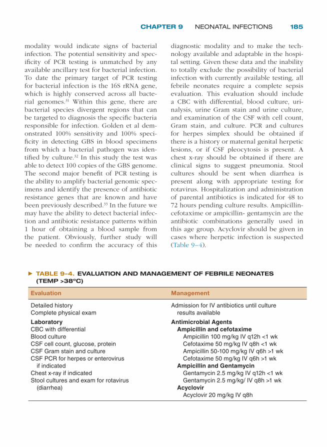
CHAPTER 9 NEONATAL INFECTIONS 185
modality would indicate signs of bacterial infection. The potential sensitivity and spec-ifi city of PCR testing is unmatched by any available ancillary test for bacterial infection. To date the primary target of PCR testing for bacterial infection is the 16S rRNA gene, which is highly conserved across all bacte-rial genomes.31 Within this gene, there are bacterial species divergent regions that can be targeted to diagnosis the specifi c bacteria responsible for infection. Golden et al dem-onstrated 100% sensitivity and 100% speci-fi city in detecting GBS in blood specimens from which a bacterial pathogen was iden-tifi ed by culture.32 In this study the test was able to detect 100 copies of the GBS genome. The second major benefi t of PCR testing is the ability to amplify bacterial genomic spec-imens and identify the presence of antibiotic resistance genes that are known and have been previously described.33 In the future we may have the ability to detect bacterial infec-tion and antibiotic resistance patterns within 1 hour of obtaining a blood sample from the patient. Obviously, further study will be needed to confi rm the accuracy of this
diagnostic modality and to make the tech-nology available and adaptable in the hospi-tal setting. Given these data and the inability to totally exclude the possibility of bacterial infection with currently available testing, all febrile neonates require a complete sepsis evaluation. This evaluation should include a CBC with differential, blood culture, uri-nalysis, urine Gram stain and urine culture, and examination of the CSF with cell count, Gram stain, and culture. PCR and cultures for herpes simplex should be obtained if there is a history or maternal genital herpetic lesions, or if CSF pleocytosis is present. A chest x-ray should be obtained if there are clinical signs to suggest pneumonia. Stool cultures should be sent when diarrhea is present along with appropriate testing for rotavirus. Hospitalization and administration of parental antibiotics is indicated for 48 to 72 hours pending culture results. Ampicillin-cefotaxime or ampicillin- gentamycin are the antibiotic combinations generally used in this age group. Acyclovir should be given in cases where herpetic infection is suspected (Table 9–4).
TABLE 9–4. � EVALUATION AND MANAGEMENT OF FEBRILE NEONATES (TEMP >38ºC)
Evaluation Management
Detailed historyComplete physical exam
Admission for IV antibiotics until culture results available
Laboratory Antimicrobial Agents
CBC with differential Ampicillin and cefotaxime
Blood culture Ampicillin 100 mg/kg IV q12h <1 wkCSF cell count, glucose, proteinCSF Gram stain and cultureCSF PCR for herpes or enterovirus
if indicatedChest x-ray if indicatedStool cultures and exam for rotavirus
(diarrhea)
Cefotaxime 50 mg/kg IV q8h <1 wkAmpicillin 50-100 mg/kg IV q6h >1 wkCefotaxime 50 mg/kg IV q6h >1 wk
Ampicillin and Gentamycin
Gentamycin 2.5 mg/kg IV q12h <1 wkGentamycin 2.5 mg/kg/ IV q8h >1 wk
Acyclovir
Acyclovir 20 mg/kg IV q8h

186 NEONATAL EMERGENCIES
MANAGEMENT �
CURRENT THERAPEUTIC STRATEGIES FOR THE FEBRILE NEONATE
In the most recent, large-scale, prospective study of febrile neonates conducted by the Pediatric Research in Offi ce Settings (PROS) Network of the American Academy of Pediatrics, the rate of bacteremia was 3%, 1.2% of febrile neo-nates had bacterial meningitis, and 4.1% had both bacteremia and meningitis, while 9.9% of febrile neonates had UTI.34,35
While medical advances from the past 20 years have decreased the mortality of neo-natal CNS infections, the morbidity and long-term sequelae from meningitis have remained relatively unchanged to this day.36-38 The cur-rent conservative standard of clinical practice is partially born from this fact, as well as the inability to identify all febrile neonates with meningitis on the basis of history, physical examination, and current laboratory evalua-tion. The emergency physician caring for the pediatric population commonly encounters the “rule out meningitis/sepsis” patient. Current practice dictates that infants who present with a fever during the fi rst 28 days of life will be admitted following a sepsis/meningitis workup as noted previously.39,40 Understanding the epi-demiology, pathophysiology, and clinical signs and symptoms of meningitis can aid the emer-gency physician when faced with atypical pre-sentations of this entity.
BACTERIAL MENINGITIS
Epidemiology
While the morbidity of meningitis has remained relatively constant over recent decades, the incidence and mortality of bacterial men-ingitis has declined signifi cantly. There are multiple reasons for this decline including prenatal GBS surveillance and subsequent
use of peripartum antibiotic prophylaxis, and vaccine development for both Haemophilus infl uenzae and Streptococcus pneumoniae. Bacterial meningitis is more common during the fi rst 2 years of life and especially during the fi rst month of life. Factors that contribute to the increased risk of meningitis in the neo-nate include an immature immune system and exposure to a wide set of pathogens via the birth process and from potential nosocomial infections. The current incidence of neona-tal bacterial meningitis in the United States has been reported to range from 0.25 of 1000 neonates to 0.32 of 1000 neonates.41,42 Various historical factors appear to carry higher risk for infections including meningitis, and may aid the clinician in establishing a higher pre-test probability in the neonatal patient. Low birth weight,43,44 prematurity,45 premature rup-ture of membranes,46 maternal infections (her-pes, UTI), and neonatal hypoxia have all been associated with an increased risk of infection that include meningitis. As with sepsis, neo-natal meningitis can present in the fi rst week of life (early onset) while late-onset meningitis occurs after the fi rst week of life. Meningitis can occur in the absence of sepsis but is more commonly seen in conjunction with sepsis. Several studies have suggested that 25% of septic neonates have concomitant meningi-tis.47 Early-onset disease is historically believed to result from vertical transmission from the mother with late-onset meningitis resulting from vertical transmission and infection spread from outside contacts.
In a study monitoring cases of neona-tal meningitis from 1979 to 1998, GBS was the most common pathogen (50% of neo-nates with meningitis) and E coli was found in approximately 25% of cases.48 Listeria is a signifi cant, if less common, pathogen responsible for meningitis and most antibi-otic regimens include ampicillin/cefotaxime to provide Listeria coverage. Other important but less frequently isolated bacteria are in listed in Table 9–1.

CHAPTER 9 NEONATAL INFECTIONS 187
Pathophysiology
The immature immune system of the neonate is characterized by ineffi cient functioning of the complement, opsonizing, and phagocytic processes necessary for cellular and humoral defense mechanisms against encapsulated bacteria and it is responsible in part for the increased risk of meningitis in the neonate49,50 here have been several proposed avenues for bacterial violation of the blood brain barrier including transmission via the choroid plexus and through skull defects along the path of the olfactory nerves51
The basic model to explain the patho-physiology of meningitis involves host expo-sure to immunogenic particles on bacterial cell walls (peptidoglycan, endotoxins, and lipopolysaccharides), causing activation of the host immune response and release of cytok-ines and acute phase reactants resulting in local infi ltration of leukocytes into the menin-geal space, increased permeability of the blood brain barrier, and subsequent direct cellular toxicity to the brain tissue.52 One study con-ducted by Tuomanen et al was able to repro-duce clinical meningitis in an animal model using only immunogenic bacterial particles without the presence of live bacteria.53
Clinical Manifestations
Clinical signs and symptoms are nonspecifi c and often mirror septicemia.53 One of the com-mon initial presenting features of a meningi-tis neonate is temperature instability. While high fever has been a hallmark of bacterial infection, this is not often seen in neona-tal meningitis. Hypothermia is often present, but more commonly in the premature infant.54 Neurologic complaints include irritability, poor feeding, seizures, and lethargy54 (Table 9–2). In rare cases the initial complaint of respiratory distress and diarrhea have also been reported. Evaluation of the asymptomatic patient (afe-brile, happy, playful, normal feeding, and no irritability) with a high-risk stratifi cation for
sepsis/meningitis secondary to prematurity, hypoxia, very low birth weight, and galacto-semia should be carefully evaluated at the dis-cretion of the individual clinician.
Diagnosis
A high index of suspicion is necessary in the evaluation of the neonate in the ED. A full sep-tic work up is indicated on all febrile neonates and classically consists of blood culture, urine culture, and CSF culture from lumbar punc-ture with examination of CSF cell count, CSF Ggram stain, CSF culture, glucose, and pro-tein. Several studies have found the normal values for neonatal CSF to differ from those of adults and can vary with gestational age and birth weight.55-58
Treatment should be administered imme-diately once the appropriate cultures have been obtained and should not be withheld until the laboratory results are available. The most common entities responsible for causing sepsis and meningitis in the neonate are dis-cussed below.
GBS MENINGITIS
Group B streptococcus is gram-positive cocci that colonizes the gastrointestinal tract and genital tract in adults and the respiratory tract in newborn infants; it is responsible for life-threatening sepsis and meningitis in the neo-nate.59 Infants acquire the organism in utero and during the birth process. After birth, infants can acquire the organism from house-hold contacts. Early-onset disease (sepsis-meningitis) is defi ned as occurring during the fi rst 7 days and represents maternally acquired disease, whereas late-onset disease occurring after 7 days is felt to represent extrauterine infection. The incidence of early-onset disease has decreased over the past 25 years from 1.8 in 1000 live births in 1990, to 0.34 cases in 1000 live births in 2004.60 The decrease is due

188 NEONATAL EMERGENCIES
in large part to effective screening of preg-nant woman for GBS and treating colonized woman with antibiotics. The incidence of late-onset disease has remained unchanged over the past 25 years at 0.35 cases in 1000 live births.60 GBS serotypes are defi ned by the presence of specifi c polysaccharides and pro-teins on the cell surface. In the United States, serotypes IA, III, and V account for most infections in the neonatal population.60,61 In early-onset disease these 3 serotypes account for 74% to 98% of documented infections with GBS.61 Similar results are described for late-onset disease (83% to 94% of GBS infections). The type III serotype is responsible for most late-onset disease and is more likely to cause meningitis.61
HERPES INFECTIONS
Clinical Presentation
Herpes simplex virus (HSV) can cause life- threatening, disseminated, multi-organ and CNS infection in the neonate. Infected infants have 1 of 3 clinical presentations: infection localized to the skin, eyes, and mucous mem-branes; meningitis; and multi-organ dissem-inated disease.62,63 Many infants with HSV acquired in the neonatal period have a normal physical examination and no manifestations of infection. The peak incidence of CNS dis-ease occurs between 10 and 17 days of life but can occur any time during the fi rst 4 weeks of life. The initial signs of infection are often nonspecifi c and may rapidly progress to hypo-tension, apnea, and disseminated intravascular coagulation.
Diagnostic Testing
Diagnostic testing should include culture of skin or mouth lesions with polymerase chain reaction assay of the CSF.64 Disseminated infections typically present in the fi rst week of life.
Management
Acyclovir administration should be promptly administered if there is any clinical suspicion for HSV infection. The current recommen-dations for treatment are acyclovir given at 20 mg/kg/dose IV q8h for 14 days if the dis-ease is limited to the skin, eyes, and mouth, and 21 days for meningitis or disseminated infection.65
ENTEROVIRAL INFECTIONS
Epidemiology & Pathophysiology
Human enteroviruses are found throughout the world and are transmitted between indi-viduals via the fecal-oral route. The nonpolio enteroviruses viruses, including Echovirus and Coxsackievirus, are responsible for a wide spectrum of clinical presentations and disease in all patients including neonates. Coxsackievirus serotypes 2 through 5 and echovirus 11 are most frequently associated with overwhelming and life-threatening infec-tions in the neonate.66 The majority of women (70%) with infected newborns have a febrile viral illness during the week of delivery.67 Transmission of the virus to the infant may occur via infected cervical secretions or from maternal viremia.68
Clinical Presentation
Many neonates with enteroviral infection will only have fever, rash, asymptomatic hepatitis, or aseptic meningitis with complete recovery; however, some infants will develop life-threat-ening myocarditis or fulminant live failure. Most infants acquire this infection in utero and during the birth process. Symptoms of ill-ness typically develop between days 3 and 7 of life and may include fever, irritability, and poor feeding, which resolves in a few days in many infants.69 Some infants (1 of 3) who have

CHAPTER 9 NEONATAL INFECTIONS 189
these early symptoms will develop generalized infection with life-threatening complications. Systemic enteroviral infections typically occur in 2 specifi c clinical entities. Neonatal hepatitis caused by enteroviral infection usually pres-ents in explosive and life-threatening fashion characterized by sudden onset of hepatomeg-aly with jaundice, signifi cant bleeding due to live failure, and coagulation factor defi ciency and shock.67 The second entity is myocarditis with heart failure as manifested by cardiomeg-aly, tachypnea, and hepatomegaly.
Management
The current management of infants with enteroviral infections with bleeding and ful-minate liver failure is vigorous supportive care combined with transfusion of fresh fro-zen plasma, red cells, and platelets combined with administration of vitamin K (1 mg IV). Administration of diuretics and isotropic agents may be required in patients with heart failure. Experimental therapies include the use of IV IgG with reported improvement in one case.70
SUMMARY �
Although sepsis and meningitis are uncom-mon occurrences in the neonatal period, these entities remain a signifi cant risk for morbidity and mortality in this population. A focused and accurate history including the maternal history and the perinatal clinical history, and a careful physical examination will dictate the need for septic workup with prompt insti-tution of appropriate antibiotic and antiviral therapy.
References
1. Baker MD, Bell LM. Unpredictability of seri-ous bacterial illness in febrile infants from birth to 1 month of age. Arch Pediatr Adolesc Med. 1999;153(5):508-511.
2. Chiu CH, Lin TY, Bullard MJ. Identifi cation of febrile neonates unlikely to have bacterial infec-tions. Pediatr Infect Dis J. 1997;16(1):59-63.
3. Kadish HA, Loveridge B, Tobey J, et al. Applying outpatient protocols in febrile infants 1-28 days of age can the threshold be lowered? Clin Pediatr. 2000;39(2):81-88.
4. Lin F-YC, Phillips JB III, Azimi PH, et al. Level of maternal antibody required to pro-tect neonates against early onset disease caused by Group B streptococcus IA: A mul-ticenter, epidemiology study. J Infect Disease. 2001;184:1022.
5. Lewis DB, Wilson CB. Developmental immu-nology and the role of host defenses to neona-tal susceptibility to in infection. In: Remington JS, Klein JO, eds. Infectious Diseases of the Fetus and Newborn Infant. 5th ed. Philadelphia, PA: WB Saunders; 2001:25-138.
6. Levy O. Impaired innate immunity at birth: defi -ciency of bacteriacidal/permeability-increasing protein (BPI) in the neutrophils of newborns. Pediatr Res. 2002;51:667.
7. Craig JV, Lancaster GA, Taylor S, et al. Infrared ear thermometry compared with rectal ther-mometry in children: a systemic review. Lancet. 2002;360:603-609.
8. Craig JV, Lancaster GA, Williamson PR, et al. Temperature at the axilla compared with rectal thermometry in children: a systemic review. Br Med J. 2000;329:1174-1178.
9. Grover G, Berkowitz CD, Lewis RJ, et al. The effects of bundling on infant temperatures. Pediatrics. 1994;94:669.
10. Jaskiewicz JA, McCarthy CA, Richardson AC, et al. Febrile infants at low risk for serious bac-terial infection: an appraisal of the Rochester criteria and implications for management. Pediatrics. 1994;94(3):390-396.
11. Baker MD, Bell LM, Avner JR. Outpatient man-agement without antibiotics of fever in selected infants. N Engl J Med. 1993;329:1437-1441.
12. Baskin MN, O’Rourke EJ, Fleisher GR. Outpatient treatment of febrile infants 28 to 89 days if age with intramuscular administration of ceftriaxone. J Pediat. 1992;120:22-27.
13. Ferrera PC, Bartfi eld JM, Snyder HS. Neonatal fever: Utility of the Rochester criteria in deter-mining low risk for serious bacterial infections. Am J Emerg Med. 1997;15(3):299-302.

190 NEONATAL EMERGENCIES
14. Ng PC. Diagnostic markers of infection in neonates. Arch Dis Child Fetal Neonatal Ed. 2004;89:F229-F235.
15. Bonsu BK, Harper MB. Identifying febrile young infants with bacteremia: Is the periph-eral white cell count an accurate screen? Ann Emerg Med. 2003;42(2):216-225.
16. Bonsu BK, Harper MB. Utility of the periph-eral white blood cell count for identifying sick young infants who need lumbar puncture. Ann Emerg Med. 2003;41(2):206-214.
17. Zorc JJ, Levine DA, Platt SL, et al. Clinical and demographic factors associated with uri-nary tract infections in young febrile infants. Pediatrics. 2005;116:664.
18. Bachur R, Harper MB. Reliability of the urinal-ysis for predicting urinary tract infections in young febrile children. Arch Pediatr Adolesc Med. 2001;155:60-65.
19. Gorelick MH, Shaw KN. Screening tests for urinary tract infections in children: a meta- analysis. Pediatrics. 1999;104(5):E54.
20. Shaw KN, McGowan KL, Gorelick MH, et al. Screening for urinary tract infection in infants in the emergency department: which test is best? Pediatrics. 1998;101(6):e1.
21. Committee on Quality improvement, Sub-committee on Urinary Tract Infection. Practice Parameter. The diagnosis, treatment and eval-uation of the initial urinary tract infection in febrile infants and young children. Pediatrics. 1999;103(4):843-852.
22. McGillivray D, Mok E, Mulronney E, et al. A head-to-head comparison: “clean-void” bag catheter urinalysis in the diagnosis of urinary tract infection in young children. J Pediatr. 2005;147(4):451-456.
23. Schroeder AR, Newman TB, Wasserman RC, et al. Choice of urine collection methods for the diagnosis of urinary tract infection in young, febrile infants. Arch Pediatr Adolesc Med. 2005;159(10):915-922.
24. Fowlie PW, Schmidt B. Diagnostic tests for bacterial infection from birth to 90 days: a sys-temic review. Arch Dis Child Fetal Neonatal Ed. 1998;78:F92-F98.
25. Garland SM, Bowman ED. Reappraisal of C-reactive Protein as a screening tool for diagno-sis for neonatal sepsis. Pathology. 2003;35:240-243.
26. Dandona P, Nix D, Wilson MF, et al. Procalcitonin increase after endotoxin injection in normal sub-jects. J Clin Endocrinol Metab. 1994;79:1605-1608
27. Guibourdenche J, Bedu A, Petzold L, et al. Biochemical markers of neonatal sepsis: value of Procalcitonin in the mergency setting. Ann Clin Biochem. 2002;39:130-135.
28. Fjaertoft G, Hakansson L, Ewald U, et al. Neutrophils from term and and preterm new-born express the high affi nity Fc gamma- receptor I (CD64) during bacterial infections. Pediatr Res. 1999;45:871-876.
29. Weirich E, Rabin RL, Maldonado Y, et al. Neutrophil CD11b expression as a diagnos-tic marker for early-onset neonatal infection. J Pediatr. 1998;132:445-451.
30. Ng PC, Li G, Chui KM, et al. Neutrophil CD64 is a sensitive diagnostic marker for early-onset neonatal infection. Pediatr Res. 2004;56:796-803.
31. Maiwald M. Broad-range PCR for the detec-tion and identifi cation of bacteria. In Persing DH, Tenover FC, Versalovic J, eds. Molecular Microbiology: Diagnostic Principles and Practice. 2nd ed. Washington, DC: American Society of Microbiology; 2004:379-390.
32. Golden SM, Stamilio DM, Faux BM, et al. Evaluation of a real-time fl uorescent PCR assay for the rapid detection of Group B Streptococci in neonatal blood. Diagn Microbiol Infect Dis. 2004;50:7-13.
33. Jaffe RI, Lane JD, Albury SV, et al. Rapid extrac-tion from and direct identifi cation in clinical sam-ples of methicillin-resistant staphylococci using the PCR. J Clin Microbiol. 2000;38:3407-3412.
34. Pantell R, Newman T, Bernzweig J, et al. Management and outcomes of care of fever in early infancy. JAMA. 2004;291:1203-1212.
35. Newman T, Bernzsweig J, Takayama J, et al. Urine testing and urinary tract infections in febrile infants seen in offi ce settings: The Pediatric Research in Offi ce Settings’ Febrile Infant Study. Arch Pediatr Adolesc Med. 2002;156:44-54.
36. Bell AH, Brown D, Halliday HL, et al. Meningitis in the newborn—a 14 year review. Arch Dis Child. 1989;64:873-874.
37. Harvey D, Holt DE, Bedford H. Bacterial men-ingitis in the newborn: a prospective study of mortality and morbidity. Semin Perinatol. 1999;23:218-225.

CHAPTER 9 NEONATAL INFECTIONS 191
38. Wegner JD, Hightower AW, Facklam RR, Gaventa S, Broome CV. Bacterial Meningitis Study Group. Bacterial meningitis in the United States, 1986: report of a multistate surveillance study. J Infect Dis. 1990;162;1316-1323.
39. Berger JE ed, Deitschel CH Jr, ed. Committee on Medical Liability, American Academy of Pediatrics. Medical Liability for Pediatricians. 6th ed. 2004:163, 169.
40. Karcz A, Holbrook J, Auerbach BS, et al. Preventability of malpractice claims in emer-gency medicine: a closed claims study. Ann Emerg Med. 1990;19(8):865-873.
41. Bell AH, Brown D, Halliday HL, et al. Meningitis in the newborn—a 14 year review. Arch Dis Child. 1989;64:873-874.
42. de Louvois J. Acute bacterial meningitis in the newborn. J Antimicrob Chemother. 1994;34(Suppl A):61.
43. Escobar GJ, De-kun L, Armstrong MA, et al. for the Neonatal Infection Study Group. Neonatal sepsis workups in infants ≥2000 grams at birth: a population-based study. Pediatrics. 2000;106(2):256-263.
44. Fanaroff AA, Korones SB, Wright LL, et al. The incidence, presenting features, risk fac-tors and signifi cance of septicemia in low birth weight infants. Pediatr Infect Dis J. 1998;17(7):593-598.
45. Voora S, Srinivasan G, Lilien LD, et al. Fever in full-term newborns in the fi rst four days of life. Pediatrics. 1982;69:40-44.
46. St. Geme Jr JW, Murray DL, Carter J, et al. Perinatal infection after prolonged rupture of membranes: an analysis of risk and manage-ment. J Pediatr. 1984;104:608-613.
47. Feigin RD. Diagnosis and management of men-ingitis. Pediatr Infect Dis J. 1992;11(9):785-814.
48. Klinger G, Chin CN, Beyene J, Perlman M. Predicting the outcome of neonatal bacterial meningitis. Pediatrics. 2000;106:477-482.
49. Schelonka RL, Infante AJ. Neonatal immunol-ogy. Semin Perinatol. 1998;22:2-14.
50. Berger M. Complement defi ciency and neu-trophil dysfunction as risk factors for bacterial infection in newborns and the role of granu-locyte transfusion in therapy. Rev Infect Dis. 1990;12(Suppl 4):S1401-S1409.
51. Weber JR, Tuomanen EI. Cellular damage in bacterial meningitis: an interplay of bacterial
and host driven toxicity. J Neuroimmunol. 2007;184(1–2):45-52.
52. Volpe JJ. Bacterial and fungal intracranial infec-tions. In: Volpe JJ, ed. Neurology of the Newborn. 4th ed. Philadelphia, PA: WB Saunders Co.; 2001:774-781.
53. Tuomanen E, Liu H, Hengstler B, Zak O, Tomasz A. The induction of meningeal infl am-mation by components of the pneumococcal cell wall. J Infect Dis. 1985;151:859–868.
54. Pong A, Bradley JS. Bacterial meningitis and the newborn infant. Infect Dis Clin North Am. 1999;13:711.
55. Anderson SG, Gilbert GL. Neonatal gram nega-tive meningitis: A 10-year review, with reference to outcome and relapse of infection. J Paediatr Child Health. 1990;26:212-216.
56. Palazzi DL, Klein JO, Baker CJ. Bacterial sep-sis and meningitis. In: Remington JS, Klein JO, Wilson CB, Baker CJ, eds. Infectious Diseases of the Fetus and Newborn Infant. 6th ed. Philadelphia, PA: Elsevier Saunders; 2006:247.
57. Sarff LD, Platt LD, McCracken GH. Comparison of high risk neonates with and without menin-gitis. J Pediatr. 1976;88:473-477.
58. Gerdes JS. Diagnosis and management of bac-terial infections in the neonate. Pediatr Clin North Am. 2004;51(4):939-959.
59. Edwards MS, Nizet V, Baker CJ. Group B Streptococcal Infections. In: Remington JS, Klein JO, Wilson CB, Baker CJ, eds. Infectious Diseases of the Fetus and Newborn Infant. 6th ed. Philadelphia, PA: Elsevier Saunders; 2006:403.
60. Centers for Disease Control and Prevention (CDC). Early-onset and late-onset neonatal group B streptococcal disease—United States 1996-2004. MMWR Morb Mortal Wkly Rep. 2005;54:1205-1208.
61. Lin FY, Clemons JD, Azimi PH, et al. Capsular polysaccharide types of group B streptococcal isolates from neonates with early-onset sys-temic infection. J Infect Dis. 1998;177:790.
62. Kimberlin DW, Lin CY, Jacibs RF, et al. Natural history of neonatal herpes simplex infections in the acyclovir era. Pediatrics. 2005;115:795.
63. Kohl S. Neonatal herpes simplex infection. Clin Perinatol. 1997;24:129.
64. Kimberlin DW, Lakeman FD, Arvin AM, et al. Application of the polymerase chain reaction to the diagnosis and management of neonatal

192 NEONATAL EMERGENCIES
herpes simplex virus disease: National Institute of Allergy and Infectious Diseases Collaborative Study Group. J Infect Dis. 1996;174:1162.
65. American Academy of Pediatrics. Herpes sim-plex. In: Pickering LK, ed. Red Book: 2006 Report of the Committee on Infectious Diseases. 27th ed. Elk Grove Village, IL: Author; 2006:361.
66. Kinney JS, McCray E, Kaplan JE, et al. Risk fac-tors associated with echovirus 11 infection in a hospital nursey. Pediatr Infect Dis. 1986;5:192.
67. Modlin JF. Perinatal echovirus infection: insights from a literature of 61 cases of serious infection and 16 outbreaks in nurseries. Rev Infect Dis. 1986;8:918.
68. Reyes MP, Osrea EM, Roskamp J, Lerner AM. Disseminated echovirus 11 disease fol-lowing antenatal maternal infection with a virus- positive cervix and virus-negative gastro-intestinal tract. J Med Virol. 1983;12:155.
69. Kaplan MH, Klein SW, McPhee J, Harper RG. Group B coxsackievirus infections in infants younger than three months of age: a serious childhood illness. Rev Infect Dis. 1983;5:1019.
70. Johnston JM, Overall JC, Jr. Intravenous immuno globulin in disseminated neonatal echovirus 11 infection. Pediatr Infect Dis J. 1989;8:254.

193
ANEMIA 193 �
BLEEDING IN THE NEONATE 202 �
SUMMARY 212 �
The neonatal period is marked by a series of rapid physiologic changes in the hematopoietic and coagulation pathways as the infant adapts to the extrauterine environment. For a variety of reasons the neonate is at signifi cant for risk for life-threatening anemia or hemorrhage dur-ing this time. This chapter summarizes the key diagnostic and therapeutic pathways needed to carefully evaluate the neonate presenting to the emergency department (ED) with anemia or bleeding.
ANEMIA �
The newborn period is characterized by a rapid transition from the hypoxic environment pre-sent in the uterus to the extra-uterine setting. This physiologic change leads to rapid altera-tions in several organ systems including the pulmonary, cardiovascular, and hematopoietic systems. In the absence of the hypoxia pre-sent within the uterus, neonatal erythrocyte production rapidly decreases during the fi rst
several days after birth with changes in hemo-globin concentration in the fi rst month of life, which are summarized in Table 10–1.1-3
Anemia is defi ned as the inability of cir-culating red cells to meet the oxygen demands of the body. The etiology of anemia can result from 1 of 3 causes: failure of red cell produc-tion, hemolysis, or blood loss. The need for rapid diagnosis and treatment dictates that an organized workup be rapidly and precisely undertaken. The key historical elements, physical examination fi ndings, and diagnostic
chapter 10
Hematologic Emergencies in the Neonate
P. David Sadowitz, MD
Trisha Tavares, MD
TABLE 10–1. � HEMOGLOBIN AND HEMATOCRIT CHANGES AFTER BIRTH
Age Hemoglobin Hematocrit
Birth 18.4 g/dL 58Day 3 17.8 g/dL 55Day 7 17.0 g/dL 54Day 14 15.6 g/dL 46Day 21 14.2 g/dL 43Day 28 12.7 g/dL 36

194 NEONATAL EMERGENCIES
testing in the neonate with anemia are out-lined below and summarized in Table 10–2.
HISTORY
The history can be a powerful and useful tool in the diagnosis and treatment of neonatal
anemia. A complete maternal history is vital in this setting. Maternal conditions predating the pregnancy are important to note and would include a history of any bleeding disorders or collagen vascular disease such as lupus. Important events during the pregnancy would include bleeding from the vagina during preg-nancy, placenta previa, placental abruption,
TABLE 10–2. � KEY FINDINGS IN THE NEONATE WITH ANEMIA
History Laboratory Evaluation Physical Examination
Family historyEthnicityMaternal history (ie, infections,
chronic diseases, medications)Obstetrical historyNeonatal disease (eg, malignancy,
hemophilia)
CBC/differential/platelet countRed cell morphologyReticulocyte countCoombs testPT/PTT/TT
ABCsJaundicePetechiaeHepatosplenomegaly
Coombs Negative Coombs Positive
Elevated Reticulocyte CountRed Cell Morphologic Abnormalities
SpherocytesStomatocytesElliptocytesFragmented red cells
Infection
CBV, Enterovirus, herpes, HIVBacterial infectionSyphilisToxoplasmosis
Red Cell Enzyme Defi ciency
G6PD defi ciencyPyryvate kinase defi ciency
Miscellaneous
GalactosemiaHemoglobinopathy (alpha chain and gamma chain)
Decreased Reticulocyte CountBone Marrow Failure
Blackfan diamond syndromeFanconi aplastic anemiaSideroblastic anemiaLeukemia, metastatic tumor
Nutrition
Maternal vitamin B12 defi ciencyChronic blood loss (fetal)Maternal iron defi ciency
ABO-Rh incompatibilityMaternal autoimmune hemolytic
anemia

CHAPTER 10 HEMATOLOGIC EMERGENCIES IN THE NEONATE 195
caesarian section or twin delivery. Other important questions to ask regarding the deliv-ery would include early cord clamping or sud-den traumatic birth.4 The family history and ethnicity may be signifi cant in determining the etiology of neonatal anemia. For example, there is a high prevalence of G6PD defi ciency in populations from the Mediterranean region and in the Black population in Africa, while populations from Southeast Asia have a high incidence of alpha thalassemia, which could produce anemia in the neonate. The presence of anemia in other family members in conjunc-tion with jaundice, gallstones, and splenec-tomy might suggest the diagnosis of hereditary spherocytosis. The age at which anemia pres-ents in the neonate gives an important clue to the cause of the anemia. Severe anemia in the fi rst 1 to 2 days of life is almost always the result of hemorrhage or severe ABO-Rh incompatibility. Anemia presenting after the fi rst 2 days of life is usually caused by hemo-lysis from ABO and Rh incompatibility and is characterized by jaundice and anemia.
PHYSICAL EXAMINATION
A careful physical examination may give impor-tant clues to the etiology of the anemia. A rapid assessment of vital signs (ABCs) is imperative, as the presence of tachycardia, tachypnea, and hypotension may indicate a rapid loss of circu-lating blood volume from hemolysis or acute blood loss and requires immediate resuscita-tive efforts. Skin examination may reveal the presence of pallor or jaundice that can be seen with blood loss and hemolytic anemia, respectively. The presence of hepatosplenom-egaly can be seen with hemolytic processes but also is seen with congenital viral infection and malignancies. Abnormalities involving the extremities such as absent thumbs or absent radii are seen with Fanconi aplastic anemia, characterized by marrow dysfunction with defi cient production of red cells.5-10
DIAGNOSTIC TESTS
The initial laboratory evaluation should include a complete CBC with red cell indices, white cell count with differential, platelet count, pro-thrombin time (PT), partial thromboplastin time (PTT), and thromboplastin time (TT) if bleeding is present, blood type and Coombs test, reticulocyte count, bilirubin (direct and indirect), and a careful review of the smear for morphologic red cell abnormalities. If the reticulocyte count is elevated, a hemolytic pro-cess or hemorrhage represents the most likely etiology. If the reticulocyte count is low in the face of severe anemia, this fi nding suggests a failure of the marrow to produce adequate numbers of red cells.
A second consideration in the neonate with low or absent reticulocytes is the pres-ence of a marrow infi ltrative process such as leukemia. A positive Coombs test identifi es infants with antibody-mediated destruction of the red cells. These antibodies are produced in the mother (IgG subclass), cross the placenta, and bind to the fetal red cells with resultant destruction of these cells. In most case these antibodies are produced in response to ABO or Rh incompatibilities between the mother and fetus. On rare occasions the mother may have unrecognized autoimmune hemolytic anemia, which leads to transfer of maternal antibody to the neonate and hemolytic anemia.
The smear should be carefully exam-ined for morphologic abnormalities. If there are numerous spherocytes on the smear and if the Coombs test is negative, the diagno-sis of hereditary spherocytosis is suggested (Figure 10–1). Numerous elliptocytes on the peripheral smear suggests the diagnosis of hereditary elliptocytosis (Figure 10–2).
The presence of hypochromic red cells seen on smear with a corresponding low MCV <95 fl suggests chronic fetal-maternal hemorrhage but can also be seen with alpha thalassemia trait. Infants with sudden onset of severe anemia, normal red cell indices, and

196 NEONATAL EMERGENCIES
absence of jaundice have hemorrhage until proven otherwise. Typically infants with hem-orrhage present to the ED with pallor and signs of impending hypovolemic shock, which include tachycardia, tachypnea, delayed cap-illary refi ll, skin mottling, and hypotension. The usual sites for hemorrhage include the head (cephalohematoma), liver, spleen, and, rarely, lung. In today’s world the diagnosis of nonaccidental trauma (NAT) must be consid-ered in the neonate presenting to the ED with hypovolemic shock and evidence of hemor-rhage. Infants with jaundice and hemolytic
anemia may be infected by a variety of organ-isms including cytomegalovirus, herpes, toxo-plasmosis, syphilis, or bacterial agents. As discussed previously, disseminated intravascu-lar coagulation (DIC) can present in this set-ting with anemia as a byproduct. The most common entities associated with anemia are briefl y discussed below and are summarized in Table 10–3.
DIFFERENTIAL DIAGNOSIS
Decreased Red Cell Production
Neonates with anemia caused by decreased red cell production represent a minority of patients. The hallmark of this diagnosis is a low or absent reticulocyte count in the pres-ence of anemia.
Congenital Viral Infection
Parvovirus B19 is a single-stranded DNA virus that can produce disease in adults and chil-dren and is the etiologic agent in erythema infectiosum (“fi fth disease”).11,12 Parvovirus B19 preferentially infects rapidly dividing cells and is cytotoxic for erythroid progenitor cells.12-18 Maternal infection with parvovirus B19 during pregnancy may lead to fetal loss with the great-est in the fi rst 20 weeks of the pregnancy.17,18
Infection of the fetal red cells can produce severe anemia and lead to nonimmune hydrops fetalis. The diagnosis of parvovirus B19 infection can be accurately established by polymerase chain reaction (PCR) for the par-vovirus genome.19-21 This diagnosis should be considered in the neonatal period in an infant with anemia and absent reticulocyte count. Transfusion support may be required in neo-nates with severe anemia until red cell pro-duction resumes.
Congenital Marrow Dysfunction
Diamond-Blackfan anemia (DBA) is a het-erogeneous mix of pure red cell aplasias
Figure 10–1. Spherocytes.
Figure 10–2. Elliptocytes. Source: From Lichtman MA, Beutler E, Kipps TJ, et al. Williams’ Hematology, 7ed, 2006, New York, NY: McGraw-Hill.

CHAPTER 10 HEMATOLOGIC EMERGENCIES IN THE NEONATE 197
that typically presents in the fi rst year of life and is characterized by the presence of a macrocytic anemia, reticulocytopenia, and a marrow devoid of red cell precursors.22,23
Thirty-fi ve percent of patients will have the diagnosis established in the fi rst month of life on presentation to the primary care phy-sician or ED.23 Physical abnormalities are
TABLE 10–3. � ETIOLOGY OF NEONATAL ANEMIA
Bleeding
Increased Red Cell
Destruction
Decreased Red Cell
Production
Prior to Delivery
Acute and chronic fetal maternal hemorrhage
Acute and chronic fetal-placental hemorrhage
Acute and chronic twin-twin hemorrhage
Traumatic amniocentesisUmbilical vein sampling
in uteroMaternal trauma
At Delivery
Placenta abruptionPlacenta previaVelamentous cord insertionRupture of a normal or
abnormal umbilical cordHemorrhage Following
Delivery
CephalohematomaSubgaleal hematomaIntracranial bleedDICLiver, spleen, adrenal, or renal
traumaPulmonary hemorrhageLine accidents (umbilical,
arterial)Neonate with
hemophilia-factor defi ciencies
Immune Mediated
ABO incompatibilityRh incompatibilityMinor blood group incompatibilityMaternal autoimmune hemolytic
anemiaDrug-induced hemolytic anemia
Anatomic
Hemolysis on abnormal heart valvesCentral line-induced hemolysisRenal artery stenosisAortic stenosis
Infection
HIVCMVRubellaHerpesToxoplasmosisSyphilisBacterial
DIC
Bacterial/viral/fungal infectionsMicroangiopathic hemolytic anemiaCavernous hemangioma
(Kasabach-Merritt syndrome)Red Cell Membrane Abnormalities
Hereditary spherocytosisHereditary elliptocytosisHereditary stomatocytosis
Red Cell Enzyme Defi ciencies
G6PD defi ciencyPyryvate kinase defi ciency
Hemoglobinopathy
Alpha thalassemia syndromesGamma thalassemia
Metabolic Abnormalities
GalactosemiaDrug Induced
DepictedSulfa drugs (G6PD defi ciency)
Infection
HIVParvovirusRubellaCytomegalovirusBacterial
Nutrition
Maternal vitamin B12 defi ciency
Maternal iron defi ciencyMaternal folate
defi ciencyMarrow Dysfunction
Diamond-Blackfan anemia
Fanconi aplastic anemiaCongenital leukemiaCongenital neuro-
blastoma with marrow replacement

198 NEONATAL EMERGENCIES
present in one-third of patients and include short stature, thumb abnormalities, congen-ital heart disease, short or webbed neck, cleft palate, and ocular abnormalities.23,24 The pathogenesis of DBA is uncertain. Most investigators believe that the erythroid pro-genitor cell is unable to proliferate.22,24-32 The diagnosis should be suspected in the neo-nate with macrocytic anemia, reticulocy-topenia, and the morphologic abnormalities noted previously.
Fanconi Aplastic Anemia
Fanconi aplastic anemia is an autosomal reces-sive marrow disorder leading to marrow fail-ure and increased risk for malignancy.5-10 To date 13 genes have been identifi ed, which are widely dispersed among several chromosomes including the X chromosome 31 Many of the normal genes have tumor suppressor activity that includes maintaining genomic stability, promoting DNA repair, and maintaining apop-tosis.32 Characteristic morphologic changes are seen in two-thirds of neonates, including elfi n facies, skin hyperpigmentation, café-au-lait spots, renal abnormalities, and thumb-radius abnormalities. The diagnosis is confi rmed by the presence of increased chromosomal breakage in lymphocytes in the presence of DNA cross-linking agents such as mitomycin C or diepoxybutane. Five percent of children are diagnosed in the fi rst year of life and the diag-nosis should be suspected in the neonate with macrocytic anemia and the described physical abnormalities.
Malignancy
Congenital malignancies such as leukemia or metastatic neuroblastoma may present with anemia and low reticulocyte count. Typically these patients have hepatosplenomegaly on physical examination. Nucleated red cells may be present on the peripheral smear and imma-ture white cells will be present in neonates with leukemia (Figure 10–3).
Bleeding
The neonate can experience bleeding prior to birth, during delivery, and following delivery (Table 10–4). Selected entities are briefl y dis-cussed below.
Bleeding Prior to Delivery
Fetal-maternal bleeding occurs in 50% of pregnancies and can occur throughout the pregnancy, but in the vast majority, the bleed-ing is insignifi cant and does not result in anemia for the newborn. Approximately 1 of 400 pregnancies have signifi cant bleeding (>30 mL) resulting in signifi cant anemia in 30% of affected infants and most commonly occurs just prior to birth.33,34 The source of the bleeding is typically via the placenta and chorionic villi.
Twin-twin transfusion can occur in up to 20% of twin gestations. This entity occurs as the result of placental anastamoses that allows rapid transfer of blood between the twins. There is a signifi cant risk of mortality in this setting; this syndrome causes 15% of prenatal mortality in twins.35,36
Bleeding During Delivery
Obstetrical complications can lead to life-threatening bleeding and anemia. Placental
Figure 10–3. Leukemic cells and nucleated red cells.

CHAPTER 10 HEMATOLOGIC EMERGENCIES IN THE NEONATE 199
rupture and cord malformations have also been associated with severe intrapartum bleeding. Velamentous insertion of the cord complicates 1% of all pregnancies and is associated with an increased risk of bleeding. In this entity the vessels exit the umbilical cord and insert into the placenta with no protection from the cord. These vessels are therefore fragile and subject to tearing during delivery.37,38 Placenta previa, vas previa (placental vessels lying across the cervical os), and placental abruption carry an increased risk for bleeding and vas previa is associated with a high mortality rate.39,40
Birth-Process Bleeding
The birth process itself can be a traumatic event and lead to internal hemorrhage in the newborn. Subgaleal bleeding and cepha-lohematoma are the most common sites of birth-process hemorrhage. Subgaleal bleeds can extend throughout the soft tissues of scalp and produce life-threatening anemia, while cephalohematoma is well contained by the periosteum and rarely produces life-threatening bleeding.41 Intracranial bleed-ing is uncommon in full-term deliveries but can occur and should be suspected in the apneic, lethargic newborn with a tense fontanel. Emergent CT scan is indicated in these settings. Less common sites of internal
hemorrhage include liver, spleen, adrenal glands, and lungs.
Bleeding After Birth
Nonaccidental trauma must be included in the differential diagnosis of the neonate who presents to the ED with unexplained bruis-ing or internal bleeding involving the CNS, liver, spleen, or lung. DIC, hemophilia and other bleeding disorders will be discussed separately.
Anemia—Hemolytic
Hemolytic anemia in the fi rst few weeks of life is a common entity and can occur as the result of intrinsic red cell defects or, more commonly, as the results of factors extrinsic to the red cell. In either case, the hallmark of hemolysis is a shortened red cell life span (normally 60-90 days in the newborn), ele-vated reticulocyte count, morphologic abnor-malities (spherocytes, fragmented red cells) seen on the peripheral smear, and jaundice (Figure 10–4).
In the neonate, hemolysis can be divided into 3 major categories: immune mediated red cell destruction, intrinsic red cell defects, and factors extrinsic to the red cell (ie, such as sepsis).
TABLE 10–4. � ETIOLOGY OF NEONATAL BLEEDING
Prior to Birth Bleeding at Delivery Internal Hemorrhage
Fetal-Maternal
Traumatic amniocentesisChorioangioma of the
placentaUse of oxytocinManual placenta extractionSpontaneous
Fetal-Placental
Placental hematomaCesarean sectionTight nuchal cord
Twin-Twin Bleed
Rupture Normal Umbilical Cord
Precipitous deliveryCord entanglement
Rupture Abnormal Umbilical Cord
VaricesAneurysmAberrant vessels in the cordVelamentous cord insertion
Placental Laceration During C-Section
Placenta Previa
Abruptio Placenta
Vasa Previa
IntracranialCephalohematomaSubgaleal hematomaAdrenal-retroperitonealLiver ruptureSplenic rupturePulmonary

200 NEONATAL EMERGENCIES
Immune-Mediated Red Cell Destruction
Hemolytic disease of the newborn is caused by the destruction of fetal and neonatal red cells by maternal IgG antibodies. These anti-bodies are produced when fetal red cells enter the maternal circulation and express antigens not found on maternal red cells.42 There are many antigens that have the potential to stim-ulate an antibody response in the newborn and include the antigens in the ABO, Rh, Kell, and Duffy systems. The vast majority of immune-mediated red cell destruction occurs in response to ABO and Rh incompatibility between the infant and the mother and are discussed below.
ABO Incompatibility
This entity was fi rst described by Halbrecht in 1944.43 In general the hemolysis induced by fetal-maternal ABO incompatibility is not severe, as the level of IgG antibody in the maternal circu-lation against the A and B antigens on the fetal cells is not markedly elevated, thus the amount of antibody transferred to the infant is low. Hemolysis in this setting is minimal to moder-ate, and is typically seen only when the mother is type O and the infant is either A or B. The
incidence of ABO mismatch with the fetus in a mother with blood type O is 15%. Only 20% of infants will become signifi cantly jaundiced for the reasons noted previously.44,45
Clinically apparent jaundice appears on the fi rst day of life in 20% of infants with ABO incompatibility.44 Anemia and subsequent pallor are rarely seen. Typically the physical examination is normal except for jaundice and possibly mild hepatomegaly. The periph-eral smear is remarkable for the presence of spherocytes and the Coombs test is positive.46 It is important to note that the direct Coombs test is positive only 40% of the time, thus it is vital to obtain the indirect Coombs test to detect small amount of antibody bound to the red cell membrane. Treatment for infants with hyperbilirubinemia involves admission to the hospital for phototherapy.
Rh Hemolytic Disease
The widespread availability and use of Rhogham in Rh-negative women has dramat-ically decreased the incidence of this poten-tially life-threatening cause of hemolytic anemia. Prior to advent of Rhogham, 1% of women developed signifi cant sensitization to the Rh antigen leading to severe anemia and hydrops in the developing fetus.47 Sensitization of the Rh-negative mother rarely occurs (1 in 1000 pregnancies) in the Rhogham era.48 Nevertheless this entity still appears and need immediate recognition to prevent potential life-threatening anemia.
This entity was fi rst described in 1934 by Diamond who noted a condition charac-terized by fetal hydrops, anemia, and circu-lating erythroblasts.49 Weiner and coworkers discovered the Rh system in 1940 and found that 15% of the population was Rh negative.50 Subsequent studies revealed that this antigen was responsible for many transfusion reac-tions and was the most common entity associ-ated with the development of severe hemolytic disease of the fetus and newborn.
Figure 10–4. Fragmented red cells. Source: From Lichtman MA, Beutler E, Kipps TJ, et al. Williams’ Hematology, 7ed, 2006, New York, NY: McGraw-Hill.

CHAPTER 10 HEMATOLOGIC EMERGENCIES IN THE NEONATE 201
The antigens of the Rh system are found at 3 loci: Dd, Cc, and Ee.51 Most signifi cant anti-body responses are related to the Dd system.51 In infants with the DD or Dd phenotype born to a mother with the dd genotype, the pres-ence of fetal red cells can induce an antibody response. The risk of sensitization in the fi rst pregnancy ranges from 11% to 16% and the risk increases in subsequent pregnancies.52,53 Because the antibody generated is of the IgG subclass, subsequent pregnancies have a much higher risk of severe hemolysis due to the heightened antibody response on re-exposure to the antigen with subsequent pregnancies. Severe hemolysis can occur in the fetus lead-ing to hydrops and fetal demise.
Fifty percent of infants with Rh incompati-bility have mild disease with a normal physical examination other than jaundice. Phototherapy may be required; tranfusional support is rarely required. Moderately severe hemolysis devel-ops in 30% of infants in this setting. These infants have a normal physical examination at birth except for jaundice and rapidly increas-ing bilirubin that necessitates immediate hos-pitalization and treatment with phototherapy. Severely affected infants present at birth with severe anemia and varying degrees of hydrops and are managed in the neonatal nursery with exchange transfusion. It is important to note that severely affected infants who undergo exchange transfusion may continue to require tranfusional support for weeks due to ongo-ing destruction of red cells including the trans-fused red cells. These infants may present to the ED in the fi rst 6 weeks of life with severe anemia and decreased reticulocyte count due to the persistent presence of maternal antibody that destroys the immature red cells in vitro.
Hereditary Spherocytosis
Hereditary spherocytosis is an autosomal dominant condition characterized by defects in the membrane surface proteins––spectrin and ankyrin––producing a red cell membrane
that is less deformable leading to red cell membrane loss, spherocyte formation, and premature red cell destruction in the spleen.54 This entity is present in 1 in 4000 births in the Caucasian population; the frequency in the Black population is signifi cantly less.54
Many children remain asymptomatic for years; however, neonates can present with jaundice, anemia, and splenomegaly. Fifty per-cent of neonates with hereditary spherocytosis have jaundice in the neonatal period.55
The diagnosis can be strongly suspected in the neonate with a positive family history of hereditary spherocytosis, jaundice on physical examination in an otherwise well-appearing infant, spherocytes on the peripheral blood smear, and a negative Coombs test.
Little intervention is required in the neonatal period other than phototherapy for jaundice. Most children with hereditary spherocytosis will eventually require sple-nectomy. Gallstones and subsequent need for cholecystectomy may occur during childhood due to ongoing red cell hemolysis.
Glucose-6-Phosphate Dehydrogenase Defi ciency (G6PD Defi ciency)
G6PD defi ciency is an X-linked trait and rep-resents the most common inherited red cell enzyme defect affecting nearly 500 million peo-ple worldwide.56 This entity is most commonly seen in Africa but is present throughout the world. G6PD is most prevalent in regions of the world where malaria is endemic. G6PD-defi cient red cells are not able to produce an adequate quantity of reduced glutathione; thus, in the presence of oxidant stress, the red cells will hemolyze.
Many infants in the newborn period will develop jaundice if G6PD defi ciency is pre-sent.57 Presently the pathophysiology of jaun-dice in this setting is unclear. Management of neonatal jaundice in G6PD defi ciency is no different than the management of infants

202 NEONATAL EMERGENCIES
with immune-mediated red cell destruction. Phototherapy is used if indicated and exchange transfusion is rarely required. Testing for G6PD defi ciency should be considered in all jaundiced infants who do not have immune-mediated red cell destruction, especially in populations with a high prevalence of this entity.
Nonaccidental Trauma (NAT)
Neonates with unexplained bruising and pete-chiae need a careful evaluation to exclude an underlying bleeding disorder such as hemo-philia, thrombocytopenia, or DIC. In the absence of these entities, NAT must be consid-ered, especially in the setting of unexplained subarachnoid or intracranial bleeding or unexplained facial bruising. Evaluation would include a complete skeletal survey, bone scan, dilated eye examination, and CT and MRI of the brain.
SUMMARY
Neonates presenting to the ED with symp-toms of anemia need a complete and focused history, a careful physical examination, and appropriate laboratory testing to determine the etiology of the anemia. Transfusional sup-port may be required to support the cardiovas-cular system and may be life-saving in some neonates.
BLEEDING IN THE �NEONATE
For a variety of reasons the neonate is at increased risk for potentially life-threatening bleeding. Maternal diseases and medication use, the fragility of newborn vessels, trauma associated with birth, physiologic coagulation factor defi ciencies present at birth, along with hypoxia and sepsis are all contributing factors toward bleeding in this group of children.
PHYSIOLOGY OF NEONATAL CIRCULATION
The platelet-endothelial cell interaction repre-sents the initial hemostatic response to dam-aged endothelium. This response is diminished in neonates due in part to decreased produc-tion of thromboxane, which promotes vaso-constriction and adherence of platelets to the vessel wall. Neonatal endothelium and vessels are more fragile and susceptible to injury than in older children. Several soluble proteins are present in the plasma, which is activated in the presence of endothelial cell injury gener-ating fi brin, which then binds to the platelet complex adherent to the damaged endothe-lium. Many of these proteins are present in diminished concentration in the neonate and include factor 11, and the vitamin K-dependent factors 2, 7, 9, and 10.58 These defi ciencies lead to prolongation of both the PT and PTT, and place the neonate at risk for bleeding. In the normal neonate the levels of factors promoting fi brinolysis and inhibiting thrombus formation are diminished and include decreased levels of antithrombin III, plasminogen, protein S, and protein C.58 Most neonates who are other-wise healthy will not experience bleeding or thrombus formation. Table 10–5 summarizes the reference values for the coagulation tests and factors in the fi rst month of life.59
HISTORY
A complete and accurate history is often the key to the diagnosis in the bleeding neonate and the important historical elements are summarized in Table 10–6. The key informa-tion needed includes the presence of mater-nal infection or disease, medication use in the mother, trauma during the birth process, family history of bleeding disorders, the pres-ence of fever or other signs of systemic ill-ness in the infant, and history of vitamin K administration.

CHAPTER 10 HEMATOLOGIC EMERGENCIES IN THE NEONATE 203
PHYSICAL EXAMINATION
The bleeding infant requires a careful and rapid assessment to determine if the bleeding is secondary to an underlying disease process or if it is related to a primary bleeding disor-der. Once the initial assessment of the infant is completed, 2 groups of infants are identi-fi ed. The fi rst group is comprised of the “sick” infant who needs immediate therapy prior to availability of laboratory testing, while infants in the second group appear clinically well and do not require emergent treatment. Table 10–7 lists the common clinical and laboratory fi nd-ings and possible diagnoses in each group of patients.
The presence of shock, hypoxia, fever, hypotension, hypothermia, acidosis, and peri-natal infection suggests that bleeding is sec-ondary to DIC. Hepatosplenomegaly in the bleeding neonate suggests the diagnosis of congenital viral infection or leukemia. The presence of congenital abnormalities involving the skin, face, heart, radius, or thumbs should suggest the possibility of Fanconi aplas-tic anemia, Wiskott-Aldrich syndrome, and
TABLE 10–5. � REFERENCE VALUES FOR COAGULATION TESTS, FACTOR LEVELS, AND ANTICOAGULANTS IN THE FIRST MONTH OF LIFE AND IN ADULTS
Coagulation Tests 5 Days Old 30 Days Old Adult
PT (sec)INRPTT (sec)Factor Levels (U/mL)
Factor 2Factor 7Factor 8Factor 9Factor 10Factor 11
Anticoagulants (U/mL)
Protein CProtein SAntithrombin III
12.4 (10-15.3)0.89 (0.53-1.48)42.6 (25.4-59.8)
0.63 (0.33-0.93)0.89 (0.35-1.43)0.88 (0.50-1.54)0.53 (0.15-0.91)0.49 (0.19-0.79)0.55 (0.23-0.87)
0.42 (0.20-0.64)0.50 (0.22-0.78)0.56 (0.30-0.82)
11.8 10-14.3)0.79 (0.53-1.26)40.4 (32-55.2)
0.68 (0.34-1.02)0.90 (0.42-1.38)0.91 (0.50-1.57)0.51 (0.21-0.81)0.59 (0.31-0.87)0.53 (0.27-0.79
0.43 (0.21-0.65)0.63 (0.33-0.93)0.59 (0.37-0.81)
12.4 (10.8-13.9)0.89 (0.64-1.17)33.5 (26.6-40.3)
1.08 (0.70-1.461.05 (0.67-1.43)0.99 (0.50-1.49)1.09 (0.55-1.63)1.06 (0.70-1.52)0.97 (0.67-1.27)
0.96 (0.64-1.28)0.92 (0.60-1.24)1.05 (0.79-1.31)
TABLE 10–6. � HISTORICAL AND PHYSICAL EXAMINATION KEYS IN EVALUATING THE BLEEDING NEONATE
Maternal Factors Neonatal Factors
Maternal Disease
SLEITPMalignancy
Maternal Medica-
tion Use
AspirinMotrin
Pregnancy Compli-
cations
PreeclampsiaAbruptio placentae
Maternal InfectionFamily History
of Bleeding
Disorders
Neonatal Birth
Complications
Birth traumaPrematurityHypoxiaHypotensionRespiratory distress
syndromeNeonatal Physical
Abnormalities
FeverAbsent radii/thumbsHepatosplenomegalyHemangiomas
Neonatal Infection
Vitamin K
Administration
Nonaccidental
Trauma (NAT)

204 NEONATAL EMERGENCIES
thrombocytopenia absent radii syndrome (TAR syndrome). Well-appearing infants with stable vital signs and no observable physical abnor-malities typically have a primary bleeding dis-order. Diagnostic considerations in this group include immune thrombocytopenia, hemo-philia, vitamin K defi ciency, von Willebrand disease, or a localized vascular lesion.
Type of Bleeding
The presence of petechiae, bruising, or GI tract bleeding is most commonly associated with thrombocytopenia. Localized bleeding
following trauma as manifested by cephalohe-matoma, postcircumcision bleeding, or hema-toma formation at injection sites points toward the diagnosis of a specifi c factor defi ciency.
DIAGNOSTIC TESTS
The CBC with platelet count, PT, and PTT often help identify the reason for bleeding. The platelet count can be accurately measured using an automated electronic cell counter and quickly estimated by viewing a well-prepared peripheral smear. Under microscopic review
TABLE 10–7. � CLINICAL AND LABORATORY FINDINGS IN THE BLEEDING NEONATE
Platelet Count PTT PT Potential Diagnoses
Well Neonate
Decreased
NormalNormal
Normal
Sick Neonate
DecreasedDecreased
NormalNormal
Normal
ProlongedProlonged
Normal
IncreasedNormal
IncreasedNormal
Normal
ProlongedNormal/prolonged
Normal
IncreasedNormal
IncreasedNormal
Antibody-Mediated Conditions
Maternal ITPAlloimmune thrombocytopenia
Rare Conditions
Marrow hypoplasiaLeukemiaMetastatic malignancy
Vitamin K defi ciencyInherited factor defi ciencies(hemophilia)TraumaAnatomic abnormalities(hemangioma)Platelet functional abnormalities
Disseminated intravascular coagulationPlatelet Consumption
Catheter relatedInfectionNECRenal vein thrombosisKasabach-Merritt syndrome
Vitamin K defi ciencyCompromised Vascular Integrity
HypoxiaAcidosisPrematurity

CHAPTER 10 HEMATOLOGIC EMERGENCIES IN THE NEONATE 205
with an oil immersion lens at 100x, each plate-let represents 10-15,000 platelets. A child with 10 or more platelets in each fi eld would have a platelet count. >100,000/μL. It is important to recognize causes of “false” thrombocytope-nia, which commonly occurs and should be suspected in a clinically well neonate with no evidence of bleeding. Diffi cult blood draws with slow blood fl ow and the presence of anti-bodies that are activated by the anticoagulant EDTA can both cause platelet clumping and a falsely low platelet count.
The PTT and PT provide a measure of the soluble clotting proteins and thus represent a valuable screening test for blood coagulation. The PTT measures the intrinsic pathway, which contains all of the clotting factors except for factors 7 and 13. The PT measures the extrin-sic pathway and does not measure factors 13, 12, 11, 9, and 8. The presence of a normal PT and PTT effectively rules out a coagulation factor defi ciency as a cause of bleeding.
Prolongation of the PTT and PT can occur for several reasons. This can occur when the level of 1 or more factors is decreased by 40%. Reduced levels of the so-called contact factors such as factor 12, prekallikrein, and high molecular weight kininogen can prolong the PTT without causing increased bleeding, as these factors are not the primary factors needed in activation of the coagulation cas-cade. Several technical factors can produce false prolongations of the PT and PTT. A long, diffi cult blood draw can release tissue throm-boplastin and trigger production of small fi brin clots, which will prolong the PT and PTT. The presence of heparin in a catheter from which blood is drawn will markedly elevate the PTT with minimal effect on the PT. The presence of a hematocrit >65% or an inadequate amount of blood placed in the citrate tube used for the PT and PTT will increase the ratio of citrate to plasma and prolong the PT and PTT.
In cases where the PT and PTT are truly prolonged, the fi brinogen and fi brin split products should be obtained. The presence
of elevated fi brin split products and decreased fi brinogen with a prolonged PT and PTT estab-lishes the diagnosis of DIC.
DIFFERENTIAL DIAGNOSIS AND MANAGEMENT
Disseminated Intravascular Coagulation (DIC)
DIC represents the most common and poten-tially life-threatening cause of impaired hemostasis in the neonate. This entity is char-acterized by endothelial damage from a vari-ety of factors (Table 10–8) with activation of the coagulation cascade, consumption of clot-ting factors, intravascular thrombi formation,
TABLE 10–8. � DISSEMINATED INTRAVASCULAR COAGULATION: ETIOLOGIC FACTORS
Endothelial Cell Injury
Bacterial infectionViral infectionFungal InfectionAcidosisHypoxemiaVascular cathetersCavernous hemangioma
Red Cell/Platelet Injury
Hemolytic disease of the newbornTissue Injury/Release of Tissue Factor
Birth asphyxiaRespiratory distress syndromeMeconium aspirationCongenital leukemiaNeuroblastomaPre-eclampsiaAbruption placentaeSevere brain injuryNecrotizing enterocolitisCrush injury
Congenital Thrombotic Disorders
Protein S defi ciencyProtein C defi ciencyAntithrombin III defi ciency

206 NEONATAL EMERGENCIES
and hemorrhage.60 When DIC is present the underlying disease state must be identifi ed, as DIC is always a secondary event. Table 10–8 summarizes the entities most commonly asso-ciated with DIC.
The initial pathophysiologic event in most cases of DIC involves injury to the endothelial cell and surrounding tissue, which activates both the intrinsic and extrinsic coagulation systems with production of intravascular thrombi and depletion of coagulation factors. The fi brin clot present in the microcirculation produces a microangiopathic hemolytic pro-cess as the red cells are impaled on the fi brin clots. Fibrin formation activates the fi brinolytic system producing plasminogen that cleaves fi brinogen and fi brin clot, producing fi brin degradation products. The fi brin degradation products interfere with platelet aggregation and impair fi brin polymerization. The fi brin clot impairs oxygen delivery to end organs causing ischemic injury.
Clinical Presentation
DIC occurs in a variety of settings, which are summarized in Table 10-8. The main causes of DIC in neonates are briefl y discussed below. Neonatal viral infections (rubella, her-pes, enterovirus, and cytomegalovirus) and bacterial infections (gram-negative organ-isms and Group B streptococcal infections) are responsible for most cases of infection- triggered DIC.61 Abruptio placentae, pre-eclampsia, and eclampsia may cause fetal anoxia and anoxic-tissue injury triggering DIC.62 Necrotizing enterocolitis and respira-tory distress syndrome are 2 neonatal condi-tions characterized by hypoxic tissue injury leading to the release of tissue factor that triggers DIC. Congenital defi ciency of the antithrombotic factors protein C, S, or anti-thrombin III defi ciency can lead to DIC. The presence of large hemangiomas can lead to Kasabach-Merritt syndrome where platelets, fi brinogen and coagulation factors are con-sumed within the hemangioma.
The clinical presentation of DIC will vary depending on the underlying entity triggering DIC. Typically the neonate with DIC may pre-sent with oozing from punctures, bruising, and purpura, and can present with internal bleed-ing (CNS, GI tract). Thrombosis of peripheral or central vessels may occur with tissue necro-sis, organ dysfunction, or stroke if the vessels of the CNS are involved.
Laboratory Findings
These are characteristic and support this diag-nosis in the setting of an ill-appearing infant with 1 or more of the clinical presentations noted above. The laboratory abnormali-ties include prolongation of the PT and PTT, decreased fi brinogen, increased fi brin split products (variable in the fi rst month of life), and a decreased platelet count (<100,00/μL). The D-dimer (antigen produced when plas-minogen cleaves fi brin) is elevated in >90% of neonates with DIC and is the most sensitive single test available for the diagnosis of DIC.63-65 Microangiopathic changes are seen in the red cells, which are fragmented by fi brin strands in the capillary beds leading to the production of schistocytes. The neutrophils often demon-strate toxic granulations especially in the set-ting of sepsis. Immature myeloid elements are often seen in the setting of sepsis, hypoxia or shock.
Treatment
Treatment must be directed toward the under-lying disease process that triggered DIC. Simple replacement of the missing coagulation factors, platelets, and blood represents inade-quate therapy as consumption of factors will continue until the underlying disease process is identifi ed and treated.
In the setting where sepsis is suspected, appropriate broad-spectrum antibiotic cover-age should be instituted. In the child who is stable with minimal bleeding or bruising, tran-fusional support is not required if the PT, PTT, and platelet count are minimally decreased.

CHAPTER 10 HEMATOLOGIC EMERGENCIES IN THE NEONATE 207
In the child with obvious bleeding who is ill with unstable vital signs, immediate resusci-tation efforts should be employed to include tranfusional support with fresh frozen plasma (15-20 mL/kg), platelets (single donor and leu-ko-depleted-irradiated, and red cells.66 Clearly infants with a platelet count less than 10,000/μL with bleeding or infants with a PT greater than 30 (INR >2) who are bleeding should receive platelets or FFP, respectively. Cryoprecipitate contains fi brinogen, von Willebrand factor, and factor 8 coagulant. Infusion of 10 mL/kg of body weight will temporarily restore the factor 8 activity and fi brinogen to near nor-mal levels. The advantage of cryoprecipitec is the small volume that needs to be infused, as each bag is only 25-30 mL. The disadvantage of cryoprecipitate is the absence of the vita-min K dependent factors 2, 7, 9, and 10, which are present in FFP and are often depleted in the infant with DIC.
It is important to remember that the goal of tranfusional therapy with FFP, cryoprecipi-tate, and platelets in the setting of DIC is to control bleeding. The tests results will not be normalized due to the ongoing consumption of coagulation factors until the underlying disease state is controlled. Reasonable trans-fusion goals are to achieve a platelet count >50,000 μL and a fi brinogen >100 mg/dL.
Anticoagulation therapy in theory is a log-ical approach to prevent the life-threatening effects of DIC. Two approaches have been stud-ied to date. The fi rst approach is the use of exog-enous anticoagulants such as heparin. To date there are no controlled trials that support the routine use of heparin in DIC. Heparin use is absolutely contraindicated when CNS bleeding or liver failure is present in the setting of DIC. Heparin may be useful in the setting of signifi -cant, life-threatening thrombi in the absence of bleeding. Replacement of the naturally occur-ring anticoagulants antithrombin III and pro-tein C are attractive options given the decreased levels of these anticoagulants in patients with DIC. Antithrombin III infusions have been
used to treat adults with sepsis and DIC with no proven benefi t. There are no large studies in children to suggest a benefi t for the use of antithrombin III in this setting.67 Protein C infu-sions have proven useful to treat DIC in specifi c settings. Clearly protein C infusions have been life saving in neonates with DIC secondary to protein C defi ciency in the newborn period.68 Protein C infusions have also been used in chil-dren with DIC associated with meningococcal sepsis with signifi cant decreases in morbidity and mortality including a reduced amputation rate. Recombinant soluble thrombomodulin is currently under investigation in treating patients with DIC. Thrombomodulin inhibits the coag-ulation cascade by binding to and inactivating thrombin and by activating protein C. A recent phase 3 clinical trial in Japan demonstrated improved survival in patients with DIC from sepsis or malignancy.69
Hemorrhagic Disease of the Newborn
The normal newborn has diminished levels of the vitamin K-dependent factors that are 3-60% of normal adult values in the fi rst 2 months of life.70 The defi ciency of factors 2, 7, 9, and 10 is directly related to the imma-turity of the liver and defi ciency of vitamin K in the newborn. Vitamin K is an essential factor required for activation of factors 2, 7, 9, and 10 and proteins S and C. In the absence of vitamin K, these factors are not functional. Vitamin K administration at birth leads to rapid activation of these factors. Without vita-min K administration, the levels of the vitamin K-dependent factors are markedly decreased for the fi rst 6 to 8 weeks of life. The adminis-tration of vitamin K, 1 mg IM at birth, activates the vitamin K-dependent factors and markedly diminishes the risk of life-threatening bleeds that were commonplace in the era prior to routine administration of vitamin K.
The infant diet represents the most important source of vitamin K. Proprietary

208 NEONATAL EMERGENCIES
formulas contain 50 μg/L of vitamin K. The other source of vitamin K in the neonate is production of vitamin K in the intestinal tract by certain strains of bacteria including Escherichia coli. In contrast to the formula-fed infant, the breast-fed infant receives milk with little vitamin K (5 μg/L) and the intesti-nal fl ora in breast-fed infants is not effi cient in producing vitamin K. In the absence of vita-min K administration at birth, the breast-fed infant is very susceptible to bleeding as the result of vitamin K defi ciency. Three forms of vitamin K defi ciency have been described. Early hemorrhagic disease of the newborn is a rare entity occurring in utero or in the fi rst 24 hours following birth. Invariably the mother is on medication, which adversely affects vitamin K metabolism leading to signif-icant depletion of this essential vitamin in the newborn. This list of medications associated with this complication includes warfarin, iso-niazid, rifampin, dilantin, and barbiturates.71 Clinically, infants in this setting can have mild bleeding or life-threatening intracranial or pulmonary hemorrhage.
Classic hemorrhagic disease of the new-born presents in the fi rst 2 to 5 days follow-ing birth and occurs in 1% to 2 % of infants prior to routine administration of vitamin K at birth.72 Affected infants typically have bruis-ing or GI bleeding. Less common presenta-tions include bleeding from the umbilicus or circumcision site. Massive, life-threatening intracranial bleeding has been reported. Late-onset disease occurs after 3 weeks of age and is usually associated with a disease state char-acterized by intestinal malabsorption or poor dietary intake (breast milk with decreased vitamin K content). The entities associated with late onset hemorrhagic disease include cystic fi brosis, chronic diarrhea, biliary atresia, hepatitis, and celiac disease.
Treatment of bleeding in this setting includes rapid administration of vitamin K, 1 mg IV with transfusion of 15-20 mL/kg of fresh frozen plasma (FFP).
Factor Defi ciencies
Congenital defi ciencies of fi brinogen and factors 2, 7, 8, 9, 10, 11, and 13 have been described and may present as life-threatening bleeding in the fi rst month of life. Factors 8 and 9 defi ciency represent more than 90% of the reported bleeding episodes due to factor defi ciency in this age group.
Hemophilia
Hemophilia is an X-linked trait characterized by a variable decrease in the activity of fac-tors VIII and IX in the blood, which can lead to life-threatening spontaneous bleeding. In the United States, hemophilia A (factor VIII defi ciency) occurs in 1 in 5000-10,000 males while hemophilia B (factor IX defi -ciency) is found in 1-25,000-30,000 males.73 Approximately one-third of patients have a negative family history for hemophilia and thus represent de novo mutations.74 Maternal factor 8 and 9 do not cross the placenta. It is vital to remember that only 50% of patients with severe hemophilia bleed in the neo-natal period. The most common bleeding site in the neonate is the circumcision site. Intracranial bleeds, scalp hematomas, and umbilical cord stump bleeding can also occur. The level of factor present determines the severity of hemophilia A and B. Mild hemophilia is defi ned by the presence of >5% factor activity in the patient’s plasma. Patients with moderate hemophilia have 1% to 5% factor activity, and patients with severe hemophilia have <1% factor activity in the plasma. Of patients with diagnosed hemo-philia A, approximately 60% have severe dis-ease (<1% factor activity), 30% have moderate disease (1% to 5% factor activity), and 10% have mild hemophilia (>5% activity). In any given kindred, the severity of hemophilia is relatively constant.75 Patients with severe hemophilia can bleed spontaneously; spon-taneous bleeding is rare in patients with moderate or mild hemophilia.

CHAPTER 10 HEMATOLOGIC EMERGENCIES IN THE NEONATE 209
In normal neonates, factor VIII levels at birth are normal, but factor IX levels are decreased as noted previously. The PTT is 50% longer in normal term infants when com-pared to values in normal adults (Table 10–5). Specifi c factor assay for factors 8 and 9 repre-sent the defi nitive test for the diagnosis of hemophilia. Hemophilia A can be diagnosed with certainty, as can severe and moderate hemophilia B. Circumcision can routinely be performed if the child has been appropriately replaced with factor.
Infused factor VIII has a biologic half-life of 8 to 12 hours; 1 U/kg of infused VIII raises the plasma level of factor 8 activity by 1.5-2%. Infused factor IX has a biologic half-life of 18-24 hours; 1 U/kg of infused IX raises the plasma level by 1%. In an emergent setting with an unstable bleeding neonate with a pro-longed PTT and in the absence of a specifi c factor assay, FFFP 15 mL/kg can be given IV immediately. If available, solvent detergent-treated FFP (purifi ed product with decreased potential of viral transmission) should be administered. Due to the potential risk of viral transmission (hepatitis, HIV) in blood prod-ucts, and the availability of recombinant fac-tors, every attempt should be made to obtain a specifi c diagnosis on a STAT basis so that a specifi c recombinant factor can be infused. If there is a known family history of hemo-philia A or B, and the patient is theoretically at risk, then a presumptive diagnosis of either VIII or IX defi ciency can be made, and the patient treated with a recombinant factor as long as blood has been obtained for factor assays. Patients with the diagnosis of factor VIII defi ciency would receive 50 U/kg IV; a patient with factor 9 defi ciency would receive 100 U/kg IV.
Thrombocytopenia
Effective hemostasis in the newborn is depen-dent on the interaction among platelets, blood vessels, and the plasma coagulation proteins.
Impaired platelet function or thrombocytope-nia (platelet count <150,000/μL) can lead to hemorrhage in the neonate and should be sus-pected in infants that present with petechiae, bruising, or mucosal bleeding.76
It is essential to obtain a complete history from the mother regarding a maternal bleed-ing disorder, underlying maternal disease (ie, systemic lupus), and any maternal medications that might cross the placenta leading to plate-let dysfunction in the neonate and bleeding (this would include aspirin and Motrin). The details of the birth history are also important regarding the presence of maternal infection at delivery or birth asphyxia.
Classic thrombocytopenic bleeding most frequently presents with cutaneous and mucosal petechiae and cutaneous bruising. Signifi cant bleeding rarely occurs until the platelet count falls below 50,000/μL. Spontaneous intracra-nial bleeding can be seen in neonate with platelet counts <10,000 μL. Thrombocytopenia in the neonate has been reported in 1% to 2% of healthy appearing newborns and 20% to 30% of sick preterm infants. It occurs as the result of increased platelet destruction, platelet sequestration, or decreased platelet production.77-79 Table 10–9 summarizes the causes of thrombocytopenia in the neonate.
Neonatal Alloimmunization Thrombocytopenia
Neonatal alloimmunization thrombocyto-penia represents the single most common cause of thrombocytopenia in an otherwise healthy neonate and occurs in approximately 1 in 1000 newborns.80 The thrombocytope-nia occurs as the result of maternal sensitiza-tion to paternal antigens present on the fetal platelets that are not expressed on maternal platelets. These antibodies, which are of the IgG class, can cross the placenta and produce thrombocytopenia in the fetus and signifi -cant bleeding in utero.81 The typical newborn with this entity is a well- appearing child with mucosal or cutaneous petechiae born after a

210 NEONATAL EMERGENCIES
normal pregnancy and delivery. Life threaten-ing bleeding (CNS) is the initial presentation in 15% of neonates in this setting.82 It is impor-tant to note that alloimmune thrombocytope-nia can present in the fi rst pregnancy and that there is no screening process to test for this potential problem. This is in contrast to the standardized testing and available treatment with Rhogham to prevent Rh sensitization in Rh-negative mothers. Treatment modali-ties would include transfusion of maternal leuko-depleted, irradiated platelets (15 mL/kg), IV IgG (1 g/kg) or IV solumedrol 1 mg/kg IV q6h.83 A CT scan or ultrasound of the brain is indicated to rule out intracranial hemorrhage.76
Autoimmune Thrombocytopenia
Autoimmune thrombocytopenia occurs in children born to mothers with an underly-ing immune-mediated destruction thrombo-cytopenia such as maternal ITP or systemic lupus. Children born in this setting are typ-ically well appearing with some cutaneous and occasional mucosal petechiae. The plate-let count may be normal at birth in contrast to infants with alloimmune thrombocytopenia, and generally the platelet count normalizes within 3 weeks of birth. These IgG antibod-ies are produced by the mother in response to specifi c platelet antigens present on both the maternal and fetal platelets. At present there is no way to predict the severity of the
TABLE 10–9. � NEONATAL THROMBOCYTOPENIA
Increased Destruction Decreased Production
Immune Mediated Bone Marrow Replacement
Neonatal alloimmune thrombocytopenia Congenital leukemia(fetal maternal platelet incompatibility) NeuroblastomaUnrecognized maternal ITP OsteopetrosisMaternal collagen vascular disease Bone Marrow Aplasia
Fanconi aplastic anemiaThrombocytopenia absent radiiWiskott-Aldrich syndromeAmegakaryocytic thrombocytopeniaTrisomy 13 and 18
Maternal Factors
HyperthyroidismPreeclampsiaMaternal medications
Nonimmune
AsphyxiaAspirationRespiratory distress syndromeNecrotizing enterocolitisNeonatal thrombosisHemangiomas (Kasabach-Merrittt syndrome)
Splenomegaly
InfectionPortal hypertensionStorage disease (Gaucher, Nieman-Pick)
Unknown
HyperbilirubinemiaPhototherapyPolycythemiaRh hemolytic diseaseTotal parenteral nutrition

CHAPTER 10 HEMATOLOGIC EMERGENCIES IN THE NEONATE 211
thrombocytopenia in the infant, as there is no correlation between the maternal platelet count and the platelet count in the infant. The thrombocytopenia in autoimmune throm-bocytopenia is usually less severe than in alloimmune thrombocytopenia. Only 10% to 15% of affected infants will have platelet counts less than 50,000/μL.84 ICH can occur but is extremely rare. Treatment modalities would include IV IgG and IV steroid as out-lined previously for alloimmune thrombocy-topenia. A CT scan of the brain should be performed. Platelet transfusions are not a use-ful treatment modality given that the antibody will react with and destroy all platelets.
Infection-Thrombocytopenia
Thrombocytopenia occurring in the fi rst few days after birth in a “sick” neonate is usually associated with a perinatal infection and/or DIC as discussed previously.
Kasabach-Merritt Syndrome
First described in 1940 as a consumptive coag-ulopathy in conjunction with the presence of a capillary hemangioma,85 this entity is asso-ciated with a kaposiform hemangioendothe-lioma, which is a form of giant hemangioma and not a simple capillary hemangioma.86
Platelets are sequestered and trapped in this vascular malformation leading to throm-bocytopenia. Lesions are noted at birth in 50% of patients. Visceral lesions occur in the trunk, extremities, and cervical facial area and often require imaging studies for detection (Figure 10–5).
This entity should be considered in the neonate with bleeding in the setting of severe thrombocytopenia and fragmented red cells (microangiopathic red cell changes such as schistocytes and helmet cells). Treatment is directed at hastening resolution of the hem-angioma while supporting hemostasis by transfusion of the appropriate blood products. Therapeutic modalities include embolization,
surgery, steroids, interferon, and chemother-apeutic agents such as vincristine and cyclo-phosphamide, which all are designed to hasten resolution of the hemangioma.87,88
Necrotizing Enterocolitis (NEC)
Thrombocytopenia is a frequent fi nding in neonates with NEC and can result in sig-nifi cant bleeding. The primary mechanism for thrombocytopenia in NEC is platelet destruction.89
Thrombocytopenia Absent Radii (TAR) Syndrome
TAR syndrome is an autosomal recessive condition characterized by severe thrombo-cytopenia with bilateral absent radii. Despite the absent radii, the thumbs are present.90 Absence or hypoplasia of the ulna and humerus may also be present. Atrial sep-tal defect or tetralogy of Fallot are present in 30% of neonates with TAR syndrome.90
Severe thrombocytopenia is present in 60% of patients with TAR syndrome in the neona-tal period (platelet count <10,000/μL), which leads to signifi cant mortality in the neona-tal period as the result of CNS hemorrhage.91 Prophylactic platelet transfusions are usually needed for the fi rst several months of life to
Figure 10–5. Hemangioma of the buttock.

212 NEONATAL EMERGENCIES
prevent life-threatening bleeding. Marrow production of platelets usually normalizes after the fi rst year of life.
Congenital Leukemia
Congenital leukemia should be suspected when the CBC reveals abnormal immature WBCS in conjunction with thrombocytope-nia. These patients often have cutaneous leu-kemic cell infi ltrates and hepatosplenomegaly. Neuroblastoma may also present in the neona-tal period with thrombocytopenia, skin infi l-tration with neuroblastoma cells, abdominal mass, or intrathoracic mass.
Table 10–10 summarizes the evaluation of the thrombocytopenic neonate prior to trans-fusion of platelets or blood.
SUMMARY �
The bleeding neonate is at signifi cant risk for life-threatening morbidity and death. A rapid, focused and accurate history including the maternal history and the perinatal clin-ical course, a careful physical examination, and appropriate laboratory testing are essen-tial in providing appropriate treatment in this
TABLE 10–10. � EVALUATION AND TREATMENT OF NEONATAL THROMBOCYTOPENIA
History Laboratory Physical Examination
Family historyMaternal history(ITP, CVD, chronic disease)Maternal medicationsObstetrical history(birth asphyxia)Neonatal disease(leukemia, sepsis)
CBC/platelet countPT, PTT, TTMaternal CBCRed cell morphology
ABCSPetechiae, bruisingPallorHepatomegalySplenomegalyAbsent radiiSkin nodules (leukemia,
neuroblastoma)
Well-Appearing Neonate Ill-Appearing Neonate
Normal Maternal Platelet Count
Alloimmune thrombocytopeniaTreatment:Maternal platelet transfusion(irradiated)IV IgGIV steroidsPediatric hematology consultation
SepsisDICKasabach-MerrittMalignancy
Treatment:Treat underlying conditionTransfusion of platelets, FFP, blood as
indicatedPediatric hematology consultation
Decreased Maternal Platelet Count
Maternal ITPMaternal CVD
Treatment:IV IgGIV SteroidsPediatric hematology consultation

CHAPTER 10 HEMATOLOGIC EMERGENCIES IN THE NEONATE 213
setting prior to administration of any blood products.
REFERENCES
1. Matoth Y, Zaizov R, Varsano I. Postnatal changes in some red cell parameters. Acta Paediat Scand. 1971;60(3):275.
2. Zaizov R, Mamoth Y. Red cell values on the fi rst day of life during the last 16 weeks of gestation. Am J Hematol. 1976;1(2):275.
3. Oski FA. Normal blood values in the newborn period. In: Nathan DG, Oski FA, eds. Nathan and Oski’s Hematologic Problems in the Newborn. 3rd ed. Philadelphia: WB Saunders; 1982:12.
4. Linderkamp O, Nelle M, Kraus M, Zilow EP. The effect of early and late cord-clamping on blood viscosity and other hemorrheological param-eters in full term neonates. Acta Pediatrica. 1992;81:745.
5. Alter BP. Fanconi’s anemia and its variability. Br J Haematol. 1993;85:9.
6. D’Andrea AD, Grompe M. Molecular biology of Fanconi anemia: implications for diagnosis and therapy. Blood. 1997;90:1725.
7. Tischkowitz M, Dokal I. Fanconi anaemia and leukaemia—clinical and molecular aspects. Br J Haematol. 2004;126:176.
8. D’Andrea AD, Grompe M. The Fanconi anaemia/BRCA pathway. Nat Rev Cancer. 2003;3:23.
9. Levitus M, Waisfi sz Q, Godthelp BC, et al. The DNA helicase BRIP1 is defective in Fanconi anemia complementation group. J Nat Genet. 2005;37:934.
10. Levran O, Attwooll C, Henry RT, et al. Addendum: the BRCA1-interacting helicase BRIP1 is defi cient in Fanconi anemia. Nat Genet. 2005;37:1296.
11. Anderson LJ. Role of parvovirus B19 in human disease. Pediatr Infect Dis J. 1987;6:711.
12. Anderson MJ, Higgins PG, Davis LR, et al. Experimental parvoviral infection in humans. J Infect Dis. 1985;152:257.
13. Young N, Mortimer P. Viruses and bone mar-row failure. Blood. 1984;63:279.
14. Anand A, Gray ES, Brown T, et al. Human par-vovirus in Pregnancy and hydrops fetalis. N Engl J Med. 1987;316:183.
15. Schwartz TF, Roggendorf M, Hottentrager B, et al. Human parvovirus B19 infection in preg-nancy. Lancet. 1988;2:566.
16. Jordan J. Identifi cation of human parvovirus B19 infection in idiopathic non-immune hydrops fetalis. Am J Obstet Gynecol. 1996;174:37.
17. Enders M, Weidner A, Zoellner I, et al. Fetal mor-bidity and mortality after acute parvovirus B19 infection in pregnancy: Prospective Evaluation of 1018 cases. Prenat Diagn. 2004;24:513.
18. Petrikovsky BW, Baker D, Schneider E. Fetal hydrops secondary to human parvoviral infection in early pregnancy. Prenat Diagn. 1996;16:342.
19. Torok TS, Wang QY, Gary GW, et al. Prenatal diagnosis on intrauterine infection with parvo-virus B19 by the polymerase in reaction tech-nique. Clin Infect Dis. 1992;14:149.
20. Clewley JP. Polymerase chain reaction assay of parvovirus B19 DNA in clinical specimens. J Clin Microbiol. 1989;27:2647.
21. Yamakawa Y, Oka H, Hori S, et al. Detection of Human Parvovirus B19 DNA by nested polymerase chain reaction. Obstet Gynecol. 1995;86:126.
22. Willig TN, Gazda H, Sieff CA. Diamond-Blackfan anemia. Curr Opin Hematol. 2000;7:85.
23. Ball SE, McGuckin CP, Jenkins G, Gordon-Smith EC. Diamond-Blackfan anaemia in the U.K.: analysis of 80 cases from a 20-year birth cohort. Br J Haematol. 1996;94:645.
24. Alter B. Inherited bone marrow failure. In: Nathan DG, Orkin SH, Ginsberg D, Look AT, eds. Nathan and Oski’s Hematology of Infancy and Childhood. 6th ed. Philadelphia: WB Saunders; 2003:280.
25. Gazda HT, Sieff CA. Recent insights into the pathogenesis of Diamond-Blackfan anaemia. Br J Haematol. 2006;135:149.
26. Bagnara GP, Zauli G, Vitale L, et al. In vitro growth and regulation of bone marrow enriched CD34+ hematopoietic progenitors in Diamond-Blackfan anemia. Blood. 1991;78:2203.
27. Ohene-Abuakwa Y, Orfali KA, Marius C, Ball SE. Two-phase culture in Diamond Blackfan anemia: localization of erythroid defect. Blood. 2005;105:838.
28. Ebert BL, Lee MM, Pretz JL, et al. An RNA inter-ference model of RPS19 defi ciency in Diamond-Blackfan anemia recapitulates defec tive hemato poiesis and rescue by dexamethasone:

214 NEONATAL EMERGENCIES
identifi cation of dexamethasone-responsive genes by microarray. Blood. 2005;105:4620.
29. Flygare J, Karlsson S. Diamond-Blackfan ane-mia: erythropoiesis lost in translation. Blood. 2007;109:3152.
30. Abkowitz JL, Sabo KM, Nakamoto B, et al. Diamond-Blackfan anemia: In vitro response of erythroid progenitors to the ligand for c-kit. Blood. 1991;78:2198.
31. Taniguchi T, D’Andrea AD. Molecular path-ogenesis of Fanconi anemia: recent progress. Blood. 2006;107:4223.
32. Redi S, Schindler D, Hanenberg H, et al. Biallelic mutations in PALB2 cause Fanconi anemia sub-type FA-N and predispose to childhood cancer. Nat Genet. 2007;39:162.
33. Scott JR, Warenski JC. Tests to detect and quantitate fetal maternal bleeding. Clin Obstet Gynecol. 1982;25:277.
34. Medearis Al, Hensleigh PA, Parks DR, Herzenber LA. Detection of fetal erythrocytes in mater-nal blood post partum with the fl uorescence-activated cell sorter. Am J Obstet Gynecol. 1984;148:290.
35. Lopriore E, Vandenbussche FP, Tiersma ES, et al. Twin-to-twin transfusion syndrome: new perspective. J Pediatr. 1995;127:675.
36. Leung WC, Jouannic JM, Hyett J, et al. Procedure-related complications of rapid amniodrainage in the treatment of polyhydramnios. Ultrasound Obstet Gynecol. 2004;23:154.
37. Heinonnen S, Ryynanen M, Kirkinen P, Saarikoski S. Perinatal diagnostic evaluation of velamentous umbilical cord insertion: clin-ical, Doppler, and ultrasound fi ndings. Obstet Gynecol. 1996;87:112.
38. Feldman DM, Borgida AF, Trymbulak WP, et al. Clinical implications of velamentous cord inser-tion in triplet gestations. Am J Obstet Gynecol. 2002;186:809.
39. Lee W, Lee VL, Kirk JS, et al. Vasa previa: pre-natal diagnosis, natural evolution and clinical outcome. Obstet Gynecol. 2000;95:572.
40. Oyelese Y, Smulian JC. Placenta previa, pla-centa accretia, and vasa previa. Obstet Gynecol. 2006;107:927.
41. Chadwick LM, Pemberton PJ, Kurinczuk JJ. Neonatal subgaleal hematoma: associated risk factors, complications and outcome. J Paediatr Child Health. 1996;32:228.
42. Halbrecht I. Role of hemagglutinins anti-A and anti-B in pathogenesis of jaundice of the new-born (icterus neonatorum precox). Am J Dis Child. 1944;64:248.
43. Desjardins L, Blajchman MA, Chintu C, et al. The spectrum of ABO hemolytic disease of the newborn. J Pediatr. 1979;95:447.
44. Grundbacher FJ. The etiology of ABO hemo-lytic disease of the newborn. Transfusion. 1980;20:563.
45. Zipursky A. Isoimmune hemolytic diseases. In: Nathan DG, Oski FA, eds. Hematology of Infancy and Childhood. Philadelphia: WB Saunders; 1987:44-73.
46. Bowman JM. Hemolytic disease (erythro-blastosis fetalis). In: Creasy RK, Resnick R, eds. Maternal-Fetal Medicine: Principles and Practice. Philadelphia: WB Saunders; 1994:711-743.
47. Chavez GF, Mulinare J, Edmonds LD. Epi-demiology of Rh hemolytic disease of the newborn in the United States. JAMA. 1991; 265:3270.
48. Diamond LK, Blackfan KD, Baty JM. Erythroblastosis fetalis and its association with universal edema of the newborn. J Pediatr. 1932;1:269.
49. Landsteiner K, Wiener AS. An agglutinable fac-tor in human blood recognized by immune sera for rhesus blood. Proc Soc Exp Biol Med. 1940;43:223.
50. Race RR. The Rh genotype and Fisher’s theory. Blood. 1948;3:27.
51. Chown B. Anemia from bleeding of the fetus into the mother’s circulation. Lancet. 1954;1:1213.
52. Nevanlinna HR. Factors affecting maternal Rh immunization. Ann Med Exp Biol. 1953;31:1.
53. Miraglia del Gludice E, Francese M, Nobilli B, et al. High frequency of de novo mutations in ankyrin gene (ANK1) in children with heredi-tary spherocytosis. J Pediatr. 1998;132:117.
54. Eber SW, Armbrust R, Schroter W. Variable clinical severity of hereditary spherocytosis: Relation to erythrocyte spectrin concentration, osmotic fragility, and autohemolysis. J Pediatr. 1990;117:409.
55. Beutler E. G6PD defi ciency. Blood. 1994;84:3613.56. Huang CS, Hung KI, Huang MJ, et al.
Neonatal jaundice and molecular mutations in

CHAPTER 10 HEMATOLOGIC EMERGENCIES IN THE NEONATE 215
glucose-6-phosphate dehydrogenase defi cient newborn infants. Am J Hematol. 1996;51:19.
57. Fessas PH, Doxiadis SA, Valaes T. Neonatal jaundice in glucose-6-phosphate dehydroge-nase defi cient infants. Brit Med J. 1962;2:1359.
58. Andrew M, Paes B, Milner R, et al. Development of the human coagulation system in the full-term infant. Blood. 1988;72:1651.
59. Andrew M, Paes B, Johnston M. Development of the hemostatic system in the neonate and young infant. Am J Pediatr Oncol. 1990;12:95.
60. Levi M. Disseminated intravascular coagula-tion. Crit Care Med. 2007;35:2191.
61. Kreuz W, Veldman A, Fischer D, et al. Neonatal sepsis: a challenge in hemostasis. Semin Thromb Hemost. 1999;25:531.
62. Woods WG, Luban NL, Hilgartner MW, Miller DR. Disseminated intravascular coagulation in the newborn. Am J Dis Child. 1979;133:44.
63. Taylor FB, Toh CH, Hoots WK, Wada H, Levi M; Scientifi c Subcommittee on Disseminated Intravascular Coagulation (DIC) of the International Society on Thrombosis and Haemostasis (ISTH). Towards defi nition, clin-ical and laboratory criteria and a scoring sys-tem for disseminated intravascular coagulation. Thromb Haemost. 2001;86:1327.
64. Bakhtiari K, Meijers JC, de Jonge E, Levi M. Prospective validation of the International Society of the Thrombosis and Haemostasis scoring system for disseminated intravascular coagulation. Crit Care Med. 2004;32:2416.
65. Toh CH, Hoots WK. The Scoring System of the Scientifi c and Standardization Committee for Disseminated Intravascular Coagulation of the International Society on Thrombosis and Haemostasis: a fi ve year overview. J Thromb Haemost. 2007;5:604.
66. Williams MD, Chalmers EA, Gibson BE. The investigation and management of neonatal haemostasis and thrombosis. Br J Haematol. 2007;5:604.
67. Hanada T, Abe T, Takita. Antithrombin III con-centrates for the treatment of disseminated intravascular coagulation in children. Am J Pediatr Hematol Oncol. 1985;7:3.
68. Dreyfus M, Magny JF, Bridey F, et al. Treatment of homozygous protein C defi ciency and neo-natal purpura fulminans with a purifi ed protein C concentrate. N Engl J Med. 1991;325:1565.
69. Saito H, Maruyuma I, Shimazaki S, et al. Effi cacy and safety of recombinant human sol-uble thrombomodulin (ART-123) in dissem-inated intravascular coagulation: results of a phase III, randomized, double-blind clinical trial. J Thromb Haemost. 2007;5:31.
70. Motohara K, Endo F, Matsuda I. Effect of vita-min K administration on acarboxy Prothrombin (PIVKA-II) levels in newborns. Lancet. 1985;2:242.
71. Cornelissen M, Steegars-Theunissen R, Kollee L, et al. Increased incidence of neonatal vitamin K defi ciency resulting from maternal anticonvul-sant therapy. Am J Obstet Gynecol. 1993;168:923.
72. Sutor AH. Vitamin K defi ciency bleeding in infants and children. Semin Thromb Hemost. 1995;21:317.
73. Hoyer LW. Hemophilia A. N Engl J Med. 1994;330:38.
74. Lawn RM. The molecular genetics of hemo-philia: blood clotting factors VIII and IX. Cell. 1985;42:405.
75. White GC II, Rosendaal F, Aledort LM, et al. Defi nitions in hemophilia. Recommendation of the scientifi c subcommittee on factor VIII and factor IX of the scientifi c and standardiza-tion of the International Society on Thrombosis and Haemostasis: Factor VIII and Factor IX Subcommittee. Thromb Haemost. 2001;85:560.
76. Sola MC, Del Vecchio A, Rimsza LM. Evaluation and treatment of thrombocytopenia in the neonatal intensive care unit. Clin Perinatol. 2000;27:655.
77. Dreyfus M, Kaplan C, Verdy E, et al. Frequency of immune thrombocytopenia in newborns: a prospective study. Immune Thrombocytopenia Working Group. Blood. 1997;89:4402.
78. Saino S, Jarvenpaa AL, Renlund M, Riikonen S. Thrombocytopenia in term infants: a popula-tion-based study. Obstet Gynecol. 2000;95:441.
79. Christensen RD, Henry E, Wiedmeier SE, et al. Thrombocytopenia among extremely low birth weight neonates: data from a multihospital healthcare system. J Perinatol. 2006;26:348.
80. Bussel JB, Zabusky MR, Berkowitz RL, et al. Fetal alloimmune thrombocytopenia. N Engl J Med. 1997;337:22.
81. Murphy MF, Bussel JB. Advances in the man-agement of alloimmune thrombocytopenia. Br J Haematol. 2007;136:366.

216 NEONATAL EMERGENCIES
82. Ghevaert C, Campbell K, Walton J, et al. Management and outcome of 200 cases of fetomaternal alloimmune thrombocytopenia. Transfusion. 2007;47:901.
83. Bassler D, Greinacher A, Okascharoen C, et al. A systematic review and survey of the management of unexpected neonatal alloimmune thrombocytopenia. Transfusion. 2008;48:92.
84. Burrows RF, Kelton JG. Pregnancy in patients with idiopathic thrombocytopenic purpura: assessing the risks for the infant at delivery. Obstet Gynecol Surv. 1993;48:781.
85. Kasabach HH, Merritt KK. Hemangioma with extensive purpura. Arch Dis Child. 1940;59:1063.
86. Blei F. New clinical observations in heman-giomas. Semin Cutan Med Surg. 1999;18:187.
87. Blei F, Karp N, Rofsky N, et al. Successful mul-timodal therapy for kaposiform hemangioen-dothelialoma complicated by Kasabach-Merritt phenomenon: case report and review of the lit-erature. Pediatr Hematol Oncol. 1998;15:295.
88. Hall GW. Kasabach-Merritt syndrome: path-ogenesis and management. Br J Haematol. 2001;112:851.
89. Ververidis M, Kiely EM, Spitz L, et al. The clin-ical signifi cance of thrombocytopenia in neo-nates with necrotizing enterocolitis. J Pediatr Surg. 2001;36:799.
90. Jones KL. Smith’s Recognizable Patterns of Human Malformation. 6th ed. Philadelphia: Elsevier Saunders; 2006.
91. Hedberg VA, Lipton JM. Thrombocytopenia with absent radii. A review of 100 cases. Am J Pediatr Hematol Oncol. 1988;10:51.

217
MANAGEMENT OF �
NEONATAL SEIZURES 217
APPROACH TO THE SEPTIC �
NEONATE 220
SEDATION AND �
ANALGESIA IN THE YOUNG INFANT 223
SUCROSE 224 �
PHARMACOLOGIC �
CONSIDERATIONS IN NEONATAL ABSTINENCE SYNDROMES 225
DRUGS UTILIZED IN �
CARDIOVASCULAR EMERGENCIES 227
PHARMACOLOGIC �
CONSIDERATIONS IN THE MANAGEMENT OF SHOCK 227
MANAGEMENT OF �NEONATAL SEIZURES
Seizures are a reported complication in approx-imately 0.1% to 2% of neonates.1-3 The natural history of neonatal seizures is unknown; how-ever, signifi cant mortality and long-term disabil-ity4 have persuaded most clinicians to practice conservatively and manage clinical and elec-trographically documented seizures pharmaco-logically. As is the case with most drug therapy in this particular cohort, randomized, placebo-controlled trials demonstrating the superiority of 1 anticonvulsant versus another are lacking and it remains unclear if any particular ther-apy is decidedly better than observation alone.
Preliminary animal data have even suggested anticonvulsants may have deleterious effects on the developing brain,5-7 although lack of long-term controlled trials limits the ability to extrapolate risk in humans.
INITIAL PHARMACOLOGIC APPROACH
Despite inconclusive data demonstrating improved response rates with phenobarbital (PHB) compared to other anticonvulsants, PHB is often used as the fi rst-line anticonvulsant to manage neonatal seizures.8-10 Unfortunately, neonatal seizures are often refractory to single
chapter 11
Selected Topics in Neonatal Pharmacology
Jeanna Marraffa, PharmD
Jamie Nelsen, PharmD

218 NEONATAL EMERGENCIES
drug therapy and approximately half of treated patients will need a second-line agent.9,11,12 For patients with continued seizures despite addi-tional therapy, a trial of IV pyridoxine may be reasonable. Due to the often-observed resolu-tion of clinical but not electrographic seizures in neonates,4,9,12-14 drug therapy should be accompanied by EEG monitoring regardless of clinical response observed with any of the agents discussed.15 Certainly seizures caused by a specifi c metabolic disturbance should be addressed before anticonvulsant therapy is ini-tiated. Because many pediatric neurologists are reluctant to prescribe continuous anticon-vulsant therapy for newborns after symptoms of brain injury have resolved or the underly-ing disease process has been identifi ed and treated,16 this review will focus on pharmaco-logic considerations during the acute manage-ment period.
PHENOBARBITAL
Therapeutic PHB plasma concentrations are considered to be in the range of 20-40 mg/L.9,17,18 Assuming drug distribution in neonates approx-imates 1 L/kg,10,19 an initial PB loading dose of 20 mg/kg should be administered IV at a rate of 1-2 mg/min to avoid rate-related hypoten-sion. Intramuscular dosing has shown to be 90% to 100% bioavailable,10,19,20 and the initial loading dose need not be adjusted for this route of administration. If seizures persist, additional doses of PHB up to 40 mg/kg may be admin-istered before considering initiating a second agent. Plasma concentrations >40 mg/L have not been associated with improved success and increase the risk for drug toxicity.18,21 The best time to sample PHB concentrations in an acute scenario is undefi ned. If PHB is adminis-tered IV, a sample should be obtained at least 1 hour after the infusion to avoid the drug’s dis-tribution phase, although clearance is generally slow in this population and obtaining a level 12 to 24 hours following initial seizure control
to determine a maintenance dose is generally recommended. If the patient continues to seize after the initial load, it is impractical to wait to redose pending a drug level. Initial maintenance doses are typically initiated at 2.5-4 mg/kg per day.17,19
Despite its accepted utility as a fi rst-line agent, PHB has a moderate success rate in seizure termination when evaluated via EEG. Painter et al found that only 43% of patients treated with PHB had both clinical and elec-trographic seizure resolution. This is consistent with other reports.14,21 Phenobarbital exerts its antiseizure effect by enhancing chloride infl ux through the gamma-aminobutyric acid (GABA) receptor.22 Excessive CNS depression and diminished feeding occur with increas-ing plasma concentrations, particularly at PHB concentrations >50 mg/L. Interestingly, Boylan et al13 found that PHB administration actually increased electrographic seizures fol-lowing PHB administration, although this was not a consistent effect and the sample size was small. This fi nding reiterates the need for EEG monitoring in the acute management period.
HYDANTOINS
Phenytoin (PHT) antagonizes voltage- dependent sodium channels, thereby blocking high-frequency, repetitive fi ring, resulting in its anticonvulsant activity.22 Therapeutic PHT con-centrations are considered to be in the range of 15-25 mg/L.9,19 As expected, drug distribu-tion in neonates is considerably larger than in adult patients, approximately 1.2 L/kg vs 0.7 L/kg, respectively.19 An initial loading dose of 20 mg/kg of PHT should be administered IV at a rate not exceeding 1 mg/min to avoid rate-limiting hypotension and cardiac depression. The pharmaceutic characteristics of PHT do not make the drug amenable to IM adminis-tration, which may be painful and cause local-ized tissue damage. If seizures persist despite an initial load, additional doses of 5 mg/kg, up

CHAPTER 11 SELECTED TOPICS IN NEONATAL PHARMACOLOGY 219
to a total of 30 mg/kg, may be administered before considering additional therapy. If main-tenance therapy is initiated, current evidence suggests poor oral bioavailability,19,23 and high oral doses, 30-40 mg/kg day, may be nec-essary in conjunction with close therapeutic drug monitoring.24
Although the overwhelming majority of experience with managing seizures has come from using PHT, fosphenytoin (FPT), a prod-rug of PHT, may be a more desirable agent due to its side-effect profi le. The pharmaco-logic activity of FPT is derived almost entirely from its conversion to PHT via hydrolyses. FPT may be administered as an initial dose of 30 mg/kg IV at a rate not to exceed 0.5-3 PHT equivalents/min (approximately 1.5 mg/kg of FPT is equivalent to 1 mg/kg of PHT).25 When administered IM, bioavailability of FPT is approximately 100% and no dose adjustments need to be made.
BENZODIAZEPINES
Benzodiazepines (BDZs) enhance inhibi-tory tone via their effect on GABA-mediated chloride transmission.22 Despite their well- recognized role as fi rst-line therapy for acute seizure termination in adults, BDZs are gener-ally reserved for add-on therapy in neonates and their effi cacy has been poorly documented in controlled studies using EEG monitoring. An initial loading dose of lorazepam (LZP) of 0.05 mg/kg given over 2-5 minutes, repeated up to a total of 0.15 mg/kg, has been shown to be an effective adjunct agent in seizure ter-mination. Similarly effective, diazepam (DZP) may be administered 0.25 mg/kg IV over 2-5 minutes, followed by an IV infusion starting at 0.3 mg/kg/h. If IV access is limited, DZP may be administered rectally, 0.5 mg/ kg.26 Either agent may be administered IM. Compared to LZP, DZP has a quicker redistribution from the central compartment and a longer half-life pre-disposing to drug accumulation, which may
make LZP a more desirable agent, although the 2 drugs have never been formally compared in this scenario. Current literature suggests midazolam (MDZ) may have limited utility in this setting, and that response is largely dose dependent. Castro Conde et al found that MDZ effectively controlled refractory seizures in 77% of neonates as documented by EEG.27 The doses used in this study were consider-ably higher than comparative evaluations that found no benefi t,28 0.15 mg/kg IV bolus, fol-lowed by an infusion of 0.06 mg/kg/h titrated up to a maximum dose of 1 mg/kg/h.
Generally BZD therapy is well tolerated in neonates, although compared to other anti-convulsants, respiratory depression may be a more common fi nding.29,30 Paridoxical move-ment disorders have been described in several case reports detailing myoclonic activity in the minutes following BZD administration.31-33 These fi ndings have not been documented in concordance with EEG monitoring and the clinical signifi cance of this adverse event is unknown. In all cases these movement dis-orders dissipated over the ensuing hours with drug discontinuation.
PYRIDOXINE
Pyridoxine (vitamin B6), is a water-soluble vitamin that is rapidly metabolized to its active form, pyridoxal, in vivo. Pyridoxal is an important cofactor in the synthesis of GABA, a primary inhibitory amino acid.34 Pyridoxine-dependent seizures are a result of inborn errors of metabolism and are an infrequent but well-described cause of intractable seizures in neonates. Diagnosis is often delayed and treatment in the acute setting is empiric. In patients with refractory seizures not respond-ing to PHB, PHT or BZD, pyridoxine 100 mg IV should be administered. The dose may be repeated up to 500 mg. If a response is noted, an oral pyridoxine dose of 5 mg/kg per day should be initiated and titrated to response.35

220 NEONATAL EMERGENCIES
Side effects related to the acute administration of pyridoxine have not been described.
APPROACH TO THE �SEPTIC NEONATE
Emergent antibiotic therapy for the treat-ment of neonates with suspected sepsis and/or meningitis is largely empiric and the ini-tial choice of therapy is based on knowledge of probable pathogens and perinatal his-tory. Antibiotics should be initiated imme-diately after appropriate cultures and IV access has been obtained.36 The most com-mon bacterial organisms responsible for neo-natal sepsis include Group B Streptococcus (GBS), Escherichia coli, Staphylococcus spp. coagulase positive and negative, and Listeria monocytogenes.37,38 The choice of antibiotic treatment for neonatal sepsis must be driven by hospital-specifi c guidelines based on prev-alent organisms and their susceptibility pat-terns. Ampicillin plus ceftazidime is the initial therapy of choice for a neonate with sepsis or meningitis of unknown etiology, unless perti-nent history dictates otherwise.38 Empiric van-comycin therapy should be withheld, unless strong evidence indicates an infection attrib-utable to gram-positive microorganisms and the prevalence of infections attributable to methicillin-resistant S aureus (MRSA) in the hospital is signifi cant.39 An uncommon cause of meningitis that should be considered in all cases is herpes simplex, in which case acy-clovir should be started promptly. Continued antibiotic therapy should be guided by culture and susceptibility results, as well as the type and location of the infection.36,40
PENICILLINS
Penicillins are time-dependent antimicrobial agents that mitigate their effect by inhibit-ing cell wall synthesis.41 Ampicillin plus an
aminoglycoside is the initial treatment of choice for a neonate with presumptive inva-sive GBS infection.42 Although resistance of GBS strains to both penicillin G and ampicillin remain universally low, penicillin G should be reserved until GBS has been identifi ed as the cause of the infection and when clinical and microbiologic responses have been document-ed.42 Compared to penicillin G, ampicillin has a broader spectrum of antimicrobial activity, including Haemophilus infulenzae and E coli. In neonates suspected of having GBS meningi-tis, experts have advocated using higher doses of penicillins due to poor penetration across meninges.36,38,41,42 Both penicillin G and ampi-cillin are generally well tolerated, although rare reports of neurotoxicity thought to be mediated by gamma-aminobutyric acid inhi-bition have been reported. In neonates with age-adjusted renal impairment, the dosing interval of penicillins may be extended to mit-igate accumulation, although formal guidelines have not been adopted.
CEPHALOSPORINS
Cephalosporins are structurally similar to pen-icillins in that they contain a ß-lactam ring, although modifi cations of substituent groups off the ß-lactam ring confer a different spec-trum of antibacterial activity. Third-generation cephalosporins, including cefotaxime, differ from fi rst-generation agents in that they are gen-erally less active against gram-positive cocci, but much more active against gram-negative rods. Cefotaxime has the added advantage of having the greatest activity against S aureus and Streptococcus pyogenes.41 In patients with presumed meningitis, cefotaxime therapy should be added to ampicillin plus an amino-glycocide. Although cefotaxime has increased penetration of meninges compared to conven-tional therapies and resultantly increases the proportion of patients who will have sterile cerebral spinal fl uid cultures 48-72 hours into

CHAPTER 11 SELECTED TOPICS IN NEONATAL PHARMACOLOGY 221
treatment, improved morbidity and mortality have not been demonstrated.38,43 Ceftriaxone, another third-generation cephalosporin, has a spectrum of activity similar to cefotaxime although the toxicity profi le in neonates is signifi cant. Approximately 50% of ceftriaxone is removed via biliary excretion and conse-quently can cause substantial displacement of bilirubin at concentrations obtained during therapeutic use. Theoretically, the drug may increase the risk of kernicterus and should be used with caution in high-risk neonates with jaundice.44 Recently the manufacturer revised the ceftriaxone (Rocephin, Roche Laboratories Inc) labeling to refl ect a specifi c contraindi-cation in neonates concomitantly receiving calcium-containing products, including paren-teral nutrition, due to complexation and pre-cipitation of a ceftriaxone-calcium salt in lung and kidneys resulting in fatality.45
AMINOGLYCOCIDES
Aminoglycosides inhibit bacterial protein synthesis and are used primarily to treat gram-negative bacterial infections. Neonates presenting with sepsis of unknown etiology should empirically receive an aminoglycocide in combination with a penicillin antibiotic.
Gentamicin, due in part to demonstrated synergistic activity against GBS, staphylococcal species, and enterocci,41,46 has traditionally been the most common empirically prescribed amin-oglycocide. For optimal bactericidal activity, peak gentamicin levels (drawn ~30 min postin-fusion) should be targeted to achieve ~8 mg/L. To minimize the potential for ototoxicity and nephrotoxicity, trough levels (immediately prior to next dose) should be <2 mg/L. In an effort to reduce the potential for drug toxicity, once-daily dosing using higher doses (4-5 mg/kg) and longer dosing intervals (24 hours) has been advocated in term neonates.47 The dos-ing refl ected in Table 11–1 is consistent with American Academy of Pediatrics (AAP) dosing
guidelines.48 The incidence of aminoglycoside nephrotoxicity in neonates is not well defi ned but is thought to be considerably lower than in the adult population. Although reversible tubu-lar dysfunction has been shown in many stud-ies involving neonates, persistent glomerular fi ltration impairment has not been conclusively shown in prospective studies.49
VANCOMYCIN
Vancomycin is a glycopeptide antibiotic that acts similarly to penicillins by inhibiting bacte-rial cell wall synthesis.41 Vancomycin is primarily active against gram-positive bacteria, but due to increasing resistance, empiric use in septic neo-nates is typically reserved for cases in which there is strong suspicion that the infection is attributable to a gram-positive organism and the prevalence of infections attributable to MRSA in the hospital is signifi cant. Vancomycin exhib-its concentration-independent killing, and spe-cifi c peak plasma concentrations have not been correlated with effi cacy. The appropriate dos-ing regimen of vancomycin is best determined by volume of distribution and ability to clear drug. The volume of distribution of vancomycin changes with the amount of body water and is larger in premature neonates, ranging from 0.57–0.69 L/kg in term neonates to as high as 0.97 L/kg in premature neonates.49 Vancomycin clearance increases with postconceptional age. Determination of dosing frequency should be guided by trough concentrations, ideally in the range of 5-15 mg/L.50 If serum trough concentrations are <5 mg/L, increasing the dosing frequency is advised. Vancomycin has been associated with various adverse effects including eosinophilia, thrombocytopenia, oto-toxicity, and nephrotoxicity.46 The occurrence of nephrotoxicity, similar to aminoglycocides, appears to be rare and reversible. Unlike amin-oglycocides, however, the relationship between ototoxicity and nephrotoxicity and serum drug concentrations is unclear.51 Red man syndrome

22
2
NE
ON
ATA
L E
ME
RG
EN
CIE
S
TABLE 11–1. � SUGGESTED ANTIBIOTIC DRUGS AND DOSING FOR THE YOUNG INFANT
< 7 days > 7 days
Drug Indication BW <1200 g BW 1200-2000 g BW >2000 g BW 1200-2000 g BW >2000 g
Acyclovir, IV Sepsis/ meningitis
20 mg/kg q8h 20 mg/kg q8h 20 mg/kg q8h 20 mg/kg q8h 20 mg/kg q8h
Ampicillin, IV/ IM Sepsis 50 mg/kg q12h 50 mg/kg q12h 50 mg/kg q8h 50 mg/kg q8h 50 mg/kg q6hMeningitis 100 mg q8h 100 mg q8h
Cefotaxime, IV/ IM Meningitis 50 mg/kg q12h 50 mg/kg q 12 h 50 mg/kg q 8-12h
50 mg/kg q 8 h 50 mg/kg q6-8h
Penicillin G, IV/ IM Sepsis 125,000 U/kg q12h
125,000 U/kg q8-12h
150,000 U/kg q8h
100,000-125,000 U/kg q 6 h
100,000-125,000 U/kg q 6 h
Meningitis 125,000 U/kg q 12 h
125,000 U/kg q 8-12 h
150,000 U/kg q8 h
100,000-125,000 U/kg q 6 h
100,000-125,000 U/kg q 6 h
Gentamicin, IV/ IM Sepsis/ meningitis
2.5 mg/kd q 24h 2.5 mg/kg q12h 2.5 mg/kg q12h
2.5 mg/kg q 8-12 h
2.5 mg/kg q8h
Tobramycin, IV/ IM Sepsis/ meningitis
2.5 mg/kd q24h 2.5 mg/kg q12h 2.5 mg/kg q12h 2.5 mg/kg q8-12h
2.5 mg/kg q8h
Vancomycin, IV Sepsis/ meningitis
15 mg/kg q24h 10-15 mg/kg q12-18h
10-15 mg/kg q8-12h
10-15 mg/kg q8-12h
10-15 mg/kg q6-8h

CHAPTER 11 SELECTED TOPICS IN NEONATAL PHARMACOLOGY 223
is a histamine-mediated, infusion-related phe-nomenon characterized by a erythematous rash that is self-limiting. This reaction does not pro-hibit further therapy, but does warrant reducing the rate of infusion.
ACYCLOVIR
Acyclovir is an antiherpesvirus agent that inhibits viral DNA synthesis via its action on herpes simplex virus (HSV), thymidine kinase, and DNA polymerase.41 Parenteral acyclovir is the treatment of choice for neonatal HSV infec-tions.52 The currently recommended dose of acyclovir in neonates, regardless of gestational age, is 60 mg/kg per day in 3 divided doses for 14 to 21 days. Acyclovir is generally well tol-erated in neonates.53 Neurotoxicity and renal insuffi ciency, secondary to drug crystallization and deposition in proximal renal tubules, have been observed in approximately 1% to 5% of adult patients.41 Baker et al described inad-vertent administration of 220 mg/kg of acy-clovir in an 11-day-old patient.54 The patient was observed to have a transient increase in serum creatinine, which resolved with ade-quate hydration. Neurotoxicity, including trem-ors, myoclonus, seizures, or coma were not observed. This is consistent with other reports of inadvertent overdose.55
SEDATION AND ANALGESIA �IN THE YOUNG INFANT
The importance of adequate and appropriate pain management in neonates has garnered signifi cant attention in the last decade. It is becoming increasingly apparent that inade-quately controlled pain may have long-term consequences on subsequent child develop-ment, although this concern is wholly based on animal data.56-58
A comprehensive review of pain manage-ment in neonates is beyond the scope of this
chapter. The following discussion will focus on acute pharmacologic treatment of pain related to interventional procedures. The approach to pain management for minor procedures involves a variety of nonpharmacologic pain-prevention techniques (nonnurtive sucking, skin-to-skin contact, facilitated tucking, swad-dling), oral sucrose administration, and topical anesthetics.59 Effective pain control should be guided by ongoing pain assessment. A number of scales to assess pain have been developed and validated to varying degrees. These scales may vary given differences in types of pain (procedural, postoperative, ventilation) and the gestational age of the patient. Regardless of the scale used, attention should be paid to physiologic indicators of pain including, but not limited to, changes in heart rate, respira-tory rate, blood pressure, oxygen saturation, and palmar sweating.59 Behavioral indicators of pain, including changes in facial expres-sions, body movements, and crying, although helpful when present, may often be absent for a variety of reasons.
ACETAMINOPHEN
Acetaminophen is a commonly used analgesic for fever reduction and mild procedural pain in neonates, although there is insuffi cient evidence that it has any benefi t in reducing pain associ-ated with heel prick66 or reducing opioid require-ments after surgery.67 Acetaminophen mediates its effects though cyclooxygenase (COX) inhibi-tion, although it has reduced activity in the pres-ence of infl ammation and its major use is as an antipyretic.41 Acetaminophen may be adminis-tered orally or rectally, 10-15 mg/kg every 6 to 8 hours.68 The most concerning toxicity of acetaminophen is its potential for hepatotoxic-ity. Hepatotoxicity is dependent on the balance between: (1) rate of formation of N-acetyl-p-benzoquinoneimine (NAPQI), acetaminophen’s toxic metabolite; (2) capacity of nontoxic path-ways of acetaminophen elimination; and (3)

224 NEONATAL EMERGENCIES
endogenous glutathione stores. Neonates can produce hepatotoxic metabolites (eg, NAPQI), although they have decreased enzyme activity compared with older infants. Despite reduced capacity for NAPQI formation, neonates are still at risk for hepatotoxicity when acetaminophen is used chronically, as neonates have markedly reduced clearance of the parent drug, which varies with gestational age.68,69
NONSTEROIDAL ANTIINFLAMATORY DRUGS (NSAIDs)
Despite extensive use for patent ductus arteri-osus closure in preterm neonates, there are no studies investigating NSAIDs for the purpose of analgesia in neonates.68 As a class, NSAIDs inhibit COX enzymes (COX-1 and COX-2), which are responsible for prostaglandin gen-eration. Adverse effects associated with NSAID use include a reduction in glomerular fi ltration rate (~20%), gastrointestinal bleeding, neutro-phil dysfunction, and bronchoconstriction.68,70
LOCAL ANESTHETICS
When compared to placebo, topically applied local anesthetics have been shown to reduce acute pain in neonates during most commonly performed medical procedures, with the excep-tion of heel lances.71 Local anesthetics can be injected SC or applied topically on intact skin. Several preparations of topical anesthetics are currently available. Lidocaine-prilocaine cream (equal parts 2.5%) has been demonstrated to reduce pain from various cutaneous proce-dures compared to placebo, although it takes approximately 60 min for onset of clinical effect, which may therefore limit applicability in providing emergent care.71 Lehr and Taddio have reviewed available clinical data evaluat-ing the analgesic effi cacy of local anesthetics used for various procedures.71 Interestingly,
2 more-recent trials found local anesthetics provided no benefi t in reducing pain caused by venipuncture when compared to or added to oral sucrose.62,72
Local anesthetics are not routinely used for lumbar puncture and their bene-fi t for this purpose has demonstrated incon-sistent effi cacy in pain relief. Lidocaine 1%, 0.1-0.4 mL/kg SC, lidocaine-priolcaine cream, and liposomal lidocaine 4%, are all reasonable options if warranted given the clinical situa-tion. Compared to lidocaine-prilocaine cream, liposomal lidocaine cream has a quicker onset of action (30 min), although in emergent situa-tions SC lidocaine should be used.71
KETAMINE
Ketamine is an N-methy-D-aspartate antago-nist resulting in dissociative analgesia.41 Despite extensive clinical use in children for induction and maintenance anesthesia, minimal data exist describing its use in neonates, and its use as a sedative and/or analgesic cannot be advo-cated at this time. In preterm neonates, pain associated with tracheal suctioning was mod-erately reduced with 1 mg/kg of IV ketamine, although changes in heart rate and blood pres-sure were not attenuated even with 2 mg/kg.73 Ketamine causes mild increases in blood pres-sure, heart rate, and bronchodilation.41
SUCROSE �
A growing body of literature advocates concen-trated oral sucrose as an effective modality of minimizing neonatal pain and this intervention is currently endorsed by both the AAP and the American College of Emergency Physicians.59,60 In conjunction with nonpharmacologic pain reduction measures, oral sucrose adminis-tration is a reasonable fi rst-line approach to minor pain-provoking interventions, including heel stick and venipuncture.61,62 Fernadez et al

CHAPTER 11 SELECTED TOPICS IN NEONATAL PHARMACOLOGY 225
demonstrated that oral sucrose eliminated EEG changes associated with procedural pain.63 Typically 0.05-0.5 mL of a 24% solution is administered via a syringe onto the tongue or by dipping a pacifi er into the sucrose solu-tion and onto the tongue or cheek. The onset of action is almost immediate with maximal analgesia observed at 120 sec.64 The duration of effect is ~5 to 10 min. There appears to be no difference between the analgesic proper-ties of sucrose or glucose.65
PHARMACOLOGIC �CONSIDERATIONS IN NEONATAL ABSTINENCE SYNDROMES
In utero exposure to drugs may result in neo-natal withdrawal upon delivery, commonly referred to as neonatal abstinence syndrome (NAS). The use of both prescription and illicit drugs in women of childbearing age is com-mon. The National Household Survey on Drug Abuse reports nearly 4.1 million women of child-bearing age abuse drugs of which approx-imately 4.1% are believed to continue drug use during pregnancy (Substance Abuse and Mental Health Services Administration. Results from the 2006 National Survey on Drug Use and Health: National Findings 2006). Many sub-stances are implicated in causing withdrawal symptoms including opioids, ethanol, barbitu-rates, caffeine, tobacco, benzodiazepines, and, most recently, the serotonin reuptake inhibi-tors (SSRIs).74-82 Stimulants, including cocaine, are more likely to cause symptoms in neonates consistent with continued drug effect rather than withdrawal symptoms.83,84
The clinical presentation of NAS varies with individual xenobiotics, the frequency, tim-ing and dose of the last maternal use, and both mother and neonatal metabolism. CNS irrita-bility is a common fi nding in NAS, secondary to opioid and nonopioid withdrawal. Seizures can be present in up to 11% of neonates
withdrawing from opiates85 and represent a true neonatal emergency. The mechanism for the seizure activity remains unclear. Indications for drug therapy are seizures, poor feeding, vomit-ing, and diarrhea with resultant hypovolemia, inability to sleep, and fever.
In a neonate presenting with signs of withdrawal, it is imperative to rule out other causes of irritability including infectious or metabolic disorders.
This section will focus on the major cul-prits of NAS and the acute management of the neonate in opioid withdrawal. The manage-ment of withdrawal syndromes is primarily supportive and a full discussion is outside the scope of this chapter.
OPIOID WITHDRAWAL
Newborns exposed to opioids in utero have approximately a 50% to 90% incidence of developing signs of withdrawal.85-87 Although tolerance can develop with chronic use of any opioid, withdrawal from heroin and meth-adone has been most rigorously described. Though the incidence of withdrawal sec-ondary to heroin is reported to be less than that of methadone, logic states that metha-done maintenance programs in opiate-ad-dicted pregnant women should be employed. Methadone has been shown to be associ-ated with increased compliance and prenatal care.88 There are confl icting data on whether higher doses of methadone are associated with increased incidences of NAS. Berghella et al76 and McCarthy et al88 all performed retrospective studies on pregnant women on methadone-maintenance programs and found no difference in the severity, duration, or treatment of NAS in high- versus low-dose methadone. Withdrawal signs secondary to heroin generally occur within 24 hours of birth and nearly always by 72 hours of birth. Methadone withdrawal generally begins at 72 hours after birth.89 Ebner et al prospectively

226 NEONATAL EMERGENCIES
evaluated 52 neonates born to opioid-main-tained mothers and found that the mean duration from birth to requirement of NAS treatment was 33 hours for the morphine group, 34 hours for the buprenorphine group, and 58 hours for the methadone group.90
Buprenorphine, a partial μ-receptor ago-nist, is beginning to have a role in the treat-ment of opiate addiction and is an alternative to methadone-maintenance programs.91 Though not FDA-approved in pregnancy, buprenor-phine has been used in this regard and has been reported to have less incidence of NAS compared to methadone.92-94 Despite this, sev-eral cases have described NAS in newborns born of buprenorphine-treated mothers.92,95-97 Kayemba-Kay et al95 describe 13 infants born to mothers on buprenorphine maintenance: 11 of the 13 infants experienced NAS, with 10 of the 11 requiring treatment. Compared to meth-adone, the onset of NAS varied among these patients, ranging from day 1 through day 5. The duration of NAS in these infants was quite prolonged, with a range of 14 to 30 days.95 Further studies on buprenorphine and its effect on neonates are warranted. Until then, providers need to be familiar with buprenor-phine use and its ability to induce NAS.
PHARMACOLOGIC CONSIDERATIONS FOR THE TREATMENT OF OPIOID-INDUCED NAS
Pharmacologic intervention must be indi-vidualized and based on the severity of NAS symptoms. Seizures represent a true emer-gency. Scoring tools are available and should be employed in the nursery to direct ther-apy. Well-recognized scoring tools include the Lipsitz tool98 and the Finnegan method.99 Nonpharmacologic strategies such as minimiz-ing excessive noise and swaddling have been successfully employed.100 Though managing these patients in the ED is extremely rare, it is
worth a discussion on the available pharmaco-logic interventions. Very few studies compare the effi cacy of different treatments for NAS. Management of opioid-induced NAS is pre-dominantly treated with opioids. Tincture of opium is recommended over paregoric by the AAP.101 Tincture of opium contains 10 mg/mL of morphine and 30% ethanol, and needs to be diluted prior to administration. A 25-fold dilution of tincture of opium (DTO) provides 0.4 mg/mL morphine. The dosing of DTO recommended by the AAP is 0.1 mL/kg or 2 drops/kg with feedings every 4 hours. Dosing can be increased by 2 drops/kg every 4 hours as needed to control withdrawal symptoms.101 Paregoric has been used for many years for the treatment of NAS. Its primary active ingre-dient is anhydrous morphine (0.4 mg/mL) and it is dosed at 0.1 mL/kg (2 drops/kg) every 4 hours and can be titrated by 2 drops/kg every 4 hours as needed to control withdrawal symp-toms.101 In addition to containing anhydrous morphine, paregoric also contains antispas-modics, camphor, ethanol, and benzoic acid all of which are not without inherent toxicity. Because of these additives, the use of paregoric has declined in recent years. If oral morphine is employed, often supplied as 2 mg/mL and 4 mg/mL concentrations, it should be dosed to deliver a morphine equivalent as supplied by DTO or paregoric. Phenobarbital (PBT) has also been employed for opioid-induced NAS, although it is considered a second-line agent. In neonates withdrawing simultaneously from several drugs, however, PBT is considered the drug of choice. Despite identifi ed therapeutic serum concentrations of PBT for the manage-ment of seizures, the serum concentration nec-essary for the control of withdrawal is unknown. Dose-dependent respiratory and mental sta-tus depression may accompany PBT admin-istration and neonates should be monitored accordingly. Few studies demonstrate the effi -cacy of one particular treatment regimen over another. Ebner et al 90 and Jackson et al90,102,103 prospectively compared morphine vs PBT

CHAPTER 11 SELECTED TOPICS IN NEONATAL PHARMACOLOGY 227
for the treatment of NAS. The infants treated with morphine, in both studies, had a shorter duration of treatment and a reduced need for second-line agents.
DRUGS UTILIZED IN �CARDIOVASCULAR EMERGENCIES
PHARMACOLOGIC CONSIDERATIONS FOR THE MANAGEMENT OF PATENT DUCTUS ARTERIOSUS (PDA)
Congenital heart disease occurs in 0.01% of all live births.104 (See Chapter 4 for a complete dis-cussion on the pathophysiology and manage-ment of congenital heart disease.)
In a neonate presenting to the ED with cyanosis and respiratory distress and a ductal- dependent lesion is suspected, immediate treatment is required. In the preterm neonate, pharmacologic therapy for closure of patent ductus arteriosus (PDA) is achieved using indo-methacin.105 For the purpose of this chapter, we will focus on the emergent management of PDA in the neonate presenting to the ED.
Prostaglandin E1 (Alprostadil) (PGE
1) is
an essential therapeutic option in the man-agement of PDA. It was fi rst studied by Olley et al106 and Coceani et al107 and was shown to be involved in the patency of the ductus arte-riosus. PGE
1 dilates the smooth muscle layer of
the ductus arteriosus, thereby increasing the right-to-left or left-to-right blood fl ow and sub-sequently increasing pulmonary or systemic blood fl ow and decreasing pulmonary vascu-lar resistance.
PGE1 should be initiated in the ED as soon
as the suspicion of a ductal-dependent lesion is entertained. PGE
1 infusion should be initi-
ated at a rate of 0.05 μg/kg/min and can be increased to 0.1 μg/kg/min.104,108
The use of PGE1 is not without risk and
adverse events. Apnea can occur in up to 12%
of patients following initiation of PGE1 infu-
sion.104 Talosi et al retrospectively reviewed 49 neonates that received PGE
1 infusions.
The main adverse effect of the infusion was apnea in 15 of 49 cases; 5 of these 15 cases required mechanical ventilation. Fever was also fairly common in these patients and was managed by lowering the incubator temper-ature. One patient had a seizure during the infusion.108
Although aminophylline has been inves-tigated and shown to be effective for the pre-vention of apnea during PGE
1 infusion via its
bronchodilating effects,109 this intervention may be associated with signifi cant toxicity with small dosing errors and improper mon-itoring. Due to the risks, we do not recom-mend the administration of aminophylline in this setting.
Neonates given PGE1 infusions in the ED
should be monitored closely for apnea and mechanical ventilation should be performed if necessary. The administration of sedatives increases the incidence of apnea and should be used with caution. Frequent vital signs including blood pressure and temperature are imperative due to the risk of fever and hypo-tension secondary to the infusion.
After initial stabilization in the ED, the neonate should be monitored in a neonatal or pediatric intensive care unit.
PHARMACOLOGIC �CONSIDERATIONS IN THE MANAGEMENT OF SHOCK
There are several causes of shock. The mecha-nisms and causes of shock are discussed in detail in Chapters 4 and 5 of this text. This review will discuss the management of shock with the assumption that the underlying cause is being addressed.
Though hypovolemia is a rare cause of hypotension in neonates, volume expansion is often used as a fi rst-line intervention.110,111

228 NEONATAL EMERGENCIES
Crystalloid fl uids should be the mainstay of volume expansion. The administration of colloids has been associated with increased morbidity and mortality and, when compared to normal saline, has shown no additional benefi t.112,113
A single IV bolus over 10 min of 10-20 mL/kg of 0.9% sodium chloride should be admin-istered for hypovolemia-induced shock.114
Vasopressors have been used for many years for the management of hypotension and shock of many different etiologies. There is little evidence suggesting that the use of vasopressors improves mortality in the neonatal population, although it makes reasonable sense that improving hypoten-sion will improve overall outcomes as well as neurologic outcomes by increasing cere-bral blood fl ow. Similarly, vasopressors have never been shown to improve mortality in the adult population, yet they are the stan-dard of care.
DOPAMINE
Dopamine is the most commonly used agent in the treatment of hypotension in neonates.111,115,116 Dopamine is an indirect vasopressor and is a precursor to norepi-nephrine. It exerts its pharmacologic action by a dose-dependent stimulation of dopami-nergic receptors and alpha- and beta-adren-ergic receptors. At low doses, dopaminergic receptor effects predominate and as the dose increases, beta-and alpha-receptor effects pre-dominate. When the alpha- and beta-adren-ergic receptors are stimulated, peripheral vasoconstriction, increased myocardial con-tractility, and increased cardiac output occur. Dopamine is administered by a continuous IV infusion and the dose is titratable from 2.5 μg/kg/min up to 20 μg/kg/min. Doses exceeding 20 μg/kg/min are not recommended due to profound alpha-receptor agonism and periph-eral vasoconstriction.111,115,116
DOBUTAMINE
Dobutamine exerts its pharmacologic action by stimulating beta 1- and beta 2-adrenergic receptors, thereby increasing cardiac output by increasing stroke volume. It also causes a decrease in total peripheral vascular resistance by beta 2-adrenergic stimulation. Dopamine has been shown to be superior over dobutamine in the neonatal patient population.112,117,118 Dobutamine should be used as adjunctive therapy in patients with known myocardial dysfunction.119 Dobutamine is administered by a continuous IV infusion and the dose is titrat-able from 5-20 μg/kg/min.
EPINEPHRINE
Epinephrine is a direct-acting vasopressor and stimulates both alpha- and beta-adrenergic receptors. At lower doses, the beta-adrenergic receptor agonism predominates with a resultant increased myocardial contractility and periph-eral vasodilatation. As the dose is increased, the alpha-adrenergic receptor agonism results in peripheral vasoconstriction and an increase in systemic vascular resistance. Epinephrine is widely used to treat neonatal resuscitation and refractory hypotension.120,121 Epinephrine is administered by a continuous IV infusion and the dose is titratable from 0.05-2.5 μg/kg/min. Adverse effects of epinephrine result from excessive peripheral vasoconstriction and can include decreased cardiac output and impaired tissue perfusion. Norepinephrine is rarely used in neonates and therefore will not be discussed in detail.
CORTICOSTEROIDS
Adrenal insuffi ciency should also be a consid-ered etiology of hypotension, particularly in the preterm infant.122 When adrenal insuffi ciency is thought to be the cause of hypotension,

CHAPTER 11 SELECTED TOPICS IN NEONATAL PHARMACOLOGY 229
corticosteroids should be administered. Corticosteroids increase the responsiveness to circulating catecholamines and subsequently can increase vascular tone.111 Hydrocortisone is administered as an IV bolus of 2.5 μg/kg and repeated every 6 hours. Despite the known adverse events associated with long-term cor-ticosteroid use, there are minimal risks associ-ated with short-term use.111
REFERENCES
1. Painter M. Neonatal seizure disorders. In: Levene M, Chervenak F, Whittle MJ, Bennett MJ, Punt J, eds. Fetal and Neonatal Neurology and Neurosurgery. 3rd ed. New York: Churchill Livingstone; 2001:547-564.
2. Evans D, Levene M. Neonatal seizures.[erratum appears in arch dis child fetal neonatal ed 1998 jul;79(1):F80]. Arch Dis Child Fetal Neonatal Ed. 1998;78:F70-F75.
3. Volpe J. Neonatal seizures. In: Volpe J, ed. Neurology of the Newborn. 4th ed. Philadelphia, PA: WB Saunders; 2001:178-214.
4. Scher MS, Aso K, Beggarly ME, Hamid MY, Steppe DA, Painter MJ. Electrographic seizures in preterm and full-term neonates: clinical cor-relates, associated brain lesions, and risk for neu-rologic sequelae. Pediatrics. 1993;91:128-134.
5. Olney JW, Wozniak DF, Jevtovic-Todorovic V, Farber NB, Bittigau P, Ikonomidou C. Drug-induced apoptotic neurodegeneration in the developing brain. Brain Pathol. 2002;12:488-498.
6. Bittigau P, Sifringer M, Genz K, et al. Antiepileptic drugs and apoptotic neurodegeneration in the developing brain. Proc Natl Acad Sci USA. 2002;99:15089-15094.
7. Diaz J, Schain RJ, Bailey BG. Phenobarbital-induced brain growth retardation in artifi cially reared rat pups. Biol Neonate. 1977;32:77-82.
8. Bartha AI, Shen J, Katz KH, et al. Neonatal sei-zures: multicenter variability in current treatment practices. Pediatr Neurol. 2007;37:85-90.
9. Painter MJ, Scher MS, Stein AD, et al. Phenobarbital compared with phenytoin for the treatment of neonatal seizures. N Engl J Med. 1999;341:485-489.
10. Lockman LA, Kriel R, Zaske D, Thompson T, Virnig N. Phenobarbital dosage for control of neonatal seizures. Neurology. 1979;29:1445-1449.
11. Boylan GB, Rennie JM, Chorley G, et al. Second-line anticonvulsant treatment of neo-natal seizures: a video-EEG monitoring study. Neurology. 2004;62:486-488.
12. Scher MS, Alvin J, Gaus L, Minnigh B, Painter MJ. Uncoupling of EEG-clinical neonatal sei-zures after antiepileptic drug use. Pediatr Neurol. 2003;28:277-280.
13. Boylan GB, Rennie JM, Pressler RM, Wilson G, Morton M, Binnie CD. Phenobarbitone, neo-natal seizures, and video-EEG. Arch Dis Child Fetal Neonatal Ed. 2002;86:F165-F170.
14. Boylan GB, Rennie JM, Chorley G, et al. Second-line anticonvulsant treatment of neo-natal seizures: a video-EEG monitoring study. Neurology. 2004;62:486-488.
15. McBride MC, Laroia N, Guillet R. Electrographic seizures in neonates correlate with poor neu-rodevelopmental outcome. Neurology. 2000; 55:506-513.
16. Ferriero DM. Neonatal brain injury.[see com-ment]. N Engl J Med. 2004;351:1985-1995.
17. Touw DJ, Graafl and O, Cranendonk A, Vermeulen RJ, van Weissenbruch MM. Clinical pharmacokinetics of phenobarbital in neonates. Eur J Pharm Sc. 2000;12:111-116.
18. Gal P, Toback J, Boer HR, Erkan NV, Wells TJ. Effi cacy of phenobarbital monotherapy in treatment of neonatal seizures—relationship to blood levels. Neurology. 1982;32:1401-1404.
19. Painter MJ, Pippenger C, MacDonald H, Pitlick W. Phenobarbital and diphenylhydantoin levels in neonates with seizures. J Pediatr. 1978;92:315-319.
20. Wilensky AJ, Friel PN, Levy RH, Comfort CP, Kaluzny SP. Kinetics of phenobarbital in nor-mal subjects and epileptic patients. Eur J Clin Pharmacol. 1982;23:87-92.
21. Gilman JT, Gal P, Duchowny MS, Weaver RL, Ransom JL. Rapid sequential phenobarbi-tal treatment of neonatal seizures. Pediatrics. 1989;83:674-678.
22. Kaindl AM, Asimiadou S, Manthey D, Hagen MV, Turski L, Ikonomidou C. Antiepileptic drugs and the developing brain. Cell Mol Life Sc. 2006;63:399-413.

230 NEONATAL EMERGENCIES
23. Sicca F, Contaldo A, Rey E, Dulac O. Phenytoin administration in the newborn and infant. Brain Dev. 2000;22:35-40.
24. Riviello JJ. Drug therapy for neonatal seizures: Part 1. NeoReviews. 2004;5:e215.
25. Fischer JH, Patel TV, Fischer PA. Fosphenytoin: clinical pharmacokinetics and comparative advantages in the acute treatment of seizures. Clin Pharmacokinet. 2003;42:33-58.
26. Langslet A, Meberg A, Bredesen JE, Lunde PK. Plasma concentrations of diazepam and N-desmethyldiazepam in newborn infants after intravenous, intramuscular, rectal and oral administration. Acta Paediatr Scand. 1978;67:699-704.
27. Castro Conde JR, Hernandez Borges AA, Domenech Martinez E, Gonzalez Campo C, Perera Soler R. Midazolam in neonatal seizures with no response to phenobarbital. Neurology. 2005;64:876-879.
28. Shany E, Benzaqen O, Watemberg N. Com-parison of continuous drip of midazolam or lidocaine in the treatment of intractable neona-tal seizures. J Child Neurol. 2007;22:255-259.
29. Orr RA, Dimand RJ, Venkataraman ST, Karr VA, Kennedy KJ. Diazepam and intubation in emer-gency treatment of seizures in children. Ann Emerg Med. 1991;20:1009-1013.
30. Ng E, Klinger G, Shah V, Taddio A. Safety of ben-zodiazepines in newborns. Ann Pharmacother. 2002;36:1150-1155.
31. Sexson WR, Thigpen J, Stajich GV. Stereotypic movements after lorazepam administration in premature neonates: a series and review of the literature. J Perinatol. 1995;15:146-149.
32. Lee DS, Wong HA, Knoppert DC. Myoclonus associated with lorazepam therapy in very-low-birth-weight infants. Biol Neonate. 1994;66:311-315.
33. Chess PR, D’Angio CT. Clonic movements following lorazepam administration in full-term infants. Arch Pediatr Adolesc Med. 1998;152:98-99.
34. Flomenbaum NE, Goldfrank LR, Hoffman RS, Howland MA, Lewin NA, Nelson LS, eds. Goldfrank’s Toxicologic Emergencies. 8th ed. New York: McGraw-Hill; 2006.
35. Gospe SM. Pyridoxine-dependent seizures: fi ndings from recent studies pose new ques-tions. Pediatr Neurol. 2002;26:181-185.
36. Gerdes JS. Diagnosis and management of bac-terial infections in the neonate. Pediatr Clin North Am. 2004;51:939-959.
37. Sinha A, Yokoe D, Platt R. Intrapartum antibiot-ics and neonatal invasive infections caused by organisms other than group B streptococcus. J Pediatr. 2003;142:492-497.
38. Heath PT, Nik Yusoff NK, Baker CJ. Neonatal meningitis. Arch Dis Child Fetal Neonatal Ed. 2003;88:F173-F178.
39. Principles of appropriate use of vancomycin. In: Pickering L, Baker C, Long S, McMillan JA, eds. Red Book: 2006 Report of the Committee on Infectious Diseases. 27th ed. Elk Grove Village, IL: American Academy of Pediatrics; 2006:740-741.
40. McCollough M, Sharieff GQ. Common com-plaints in the fi rst 30 days of life. Emerg Med Clin North Am. 2002;20:27-48.
41. Brunton LL, Lazo JS, Parker KL, eds. Goodman & Gilman’s: The Pharmacological Basis of Therapeutics. 11th ed. New York: McGraw-Hill; 2006.
42. Group B streptococcal infections. In: Pickering L, Baker C, Long S, McMillan JA, eds. Red Book: 2006 Report of the Committee on Infectious Diseases. 27th ed. Elk Grove Village, IL: American Academy of Pediatrics; 2006:620-627.
43. Odio CM, Faingezicht I, Salas JL, Guevara J, Mohs E, McCracken GH, Jr. Cefotaxime vs. con-ventional therapy for the treatment of bacte-rial meningitis of infants and children. Pediatr Infect Dis. 1986;5:402-407.
44. Henry NK, Hoecker JL, Rhodes KH. Antimicrobial therapy for infants and children: guidelines for the inpatient and outpatient prac-tice of pediatriac infectious diseases. Mayo Clin Proc. 2000;75:86-97.
45. Rocephin (Ceftriaxone Sodium) Package Insert. Nutley, New Jersey: Roche Laboratories; 2007; No. May.
46. Dawson PM. Vancomycin and gentamicin in neonates: hindsight, current controversies, and forethought. J Perinat Neonatal Nurs. 2002;16:54-72.
47. Nestaas E, Bangstad HJ, Sandvik L, Wathne KO. Aminoglycoside extended interval dosing in neonates is safe and effective: a meta-analysis.[see comment]. Arch Dis Child Fetal Neonatal Ed. 2005;90:F294-F300.

CHAPTER 11 SELECTED TOPICS IN NEONATAL PHARMACOLOGY 231
48. Tables of antibacterial drug dosages. In: Pickering L, Baker C, Long S, McMillan JA, eds. Red Book: 2006 Report of the Committee on Infectious Diseases. 27th ed. Elk Grove Village, IL: American Academy of Pediatrics; 2006:750-765.
49. de Hoog M, Mouton JW, van den Anker JN. New dosing strategies for antibacterial agents in the neonate. Semin Fetal Neonatal Med. 2005;10:185-194.
50. Winter M, ed. Basic Clinical Pharmacokinetics. 4th ed. Baltimore, MD: Lippincott Williams & Wilkins; 2004.
51. de Hoog M, Mouton JW, van den Anker JN. Vancomycin: pharmacokinetics and administra-tion regimens in neonates. Clin Pharmacokinet. 2004;43:417-440.
52. Herpes simplex. In: Pickering L, Baker C, Long S, McMillan JA, eds. Red Book: 2006 Report of the Committee on Infectious Diseases. 27th ed. Elk Grove Village, IL: American Academy of Pediatrics; 2006:361-371.
53. Rudd C, Rivadeneira ED, Gutman LT. Dosing considerations for oral acyclovir follow-ing neonatal herpes disease. Acta Paediatr. 1994;83:1237-1243.
54. Baker KL, Baker SD, Morgan DL. Largest dose of acyclovir inadvertently administered to a neonate. Pediatr Infect Dis J. 2003;22:842.
55. McDonald LK, Tartaglione TA, Mendelman PM, Opheim KE, Corey L. Lack of toxicity in two cases of neonatal acyclovir overdose. Pediatr Infect Dis J. 1989;8:529-532.
56. Fitzgerald M, Beggs S. The neurobiology of pain: developmental aspects. Neuroscientist. 2001;7:246-257.
57. Maroney DI. Recognizing the potential effect of stress and trauma on premature infants in the NICU: How are outcomes affected? J Perinatol. 2003;23:679-683.
58. Anand KJ, Scalzo FM. Can adverse neonatal experiences alter brain development and sub-sequent behavior? Biol Neonate. 2000;77:69-82.
59. American Academy of Pediatrics Committee on Fetus and, Newborn, American Academy of Pediatrics Section on, Surgery, Canadian Paediatric Society Fetus and Newborn, Committee, Batton DG, Barrington KJ, Wallman C. Prevention and management of pain in the neonate: an update. Pediatrics. 2006;118:2231-2241.
60. Mace S, Brown L, Clark R, et al. Clinical policy: critical issues in sedation of pediatric patients in the emergency department. J Emerg Nurs. 2005;34:e33-e107.
61. Lefrak L, Burch K, Caravantes R, et al. Sucrose analgesia: identifying potentially better prac-tices. Pediatrics. 2006;118:S197-S202.
62. Gradin M, Eriksson M, Holmqvist G, Holstein A, Schollin J. Pain reduction at venipuncture in newborns: oral glucose compared with local anesthetic cream. Pediatrics. 2002;110:1053-1057.
63. Fernandez M, Blass EM, Hernandez-Reif M, Field T, Diego M, Sanders C. Sucrose attenuates a negative electroencephalographic response to an aversive stimulus for newborns. J Dev Behav Pediatr. 2003;24:261-266.
64. Blass EM, Shah A. Pain-reducing properties of sucrose in human newborns. Chem Senses. 1995;20:29-35.
65. Blass EM, Shide DJ. Some comparisons among the calming and pain-relieving effects of sucrose, glucose, fructose and lactose in infant rats. Chem Senses. 1994;19:239-249.
66. Shah V, Taddio A, Ohlsson A. Randomised con-trolled trial of paracetamol for heel prick pain in neonates. Arch Dis Child Fetal Neonatal Ed. 1998;79:F209-F11.
67. van der Marel CD, Peters JW, Bouwmeester NJ, Jacqz-Aigrain E, van den Anker JN, Tibboel D. Rectal acetaminophen does not reduce mor-phine consumption after major surgery in young infants. Br J Anaesth. 2007;98:372-379.
68. Jacqz-Aigrain E, Anderson BJ. Pain control: non-steroidal anti-infl ammatory agents. Semin Fetal Neonatal Med. 2006;11:251-259.
69. Walls L, Baker CF, Sarkar S. Acetaminophen-induced hepatic failure with encephalopathy in a newborn. J Perinatol. 2007;27:133-135.
70. Anand KJ, Hall RW. Pharmacological therapy for analgesia and sedation in the newborn.[erratum appears in arch dis child fetal neo-natal ed. 2007 mar;92(2):F156 note: Dosage error in text]. Arch Dis Child Fetal Neonatal Ed. 2006;91:F448-F453.
71. Lehr VT, Taddio A. Topical anesthesia in neo-nates: clinical practices and practical consider-ations. Semin Perinatol. 2007;31:323-329.
72. Lemyre B, Hogan DL, Gaboury I, Sherlock R, Blanchard C, Moher D. How effective is tetracaine 4% gel, before a venipuncture, in

232 NEONATAL EMERGENCIES
reducing procedural pain in infants: a random-ized double-blind placebo controlled trial. BMC Pediatrics. 2007;7:7.
73. Saarenmaa E, Neuvonen PJ, Huttunen P, Fellman V. Ketamine for procedural pain relief in new-born infants. Arch Dis Child Fetal Neonatal Ed. 2001;85:F53-F56.
74. Osborn DA, Jeffery HE, Cole M. Opiate treat-ment for opiate withdrawal in newborn infants.[update of cochrane database syst rev. 2002;(3):CD002059; PMID: 12137642]. Cochrane Database Syst Rev. 2005:002059.
75. Richardson KA, Yohay AL, Gauda EB, McLemore GL. Neonatal animal models of opi-ate withdrawal. ILAR J. 2006;47:39-48.
76. Subhedar NV. Treatment of hypotension in newborns. Semin Neonatol. 2003;8:413-423.
77. Pichini S, Garcia-Algar O. In utero exposure to smoking and newborn neurobehavior: How to assess neonatal withdrawal syndrome?. Ther Drug Monit. 2006;28:288-290.
78. Reynolds EW, Riel-Romero RM, Bada HS. Neonatal abstinence syndrome and cere-bral infarction following maternal codeine use during pregnancy. Clin Pediatr (Phila). 2007;46:639-645.
79. Schempf AH. Illicit drug use and neonatal out-comes: a critical review. Obstet Gynecol Surv. 2007;62:749-757.
80. Noori S, Seri I. Pathophysiology of newborn hypotension outside the transitional period. Early Hum Dev. 2005;81:399-404.
81. Pladys P, Wodey E, Beuchee A, Branger B, Betremieux P. Left ventricle output and mean arterial blood pressure in preterm infants during the 1st day of life. Eur J Pediatr. 1999;158:817-824.
82. Seri I, Somogyvari Z, Hovanyovszky S, Kiszel J, Tulassay T. Developmental regulation of the inhibitory effect of dopamine on prolactin release in the preterm neonate. Biol Neonate. 1998;73:137-144.
83. Weinstein LB. Mothers and methadone. Am J Nurs. 2000;100:13-14.
84. Watterberg KL. Adrenal insuffi ciency and car-diac dysfunction in the preterm infant. Pediatr Res. 2002;51:422-424.
85. Welsh CJ, Cargiulo TP. Response to “detoxifi ca-tion with buprenorphine of a pregnant heroin addict.” Am J Addict. 2000;9:340-341.
86. Van Overmeire B, Chemtob S. The pharmaco-logic closure of the patent ductus arteriosus. Semin Fetal Neonatal Med. 2005;10:177-184.
87. McCollough M, Sharieff GQ. Common com-plaints in the fi rst 30 days of life. Emerg Med Clin North Am. 2002;20:27-48.
88. Schneider DJ, Moore JW. Patent ductus arterio-sus. Circulation. 2006;114:1873-1882.
89. Davids E, Gastpar M. Buprenorphine in the treatment of opioid dependence. Eur Neuropsychopharmacol. 2004;14:209-216.
90. Pacifi co P, Nardelli E, Pantarotto MF. Neonatal heroin withdrawal syndrome; evaluation of dif-ferent pharmacological treatments. Pharmacol Res. 1989;21:63-64.
91. Ebner N, Rohrmeister K, Winklbaur B, et al. Management of neonatal abstinence syndrome in neonates born to opioid maintained women. Drug Alcohol Depend. 2007;87:131-138.
92. Seri I, Noori S. Diagnosis and treatment of neonatal hypotension outside the transitional period. Early Hum Dev. 2005;81:405-411.
93. Ceger P, Kuhn CM. Opiate withdrawal in the neonatal rat: relationship to duration of treat-ment and naloxone dose. Psychopharmacology (Berl). 2000;150:253-259.
94. Dysart K, Hsieh HC, Kaltenbach K, Greenspan JS. Sequela of preterm versus term infants born to mothers on a methadone maintenance pro-gram: differential course of neonatal abstinence syndrome. J Perinat Med. 2007;35:344-346.
95. So KW, Fok TF, Ng PC, Wong WW, Cheung KL. Randomised controlled trial of colloid or crys-talloid in hypotensive preterm infants. Arch Dis Child Fetal Neonatal Ed. 1997;76:F43-F46.
96. Smith GC. The pharmacology of the ductus arteriosus. Pharmacol Rev. 1998;50:35-58.
97. Osborn DA, Jeffery HE, Cole MJ. Sedatives for opiate withdrawal in newborn infants.[update of cochrane database syst rev. 2002;(3):CD002053; PMID: 12137641]. Cochrane Database Syst Rev. 2005:002053.
98. Dysart K, Hsieh HC, Kaltenbach K, Greenspan JS. Sequela of preterm versus term infants born to mothers on a methadone maintenance pro-gram: differential course of neonatal abstinence syndrome. J Perinat Med. 2007;35:344-346.
99. Ebbesen F, Joergensen A, Hoseth E, et al. Neonatal hypoglycaemia and withdrawal symp-toms after exposure in utero to valproate. Arch

CHAPTER 11 SELECTED TOPICS IN NEONATAL PHARMACOLOGY 233
Dis Child Fetal Neonatal Ed. 2000;83:F124-F129.
100. Burns L, Mattick RP. Using population data to examine the prevalence and correlates of neo-natal abstinence syndrome. Drug Alcohol Rev. 2007;26:487-492.
101. Coyle MG, Ferguson A, Lagasse L, Oh W, Lester B. Diluted tincture of opium (DTO) and phenobarbital versus DTO alone for neonatal opiate withdrawal in term infants. J Pediatr. 2002;140:561-564.
102. Sinha C, Ohadike P, Carrick P, Pairaudeau P, Armstrong D, Lindow SW. Neonatal outcome following maternal opiate use in late preg-nancy. Int J Gynaecol Obstet. 2001;74:241-246.
103. Seri I, Noori S. Diagnosis and treatment of neonatal hypotension outside the transitional period. Early Hum Dev. 2005;81:405-411.
104. Greenough A, Cheeseman P, Kavvadia V, Dimitriou G, Morton M. Colloid infusion in the perinatal period and abnormal neurode-velopmental outcome in very low birth weight infants. Eur J Pediatr. 2002;161:319-323.
105. Zhu H, Barr GA. Naltrexone-precipitated morphine withdrawal in infant rat is attenu-ated by acute administration of NOS inhibi-tors but not NMDA receptor antagonists. Psychopharmacology (Berl). 2000;150:325-336.
106. Haddad PM, Pal BR, Clarke P, Wieck A, Sridhiran S. Neonatal symptoms following maternal paroxetine treatment: Serotonin tox-icity or paroxetine discontinuation syndrome?. J Psychopharmacol. 2005;19:554-557.
107. Heckmann M, Trotter A, Pohlandt F, Lindner W. Epinephrine treatment of hypotension in very low birthweight infants. Acta Paediatr. 2002;91:566-570.
108. Neonatal drug withdrawal. American academy of pediatrics committee on drugs. Pediatrics. 1998;101:1079-1088.
109. Abrahams RR, Kelly SA, Payne S, Thiessen PN, Mackintosh J, Janssen PA. Rooming-in com-pared with standard care for newborns of mothers using methadone or heroin. Can Fam Physician. 2007;53:1722-1730.
110. Hermes-DeSantis ER, Clyman RI. Patent ductus arteriosus: Pathophysiology and management.[see comment]. J Perinatol. 2006;26:S14-S18.
111. Jackson L, Ting A, McKay S, Galea P, Skeoch C. A randomised controlled trial of morphine
versus phenobarbitone for neonatal abstinence syndrome. Arch Dis Child Fetal Neonatal Ed. 2004;89:F300-F304.
112. Jansson LM, Dipietro JA, Elko A, Velez M. Maternal vagal tone change in response to methadone is associated with neonatal absti-nence syndrome severity in exposed neonates. J Matern Fetal Neonatal Med. 2007;20:677-685.
113. Jazz Pharmaceuticals I, ed. Antizol (R) (Fomepizole) Injection [Product Information]. Palo Alto, CA: Author; 2007.
114. Boluyt N, Bollen CW, Bos AP, Kok JH, Offringa M. Fluid resuscitation in neonatal and pediatric hypovolemic shock: a Dutch Pediatric evidence-based clinical practice guideline. Intens Care Med. 2006;32:995-1003.
115. Greenberg M. Ultrarapid opioid detoxifi cation of two children with congenital heart disease. J Addict Dis. 2000;19:53-58.
116. Fischer G. Treatment of opioid dependence in pregnant women. Addiction. 2000;95:1141-1144.
117. Johnson K, Gerada C, Greenough A. Treatment of neonatal abstinence syndrome. Arch Dis Child Fetal Neonatal Ed. 2003;88:F2-F5.
118. Johnson RE, Jones HE, Jasinski DR, et al. Buprenorphine treatment of pregnant opioid—dependent women: maternal and neonatal outcomes. Drug Alcohol Depend. 2001;63:97-103.
119. Kahila H, Kivitie-Kallio S, Halmesmaki E, Valanne L, Autti T. Brain magnetic resonance imaging of infants exposed prenatally to buprenorphine. Acta Radiol. 2007;48:228-231.
120. Jones HE, Johnson RE, Jasinski DR, et al. Buprenorphine versus methadone in the treat-ment of pregnant opioid-dependent patients: effects on the neonatal abstinence syndrome. Drug Alcohol Depend. 2005;79:1-10.
121. Jones HE, Johnson RE, Jasinski DR, et al. Buprenorphine versus methadone in the treat-ment of pregnant opioid-dependent patients: effects on the neonatal abstinence syndrome. Drug Alcohol Depend. 2005;79:1-10.
122. Jones HE, Suess P, Jasinski DR, Johnson RE. Transferring methadone-stabilized pregnant patients to buprenorphine using an immedi-ate release morphine transition: an open-label exploratory study. Am J Addict. 2006;15:61-70.

This page intentionally left blank

235
iNDEX
Page numbers followed by t indicate tables; page numbers followed by f indicate fi gures.
Abdominal exam, 34diaper rashes, 34Hirschsprung disease, 34
Abnormal breathing, 58–61respiratory distress, 58
ABO incompatibility, 200Absent red refl ex, 14–17
leukocoria, 14–17Acetaminophen and neonatal
pain, 223Acne neonatorum, 4–5, 5f, 165–166,
166fAcrocyanosis, 59Acyclovir, septic neonates, 223Afebrile neonate, 66–67Ambiguous genitalia, 142
clinical presentation, 142diagnosis, 142management, 142
Aminoglycocides, for septic neonates, 221
Anal atresia, 122Anal fi ssure, 126Anal stenosis, 122Anemia, 193–202
bleeding, 198congenital viral infection, 196decreased red cell production,
196defi nition, 193diagnostic tests, 195–196differential diagnosis, 196–202elliptocytes, 196fetiology, 197Fanconi aplastic anemia, 198hemolytic, 199history, 194key fi ndings, 194
malignancy, 198marrow dysfunction, 196–198physical examination, 195spherocytes, 196f
Anemia. See also BleedingAnesthetics, local, for neonatal
pain, 224Anorectal malformations, 122
anal atresia, 122anal stenosis, 122
Anterior urethral valves, 137Antibiotics, 66Antimicrobial agents, 68
azithromycin, 68APGAR scores, 27Apnea, 48, 59
acrocyanosis, 59causes, 60tcentral cyanosis, 59clinical manifestations, 59diagnostic testing, 60
sepsis, 60management of, 60mixed, 59obstructive, 59premature, 59respiratory syncytial virus, 60
Arrhythmias, 98–99bradyarrhythmias, 94tachyarrhythmias, 94–97
Arterial partial pressure of oxygen (PaO
2), 75
Arthritis. See Septic arthritisAsymptomatic murmur, 92–93Atrial septal defect (ASD), 91, 91fAtrioventricular septal defect, 88,
90, 99fAutoimmune thrombocytopenia, 210
Axonotmesis, 147Azithromycin, 68
BabyBIG, 40Bacterial disease
identifi cation, 184cell surface markers, 184C-reactive protein (CRP), 184polymerase chain reaction
(PCR), 184–186procalcitonin, 184
Bacterial meningitis, 186–187clinical manifestations, 186diagnosis, 187epidemiology, 186pathophysiology, 187
Barlow maneuver, 151, 152fBenign congenital hypotonia, 41Benign familial neonatal
seizures, 51Benign idiopathic neonatal
convulsions, 51hyperkerkplexia, 51sleep myoclonus, 51
Benign sleep myoclonus, 51Benzodiazepines, 53
neonatal seizures, 219Beta agonists, 64Bilirubin encephalopathy, 123Birthing process injuries, 1–3
caput succedaneum, 1–2cephalohematoma, 2subgaleal hematoma, 2–3
Birthmarks, 158–164café-au-lait spots, 159, 159fdermal melanosis (Mongolian
spots), 159, 159fpigmented, 158–159

236 INDEX
Birthmarks (cont.)vascular, 159–164vascular malformations, 161–163vascular, hemangiomas, 160
Bladder abnormalitiesurinary tract obstruction, 137
Bladder anomalies, 132–137Bladder neck obstruction, 136Bleeding
ABO incompatibility, 200after birth, 199birth-process, 199diagnostic tests, 204–205disseminated intravascular
coagulation (DIC), 205–207during delivery, 198–199etiology, 199factor defi ciencies, 208G6PD defi ciency, 201–202hemophilia, 208–209hemorrhagic disease, 207–208history, 202, 203immune–mediated red cell
destruction, 200jaundice, 200Kasabach–Merritt syndrome, 211laboratory fi ndings, 204necrotizing enterocolitis (NEC), 211nonaccidental trauma (NAT), 202physical examination, 203physiology, 202prior to delivery, 198reference values, 203Rh hemolytic disease, 200–201spherocytosis, 201thrombocytopenia, 209–211type of, 204
Blood, adult circulation, 73fBlood culture, infections, 182–183Boot-shaped heart, 81fBordetella pertussis, 66Bowel obstruction, 112fBPD. See Bronchopulmonary
dysplasiaBrachial plexus injuries, 147–149
clinical presentation, 148diagnosis, 148epidemiology, 147Erb palsy, 148, 148fHorner syndrome, 148management, 148–149Moro refl ex, 148pathophysiology, 147phrenic nerve injury, 148types of, 147
Bradyarrhythmias, 94Bradycardia, 98tBrain insults, 37–38Brain structure, 38Breast milk jaundice, 124Breathing
effi ciency, 76newborn, 58
Brochodilators, 64Bronchiolitis, 61–63
clinical manifestations, 62diagnostic studies, 62
chest, radiograph, 62fever, 62
management, 62–63bronchodilators, 63nebulized beta agonist, 63oxygen, 62steroids, 63
respiratory syncytial virus, 61Bronchodilators, 63Bronchopulmonary dysplasia (BPD),
63–65chronic lung disease, 63clinical manifestations, 63–64
chest radiograph, 64pulmonary hypertension, 64
management, 64–65beta agonists, 64bronchodilators, 64corticosteroids, 64diuretics, 64–65inhaled steroids, 64oxygen, 64prednisolone, 64
premature infants, 63Bullous impetigo, 171
Café–au–lait spots, 159, 159fCandidal diaper dermatitis, 174, 173fCaphalohematomas, 29Caput secundum, 29Caput succedaneum, 1–2Cardiac emergencies, 73–98
classic arrhythmias, 93–98congenital heart defects, 75–84congestive heart failure, 87–93epidemiology, 73neonatal circulation, 73–75Cardiovascular emergencies,
pharmacologic considerations, 227
Cataract, 15–16Catharral phase, 67Cell surface markers, 184
Central apnea, 59Central cyanosis, 59Central cyanotic CHD, 77Central disorders, 36–37Central hypotonia, 37Cephalohematoma, 2, 2fCephalosporins, septic neonates,
220–221Cerebral spinal fl uid (CSF), 7CHD. See Congenital heart defectsChemical conjunctivitis, 11Chemotaxis, 180Chest exam, 34Chest radiograph, 58, 62, 64, 65Chest x–ray, 89CHF. See Congestive heart failureChild abuse, 3–4, 21–22Chlamydial conjunctivitis, 11fChoanal atresia, 19–20Chordee, 138Chromosomal disorders, 38–41
benign congenital hypotonia, 41congenital heart defect, 38Down syndrome, 38infantile botulism, 39–40local peripheral nerves, 40muscle tone disorders, 40neuromuscular junction, 39peripheral nervous system
disorders, 38–39Pompe disease, 40–41Prader–Willi syndrome, 38spinal cord injuries, 38trisomy 21, 38
Chronic lung disease (CLD), 63Circulation, 73
fetal, 74pulmonary vascular resistance, 74
Circumcision complications, 138clinical presentation, 138management, 138
Classic arrhythmias, 98–98Clavicle fracture, 146
clinical presentation, 146diagnosis, 146epidemiology, 146management, 146Moro refl ex, 146pathophysiology, 146
CLD. See Chronic lung diseaseClonic seizure, 48CMV. See CytomegalovirusCoarctation of the aorta, 86, 86fCocaine, 44–45Colic, 35–36

INDEX 237
Collecting system anomalies, 132–137Collodion newborns, 174Colonic atresia,122–123Colonic stenosis, 122–123Complete blood count (CBC),
infections, 183Congenital anomalies, lung, 68, 68tCongenital brain malformations,
50–51Congenital heart defect, 38Congenital heart defects (CHD),
75–85common types, 76–84, 77t
cyanosis, 76decreased pulmonary blood fl ow,
78–81ductal dependent lesions, 75–84ductus arteriosus, 75examination, 76
appearance, 76breathing effi ciency, 76skin color, 76
general assessment, 75–76history, 75–76, 76thypercyanotic spells, 84–85increased pulmonary blood fl ow,
81–84management, 84
PGE1 side effects, 84profound shock, 85–87pulmonary vascular resistance,
75–84Congenital hip dislocation. See
Developmental hip dysplasiaCongenital nevi, 158, 158fCongenital nevi, melanocytic, 158Congestive heart failure (CHF),
87–93, 88tasymptomatic murmur, 92–93atrial septal defect, 91atrioventricular septal defect,
88, 90differential diagnosis, 88management, 91–92patent ductus arteriosus, 90–91presentations of, 89tventricular septal defect, 88
Conjunctivitis, 11–12Constipation, 118–123
emergent causes, 118–123anorectal malformations, 122colonic atresia, 122–123colonic stenosis, 122–123Hirschsprung disease, 118–120meconium ileus, 120–121
meconium plug syndrome, 121–122
nonemergent causes, 118Corneal abrasion, 13–14,13fCorticosteroids, 64Corticosteroids, shock and,
228–229Cow’s milk sensitivity, 117–118Coxsackie, 49Cradle cap, 3f, 4, 167–168fCranial nerve examination, 30, 30tCranial nerves
eye movement, 30funduscopic examination, 30pupil, 30visual patency, 30
Craniosynostosis, 6–7fontanelle closure, 6t
Craniosynostosis, suture closure, 6tC–reactive protein (CRP), 184Critical aortic stenosis, 86Crossed–fusion renal ectopia, 132Crying infant, 32–36Crying infant
colic, 35–36diagnostic tests, 35differential diagnosis, 32–33
electrolyte disturbances, 33illness, 32–36management, 35–36pathologic causes of, 33, 33tphysical exam, 33–35
abdominal, 34chest, 34general appearance, 34head, 34musculoskeletal, 35
regular period of, 32CSF pleocytosis, 185Culture, 66, 67Cutis marmorata, 163–164, 163fCyanosis, 59Cyanotic CHD, 76–78
central, 77etiologies of, 77
Cytomegalovirus (CMV), 49
Dacryocystitis, 18Dacryostenosis, 17–18Dacryostenosis
nasolacrimal duct obstruction, 17–18
Decreased pulmonary blood fl ow, 78–81
Ebstein anomaly, 79
hypoxia test results, 78oxygen challenge test, 78pulmonary atresia, 78–79severe pulmonary stenosis, 78tetralogy of Fallot, 80–81tricuspid atresia, 79–80
Dermal melanosis (Mongolian spots), 159, 159f
Dermatologic disorders, 157–174See also Birthmarks, 158–164
Developmental hip dysplasia (DHD), 150–153, 153f
Barlow maneuver, 151, 152fclinical presentation, 151diagnosis, 151epidemiology, 150factors, 151Galeazzi sign, 151management, 151Ortolani test, 151, 152fpathophysiology, 151risk, 151specifi c entities, 150
Diagnostic tests, 35Diamond–Blackfan anemia (DBA),
196–198Diaper candidasis, 173Diaper dermatitis, 172
candidal, 174, 173fDiaper rashes, 34Diaphragmatic hernia, 68fDiarrhea, 115–118
emergent causes, 116–118cow’s milk sensitivity,
117–118formula intolerance, 117–118infectious enteritis, 116–117Salmonella, 117
nonemergent causes, 15–116overfeeding, 116stool change, 115–116
DiGeorge syndrome, 44Direct bilirubin, 123Direct brain insults, 37–38
central hypotonia, 37Direct fl uorescent antibody, 67Dislocatable hip, 150Dislocation of the hip, 150Disseminated intravascular
coagulation (DIC)clinical presentation, 206etiology, 205laboratory fi ndings, 206treatment, 206–207
Diuretics, 64–65

238 INDEX
Dobutamine, shock and, 228Dopamine, shock and, 228Double track sign, pyloric stenosis
and, 107Down syndrome, 30, 38Drug exposure, jittery neonate and,
44–46Drug withdrawal, 45–46d–TGA. See d–transposition of the
great arteriesd–transposition of the great arteries
(d–TGA), 81, 82–83truncus arteriosus, 83
Ductal dependent lesions, 75–84Ductus arteriosus, 74, 75Ductus venosis, 74Duodenal atresia, 113–114
clinical manifestations, 113diagnostic studies, 113–114epidemiology, 113GI abnormalities, 113history, 113management, 114signs of, 113f
Duplication anomalies, ureter, 133Ductus arteriosus, prostaglandin
E1, 75
Dysgenesis, renal, 129Dysplastic kidney, multicystic, 130,
139f
Eagle–Barrett syndrome, 135Early hypocalcaemia, 44Ebstein anomaly, 79
chest x–ray, 89fEctopic ureter, 135
diagnosis, 135females, 135males, 135
Electrolyte abnormalities, 49Electrolyte disturbances, 33Elliptocytes, 196fEnteroviral infections, 188–189
clinical presentation, 188epidemiology, 188management, 189pathophysiology, 188
Epinephrine, shock and, 228Epiphora, 17–20
dacryocystitis, 18dacryostenosis, 17–18glaucoma, 18
Erb palsy, 148, 148fErythema toxium neonatorum,
164–165, 165f
Escherichia coli, 65, 116Esophageal atresia, 115Extragastrointestinal causes of
vomiting, 115, 115tExtravaginal torsion, 141Eye movement, 30
facial asymmetry, 30
Face bruising injuries, 3–4, 3fFace rashes, 4–6Facial asymmetry, 30Factor defi ciencies, 208Fanconi aplastic anemia, 198Febrile neonate
management, 186evaluation and management, 185risk stratifi cation 183
Fetal circulation, 74ductus arteriosus, 74ductus venosis, 74foramen ovale, 74
Fetus circulation, 73fFever, 62, 181Floppy baby syndrome, 36Fluoroscein exam, 34Fontanelle closure, 6tFontanelles, 29, 34Foramen ovale, 74Formula intolerance, 117–118Fractures, 146
brachial plexus injuries, 147–149clavicle, 146long bone, 146
Funduscopic examination, 30Fusion abnormalities. See Renal
ectopia
Galeazzi sign, 151Gartner duct cyst, 143Gastroesophageal refl ux disease
(GERD), 104Gastrointestinal emergencies,
101–126constipation, 118–123GI bleeding, 125–126diarrhea, 115–116jaundice, 123–125lower, 126upper, 125–126vomiting, 103–110
Gene defects, 50Genitourinary emergencies, 129–143GERD. See Gastroesophageal refl ux
diseaseGlaucoma, 18
Glucose-6-phosphate dehydrogenase (G6PD) defi ciency, 201–202
Gonococcal conjunctivitis, 12, 13fGroup A streptococcal infections, 172Group B streptococcus, 180Group B streptococcus meningitis,
187–188Guillain–Barré syndrome, 40Gynecologic abnormalities, 142–143
Gartner duct cyst, 143imperforate hymen, 143masses, 143mastitis, 142–143sarcoma botryoides, 143
Gynecologic masses, 143
Harlequin color change, 164, 163fHead bruising injuries, 3–4Head examination, 28–30, 34
cranial nerves, 30, 30tfl uorescein exam, 34fontanelles, 29, 34head shape, 29macrocephaly, 28tmicrocephaly, 28tnonaccidental head trauma, 30size, 28–29trauma, 34
Head shape, 29caphalohematomas, 29caput secundum, 29
Head size, deviation in, 28tHeart blocks, 95fHeat rash, 167, 167fHEENT emergencies, 1–22
absent red refl ex, 14–17birthing process injuries, 1–3child abuse, 3–4head and face bruising injuries,
3–4nasal, 18–20nonaccidental bruising, 3ophthalmic, 10–14oral, 20–22rashes, 4–6skull malformations, 6–10
Hemangioma, 160–161, 169f, 211fcavernous, 161, 161f
Hematocrit changes after birth, 193Hematologic emergencies, 193–213Hemoglobin changes after
birth, 193Hemolytic anemia, 199Hemophilia, 208–209Hemorrhages, 27

INDEX 239
Hemorrhagic disease, 50, 207–208Hernias, inguinal, 142Herpes infections, 188
clinical presentation, 188diagnostic testing, 188management, 188
Herpes simplex virus (HSV), 49, 168–169, 169f
clinical signs and symptoms, 169Herpetic conjunctivitis, 13
herpetic keratitis, 13fHerpetic ketatitis, 13fHIE. See Hypoxic–ischemic
encephalopathyHip dysplasia, developmental. See
Developmental hip dysplasiaHirschsprung disease, 34, 118–120
radiographs, 129fHLHS. See Hypoplastic left heart
syndromeHorner syndrome, 148HSV. See Herpes simplex virusHydantoins, neonatal seizures,
218–219Hydrocalycosis, 134Hydrocele, 141Hydrocephalus, 7–10, 8f, 27
cerebral spinal fl uid, 7intracranial shunts, 9survival rate, 8–9
Hydronephrosis, 134Hyperbilirubinemia, 125
etiology of, 125tmanagement of, 125t
Hypercyanotic spells, 84–85Hyperkerkplexia, 51Hypertension, renin–mediated, 131Hypocalcaemia, 43–44
duration of, 44early, 44late, 44
Hypoglycemia, 43, 47Hypoplastic left heart syndrome
(HLHS), 85–86Hypospadias, 137, 138f
clinical presentation, 137epidemiology, 137management, 137pathophysiology, 137
Hypotonia, 37Hypotonic baby, 30Hypotonic neonate, 36–43
chromosomal disorders, 38–41diagnostic tests, 42
Narcan, 42
differential diagnosis, 36–41, 37–38tbrain structure, 38central disorders, 36–37direct brain insults, 37–38metabolic disorders, 37nonneurologic disorders, 37peripheral disorders, 37
fl oppy baby syndrome, 36management, 42–43
respiratory status, 42patient history, 41–42physical exam, 41–42
Hypoxia test results, 78Hypoxic–ischemic encephalopathy
(HIE), 38, 48–49
Ichthyosis, 174Imperforate hymen
clinical presentation, 143diagnostic testing, 143
Impetigo, 170–171, 171fInconspicuous penis, 139Increased pulmonary blood fl ow,
81–84d–transposition of the great
arteries, 82–83total anomalous pulmonary venous
return, 81–82Indirect bilirubin, 123Infant formulas and composition,
102–103tInfantile botulism, 39–40
BabyBIG, 40Infantile fontanelles, 6fInfantile psoriasis, 173–174Infections, 179–189
chemotaxis, 180clinical presentation, 181common etiologic agents, 180coxsackie, 49cytomegalovirus, 49fever, 179, 181herpes simplex virus, 49history, 181immature immune system, 179–180initial signs and symptoms, 182intrapartum, 181laboratory tests, 182–184
complete blood count (CBC), 183lumbar puncture, 183–184urine studies, 183
nonbacterial TORCH, 49pathogenesis, 180–181pathophysiology, 179–180physical examination, 181
seizures and, 49Infectious enteritis, 116–117
Escherichia coli, 116Infectious enteritis, rotavirus, 116Infl uenza, 66Inguinal hernia, 110–112, 142
bowel obstruction, 112fclinical manifestations, 111diagnostic studies, 111–112epidemiology, 110–111history, 111illustration of, 111fmanagement, 112risk factors, 111f
Inhaled steroids, 64Interrupted aortic arch, 86–87Intertrigo, 173fIntracranial hemorrhage, 50
intraventricular, 50nonaccidental trauma, 50
Intracranial shunts, 9Intrapartum infection, 181Intravenous fl uid therapy, 67Intraventricular hemorrhage (IVH),
26, 50Irritant contact dermatitis, 173IVH. See Intraventricular hemorrhage
Jaundice, 123–125bilirubin encephalopathy, 123breast milk, 124direct bilirubin, 123indirect bilirubin, 123unconjugated bilirubin, 123kernicterus, 123nonemergent causes, 123other causes, 124–125, 124tphysiologic, 123–124
Jejunal atresia, 114Jittery neonate, 43–47
differential diagnosis, 43–46metabolic causes, 43
drug exposure, 44–46cocaine, 44–45marijuana, 45selective serotonin reuptake
inhibitors, 45withdrawal, 45–46
management of, 46–47hypoglycemia, 47social situation, 47temperature, 46
metabolic causes, 43–44hypocalcaemia, 43–44hypoglycemia, 43

240 INDEX
Jittery neonate (cont.)patient history, 46physical examination, 46seizures vs. 43t
Jaundice, other causes, hyperbilirubinemia, 125
Kasabach–Merritt syndrome, 211Kernicterus, 123Ketamine, and neonatal pain,
224–225Kidney, development in utero, 129Klebsiella pneumoniae, 65Klippel–Feil syndrome, 149
Laboratory tests, infections, 182–184
blood culture, 182–183complete blood count (CBC), 183lumbar puncture, 183–184urine studies, 183
Lacrimal duct apparatus, 17Large atrial septal defect, 91Large for gestational age (LGA), 27Laryngomalacia
clinical manifestations, 69–70diagnostic studies, 69–70management of, 69–70racemic epinephrine, 69–70
Late hypocalcaemia, 44DiGeorge syndrome, 44
Leukemia, congenital, 212Leukemic cells, 198fLeukocoria, 14–17
cataract, 15–16persistent fetal vasculature, 16retinoblastoma, 15retinopathy of prematurity, 16toxocariasis, 16vitreous hemorrhage, 16–17
LGA. See Large for gestational ageLocal peripheral nerve disorders, 40Long bone fractures, 146
clinical presentation, 146diagnosis, 146epidemiology, 146management, 146pathophysiology, 146
Lower GI bleeding, 126anal fi ssure, 126
Lumbar puncture, 183–184Lung congenital anomalies, 68
diaphragmatic hernia, 68fLung development, 57
delivery transition, 57–58
Macrocephaly, 28tMalrotation of gut, 108–110
clinical manifestations, 109diagnostic studies, 109epidemiology, 108history, 108–109management, 109radiograph view, 109fSee also Midgut volvulus
Mammary glands, extra, 162–163Marijuana, 45Marrow dysfunction, anemia,
196–198Mastitis, 142–143
clinical presentation, 142–143diagnostic testing, 143management, 143
Meatal stenosis, 139Meconium ileus, 120–121
failure to pass, 120tperforation, 121
Meconium plus syndrome, 121–122Megaureter, 135, 136fMeningitis
bacterial. See Bacterial meningitisGroup B streptococcus (GBS),
187–188Metabolic disorders, 37, 49
family history, 50gene defects, 50
Metatarsus adductus, developmental hip dysplasia (DHD) and, 151
Microcephaly, 28tMidgut volvulus, 108–110
clinical manifestations, 109diagnostic studies, 109epidemiology, 108history, 108–109management, 109radiograph view, 119f
Midureteral obstruction, 134–135Milia, 5, 5f, 166, 166fMiliaria crystallina, 167Miliaria rubra, 167Miliaria, 5–6, 167, 167fMixed apnea, 59Mongolian spots, 159, 159fMoro refl ex, 148Motor examination, 30Multicystic dysplastic kidney, 130,
139fMurmur, 92–93Muscle tone disorders, 40Muscular torticollis, 149–150
clinical presentation, 149
diagnosis, 149–150epidemiology, 149Klippel–Feil syndrome, 149management, 150Noonan syndrome, 149pathophysiology, 149pterygium colli, 149Sprengel deformity, 149trisomy 18, 149types of, 149
Musculoskeletal exam, 35Myasthenia gravis, 39Myoclonic seizures, 48
Narcan, 42Nasal problems, 18–20
choanal atresia, 19–20Nasolacrimal duct obstruction, 17–18Natal teeth, 21, 21fNebulized beta agonist, 63NEC. See Necrotizing enterocolitisNecrotizing enterocolitis (NEC),
104–106, 211clinical manifestations, 105diagnostic studies, 105epidemiology, 104–105history, 105management, 106pneumatosis intestinalis, 105frisk factors, 105f
Neonatal abstinence syndrome (NAS), 225–227
Neonatal alloimmunization thrombocytopenia, 209–210
Neonatal behavioralert state, 25normal, 25–26recognition memory, 26sleep–wake cycle, 26stimuli, 25
Neonatal circulation, 73–75Neonatal emergencies, 25–53
crying infant, 32–36hypotonic, 36–43intensive care, 26–27jittery neonate, 43–47neurologic examination, 28–32neurologic history, 27–28normal behavior, 25–26seizures, 47–53Tet spells, 84–85
Neonatal intensive care unit (NICU) patient, 260–27
common outcomes, 26premature, 26–27


242 INDEX
Posterior urethral valves, 136prenatal diagnosis, 136
Postural torticollis, 149Prader–Willi syndrome (PWS), 38Prednisolone, 64Pregnancy, historical features, 27Premature apnea, 59Premature neonates, 26–27
bronchopulmonary dysplasia and, 63
complications, 26–27neurologic sequelae, 26–27
hospital discharge, 26normal functioning of, 26
Procalcitonin, 184Profound shock, 85–87
coarctation of the aorta, 86, 86fcritical aortic stenosis, 86differential diagnosis, 85–87hypoplastic left heart syndrome,
85–86interrupted aortic arch, 86–87management, 87
Prostagladin E1 (PGE
1), 75
Prostaglandin E1, side effects of, 84
Prune-belly syndrome, 135PS. See Pyloric stenosisPsoriasis, infantile, 173–174Pterygium colli, 149Pulmonary atresia, 78–79Pulmonary blood fl ow (PBF), 75
decreased, 78–81Ebstein anomaly, 79hypoxia test results, 78increased, 81–84oxygen challenge test, 78pulmonary atresia, 78–79severe pulmonary stenosis, 78tetralogy of Fallot, 80–81tricuspid atresia, 79–80
Pulmonary hypertension, 64Pulmonary vascular resistance (PVR),
74, 75–85PVR. See Pulmonary vascular
resistancePWS. See Prader–Willi syndromePyloric atresia, 115Pyloric stenosis (PS), 106–107
clinical manifestations, 106diagnostic studies, 106–107double track sign, 107epidemiology, 106history, 106management of, 107string sign, 107f
Pyridoxine defi ciency, 51Pyridoxone, neonatal seizures,
219–220
Racemic epinephrine, 69–70Rashes, 4–6
acne neonatorum, 4–5face, 4–6milia, 5miliaria, 5–6scalp, 4–6seborrhea, 4
Recognition memory, 26Red cell destruction, immune
mediated, 200Red cell production, decreased, 196Red cells, fragmented, 209fRed eye, 11–13
chemical conjunctivitis, 11chlamydial conjunctivitis, 11–12gonococcal conjunctivitis, 12herpetic conjunctivitis, 13ophthalmia neonatorum, 11
Red refl ex, 10, 19fReducible hip, 150Refl exes, 30–32
common, 31, 31tprimitive, 31, 31t
Renal abnormalities, 129Renal agenesis
true, 130unilateral, 129–130, 139f
clinical presentation, 130diagnostic testing, 130epidemiology, 129–130management, 130
Renal architecture, normal, 132fRenal dysplasia, 130–131
clinical presentation, 130diagnostic testing, 130management, 130–131multicystic, 130, 139fpathophysiology, 130
Renal ectopia, 131–132clinical presentation, 132crossed–fusion ectopia, 132diagnostic testing, 132pathophysiology 131–132
Renal hypoplasia, 131clinical presentation, 131management, 131pathophysiology, 131
Respiration, hypotonic neonate, 42Respiratory distress, 58
chest radiograph, 58
Respiratory emergencies, 57–70abnormal breathing, 58–61apnea, 59bronchiolitis, 61–63bronchopulmonary dysplasia,
63–65lung congenital anomalies, 68lung development, 57neonatal pneumonia, 65–66newborn breathing, periodic, 58pertussis, 66–67stridor, 68–69tachypnea, 61wheezing, 61
Respiratory syncytial virus (RSV), 60, 61
Retinal hemorrhages, 3fRetinoblastoma, 15, 15fRetinopathy of prematurity (ROP), 16Rh hemolytic disease, 200–201ROP. See Retinopathy of prematurityRotavirus, 116RSV, 66RSV. See Respiratory syncytial virus
Salmon patches, 162, 162fSalmonella, 117Sarcoma botryoides, 143Scalp rashes, 4–6Scrotal abnormalities, 140–142
enlarged/swollen scrotum, 141–142undescended testes, 140–141
Scrotal hematoma, 141Scrotum, enlarged or swollen,
141–142diagnosis, 141hydrocele, 141inguinal hernias, 142management, 141neonatal testicular tumors, 141scrotal hematoma, 141testicular torsion, 141
Seborrhea, 4Seborrheic dermatitis, 167–168fSedation and analgesia of young
infant, 223–225Seizures, 47–53
apnea, 48clonic, 48diagnostic tests, 51–52differential diagnosis, 48–51
benign idiopathic neonatal convulsions, 51
congenital brain malformations, 50–51

INDEX 243
Seizures (cont.)electrolyte abnormalities, 49hypoxic–ischemic
encephalopathy, 48–49infections, 49intracranial hemorrhage, 50perinatal asphyxia, 48–49pyridoxine defi ciency, 51
infant’s susceptibility to, 48jittery neonate vs., 43tmanagement of, 52–53
benzodiazepines, 53phenobarbital, 52phenytoin, 52–53
metabolic disorders, 49–50mortality rate, 47myoclonic, 48patient history, 48physical exam, 48subtle, 48timing of, 53, 53ttonic, 48types, 48t
Selective serotonin reuptake inhibitors (SSRIs), 45
Sensory examination, 30Sepis evaluation, 60Septic arthritis, 153–155
clinical presentation, 153complications, 155diagnosis, 153–154epidemiology, 153hip effusion, 153fjoint effusion, 153flaboratory studies, 153–154management, 154–155microbiology, 153pathophysiology, 153radiographic studies, 154, 153f
Septic neonate, 220–223acyclovir, 223aminoglycocides, 221cephalosporins, 220–221penicillins, 220suggested antibiotic dosing, 222vancomycin, 221–223
Severe pulmonary stenosis, 78SGA. See Small gestational ageShock, pharmacologic considerations,
227–229corticosteroids, 228–229dobutamine, 228dopamine, 228epinephrine, 228
Size of head, 28–29
deviation in, 28tSkin anatomy, 157Skin color, circulation to, 76Skin eruptions, transient. See
Transient skin eruptionsSkin examination, 157Skin, bacterial infections, 170–172
bullous impetigo, 171Group A streptococcal infections,
172impetigo, 170–171, 171fintertrigo, 173fnonbullous impetigo, 171staphylococcal scalded skin
syndrome, 171–172, 173fSkin, infectious conditions, 168–172
herpes simplex virus (HSV), 168–169, 169f
varicella–zoster virus (VZV), 169–170
Skull malformations, 6–10craniosynostosis, 6–7hydrocephalus, 7–10
Sleep–wake cycle, 26SMA. See Spinal muscular atrophySmall bowel atresias, 112–113Small gestational age (SGA), 27Social situation, jittery neonate
and, 47Sphereocytosis, 201Spherocyctes, 196fSpinal cord injuries, 38
hypoxic–ischemic encephalopathy, 38
Spinal muscular atrophy (SMA), 39Spitting up, 103–104Sprengel deformity, 149SSRIs. See Selective serotonin
reuptake inhibitorsStaphylococcal scalded skin
syndrome, 171–172, 173fSternomastoid torticollis, 149Steroids, 63Stool, changes to, 115–116Streptococcus, 65Stridor, 68–69
etiologies, 69tlaryngomalacia, 69–70tracheomalacia, 69–70
String sign, pyloric stenosis and, 107f
Subgaleal hematoma, 2–3, 2fSubluxatable hip, 150Subluxation of the hip, 150Subtle seizures, 48
Sucking blisters, 167Sucrose and neonatal pain,
224–225Supernumerary nipples, 162–163Supraventricular tachycardia, 93fSuture closure, 6t
TA. See Tricuspid atresiaTachyarrhythmias, 94–97
Wolff–Parkinson–White syndrome, 95
Tachycardia, 97tTachypnea, 61TAPVR. See Total anomalous
pulmonary venous returnTAR (thrombocytopenia absent radii)
syndrome, 211–212Tearing, 17–20
epiphora, 17–20lacrimal duct apparatus, 17
Teeth, 21Temperature, 46Testicular torsion, 141Tet spells, 84–85Tetralogy of Fallot (TOF), 80–81, 81f
Boot-shaped heart, 81fThrombocytopenia absent radii
(TAR) syndrome, 211–212Thrombocytopenia, 209–211
autoimmune, 210evaluation and treatment, 212infection caused, 211neonatal alloimmunization,
209–210Thrush, 20–21
oropharyngeal candidiasis, 20–21TOF. See Tetralogy of FallotTonic seizure, 48TORCH infection, 49Torticollis, developmental hip
dysplasia (DHD) and, 151Total anomalous pulmonary venous
return (TAPVR), 81–82, 82fToxocariasis, 16Tracheoesophagel fi stula, 115Tracheomalacia, 69–70
clinical manifestations, 69–70diagnostic studies, 69–70management of, 69–70racemic epinephrine, 69–70
Transient neonatal pustular melanosis, 165, 165f
Transient skin eruptions, 164–168acne neonatorum, 165–166, 166fcradle cap, 167–168, 168f

244 INDEX
Transient skin eruptions (cont.)erythema toxium neonatorum,
164–165, 165fheat rash, 167, 167fmilia, 166, 166fmiliaria crystallina, 167miliaria rubra, 167miliaria, 167, 167fseborrheic dermatitis, 167–168, 168fsucking blisters, 167transient neonatal pustular
melanosis, 165, 165fTrauma, 34Triad syndrome, urinary tract, 135Tricuspid atresia (TA), 79–80, 89fTrisomy 18, 149Trisomy 21, 38Truncus arteriosus, 83, 83f
Unconjugated bilirubin, 123Undescended testes
clinical presentation, 140diagnosis, 140–141epidemiology, 140management, 141orchiopexy, 141pathophysiology, 140
Upper GI bleeding, 125–126Ureter/bladder anatomy, 132fUreteral bud, 129Ureteral pelvic junction (UPJ)
obstruction, 134, 133fUreterocele, 135Urethral abnormalities. See Penile
abnormalitiesUrethral atresia, 136Urethral stricture, 136–137, 137fUrethral valves
anterior, 137posterior, 136
Urinary tract infection (UTI), 139–140clinical presentation, 139diagnostic testing, 139epidemiology, 139management, 139pathophysiology, 139vesicoureteral refl ux, 133
Urinary tract obstruction, 134–137anterior urethral vales, 137bladder abnormalities, 137bladder neck obstruction, 136clinical presentation, 134diagnostic testing, 134differential diagnosis, 134–137Eagle–Barrett syndrome, 135ectopic ureter, 135
females, 135males, 135
hydrocalycosis, 134megaureter, 135, 136fmidureteral obstruction, 134–135neurogenic bladder, 137pathophysiology, 134posterior urethral valves, 136prune-belly syndrome, 135triad syndrome, 135ureteral pelvic junction (UPJ)
obstruction, 134, 133fureterocele, 135urethral atresia, 136urethral stricture, 136–137, 137f
Urine studies, infections, 183
Vancomycin, septic neonates, 221–223Varciella-zoster virus (VZV), 169–170
diagnosis, 170Varciella–zoster immune globulin
(VZIG), 170Vascular malformations, port-wine
stains, 161–162, 162fVascular birthmarks, 159–164
cavernous hemangiomas, 161, 161fhemangiomas, 160–161, 169f
Vascular malformations, 161–163extra mammary glands, 162–163nevus fl ammeus, 161–162, 162fnevus simplex (salmon patches),
162, 162fsupernumerary nipples, 162–163transient vascular phenomena. See
Vascular phenomena, transientVascular phenomena, transient,
162–168cutis marmorata, 163–164, 163f
harlequin color change, 164, 163fVentricular septal defect (VSD),
88, 99fVesicoureteral refl ux, 132–133, 133f
clinical presentation, 133duplication anomalies, 133epidemiology, 132genetic link, 133grading, 133management, 133–134pathophysiology, 132urinary tract infection, 133
Viral infection, congenital, 196Visual patency, 30Vitreous hemorrhage, 16–17Vomiting, 103–110Vomiting, emergent cause
duodenal atresia, 113–114esophageal atresia, 115inguinal hernia, 110–112jejunal atresia, 114malrotation, 108–110midgut volvulus, 108–110necrotizing enterocolitis,
104–106pyloric atresia, 115pyloric stenosis, 106–107small bowel atresias, 112–113tracheoesophageal fi stula, 115
Vomiting, extragastrointestinal causes, 115
Vomiting, nonemergent causes, 103–104
gastroesophageal refl ux disease, 104
gastroesophageal refl ux, 104spitting up, 103–104
VSD. See Ventricular septal defect
Wheezing, 61Wilms tumor, 131Withdrawal symptoms, jittery
neonate and, 45–46Wolffi an duct, 129Wolff–Parkinson–White (WPW)
syndrome, 95