NCT Title: Resin Infiltration to Arrest Early Tooth Decay · 2011-10-04 Page 5 of 42 1 SYNOPSIS OF...
44
NCT Title: Resin Infiltration to Arrest Early Tooth Decay NCT Number: NCT01584024 IRB Approval Date: 10/18/2011 Content: 1. Clinical Investigation Plan 2. Statistical Analysis Plan
Transcript of NCT Title: Resin Infiltration to Arrest Early Tooth Decay · 2011-10-04 Page 5 of 42 1 SYNOPSIS OF...

NCT Title: Resin Infiltration to Arrest Early Tooth Decay
NCT Number: NCT01584024
IRB Approval Date: 10/18/2011
Content:
1. Clinical Investigation Plan
2. Statistical Analysis Plan
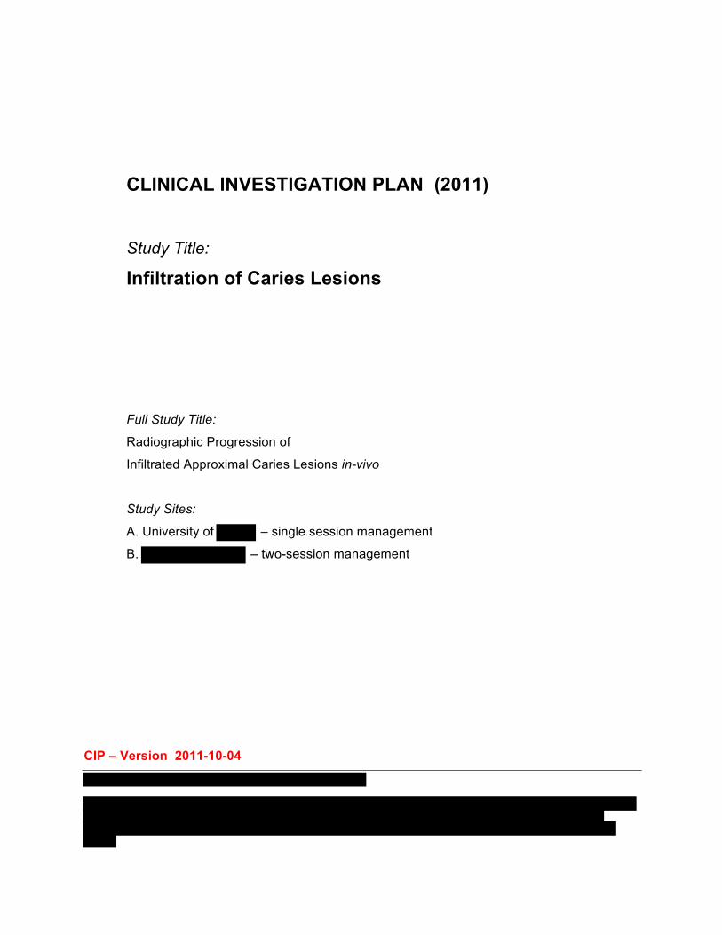
CLINICAL INVESTIGATION PLAN (2011) Study Title:
Infiltration of Caries Lesions
Full Study Title:
Radiographic Progression of
Infiltrated Approximal Caries Lesions in-vivo
Study Sites:
A. University of – single session management
B. – two-session management
CIP – Version 2011-10-04

2011-10-04 Page 2 of 42
CONTENT
APPENDICES TO CIP (2011) .................................................................................................................... 4
1 SYNOPSIS OF THE CLINICAL INVESTIGATION ....................................................................... 5
2 OBJECTIVES OF THE CLINICAL INVESTIGATION PLAN (CIP) ........................................... 7
3 THE STUDY ORGANIZATION ......................................................................................................... 8
4 PRELIMINARY INVESTIGATIONS AND JUSTIFICATION .................................................... 13
4.1 BACKGROUND AND RATIONALE ....................................................................................................... 134.2 HYPOTHESES ...................................................................................................................................... 144.2.1 HYPOTHESIS 1 .................................................................................................................................. 144.2.2 HYPOTHESIS 2 .................................................................................................................................. 144.3 PREVIOUS EXPERIENCE WITH STUDY MATERIALS ......................................................................... 154.3.1 INFILTRATION PROCEDURE MATERIALS (KIT) .................................................................................. 154.3.2 PRECLINICAL TESTING ..................................................................................................................... 154.3.3 PREVIOUS CLINICAL EXPERIENCE .................................................................................................... 164.3.4 DEVICE RISK ANALYSIS AND RISK ASSESSMENT .............................................................................. 16
5 OBJECTIVES OF THE CLINICAL INVESTIGATION ............................................................... 17
5.1 OBJECTIVES ....................................................................................................................................... 175.2 SPECIFIC AIMS (PRIMARY ENDPOINTS) ........................................................................................... 175.2.1 SPECIFIC AIM 1 ................................................................................................................................ 175.2.2 SPECIFIC AIM 2 ................................................................................................................................ 175.3 SECONDARY ENDPOINTS ................................................................................................................... 185.4 STUDY OVERVIEW ............................................................................................................................. 18
6 DESIGN OF THE CLINICAL INVESTIGATION ......................................................................... 20
6.1 CLINICAL INVESTIGATION PROTOCOL ............................................................................................ 206.2 PARTICIPANT SELECTION ................................................................................................................. 206.2.1 INCLUSION CRITERIA ........................................................................................................................ 206.2.2 EXCLUSION CRITERIA ...................................................................................................................... 216.3 RANDOMIZATION AND BIAS .............................................................................................................. 216.4 ENDPOINTS ......................................................................................................................................... 21
7 CLINICAL EVALUATION ............................................................................................................... 22
7.1 RADIOGRAPHS ................................................................................................................................... 227.2 CLINIC VISITS .................................................................................................................................... 23

2011-10-04 Page 3 of 42
7.3 EVALUATION CATEGORIES ............................................................................................................... 247.3.1 HISTORY TAKING (INTERVIEW) ........................................................................................................ 247.3.2 DIET ................................................................................................................................................. 247.3.3 LOCAL ECOLOGY OF LESION ENVIRONMENT (ADJACENT EMBRASURE) .......................................... 247.3.4 CARIES EXPERIENCE ........................................................................................................................ 247.3.5 INDIVIDUALLY STANDARDIZED RADIOGRAPHS .............................................................................. 247.3.6 CONFIRMATION OF LESION STATUS .............................................................................................. 247.4 EVALUATION CRITERIA .................................................................................................................... 257.4.1 ELIGIBILITY CRITERIA ..................................................................................................................... 257.4.2 LOCAL ECOLOGY OF LESION ENVIRONMENT (ADJACENT EMBRASURE) ......................................... 26
8 CLINICAL MEASUREMENTS AND PROCEDURES .................................................................. 27
8.1 SCREENING VISIT .............................................................................................................................. 278.2 PLACEMENT OF SEPARATORS .......................................................................................................... 288.3 SELECTION OF LESIONS (SCREENING VISIT ONLY) ......................................................................... 288.4 RANDOMIZATION ............................................................................................................................... 288.5 INTERVENTION VISIT ........................................................................................................................ 298.5.1 INTERVENTION SITE: ‘INFILTRATION’ ............................................................................................. 298.5.2 INTERVENTION SITE: ‘CONTROL’ .................................................................................................... 298.6 FOLLOW-UP EVALUATIONS ............................................................................................................. 308.7 CONCOMITANT TREATMENT ............................................................................................................ 31
9 BIOSTATISTICAL DESIGN ............................................................................................................. 31
9.1 PROSPECTIVE BI-LATERAL INTRA-ORAL DESIGN ............................................................................ 319.2 DETERMINATION OF SAMPLE SIZE .................................................................................................... 319.3 STATISTICAL ANALYSES ................................................................................................................... 329.3.1 DESCRIPTIVE STATISTICS .............................................................................................................. 329.3.2 BLINDING ......................................................................................................................................... 329.3.3 EVALUATION OF RADIOGRAPHS .................................................................................................... 339.3.4 DIGITAL SUBTRACTION RADIOGRAPHY – LESION EXTENSION AND DENSITY ............................. 339.3.5 INDEPENDENT VISUAL READING – LESION SIZE .......................................................................... 339.3.6 DIRECT VISUAL COMPARISON – LESION EXTENSION .................................................................. 339.3.7 MISSING DATA ................................................................................................................................ 349.3.8 STATISTICAL SOFTWARE ................................................................................................................ 34
10 METHOD OF REPORTING ........................................................................................................... 34
10.1 PERIODIC REPORTS TO THE SPONSOR ........................................................................................... 3410.1.1 FINAL REPORT ............................................................................................................................... 3410.2 DEVIATIONS FROM THE CLINICAL INVESTIGATION PLAN (CIP) .................................................. 3410.2.1 DEVIATIONS FROM THE CIP ........................................................................................................... 3410.2.2 WITHDRAWAL / DROPOUT OF SUBJECTS ....................................................................................... 3510.2.3 AMENDMENTS TO THE CIP ............................................................................................................ 3510.2.4 ADVERSE EVENTS .......................................................................................................................... 3610.2.5 EARLY TERMINATION OR SUSPENSION OF THE INVESTIGATION .................................................... 3610.2.6 LENGTH OF FOLLOW-UP (3 YEARS) – POTENTIAL STUDY EXTENSION ........................................... 36

2011-10-04 Page 4 of 42
11 ETHICAL CONSIDERATIONS ..................................................................................................... 37
12 ADMINISTRATIVE ARRANGEMENTS ...................................................................................... 38
12.1 THE RESEARCH TEAM AND ITS RESPONSIBILITIES ........................................................................ 3812.2 PROJECTED TIME FRAME ............................................................................................................... 3812.3 FINANCING ....................................................................................................................................... 3912.4 REIMBURSEMENT TO SUBJECTS ..................................................................................................... 3912.5 INSURANCE OF THE SUBJECTS ........................................................................................................ 3912.6 CONFIDENTIALITY ........................................................................................................................... 3912.7 PUBLICATIONS ................................................................................................................................. 39
13 REFERENCES .................................................................................................................................. 40
(2011)

2011-10-04 Page 5 of 42
1 SYNOPSIS OF THE CLINICAL INVESTIGATION
Introduction
Dental caries is the most widespread of all diseases. Caries is coming to be more broadly
understood as a chronic bacterial infection. The resultant caries lesion causes destruction of
tooth structure by dissolving the enamel on the outside of the tooth first. If not effectively arrested
by remineralization therapy, it continues with dissolution progressing into the dentin. While it is
possible to use traditional dental fillings to replace diseased tooth structure, it is far better to slow
down or reverse the disease process so that no fillings are needed. One of the most difficult
places to use preventive or non-surgical treatment is at the contact area between
teeth. Recently there is evolving interest in using plastic resins to infiltrate enamel and dentin for
teeth that have suffered some initial damage from caries but have their proximal surfaces intact.
For uncavitated proximal surfaces, the infiltration arrests the disease progression and assists in
repair of the damage already done.
Infiltration lesion management would be indicated when remineralization efforts either didn’t
provide the expected result or have a high likeliness of failure due to lack of monitoring and
follow-up, or due to potentially drastical changes in attention to oral health.
Objective
The purpose of this study is to assess the clinical effectiveness of using resin to infiltrate initial
caries lesions below the uncavitated tooth surfaces that exist on the contact surfaces between
posterior teeth as a means of stabilizing diseased tooth structure and arresting further lesion
development.
Materials and Methods
Young volunteers (14-35 years old) with two early lesions in posterior teeth will be enrolled into a
clinical trial to evaluate the clinical effectiveness of arresting lesion progression by infiltrating the
lesions as compared to current watch-and-wait approaches that are combined with good oral
hygiene and fluoride supplementation. Each subject will have a treated lesion and a control
lesion. Only small early lesions without clinical signs of surface cavitation will be selected. The
control lesions will be stabilized through a normal preventive regimen, while the treatment
lesions will be infiltrated with a resin. Lesion status will be monitored at six month intervals for
the first year by clinical examination and bitewing radiographs. If serial radiographic and clinical

2011-10-04 Page 6 of 42
recall demonstrates no lesion progression then the next bitewings will be taken at annual
intervals fort he remaining two years of surveillance.
Clinical Significance
Infiltrating a caries lesion is a potentially effective strategy to strengthen damaged tooth structure
and to arrest caries progression without any surgical intervention, while protecting the surface
against renewed caries attacks.
The present CIP is the most recent version of the Clinical Investigation Plan
(2011) for the infiltration trial. It supersedes the former version including the
trial. Where applicable, the current CIP includes an update of the
study site (enrollment concluded, 1-yr recall ongoing).
Advancing insight in the novel procedure and emergence of the first short-term
clinical data using a similar technique, warranted the start of a new patient group.
Hence, a new supplemental part of this study investigating the infiltration
technique is proposed. This part is based on a largely similar clinical protocol.
The slight modifications are related to patient pool and clinical application
technique as described in this CIP.
September 2011,

2011-10-04 Page 7 of 42
2 OBJECTIVES OF THE CLINICAL INVESTIGATION PLAN (CIP)
The objectives of the CIP are to provide a full description of the planned clinical trial.
The preceding short narrative (Synopsis) introduced the goal of the project and its approach to
investigate the clinical effectiveness of management of early caries lesions by resin infiltration.
The Background and Rationale section addresses the relevance of the project to oral health and
the rationale for the proposed clinical trial. The existing knowledge is briefly stated, including
literature citations and highlights of relevant data, indicating the early progressive caries
management gap that the project is intended to fill. The research plan includes the Study Design
and Methods addressing the hypotheses to be tested, followed by Specific Aims. The data
collection and analysis (including assessment of statistical significance) will be described and
used to prove or disprove the hypotheses.
Previous experience with the materials involved is described including attention to risk analysis.
Expected results, potential difficulties and limitations are discussed and solutions or alternative
approaches indicated.
Project Overview and Professional Support
Based on internationally accepted protocols we have designed a prospective, randomized
controlled clinical trial (RCT) to investigate the effect of a novel infiltration method as an
alternative management option for the treatment of early, progressing, non-cavitated caries
lesions.
The Clinical Investigation Protocol (CIP) for this study includes all components and
documentation as required to conform to international standards for clinical research
(CONSORT [1, 2, 3], ISO/DIS [4], and Good Clinical Practice Guidelines [5]. An overview of terms,
definitions and acronyms used in this document are included in Appendix 9.


2011-10-04 Page 12 of 42
trial documentation at the clinical site and at the CDC under the condition that confidentiality
concerning the identification of subjects is guaranteed. Access to clinical records and clinical
areas can only be granted with the permission of the PCI, PD/PI and in accordance with the
recognized procedure of the University of Michigan. Reasonable requests will not normally be
refused.
Oversight of human subject research
Approval of the appropriate ethics committees is required (see Human Subjects appendix: A3).
In addition to approval of the investigation by the ethics committee of the central administrative
core at the University of Michigan (IRBMED), the application for the clinical site will be submitted
for the opinion and approval by the local governing ethics committee.
Approval by both central and local ethics committee is required
to initiate the clinical phase and the start of patient recruitment.
The Human Subjects appendix (A3) describes the issues involved in human subjects research.
This includes target recruitment tables, certifications and FDA information of the materials that
will be used in the investigation. The Data Safety and Monitoring Plan (DSMP; A3.4) describes
the reporting of clinical complications (Adverse Events (AE) and Other Reportable Information
and Occurrences (ORIO): A3.5) and the data quality and management.

2011-10-04 Page 13 of 42
4 PRELIMINARY INVESTIGATIONS AND JUSTIFICATION
4.1 BACKGROUND AND RATIONALE
Fissure sealing has been shown to inhibit not only the formation of occlusal caries but also to
impede the progression of existing caries lesions [6, 7, 8]. Pit-and-fissure sealants are an effective
way of preventing caries in children and adults—even in early noncavitated (incipient) lesions,
according to a new set of ADA evidence-based clinical recommendations [9, 10, 11].
Lately, the concept of sealing caries to arrest lesion progression has been transferred to
approximal surfaces [12, 13]. In a clinical study sealed approximal lesions showed significantly
reduced progression after 18 month compared with those that were treated only with preventive
measures [13]. In addition, it has been shown in contemporary European populations that lesions,
radiographically progressed into dentin, were not cavitated in 60% of the cases [14]. The literature
suggests that, when appropriate, a less-invasive management of progressing, non-cavitated
caries lesions may be reasonably preferred above currently utilized “conventional” but, more
invasive operative-restorative management options.
The pores of enamel caries lesions provide diffusion pathways for acids and dissolved minerals.
The aim of lesion infiltration is to occlude these pores by infiltration with light curing resins in
order to block the diffusion of acids into the lesion body [15, 16]. In contrast to the sealing of caries,
caries lesion infiltration aims to occlude the pores within the lesion rather then placing a diffusion
barrier on the lesion surface. Several in-vitro studies show significantly reduced lesion
progression within and peripheral to infiltrated enamel lesions in demineralizing environments [15,
16]. Emerging early data from clinical trials are promising. Resin infiltration in combination with
self-applied non-invasive measures was shown to be more efficacious in reducing lesion
progression compared with selfapplied non-invasive measures alone. [16A]
Clinical Significance: Infiltrating a caries lesion is a potentially effective strategy to strengthen
damaged tooth structure and to arrest caries progression without any surgical intervention.
Successful management of early progressing, non-cavitated caries lesions by resin infiltration
instead of immediate or or postponed restorative treatment may have a great impact in
improving oral health care by means of its non-invasive nature. It may drastically lengthen the

2011-10-04 Page 14 of 42
life-cycle of an at-risk tooth. Confirmation of the clinical efficacy of infiltration resins would yield a
simple and cost-effective caries management treatment option with the least anticipated
iatrogenic effect and a diminished need for future re-treatment due to the deterioration of
conventional restoration margins.
There are currently no USA-based clinical data reported comparing the recently available option
to infiltrate lesions with a standard preventive regimen. Therefore, a randomized controlled
clinical trial is warranted to study lesion progression with and without resin infiltration, while the
participating patients concurrently receive standard-of-care hygiene treatment, diet counselling
and the appropriate fluoride regimen.
4.2 HYPOTHESES
The objectives of the investigation are to study the short-term clinical performance of resin-
infiltrated teeth in a caries-active environment. We hypothesize that, in a high caries risk
population and with a regular preventive regimen as control management, the infiltration of early
approximal caries lesions leads to arrest of the lesion and a reduction of lesion progression.
4.2.1 HYPOTHESIS 1 Infiltrated early caries lesions in a high caries risk population have a lower incidence of
lesion progression when compared to lesions managed by regular preventive regimen.
4.2.2 HYPOTHESIS 2 Infiltrated early caries lesions in a high caries risk population have a lower rate of lesion
progression when compared to lesions managed by regular preventive regimens.
To confirm or reject the hypotheses a prospective, randomized controlled trial (RCT) is designed
to investigate the clinical performance of resin-infiltration as early caries management in a
caries-prone population.
Definitions
Caries detection and diagnosis is an important part of the dentist’s daily work. Caries risk
assessment is the assessment of a patient’s risk of developing new lesions in the near future [17,

2011-10-04 Page 15 of 42
18]. At present, the literature shows inconsistent use of criteria defining “high caries risk”. The
target population for the current study is a caries-prone population. Recruitment and selection
will focus on previous disease (DMFT). During the study the oral environment will be monitored
by DMFT assessment using fibre-optic transillumination. The local environment at the embrasure
of the included approximal lesion surfaces will be assessed in terms of (1) presence of plaque
and gingivitis, and (2) visually using collapsed ICDAS-II scores (see Clinical Evaluation Criteria).
In this clinical study the term “high caries-risk” is defined by clinical evidence of previous
disease. The caries prevalence is expressed in DMFT. In this study, a DMFT ≥ 3 is chosen to
define a patient’s status of “high caries-risk”.
4.3 PREVIOUS EXPERIENCE WITH STUDY MATERIALS
The proposed components of the infiltration kit are substantially equivalent to a variety of
currently marketed dental materials in terms of physical and mechanical properties.
4.3.1 INFILTRATION PROCEDURE MATERIALS (KIT) Infiltrant The infiltration resin is substantially equivalent to an FDA-cleared – 510(k) K992326 – light-curing sealant material.
Etching gel The HCl Etching Gel (15%) is substantially equivalent to an FDA-cleared – 510(k) K891536 – enamel microabrasion compound.
The indication for use of the DMG “Infiltration kit for caries lesions” is micro-invasive treatment of
early approximal caries. The materials will be used according to the label.
4.3.2 PRECLINICAL TESTING The materials to be used for infiltration (etchant and infiltrant) have received EEC market
authorization for medical devices on 2007/04/03. Therefore, complete documentation of
preclinical testing and biological evaluation of the device and its results has passed the
appropriate regulatory bodies. Documentation and approvals are on file with the sponsor.

2011-10-04 Page 16 of 42
4.3.3 PREVIOUS CLINICAL EXPERIENCE The devices used in this study have seen clinical application for several years for sealant
indication with excellent results. The materials in the infiltration kit will be used according to label
and for approved indication (micro-invasive treatment of early approximal caries). The materials
have a low incidence of adverse events and no prior serious adverse events are known.
A recently reported in-vitro study showed that active management of incipient lesions with
sealant application reduced lesion size [20]. Investigations of clinical performance of sealed
approximal lesions [12, 13] revealed that application of a sealant acted as a physical barrier and
reduced lesion size. An 18-month clinical study demonstrated that only 22% of the sealed
incipient proximal caries lesions progressed, compared with the 47% of the group left with no
intervention other than instructions for patients to floss regularly [13]. However, only limited in-vivo
data is available evaluating the clinical performance of infiltrated early caries lesions in a
cariogenic oral environment. A recently published abstract concluded that for proximal caries
lesions extending around the enamel-dentin junction (E2, D1) resin infiltration in combination with
self-applied non-invasive measures was more efficacious in reducing lesion progression
compared with self-applied non-invasive measures alone.[16A]
4.3.4 DEVICE RISK ANALYSIS AND RISK ASSESSMENT
There is „no more than minimal“ risk associated with the device itself and the procedures
involved in its use, as identified by risk assessment and post-market experience of
substantially equivalent materials, beyond the common risks related to standard dental
treatment. The FDA 510(k) information of the products used is included in A3.4: FDA–510(k)
Database Excerpts.
Definition: Minimal risk is the probability and magnitude of harm or discomfort
anticipated in the research and not greater in and of themselves than
those ordinarily encountered in daily life or during the performance of
routine physical or psychological examinations or tests (45 CFR
46.102(i)).
The entire treatment in each aspect, including lesion size and location, all materials, equipment
and techniques used, is part of regular daily life (see also A3.1 Risks). Dentist visits, like in this
study, resulting in caries detection, diagnosis and management by regular preventive regimen or
sealant are not at all out of the ordinary. It is –alas– rather ubiquitous and a common part of daily
oral health care provisions that our profession provides to the public. Therefore, the risk is

2011-10-04 Page 17 of 42
NOT greater than routine dental practice. The care provided meets this definition of
minimal risk.
Although the exclusion criteria mention allergy to methylmethacrylate (a commonly used
monomer as a component in many commercial medical, dental, and non-medical applications),
this allergy has a low prevalence and materials containing methylmethacrylate or its analogues
are managed routinely in dental practices around the world every day. In case of the rare
occurrence of an allergy, patients are isolated immediately from contact, treated using well-
known emergency procedures, and recover quickly. In almost every case, a patient knows in
advance that they have these types of allergies because they must avoid contacts with a huge
number of acrylic materials present normally in the world as parts of garments, containers, and
common household items. No other adverse effects are known, and none are anticipated.
5 OBJECTIVES OF THE CLINICAL INVESTIGATION
5.1 OBJECTIVES
The aim of the study is to evaluate the efficacy of infiltrating early approximal caries lesions in a
caries-prone environment.
5.2 SPECIF IC A IMS (PRIMARY ENDPOINTS)
The investigation will address the following specific aims (primary endpoints):
5.2.1 SPECIFIC AIM 1 To investigate in a high caries risk population of young volunteers (14-35 years old) the
incidence of radiographic progression of infiltrated early caries lesions, when compared to
lesions managed by a standard preventive regimen, evaluated by dental subtraction radiography
after 12, 24 and 36 months.
5.2.2 SPECIFIC AIM 2 To investigate in a high caries risk population of young volunteers (14-35 years old) the rate of
radiographic progression of infiltrated early caries lesions, when compared to lesions managed
by a standard preventive regimen, evaluated by dental subtraction radiography after 12, 24 and
36 months.

2011-10-04 Page 18 of 42
5.3 SECONDARY ENDPOINTS
In addition to the Specific Aims that describe the use of subtraction radiography, the digital
radiographs also will be evaluated visually under standardized conditions (using low-level
magnification (magnifier, loupes) on a computer screen (that can be magnified with the
program). The details of the digitizing process and use of procedures involved in image
enhancement techniques are described later.
– Radiographic size of lesions, evaluated visually by independent assessment of single
bitewing radiographs taken at baseline and after 12, 24 and 36 months
– Radiographic progression of lesions (incidence and rate), evaluated visually by direct
pair-wise comparison of sets of two bitewing radiographs taken at different intervals (at
baseline and after 12, 24 and 36 months).
5.4 STUDY OVERVIEW
In view of prevalence of the type of early lesions to be included in this study and the availability
for recalls for a 3-year period, our focus for recruitment will be adolescents/young adults (age
range of 14-35 years) with a high caries risk status. After receiving introductory study information
at the start of the screening visit the parent/guardian and/or the patient will sign the appropriate
consent/assent form. A dental examination including a caries risk assessment will confirm
eligibility for the study and individually standardized bitewing radiographs will be taken. The
subjects will receive the standard preventive regimen for high caries risk patients. The risk status
will be monitored throughout the study using determinants as DMFT, plaque and gingiva status
and ICDAS-II scores.
Clinical and radiographic examinations of the study teeth are indispensable components of
clinical caries studies in order to diagnose and properly monitor change in approximal caries
lesions. [21, 22, 23, 24] Clinical evaluations will include history taking and diet counseling, and clinical
examination (plaque, gingiva and caries status).
The infiltration procedure will be conducted at the second visit. Contact with the patient will be
assured twice a year following a regular 6-months recall schedule, to monitor the study lesions
by clinical examination, to update demographic registration information and to maintain

2011-10-04 Page 19 of 42
participants’ interest in the study. Standard preventive measures will be provided at each recall
and individually standardized bitewing radiographs will be taken annually.
It is expected that after 3 years of clinical service the safety and efficacy is maintained and both
study teeth are considered satisfactory for continued clinical service.
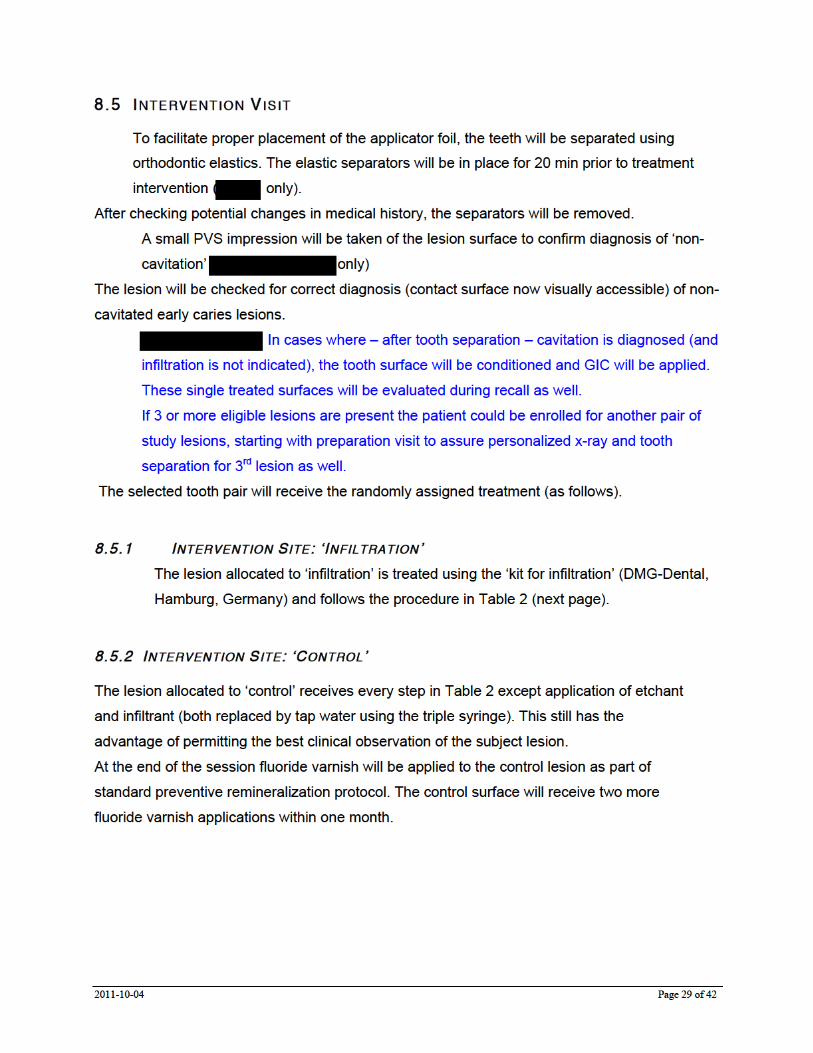
NOTE –– Status Sep 2011: This protocol was originally designed for a clinical site . However, the study had to be moved due to unexpected change in local circumstances (administrative changes, not study-related). Subsequently the CIP was adapted for the clinical site in . Advancing insights and emerging data led to a new study group, now planned for the clinical site in . The study includes minor adjustments to the CIP. The most important differences compared to the study group are:
• Study population: o Patients attending the Dental Clinic at o Young adults (age range 18-24 yrs) previously identified having
two approximal early caries lesions • Clinical protocol modifications:
o Instead of a one-session treatment (incl 20 min separation), the treatment will take place during two sessions (insertion of small tooth-separating elastics at 3 days prior to treatment session).
o A small pre-treatment impression (included approximal surfaces only) will be taken to confirm ‘non-cavitation’ status
o In cases where – after tooth separation – cavitation is diagnosed (and infiltration is no longer indicated), the tooth surface will be conditioned and GIC will be applied. These surfaces will be evaluated during recall as well.
o At conclusion of treatment the occlusal surface will be sealed.

2011-10-04 Page 20 of 42
6 DESIGN OF THE CLINICAL INVESTIGATION
6.1 CLINICAL INVESTIGATION PROTOCOL
This CIP describes a three-year longitudinal, prospective, randomized controlled trial
incorporating a bilateral intra-oral design. The investigation is designed as a proof-of-principle
trial in anticipation of a future multi-site controlled trial. The study includes a clinical team involving
one on-site Principal Clinical Investigator (Site-PCI)/operator and one or two Clinical Investigator
(CI).
The clinical site will enroll 50 subjects (in the age range of 14-35 years (U and
18-24 ( )) with two study lesions. If the lesion management provided in the
study appears to be insufficient within the investigation period ( ;
), the standard traditional restoration will be provided. The patients will be evaluated at
six time points over a period of 3 years. Lesion status and caries risk will be radiographically and
clinically monitored at 6-month intervals during the first year, with radiographic evaluation then
conducted at 12-month intervals (at 1-, 2- and 3-year recall visits).
6.2 PARTICIPANT SELECTION
6.2.1 INCLUSION CRITERIA
Subject level
• Young adolescents/adults with good general health • High caries risk status based on past experience: DMFT ≥ 3 • Reliable for recall attendance for a 3-year follow-up period • At least two early caries lesions in approximal posterior surfaces
Tooth level
• Vital, non-symptomatic tooth • Approximal caries lesion into the inner enamel or outer dentin (E2/D1 lesion) • Lesion visible on radiograph • Tooth routinely in contact with adjacent tooth • Tooth with independent lesion or restoration (PRR, sealant, amalgam) at another
surface is allowed
Lesion level
• Location: Lesion not adjacent to a lesion to be treated,
2011-10-04 Page 21 of 42
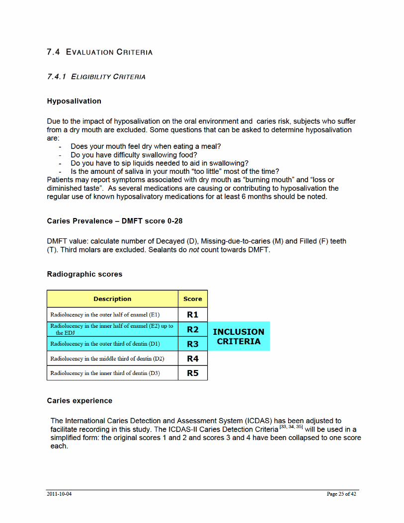
• Depth: Radiographic score R2-R3 (lesion depth around EDJ)
6.2.2 EXCLUSION CRITERIA
Subject level
• Current participation in another clinical study • Medically compromised subjects • Hyposalivation • Pregnancy • Allergic to methylmethacrylates
Tooth level
• Symptomatic tooth
Lesion level
• Location and size outside targeted lesion description
If patients, teeth or lesions fall outside the scope of this study the patient will be referred back to
their regular dental provider. The enrollment will continue until the required number of subjects
for study population has been met.
6.3 RANDOMIZATION AND BIAS
Using a computer-generated random list, the treatment (infiltration or control) is randomly
assigned to each tooth between the screening visit and the intervention visit. The randomization
is recorded at the Patient-Code link list and at the Case Report Form (CRF).
The operator will not be blinded. To avoid bias during the evaluation phase the assessment of the
patient’s radiographs after the 1-, 2- and 3- year recall will be performed by non-operator
examiners/evaluators. The evaluators will be masked to the treatment provided.
6.4 ENDPOINTS
The primary endpoints will be:
• Incidence of lesion progression as analyzed by subtraction radiography • Rate of lesion progression as analyzed by subtraction radiography
The secondary endpoints will be:
• Radiographic lesion size as evaluated visually by independent reading of digital radiographs


2011-10-04 Page 24 of 42
7.3 EVALUATION CATEGORIES
Following is a description of the different evaluation categories that will be used to determine caries risk status, to monitor the lesions and to determine the primary and secondary endpoints of this investigation.
7.3.1 HISTORY TAKING ( INTERVIEW) • Fluoride history • Hyposalivation • Restorations for the last 3 years
7.3.2 DIET • Snacking: frequency and content • Softdrinks: frequency and sipping
7.3.3 LOCAL ECOLOGY OF LESION ENVIRONMENT (ADJACENT EMBRASURE) • Plaque score • Gingiva score • : A separate, local periodontal examination of study teeth and
adjacent teeth will be performed by a periodontist specialist at baseline and 3-month post-treatment
7.3.4 CARIES EXPERIENCE • DMFT (using fibre-optic transillumination) • Collapsed ICDAS score for included approximal lesion surfaces
7.3.5 INDIVIDUALLY STANDARDIZED RADIOGRAPHS • Early caries lesions at approximal surfaces (E2/D1)
7.3.6 CONFIRMATION OF LESION STATUS
• : After tooth separation and prior to treatment a small impression will be taken of the approximal surface to confirm the clinical diagnosis of ‘non-cavitated’ lesion.


2011-10-04 Page 28 of 42
Fig. 1: Bitewing film holder with acrylic impressions
Fig. 2: Radiographic examination
8.2 PLACEMENT OF SEPARATORS
protocol: To facilitate proper diagnosis of non-cavitated lesion the
approximal contact surface of the study tooth will be separated using orthodontic elastics. The
elastic separators will remain in place until just prior to treatment intervention (next session).
8.3 SELECTION OF LESIONS (SCREENING VIS IT ONLY)
Using the study radiographs two lesions will be selected for inclusion in the study:
1) The bitewing radiographs are evaluated by the clinical investigator
2) Of those approximal caries lesions with radiographic scores R2 (inner enamel – E2)
and R3 (outer dentin – D1) the study investigator selects two eligible lesions.
3) If more than two lesions are present, the following priorities will be applied to
determine those to be included in the study: (1) lesion depth R2/R3 (E2 or D1); (2)
tooth type (premolar or molar); (3) lesion location (preference for same arch left and
right over two arches left and right over same quadrant).
8.4 RANDOMIZATION
Randomization for this study is associated only with the allocation of lesions to the two
management groups. Prior to the intervention visit, the two study lesions will be randomized
and assigned to either ‘infiltration’ or ‘control’ group by means of a predetermined randomization
table.

2011-10-04 Page 30 of 42
Table 2 Infiltration protocol
CLINICAL STEPS: 1. Apply a topical anesthetic gel to tooth to be clamped and papilla to be wedged. 2. Remove orthodontic separator.
( Pre-wedging: Expand the interdental space using a wedge.) 3. Clean the affected tooth and adjacent tooth with non-fluoride containing slurry/paste. 4. Take a small impression of lesion surface
( Remove wedge.) 5. Apply rubber dam. Remove any residue with water spray. 6. Expand the interdental space using a flattened wedge and place the applicator. 7. Apply HCl gel on the lesion (1.5-2 turns) using the foil applicator and let set for 2
minutes. 8. While rinsing, remove foil applicator. Rinse-off HCl gel immediately with water spray
for at least 30 seconds. 9. Then dry with oil-free and water-free air.
* The etched enamel should have a chalky white appearance. If this is not the case, the etching process must be repeated.
* The etched surface must not be touched or contaminated with saliva until the treatment resumes. If contamination occurs after drying re-etch for approx. 10 seconds.
10. Apply approximately half of the syringe content of ethanol (96%) on the lesion for 30 seconds. Dry with oil-free and water-free air for 30 seconds.
11. Place a fresh foil applicator between the separated teeth. 12. Apply infiltrant on the lesion using the foil applicator and let set for 3 minutes. 13. Remove excess material using air (triple syringe) and high-vacuum suction. 14. Remove foil applicator and wedge. 15. Remove excess material with floss. 16. Light-cure infiltrant for ≥ 40 seconds total from buccal and lingual direction
Place the light-tip as close to the material as possible. 17. Repeat the application (using a fresh foil applicator and wedge; 1 minute setting
time) and ≥40 s light-curing of the infiltrant (steps 11 – 16). 18. Application of sealant to or perform PRR in occlusal surface 19. Remove rubber dam.
* Light-curing unit should have a minimum standard output of 450 nm and should be checked regularly with a handheld calibrated radiometer. The light intensity should be at least 400 mW/cm2.
8.6 FOLLOW-UP EVALUATIONS
All procedures for the yearly follow-up examinations are itemized in Table 1. In addition,
intervening 6-month recalls will be conducted to reinforce oral hygiene and to monitor lesion
status. Specific standard instructions for good oral hygiene and fluoride varnish will be provided
at each recall to both study teeth. Due to the non-invasive preventive approach for controls, the
patient will be on a strict 6-month recall schedule to closely monitor any changes in caries lesion
status and caries risk assessment.
At the 6-month intervals any visual signs of lesion progression are noted and an additional set of
2BW radiographs will be taken to confirm any change in lesion status by radiographical
assessment and diagnosis. This procedure is current standard-of-care clinical practice.

2011-10-04 Page 31 of 42
8.7 CONCOMITANT TREATMENT
The clinical study is concerned with two lesions per patient. Within the study no additional
concomitant treatment will be offered to the subjects outside the normal clinical routines.
At the recall appointments, normal recall procedures will be applied.
If lesion progression is suspected or detected by clinical examination during the 1.5 or 2.5 year
recall, 2 BW radiographs will be taken to confirm the diagnosis.
If lesion progression is detected at any interval, intervention with a management response that is
in the best interest of the patient will be indicated and performed. If this intervention deviates
from the study’s hygiene or fluoride measures (fluoride varnish application) the patient will be
transferred out of the study. The treatment provided will be recorded and the patient’s study
participation is completed.
9 BIOSTATISTICAL DESIGN
9.1 PROSPECTIVE BI-LATERAL INTRA-ORAL DESIGN
Study designs, as proposed, where the patient serves as her/his own control are recognized as
having the ability to greatly facilitate the interpretation of trials by minimizing the effects of inter-
patient variability [38]. In such a design it is possible to subtract out the influence of individual
patient characteristics and obtain a more powerful estimate of treatment effect with smaller
sample size.
9.2 DETERMINATION OF SAMPLE SIZE
Sample size calculations were calculated based on clinical data from [13].
Input data for the power analysis:
- two-sided test (paired responses McNemar Chi-square test)
- alpha: 1%
- power 90%
Sample size calculation
Parameters:
- p0: 0.841 - p1: 0.435


2011-10-04 Page 33 of 42
The teeth will be randomly allocated to either the „infiltration“ or „control“ group. The study
materials are used “on-label”, and have been on the market for a considerable period of time. It
is unlikely that the study needs to be terminated on statistical grounds as the safety and efficacy
of the materials have already been established. We do expect to find similar or larger differences
between the two management approaches in this study when compared to the Martignon study
[13]. Based on emerging clinical data (2011) a higher difference in discordant pairs is to be
expected.
9.3.3 EVALUATION OF RADIOGRAPHS
The radiographs will be examined and assessed by evaluators who are blinded to both
sequence of radiographs and allocation of lesions to ‘infiltration’ and ‘control’ group.
9.3.4 DIGITAL SUBTRACTION RADIOGRAPHY – LESION EXTENSION AND DENSITY
After image enhancement the digital radiographical image will be assessed for positive, none or
negative change in size and density using digital subtraction radiography.
Digital subtraction using a set threshold will be performed by two independent examiners who
are masked to the origin of the radiographs. The images are read independently and
discrepancies between the readings will be solved by consensus. The examiners determine the
size of the lesion and record whether the lesion progressed, did not change or regressed.
Chronological and other sets of radiographs will be compared: BL vs. 1Y, 1Y vs. 2Y and 2Y vs.
3Y; and also BL vs. 2Y, BL vs. 3Y and 1Y vs. 3Y.
9.3.5 INDEPENDENT VISUAL READING – LESION SIZE
The radiographs are read independently using the standard radiographic scoring system as
described in Evaluation Criteria (7.3).
9.3.6 DIRECT VISUAL COMPARISON – LESION EXTENSION
At least one week later the bitewing radiographs are read pair-wise (Scores: “A deeper then B”;
“B deeper then A”; “A and B same lesion extension”). The examiner is blinded regarding the date
of record (baseline or follow-up).


2011-10-04 Page 36 of 42
However, when there are changes to the initial list of clinical investigators this list will not be
formally updated by amendments at each change; the PD/PI will maintain an updated list, which
will be available on request. The definitive list of all investigators shall be provided with the final
report.
10.2.4 ADVERSE EVENTS Emergency contact details for reporting of serious adverse events (Appendix 4) are included in
the CIP (see A4: work and residential addresses and phone numbers of PCIs).
All materials and devices used in this study have been approved by local regulatory bodies
(including FDA) and are currently on the market in various parts of the world. Their clinical use is
according to label. Therefore, no foreseeable adverse events are expected.
In case of a serious adverse event and subsequent need for un-blinding the name-code list can
be accessed immediately and breaking the code will not cause any further problem. The data
recorded until the report of adverse event will remain included in the dataset.
10.2.5 EARLY TERMINATION OR SUSPENSION OF THE INVESTIGATION All materials and devices used in this study have been approved by local regulatory bodies and
are currently on the market in various parts of the world. Their clinical use in this investigation is
according to label. Therefore, early termination or suspension of the investigation due to
problems with the restorative materials is not anticipated.
If the investigation is terminated prematurely or suspended, the PD/PI will promptly inform the
clinical investigators/investigation center of the termination or suspension and the reason(s) for
this. The ethics committees will also be informed promptly and provided with the reason(s) for
the termination or suspension by the PD/PI or by the Site-PCI.
10.2.6 LENGTH OF FOLLOW-UP (3 YEARS) – POTENTIAL STUDY EXTENSION At the 3-year recall the number of subjects and the outcomes of the study will be assessed
specifically in view of study continuation. Based on an informed discussion concerning patient
retention data and available results an assessment regarding continuation or closing of the
investigation will be made by the PD/PI. The PD/PI will inform the Sponsor whether continuation
2011-10-04 Page 37 of 42
of the evaluation period to the 4- or 5-year recall is expected to provide scientific valuable
results. The decision for continuation will be made by the Sponsor.
11 ETHICAL CONSIDERATIONS
The Human Subjects appendix (Appendix 3) describes the issues involved in human subjects
research.
Institutional ethics applications, including proper informed consent (parents/guardians) with
informed assent (teenagers 14-17 years old) or adult informed consent (18-35 years), will be
submitted for approval according to the international rules and regulations for Federal-Wide
Assurances of Protection for Human Subjects (FWAs). See also webpage:
http://www.hhs.gov/ohrp/assurances/assurances index.html . The study protocol will be
submitted to the ethics committee (Institutional Review Board: IRBMED) at the University of
Michigan (location of the central administrative core) and the respective IRBs in and
IRB-KACH). The participating clinical centers ) have each an
established, registered IRB and FWA (or equivalent).
Current information: Michigan: FWA00004969
FWA00007742
USAMEDDAC assurance: DoD A10027
Full registration information of the governing Assurances and IRBs is included in Appendix 3.8.
All investigators involved are required to complete the PEERRS certification (or equivalent) prior
to the start of the study (Appendix 11).

2011-10-04 Page 39 of 42
12.3 FINANCING
Financial agreements regarding funding of the different aspects of the investigation are part of
separate contracts between the Sponsor and the Study Team / University of Michigan.
12.4 REIMBURSEMENT TO SUBJECTS
Subject compensation and incentives being part of the subject retention package for the clinical
site will be part of a separate contract and fall outside the scope of the CIP. (not applicable in
12.5 INSURANCE OF THE SUBJECTS
The study materials in this investigation are cleared by the FDA (USA) and available on the
market. Therefore, the subjects taking part in the trial are insured by the Sponsor against any
injury caused by the study materials under investigation.
12.6 CONFIDENTIALITY
All unpublished information concerning this trial and the materials supplied to the PD/PI and the
Investigators by the Sponsor will be treated confidentially by all parties involved until the Sponsor
gives written consent that the information may be published or handed over to third parties.
The Sponsor has the rights on all data and information acquired during the investigation.
12.7 PUBLICATIONS
Publication of (parts of) the trial by the Study Team will take place only with the written consent
of the Sponsor, or following a period of one year from the date the Sponsor receives the related
report.

2011-10-04 Page 40 of 42
13 REFERENCES
1. Begg C, et al., Improving the quality of reporting of randomized controlled trials: The CONSORT statement. J
Amer Med Assoc, 1996. 276(8): 637-639.
2. Needleman I, CONSORT. Consolidate Standards of Reporting Trials. Br Dent J, 1999a. 186(5): 207.
3. Needleman I, Clinical research at cross roads. Br Dent J, 1999b. 187(9): 459.
4. ISO (International Organization for Standardization), Clinical Investigation of medical devices for human subjects – #14155. 2001, ISO Central Secretariat: Geneva, Switzerland.
5. ICH (Int Confer Harmonisation). Harmonised Tripartite Guideline for Good Clinical Practice. in International Conference on Harmonisation of Technical Requirements for Registration of Pharmaceuticals for Human Use (ICH). 1996.
6. Swift EJ, The effect of sealants on dental caries: a review. J Am Dent Assoc, 1988. 116: 700-704.
7. Going RE, et al., The viability of microorganisms in carious lesions five years after covering with a fissure sealant. J Am Dent Assoc, 1978. 97: 455-462.
8. Mertz-Fairhurst E, et al., Ultraconservative and cariostatic sealed restorations: results at Year 10. J Am Dent Assoc, 1998. 129: 55-66.
9. Garvin J, Sealant use examined in JADA Journal of the American Dental Association 2008. http://www.ada.org/prof/resources/pubs/adanews/printarticle.asp?articleid=2911
10. Griffin SO, et al., The effectiveness of sealants in managing caries lesions. CDC Dental Sealant Systematic Review Work Group, Bader J, Clarkson J, Fontana MR, Meyer DM, Rozier RG, Weintraub JA, Zero DT. J Dent Res, 2008. 87(2): 169-174.
11. Beauchamp J, et al., Evidence-Based Clinical Recommendations for the Use of Pit-and-Fissure Sealants: A Report of the American Dental Association Council on Scientific Affairs. J Am Dent Assoc, 2008. 139: 257-268.
12. Gomez SS, CP Basili, and CG Emilson, A 2-year clinical evaluation of sealed noncavitated approximal posterior carious lesions in adolescents. Clin Oral Investig, 2005. 9: 239-243.
13. Martignon S, KR Ekstrand, and R Ellwood, Efficacy of sealing proximal early active lesions: an 18-month clinical study evaluated by conventional and subtraction radiography. Caries Res, 2006. 40: 382-388.
14. Kidd EAM, et al., Why restore teeth? – Diagnosis of Dental Caries:12-18. Pickard’s Manual of Operative Dentistry, 2003. 8th Edition.
15. Paris S, H Meyer-Lueckel, and AM Kielbassa, Resin infiltration of natural caries lesions. J Dent Res, 2007. 86: 662-666.
16. Meyer-Lueckel H, et al., Influence of the application time on the penetration of different dental adhesives and a fissure sealant into artificial subsurface lesions in bovine enamel. Dent Mater, 2006. 22: 22-28.
16A. Meyer-Lueckel H, Bitter K, Paris S. Placebo-Controlled Randomized Clinical Trial on Proximal Caries Infiltration: 3-Year Follow-Up. Caries Res 2011;45:183-184.
17. Bratthall D, Dental caries: Intervened - Interrupted - Interpreted. Eur J Oral Sci, 1996. 104(4): 486-491.

2011-10-04 Page 41 of 42
18. Bratthall D, Introducing the Significant Caries Index together with a proposal for a new global oral health goal for 12-year-olds. Int Dent J, 2000. 50(6): 378-384.
19. Wierzbicka M and et al, Changing oral health status and oral health behaviour of school children in Poland. Comm Dent Health, 2002. 19: 243-250.
20. Trairatvorakul C, S Kladkaew, and S Songsiripradabboon, Active Management of Incipient Caries and Choice of Materials. J Dent Res, 2008. 87(3): 228-232.
21. Pitts NB, The use of bitewing radiographs in the management of dental caries: scientific and practical considerations. Dent Max Facial Radiol, 1996. 25(1): 5-16.
22. Kidd EA, DN Ricketts, and NB Pitts, Occlusal caries diagnosis: a changing challenge for clinicians and epidemiologists. J Dent, 1993. 21: 323-331.
23. White SC, et al., Efficacy of FDA guidelines for ordering radiographs for caries detection. Oral Surg Oral Med Oral Pathol, 1994. 77: 531-540.
24. Reis IM, et al., Findings of clinical and radiographic caries among several adult age groups. Oral Surg Oral Med Oral Pathol, 1998. 86(6): 760-764.
25. Pitts NB and EAM Kidd, The prescription and timing of bitewing radiography in the management of dental caries. Br Dent J, 1992. 172: 225-227.
26. American Academy of Pediatric Dentistry, Endorsement: Prescribing dental radiographs for infants, children, adolescents and persons with special health care needs, in 2005-06 Oral Health Policies and Clinical Guidelines (AAPD). 2005.
27. Klinger HG, et al., Möglichkeiten der digitalen Bildverarbeitung zur Informationsverstärkung in BissFlügelaufnahmen. Z Stomatol, 1989. 86: 1-11.
28. Wenzel A, PN Anthonisen, and MB Juul, Reproducibility in the assessment of caries lesion behaviour: a comparison between conventional film and subtraction radiography. Caries Res, 2000. 34(3): 214-218.
29. Wenzel A, Current trends in radiographic caries imaging. Oral Surg Oral Med Oral Pathol Oral Radiol Endodont, 1995. 80(5): 527-539.
30. Ricketts NJ, et al., Accuracy and reproducibility of radiographic assessment and subtraction radiography in detecting demineralization in occlusal surfaces. Caries Res, 2003. 37: (Abstr #66).
31. Ekstrand KR, et al., The performance of two different subtraction radiography methods in detecting demineralization in occlusal surfaces. Caries Res, 2003. 37: (Abstr #67).
32. Maltz M, et al., A clinical, microbiologic, and radiographic study of deep caries lesions after incomplete caries removal. Quintessence Int, 2002. 33: 151-159.
33. Ekstrand KR, DN Ricketts, and EA Kidd, Reproducibility and accuracy of three methods for assessment of demineralization depth of the occlusal surface: an in vitro examination. Caries Res, 1997. 31: 224-231.
34. Ekstrand KR, et al., Detection and activity assessment of primary coronal caries lesions: a methodologic study. Oper Dent, 2007. 32: 225-235.
35. ICDAS Coordinating Committee, ICDAS II Appendix Criteria Manual (Workshop Baltimore 2005), in The International Caries Detection and Assessment System (ICDAS II), G IAT, Editor. 2006. p. 28.
36. Ekstrand KR, DN Ricketts, and EA Kidd, Occlusal caries: pathology, diagnosis and logical management. Dent Update, 2001. 28(8): 380-387.

2011-10-04 Page 42 of 42
37. Ekstrand KR, G Bruun, and M Bruun, Plaque and gingival status as indicators for caries progression on approximal surfaces. Caries Res, 1998. 32: 41-45.
38. Antczak-Bouckoms AA, JF Tulloch, and CS Berkey, Split-mouth and cross-over designs in dental research. J Clin Periodontol, 1990. 17: 446-453.
39. PASW (SPSS v.18.) 2011: Chicago, IL.
40. SAS Institute, SAS/STATA v.11.0 SE. 2004, Cary, NC: SAS Publishing.
41. SAS Institute, SAS® Release 9.2. 2003: Cary, NC.

Statistical Analysis Plan Study Title: Infiltration of Caries Lesions
NCT Number: NCT01584024
NCT Title: Resin Infiltration to Arrest Early Tooth Decay
Sample size calculation
Sample size calculation for paired observations (split-mouth design, McNemar’s test) was based
on the following parameters from a previous infiltration study of Martignon et al. [25]. Assuming a
difference of proportions of 41% in lesion progression between control (84%) vs. test (43%)
group, and a proportion of discordant pairs of 56%, alfa = 0.05, and 1-beta = 0.8, the calculated
sample size was 22 lesion pairs. Allowing for a potentially high attrition rate over a period of 3
years, 42 participants were enrolled to find significant differences using McNemar’s test.
Statistical analysis
Intra/inter-examiner reliability for independent radiograph evaluations was analyzed with Kappa
statistics. The primary outcome was proportion of lesion progression (comparative pairwise
assessment: continuous progression), whereas change in categorical lesion depth (progression
to next depth category) was considered a secondary outcome. Differences between groups were
tested using Chi-square test and Fisher’s exact test. Differences in proportion of progressing
lesions were analyzed with McNemar’s test with 95% confidence interval (95% CI) and P = .05 for
significance level using SAS software. Pairwise comparison data and cumulative lesion
progression were used to calculate the therapeutic effect (absolute value) and the relative risk
reduction (RRR) that indicates efficacy of treatment.