
natural course of AK_diss
85
Aus der Division of Evidence based Medicine (dEBM) der Klinik für Dermatologie, Venerologie und Allergologie, Medizinische Fakultät Charité – Universitätsmedizin Berlin DISSERTATION Systematic Review of the Natural Course of Actinic Keratosis and their Relation to Squamous Cell Carcinoma zur Erlangung des akademischen Grades Doctor medicinae (Dr. med.) vorgelegt der Medizinischen Fakultät Charité – Universitätsmedizin Berlin von Ricardo Niklas Werner aus Iserlohn Datum der Promotion: 12.09.2014
Transcript of natural course of AK_diss

Aus der Division of Evidence based Medicine (dEBM) der Klinik für Dermatologie, Venerologie und Allergologie, Medizinische Fakultät
Charité – Universitätsmedizin Berlin
DISSERTATION
Systematic Review of the Natural Course of Actinic Keratosis and their Relation to Squamous Cell Carcinoma
zur Erlangung des akademischen Grades Doctor medicinae (Dr. med.)
vorgelegt der Medizinischen Fakultät Charité – Universitätsmedizin Berlin
von
Ricardo Niklas Werner
aus Iserlohn
Datum der Promotion: 12.09.2014
jacobsa
Schreibmaschinentext

2
Systematic Review of the Natural Course of Actinic Keratosis and their Relation to Squamous Cell Carcinoma
LIST OF FIGURES..................................................................................................... 5
LIST OF TABLES ...................................................................................................... 5
ABBREVIATIONS...................................................................................................... 5
1. ABSTRACT / ABSTRAKT .................................................................................. 6
1.1. Abstract ..................................................................................................................................... 6
1.2. Abstrakt (German) .................................................................................................................... 7
2. BACKGROUND .................................................................................................. 9
2.1. Introduction............................................................................................................................... 9
2.2. Terminology and significance of AK ...................................................................................... 9
2.3. Etiology of AK......................................................................................................................... 10
2.4. Risk factors for the development of AK............................................................................... 11
2.5. Epidemiology of AK ............................................................................................................... 11
2.6. Diagnosis of AK...................................................................................................................... 12 2.6.1. Clinical features ............................................................................................................... 12 2.6.2. Clinical diagnosis ............................................................................................................. 13 2.6.3. Histological features ........................................................................................................ 14 2.6.4. Other diagnostic means................................................................................................... 16
2.7. Treatment options and recommendations........................................................................... 16 2.7.1. Lesion-directed interventions........................................................................................... 16 2.7.2. Field-directed interventions.............................................................................................. 17 2.7.3. Recommended treatment algorithms .............................................................................. 17
2.8. Rationale and objectives of the present study.................................................................... 18
3. METHODS ........................................................................................................ 20
3.1. Introduction............................................................................................................................. 20
3.2. Search for literature ............................................................................................................... 20
3.3. Outcomes of interest.............................................................................................................. 20
3.4. Eligibility criteria..................................................................................................................... 21
3.5. Selection of eligible studies .................................................................................................. 22
3.6. Data extraction........................................................................................................................ 22
3.7. Assessment of the methodological quality of included trials ........................................... 22

3
3.7.1. Methodological quality of the included prospective longitudinal studies ......................... 22 3.7.2. Methodological quality of the included cross-sectional/ retrospective studies ................ 23
3.8. Analysis and presentation of the results ............................................................................. 23 3.8.1. Presentation of the results of the systematic review ....................................................... 23 3.8.2. Extrapolation of the overall risk of the ‘standard AK patient’ to develop an SCC............ 24
4. RESULTS.......................................................................................................... 26
4.1. Study selection ....................................................................................................................... 26
4.2. Characteristics of the included studies ............................................................................... 27
4.3. Quality and risk of bias assessment .................................................................................... 29 4.3.1. Quality assessment and risks of bias within the prospective longitudinal studies........... 29 4.3.2. Quality assessment and risk of bias within the cross-sectional / retrospective studies .. 30
4.4. Results of individual studies: Data on the relation of AK to SCC..................................... 31 4.4.1. Progression rates of single AK lesions to SCC ............................................................... 31 4.4.2. Risk or odds ratios for developing SCC relative to the number of pre-existing AK ......... 33 4.4.3. Rates of contiguous AK lesions in SCC specimens ........................................................ 34
4.5. Results of individual studies: Data on the natural history of AK...................................... 35 4.5.1. Regression and recurrence rates of single AK lesions.................................................... 35 4.5.2. Rates of complete field regression und subsequent recurrences ................................... 38 4.5.3. Change in total AK counts over time ............................................................................... 40
4.6. Extrapolation of the risk of development of an SCC for the ‘standard’ patient............... 43 4.6.1. Patients presenting with AK without history of NMSC or immunosuppression ............... 43 4.6.2. Patients presenting with AK with a history of NMSC....................................................... 43 4.6.3. Organ transplant recipients presenting with AK .............................................................. 44
5. DISCUSSION.................................................................................................... 45
5.1. Brief summary of the main findings ..................................................................................... 45
5.2. Overall limitations of the systematic review ....................................................................... 45 5.2.1. Inclusion of control arms from clinical trials ..................................................................... 45 5.2.2. Definition of patient subgroups ........................................................................................ 46 5.2.3. Assessment and differentiation of AK and SCC.............................................................. 47 5.2.4. Validity of AK lesion counts ............................................................................................. 47 5.2.5. Treatment of lesions during studies................................................................................. 48 5.2.6. Measures to avoid unprotected exposure to sun radiation.............................................. 48
5.3. Discussion of the results on a study level........................................................................... 48 5.3.1. Risk of progression of AK lesions to SCC ....................................................................... 48 5.3.2. Risk or odds ratios for developing SCC relative to the number of pre-existing AK ......... 50 5.3.3. Rates of contiguous AK lesions in SCC specimens ........................................................ 51 5.3.4. Regression and recurrence rates of single AK lesions.................................................... 51 5.3.5. Rates of complete field regression and subsequent recurrence ..................................... 52 5.3.6. Change in total AK counts over time ............................................................................... 53
5.4. Discussion of the extrapolation results of the risk of development of an SCC for the ‘standard’ patient presenting with AK............................................................................................... 53
5.5. Conclusions ............................................................................................................................ 54
6. REFERENCES.................................................................................................. 56

4
7. SUPPLEMENT.................................................................................................. 65
7.1. Tables of included studies .................................................................................................... 66 7.1.1. General characteristics of the included prospective longitudinal studies (Table 8)......... 66 7.1.2. General characteristics of the included cross-sectional / retrospective studies (Table 9) 74
7.2. Search strategies.................................................................................................................... 76 7.2.1. Search strategy used for the search in the Cochrane Library ......................................... 76 7.2.2. Search strategy used for the search in Medline .............................................................. 77 7.2.3. Search strategy used for the search in Medline in Process ............................................ 77 7.2.4. Search strategy used for the search in Embase.............................................................. 78
9. CURRICULUM VITAE....................................................................................... 82
10. LIST OF PUBLICATIONS ............................................................................. 84
11. ACKNOWLEDGEMENTS ............................................................................. 85

5
List of figures
Figure 1. Process of study identification and selection ............................................. 26 Figure 2. Types of studies, numbers of studies reporting on each outcome, and
specification of population ........................................................................ 28
List of tables
Table 1. Severity degrees of AK............................................................................... 16 Table 2. Risk of progression of single AK lesions to SCC ........................................ 32 Table 3. Relative risks and odds ratios for developing SCC relative to the number of
pre-existing AK ......................................................................................... 34 Table 4. Rates of SCC specimens with contiguous AK lesions ............................... 35 Table 5. Regression and recurrence rates of single AK lesions ............................... 37 Table 6. Rates of complete field regression and subsequent recurrence ................. 39 Table 7. Changes in the total AK lesion count over time .......................................... 41 Table 8. General characteristics and methodological quality rating of the included
prospective longitudinal studies................................................................ 66 Table 9. General characteristics and methodology rating of the included studies on
contiguous AK lesions in SCC specimens................................................ 74
Abbreviations
AK, AKs – actinic keratosis, actinic keratoses
ALA-PDT – aminolaevulinic acid-photodynamic therapy
NMSC – non-melanoma skin cancer
OTR – organ transplant recipients
Pts. – participants, patients
SCC – squamous cell carcinoma
UV radiation – ultraviolet radiation

6
1. Abstract / Abstrakt
1.1. Abstract Background: For decisions about the treatment necessity, knowledge about the
course of untreated actinic keratosis (AK) is essential, particularly with respect to
spontaneous regression rates and the risk of progression to squamous cell
carcinoma (SCC). The goal of the present study is to systematically assess the
primary data on the natural history of AK and its relation to SCC.
METHODS: A systematic literature search was conducted. Cohort studies and
control groups from clinical trials were included if they reported data on progression
or regression rates, correlational data on the subsequent risk of SCC development,
data on histologically contiguous AK in SCC specimens, changes in total AK counts
over time and/or complete field regression and recurrence rates. High risk
populations (history of non-melanocytic skin cancer (NMSC), concomitant
immunosuppression) were reported separately. An extrapolation of the risk of
development of an SCC for the ‘standard patient’, based on mean data from the
source studies, was performed.
RESULTS: 33 studies reporting on at least one of the outcomes were eligible. A
progression risk for single AK lesions of up to 0.075% per lesion-year was reported.
With a progression rate of 0.53%, the risk was higher in patients with a history of
NMSC. Extrapolating these data for the ‘standard patient’s’ ten year-risk to develop
SCC results in a disproportionally higher risk. Correlational data show a strong
relation of AK and consecutive SCC, and indicate a higher risk for organ transplant
recipients. Contiguous AK lesions were regularly found in SCC specimens.
Regression rates of AK lesions varied from 15 to 63% after 12 months, with studies
showing a subsequent recurrence of 15% to 53% of regressed lesions. Rates of
complete field regression varied from 0 to 21%, with subsequent recurrences in 57%
of the patients. Data on changes of total AK lesion counts were heterogeneous,
ranging from -53% to +99.1%.
DISCUSSION: The close relation between AK and SCC is seen in correlational data
and confirmed in observational studies following AK lesions. Several methodological
limitations apply to the data and at the current time, any estimate on progression
rates remains highly uncertain. This particularly applies to extrapolations based on

7
the respective data. Other results described below illustrate a dynamic interplay of
progression and regression. Despite the relatively high regression rates,
spontaneous complete field regressions rarely occur. Due to the low probability of
spontaneous complete regressions and the inherent risk of progression, AK requires
an appropriate treatment.
1.2. Abstrakt (German) Hintergrund: Um klinisch relevante Entscheidungen über die Behandlung zu treffen,
sind Kenntnisse über den unbehandelten Verlauf aktinischer Keratosen (AK)
unumgänglich, insbesondere hinsichtlich spontaner Regression und Progression in
spinozelluläre Karzinome (SCC). Ziel der vorliegenden Studie ist die systematische
Erfassung der Primärdaten zum natürlichen Verlauf von AK und ihrer Beziehung zum
SCC.
Methoden: Es wurde eine systematische Literaturrecherche durchgeführt.
Kohortenstudien und Kontrollgruppen randomisierter Studien wurden
eingeschlossen, wenn mindestens eines der folgenden Outcomes berichtet wurde:
Progressions- und Regressionsraten einzelner AK, korrelationale Daten zum Risiko
der Entwicklung eines SCC, Daten zu histologischen AK Korrelaten in SCC-Proben,
Veränderung der Anzahl an AKs über den zeitlichen Verlauf und Raten vollständiger
spontaner Regression und nachfolgendem Wiederauftreten. Hoch-Risiko-
Populationen (Zustand nach nicht-melanozytärem Hautkrebs (NMSC),
immunsupprimierte Patienten) wurden separat berichtet. Das theoretische
individuelle 10-Jahres-Risiko eines ‘Standardpatienten’ wurde, basierend auf den
Daten aus den entsprechenden Quellstudien, berechnet.
Ergebnisse: 33 den Einschlusskriterien entsprechende Studien wurden identifiziert.
Es zeigte sich eine Progressionswahrscheinlichkeit einzelner AK-Läsionen von
0,075%/Jahr. Mit 0,53% pro Läsion und Jahr war die Progressionswahrscheinlichkeit
für Patienten mit Zustand nach multiplen NMSC deutlich höher. Die theoretische
Berechnung des 10-Jahres-Risikos eines ‘Standardpatienten’ für die Entwicklung
eines SCC ergab ein unverhältnismäßig höheres Risiko. Korrelationale Daten zeigen
eine starke statistische Beziehung des Risikos der Entwicklung eines SCC zur
Anzahl vorbestehender AKs und weisen auf eine höhere Progressions-
wahrscheinlichkeit für immunsupprimierte Patienten hin. In Biopsien aus SCC
wurden regelmäßig angrenzende AK histologisch nachgewiesen. Regressionsraten

8
von AKs nach 12-monatiger Beobachtung bewegten sich zwischen 15 und 63%, mit
einem erneuten Wiederauftreten von 15% bis 53% der regredierten Läsionen.
Vollständige spontane Regressionen wurden in 0 bis 21% der Patienten beobachtet,
ein nachfolgendes Wiederauftreten von AK in 57% der Patienten. Daten zur relativen
Änderung der Gesamtanzahl von AK im zeitlichen Verlauf zeigten ein breites
Spektrum der Ab- und Zunahme, mit relativen Veränderungen von –53% bis +99,1%.
Diskussion: Nicht nur korrelationale und histologische Daten spiegeln die enge
Verknüpfung von AKs und SCC wider, sondern auch Beobachtungsstudien, die den
Verlauf einzelner AKs nachverfolgen. Verschiedene methodologische Aspekte
schränken die Interpretierbarkeit der Ergebnisse ein und zum jetzigen Zeitpunkt
bleiben Schätzungen zur Progressionsrate von AK in hohem Maße unsicher. Dies gilt
insbesondere für die theoretische Berechnung des individuellen Progressionsrisikos.
Insgesamt zeigt sich ein dynamischer Wechsel von qualitativer und quantitativer
Progression und Regression aktinischer Keratosen. Trotz der relativ hohen
Regressionsraten einzelner AK-Läsionen sind vollständige spontane Remissionen
seltene Ereignisse. Aufgrund der geringen Wahrscheinlichkeit kompletter spontaner
Regressionen und des inhärenten Progressionsrisikos, erfordern AKs eine adäquate
und konsequente Behandlung.

9
2. Background
2.1. Introduction Actinic keratoses (AKs, also referred to as solar or senile keratoses) are lesions of
the skin that develop on areas chronically exposed to ultraviolet (UV) radiation. AKs
are the most common skin lesions that have a supposed potential of progression into
invasive, malignant skin cancer. AK lesions present as scaly, rough papules that may
feel like patches of dry skin. Usually AKs are not symptomatic, but pruritus, pain and
bleeding may occur. Different treatment options have been described for the
management of AKs, including interventions aimed at the elimination of manifest
areas (lesion-directed interventions) and interventions intended to reduce both
manifest and latent areas of dysplastic skin (field-directed interventions). The
treatment necessity itself is assumed to be due to the risk of malignant progression of
AK lesions into squamous cell carcinoma (SCC) of the skin. Yet, the natural history of
AK remains a matter of debate and especially the risk of progression into malignant
disease has not been assessed systematically.
2.2. Terminology and significance of AK Several expressions have been in use synonymously for AK, including ‘solar
keratosis’, ‘senile keratosis’, ‘senile keratoma’, ‘keratinocytic intraepidermal
neoplasia’,1 and ‘in situ squamous cell carcinoma Type AK’.2 AKs on the lips are
termed as ‘actinic cheilitis’.3
AK is a medical condition of the skin that already had been described by the end of
the nineteenth century1,4 and the debate about their malignant character or potential
almost reaches back to that time.1,5 AKs can be considered as keratinocytic dysplasia
(‘precancerous lesion’) or as in situ SCC (‘neoplasia’, ‘carcinoma’). The former view
tends to describe AK lesions as a premaligancy that does not necessarily have to be
treated unless a malignant ‘transformation’ occurs, whereas the latter view
characterizes AKs themselves as malignancies with the risk of ‘progression’ into
invasive stages, imposing an absolute treatment indication.5 On the level of cytology,
AKs are hardly distinguishable from SCCs2,5,6 and thus, more recent
characterizations of AKs tend to accentuate the view of AK as ‘superficial SCC’.1,2,7
This perspective characterizes AKs analogously to other epithelial in situ lesions that
may progress into invasive disease, such as the cervical intraepithelial neoplasia

10
(CIN).8 Suggested clinical and histological classifications of AK attempt to adapt the
nomenclature, so that the role of AK not as a ‘dysplastic’, ‘precancerous’ lesion but
as an actual ‘carcinoma’ in situ is emphasized: suggestions include the classification
of AK as “keratinocytic intraepidermal neoplasia (KIN) 1-3”9 and “in situ squamous
cell carcinoma Type AK I-III”.2 However the debate on the terminology of AK is
solved, studies evaluating molecular genetics and gene expression patterns clearly
demonstrate the strong entanglement of AK and SCC. On this level, AK is
characterized as a step on a continuous spectrum of accumulation of genetic
alterations from (clinically) healthy skin to SCC.10,11
2.3. Etiology of AK Chronic and continuous unprotected exposure to sunlight has been found to be a
major factor in the pathogenesis of AK,12-15 a fact that is also reflected by the term
‘actinic’ (referring to ‘radiation’) and the synonym ‘solar’ keratosis. UV radiation has
three major effects promoting the development of AK and cancerogenesis in the
affected keratinocytes: affection of the DNA, alteration of intracellular signaling
cascades, and inflammatory effects.16
Direct DNA alterations in the keratinocytic cells of the epidermis are caused by the
exposure to UVB radiation that leads to the development of UV photoproducts such
as cyclobutane pyrimidine dimmers.17,18 DNA mutations owing to UVB radiation result
in characteristic C-T and CC-TT transitions and frequently cause a suppression of
the function of tumor suppressor proteins such as p53, which plays a central role in
the regulation of the physiologic cell cycle.16 The suppression of p53 leads to a clonal
expansion of the affected keratinocytes and thus to the development of AK
lesions.19,20 The dysregulation of the p53 pathway not only seems to play the most
important role in the development of AK lesions, but also in the further possible
development of SCC.6,11 Excessive exposure to sunlight radiation furthermore
causes alterations of signaling pathways. Modifications of membrane tyrosine
kinases, epidermal growth factor receptors, and activation of Ras, Raf and nuclear
factor ĸB may occur. Inducing cytokine production (e. g. tumor necrosis factor, IL-6),
the arachidonic acid cascade, and translocation of transcription factors into the
nucleus, these events change the cellular gene expression and cause inflammatory
effects.16 In consequence of the generation of reactive oxygen species (ROS), UVA
radiation may increase DNA alterations that were caused by UVB radiation, as

11
described in the pathophysiology of melanoma.21,22 ROS furthermore induce lipid
peroxidation and enhance cellular destruction, both factors that have been
associated with a neoplastic development of cells.16
Human papilloma viruses appear to play a role as cofactors in the development of
AKs,23 particularly in cooperation with DNA alterations related to UV radiation.24,25
2.4. Risk factors for the development of AK The major risk factors that have been described to contribute to the susceptibility to
develop actinic keratoses are cumulative exposure to sun light, fair skin type,
increasing age and male sex.13,26,27 The medical condition of immunosuppression
increases the risk for the development of AK. Several studies on immunosuppressed
organ transplant recipients demonstrated the participants’ manifold increased risk for
developing AK and/or NMSC as compared to populations of patients without history
of immunosuppression.28-31 Melanin deficiency syndromes and insufficient DNA
repair mechanisms increase the vulnerability of the keratinocytes towards UV
radiation and thus account for an increased risk for developing AK and NMSC. The
underlying genetic syndromes primarily include Xeroderma pigmentosum32 and
Albinism,33 but further genetic defects have been described to increase the risk for
developing AKs: Bloom´s syndrome, Cockayne´s syndrome, and Rothmund-
Thomson syndrome.26 Vitiligo, as a skin disease that goes along with melanin
deficiency, has been described to be related to AK development in a case report,34
but generally, patients with vitiligo seem to be at a decreased risk for NMSC.35
In a randomized trial, a continued ‘normal’ diet has also been shown to play a role as
a risk factor for the development of AK as compared to a group that started a low-fat
diet.36
2.5. Epidemiology of AK The worldwide epidemiology of AK has a wide range from country to country due to
the variability of the various risk factors, with exposition to UV radiation and the
dominant skin type of the inhabitants playing a major role for country-specific
prevalence and incidence rates. Within countries, variations of prevalence and
incidence rates are mainly attributable to age and sex.
Owing to its close proximity to the equator and its large percentage of fair-skinned
inhabitants, the highest prevalence of AK has been described for Australia: Up to

12
60% of Australians over the age of 40 were found to have at least one AK
lesion.27,37,38 In a population from the UK, aged 60 years or more, a prevalence of
23% and an incidence rate of 149 AK lesions in 1000 person-years were seen.39
Another study on the prevalence of AK in the UK reported 34% of the men and 18%
of the women in the age of 70 years or more to have AK.40 In the USA, an age-
adjusted prevalence of AK of 6.5% was calculated. In a subpopulation of the same
study restricted to men aged 65 to 74 years, the prevalence of AK for participants
with high and low sunlight exposure was 55% and 19%, respectively.12 In an Italian
population, a point prevalence of 1.4% was reported, with a prevalence correlated to
the age of the participants: in the subgroup of participants aged 75 years or older, a
prevalence of 3.0% was seen.41 A study on a Japanese population reported
prevalence rates for an urban area (Kasai City) and a rural area with a higher
occupational exposure to UV radiation (Ie Island) of 203 and 756 per 100,000,
respectively.42
2.6. Diagnosis of AK
2.6.1. Clinical features AK lesions typically manifest as keratotic or scaly papules with an erythematous
base. AKs may be colored as the surrounding skin, or have a pigmented, red or pink
surface. Thus, they may be visible or only palpable, presenting as sandpaper-like
patches. Lesions usually do not present with a diameter of more than 1.0cm.2,6
Wrinkles, teleangiectasias and elastosis of the surrounding skin may display the
chronic exposure to UV radiation.43
Symptomatic lesions can present with symptoms such as pruritus, burning, and
tenderness; however, AKs may also be subjectively symptom-free. The impact of the
presence of AK lesions on the quality of life of affected patients has not been well
studied. One study could reveal a negative correlation between the total AK lesions
count and the quality of life as measured by the Skindex-29 and KC (keratinocyte
cancer)-specific questionnaire.44
Different types of AK have been described, referring to their clinical and histological
patterns. These different types of AK include pigmented, atrophic, bowenoid,
lichenoid, and hyperkeratotic lesions.2,26 This differentiation is not consistent with an
important impact on the clinical management of AK: Recent guidelines on the

13
management of AK do not refer to these criteria as criteria that are necessary for a
differential therapeutic strategy.45,46
The importance of the exposure to UV radiation for the pathogenesis of AK serves as
an explanation for the anatomic distribution of AK lesions: the majority of lesions is
located on typically sun exposed areas, with over 80% of AKs occurring on the upper
limbs, head and neck.26 The bald scalp, ears and face, dorsal forearms and hands
are areas that are often affected.
AKs can present as singular, non-confluent lesions (‘single lesions’), or as multiple,
disseminated or confluent lesions (‘multiple lesions’). When multiple AKs occur in a
field of chronically sun-damaged skin, possibly contiguous to lesions that are
suspected of being an SCC, the term of ‘field cancerization’ is applied. An adequate
patient care requires differential therapeutic strategies for the treatment of these
distinguishable subgroups of patients.45-48
2.6.2. Clinical diagnosis In the clinical context, the diagnosis of AK is usually based on, and restricted to, the
clinical examination of the skin. The clinical presentation mentioned above (see
2.6.1. ‘Clinical features’), along with a history of the typical risk factors (see 2.4. ‘Risk
factors for the development of AK’) serves as a sufficient condition for the diagnosis.
The clinical diagnosis of AK was shown to reach a positive predictive value of
between 74% and 94%.49-51 Nevertheless, even among dermatologists, the
interobserver reliability with respect to the diagnosis of single AK lesions was low,
with a positive agreement in 23 to 26% of the cases. With respect to the identification
of patients with multiple AK lesions, interobserver reliability was higher with a positive
agreement in 53 to 63% of the cases. Positive agreement of the assessors with
respect to the diagnosis of malignant skin lesions further rose to 65%, when the
history of patients was included in the diagnostic considerations.52 The variability of
total AK lesion counts among different assessors is high, corresponding to the poor
interobserver reliability with respect to the detection of single AK lesions.53
Comparison of primary care clinicians’ clinical diagnoses to dermatologists’
diagnoses of AK as reference, shows moderate diagnostic specifications for the
primary care clinicians’ diagnosis, with a sensitivity of 38 to 57% and a specificity of
88 to 95%.54 Using dermoscopy for the clinical evaluation of lesions that are
suspected for being an AK was shown to result in positive and negative likelihood

14
ratios of 19.74 and 0.01, respectively. The concordance between dermoscopy and
histological diagnosis was 0.917.55 Optical coherence tomography and reflectance
confocal microscopy are non-invasive imaging techniques that are being evaluated
and show promising preliminary results.56 Reflectance confocal microscopy was
shown to enable the assessor to identify subclinical AK lesions in fields of sun
damaged skin, through the detection of pleomorphism and architectural disruption in
the stratum spinosum of the affected skin.57
For the clinical assessment of AK lesions, different clinical severity scales have been
suggested: The grading system presented by Olsen et al. (1991) classifies AK
lesions with respect to the degree of palpability and visibility of keratosis: grade 1,
slightly palpable; grade 2, moderately thick and visible; and grade 3, very thick and
hyperkeratotic.58 Cockerell (2000) suggested a slightly modified scale.9
No guidelines on the clinical differentiation of AK and SCC have been established.
Inflammation and increased tenderness or pain have been described as being
possible clinical markers for AK lesions that should be suspected of progression to
SCC.59 Following analyses from a systematic review, six major clinical features were
proposed as criteria to discriminate AK lesions suspected of progression to SCC:
Induration or inflammation, diameter of more than 1 cm, rapid enlargement in
diameter, bleeding, ulceration and erythema.60 However, clinical criteria to securely
differentiate AK from SCC lesions are not well established and remain a matter of an
ongoing debate.
Differential diagnoses of AK lesions include (invasive) SCC, basal cell carcinoma,
Bowens disease, porokeratosis, and seborrheic keratosis. If the respective lesion is
pigmented, another possible differential diagnosis is lentigo maligna.61,62
2.6.3. Histological features Evaluation of the histological features of an AK lesion represents the gold standard
and reference in the differentiation of AK, SCC and other differential diagnoses.
However, performing a skin biopsy with histological assessment is only
recommended for lesions that are clinically suspected of a progression to SCC.
Further indications for performing a histological assessment are cases of clinical
uncertainty of the diagnosis or an unresponsiveness of the respective lesion to
therapy.

15
The presence of atypical keratinocytes is an indication of an architectural disorder of
the epidermis, which is the main feature of the histopathology of AK lesions. This can
be a focal aspect, affecting only the basal and / or suprabasal layers, or extend to the
whole thickness of the epidermis.2,6 Typical characteristics of atypical keratinocytes
are variability in size and shape, nuclear atypia and an alteration of the cellular
polarity with respect to their orientation in the epidermis. Further histopathological
characteristics as described by Röwert-Huber et al.2,6 include the following:
• parakeratosis alternating with hyperkeratosis as a result of defective
maturation of the keratinocytes within the epidermis
• facultative small round buds at the basal layer that protrude into the papillary
dermis
• epidermal keratinocytes of the acrosyringia and acrotrichia are spared and
thus the cornified layer is orthokeratotic and normal
• an acantholysis with suprabasal clefts may occur
Due to their pathogenesis, histological features of the epidermis typical for AK are
usually accompanied with an elastosis of the dermis. A lymphocellular and plasma
cellular dermal infiltrate may occur.2,6
The degree of extension of atypical keratinocytes within the epidermis has been
suggested as the most relevant histopathological feature for a histological
classification of AK lesions with respect to severity. The following classification has
been published by Cockerell (2000)9 and Röwert-Huber et al. (2007)2 (Table 1.
Severity degrees of AK):

16
Table 1. Severity degrees of AK
Cockerell (2000)9:
Keratinocytic intraepidermal neoplasia (KIN)
Röwert-Huber et al. (2007)2:
In situ SCC Type AK
Histological characterization Clinical equivalent (Cockerell, 2000)9
KIN I Early in situ SCC
type AK I
Atypical keratinocytes in the basal and suprabasal layers (the lower third) of
the epidermis
Flat macule on sun- damaged skin,
background mottling, no roughness or hyperkeratosis
KIN II Early in situ SCC
Type AK II
Atypical keratinocytes extending to the lower two thirds of the epidermis
(+ focal hyperkeratosis, alternating orthokeratosis and parakeratosis; prominent acanthosis and buds of keratinocytes in the upper papillary
dermis)
Papule or plaque with rough hyperkeratotic
surface, variable induration
KIN III In situ SCC Type
AK III
Atypical keratinocytes extending to more than two thirds of the full
thickness of the epidermis
(+ parakeratosis, acanthosis, papillomatosis, involvement of adnexal
structures)
Red, scaly, indurated plaques on sun-damaged skin, occasionally pigmented
2.6.4. Other diagnostic means There is no general diagnostic indication for performing laboratory studies including
blood testing on patients that present with AK. Melanocytic markers may play a role
for the differentiation of melanocytic proliferations and pigmented AK lesions, but the
clinical significance of this remains unclear.63-65 Other immunohistochemical tests do
not have a significant function in assessing AKs.
2.7. Treatment options and recommendations This chapter is based on the descriptions and recommendations made in the
European Dermatology Forum (EDF) “Guidelines for the management of Actinic
Keratoses”45 and in the guidelines on the treatment of AK by the German society for
dermatology (Deutsche Dermatologische Gesellschaft, DDG).46
2.7.1. Lesion-directed interventions The aim of lesion-directed treatment options for AK is the physical destruction or
removal of atypical keratinocytes that constitute a single, circumscribable AK lesion.
Consequently, only clinically manifest, visible or palpable lesions can be directed. A

17
destruction or removal of a field of multiple, confluent AK lesions or field
cancerization including areas of latent keratinocytic dysplasia, usually is not desirable
due to the massive traumatisation of huge areas of skin that this would involve.
Lesion-directed interventions include all ablative treatments that attain a direct
physical or chemical destruction of circumscribable areas of the skin: all types of
surgical excision (curettage, dermabrasion), cryotherapy and laser therapy (carbon
dioxide laser, Er:YAG laser).
2.7.2. Field-directed interventions The physical or chemical destruction, removal or regression of atypical keratinocytes
is also intended by field-directed interventions for AK. The difference between lesion-
directed and field-directed treatment options is the additional treatment of latent,
clinically not manifest areas of atypical keratinocytes that is only intended and
achieved by field-directed interventions. A huge variety of field-directed interventions
is available; this includes topical diclofenac in 2.5% hyaluronic acid, topical
imiquimod and resiquimod, topical 5-fluorouracil, ingenol mebutate, and
photodynamic therapy (PDT) using aminolevulinic acid (ALA) or methyl
aminolevulinate (MAL).
The regular use of sunscreen can be considered as an indirect topical field-directed
treatment that supports the innate capacity of the patients’ immune system to clear
areas of dysplastic keratinocytes.
2.7.3. Recommended treatment algorithms The choice of treatment mainly depends on the localization and extent of the AK
(single AK lesions, multiple lesions, or field cancerization), presence of specific risk
factors (history of skin cancers, immunosuppression), age, comorbidities and
compliance of the patients. The ablative, mainly lesion-directed treatment options
(surgical excision, cryotherapy, laser therapy) are recommended for the use in
patients presenting with single and clearly circumscribable AK lesions. In the case of
doubtful malignant progression of single lesions, a surgical excision and
histopathological assessment is recommended. Field-directed interventions can be
used both for patients presenting with single AK lesions and for those presenting with
multiple, confluent lesions or field-cancerization. For patients presenting with field
cancerization and a history of special risk factors for the development of skin cancer

18
(e.g. immunosuppressed organ transplant recipients), field-directed treatment options
such as photodynamic therapy or topical imiquimod are recommended.
As a preventive measure, all patients should be recommended to consistently avoid
the exposure towards sunlight. This includes the regular use of sunscreen and of sun
protective clothes as well as the avoidance of sun light during the hours of most
intense radiation (noon, afternoon).
2.8. Rationale and objectives of the present study As described above, there has been an ongoing debate about the terminology to be
used when referring to AK lesions. This debate refers not only to theoretical
approaches as the pathobiology, molecular genetics or terminology of AK lesions, but
it directly affects recommendations regarding the general treatment necessity of
patients presenting with AKs.
This systematic review critically appraises and discusses the available primary data
on the clinical significance of AKs. Controversies regarding the natural course of AK
are addressed, looking at the risk of progression into invasive SCC on the one hand
and the potential of regression into healthy skin on the other. For this purpose, all
available studies on the course of untreated AK matching the predefined inclusion
criteria were assessed and analyzed systematically. Furthermore, correlational data
on the prevalence/incidence of AK and SCC, and data on histologically contiguous
AK in SCC specimens were systematically assessed. Clinically relevant subgroups of
patients were defined and analyzed separately.
This is a preliminary study in preparation for an international evidence-based
guideline on interventions for actinic keratoses. It aims to establish treatment
implications for actinic keratosis: only well-founded knowledge on the course of
untreated AK lesions empowers experts to take sensible decisions on
recommendations for interventions.
Partial results of the present study were published by the British Journal of
Dermatology, entitled ‘The Natural History of Actinic Keratosis: A Systematic
Review’.66 Further partial results were submitted as an abstract and poster to the 47th
conference of the German Dermatological Society (Deutsche Dermatologische
Gesellschaft, DDG), entitled ‘Progression und Regression aktinischer Keratosen: Ein
systematischer Review’.

19
Texts on the clinical background of AK worked out for the present study will be used
as drafts for the international evidence-based guideline on interventions for actinic
keratosis. The fragments will be adapted by subgroups of the guidelines’ expert
committee.
A publication of further results of this study will be considered.

20
3. Methods
3.1. Introduction A systematic search for studies reporting primary data on different outcomes with
respect to the natural history of AK was conducted. The results of the search were
systematically checked for predefined eligibility criteria and data were systematically
extracted from included publications. All included studies were assessed for the
methodological quality and sources of bias. The results are reported in overview
tables with respect to each relevant outcome. For the risk of progression from AK to
SCC, a theoretical extrapolation of the overall risk of the ‘standard’ patient affected
by AK to develop an SCC is presented, based on the results of the systematic
review.
The systematic review was conducted in accordance with the Preferred Reporting
Items for Systematic Reviews and Meta-Analyses (PRISMA) statement.67
3.2. Search for literature An electronic systematic literature search was performed using the databases
Cochrane Library, Medline, Medline in Process and Embase. Medline, Medline in
Process and Embase were searched using OvidSP. The search covered the time
period from the beginning of the databases to end of July 2012. The detailed
electronic search strategies used for the search in each database are presented as
supplemental documents (see 7.2. ‘Search strategies’). Further eligible trials were
yielded through a systematic check of the reference lists of all included studies.
3.3. Outcomes of interest To be included in this systematic review, studies had to explicitly report on at least
one of the following predefined outcomes:
• Rate of progression of single AK lesions to SCC per time unit
• Relative risks or odds ratios for developing SCC relative to the baseline
number of AK lesions
• Rate of contiguous AK lesions in SCC specimen
• Rate of regression of single AK lesions per time unit
• Rate of subsequent recurrence of AK lesions after spontaneous regression
• Rate of participants who experience a complete AK field regression over time
(‘complete clearance rate’, ‘total clearance rate’)

21
• Rate of subsequent recurrence of AK lesions in participants after spontaneous
complete field regression
• Changes in total counts of AK lesions over time
Data from studies reporting on more than one of the selected outcomes were
reported in each category of evaluation.
3.4. Eligibility criteria Uncontrolled cohort studies and data on control groups (placebo-treated, vehicle-
treated or without intervention) from RCTs were included into this systematic review
as available data sources if the studies reported on at least one of the selected
outcomes (see 3.3. ‘Outcomes of interest’). The studies had to report primary and
original data in English or German languages to be eligible. The assessment of AK
and/or SCC lesions in the studies could be based on clinical and/or histological
diagnosis.
Data from studies reporting on populations with varying degrees of susceptibility to
the development of AK and SCC were reported separately. Depending on the
respective characteristics of the included population, studies reporting on a
population of participants without a history of NMSC or immunosuppression, studies
reporting on organ transplant recipients, and studies reporting on participants with
pre-existing or preceding NMSC were differentiated.
Studies were excluded from the review if they reported on participants with a
particular predisposition for the development of NMSC and melanoma (e. g.
Xeroderma pigmentosum), if they had a follow-up interval of less than 6 months, or if
the sample size was less than 10 participants. Studies that reported on subsequent
recurrence rates in participants who received an explicit treatment at study entry
(e. g. recurrence of AK in patients with field cancerization after photodynamic
therapy) were also excluded.
An exception with respect to the exclusion criteria was only made for studies
reporting on rates of contiguous AK lesions in SCC specimen: For these studies, a
cross-sectional or retrospective study design was accepted and a ‘follow-up of less
than six months’ was therefore no reason for exclusion. All other inclusion and
exclusion criteria were applied to these studies as to the other identified publications.

22
3.5. Selection of eligible studies All search results were individually and independently screened for eligibility by two
assessors (Dr. med. Adel Sammain, Dr. med. Vanessa Hartmann / Ricardo Niklas
Werner), checking titles and abstracts for the inclusion and exclusion criteria. Full
texts of all publications that were considered as potentially relevant during the
abstract screening were obtained. Eligibility of the full text studies was similarly
checked by two independent assessors (Ricardo Niklas Werner, Dr. med. Adel
Sammain). Cases of dissent during the independent abstract or full text screening
were discussed, involving a third assessor (PD Dr. med. Alexander Nast).
3.6. Data extraction Extraction of the data was done using a standardized data extraction form for all
studies. Two assessors (Ricardo Niklas Werner, Dr. med. Adel Sammain)
independently extracted the relevant data from the publications. The two data sheets
were compared with each other and any mismatch was reviewed and discussed.
Collected data included author and year, study characteristics (study type, country of
the study, number of the included participants, reported outcomes, period of inclusion
of patients, length of follow-up), characteristics of the included population (age, sex,
number of AK lesions at baseline, medical history of NMSC or immunosuppression),
assessment of the methodological quality (see 3.7. ‘Assessment of the
methodological quality of included trials’), and results (with respect to the reported
relevant outcomes).
3.7. Assessment of the methodological quality of included trials
3.7.1. Methodological quality of the included prospective longitudinal studies
To assess the methodological quality of the included studies, a modified version of
the Newcastle-Ottawa-Scale68 was used. The Newcastle-Ottawa-Scale is a tool
suggested by the Cochrane Handbook for Systematic Reviews of Interventions69 to
assess the risk of bias in non-randomized studies. In this systematic review, 5 of the
criteria suggested by the Newcastle-Ottawa-Scale were suitable for assessing the
risk of bias in the included trials:
• Representativeness of the study population
• Ascertainment of the exposure (clinical diagnosis of AK at baseline)

23
• Assessment of the outcome (clinical or histological diagnosis with a real
participant contact)
• Sufficiently long follow-up (at least 12 months)
• Adequacy of follow-up (less than 20% of participants lost)
In addition to these items suggested by the Newcastle-Ottawa-Scale, the included
publications were checked for additional risks of confounding:
• Provision of data on sunscreen use and sun exposure (or alternatively
provision of information on recommendations that were given to the
participants at the beginning of the study)
• Provisions of data on therapies applied during the follow-up period
3.7.2. Methodological quality of the included cross-sectional/ retrospective studies
For publications reporting data on contiguous AK lesions in SCC specimens,
modified methodological quality characteristics had to be applied due to the different
study design. Methodological quality assessment for these studies consisted of a
check for the representativeness of the selection of SCC specimens, the mode of the
ascertainment of contiguous AK in the specimens (high quality: histological
(re)evaluation of the specimen, low quality: chart review), the provision of a definition
of the meaning of ‘contiguous’, the provision of a definition of the histological
differences between AK and SCC, and check for a blinding of the assessor to the
original chart diagnosis. Studies were further evaluated for the provision of
information on the rate of incomplete excisions of the analyzed specimens.
3.8. Analysis and presentation of the results
3.8.1. Presentation of the results of the systematic review Data on the study characteristics and the methodological quality of the prospective
longitudinal studies included into the systematic review are presented in Table 8 (see
7.1.1. ‘General characteristics of the included prospective longitudinal studies (Table
8)’). Data on study characteristics and the methodological quality of cross-
sectional/retrospective studies reporting on rates of contiguous AK lesions in SCC
specimens are presented in a separate table due to the different quality assessment
for these studies (see 7.1.2. ‘General characteristics of the included cross-sectional/
retrospective studies (Table 9)’).

24
The results of the systematic literature search and appraisal with respect to the
defined relevant outcome data are presented using overview tables concerning each
outcome:
• Data on the progression of AK to SCC or on the relation of AK to SCC
o Rates of progression of single AK lesions to SCC per year (Table 2,
see chapter 4.4.1.)
o Risk or odds ratios for developing SCC relative to the number of
preexisting AK (Table 3, see chapter 4.4.2.)
o Rates of contiguous AK lesions in SCC specimens (Table 4, see
chapter 4.4.3.)
• Data on the natural history of AK with respect to quantitative changes
o Rates of regression of single AK lesions per time unit and rates of
subsequent recurrence (Table 5, see chapter 4.5.1.)
o Rates of complete field regression of AK lesions over time and rates of
subsequent recurrence of AK lesions after spontaneous complete field
regression (Table 6, see chapter 4.5.2.)
o Changes in total counts of AK lesions over time (Table 7, see chapter
4.5.3.)
Due to the expectably low number of comparable high quality studies, a meta-
analysis of the data was not carried out. The results obtained from the systematic
assessment were analyzed descriptively, taking into account the risk of bias of the
individual studies.
3.8.2. Extrapolation of the overall risk of the ‘standard AK patient’ to develop an SCC
Rates of a single AK lesion to progress to SCC are not the most interesting
information for healthcare providers, physicians and patients. As most patients with
AKs are affected by multiple AK lesions and have them over a time interval longer
than 12 months, the risk of development of SCC in the individual patient is
considerably higher.

25
A statistical method of extrapolating the risk of a patient with the condition of multiple
AK to develop at least one SCC over a certain time period has been outlined by
Dodson et al.70:
])1[(1__ nmROPSCCofRisk −−=
This calculation extrapolates the risk of a patient to develop at least one SCC
(Risk_of_SCC) within a certain time period (m = number of years), based on a given
risk of a single AK lesion to progress to SCC per year (ROP), and a given number of
AK lesions per patient (n).
Dodson et al.70 derived the equation as follows: If the risk of progression for one
lesion in a year of follow-up is ROP, then the probability of no progression within that
year is 1-ROP. The probability of no progression within one year for more than 1 AK
lesion is (1-ROP)n, where n is the number of lesions in a patient. Exponentiation of
that probability with the number of years (m) yields the probability that none of the n
AK lesions progresses to SCC over the time period of m years: (1-ROP)nm. Finally,
the probability that at least one of the n AK lesions progresses to SCC within the time
period of m years (Risk_of_SCC) is calculated as the complementary probability: 1-
[(1-ROP)nm].
In the present study, the equation was applied for the obtained data on the risk of
progression of a single AK lesion to SCC (ROP) for the respective populations. As
number of AK lesions (n) in the ‘standard patient’, the data on mean baseline AK
counts from the studies were used. A time interval of 10 years (m = 10) was chosen
for the calculation.

26
4. Results
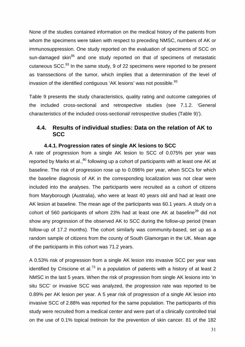
4.1. Study selection The systematic literature search generated 4741 hits (3090 after removal of
duplicates). 2967 of these were excluded during the screening of the titles and
abstracts. After assessment of the full texts of the remaining 123 publications, 31
were evaluated as eligible for inclusion into this systematic review. Three further
studies were identified through reviews of the reference lists of the assessed full text
articles. Two of these three additional studies were included into the analysis.
Altogether, 33 publications were included into the evaluation. Figure 1 illustrates the
process of study selection.
4741 records identifiedthrough database searching(Medline, Embase, Cochrane Libr.)
3090 records afterduplicates removed
3090 recordsscreened
2967 recordsexcluded
123 full text articlesassessed for eligibility
92 full text articles excluded
33 studies includedinto the qualitative
synthesisIncl
uded
Elig
ibili
tyS
cree
ning
Iden
tific
atio
n
3 recordsidentified
through review of reference lists
2
31
Figure 1. Process of study identification and selection

27
4.2. Characteristics of the included studies The total of 33 included studies37-39,50,71-99 contained fifteen RCTs with data that were
obtained from the non-interventional (or placebo/vehicle) group,71,72,75-79,81-84,86,88,89,99
two RCTs50,74 and one non-randomized controlled trial85 comparing the effects of
sunscreen versus no sunscreen use, nine prospective cohort studies37-39,73,80,87,91,94,97
and six cross-sectional/retrospective studies.90,92,93,95,96,98
As several studies reported on more than one outcome, the total number of studies
reporting on each of the relevant outcomes was higher than the number of included
studies. The following list summarizes the number of publications identified for each
outcome:
• 4 of the identified publications reported data on rates of progression of a single
AK lesion to SCC39,73,80,86
• 7 studies contained data on remission and recurrence rates of single AK
lesions37-39,50,82,87,99
• 16 studies reported on the change of total AK lesion counts38,50,71,72,74-77,79,81,83-
85,87-89
• 5 studies on complete field regression and/or subsequent recurrence
rates72,78,79,83,84
• 3 studies on relative risk / odd ratios for the development of SCC91,94,97
• 6 studies on contiguous AK lesions in SCC specimen90,92,93,95,96,98
Figure 2 further illustrates numbers of study types, reported outcomes and population
characteristics.

28
33 studies included into the qualitative
analysis
10 studies reporting on the course of single AK
lesions
17 studies reporting on the development of AK in
an area of interest
4 on progression rates from AK to
SCC
7 on remission and recurrence rates of
single lesions
16 on change of total AK counts over
time
5 on field regression rates and
subsequent recurrence rate
3
13
5
7
4
2 GPP1 prior NMSC1 OTR
7 GPP
10 GPP4 prior NMSC2 OTR
4 GPP1 OTR
6 cross-sectional or retrospective studies on rates of contiguous AK
lesions in SCC specimen
3 on the relative risk or odds ratio for development of
SCC
1 GPP2 OTR
3
6 on rates of contiguous AK lesions in SCC
specimen
5 prior SCC1 metastatic SCC
6
27 prospective longitudinal studies
reporting on the development of AK
Figure 2. Types of studies, numbers of studies reporting on each outcome, and specification of population
GPP = general patient
population (population of
participants without explicit
history of prior NMSC or
history of NMSC in less than
50% of participants);
prior NMSC = study
population includes at least
50% of participants with
preceding or preexisting
NMSC;
OTR = study population of
immunosuppressed organ
transplant recipients;
prior SCC = all participants
had an SCC excised;
metastatic SCC = SCC
specimens from patients
with metastatic SCC.

29
4.3. Quality and risk of bias assessment
4.3.1. Quality assessment and risks of bias within the prospective longitudinal studies
The methodological quality of the included prospective trials was generally low. The
following list summarizes the most important methodological limitations:
• Study population with risk of selection bias and/or missing information on the
selection/recruitment of participants71-73,75-79,81-84,86-89,91,97,99 (19/27)
• Missing information on intermittent treatment of lesions during the observation
period of the study71,72,76,77,79,81-84,86-89,91,94,97 (16/27)
• Missing information on sunscreen use or sun avoidance recommendations
given to the participants and/or on the participants’ respective
behavior38,39,71,78-80,82-84,86-89,91,94,99 (16/27)
• Follow-up period of less than twelve months50,56,71,75,76,79,81,83,87-89,91 (12/27)
• Dropout rate of more than 20% of the participants or not
reported39,50,72,74,77,78,80,84,87,89 (10/27)
• The mode of the outcome assessment was not reported in one study87 and
outcome assessment was based on a questionnaire survey in one study94
(2/27)
• The mode of the ascertainment of the baseline diagnosis was not reported in
one study87 (1/27)
Treatment of lesions became necessary during the observation period of some
studies, because clinical assessment could not always clearly differentiate between
AK and invasive SCC, and participants with treatment during the course of the study
often were excluded from the analysis or participation.37-39,50,73,74,80,85 Excluding these
potentially more severely affected participants from participation or evaluation
introduces a risk of selection and/or reporting bias. The treatment itself may have an
impact on the surrounding skin and therefore influence the development of other
lesions in the area.
Sixteen of the 27 prospective studies did not report whether a recommendation with
respect to sunscreen use or sun avoidance during the study period was given to the
participants38,39,71,78-80,82-84,86-89,91,94,99 (thirteen of these reporting on a population of
participants without history of prior NMSC or immunosuppression, three on organ
transplant recipients). Eight studies explicitly reported to have encouraged the use of

30
sunscreen or the avoidance of sun exposure37,72,73,75-77,81,97 (three reporting on a
population of participants without a history of prior NMSC or immunosuppression,
four on participants with a history of NMSC and one on organ transplant recipients).
Three trials compared the effects of regular use of sunscreen with no sunscreen
use50,74,85 (one study reporting on a population of participants without history of prior
NMSC or immunosuppression, one study on participants with a history of NMSC and
one study on organ transplant recipients).
Prospective cohort studies following up specific patient populations (e.g. organ
transplant recipients) were included into the review, and some of the cohorts included
participants without AKs at baseline. When assessing the mean numbers of baseline
AKs and the mean changes of total AK counts, the participants not affected by AK
result in a reduction in the mean counts of AKs and in a lowering of the average
change in total AK counts. This effect could have played a role in three studies.74,77,85
Table 8 presents the study characteristics, quality rating and outcome categories of
the included prospective studies (see 7.1.1. ‘General characteristics of the included
prospective longitudinal studies (Table 8)’).
4.3.2. Quality assessment and risk of bias within the cross-sectional / retrospective studies
The methodological quality of the included cross-sectional or retrospective studies
reporting on rates of contiguous AK lesions in SCC specimens was low as well. The
following list summarizes the most important methodological deficits:
• No information on rates of incomplete excisions in the evaluated specimens
presented92,93,95,96,98 (5/6)
• No definition of the meaning of ‘contiguous’ provided 90,92,93,95,98 (5/6)
• No information with respect to blinding of the assessor to the chart
diagnosis90,93,95,96,98 (5/6)
• Selection bias or missing information on the mode of recruitment / selection of
the analyzed specimens93,95,96,98 (4/6)
• No definition of the histological differences of AK and SCC provided90,92,93
(3/6)
• Assessment based on chart review and not on re-evaluation of SCC
specimens90,98 (2/6)
31
None of the studies contained information on the medical history of the patients from
whom the specimens were taken with respect to preceding NMSC, numbers of AK or
immunosuppression. One study reported on the evaluation of specimens of SCC on
sun-damaged skin95 and one study reported on that of specimens of metastatic
cutaneous SCC.93 In the same study, 9 of 22 specimens were reported to be present
as transsections of the tumor, which implies that a determination of the level of
invasion of the identified contiguous ‘AK lesions’ was not possible.93
Table 9 presents the study characteristics, quality rating and outcome categories of
the included cross-sectional and retrospective studies (see 7.1.2. ‘General
characteristics of the included cross-sectional/ retrospective studies (Table 9)’).
4.4. Results of individual studies: Data on the relation of AK to SCC
4.4.1. Progression rates of single AK lesions to SCC A rate of progression from a single AK lesion to SCC of 0.075% per year was
reported by Marks et al.,80 following up a cohort of participants with at least one AK at
baseline. The risk of progression rose up to 0.096% per year, when SCCs for which
the baseline diagnosis of AK in the corresponding localization was not clear were
included into the analyses. The participants were recruited as a cohort of citizens
from Maryborough (Australia), who were at least 40 years old and had at least one
AK lesion at baseline. The mean age of the participants was 60.1 years. A study on a
cohort of 560 participants of whom 23% had at least one AK at baseline39 did not
show any progression of the observed AK to SCC during the follow-up period (mean
follow-up of 17.2 months). The cohort similarly was community-based, set up as a
random sample of citizens from the county of South Glamorgan in the UK. Mean age
of the participants in this cohort was 71.2 years.
A 0.53% risk of progression from a single AK lesion into invasive SCC per year was
identified by Criscione et al.73 in a population of patients with a history of at least 2
NMSC in the last 5 years. When the risk of progression from single AK lesions into ‘in
situ SCC’ or invasive SCC was analyzed, the progression rate was reported to be
0.89% per AK lesion per year. A 5 year risk of progression of a single AK lesion into
invasive SCC of 2.88% was reported for the same population. The participants of this
study were recruited from a medical center and were part of a clinically controlled trial
on the use of 0.1% topical tretinoin for the prevention of skin cancer. 81 of the 182

32
participants received the verum during the observational period. Mean age was 68
years.
No progression from AK to SCC was reported in a trial that included 28
immunosuppressed organ transplant recipients.86 The study was a split-patient trial
with a follow-up of 12 months, including participants with a mean age of 57 years.
The data are summarized in table 2.
Table 2. Risk of progression of single AK lesions to SCC
Publication
No. pts. / mean no. of lesions p.p. / absolute no. of lesions in
study
Follow- up time
(months)
Advice to use sun-
screen
Risk of progression to
SCC per AK lesion per year
Further results Comments
Participants without history of prior NMSC or immunosuppression
Harvey, 199639
560 / mean 1.9 AK
per affected pt. /
239 lesions
12-24 (mean:
1.43 years)
- 0% -
Prevalence of AK at baseline:
23%, unclear data on dropouts
(>30%)
Marks, 198880
1689 / no data / no data
12 -
0.075% (including
doubtful cases: 0.096%)
Percentage of SCC that developed at a site with preexistent AK (new SCC with
knowledge about the previous skin
condition): 58.8%
see comment in
overview table 8
Participants with history of NMSC
Criscione, 200973
182 / 12 lesions p.p. in affected pts.
/ 1960 lesion
4-71 (mean 42) +
risk of progression of baseline AK to invasive SCC:
0.53%; risk of
progression of BL-AK to ‘in situ’
and invasive SCC: 0.89%
Percentage of SCC that developed at a site with preexistent
AK: 65%; 5-year risk of
progression of BL-AK to invasive SCC:
2.88%
Pts. with history of NMSC;
prevalence of AK at baseline:
95%; see
comment in overview table 8
Participants with immunosuppression
Wulf, 200686
28 / mean 2.9 lesions /
80 lesions
12 - 0% - OTR
Abbreviations: AK = actinic keratosis, BL = baseline, NMSC = non-melanoma skin cancer, no. = number, OTR = organ transplant recipients, p.p. = per person, pts. = participants, SCC = squamous cell carcinoma

33
4.4.2. Risk or odds ratios for developing SCC relative to the number of pre-existing AK
On a community-based cohort of participants of whom 40% had AK at the baseline,
Green et al.94 showed age- and sex-adjusted relative risks for the development of
SCC of 1.7 for participants with 1-5 facial AK at baseline, of 4.2 for participants with
6-20 facial AK at baseline, and of 11.0 for participants with more than 20 facial AK at
baseline (reference were participants without facial AKs at baseline). The age ranged
from 20 to 69 years in the respective population, no mean age was specified.
For populations of organ transplant recipients, a relative risk of developing a
subsequent SCC of 6.991 and an odds ratio of 56.497 was reported for patients
presenting with AK at baseline (reference were patients without AK at baseline).
Ramsay et al.97 further differentiate their results, reporting an odds ratio of 17.9 for
patients presenting with 1-10 preexisting AK and of 89.4 for patients presenting with
more than 10 preexisting AK (reference were patients without preexisting AK). The
data from both studies were derived from hospital-based populations. Mean age in
one study was 49 years,91 in the other study it was not specified.97
No data were available for populations of participants with a history of NMSC.
Table 3 summarizes the data obtained on the risk or odds ratios for the development
of SCC relative to the number of preexisting AK.

34
Table 3. Relative risks and odds ratios for developing SCC relative to the number of pre-existing AK
Publication
No of pts. / Prevalence
of AK at baseline
Follow- up time
(months)
Advice to use sun-screen /
avoid sun exposure
Relative risk (RR) / odds ratio (OR) [Reference:
no pre-existing AK]
Further results Comments
Participants without history of NMSC or immunosuppression
Green, 199094
2095 pts./ prevalence of
AK at baseline:
40%
24 -
- 1-5 facial AK: RR = 1.7 - 6-20 facial AK: RR = 4.2 - >20 facial AK: RR = 11.0
- Follow-up was
based on a postal survey
Participants with immunosuppression
Caforio, 200091
300 pts./ prevalence of
AK at baseline:
25.7%
Mean 55.2 - preexisting AK: RR =
6.9 (p=.001)
Hazard ratio for developing SCC (Reference: no
preexisting AK) with preexisting AK =
6.87 (p=.001)
OTR (heart transplant);
RR only presented in
the abstract; in the full text, the hazard ratio is
referred to
Ramsay, 200097
182 pts./ prevalence of
AK at baseline:
15.4%
Mean 15.36
+
- preexisting AK: OR = 56.4 (p<.001) - 1-10 preexisting AK: OR = 17.9 (p<.001) - > 10 preexisting AK: OR = 89.4 (p<.001)
Prevalence of AK was descriptively correlated to the
time since transplantation:
- < 5y: 4.9% - 5-10y: 12.7% - > 10y: 25.9%
OTR (renal transplant)
Abbreviations: AK = actinic keratosis, BL = baseline, no. = number, OR = odds ratio, OTR = organ transplant recipients, pts. = participants, RR = relative risk, SCC = squamous cell carcinoma
4.4.3. Rates of contiguous AK lesions in SCC specimens Rates of SCC specimens with contiguous AK lesions ranged from 15.9% to
97.2%.90,92,93,95,96,98 In a study on metastatic cutaneous SCC, 44% of the specimens
had contiguous AK lesions and further 17% of the analyzed specimens had overlying
or adjacent AK lesions.93 The mean age of the patients at the time of excision ranged
from 75 years95 to 83.3 years90 in the included studies. No data on the mean age
were available in two studies.93,96
Table 4 summarizes the data obtained on the rates of SCC specimens with
contiguous AK lesions.

35
Table 4. Rates of SCC specimens with contiguous AK lesions
Publication No of pts. / no. of SCC specimens
Rate of SCC specimens with contiguous AK
lesions Comment
Ang, 200490 57pts. / 63 SCC
specimens 15.9%
Evaluation of all excised SCC during 1991 to 1995, SCC treated with other
modalities were excluded
Czarnecki, 200292
208 SCC specimens
72% -
Dinehart, 199793
22 SCC specimens
44% contiguous; 17% overlying or adjacent, but not
contiguous
Metastatic cutaneous SCC; SCC from the vermillion border of the
lips were excluded
Guenthner, 199995
859 pts. / 1011 SCC specimens
97.2% Authors report contiguous “in situ SCC”
in SCC specimens
Mittelbronn, 199896
145 pts. / 165 SCC specimens
82.4% of SCC with concomitant AK
-
Takemiya, 199098 53 SCC specimens 58.5% -
Abbreviations: AK = actinic keratosis, no. = number, pts. = participants, SCC = squamous cell carcinoma
4.5. Results of individual studies: Data on the natural history of AK
4.5.1. Regression and recurrence rates of single AK lesions In 5 studies on populations of participants without history of NMSC or conditions of
immunosuppression, the reported regression rates of single AK lesions ranged from
18 to 62.9% after a follow-up of 7, 11, 12 or 17 months.37-39,50,87 Frost et al. (2000)37
reported an at least transient regression of 74% of AK lesions present at baseline,
but 15% of the regressed lesions subsequently recurred. A calculation showed that
62.9% of AK lesions regressed without subsequent recurrence during the 12 months
follow-up. This study excluded participants with field cancerization and recommended
the participants to continue their usual sunscreen use. The age of the included
participants ranged from 30 to 69 years. Age ranges of the participants from the other
studies reporting on this outcome were 39-79,87 ≥60,39 40-99,38 and 40-9350 years.
Harvey et al. (1996)39 reported that 21% of the AK lesions present at baseline
regressed during the follow-up period (mean follow-up of 1.4 years). The authors of
the study extrapolate a lesion-year adjusted regression rate of 15% of AK lesions. In
an RCT on the effects of sunscreen use, a statistically significant difference of the
regression rates during 7 months of follow-up between the sunscreen group and the
placebo group of 25% and 18%, respectively, was shown.50 No data on regression

36
rates in populations of participants with a history of NMSC or of organ transplant
recipients were found.
With respect to rates of subsequent recurrence of AK lesions after an initial
regression, again only data on populations of participants without history of NMSC or
immunosuppression were identified. As described above, a recurrence rate of 15%
after an initial regression rate of 74% of the individual AK lesions present at baseline
was reported in one trial37 on a community-based population. In two publications
reporting on three trials following up AK lesions for 12 months after their spontaneous
regression, 15%, 53%,99 and 20.2%82 of the lesion recurred. Data on recurrences
from these trials were derived from hospital-based populations with sample sizes of
12, 22,99 and 9882 participants, respectively. In a prospective cohort study with very
limited information on methods and participants’ characteristics, 32.6% regressed
and recurred, and 10.6% of the lesions present at baseline regressed twice within a
follow-up period of 11 months.87
The available data on regression and subsequent recurrence rates of single AK
lesions are summarized in Table 5.

37
Table 5. Regression and recurrence rates of single AK lesions
Publication
No of pts. / mean no. of lesions p.p. / total no. of lesions in study (BL)
Advice to use sun-
screen
Regression of AK lesions present at
BL after 12 months
Recurrences of single lesions
Comments
Participants without history of NMSC or immunosuppression
Cantrell, 200987
27 pts. / mean 23.5 lesions /
635 lesions
- 29.7%*
Percentage of lesions present at baseline with regression and recurrence: 32.6%;
Percentage of lesions present at BL that
regressed twice: 10.6%
Pts. with extensive sundamage;
n. i. on methods and confounders; only abstract
available
Frost, 200037
96pts. / 11.49 lesions
p.p. in affected pts. /
494 lesions
(-)
74.0% (percentage of
lesions present at BL that regressed
without recurrence: 62.9%)
Percentage of regressed AK that recurred during the study period: 15%
Prevalence of AK at BL: 46%;
pts. with field cancerization or
more than 50 AK at one site were
excluded; see comment in
table 8
Harvey, 199639
560 / mean 1.9 lesions in
affected pts. / 239 lesions
-
21%***; calculated rate of
regression: 15% of AK lesions / year
-
Prevalence of AK at BL: 23%,
unclear data on dropouts (>30%)
Marks, 198638
618 pts. / mean 7.7 lesions /
4759 lesions
- 25.9% - Pts. with treatment
were excluded
Stockfleth, 201282
98 pts. / no data / no data
- -
Percentage of regressed lesions that
recurred within 12 months after
regression: 20.2%
-
Szeimies, 201099
34 pts (12 and 22) /
5.5 and 5.9 lesions /
476 lesions
- -
Percentage of regressed lesions that
recurred within 12 months after
regression: 15% and 53%
Pts. with recurrence of all lesions were
withdrawn, see comment in overview table 8
Thompson, 199350
588 pts. / mean 8.12 lesions /
3498 lesions
+/-
Sunscreen group: 25%**;
Placebo group: 18%**
-
> 20% dropouts; any lesion treated
during the course of the study was excluded from
analysis Follow-up periods: * = 11 months; ** = 7 months; *** = 12-24 months (mean = 1.43 years); Abbreviations: AK = actinic keratosis, BL = baseline, n. i. = no information, NMSC = non-melanoma skin cancer, no. = number, p. p. = per person, pts. = participants

38
4.5.2. Rates of complete field regression und subsequent recurrences Complete field regression rates from AK that were reported in the identified studies on
populations of participants without history of NMSC or concomitant immunosuppression
varied from 0% to 7.2% of the included participants for observed areas on the face or
scalp.72,79,83 A complete field regression rate from AK for an observed area on the upper
extremities of 21% was reported on a small study group of 14 participants.72 Partial field
regressions (regression of at least 75% of the lesions present at baseline) were seen in
4.3 to 13.6% of the observed participants without history of prior NMSC during the 6
months of follow-up.79,83
In a small population of organ transplant recipients (14 participants), a complete (100%)
and partial (75%) field regression rate of 0% within a time period of 6 months was
reported.84 No data on field regression rates were available for participants with a
history of NMSC.
Only one study reported data on the percentage of participants that had experienced a
recurrence after a spontaneous complete field regression: 57% of participants from a
population of 18 patients who had experienced a spontaneous complete regression of
their observed field had a recurrence within a follow-up period of 12 to 14 months.78
The mean age of the included participants was 61.6 years,84 65.9 years,79 70.9 years,83
or not reported.72,78 All data on field regression and recurrence were derived from
inpatient or outpatient populations.
The available data on rates of spontaneous complete field regression rates and rates of
subsequent recurrence are shown in Table 6.

39
Table 6. Rates of complete field regression and subsequent recurrence
Publication
No of pts. / mean no. of lesions p.p. / total
no. of lesions in study (BL)
Follow-up time
(months)
Advice to use sunscreen / avoid sun exposure
Rate of pts. with a complete field
regression (100% of AK lesions in a defined
field)
Rate of pts. with subsequent recurrence(s) after
spontaneous complete field regression
Comment
Participants without history of prior NMSC or immunosuppression
Alirezai, 199472
49 pts. / mean 7.8 (face);
9.7 (scalp); 8.4 (upper extr.) lesions
/ no data
12 + Face: 2%; Scalp: 0%;
Upper extr.: 21% -
> 20% dropouts; pts. who had skin cancer or suspect lesions at baseline
were not included; see comment in table 8
Jorizzo, 200778
18 pts. / median 6 lesions /
no data 12-14 - - 57%
> 20% dropouts; see comment in table 8
Korman, 200579
250 pts. / no data / no data
6 - 7.2%;
75%-regression: 13.6% - see comment in table 8
Szeimies, 200483
139 pts. / mean 5.5 lesions / 766 total lesions
6 - 2.2%;
75%-regression: 4.3% - see comment in table 8
Participants with immunosuppression
Ulrich, 200784
14 pts. / no data / no data
6 - 0%;
75%-regression: 0% -
OTR (kidney-, liver- or heart-Tx more than 3y prior to
entering the study); > 20% dropouts;
see comment in table 8 Abbreviations: AK = actinic keratosis, BL = baseline, NMSC = non-melanoma skin cancer, no. = number, OTR = organ transplant recipients, p. p. = per person, pts. = participants, Tx = transplant, y = years

40
4.5.3. Change in total AK counts over time Numerous publications reported data on the relative changes of the mean AK counts in
the participants over time, showing a broad range from -53% to +99.1%.38,50,71,72,74-
77,79,81,83-85,87-89
The range was narrower for studies on participants without history of NMSC or
immunosuppression, from -37 to +57%.38,50,71,72,77,79,83,87-89 For populations including
participants with history of NMSC, relative changes of total AK counts over time from -
23% to +57% were observed.74-76,81 The broadest range was found in studies on
populations of organ transplant recipients, with relative changes of total AK counts over
time ranging from -53% to +99.1%.84,85
The ‘sunscreen use and/or sun avoidance recommendations’ may serve as an
explanation for the broad ranges of reported changes: In studies that explicitly reported
having recommended participants to protect themselves from sun radiation, relative
changes of AK counts with a trend towards a reduction in the numbers of AK lesions
(range: -53% to +20%) were seen, with 7 of 8 studies reporting decreases in the total
AK counts.50,72,75-77,81,85 In studies without information on sun protection
recommendations given to the participants or in control groups of sunscreen trials, a
trend towards increasing AK counts (range: -14.3% to 99.1%) can be seen, with 8 of 11
studies showing increasing AK numbers.38,50,74,84,85,87-89 These trends hold true for
studies on populations of participants without history of prior NMSC or
immunosuppression as well as for studies on populations of participants with history of
prior NMSC and populations of organ transplant recipients.
The mean age of the participants who were included into the studies was 48 years74 to
75 years.89 Data on the mean age were not reported in three studies.71,72,87 Age was not
a consistent modifier of the relative changes of AK counts: On participants with a mean
age of 48 years and 50 years, one study reports an increase of 57%74 and another
study a decrease of 33% of the mean AK lesion counts per participant.77 In studies on
older populations, reported changes also include a broad range without any obvious
correlation to the age of the participants.
The available data on relative changes of total AK counts over time are summarized in
Table 7.

41
Table 7. Changes in the total AK lesion count over time
Publication
No of pts. / mean no. of lesions p.p. / total
no. of lesions in study (BL)
Follow-up time
(months)
Advice to use sun-screen /
avoid sun exposure
relative Difference in total AK lesion counts
from baseline to the end of study
Further results Comment
Participants without history of prior NMSC or immunosuppression
Alberts, 200071
48 pts. / mean 29.2 lesions /
1402 6 - - 2.4% - -
Alirezai, 199472
49 pts. / mean 7.8 (face);
9.7 (scalp); 8.4 (upper extr.)
lesions / no data
12 + Face: - 21.8% Scalp: - 37.1%
Upper Extr.: - 11.9% -
> 20% dropouts; pts. who had skin cancer or
suspect lesions at BL were not included
Cantrell, 200987
27 pts. / mean 23.5 lesions /
635 lesions 11 - + 57% -
Pts. with extensive sundamage; n. i. on methods and confounders;
only abstract available
Green, 199877
35 pts. / mean 3.6 lesions /
126 lesions 12 + - 33.3% -
> 20% dropouts; all pts. with ‘photoaging’
Kang, 200388 30 pts. / no data / no data
9 - Mean increase in total no.
of AK: + 1.5 -
No data for calculation of the relative difference of AK lesion counts from baseline to end of
study presented
Korman, 200579
250 pts. / no data / no data
6 - Median count reduction:
-14.3%
Rate of pts. with increase in AK count at any point in
the treatment period: 22.0%
see comment in table 8
Marks, 198638
618 pts. / mean 7.7 lesions /
4759 lesions 12 - + 21.8%
36.4% of pts. had a remission of at least 1 AK
Pts. with treatment were excluded
Szeimies, 200483
139 pts. / mean 5.5 lesions / 766 total lesions
6 - - 8.7% - -
Thompson, 199350
588 pts. / mean 8.12 lesions /
7 +/- Sunscreen group:
- 6.7%; -
> 20% dropouts; any lesion treated during the

42
3498 lesions Placebo group: +12.5% course of the study was excluded from analysis
Zeichner, 200989
20 pts. / median 9.5 lesions /
no data 7 -
Average change in total lesions number score*:
+0.07 -
> 20% dropouts; *Total lesions number score:
score 0 for 0 AKs; score 1 for 1-3 AKs; score 2 for 4-6 AKs; score 3
for > 6 AKs Participants with history of NMSC
Darlington, 200374
1621pts. / mean 3.7 lesions /
4129 lesions 30 -/+
Sunscreen group: + 20%*; Control group: + 57%*
Prevalence of AK in the study population: 1992: 46%; 1994: 50%; 1996:
53%
Prevalence of AK at baseline: 46%;
prevalence of a history of skin cancer: 27% (436 pts.);
> 20% dropouts; *adjusted rates
Elmets, 201075
118 pts. / mean 24 lesions /
2832 9 + -18.5% -
Prevalence of a history of skin cancer at baseline: 59.3%
Foote, 200976
50 pts. / mean 34.3 lesions /
1715 6 + - 23.3% -
Prevalence of a history of skin cancer: 40.5% (17 pts.)
Moloney, 201081
17 pts. / mean 23.5 lesions /
400 lesions 6 + - 22.4% -
‘sundamaged’ pts. with multiple prior skin cancers
Participants with immunosuppression
Ulrich, 200784
14 pts. / no data / no data
6 - + 99.1% -
OTR (kidney-, liver- or heart-Tx more than 3y prior to entering the
study); > 20% dropouts
Ulrich, 200985
120 pts. / mean 3.18 lesions /
382 total lesions 24 -/+
Sunscreen group: - 53%; Control group: + 43 %
Incidence of SCC in sunscreen group vs.
controls during study: 0 vs. 8 (p<.01)
OTR (heart-, liver-, and kidney-Tx, each
40 pts.)
Abbreviations: AK = actinic keratosis, BL = baseline, Extr. = extremities, NMSC = non-melanoma skin cancer, no. = number, OTR = organ transplant recipient, p. p. = per person, pts. = participants, SCC = squamous cell carcinoma, Tx = transplant

43
4.6. Extrapolation of the risk of development of an SCC for the ‘standard’ patient
Based on the calculation presented by Dodson et al.70 and on the results of the
systematic review, the risk of a hypothetical patient to develop at least one SCC over
the time of 10 years (m = 10) can be extrapolated.
4.6.1. Patients presenting with AK without history of NMSC or immunosuppression
One study presented data on the risk of progression of single AK lesions to SCC in
participants without a history of NMSC or immunosuppression.80 The risk of progression
was reported to be 0.075% per year (ROP = 0.00075). As data on the mean number of
AK lesions per participant were not presented, the respective number from an earlier
publication of the authors on an overlapping population,38 with a mean of 7.7 AK lesions
per participant was used (n = 8).
( )[ ] ( )[ ] 0583.000075.011__11__ 108 =−−=⇒−−= ×SCCofRiskROPSCCofRisk nm
For a patient without immunosuppression and without a history of NMSC who presents
with 8 AK lesions, the equation extrapolates a 10 year risk of development of SCC of
5.8%.
4.6.2. Patients presenting with AK with a history of NMSC One study presented data on the risk of progression of a single AK lesion to SCC in
patients with a history of multiple NMSC.73 For patients with at least 2 NMSC in the past
5 years, the risk of progression of a single AK lesion into invasive SCC was
demonstrated to be 0.53% in one year (ROP = 0.0053). The mean number of AK
lesions in the participants of the study was 12 AK lesions (n = 12).
( )[ ] ( )[ ] 4715.00053.011__11__ 1012 =−−=⇒−−= ×SCCofRiskROPSCCofRisk nm
For a patient presenting with 12 AK lesions who had a history of at least two NMSC in
the last 5 years, a 10 year risk for the development of at least one invasive SCC of
47.2% was extrapolated.

44
4.6.3. Organ transplant recipients presenting with AK For the risk of progression of single AK lesions to SCC in organ transplant recipients, no
applicable data were identified in the available literature. There was only one study on
28 organ transplant recipients that showed no progression within one year.86 Therefore
it is not possible to extrapolate the overall patient risk for patients with the condition of
immunosuppression.

45
5. Discussion
5.1. Brief summary of the main findings Methodological limitations of the studies identified through the systematic search for
primary data on the natural history of actinic keratoses strongly restrict the
interpretability, comparability and applicability of the results. Data on progression of
single AKs indicate a risk of progression to SCC of up to 0.075% per lesion per year,
when looking at participants without a history of NMSC or immunosuppression. The risk
was seen to be higher in participants with a history of multiple NMSC, with a
progression rate into invasive SCC of 0.53% per AK lesion and year. Because of the
methodological limitations, the data obtained on progression rates are not suitable for a
reliable estimate of the actual risk of progression. When extrapolating the data for the
individual risk of a ‘standard patient’ with multiple AKs to develop at least one SCC
within ten years, the reported lesion-based risks rise dramatically. Data on the relative
risks indicate a higher risk of SCC development for organ transplant recipients as
compared to participants without history of immunosuppression. In SCC specimens,
histological correlates of AK are regularly found to be contiguous to the SCC lesion.
Regression of AK occurs in around 20-30% of AK lesions and recurrences in 15-20% of
the regressed lesions. Despite of these relatively high regression rates, a complete field
regression is unlikely to happen.
5.2. Overall limitations of the systematic review AK is discussed as being a premalignant or malignant in situ lesion of the skin. However
this debate is resolved, performing trials on potentially malignant lesions or in situ
carcinoma raises difficult questions with respect to methodology and ethics.
5.2.1. Inclusion of control arms from clinical trials Following up cohorts of participants with skin lesions that potentially progress into
invasive skin cancer raises severe ethical questions. A small number of observational
studies on participants with AK lesions has been performed, probably for this reason.
Therefore, control arms from clinical trials were included into this systematic review and
considered as observational cohorts. The placebo effect or the possible effects of the
vehicles used or placebo interventions has to be taken into account when interpreting
the results on the natural history of AK. Especially the use of vehicle creams or gels

46
might have had an impact on the development of AK lesions, due to emollient or
moisturizing effects, especially on hyperkeratotic AK lesions.
5.2.2. Definition of patient subgroups Populations of the included studies were differentiated, due to the different risks for the
development of further AK lesions and the progression to SCC, taking clinically relevant
situations into account: Populations of participants without history of NMSC or
immunosuppression were reported separately from populations of participants with a
history of NMSC and populations of participants with concomitant immunosuppression
due to their status as organ transplant recipients. The group of the ‘organ transplant
recipients’ was well-defined, and studies reporting on this population had clear and
explicitly reported inclusion criteria. The differentiation between the group of the
‘participants without history of prior NMSC or immunosuppression’ and the ‘participants
with a history of NMSC’ was more difficult, since population-based cohort studies were
included. Studies including a ‘population of participants without history of NMSC or
immunosuppression’ usually did not report whether or how they assured that all
included participants were free of preceding or preexisting NMSC. Studies categorized
as reporting on a population of ‘participants with a history of NMSC’ included studies
with a history of multiple NMSC as an inclusion criterion for participation as well as
studies on mixed populations.74-76 Therefore, the differentiation between the ‘population
of participants without history of prior NMSC or immunosuppression’ and the ‘population
of participants with a history of NMSC’ is not very selective and missing information
could have led to an over- or underestimation of the reported outcomes.
The age is another important variable that has to be taken into account when reporting
on the natural history of AK. Unfortunately, the age specifications for the different study
populations were very overlapping and most of the studies did not provide age-stratified
data with respect to the reported endpoints. Partly, exact data on the mean age and
range of the study populations were missing. In this review, age as a main influential
factor is only discussed for outcomes where this was possible.
Since studies on ‘community-based’ populations were considered in this review,37-
39,50,74,80 the terminology of ‘patients’ does not necessarily reflect the actual situation of
some of the included individuals. Individuals presenting with the medical condition of
AKs were termed as ‘patients’ when the population was derived from an inpatient- or

47
outpatient-based collective and as ‘participants’ when studies reported on ‘community-
based’ populations. As the respective studies were included into this review with
reference to the reported outcomes in participants with prevalent AKs, ‘community-
based’ studies were not separately reported from ‘inpatient-based’ studies.
Nevertheless, the source of participants may have an effect on the reported results.
5.2.3. Assessment and differentiation of AK and SCC The diagnosis of AK lesions in the clinical trials is based on the clinical examination of
the participants in most of the included trials. The validity of this assessment was shown
in some studies,49-51 but only the histological assessment of AK lesions offers a secure
differentiation of AK and SCC. At the same time, a histological assessment would
require a removal of the lesion and impede the follow-up. A description of the natural
course of AK lesions would then be impossible.
Due to the ongoing discussions on the dignity of AK and the variety of clinical and
histological definitions, the differentiation of AK and SCC is not standardized. Since the
authors of the studies often fail to provide definitions of the terms they use with respect
to ‘AK’, ‘in situ SCC’ and ‘SCC’, some results of the single studies are not comparable
to each other, in particular concerning results on the rates of malignant progression of
AK lesions and the rate of contiguous AK in SCC specimens.
5.2.4. Validity of AK lesion counts For a substantial part of individuals affected by AK, a field cancerization with contiguous
areas of chronic actinic skin damage, latent and manifest AKs and potentially evolving
or manifest SCC can be expected. The validity of the clinical endpoint of total AK lesion
counts as used in most research on the development of AK lesions is questionable. As
described above (see 2.6.2. ‘Clinical diagnosis’), positive predictive values of clinical
diagnosis of AK were shown to be sufficient,49-51 but the interobserver reliability of the
identification of single AK lesions in patients was low, even among dermatologists.52,54
Inter-rater variability with respect to total AK lesion counts in patients is correspondingly
high, especially in the case of untrained assessors,53 and therefore the use of total AK
lesion counts as a clinical endpoint in the evaluation of AK was recommended no longer
to be used.100 Nonetheless, the total AK lesions count remains the most frequently used
clinical endpoint in research for assessing AK. Some means of assessment has to be
applied, and for this systematic review, the available data reported in the literature were

48
assessed. The development of a valid alternative to AK lesion counts for use in further
research is desirable.
5.2.5. Treatment of lesions during studies As the assessment of AK usually was based on clinical diagnosis, treatment of suspect
lesions during the observational period could become necessary in some studies.
Lacking information on the amount of treatment, details of the treatment modality,
statistical handling of treated lesions or influence of the treatment on the results
introduced a risk of bias to the majority of the included studies. This imposes a major
limitation when it comes to interpreting the results of this systematic review. The
significance of this limitation is discussed in detail with respect to the reported outcomes
in section 5.3. ‘Discussion of the results on a study level’.
5.2.6. Measures to avoid unprotected exposure to sun radiation As the continued exposition to sunlight is a major influential factor for the further
development of AK lesions, the recommendations made to the participants at the
beginning of the studies were assessed. Some authors reported that they had
recommended all participants to avoid unprotected exposure to sun radiation (e.g.
recommendation to avoid sun exposure in the afternoon or to use sunscreen on a daily
basis). However, the majority of studies neither reported whether they recommended
any behavioral aspects concerning exposure to sun radiation to their participants nor did
they provide any data on the actual sun exposure of the participants. The knowledge on
the role of sunlight as a risk factor for the development of AK and NMSC can be
assumed to be general, and therefore, the actual behavior of participants cannot be
predicted, unless the behavior is reported in the studies. The differentiation of ‘studies
with information on sunscreen use recommendations’ and those ‘without information on
sunscreen use recommendations’ is very basal, but it accounts for the low quality of the
vast majority of the included trials in this respect.
5.3. Discussion of the results on a study level
5.3.1. Risk of progression of AK lesions to SCC All of the identified studies reporting on progression rates have severe methodological
limitations.

49
Following the data presented by Marks et al. (1988),80 a risk of progression from AK to
SCC of 0.075% per AK lesion per year can be assumed for a (community-based)
population of participants without history of NMSC or immunosuppression. Due to the
design of this cohort study, no data on the number of dropouts are provided, which
introduces a strong reporting bias (on the outcome level). An important reason for
dropping out can be surgical intervention for an invasive SCC, and thus the risk of
progression of an AK lesion may have been underestimated. Furthermore, an unknown
number of participants was repeatedly reported: the authors counted each two contacts
in consecutive years as one ‘participant contact’ if the participant had at least one AK
lesion during the first presentation. When the same participant was seen on three
different occasions in two consecutive years, the respective participant was reported as
‘two occasions’. The multiple reporting of participants aggravates the possible bias
towards underreporting progression rates. On the other hand, the invitation of all
citizens from a community to participate in a skin cancer screening may have introduced
a bias towards the participation of more severely affected citizens.
Criscione et al. (2009)73 reported a progression rate of 0.53% per AK lesion per year in
a hospital-based population of patients with a history of multiple prior NMSC and a
mean age of 68 years. No histological or clinical definition of what was termed as ‘AK’,
‘in situ SCC’ and ‘invasive SCC’ is provided, which makes a comparison of the results to
other studies difficult. The rate of 0.53% per AK lesion per year refers to the progression
from an AK lesion present at baseline into invasive SCC at the 12 months follow-up
visit. 81 of the 160 reported participants received tretinoin within the framework of the
Veteran Affairs Topical Tretinoin Chemoprevention (VATTC) trial. The authors confirm
that the progression rates did not differ between the interventional and the control
groups, but do not provide subgroup analyses on that issue.
Two studies did not find a progression of the followed AK lesions at all. The number of
included participants was probably too small for the detection of this rare event in the
study by Wulf et al. (2006).86 In the other study that reported no progression, conducted
by Harvey et al. (1996),39 more than 30% of the participants dropped out. As
participants more severely affected by AK are more likely to search for surgical
treatment outside the framework of the study, this introduces a risk of bias towards an
underreporting of progression. Generally, from a methodological perspective, it is not

50
recommended to consider studies with ‘no events’ for the determination of event rates
for rare events: e. g., if the expected event rate is lower than 1%, it is recommendable to
exclude studies showing no events from metaanalyses.101
In summary, data on progression rates of single AK lesions remain highly uncertain due
to various methodological limitations of the studies reporting on this outcome. Studies
following the course of single AK lesions demonstrate the inherent potential of AK
lesions to progress to SCC, but at the current time, no reliable estimates concerning the
actual progression rates of untreated AK lesions can be given. The data obtained
indicate that the risk of malignant progression may be higher in patients with a history of
previous NMSC.
5.3.2. Risk or odds ratios for developing SCC relative to the number of pre-existing AK
Studies on different populations showed a strong correlation between the number of
baseline AK lesions and the risk of developing a subsequent SCC during the
observational period. As the individual studies refer to different locations of AK (e. g.
only facial AK), different effect measures (risk ratio or odds ratio), or different
independent variables (AK in general or certain number of AK compared to no AK), the
data obtained are not directly comparable. Generally, increasing numbers of baseline
AKs in the studies reviewed were consistent with increasing subsequent risks for the
development of an SCC. Relative risks in participants without history of NMSC or
immunosuppression were lower than relative risks for the development of SCC in organ
transplant recipients with AK.91,94,97 This finding goes along with the results of other
studies specifically reporting on this research question.28-31
The data on the relative risk for a population of participants without history of NMSC or
immunosuppression are based on follow-up data from a postal survey94 and are
therefore at a higher risk of recall bias.
Summarizing, the data on risk or odds ratios for developing SCC, relative to the number
of AK lesions at baseline, show a strong interdependency between AKs and subsequent
SCC development. The data are based on sample sizes of at least 180 participants.
Although the risk or odds ratios obtained are not comparable, the risk seems to be
higher in immunosuppressed patients than in a community-based sample from the
general population.

51
5.3.3. Rates of contiguous AK lesions in SCC specimens Rates of SCC specimens with contiguous AK lesions ranged from 15.9% to
97.2%.90,92,93,95,96,98 The main limitations to these data were missing data on the rates of
incomplete excision and lacking definitions of the meaning of ‘contiguous lesions’ in
most of the studies. The latter aspect impedes a meaningful comparison of the obtained
data on rates of contiguous AK lesions. None of the studies contained information on
the medical history of the patients; hence, a discrimination of studies on patients without
history of NMSC or immunosuppression, on patients with history of NMSC, or on
immunosuppressed patients was not possible.
Despite the low quality and comparability of the studies and their findings, histological
changes that are attributable to AK lesions were frequently found to be present in SCC
specimens. In studies including at least 150 analyzed specimens, rates of contiguous
AK lesions in SCC specimens ranged from 72%92 to 97.2%.95 This finding does not
prove a causal relationship between these two ‘entities’. Yet, it may provide support to
the hypothesis of a stepwise development on a spectrum from healthy skin to AK and
from AK to SCC.
5.3.4. Regression and recurrence rates of single AK lesions The studies included reporting data on regression rates of single AK lesions showed a
general rate of regression of 20 to 30% of the lesions from baseline until the end of the
study. One study showed a regression of 63% of AK lesions that were present at
baseline37. The latter study included the youngest participants (range from 30 to 69
years) and excluded participants with a field cancerization, which might explain the
outstanding rate of regressions in this study. No data on populations of participants with
a history of NMSC or on organ transplant recipients were available. Recurrence rates
after spontaneous regression of AK lesions of between 15% and 53% of the regressed
lesions37,82,99 and 33% of the lesions present at baseline87 were reported. Only the
single lesion recurrence rate of 20.2% within 12 months of follow-up refers to data from
more than 35 participants (the study in question82 included 98 participants).
Generally, the results of the studies concerning regression rates may have been biased
towards overreporting remission rates due to AK lesions in which the development of
invasive SCC was suspected and which therefore were excluded from analysis or
treated. In the study with the high regression rate of 63% of AK lesions, 6% of the

52
lesions were treated by a dermatologist or a GP outside the framework of the study.37 In
some studies, the drop-out rates were high and the data provided on treatment during
the course of the study insufficient.
Data on regressions of single AK lesions and consecutive recurrence of the respective
lesions yield the most consistent results within this systematic review. Regressions may
be expected in around 20-30% of AK lesions in immunocompetent patients without a
history of NMSC. Recurrences occur in around 20% of the regressed lesions within 12
months. The data underline the assumption that AK is a dynamic disease of regressions
and recurrences of lesions within a field of sun damage or field cancerization.
5.3.5. Rates of complete field regression and subsequent recurrence Rates of spontaneous complete field regressions ranged from 0% to 7.2%. The rate of
21% of complete field regressions reported by Alirezai et al.72 refers to AK lesions on
the upper extremity and is based on data from the observation of only 14 patients.
Data on spontaneous complete field regression rates are generally limited with respect
to the number of studies and to the sample sizes in some of these studies. If only data
from trials with more than 100 participants are considered, spontaneous regression of a
defined field occurred in 2.2% to 7.2% of the participants included. It should be kept in
mind that ‘complete field regression’ is an expression that was coined for the purpose of
the present study, in analogy to the expression ‘complete clearance’ that is frequently
used as an outcome in clinical trials. Clinical trials that assess ‘complete clearance’ refer
to a well-defined treatment or control area and not to all AK on the whole body of a
participant. The regression of all AK lesions on all body regions of affected individuals
should consequently be assumed to be correspondingly lower. Moreover, the low rates
of spontaneous complete field regression were counterbalanced by relatively high rates
of recurrence: The authors of one study showed that 57% of the participants who
experienced a spontaneous complete field regression developed at least one
recurrence within 12 to 14 months of follow-up.78 These data indicate that despite the
relatively high single lesion regression rates, the probability of a complete regression of
all AK lesions in a patient affected by multiple AK lesions is low and the presence of AK
lesions is a chronic condition.

53
5.3.6. Change in total AK counts over time The changes in the AK lesion counts were most frequently chosen as the primary
outcome in the studies included in this review. Very heterogeneous results were
obtained with respect to that outcome. The broad range from decreases to increases of
the total AK counts is partly explained by the sunscreen use recommendations made to
the participants of the studies. This finding is consistent with the results of various
controlled trials that show a beneficial influence of the use of sunscreen on the course
of AK lesions regardless of the population of study participants.50,74,85 For instance, one
study showed a statistically significant difference in the relative ratio of increase of the
total AK counts of 0.76 for the sunscreen group compared to sunscreen non-users.74
Another study demonstrated an even more pronounced difference between the
sunscreen group (mean decrease of 53% from baseline) and the control group (mean
increase of 43% of the baseline AK lesion counts) in a population of organ transplant
recipients.85
An effect of the participants’ age on the changes of the AK lesion counts was not seen
throughout the included studies.
Similarly, data on the change in the AK counts from baseline until the end of the study
show that AK is a dynamic condition with frequent (numeric) progressions and
regressions. Continued unprotected exposure to UV radiation seems to be a major
factor influencing the further development of AK lesions.
5.4. Discussion of the extrapolation results of the risk of development of an SCC for the ‘standard’ patient presenting with AK
The extrapolated risk of developing at least one SCC, based on the mean number of AK
and the risk of progression of a single AK lesion to SCC per year in the given studies
and on the set time of ten years, was 5.8% for a patient without immunosuppression
and without a history of NMSC who presents with 8 AK lesions. For a patient who had a
history of at least two NMSC in the last 5 years, presenting with 12 AK lesions, the
corresponding risk was calculated to be 47.2%. No data for a risk extrapolation for
organ transplant recipients were available. These data should not be used for
estimating the actual risk of patients due to the severe limitations of the validity of the
underlying data on the single AK lesions progression risk. Furthermore, the calculation
itself introduces uncertainty:

54
As stated by Dodson et al.,70 the extrapolation of risk is only valid under the hypotheses
of independent development of each lesion and stability of each lesion over time. These
are hypothetical presumptions that pose a major limitation to the interpretability of the
extrapolated data. On the other hand, the risk of progression presented in the source
studies is not differentiated over lesion age, lesion stability and interdependency of
progression. The hypothetical premises may therefore be assumed to be valid for the
specified risks of progression in the respective populations.
The calculated individual progression rate for immunocompetent patients without history
of NMSC of 5.8% in ten years lies well within the range of previous estimations of the
individual risk for the development of an SCC. Quaedvlieg et al.60 reported estimations
of rates of SCC development for patients affected by AK of between 1.6% and 14% in
five years.
However, the extrapolated data on the individual risk of progression are not directly
transferable to clinical situations. They are based on highly uncertain estimates of
progression risks of single AK lesions. Applying a theoretical statistical method further
decreases the validity of the results. The important and additional information that these
calculations may provide is to demonstrate the disproportional increase in the risk for
SCC development in an individual with several AK over a longer time period.
5.5. Conclusions The findings of this systematic review reflect that AKs are not a static skin condition. If
left untreated, AKs are a dynamic disease on the grounds of chronically sun damaged
skin. The lesions have the potential of progression and regression, with respect to
quantitative (e. g. changes in total AK lesion counts) as well as qualitative categories (e.
g. progression of an AK lesion to SCC or regression into clinically healthy skin).
Correlational data suggest a strong relation between the number of AK and the risk of
subsequent development of SCC. SCC specimens frequently contain contiguous AK
lesions, a fact that equally underlines the strong entanglement of these two ‘entities’.
When single AK lesions are followed over time, a progression to SCC can be observed
in a small number of cases. Extrapolations of the risk for an individual affected by AK
demonstrate that the apparently low rates of progression of single AK lesions are only a
fraction of the risk of development of SCC in a patient affected by several AK for several
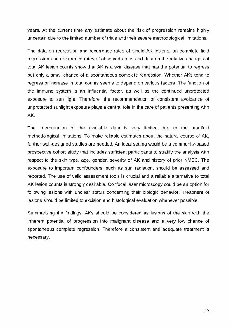
55
years. At the current time any estimate about the risk of progression remains highly
uncertain due to the limited number of trials and their severe methodological limitations.
The data on regression and recurrence rates of single AK lesions, on complete field
regression and recurrence rates of observed areas and data on the relative changes of
total AK lesion counts show that AK is a skin disease that has the potential to regress
but only a small chance of a spontaneous complete regression. Whether AKs tend to
regress or increase in total counts seems to depend on various factors. The function of
the immune system is an influential factor, as well as the continued unprotected
exposure to sun light. Therefore, the recommendation of consistent avoidance of
unprotected sunlight exposure plays a central role in the care of patients presenting with
AK.
The interpretation of the available data is very limited due to the manifold
methodological limitations. To make reliable estimates about the natural course of AK,
further well-designed studies are needed. An ideal setting would be a community-based
prospective cohort study that includes sufficient participants to stratify the analysis with
respect to the skin type, age, gender, severity of AK and history of prior NMSC. The
exposure to important confounders, such as sun radiation, should be assessed and
reported. The use of valid assessment tools is crucial and a reliable alternative to total
AK lesion counts is strongly desirable. Confocal laser microscopy could be an option for
following lesions with unclear status concerning their biologic behavior. Treatment of
lesions should be limited to excision and histological evaluation whenever possible.
Summarizing the findings, AKs should be considered as lesions of the skin with the
inherent potential of progression into malignant disease and a very low chance of
spontaneous complete regression. Therefore a consistent and adequate treatment is
necessary.

56
6. References
1 Heaphy MR, Jr., Ackerman AB. The nature of solar keratosis: a critical review in
historical perspective. J Am Acad Dermatol. 2000;43; 138-50.
2 Röwert-Huber J, Patel MJ, Forschner T, et al. Actinic keratosis is an early in situ
squamous cell carcinoma: a proposal for reclassification. Br J Dermatol.
2007;156; 8-12.
3 Ulrich M, Gonzalez S, Lange-Asschenfeldt B, et al. Non-invasive diagnosis and
monitoring of actinic cheilitis with reflectance confocal microscopy. J Eur Acad
Dermatol Venereol. 2011;25; 276-84.
4 Dubreuilh WA. Des hyperkeratosis circonscrites. Ann Dermatol Venereol.
1896;27; 1158–64.
5 Ackerman AB, Mones JM. Solar (actinic) keratosis is squamous cell carcinoma.
Br J Dermatol. 2006;155; 9-22.
6 Roewert-Huber J, Stockfleth E, Kerl H. Pathology and pathobiology of actinic
(solar) keratosis - an update. Br J Dermatol. 2007;157 Suppl 2; 18-20.
7 Lober BA, Lober CW. Actinic keratosis is squamous cell carcinoma. South Med
J. 2000;93; 650-5.
8 Grizzle WE, Srivastava S, Manne U. The biology of incipient, pre-invasive or
intraepithelial neoplasia. Cancer Biomark. 2010;9; 21-39.
9 Cockerell CJ. Histopathology of incipient intraepidermal squamous cell
carcinoma ("actinic keratosis"). J Am Acad Dermatol. 2000;42; 11-7.
10 Padilla RS, Sebastian S, Jiang Z, Nindl I, Larson R. Gene expression patterns of
normal human skin, actinic keratosis, and squamous cell carcinoma: a spectrum
of disease progression. Arch Dermatol. 2010;146; 288-93.
11 Ratushny V, Gober MD, Hick R, Ridky TW, Seykora JT. From keratinocyte to
cancer: the pathogenesis and modeling of cutaneous squamous cell carcinoma.
J Clin Invest. 2012;122; 464-72.
12 Engel A, Johnson ML, Haynes SG. Health effects of sunlight exposure in the
United States. Results from the first National Health and Nutrition Examination
Survey, 1971-1974. Arch Dermatol. 1988;124; 72-9.
13 Harvey I, Frankel S, Marks R, Shalom D, Nolan-Farrell M. Non-melanoma skin
cancer and solar keratoses II analytical results of the South Wales Skin Cancer
Study. Br J Cancer. 1996;74; 1308-12.

57
14 Karagas MR, Zens MS, Nelson HH, et al. Measures of cumulative exposure from
a standardized sun exposure history questionnaire: a comparison with histologic
assessment of solar skin damage. Am J Epidemiol. 2007;165; 719-26.
15 Kennedy C, Bajdik CD, Willemze R, De Gruijl FR, Bouwes Bavinck JN. The
influence of painful sunburns and lifetime sun exposure on the risk of actinic
keratoses, seborrheic warts, melanocytic nevi, atypical nevi, and skin cancer. J
Invest Dermatol. 2003;120; 1087-93.
16 Berman B, Cockerell CJ. Pathobiology of actinic keratosis: ultraviolet-dependent
keratinocyte proliferation. J Am Acad Dermatol. 2013;68; S10-9.
17 DiGiovanna JJ, Kraemer KH. Shining a light on xeroderma pigmentosum. J
Invest Dermatol. 2012;132; 785-96.
18 Yarosh DB. DNA repair, immunosuppression, and skin cancer. Cutis. 2004;74;
10-3.
19 Brash DE, Ziegler A, Jonason AS, Simon JA, Kunala S, Leffell DJ. Sunlight and
sunburn in human skin cancer: p53, apoptosis, and tumor promotion. J Investig
Dermatol Symp Proc. 1996;1; 136-42.
20 Nomura T, Nakajima H, Hongyo T, et al. Induction of cancer, actinic keratosis,
and specific p53 mutations by UVB light in human skin maintained in severe
combined immunodeficient mice. Cancer Res. 1997;57; 2081-4.
21 Garland CF, Garland FC, Gorham ED. Epidemiologic evidence for different roles
of ultraviolet A and B radiation in melanoma mortality rates. Ann Epidemiol.
2003;13; 395-404.
22 Wang SQ, Setlow R, Berwick M, et al. Ultraviolet A and melanoma: a review. J
Am Acad Dermatol. 2001;44; 837-46.
23 Harwood CA, Proby CM. Human papillomaviruses and non-melanoma skin
cancer. Curr Opin Infect Dis. 2002;15; 101-14.
24 Lebwohl MG, Rosen T, Stockfleth E. The role of human papillomavirus in
common skin conditions: current viewpoints and therapeutic options. Cutis.
2010;86; suppl 1-11; quiz suppl 2.
25 Queille S, Luron L, Spatz A, et al. Analysis of skin cancer risk factors in
immunosuppressed renal transplant patients shows high levels of UV-specific
tandem CC to TT mutations of the p53 gene. Carcinogenesis. 2007;28; 724-31.
26 Frost CA, Green AC. Epidemiology of solar keratoses. Br J Dermatol. 1994;131;
455-64.

58
27 Frost CA, Green AC, Williams GM. The prevalence and determinants of solar
keratoses at a subtropical latitude (Queensland, Australia). Br J Dermatol.
1998;139; 1033-9.
28 Parrish JA. Immunosuppression, skin cancer, and ultraviolet A radiation. New
England Journal of Medicine. 2005;353; 2712-3.
29 Stockfleth E, Ulrich C, Meyer T, Christophers E. Epithelial malignancies in organ
transplant patients: clinical presentation and new methods of treatment. Recent
Results Cancer Res. 2002;160; 251-8.
30 Tessari G, Girolomoni G. Nonmelanoma skin cancer in solid organ transplant
recipients: update on epidemiology, risk factors, and management. Dermatol
Surg. 2012;38; 1622-30.
31 Ulrich C, Christophers E, Sterry W, Meyer T, Stockfleth E. [Skin diseases in
organ transplant patients]. Hautarzt. 2002;53; 524-33.
32 Kraemer KH, DiGiovanna JJ. Xeroderma Pigmentosum. In: Pagon RA, Bird TD,
Dolan CR, Stephens K, Adam MP, editors. GeneReviews. Seattle (WA).
University of Washington, Seattle, 2003.
33 Lookingbill DP, Lookingbill GL, Leppard B. Actinic damage and skin cancer in
albinos in northern Tanzania: findings in 164 patients enrolled in an outreach skin
care program. J Am Acad Dermatol. 1995;32; 653-8.
34 Akimoto S, Suzuki Y, Ishikawa O. Multiple actinic keratoses and squamous cell
carcinomas on the sun-exposed areas of widespread vitiligo. Br J Dermatol.
2000;142; 824-5.
35 Teulings HE, Overkamp M, Ceylan E, et al. Decreased risk of melanoma and
nonmelanoma skin cancer in patients with vitiligo: a survey among 1307 patients
and their partners. Br J Dermatol. 2013;168; 162-71.
36 Black HS, Herd JA, Goldberg LH, et al. Effect of a low-fat diet on the incidence of
actinic keratosis. New England Journal of Medicine. 1994;330; 1272-5.
37 Frost C, Williams G, Green A. High incidence and regression rates of solar
keratoses in a queensland community. Journal of Investigative Dermatology.
2000;115; 273-7.
38 Marks R, Foley P, Goodman G, Hage BH, Selwood TS. Spontaneous remission
of solar keratoses: the case for conservative management. Br J Dermatol.
1986;115; 649-55.

59
39 Harvey I, Frankel S, Marks R, Shalom D, Nolan-Farrell M. Non-melanoma skin
cancer and solar keratoses. I. Methods and descriptive results of the South
Wales Skin Cancer Study. British Journal of Cancer. 1996;74; 1302-7.
40 Memon AA, Tomenson JA, Bothwell J, Friedmann PS. Prevalence of solar
damage and actinic keratosis in a Merseyside population. Br J Dermatol.
2000;142; 1154-9.
41 Naldi L, Chatenoud L, Piccitto R, Colombo P, Placchesi EB, La Vecchia C.
Prevalence of actinic keratoses and associated factors in a representative
sample of the Italian adult population: Results from the Prevalence of Actinic
Keratoses Italian Study, 2003-2004. Arch Dermatol. 2006;142; 722-6.
42 Araki K, Nagano T, Ueda M, et al. Incidence of skin cancers and precancerous
lesions in Japanese--risk factors and prevention. J Epidemiol. 1999;9; S14-21.
43 Schmitt JV, Miot HA. Actinic keratosis: a clinical and epidemiological revision. An
Bras Dermatol. 2012;87; 425-34.
44 Weinstock MA, Lee KC, Chren MM, Marcolivio K. Quality of life in the actinic
neoplasia syndrome: The VA Topical Tretinoin Chemoprevention (VATTC) Trial.
J Am Acad Dermatol. 2009;61; 207-15.
45 Stockfleth E, Terhorst D, Braathen L, et al. Guidelines For the Management of
Actinic Keratoses. European Dermatology Forum (EDF), 2011. (Accessed
January 21, 2013, at http://www.euroderm.org/images/stories/guidelines/
guideline_Management_Actinic_Keratoses-update2011.pdf).
46 Stockfleth E, Terhorst D, Hauschild A, et al. Leitlinie zur Behandlung der
aktinischen Keratosen C44.X. Deutsche Dermatologische Gesellschaft (DDG),
AWMF online, 2011. (Accessed January 21, 2013, at http://www.awmf.org/
uploads/tx_szleitlinien/013-041l_S1_Aktinische_Keratose_2012-01.pdf).
47 Gold MH. Pharmacoeconomic analysis of the treatment of multiple actinic
keratoses. J Drugs Dermatol. 2008;7; 23-5.
48 Vatve M, Ortonne JP, Birch-Machin MA, Gupta G. Management of field change
in actinic keratosis. Br J Dermatol. 2007;157 Suppl 2; 21-4.
49 Marks R, Rennie G, Selwood T. The relationship of basal cell carcinomas and
squamous cell carcinomas to solar keratoses. Arch Dermatol. 1988;124; 1039-
42.
50 Thompson SC, Jolley D, Marks R. Reduction of solar keratoses by regular
sunscreen use. New England Journal of Medicine. 1993;329; 1147-51.

60
51 Venna SS, Lee D, Stadecker MJ, Rogers GS. Clinical recognition of actinic
keratoses in a high-risk population: how good are we? Arch Dermatol. 2005;141;
507-9.
52 Whited JD, Horner RD, Hall RP, Simel DL. The influence of history on
interobserver agreement for diagnosing actinic keratoses and malignant skin
lesions. J Am Acad Dermatol. 1995;33; 603-7.
53 Weinstock MA, Bingham SF, Cole GW, et al. Reliability of counting actinic
keratoses before and after brief consensus discussion: the VA topical tretinoin
chemoprevention (VATTC) trial. Arch Dermatol. 2001;137; 1055-8.
54 Whited JD, Hall RP, Simel DL, Horner RD. Primary care clinicians' performance
for detecting actinic keratoses and skin cancer. Arch Intern Med. 1997;157; 985-
90.
55 Huerta-Brogeras M, Olmos O, Borbujo J, et al. Validation of dermoscopy as a
real-time noninvasive diagnostic imaging technique for actinic keratosis. Arch
Dermatol. 2012;148; 1159-64.
56 Ulrich M, Stockfleth E, Roewert-Huber J, Astner S. Noninvasive diagnostic tools
for nonmelanoma skin cancer. Br J Dermatol. 2007;157 Suppl 2; 56-8.
57 Ulrich M, Krueger-Corcoran D, Roewert-Huber J, Sterry W, Stockfleth E, Astner
S. Reflectance confocal microscopy for noninvasive monitoring of therapy and
detection of subclinical actinic keratoses. Dermatology. 2010;220; 15-24.
58 Olsen EA, Abernethy ML, Kulp-Shorten C, et al. A double-blind, vehicle-
controlled study evaluating masoprocol cream in the treatment of actinic
keratoses on the head and neck. J Am Acad Dermatol. 1991;24; 738-43.
59 Berhane T, Halliday GM, Cooke B, Barnetson RS. Inflammation is associated
with progression of actinic keratoses to squamous cell carcinomas in humans. Br
J Dermatol. 2002;146; 810-5.
60 Quaedvlieg PJ, Tirsi E, Thissen MR, Krekels GA. Actinic keratosis: how to
differentiate the good from the bad ones? Eur J Dermatol. 2006;16; 335-9.
61 Akay BN, Kocyigit P, Heper AO, Erdem C. Dermatoscopy of flat pigmented facial
lesions: diagnostic challenge between pigmented actinic keratosis and lentigo
maligna. Br J Dermatol. 2010;163; 1212-7.
62 Pock L, Drlik L, Hercogova J. Dermatoscopy of pigmented actinic keratosis--a
striking similarity to lentigo maligna. Int J Dermatol. 2007;46; 414-6.

61
63 Beltraminelli H, Shabrawi-Caelen LE, Kerl H, Cerroni L. Melan-a-positive
"pseudomelanocytic nests": a pitfall in the histopathologic and
immunohistochemical diagnosis of pigmented lesions on sun-damaged skin. Am
J Dermatopathol. 2009;31; 305-8.
64 Helm K, Findeis-Hosey J. Immunohistochemistry of pigmented actinic keratoses,
actinic keratoses, melanomas in situ and solar lentigines with Melan-A. J Cutan
Pathol. 2008;35; 931-4.
65 Wiltz KL, Qureshi H, Patterson JW, Mayes DC, Wick MR. Immunostaining for
MART-1 in the interpretation of problematic intra-epidermal pigmented lesions. J
Cutan Pathol. 2007;34; 601-5.
66 Werner RN, Sammain A, Erdmann R, Hartmann V, Stockfleth E, Nast A. The
natural history of actinic keratosis: a systematic review. Br J Dermatol. 2013;169;
502-18.
67 Moher D, Liberati A, Tetzlaff J, Altman DG. Preferred reporting items for
systematic reviews and meta-analyses: the PRISMA statement. BMJ. 2009;339;
b2535.
68 Wells GA, Shea B, O'Connell D, et al. The Newcastle-Ottawa Scale (NOS) for
assessing the quality of nonrandomised studies in meta-analyses. 2008.
69 Higgins JPT, Green S. Cochrane Handbook for Systematic Reviews of
Interventions, Version 5.1.0 [updated March 2011]. The Cochrane Collaboration,
2011. (Accessed January 25, 2013, at www.cochrane-handbook.org).
70 Dodson JM, DeSpain J, Hewett JE, Clark DP. Malignant potential of actinic
keratoses and the controversy over treatment. A patient-oriented perspective.
Arch Dermatol. 1991;127; 1029-31.
71 Alberts DS, Dorr RT, Einspahr JG, et al. Chemoprevention of human actinic
keratoses by topical 2-(difluoromethyl)-dl-ornithine. Cancer Epidemiology,
Biomarkers & Prevention. 2000;9; 1281-6.
72 Alirezai M, Dupuy P, Amblard P, et al. Clinical evaluation of topical isotretinoin in
the treatment of actinic keratoses. Journal of the American Academy of
Dermatology. 1994;30; 447-51.
73 Criscione VD, Weinstock MA, Naylor MF, et al. Actinic keratoses: Natural history
and risk of malignant transformation in the Veterans Affairs Topical Tretinoin
Chemoprevention Trial. Cancer. 2009;115; 2523-30.

62
74 Darlington S, Williams G, Neale R, Frost C, Green A. A randomized controlled
trial to assess sunscreen application and beta carotene supplementation in the
prevention of solar keratoses. Archives of Dermatology. 2003;139; 451-5.
75 Elmets CA, Viner JL, Pentland AP, et al. Chemoprevention of nonmelanoma skin
cancer with celecoxib: a randomized, double-blind, placebo-controlled trial.
Journal of the National Cancer Institute. 2010;102; 1835-44.
76 Foote JA, Ranger-Moore JR, Einspahr JG, et al. Chemoprevention of human
actinic keratoses by topical DL-alpha-tocopherol. Cancer Prevention Research.
2009;2; 394-400.
77 Green C, Orchard G, Cerio R, Hawk JL. A clinicopathological study of the effects
of topical retinyl propionate cream in skin photoageing. Clinical & Experimental
Dermatology. 1998;23; 162-7.
78 Jorizzo J, Dinehart S, Matheson R, et al. Vehicle-controlled, double-blind,
randomized study of imiquimod 5% cream applied 3 days per week in one or two
courses of treatment for actinic keratoses on the head. Journal of the American
Academy of Dermatology. 2007;57; 265-8.
79 Korman N, Moy R, Ling M, et al. Dosing with 5% imiquimod cream 3 times per
week for the treatment of actinic keratosis: results of two phase 3, randomized,
double-blind, parallel-group, vehicle-controlled trials. Archives of Dermatology.
2005;141; 467-73.
80 Marks R, Rennie G, Selwood TS. Malignant transformation of solar keratoses to
squamous cell carcinoma. Lancet. 1988;1; 795-7.
81 Moloney F, Vestergaard M, Radojkovic B, Damian D. Randomized, double-
blinded, placebo controlled study to assess the effect of topical 1% nicotinamide
on actinic keratoses. British Journal of Dermatology. 2010;162; 1138-9.
82 Stockfleth E, Zwingers T, Willers C. Recurrence rates and patient assessed
outcomes of 0.5% 5-fluorouracil in combination with salicylic acid treating actinic
keratoses. European Journal of Dermatology. 2012;22; 370-4.
83 Szeimies R-M, Gerritsen M-JP, Gupta G, et al. Imiquimod 5% cream for the
treatment of actinic keratosis: results from a phase III, randomized, double-blind,
vehicle-controlled, clinical trial with histology. Journal of the American Academy
of Dermatology. 2004;51; 547-55.
84 Ulrich C, Bichel J, Euvrard S, et al. Topical immunomodulation under systemic
immunosuppression: results of a multicentre, randomized, placebo-controlled

63
safety and efficacy study of imiquimod 5% cream for the treatment of actinic
keratoses in kidney, heart, and liver transplant patients. British Journal of
Dermatology. 2007;157 Suppl 2; 25-31.
85 Ulrich C, Jurgensen JS, Degen A, et al. Prevention of non-melanoma skin cancer
in organ transplant patients by regular use of a sunscreen: a 24 months,
prospective, case-control study. British Journal of Dermatology. 2009;161 Suppl
3; 78-84.
86 Wulf HC, Pavel S, Stender I, Bakker-Wensveen CA. Topical photodynamic
therapy for prevention of new skin lesions in renal transplant recipients. Acta
Dermato-Venereologica. 2006;86; 25-8.
87 Cantrell W, Turnham JW, Andea AA, Elmets CA. Actinic keratoses are
intermittently present during the natural course of their disease. Journal of
Investigative Dermatology. 2009;129; S53.
88 Kang S, Goldfarb MT, Weiss JS, et al. Assessment of adapalene gel for the
treatment of actinic keratoses and lentigines: a randomized trial. Journal of the
American Academy of Dermatology. 2003;49; 83-90.
89 Zeichner JA, Stern DWK, Uliasz A, Itenberg S, Lebwohl M. Placebo-controlled,
double-blind, randomized pilot study of imiquimod 5% cream applied once per
week for 6 months for the treatment of actinic keratoses. Journal of the American
Academy of Dermatology. 2009;60; 59-62.
90 Ang P, Tan AW, Goh CL. Comparison of completely versus incompletely excised
cutaneous squamous cell carcinomas. Ann Acad Med Singapore. 2004;33; 68-
70.
91 Caforio AL, Fortina AB, Piaserico S, et al. Skin cancer in heart transplant
recipients: risk factor analysis and relevance of immunosuppressive therapy.
Circulation. 2000;102; III222-7.
92 Czarnecki D, Meehan CJ, Bruce F, Culjak G. The majority of cutaneous
squamous cell carcinomas arise in actinic keratoses. J Cutan Med Surg. 2002;6;
207-9.
93 Dinehart SM, Nelson-Adesokan P, Cockerell C, Russell S, Brown R. Metastatic
cutaneous squamous cell carcinoma derived from actinic keratosis. Cancer.
1997;79; 920-3.
94 Green A, Battistutta D. Incidence and determinants of skin cancer in a high-risk
Australian population. Int J Cancer. 1990;46; 356-61.

64
95 Guenthner ST, Hurwitz RM, Buckel LJ, Gray HR. Cutaneous squamous cell
carcinomas consistently show histologic evidence of in situ changes: a
clinicopathologic correlation. J Am Acad Dermatol. 1999;41; 443-8.
96 Mittelbronn MA, Mullins DL, Ramos-Caro FA, Flowers FP. Frequency of pre-
existing actinic keratosis in cutaneous squamous cell carcinoma. Int J Dermatol.
1998;37; 677-81.
97 Ramsay HM, Fryer AA, Reece S, Smith AG, Harden PN. Clinical risk factors
associated with nonmelanoma skin cancer in renal transplant recipients. Am J
Kidney Dis. 2000;36; 167-76.
98 Takemiya M, Ohtsuka H, Miki Y. The relationship between solar keratoses and
squamous cell carcinomas among Japanese. J Dermatol. 1990;17; 342-6.
99 Szeimies RM, Stockfleth E, Popp G, et al. Long-term follow-up of photodynamic
therapy with a self-adhesive 5-aminolaevulinic acid patch: 12 months data.
British Journal of Dermatology. 2010;162; 410-4.
100 Epstein E. Quantifying actinic keratosis: assessing the evidence. Am J Clin
Dermatol. 2004;5; 141-4.
101 Bradburn MJ, Deeks JJ, Berlin JA, Russell Localio A. Much ado about nothing: a
comparison of the performance of meta-analytical methods with rare events. Stat
Med. 2007;26; 53-77.

65
7. Supplement

66
7.1. Tables of included studies
7.1.1. General characteristics of the included prosp ective longitudinal studies (Table 8)
Table 8. General characteristics and methodological quality rating of the included prospective longitudinal studies
Participants Study characteristics
Author, year,
country
No.
of p
ts. i
n st
udy
grou
p, d
ropo
uts
% (
n)
Age (mean,
±SD, range in years)
Sex (%
male) Pts. characteristics
Mean number of AK
lesions at BL, ±SD
Examined area
Study type and intervention(s)
L =
lesi
ons
follo
w-u
p
F =
fiel
d ob
serv
atio
n
Sun
scre
en u
se
reco
mm
enda
tions
Rec
ruitm
ent p
erio
d
Leng
th o
f fol
low
-up
(mon
ths)
Pro
vide
d ou
tcom
e da
ta
Met
hodo
logi
cal
Qua
lity
Rat
ing
Comments/ risks of bias
Alberts, 200071
USA
48, 12.5% (6 pts.)
n. i., inclusion criterion:
age > 30y
76% > 9 AK on each forearm
--- General pt. population *
29.2 ± 18.7
lateral forearms
placebo arm of intraindividual split-pt. RCT
(DFMO BID vs. Placebo BID) included as
prospective cohort study
F 0 n. i. 6 N
R-; AE*; AO*; F-; D*
n. i. on treatment during study; no detailed information on selection of pts.; n. i. on the source of participants
Alirezai, 199472
FR
49, 20.4%
(10 pts.)
n. i. ., inclusion criterion:
age > 20y
n. i.
≥ 5 AK at BL; pts. who had skin cancer or suspect lesions at BL were
excluded ---
General pt. population *
7.8 (face);
9.7 (scalp);
8.4 (upper extr.)
face, scalp, and upper extremities
placebo group of RCT (isotretinoin vs. vehicle) included as prospective
cohort study
F 1 n. i. 6 (24w) N, C
R-; AE*; AO*; F-; D-
n. i. on treatment during study; no detailed information on selection of pts; outpatients recruited at 5 centers
(hospital-based population)
Caforio, 200091
IT
300, -
Mean 49; SD ±15 86%
25.7% of pts. with BL AK ---
OTR (heart transplant)
n. i. entire skin Prospective cohort study, no intervention F 0 n. i. 55.2 O
R-; AE*; AO*; F*; D*
n. i. on treatment during the study; no detailed information on
selection/recruitment of pts.; hospital-based population
Cantrell, 200987
USA
27, n. i. range 39-79 n. i.
pts. with ‘extensive sun damage’
--- General pt. population *
23.5 ± 7.9 n. i. prospective cohort study;
no intervention L 0 n. i. 11 R, N
R-; AE-; AO-; F-; D-
only abstract available; n. i. on methods and confounders;
no detailed information on selection/recruitment of pts.; n. i. on the
source of pts.; n. i. on treatment during the study

67
Participants Study characteristics
Author, year,
country N
o. o
f pts
. in
stud
y gr
oup,
dro
pout
s %
(n)
Age (mean,
±SD, range in years)
Sex (%
male) Pts. characteristics
Mean number of AK
lesions at BL, ±SD
Examined area
Study type and intervention(s)
L =
lesi
ons
follo
w-u
p
F =
fiel
d ob
serv
atio
n
Sun
scre
en u
se
reco
mm
enda
tions
Rec
ruitm
ent p
erio
d
Leng
th o
f fol
low
-up
(mon
ths)
Pro
vide
d ou
tcom
e da
ta
Met
hodo
logi
cal
Qua
lity
Rat
ing
Comments/ risks of bias
Criscione, 200973
USA
182, 7.1%
(13 pts.)
mean 68; range 44-84
95%
95% of the pts. had at least 1 AK at BL;
≥ 2 NMSC during last 5y ---
Prior NMSC
12 face and ears
prospective cohort study; no intervention
L 1 1998-2002
mean 42 (range 4-
71) M
R-; AE*; AO*;
F*; D*
No diagnostic criteria or definitions of ‘in situ SCC’, and ‘invasive SCC’;
no detailed information on selection of pts.; pts. were recruited at a Veterans Affairs medical center (hospital-based
population); 81 pts. received topical tretinoin 0.1%
during the trial (authors state that groups did not differ in the rate of
malignant transformation, no subgroup analyses);
38 treatments of AKs during study
Darlington, 200374
AU
1621, 31%
(505 pts.)
Mean 48.8; range 25-74
**
43.7% **
46% of the pts. had at least 1 AK at BL;
436 (27%) had previous skin cancer **
--- Prior NMSC
3.7
complete skin
examination
RCT (Daily sunscreen SPF 16 vs. sunscreen as usual and 30mg beta carotene/d
vs. placebo)
F 2 1992-1996 54 N
R*; AE*; AO*; F*; D-
mean numbers of AK development probably refer to the whole population (prevalence of AK at baseline: 46%);
no data on intermittent treatment; no blinding (no placebo control; control
group is encouraged to continue the usual sunscreen use);
community-based population
Elmets, 201075
USA
118, 18.6%
(22 pts.)
mean 64.9 ± 10.4;
range 37-88 81%
10-40 AK at BL; 70 pts. with history of skin cancer (average
2.4 previous skin cancers)
--- Prior NMSC
23.5 ± 8.4;
range 11-41
upper extremities, neck, face, and scalp
Placebo group of RCT (200mg celecoxib BID vs. placebo BID) included as prospective cohort study
L 1 2001-2006 11 N
R-; AE*; AO*;
F- ; D*
no detailed information on selection/recruitment of pts; n. i. on the
source of pts.; no treatment of AK;
FDA terminated study due to concerns about safety of celecoxib
Foote, 200976
USA
50, 16.0% (8 pts.)
mean 68.0., inclusion criterion:
age > 30y
85.7%
>9 AK at BL, skin cancer history in 17
pts. (40.5%) ---
Prior NMSC
34.3 ± 3.8
dorsal forearm
placebo arm of intraindividual split-pt. RCT
(tocopherol 12.5g% vs. non-active cream base) included as prospective
cohort study
F 1 n. i. 6 N
R-; AE*; AO*; F-; D*
n. i. on treatment during study; no detailed information on
selection/recruitment of pts.; n. i. on the source of pts.

68
Participants Study characteristics
Author, year,
country N
o. o
f pts
. in
stud
y gr
oup,
dro
pout
s %
(n)
Age (mean,
±SD, range in years)
Sex (%
male) Pts. characteristics
Mean number of AK
lesions at BL, ±SD
Examined area
Study type and intervention(s)
L =
lesi
ons
follo
w-u
p
F =
fiel
d ob
serv
atio
n
Sun
scre
en u
se
reco
mm
enda
tions
Rec
ruitm
ent p
erio
d
Leng
th o
f fol
low
-up
(mon
ths)
Pro
vide
d ou
tcom
e da
ta
Met
hodo
logi
cal
Qua
lity
Rat
ing
Comments/ risks of bias
Frost, 200037
AU
96, 7.3%
(7 pts.) range 30-69 50%
46% of pts. had at least 1 AK at BL;
pts. with field cancerization or more than 50 AK at 1 site
were excluded ---
General pt. population *
11.5 head, neck, hands, and forearms
prospective cohort study; no intervention
L (1) 1992 12 R
R*; AE*; AO*;
F*; D*
subsample analysis of another study population (data on changes in AK
lesion counts reported by Darlington et al. (2003) are not further included in
this review); 6% of the total AK numbers in the study
were treated; 49% of pts. received ß-carotene within another trial (Darlington et al., 2003); participants were recommended to continue their usual sunscreen use
behaviors; community-based population
Green, 199877
UK
35, 37.1%
(13 pts.)
mean 50; range 25-77
13.8%
n. i. on the prevalence of AK;
all pts. with ‘photoaging’ ---
General pt. population *
3.6 face,
forearm, and hand
placebo group of RCT (retinyl proprionate cream vs. vehicle) included as prospective cohort study
F 1 n. i. 12 (48w) N
R-; AE*; AO*; F*; D-
means refer to the whole group of pts. (n. i. on the prevalence of AK); n. i. on treatment during study;
no detailed information on selection of pts.; ‘outpatients attending a
dermatology center or other volunteers’ (hospital-based population)
Green, 199094
AU
2095, 15.5%
(325 pts.)
Not reported; inclusion
criteria: 20-69
n. i.
40% of pts. had AK at BL ---
General pt. population *
n. i.
Head, neck, back of hands, forearms
Prospective cohort study F 0 1986 24 O
R*; AE*; AO-; F*; D*
Postal follow-up: pts. reported whether malignant skin lesions occurred, if so
then the treating physician was contacted for confirmation;
No detailed information on treatment of AK during study;
community-based population
Harvey, 199639
UK
560, data on dropouts unclear; ~30.2%
(169 pts.)
mean 71.2., inclusion criterion:
age > 60y
42%
Prevalence of AK: 23% of pts.
--- General pt. population *
Post hoc calc. of mean
no. of AK in
affected pts.: 1.9
head, neck, arms (from shoulder), legs (from
knees), and feet
prospective cohort study; no intervention
L 0 n. i. 12-24 (mean 1.43y)
M, R
R*; AE*; AO*; F*; D-
community-based population

69
Participants Study characteristics
Author, year,
country N
o. o
f pts
. in
stud
y gr
oup,
dro
pout
s %
(n)
Age (mean,
±SD, range in years)
Sex (%
male) Pts. characteristics
Mean number of AK
lesions at BL, ±SD
Examined area
Study type and intervention(s)
L =
lesi
ons
follo
w-u
p
F =
fiel
d ob
serv
atio
n
Sun
scre
en u
se
reco
mm
enda
tions
Rec
ruitm
ent p
erio
d
Leng
th o
f fol
low
-up
(mon
ths)
Pro
vide
d ou
tcom
e da
ta
Met
hodo
logi
cal
Qua
lity
Rat
ing
Comments/ risks of bias
Jorizzo, 200778
USA
18, 22.2% (4 pts.)
n. i. ., inclusion criterion:
age > 18y
n. i.
Prior 4-8 AK on scalp; but all pts. had a
complete field regression at entrance
to the follow-up ---
General pt. population *
5.8
contiguous 25cm²
treatment area on face or balding scalp
follow-up of placebo group of RCT (imiquimod vs. vehicle) included as
prospective cohort study
F 0
Aug 2001 and
following 6m
12-14 C
R-; AE*; AO*; F*; D-
subsample of the population from Korman et al. (2005), data on follow-up: only pts. with a spontaneous complete
field regression during the study by Korman et al. (2005) were eligible; pts.
from 13 study centers were enrolled (hospital-based population)
Kang, 200388
USA
30, 6.7%
(2 pts.)
mean 64.6; range 43-83
66.7% 5-25 AK at BL
--- General pt. population *
n. i. face, ear, and scalp
placebo group of RCT (adapalene 0.1% vs. adapalene 0.3% vs.
vehicle for 4w, followed by application for up to 9 months) included as
prospective cohort study
F 0 n. i. 9 N
R-; AE*; AO*; F-; D*
n. i. on treatment during study; n. i. on selection process; n. i. on the source of pts.
Korman, 200579
USA
250, 6.4%
(16 pts.)
mean 65.9 ± 9.9;
range 41-93 88.4%
4-8 AK at BL ---
General pt. population * n. i.
contiguous 25cm²
treatment area on face or balding scalp
placebo group of RCT (imiquimod 5% cream 3x/w vs. vehicle for up to 16w) included as prospective
cohort study
F 0
Aug 2001 and
following 6m
6 (24w) N, C
R-; AE*; AO*; F-; D*
n. i. on treatment during the study; n. i. on recruitment/selection of pts.; patients from 26 study centers were enrolled (hospital-based population); data refer to a 25cm² area on face or
balding scalp
Marks, 198638
AU
1040, 0.3%
(3 pts.)
mean 58.8; range 40-99
42.9%
59.2% of pts. had at least 1 AK
--- General pt. population *
7.7
face, head, neck,
hands, and forearms
prospective cohort study; no intervention
L 0 1983 12 R, N
R*; AE*; AO*;
F*; D*
partly same population as in Marks et al. (1988), here: data on regressions (rate of malignant transformation not
included into the review); pts. with treatment were excluded from
the analysis; community-based population

70
Participants Study characteristics
Author, year,
country N
o. o
f pts
. in
stud
y gr
oup,
dro
pout
s %
(n)
Age (mean,
±SD, range in years)
Sex (%
male) Pts. characteristics
Mean number of AK
lesions at BL, ±SD
Examined area
Study type and intervention(s)
L =
lesi
ons
follo
w-u
p
F =
fiel
d ob
serv
atio
n
Sun
scre
en u
se
reco
mm
enda
tions
Rec
ruitm
ent p
erio
d
Leng
th o
f fol
low
-up
(mon
ths)
Pro
vide
d ou
tcom
e da
ta
Met
hodo
logi
cal
Qua
lity
Rat
ing
Comments/ risks of bias
Marks, 198880
AU
1689 , n. i. (see com-
ment !)
mean 60.1 ±11.2.,
inclusion criterion:
age > 40y
42%
only pts. with 2 visits in consecutive y and at least 1 AK at the 1st visit were included in
analysis ---
General pt. population *
n. i.
face, head, neck,
hands, and forearms
prospective cohort study; no intervention
L 0 1982-1986
12 M
R*; AE*; AO*; F*; D-
pt. contacts in consecutive years were reported as 1 ‘occasion’ (e.g. pts. with 4
visits in 3 consecutive years were counted as 3 ‘occasions’);
an unknown number of AK lesions and participants are multiply reported; data on dropouts are not reported;
no treatment (in the reported participants);
community-based population
Moloney, 201081
AU
17, 11.8% (2 pts.)
mean 73; range 55-85
82.4%
≥ 4 non-hyperkeratotic AKs on face, scalp,
upper limbs; pts. with sundamaged skin and multiple prior
skin cancers ---
Prior NMSC
23.5 face, scalp,
and forearm
placebo group of RCT (nicotinamide 1% BID vs. vehicle BID) included as prospective cohort study
F 1 Mar
2008 - Jun 2008
6 N
R-; AE*; AO*; F-; D*
n. i. on treatment of lesions during study;
n. i. on selection of pts.; pts. were recruited from a hospital (hospital-
based population)
Ramsay, 200097
UK
182, 18.0%
(33pts.) n. i. 67% OTR (renal transplant) n. i.
Complete skin
examination
Prospective cohort study F 1 1997-1999
15.4 O
R-; AE*; AO*; F*; D*
n. i. on treatment during study; no detailed information on
selection/recruitment of pts.; recurrent lesion were excluded from
analysis; hospital-based population
Stockfleth, 201282
DE
98, 10.2%
(10 pts.) range 18-85 n. i.
4-10 AK grade I&II (Olsen) on face and
scalp ---
General pt. population *
n. i.
face, forehead,
or bald scalp
placebo group of RCT (5-FU 0.5% + salicylic acid
10% QD vs. 3% diclofenac/hyaluronic acid
BID vs. placebo BID) included as prospective
cohort study
F 0 2008-2009
12 R
R-; AE*; AO*;
F*; D*
n. i. on selection of pts.; pts. were recruited from 38 study centers
(hospital-based population)

71
Participants Study characteristics
Author, year,
country N
o. o
f pts
. in
stud
y gr
oup,
dro
pout
s %
(n)
Age (mean,
±SD, range in years)
Sex (%
male) Pts. characteristics
Mean number of AK
lesions at BL, ±SD
Examined area
Study type and intervention(s)
L =
lesi
ons
follo
w-u
p
F =
fiel
d ob
serv
atio
n
Sun
scre
en u
se
reco
mm
enda
tions
Rec
ruitm
ent p
erio
d
Leng
th o
f fol
low
-up
(mon
ths)
Pro
vide
d ou
tcom
e da
ta
Met
hodo
logi
cal
Qua
lity
Rat
ing
Comments/ risks of bias
Szeimies, 200483
UK, FR, ES, DE,
NL
139, 14.4%
(20 pts.)
mean 70.9; range 44-94
84.2%
5-9 histologically and clinically proven AK
--- General pt. population *
5.51 (total:
766 AK)
contiguous 25cm²
treatment area on face or balding scalp
placebo group of RCT (imiquimod vs. vehicle) included as prospective
cohort study
F 0
Jan 2002 - approx.
Sep 2002
6 (24w) N, C
R-; AE*; AO*; F-; D*
n. i. on treatment of lesions; n. i. on selection process; pts. were
enrolled at 18 centers (hospital-based population);
data refer to a 25cm² area on face or balding scalp
Szeimies, 201099
DE
34, 14.7% (5 pts.)
mean 71.5; range 51-86
***
79.5% ***
4-8 mild-to-moderate AK (Cockerell)
--- General pt. population *
5.5 and 5.9
(range: 2-8
lesions, total: 476 lesions)
***
face and/or balding scalp
follow-up of placebo groups of 2 RCT
(5-ALA-PDT vs. placebo-PDT, and
5-ALA-PDT vs. cryosurgery vs. placebo-
PDT) included as prospective cohort study
F 0 n. i. 12 R
R-; AE*; AO*;
F*; D*
n. i. on selection process; n. i. on the source of pts.;
follow-up of regressed lesions after placebo intervention;
pts. with recurrences of all lesions were withdrawn (possibly the case for 5/34
pts.)
Thompson, 199350
AU
588, 26.7%
(157 pts.)
mean 63 ± 11;
range 40-93 41.8%
1-30 AK at BL ---
General pt. population * 8.1
head, neck, hands, and forearms
RCT (sunscreen SPF 17 vs. placebo)
L 2 Sep 1991
7 R, N
R*; AE*; AO*; F-; D-
Possibly subsample of populations from Marks et al. (1986/ 1988), but different
setting and period of observation; lesions treated during the study were
excluded from analysis; a randomized subsample of subjects was selected for biopsy of a lesion, if this lesion was not present at EOS, it
was termed a remission; community-based population
Ulrich, 200784
DE, FR, UK, NL, IT, NO
14, 21.4% (3 pts.)
mean 61.64;
range 37-76 92.9%
4-10 AK; kidney-, liver- or heart-
Tx more than 3y prior to begin of study
--- Organ transplant
recipients
n. i.
100cm² area on
face, forehand or
balding scalp
placebo group of RCT (imiquimod vs. vehicle 3x/week) included as
prospective cohort study
F 0 n. i. 6 (24w) N, C
R-; AE*; AO*; F-; D-
n. i. on treatment of lesions during study;
n. i. on selection of pts.; pts. were recruited from 6 transplant centers
(hospital-based population); data refer to a 100cm² area on the face,
forehand or balding scalp

72
Participants Study characteristics
Author, year,
country N
o. o
f pts
. in
stud
y gr
oup,
dro
pout
s %
(n)
Age (mean,
±SD, range in years)
Sex (%
male) Pts. characteristics
Mean number of AK
lesions at BL, ±SD
Examined area
Study type and intervention(s)
L =
lesi
ons
follo
w-u
p
F =
fiel
d ob
serv
atio
n
Sun
scre
en u
se
reco
mm
enda
tions
Rec
ruitm
ent p
erio
d
Leng
th o
f fol
low
-up
(mon
ths)
Pro
vide
d ou
tcom
e da
ta
Met
hodo
logi
cal
Qua
lity
Rat
ing
Comments/ risks of bias
Ulrich, 200985
DE
120, 0
dropouts
median 60.7;
range 40-77 50%
prevalence of AK is not reported;
40 kidney-, 40 liver- and 40 heart-Tx
--- Organ transplant
recipients
3.2
head, neck, dorsum of hands and
lower forearms
non-randomized controlled trial (sunscreen (SPF>50)
vs. no sunscreen) F 2 n. i. 24 N
R*; AE*; AO*;
F*; D*
means and mean changes in total AK counts probably refer to the whole
group of participants (prevalence of AK at BL is not reported and AK diagnosis
is no inclusion criterion); grade III AKs were surgically removed;
outpatients from a specialized OTR skin clinic (hospital-based population)
Wulf, 200686
NL, DK
28, 14.3% (4 pts.)
mean 57; range 32-75
63%
≥2 AK; ≤10 lesions (AK, NMSC, warts);
renal OTR ≥ 3y prior to begin of study;
pts. with prior SCC were excluded
--- Organ transplant
recipients
2.9
2 circular contralat.
areas (5cm diameter), on the face or dorsal
side of the hands
placebo group of intraindividual split-pt. RCT
(ALA-PDT vs. no treatment) included as
prospective cohort study
L 0 Jul 1999
- Mar 2000
12 M
R-; AE*; AO*;
F*; D*
n. i. on treatment during the course of study;
no detailed information on selection of pts.; outpatients from two hospital
dermatology centers (hospital-based population);
data refer to a circular area (5cm diameter) on the face or dorsal side of
the hands
Zeichner, 200989
USA
20, 25.0% (5 pts.)
mean 75.4 ± 12.8,
inclusion criterion:
age > 18y
80% > 5 AK (6 to >40)
--- General pt. population *
median 9.5
20cm² treatment
area on the face, head
or scalp
placebo group of intraindividual split-pt. RCT
(imiquimod 5% 1/w vs. placebo) included as
prospective cohort study
F 0
Apr 2005 and
approx. following
5m
7 (28w) N
R-; AE*; AO*; F-; D-
n. i. on treatment of lesions; n. i. on selection process; n. i. on the
source of pts.; data refer to a 20cm² area on the face,
head or scalp
General abbreviations: - = no data, not applicable; AK = actinic keratosis; ALA = aminolaevulinic acid; approx. = approximately; Apr = april; Aug = august; BL = baseline; BID = bis in die (twice daily); d = day; DFMO = 2-(Difluoromethyl)-dl-ornithine; EOS = end of study; FDA = food and drug administration; FU = fluorouracil; GP = general practitioner; Jan = January; Jul = July; Jun = June; m = month/ months; Mar = march; n. i. = no information; NMSC = non melanocytic skin cancer; OTR = organ transplant recipients; PDT = photodynamic therapy; pt/pts. = participant/participants, RCT = randomized controlled trial; SCC = squamous cell carcinoma; Sep = September; SPF = sun protection factor; Tx = Transplantation; w = week / weeks; y = year / years. Countries : AU = Australia, DE = Germany, DK = Denmark, ES = Spain, FR = France, IT = Italy, NL = Netherlands, NO = Norway, UK = United Kingdom, USA = United States of America. Methodological Quality Rating : R = Representativeness of the population; AE = ascertainment of exposure (initial clinical diagnosis of AK); AO = assessment of outcome (clinical or histological diagnosis with a real participant contact); F = follow-up long enough (≥ 12 months); D = adequacy of follow-up (dropout rate ≤ 20%); ‘-‘ = no information / not fulfilled; ‘*’ = fulfilled criterion. Provided outcome data : M = Risk of Progression from AK lesion to SCC; R = Regression/Recurrence rates; N = Change in total number of AK lesion counts; C = Complete field regression rate/recurrences; O = odds ratio or relative risk for the development of SCC with respect to numbers of preexisting AK. Sunscreen use recommendations: 0 = no information; 1 = all pts. were recommended to use sunscreen/avoid sun exposure; 2 = sunscreen trial with placebo group.

73
* The term “General pt. population” refers to studies that included a population of participants that were randomly selected from a general population or had a history of prior NMSC as exclusion criterion. ** Data for Darlington et al. (2003) were partly extracted from Green et al., Daily sunscreen application and betacarotene supplementation in prevention of basal-cell and squamous-cell carcinomas of the skin: a randomised controlled trial. Lancet, 1999 354(9180):723-9. *** Data on participant characteristics from Szeimies et al. (2010) were extracted from Hauschild A, Stockfleth E, Popp G et al. Optimization of photodynamic therapy with a novel self-adhesive 5-aminolaevulinic acid patch: results of two randomized controlled phase III studies. Br J Dermatol. 2009 May;160(5):1066-74.

74
7.1.2. General characteristics of the included cross-sectional / retrospective studies (Table 9)
Table 9. General characteristics and methodology rating of the included studies on contiguous AK lesions in SCC specimens
Patients Study characteristics
Author, year
No.
of p
ts. (
No.
of a
naly
zed
SC
C
spec
imen
), d
ropo
uts
Age
(m
ean,
±S
D, r
ange
in y
ears
)
Sex
(%
mal
e)
Cou
ntry
Pts
. cha
ract
eris
tics
Mea
n nu
mbe
r of
AK
lesi
ons
at
BL,
±S
D
Loca
lizat
ions
of t
he S
CC
sp
ecim
en
Stu
dy ty
pe
Info
rmat
ion
on s
un e
xpos
ure
(0, n
. i.;
1,in
form
atio
n on
sun
scre
en
use
or s
un e
xpos
ure
of th
e pa
tient
s)
Rec
ruitm
ent p
erio
d
Met
hodo
logi
cal Q
ualit
y R
atin
g (m
odifi
ed N
ewca
stle
-Otta
wa-
Sca
le)
Comments/ risks of bias
Ang, 200490
57 (63), -
mean 83.3 (range 52-
104) 52.6% CN n. i. n. i.
Face, scalp, upper/lower
limbs, genitals and trunk
Retrospective cohort study
0 1991-1995
R*; AE-; DC-: DO-; AO-
Chart revue; No data on outcome assessor;
15.9% incomplete excisions
Czarnecki, 200292
208, -
males: median 76 (range 31-
94); females:
median 78 (range 55-
95)
71.2% AU n. i. n. i. Head, neck, extremities
Cross-sectional study
0 1999
R*; AE*; DC- DO-; AO*
No data on the number of outcome assessors; No data on incomplete excisions
Dinehart, 199793
22, 18.2%
n. i. n. i. USA Metastatic cutaneous
SCC n. i.
Specimens from SCC of the vermillion
border of the lip were excluded
Retrospective cohort study
0 n. i.
R-; AE*; DC-; DO-; AO-
No information on incomplete excisions; no information on the selection of specimen;
9 of the specimens were reported to be present as transsected, which means that the dignity of
the lesion was not reportable
Guenthner, 199995
859 (1011),
-
mean 75 (range 34-
99) 66.6% USA n. i. n. i. Sun damaged
skin Cross-sectional
study 0 1996-
1998
R-; AE*; DC-; DO*; AO-
No data on incomplete excisions; No information on the selection of specimen;
Authors refer to ‘in situ SCC’

75
Patients Study characteristics
Author, year
No.
of p
ts. (
No.
of a
naly
zed
SC
C
spec
imen
), d
ropo
uts
Age
(m
ean,
±S
D, r
ange
in y
ears
)
Sex
(%
mal
e)
Cou
ntry
Pts
. cha
ract
eris
tics
Mea
n nu
mbe
r of
AK
lesi
ons
at
BL,
±S
D
Loca
lizat
ions
of t
he S
CC
sp
ecim
en
Stu
dy ty
pe
Info
rmat
ion
on s
un e
xpos
ure
(0, n
. i.;
1,in
form
atio
n on
sun
scre
en
use
or s
un e
xpos
ure
of th
e pa
tient
s)
Rec
ruitm
ent p
erio
d
Met
hodo
logi
cal Q
ualit
y R
atin
g (m
odifi
ed N
ewca
stle
-Otta
wa-
Sca
le)
Comments/ risks of bias
Mittelbronn, 199896
145 (165), 5.7%
n. i. n. i. USA n. i. n. i.
Head, neck, extremities, trunk and
unspecified
Cross-sectional study
0 1996
R-; AE*; DC* DO*; AO-
No data on incomplete excisions; No information on the selection of specimen
Takemiya, 199098
53, -
mean 77.7 (range 64-
96) 69.7%
JP n. i. n. i. Head, upper
extremities Cross-sectional
study 0 1976-
1988
R-; AE-; DC-; DO*; AO-
No data on incomplete excisions; specimens were sought among all histologic skin specimens in Ehime, Japan; no information if the selected specimens represent all cases of SCC
specimen
General abbreviations: - = no data, not applicable, n. i. = no information available; no. = number; pts. = participants; SD = standard deviation; Countries : AU = Australia, CN = China, JP = Japan, USA = United states of America; Methodological quality assessment: R = representativeness of the population; AE = ascertainment of the diagnosis through (re)evaluation of specimen; DC = definition of the meaning of contiguous; DO = Definition of histological criteria to differentiate AK and SCC; AO = outcome assessment by an assessor blinded towards the original chart diagnosis; ‘-‘ = no information / not fulfilled; ‘*’ = fulfilled criterion.

76
7.2. Search strategies
7.2.1. Search strategy used for the search in the Cochrane Library 2012-07-31 (155 Hits) ID Search Hits
#1 MeSH descriptor Carcinoma, Squamous Cell explode all trees 1876
#2 (squamous cell carcino*):ti or (squamous cell carcino*):ab 1563
#3 MeSH descriptor Neoplasms, Squamous Cell explode all trees 2010
#4 (squamous cell neoplasm*):ti or (squamous cell neoplasm*):ab 10
#5 MeSH descriptor Carcinoma in Situ explode all trees 598
#6 MeSH descriptor Skin Neoplasms explode all trees 1029
#7 MeSH descriptor Neoplasm Regression, Spontaneous explode all trees 10
#8 MeSH descriptor Disease Progression explode all trees 4442
#9 (progress):ti or (progress):ab 4505
#10 (regression):ti or (regression):ab 15539
#11 (convers):ti or (convers):ab 0
#12 (decrease):ti or (decrease):ab 92333
#13 (reduc*):ti or (reduc*):ab 141000
#14 (clear*):ti or (clear*):ab 24120
#15 (natural course):ti or (natural course):ab 527
#16 (disease course):ti or (disease course):ab 4752
#17 (natural history):ti or (natural history):ab 979
#18 (transform):ti or (transform):ab 1447
#19 (evolution):ti or (evolution):ab 1693
#20 (involut*):ti or (involut*):ab 140
#21 (#1 OR #2 OR #3 OR #4 OR #5 OR #6 OR #7 OR #8 OR #9 OR #10 OR #11 OR #12 OR #13 OR #14 OR #15 OR #16 OR #17 OR #18 OR #19 OR #20)
234758
#22 MeSH descriptor Keratosis, Actinic explode all trees 54
#23 (solar* near/3 keratos?s):ti or (solar* near/3 keratos?s):ab 21
#24 (a?tinic* near/3 keratos?s):ti or (a?tinic* near/3 keratos?s):ab 221
#25 (senil* near/3 keratos?s):ti or (senil* near/3 keratos?s):ab 0
#26 (senil* near/3 keratoma*):ti or (senil* near/3 keratoma*):ab 0
#27 (solar* near/3 hyperkeratos?s):ti or (solar* near/3 hyperkeratos?s):ab 0
#28 (#22 OR #23 OR #24 OR #25 OR #26 OR #27) 237
#29 (#21 AND #28) 155

77
7.2.2. Search strategy used for the search in Medline 2012-07-31 (1557 Hits) # Searches Results
1 exp Carcinoma, Squamous Cell/ 95405
2 "squamous cell carcino*".ab,ti. 52089
3 exp Neoplasms, Squamous Cell/ 117612
4 "squamous cell neoplasm*".ab,ti. 57
5 exp Carcinoma in Situ/ 19999
6 exp Skin Neoplasms/ 89332
7 exp Neoplasm Regression, Spontaneous/ 2687
8 exp Disease Progression/ 101665
9 progress.ab,ti. 129740
10 regression.ab,ti. 303830
11 convers.ab,ti. 5
12 decrease.ab,ti. 592572
13 reduc*.ab,ti. 1828187
14 clear*.ab,ti. 503942
15 natural course.ab,ti. 5092
16 disease course.ab,ti. 5998
17 natural history.ab,ti. 31184
18 transform.ab,ti. 33378
19 evolution.ab,ti. 165488
20 involut*.ab,ti. 8220
21 1 or 2 or 3 or 4 or 5 or 6 or 7 or 8 or 9 or 10 or 11 or 12 or 13 or 14 or 15 or 16 or 17 or 18 or 19 or 20
3443204
22 exp Keratosis, Actinic/ 341
23 (solar* adj3 keratos#s).ab,ti. 385
24 (a#tinic* adj3 keratos#s).ab,ti. 1763
25 (senil* adj3 keratos#s).ab,ti. 74
26 (senil* adj3 keratoma*).ab,ti. 12
27 (solar* adj3 hyperkeratos#s).ab,ti. 2
28 22 or 23 or 24 or 25 or 26 or 27 2222
29 21 and 28 1686
30 limit 29 to (humans and (english or german)) 1557
7.2.3. Search strategy used for the search in Medline in Process 31.07.2012 (57 Hits)

78
# Searches Results
1 "squamous cell carcino*".ab,ti. 2426
2 "squamous cell neoplasm*".ab,ti. 3
3 "carcinom* in situ".ab,ti. 387
4 "skin neoplasm*".ab,ti. 16
5 "skin cancer*".ab,ti. 529
6 progress.ab,ti. 11174
7 regression.ab,ti. 18220
8 convers.ab,ti. 0
9 decrease.ab,ti. 30002
10 reduc*.ab,ti. 117560
11 clear*.ab,ti. 29828
12 natural course.ab,ti. 227
13 disease course.ab,ti. 332
14 natural history.ab,ti. 1578
15 transform.ab,ti. 9381
16 evolution.ab,ti. 19566
17 involut*.ab,ti. 278
18 1 or 2 or 3 or 4 or 5 or 6 or 7 or 8 or 9 or 10 or 11 or 12 or 13 or 14 or 15 or 16 or 17
216151
19 (solar* adj3 keratos#s).ab,ti. 6
20 (a#tinic* adj3 keratos#s).ab,ti. 78
21 (senil* adj3 keratos#s).ab,ti. 2
22 (senil* adj3 keratoma*).ab,ti. 1
23 (solar* adj3 hyperkeratos#s).ab,ti. 0
24 19 or 20 or 21 or 22 or 23 85
25 18 and 24 57
7.2.4. Search strategy used for the search in Embase 31.07.2012 - 2972 Hits # Searches Results
1 exp squamous cell carcinoma/ 93346
2 "squamous cell carcino*".ti,ab. 66740
3 "squamous cell neoplasm*".ti,ab. 64
4 exp carcinoma in situ/ 14904
5 exp skin tumor/ 176511
6 exp disease course/ 1722869
7 exp tumor regression/ 9355
8 progress.ab,ti. 178416

79
9 regression.ab,ti. 407130
10 convers.ab,ti. 11
11 decrease.ab,ti. 763845
12 reduc*.ab,ti. 2366350
13 clear*.ab,ti. 659700
14 natural course.ab,ti. 6995
15 disease course.ab,ti. 8850
16 natural history.ab,ti. 40890
17 transform.ab,ti. 43805
18 evolution.ab,ti. 216322
19 involut*.ab,ti. 10144
20 1 or 2 or 3 or 4 or 5 or 6 or 7 or 8 or 9 or 10 or 11 or 12 or 13 or 14 or 15 or 16 or 17 or 18 or 19
5643670
21 exp actinic keratosis/ 3658
22 (solar* adj3 keratos#s).ti,ab. 487
23 (a#tinic* adj3 keratos#s).ti,ab. 2574
24 (senil* adj3 keratos#s).ti,ab. 102
25 (senil* adj3 keratoma*).ti,ab. 16
26 (solar* adj3 hyperkeratos#s).ti,ab. 2
27 21 or 22 or 23 or 24 or 25 or 26 4604
28 20 and 27 3722
29 limit 28 to (human and (english or german)) 2972

80
8. Eidesstattliche Versicherung „Ich, Ricardo Niklas Werner, versichere an Eides statt durch meine eigenhändige Unterschrift, dass ich die vorgelegte Dissertation mit dem Thema: „Systematic Review of the Natural Course of Actinic Keratosis and their Relation to Squamous Cell Carcinoma“ selbstständig und ohne nicht offengelegte Hilfe Dritter verfasst und keine anderen als die angegebenen Quellen und Hilfsmittel genutzt habe. Alle Stellen, die wörtlich oder dem Sinne nach auf Publikationen oder Vorträgen anderer Autoren beruhen, sind als solche in korrekter Zitierung (siehe „Uniform Requirements for Manuscripts (URM)“ des ICMJE -www.icmje.org) kenntlich gemacht. Die Abschnitte zu Methodik (insbesondere praktische Arbeiten, Laborbestimmungen, statistische Aufarbeitung) und Resultaten (insbesondere Abbildungen, Graphiken und Tabellen) entsprechen den URM (s.o) und werden von mir verantwortet. Meine Anteile an etwaigen Publikationen zu dieser Dissertation entsprechen denen, die in der untenstehenden gemeinsamen Erklärung mit dem Betreuer, angegeben sind. Sämtliche Publikationen, die aus dieser Dissertation hervorgegangen sind und bei denen ich Autor bin, entsprechen den URM (s.o) und werden von mir verantwortet. Die Bedeutung dieser eidesstattlichen Versicherung und die strafrechtlichen Folgen einer unwahren eidesstattlichen Versicherung (§156,161 des Strafgesetzbuches) sind mir bekannt und bewusst. Datum Unterschrift Anteilserklärung an Publikationen Ricardo Niklas Werner hatte folgenden Anteil an den folgenden (geplanten) Publikationen: Publikation 1: Werner RN, Sammain A, Erdmann R, Hartmann V, Stockfleth E, Nast A. The Natural history of Actinic Keratosis: A Systematic Review. Br. J. Dermatology, 2013. Ricardo Niklas Werner conducted the title/abstract-screening partially, and was one main assessor for the eligibility check of the full texts, data extraction and quality assessment. The collected data were assembled and analyzed by Ricardo Niklas Werner. Ricardo Niklas Werner was the main author of the publication text.

81
Publikation 2: Werner RN, Sammain A, Erdmann R, Hartmann V, Stockfleth E, Nast A. Progression und Regression aktinischer Keratosen: Ein systematischer Review. Abstract / poster accepted for publication on the conference of the German dermatological society (Deutsche Dermatologische Gesellschaft, DDG) in May 2013. Ricardo Niklas Werner conducted the title/abstract-screening partially, and was one main assessor for the eligibility check of the full texts, data extraction and quality assessment. The collected data were assembled and analyzed by Ricardo Niklas Werner. Ricardo Niklas Werner was the main author of the published text and the presented poster. Publikation 3 (geplant): International (ILDS) Evidence-based Guidelines on Interventions for Actinic Keratosis and Methods report and results of the International (ILDS) Evidence-based Guidelines on Interventions for Actinic Keratosis (full version) Texts on the clinical background of AK from the present study will be used as basis for the guidelines texts on the clinical background. They will be adapted by subgroups of the guidelines’ expert panel. Unterschrift, Datum und Stempel des betreuenden Hochschullehrers Unterschrift des Doktoranden/der Doktorandin

82
9. Curriculum vitae Mein Lebenslauf wird aus datenschutzrechtlichen Gründen in der elektronischen Version meiner Arbeit nicht veröffentlicht.

83

84
10. List of publications
Werner RN , Sammain A, Erdmann R, Hartmann V, Stockfleth E., Nast A. The Natural History of Actinic Keratosis: A Systematic Review. Br J Dermatol. 2013; 169(3): 502-18. DOI: 10.1111/bjd.12420. Nast A, Sporbeck B, Rosumeck S, Pathirana D, Jacobs A, Werner RN , Schmitt J. Which Antipsoriatic Drug has the Fastest Onset of Action? Systematic Review on the Rapidity of the Onset of Action. J Invest Dermatol. 2013; 133(8):1963-70. DOI: 10.1038/jid.2013.78. Nast A, Rosumeck S, Erdmann R, Werner RN . Can physician’s self estimates be used as a valid instrument to determine prescription frequencies of anti-psoriatic drugs? Comparison of the results of a cross sectional study using self estimates with actual prescription behavior documented in a cohort study. Pharmacoepidemiol Drug Saf. 2013. DOI 10.1002/pds.3453. [Epub ahead of print]
Nast A, Mrowietz U, Kragballe K, de Jong EM, Puig L, Reich K, Warren RB, Werner RN , Kopkow C, Schmitt J. Barriers to the prescription of systemic therapies for moderate-to-severe psoriasis – a multinational cross-sectional study. Arch Dermatol Res. 2013. [Epub ahead of print]

85
11. Acknowledgements
I wish to express my very great appreciation to PD Dr. med. Alexander Nast for his
constructive suggestions, help and advice during the development of this research
work. My special thanks are addressed to Dr. med. Adel Sammain, Ricardo Erdmann
and Dr. med. Vanessa Hartmann for the great cooperation in the conduct of the
systematic review. I am very thankful towards Prof. Dr. med. Stockfleth and the
European Skin Cancer Foundation (ESCF). This work would not have been viable
without their advice and funding for the planned International Evidence-based
Guidelines for the Treatment of Actinic Keratosis. Finally, very personal thanks go to my
family, especially to my parents for their long-lasting support and to F.K. for the patience
towards the lack of time that was accountable to this work.


![TOP ROPE CLIMBING COURSE [Natural surfaces/cliffs] · Course information – Top rope climbing course VER 1.1 15 April 2009 1 TOP ROPE CLIMBING COURSE – [Natural surfaces/cliffs]](https://static.fdocuments.net/doc/165x107/5f436f5ae10be5521b1a549a/top-rope-climbing-course-natural-surfacescliffs-course-information-a-top-rope.jpg)

