Natural colorants: Pigment stability and extraction yield … · 2020-03-19 · Natural colorants:...
18
Full Terms & Conditions of access and use can be found at https://www.tandfonline.com/action/journalInformation?journalCode=bfsn20 Critical Reviews in Food Science and Nutrition ISSN: 1040-8398 (Print) 1549-7852 (Online) Journal homepage: https://www.tandfonline.com/loi/bfsn20 Natural colorants: Pigment stability and extraction yield enhancement via utilization of appropriate pretreatment and extraction methods Luxsika Ngamwonglumlert, Sakamon Devahastin & Naphaporn Chiewchan To cite this article: Luxsika Ngamwonglumlert, Sakamon Devahastin & Naphaporn Chiewchan (2017) Natural colorants: Pigment stability and extraction yield enhancement via utilization of appropriate pretreatment and extraction methods, Critical Reviews in Food Science and Nutrition, 57:15, 3243-3259, DOI: 10.1080/10408398.2015.1109498 To link to this article: https://doi.org/10.1080/10408398.2015.1109498 Accepted author version posted online: 30 Oct 2015. Published online: 23 May 2017. Submit your article to this journal Article views: 1447 View related articles View Crossmark data Citing articles: 28 View citing articles
Transcript of Natural colorants: Pigment stability and extraction yield … · 2020-03-19 · Natural colorants:...
Full Terms & Conditions of access and use can be found athttps://www.tandfonline.com/action/journalInformation?journalCode=bfsn20
Critical Reviews in Food Science and Nutrition
ISSN: 1040-8398 (Print) 1549-7852 (Online) Journal homepage: https://www.tandfonline.com/loi/bfsn20
Natural colorants: Pigment stability and extractionyield enhancement via utilization of appropriatepretreatment and extraction methods
Luxsika Ngamwonglumlert, Sakamon Devahastin & Naphaporn Chiewchan
To cite this article: Luxsika Ngamwonglumlert, Sakamon Devahastin & Naphaporn Chiewchan(2017) Natural colorants: Pigment stability and extraction yield enhancement via utilization ofappropriate pretreatment and extraction methods, Critical Reviews in Food Science and Nutrition,57:15, 3243-3259, DOI: 10.1080/10408398.2015.1109498
To link to this article: https://doi.org/10.1080/10408398.2015.1109498
Accepted author version posted online: 30Oct 2015.Published online: 23 May 2017.
Submit your article to this journal
Article views: 1447
View related articles
View Crossmark data
Citing articles: 28 View citing articles
Natural colorants: Pigment stability and extraction yield enhancement via utilizationof appropriate pretreatment and extraction methods
Luxsika Ngamwonglumlert, Sakamon Devahastin , and Naphaporn Chiewchan
Advanced Food Processing Research Laboratory, Department of Food Engineering, Faculty of Engineering, King Mongkut’s University of TechnologyThonburi, Tungkru, Bangkok, Thailand
ABSTRACTNatural colorants from plant-based materials have gained increasing popularity due to healthconsciousness of consumers. Among the many steps involved in the production of natural colorants,pigment extraction is one of the most important. Soxhlet extraction, maceration, and hydrodistillation areconventional methods that have been widely used in industry and laboratory for such a purpose. Recently,various non-conventional methods, such as supercritical fluid extraction, pressurized liquid extraction,microwave-assisted extraction, ultrasound-assisted extraction, pulsed-electric field extraction, andenzyme-assisted extraction have emerged as alternatives to conventional methods due to the advantagesof the former in terms of smaller solvent consumption, shorter extraction time, and more environment-friendliness. Prior to the extraction step, pretreatment of plant materials to enhance the stability of naturalpigments is another important step that must be carefully taken care of. In this paper, a comprehensivereview of appropriate pretreatment and extraction methods for chlorophylls, carotenoids, betalains, andanthocyanins, which are major classes of plant pigments, is provided by using pigment stability andextraction yield as assessment criteria.
KEYWORDSAnthocyanin; betalains;carotenoids; chlorophylls;natural products; non-conventional extraction
Introduction
Among many characteristics of food, color is one of the mostimportant one, as it can lead to a good first impression of aproduct. Unfortunately, most food processing operations, espe-cially those involving the use of heat, generally causes alter-ation, degradation, or even loss of food color. For this reason, awide array of food colorants (i.e. pigments and dyes) has beenadded into food to enhance or intensify its original color. Colo-rants can also be added to ensure color uniformity or to obtainthe best food appearance, or even to provide color to otherwiseuncolored food (Delgado-Vargas and Paredes-L�opez, 2003).
Most food colorants that are in use today are derived fromminerals or synthetic processes. Seven types of synthetic pig-ments, which are Brilliant Blue FCF (FD&C Blue No. 1), Indi-gotine (FD&C Blue No. 2), Fast Green (FD&C Green No. 3),Erythrosine (FD&C Red No. 3), Allura Red AC (FD&C RedNo. 40), Tartrazine (FD&C Yellow No. 5), and Sunset YellowFCF (FD&C Yellow No. 6), are permitted in foods under theFood and Drug Administration (FDA) regulations (Harp andBarrows, 2015). Although synthetic colorants possess higherstability, more diverse hue, and vibrant color, their consump-tion has been reported to be related to many adverse healtheffects such as attention problems, hyperactivity, irritability,sleep disorders, and aggressiveness in children (Masone andChanforan, 2015). Due to health consciousness of modern con-sumers, colorants produced from natural sources (natural colo-rants) are an attractive alternative and have gained increasing
popularity. Chlorophylls, carotenoids, betalains, and anthocya-nins are four important classes of natural pigments that areabundant in nature and their extracts have been approved asfood additives by FDA and Codex (Francis, 1996). Neverthe-less, less stability and limited diversity in terms of color shadeare still problems of natural colorants (Delgado-Vergas andParedes-L�opez, 2003).
In general, to produce a natural colorant, extraction processis first required to extract crude pigment from a starting mate-rial, which most of the time is a plant material. Extraction of aplant material can be done by various extraction techniques.Normally, conventional methods such as Soxhlet extraction areused. More recently, however, many non-conventional meth-ods, such as supercritical fluid extraction (SFE), pressurized liq-uid extraction (PLE), microwave-assisted extraction (MAE),ultrasound-assisted extraction (UAE), pulsed-electric field(PEF) extraction, and enzyme-assisted extraction (EAE), havebeen proposed due to their enhanced extraction efficiency andenvironment-friendliness (Azmir et al., 2013). A suitableextraction technique helps to increase extraction yield and pre-vent the degradation of extracted pigments, leading to an abilityto produce natural colorants of higher quality.
A number of researchers have also proposed the use of vari-ous pretreatment methods prior to extraction to increaseextraction yield and/or enhance the stability of pigments. Ther-mal pretreatments (e.g., steam and hot water blanching) andchemical soaking are among the most popular methods that
CONTACT Sakamon Devahastin [email protected] Advanced Food Processing Research Laboratory, Department of Food Engineering, Faculty ofEngineering, King Mongkut’s University of Technology Thonburi, 126 Pracha u-tid Road, Tungkru, Bangkok 10140, Thailand.© 2017 Taylor & Francis Group, LLC
CRITICAL REVIEWS IN FOOD SCIENCE AND NUTRITION2017, VOL. 57, NO. 15, 3243–3259https://doi.org/10.1080/10408398.2015.1109498
can be used to pre-treat a plant material. Thermal treatmentsare mainly used to inactivate deleterious enzymes, which causepigment degradation; these treatments also help inducechanges in the physical structure of a plant material, resultingin an enhanced extraction yield (Heaton and Marangoni, 1996;Stamatopoulos et al., 2012). Enhancement of pigment stabilityby using chemical solutions (e.g., organic acid solutions, alka-line solutions, and ionic solutions) is also well recognized(Maharaj and Sankat, 1996; Reynoso et al., 1997; Bakowskaet al., 2003; Hiranvarachat et al., 2011). However, selecting aproper pretreatment method for each pigment group is of para-mount importance and must always be done with great care.
This paper aims to provide a comprehensive review ofappropriate pretreatment and extraction methods for eachgroup of four important pigments, namely, chlorophylls, caro-tenoids, betalains, and anthocyanins, by using stability andextraction yield of pigments as assessment criteria.
Important classes of natural pigments and theirstability
Two systems have been used to classify natural pigments. Thefirst one is based on structural affinities; natural pigments aredivided into six major structural classes, which are tetra-pyroles, tetra-terpenoids, quinones, N-heterocyclic, O-hetero-cyclic, and metallo-proteins (Hendry, 1996). This system hasan advantage of brevity, but biosynthetic compounds, whichare quite unrelated, are grouped together. The other classifica-tion system is based on natural occurrence of pigments(Hendry, 1996), and is adopted in the present paper. Accordingto this system of classification, pigments in plant materials areclassified into chlorophylls, carotenoids, betalains, andanthocyanins.
Chlorophylls
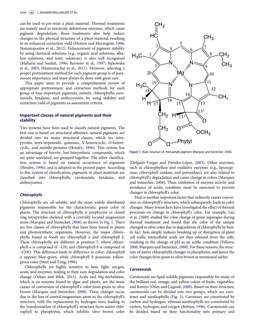
Chlorophylls are oil-soluble, and the most widely distributedpigments responsible for the characteristic green color ofplants. The structure of chlorophylls is porphyrins or closedring tetrapyroles chelated with a centrally located magnesiumatom (Marquez and Sinnecker, 2008), as shown in Fig. 1. Thereare five classes of chlorophylls that have been found in plantsand photosynthetic organisms. However, the major chloro-phylls found in foods are chlorophyll a and chlorophyll b.These chlorophylls are different at position 7, where chloro-phyll a is composed of –CH3 and chlorophyll b is composed of–CHO. This difference leads to difference in color; chlorophylla appears blue–green, while chlorophyll b possesses yellow-green color (Steet and Tong, 1996).
Chlorophylls are highly sensitive to heat, light, oxygen,acids, and enzymes, leading to their easy degradation and colorchange (€Ozkan and Bilek, 2015). Acids and Mg-dechelatase,which is an enzyme found in algae and plants, are the maincauses of conversion of chlorophyll’s color from green to olivebrown (Marquez and Sinnecker, 2008). These changes occurdue to the loss of central magnesium atom in the chlorophyll’sstructure, with the replacement by hydrogen ions, leading tothe transformation of chlorophyll’s structure from native chlo-rophyll to pheophytin, which exhibits olive brown color
(Delgado-Vargas and Paredes-L�opez, 2003). Other enzymes,such as chlorophyllase and oxidative enzymes (e.g., lipoxyge-nase, chlorophyll oxidase, and peroxidase), are also related tochlorophyll’s degradation and cause change in colors (Marquezand Sinnecker, 2008). Thus, inhibition of enzyme activity andavoidance of acidic condition must be exercised to preventchanges in chlorophyll’s color.
Heat is another important factor that indirectly causes conver-sion in chlorophyll’s structure, which subsequently leads to colorchanges. Many researchers have investigated the effect of thermalprocesses on change in chlorophyll’s color. For example, Lauet al. (2000) studied the color change of green asparagus duringthermal treatment and found that the color of the samplechanged to olive color due to degradation of chlorophylls by heat.In fact, heat simply induces breaking up or disruption of plantcell walls; intracellular acids are thus released from the cells,resulting in the change of pH to an acidic condition (Fellows,2000;Marquez and Sinnecker, 2008). For these reasons, the struc-ture of native chlorophylls changes to pheophytin, and hence thecolor changes from green to olive brown as mentioned earlier.
Carotenoids
Carotenoids are lipid-soluble pigments responsible for many ofthe brilliant red, orange, and yellow colors of fruits, vegetables,and flowers (€Otles and Cagindi, 2008). Based on their structure,carotenoids can be divided into two groups, which are caro-tenes and xanthophylls (Fig. 2). Carotenes are constituted bycarbon and hydrogen, whereas xanthophylls are constituted bycarbon, hydrogen, and oxygen (Britton, 1996). Carotenoids canbe divided based on their functionality into primary and
Figure 1. Basic structure of chlorophylls pigment (Marquez and Sinnecker, 2008).
3244 L. NGAMWONGLUMLERT ET AL.
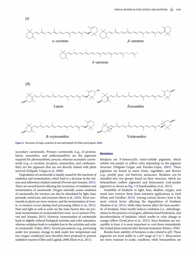
secondary carotenoids. Primary carotenoids (e.g., b-carotene,lutein, zeaxanthin, and antheraxanthin) are the pigmentsrequired for photosynthetic process, whereas secondary carote-noids (e.g., a-carotene, lycopene, astaxanthin, and canthaxan-thin) are the pigments that are not directly related with plantsurvival (Delgado-Vargas et al., 2000).
Degradation of carotenoids is mainly caused by the reactions ofoxidation and isomerization, which lead to a decrease in the red-ness and yellowness of plant material (Provesi and Amante, 2015).There are several factors affecting the occurrence of oxidation andisomerization of carotenoids. Oxygen naturally causes oxidationof carotenoids; the reaction can also be stimulated by light, heat,peroxide, metal ions, and enzymes (Boon et al., 2010). Most caro-tenoids in plants are trans-isomers, and the isomerization of trans-to cis-isomers occurs during food processing (Khoo et al., 2011).Heat and light as well as acids are the main factors that can pro-mote isomerization of carotenoids from trans- to cis-isomers (Pro-vesi and Amante, 2015). However, isomerization of carotenoidsleads to slightly reduced biological activities and color saturation,whereas oxidation leads to complete loss in the activities and colorof carotenoids (Takyi, 2001). Several precautions (e.g., processingunder low pressure, storage in dark under low temperature andlow oxygen conditions) have therefore been proposed to preventoxidation reaction (€Otles and Cagindi, 2008; Khoo et al., 2011).
Betalains
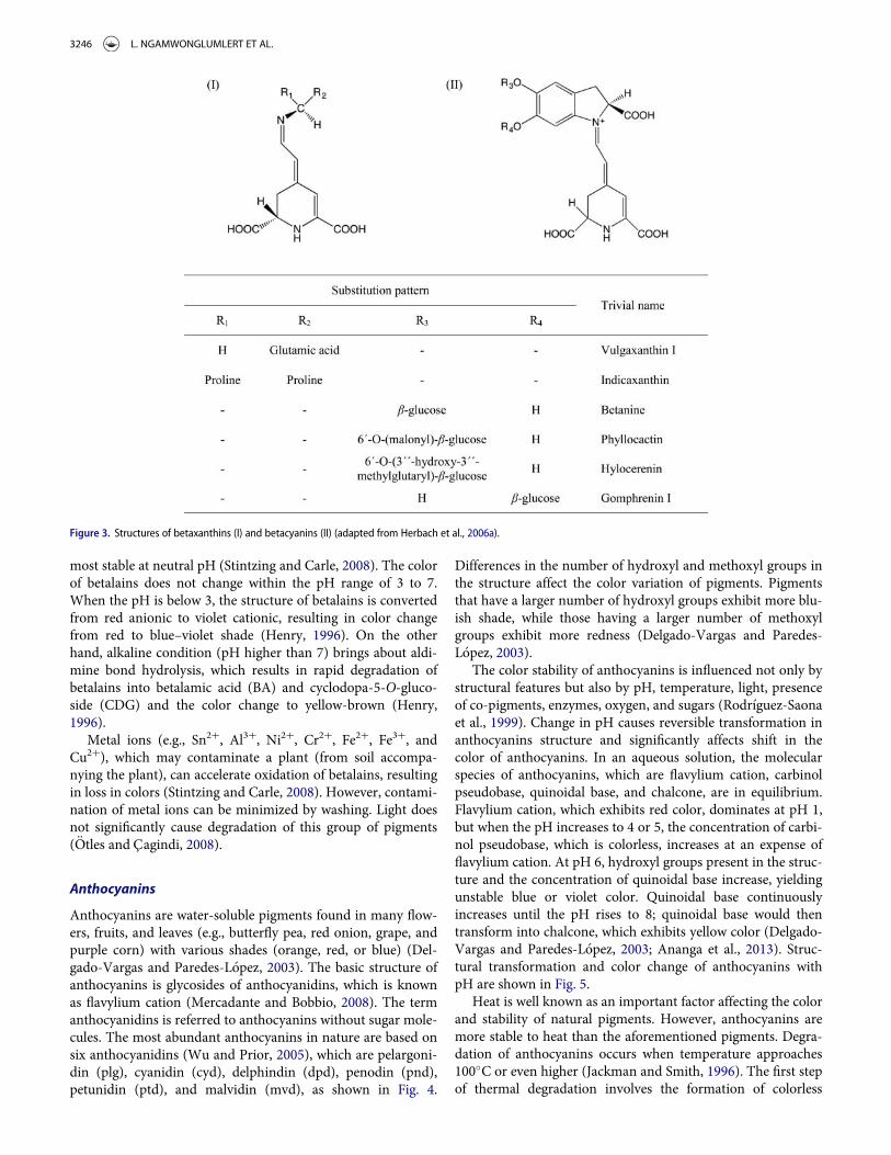
Betalains are N-heterocyclic water-soluble pigments, whichexhibit red–purple or yellow color depending on the pigmentstructure (Delgado-Vargas and Paredes-L�opez, 2003). Thesepigments are found in many fruits, vegetables, and flowers(e.g., prickly pear, red beetroot, aizoaceae). Betalains can beclassified into two groups based on their structure, which arebetaxanthins (yellow pigment) and betacyanins (red–purplepigment) as shown in Fig. 3 (Chandrasekhar et al., 2015).
Instability of betalains to light, heat, alkaline, oxygen, andmetal ions restricts them from extensive applications in food(Khan and Giridhar, 2014). Among various factors, heat is themost critical factor affecting the degradation of betalains(Reshmi et al., 2012), while other factors affect the heat sensitiv-ity of betalains. Heat mostly induces oxidation (i.e., dehydroge-nation in the presence of oxygen), aldimine bond hydrolysis, anddecarboxylation of batalains, which results in color change toorange-yellow (GonCalves et al., 2013). Since betalains are sus-ceptible to heat, it is most important to cool down immediatelythe treated plant material after thermal treatment (Henry, 1996).
Besides heat, stability of betalains is also related to pH. Thesepigments are most stable in a pH range of 3 to 7. Betacyaninsare more resistant to acidic condition, while betaxanthins are
Figure 2. Structures of major carotenes (I) and xanthophylls (II) (€Otles and Cagindi, 2008).
CRITICAL REVIEWS IN FOOD SCIENCE AND NUTRITION 3245
most stable at neutral pH (Stintzing and Carle, 2008). The colorof betalains does not change within the pH range of 3 to 7.When the pH is below 3, the structure of betalains is convertedfrom red anionic to violet cationic, resulting in color changefrom red to blue–violet shade (Henry, 1996). On the otherhand, alkaline condition (pH higher than 7) brings about aldi-mine bond hydrolysis, which results in rapid degradation ofbetalains into betalamic acid (BA) and cyclodopa-5-O-gluco-side (CDG) and the color change to yellow-brown (Henry,1996).
Metal ions (e.g., Sn2C, Al3C, Ni2C, Cr2C, Fe2C, Fe3C, andCu2C), which may contaminate a plant (from soil accompa-nying the plant), can accelerate oxidation of betalains, resultingin loss in colors (Stintzing and Carle, 2008). However, contami-nation of metal ions can be minimized by washing. Light doesnot significantly cause degradation of this group of pigments(€Otles and Cagindi, 2008).
Anthocyanins
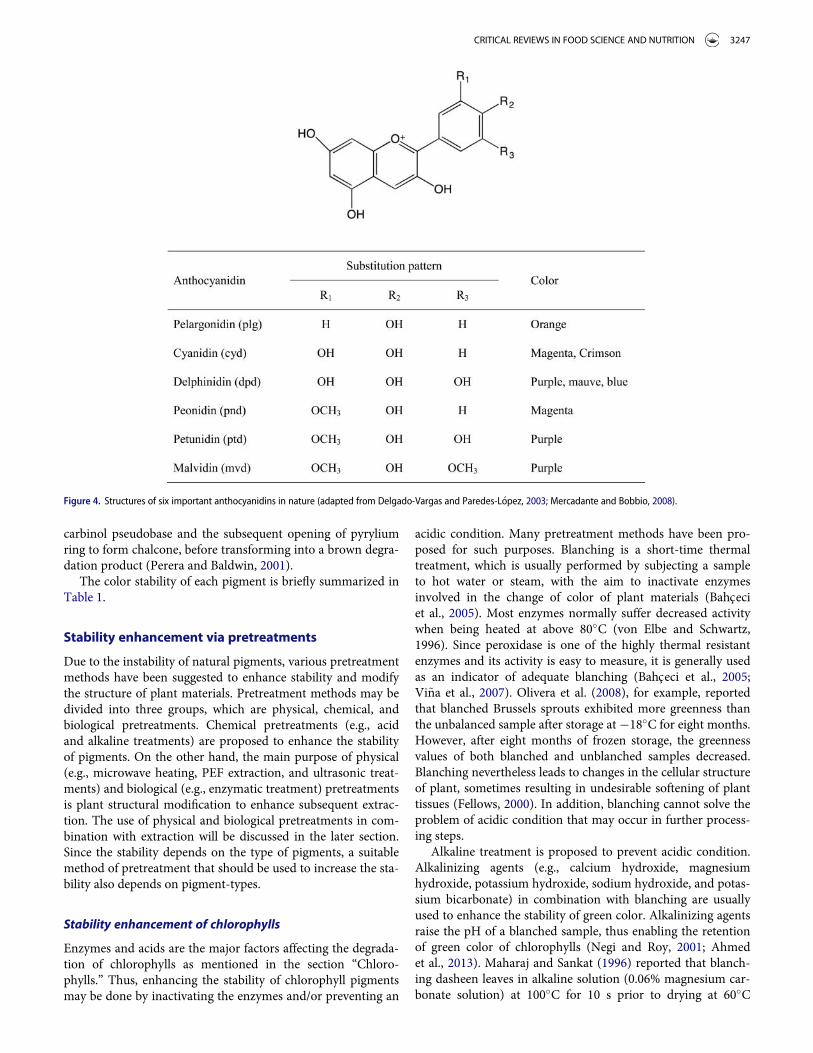
Anthocyanins are water-soluble pigments found in many flow-ers, fruits, and leaves (e.g., butterfly pea, red onion, grape, andpurple corn) with various shades (orange, red, or blue) (Del-gado-Vargas and Paredes-L�opez, 2003). The basic structure ofanthocyanins is glycosides of anthocyanidins, which is knownas flavylium cation (Mercadante and Bobbio, 2008). The termanthocyanidins is referred to anthocyanins without sugar mole-cules. The most abundant anthocyanins in nature are based onsix anthocyanidins (Wu and Prior, 2005), which are pelargoni-din (plg), cyanidin (cyd), delphindin (dpd), penodin (pnd),petunidin (ptd), and malvidin (mvd), as shown in Fig. 4.
Differences in the number of hydroxyl and methoxyl groups inthe structure affect the color variation of pigments. Pigmentsthat have a larger number of hydroxyl groups exhibit more blu-ish shade, while those having a larger number of methoxylgroups exhibit more redness (Delgado-Vargas and Paredes-L�opez, 2003).
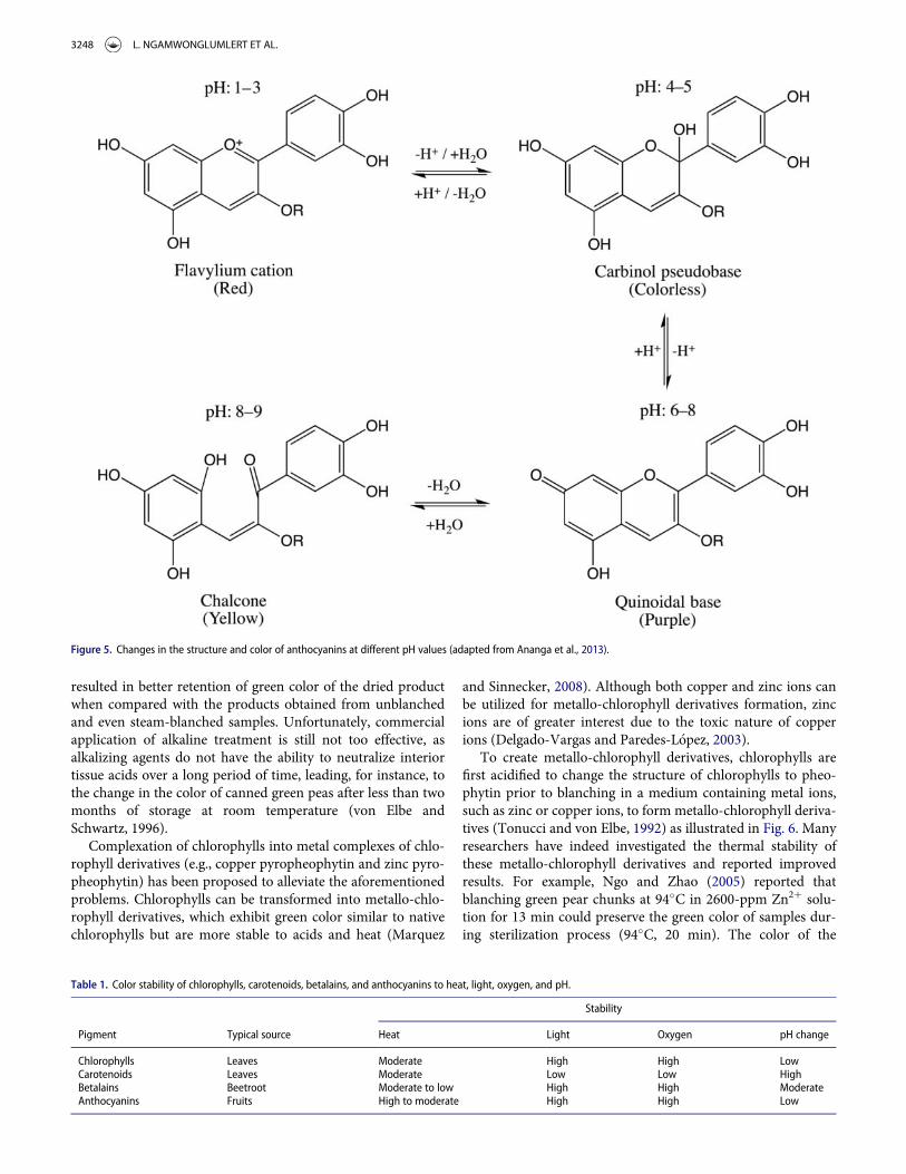
The color stability of anthocyanins is influenced not only bystructural features but also by pH, temperature, light, presenceof co-pigments, enzymes, oxygen, and sugars (Rodr�ıguez-Saonaet al., 1999). Change in pH causes reversible transformation inanthocyanins structure and significantly affects shift in thecolor of anthocyanins. In an aqueous solution, the molecularspecies of anthocyanins, which are flavylium cation, carbinolpseudobase, quinoidal base, and chalcone, are in equilibrium.Flavylium cation, which exhibits red color, dominates at pH 1,but when the pH increases to 4 or 5, the concentration of carbi-nol pseudobase, which is colorless, increases at an expense offlavylium cation. At pH 6, hydroxyl groups present in the struc-ture and the concentration of quinoidal base increase, yieldingunstable blue or violet color. Quinoidal base continuouslyincreases until the pH rises to 8; quinoidal base would thentransform into chalcone, which exhibits yellow color (Delgado-Vargas and Paredes-L�opez, 2003; Ananga et al., 2013). Struc-tural transformation and color change of anthocyanins withpH are shown in Fig. 5.
Heat is well known as an important factor affecting the colorand stability of natural pigments. However, anthocyanins aremore stable to heat than the aforementioned pigments. Degra-dation of anthocyanins occurs when temperature approaches100�C or even higher (Jackman and Smith, 1996). The first stepof thermal degradation involves the formation of colorless
Figure 3. Structures of betaxanthins (I) and betacyanins (II) (adapted from Herbach et al., 2006a).
3246 L. NGAMWONGLUMLERT ET AL.
carbinol pseudobase and the subsequent opening of pyryliumring to form chalcone, before transforming into a brown degra-dation product (Perera and Baldwin, 2001).
The color stability of each pigment is briefly summarized inTable 1.
Stability enhancement via pretreatments
Due to the instability of natural pigments, various pretreatmentmethods have been suggested to enhance stability and modifythe structure of plant materials. Pretreatment methods may bedivided into three groups, which are physical, chemical, andbiological pretreatments. Chemical pretreatments (e.g., acidand alkaline treatments) are proposed to enhance the stabilityof pigments. On the other hand, the main purpose of physical(e.g., microwave heating, PEF extraction, and ultrasonic treat-ments) and biological (e.g., enzymatic treatment) pretreatmentsis plant structural modification to enhance subsequent extrac-tion. The use of physical and biological pretreatments in com-bination with extraction will be discussed in the later section.Since the stability depends on the type of pigments, a suitablemethod of pretreatment that should be used to increase the sta-bility also depends on pigment-types.
Stability enhancement of chlorophylls
Enzymes and acids are the major factors affecting the degrada-tion of chlorophylls as mentioned in the section “Chloro-phylls.” Thus, enhancing the stability of chlorophyll pigmentsmay be done by inactivating the enzymes and/or preventing an
acidic condition. Many pretreatment methods have been pro-posed for such purposes. Blanching is a short-time thermaltreatment, which is usually performed by subjecting a sampleto hot water or steam, with the aim to inactivate enzymesinvolved in the change of color of plant materials (Bahceciet al., 2005). Most enzymes normally suffer decreased activitywhen being heated at above 80�C (von Elbe and Schwartz,1996). Since peroxidase is one of the highly thermal resistantenzymes and its activity is easy to measure, it is generally usedas an indicator of adequate blanching (Bahceci et al., 2005;Vi~na et al., 2007). Olivera et al. (2008), for example, reportedthat blanched Brussels sprouts exhibited more greenness thanthe unbalanced sample after storage at¡18�C for eight months.However, after eight months of frozen storage, the greennessvalues of both blanched and unblanched samples decreased.Blanching nevertheless leads to changes in the cellular structureof plant, sometimes resulting in undesirable softening of planttissues (Fellows, 2000). In addition, blanching cannot solve theproblem of acidic condition that may occur in further process-ing steps.
Alkaline treatment is proposed to prevent acidic condition.Alkalinizing agents (e.g., calcium hydroxide, magnesiumhydroxide, potassium hydroxide, sodium hydroxide, and potas-sium bicarbonate) in combination with blanching are usuallyused to enhance the stability of green color. Alkalinizing agentsraise the pH of a blanched sample, thus enabling the retentionof green color of chlorophylls (Negi and Roy, 2001; Ahmedet al., 2013). Maharaj and Sankat (1996) reported that blanch-ing dasheen leaves in alkaline solution (0.06% magnesium car-bonate solution) at 100�C for 10 s prior to drying at 60�C
Figure 4. Structures of six important anthocyanidins in nature (adapted from Delgado-Vargas and Paredes-L�opez, 2003; Mercadante and Bobbio, 2008).
CRITICAL REVIEWS IN FOOD SCIENCE AND NUTRITION 3247
resulted in better retention of green color of the dried productwhen compared with the products obtained from unblanchedand even steam-blanched samples. Unfortunately, commercialapplication of alkaline treatment is still not too effective, asalkalizing agents do not have the ability to neutralize interiortissue acids over a long period of time, leading, for instance, tothe change in the color of canned green peas after less than twomonths of storage at room temperature (von Elbe andSchwartz, 1996).
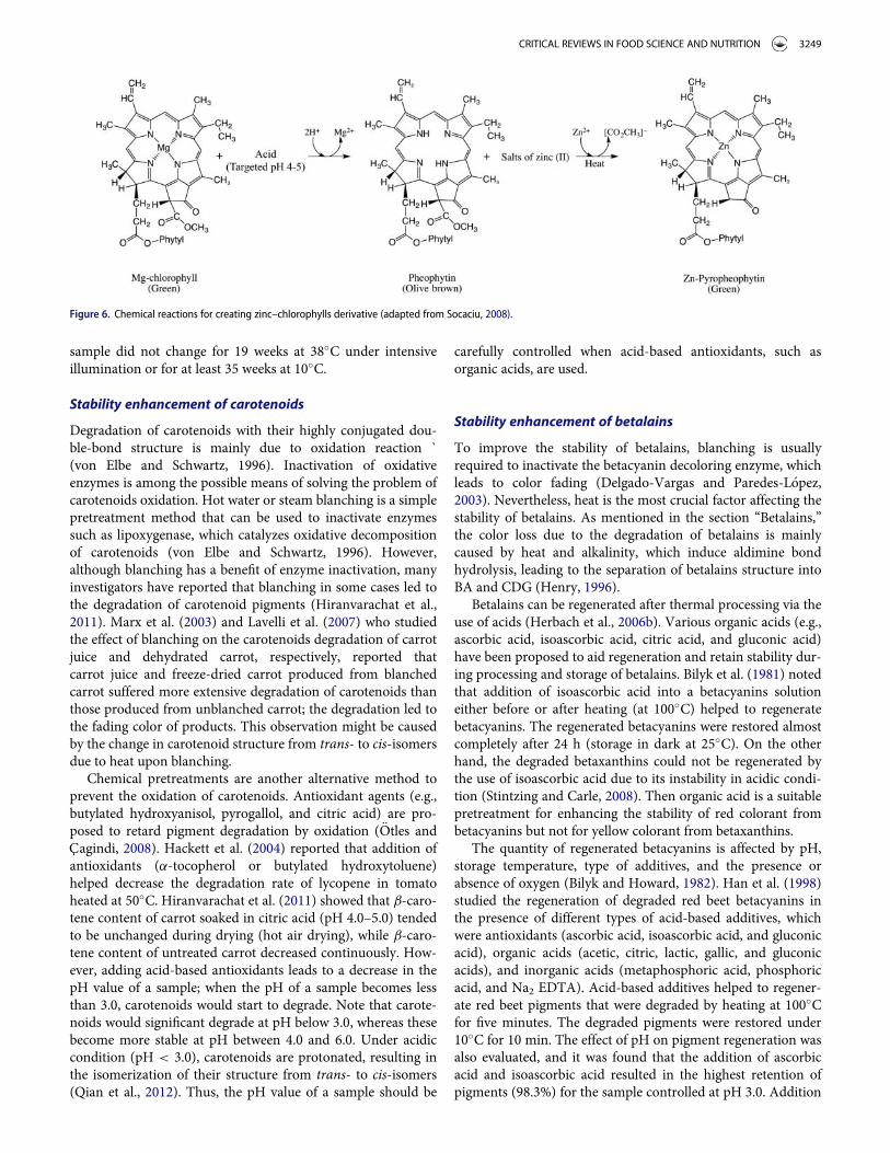
Complexation of chlorophylls into metal complexes of chlo-rophyll derivatives (e.g., copper pyropheophytin and zinc pyro-pheophytin) has been proposed to alleviate the aforementionedproblems. Chlorophylls can be transformed into metallo-chlo-rophyll derivatives, which exhibit green color similar to nativechlorophylls but are more stable to acids and heat (Marquez
and Sinnecker, 2008). Although both copper and zinc ions canbe utilized for metallo-chlorophyll derivatives formation, zincions are of greater interest due to the toxic nature of copperions (Delgado-Vargas and Paredes-L�opez, 2003).
To create metallo-chlorophyll derivatives, chlorophylls arefirst acidified to change the structure of chlorophylls to pheo-phytin prior to blanching in a medium containing metal ions,such as zinc or copper ions, to form metallo-chlorophyll deriva-tives (Tonucci and von Elbe, 1992) as illustrated in Fig. 6. Manyresearchers have indeed investigated the thermal stability ofthese metallo-chlorophyll derivatives and reported improvedresults. For example, Ngo and Zhao (2005) reported thatblanching green pear chunks at 94�C in 2600-ppm Zn2C solu-tion for 13 min could preserve the green color of samples dur-ing sterilization process (94�C, 20 min). The color of the
Figure 5. Changes in the structure and color of anthocyanins at different pH values (adapted from Ananga et al., 2013).
Table 1. Color stability of chlorophylls, carotenoids, betalains, and anthocyanins to heat, light, oxygen, and pH.
Stability
Pigment Typical source Heat Light Oxygen pH change
Chlorophylls Leaves Moderate High High LowCarotenoids Leaves Moderate Low Low HighBetalains Beetroot Moderate to low High High ModerateAnthocyanins Fruits High to moderate High High Low
3248 L. NGAMWONGLUMLERT ET AL.
sample did not change for 19 weeks at 38�C under intensiveillumination or for at least 35 weeks at 10�C.
Stability enhancement of carotenoids
Degradation of carotenoids with their highly conjugated dou-ble-bond structure is mainly due to oxidation reaction `(von Elbe and Schwartz, 1996). Inactivation of oxidativeenzymes is among the possible means of solving the problem ofcarotenoids oxidation. Hot water or steam blanching is a simplepretreatment method that can be used to inactivate enzymessuch as lipoxygenase, which catalyzes oxidative decompositionof carotenoids (von Elbe and Schwartz, 1996). However,although blanching has a benefit of enzyme inactivation, manyinvestigators have reported that blanching in some cases led tothe degradation of carotenoid pigments (Hiranvarachat et al.,2011). Marx et al. (2003) and Lavelli et al. (2007) who studiedthe effect of blanching on the carotenoids degradation of carrotjuice and dehydrated carrot, respectively, reported thatcarrot juice and freeze-dried carrot produced from blanchedcarrot suffered more extensive degradation of carotenoids thanthose produced from unblanched carrot; the degradation led tothe fading color of products. This observation might be causedby the change in carotenoid structure from trans- to cis-isomersdue to heat upon blanching.
Chemical pretreatments are another alternative method toprevent the oxidation of carotenoids. Antioxidant agents (e.g.,butylated hydroxyanisol, pyrogallol, and citric acid) are pro-posed to retard pigment degradation by oxidation (€Otles andCagindi, 2008). Hackett et al. (2004) reported that addition ofantioxidants (a-tocopherol or butylated hydroxytoluene)helped decrease the degradation rate of lycopene in tomatoheated at 50�C. Hiranvarachat et al. (2011) showed that b-caro-tene content of carrot soaked in citric acid (pH 4.0–5.0) tendedto be unchanged during drying (hot air drying), while b-caro-tene content of untreated carrot decreased continuously. How-ever, adding acid-based antioxidants leads to a decrease in thepH value of a sample; when the pH of a sample becomes lessthan 3.0, carotenoids would start to degrade. Note that carote-noids would significant degrade at pH below 3.0, whereas thesebecome more stable at pH between 4.0 and 6.0. Under acidiccondition (pH < 3.0), carotenoids are protonated, resulting inthe isomerization of their structure from trans- to cis-isomers(Qian et al., 2012). Thus, the pH value of a sample should be
carefully controlled when acid-based antioxidants, such asorganic acids, are used.
Stability enhancement of betalains
To improve the stability of betalains, blanching is usuallyrequired to inactivate the betacyanin decoloring enzyme, whichleads to color fading (Delgado-Vargas and Paredes-L�opez,2003). Nevertheless, heat is the most crucial factor affecting thestability of betalains. As mentioned in the section “Betalains,”the color loss due to the degradation of betalains is mainlycaused by heat and alkalinity, which induce aldimine bondhydrolysis, leading to the separation of betalains structure intoBA and CDG (Henry, 1996).
Betalains can be regenerated after thermal processing via theuse of acids (Herbach et al., 2006b). Various organic acids (e.g.,ascorbic acid, isoascorbic acid, citric acid, and gluconic acid)have been proposed to aid regeneration and retain stability dur-ing processing and storage of betalains. Bilyk et al. (1981) notedthat addition of isoascorbic acid into a betacyanins solutioneither before or after heating (at 100�C) helped to regeneratebetacyanins. The regenerated betacyanins were restored almostcompletely after 24 h (storage in dark at 25�C). On the otherhand, the degraded betaxanthins could not be regenerated bythe use of isoascorbic acid due to its instability in acidic condi-tion (Stintzing and Carle, 2008). Then organic acid is a suitablepretreatment for enhancing the stability of red colorant frombetacyanins but not for yellow colorant from betaxanthins.
The quantity of regenerated betacyanins is affected by pH,storage temperature, type of additives, and the presence orabsence of oxygen (Bilyk and Howard, 1982). Han et al. (1998)studied the regeneration of degraded red beet betacyanins inthe presence of different types of acid-based additives, whichwere antioxidants (ascorbic acid, isoascorbic acid, and gluconicacid), organic acids (acetic, citric, lactic, gallic, and gluconicacids), and inorganic acids (metaphosphoric acid, phosphoricacid, and Na2 EDTA). Acid-based additives helped to regener-ate red beet pigments that were degraded by heating at 100�Cfor five minutes. The degraded pigments were restored under10�C for 10 min. The effect of pH on pigment regeneration wasalso evaluated, and it was found that the addition of ascorbicacid and isoascorbic acid resulted in the highest retention ofpigments (98.3%) for the sample controlled at pH 3.0. Addition
Figure 6. Chemical reactions for creating zinc–chlorophylls derivative (adapted from Socaciu, 2008).
CRITICAL REVIEWS IN FOOD SCIENCE AND NUTRITION 3249
of gluconic acid and metaphosphoric acid also resulted in thehigher retention of pigments, with the retention of 81.7% and75.4%, respectively for the sample controlled at pH 6.8.
Besides the ability to regenerate the pigment after heating,ascorbic acid and isoascorbic acid could also help stabilize beta-cyanins during food processing and storage. Reynoso et al.(1997) reported that garambullo juice that was added with0.1% ascorbic acid exhibited higher redness than the untreatedsample after sterilization at 121�C for 15 min. The presence ofthe acid in red beet juice and garambullo juice (adjusted pH5.5) also resulted in an increase in the stability of betalains dur-ing storage at 25�C for five days. Moreover, ascorbic acid couldimprove the stability of betalains when metal ions (chromiumand iron) are present, since the acid could act as a chelatingagent (Pokorny, 2007).
Stability enhancement of anthocyanins
Anthocyanins are highly sensitive to change in pH, whichresults in shift of color. Using anthocyanins as a natural color-ant is suggested at low pH (pH < 4.0). At pH below 4, antho-cyanins primarily are in the form of flavylium cation, which ismore stable than other structures (Tan et al., 2014). Cevallos-Casals and Cisneros-Zevallos (2004), for instance, reported thatat a pH in the range of 0.9 to 4.0, the colorant samples from redsweet potato and purple carrot showed high stability duringstorage at 20�C for 134 days; hue angle of the red sweet potatocolorant at pH 4.0 still exhibited red–violet color after storage.In contrast, the color of the samples that were controlled at pHabove 4.0 shifted from purple–blue to brown and yellow, andafter 134 days the color of all the samples was predominated bythe yellow-colored chalcone.
Besides a proper pH adjustment, the source of anthocyaninsmust be considered to obtain the most stable colorant. Differentsources of plant materials contain different anthocyanin struc-tures, which affect their stability. Anthocyanins from red cab-bage, black carrot, red radish, and red sweet potato are reportedto be more stable to heat and pH change than anthocyaninsfrom other sources due to their acylation of structure(Bakowska-Barczak, 2005). Acylation of anthocyanin moleculescan enhance stability through intramolecular co-pigmentation(Rodr�ıguez-Saona et al., 1999). Higher stability of acylatedanthocyanins is attributed to the stacking of acyl group with thepyrylium ring of flavalium cation, thus preventing the nucleo-phile attack of water and subsequent formation of chalcone(Bakowska-Barczak, 2005). Many researchers have indeed dem-onstrated the stability of acylated anthocyanins in comparisonwith that of non-acylated anthocyanins. For example, Cevallos-Casals and Cisneros-Zevallos (2004), who studied the stability ofanthocyanins from red sweet potato, purple carrot, purple corn,and red grape, reported that anthocyanins from red sweet potatoand purple carrot, which mainly consist of acylated anthocya-nins, were more stable than those from purple corn and redgrape, which mainly consist of non-acylated anthocyanins.Anthocyanins extracted from red sweet potato exhibited longerhalf-life than those from purple carrot, purple corn, and redgrape when heated at 98�C and controlled at pH 3.0.
Intercellular co-pigmentation has been suggested to improvethe stability of anthocyanins. Intercellular co-pigmentation is
defined as interactions between anthocyanin molecules and othermolecules, e.g., flavonoids, alkaloids, amino acids, organic acids,metals, and other anthocyanins (Casta~neda-Ovando et al., 2008).The basic role of intercellular co-pigmentation is the same as thatof intracellular co-pigmentation, which is to protect flavalium cat-ion from the nucleophile attack of water molecules (Mazza andBrouillard, 1990; Casta~neda-Ovando et al., 2008). Gauche et al.(2010) reported that addition of organic acids (caffeic, ferulic, gal-lic, and tannic acids) into an anthocyanins solution could retardcolor change when the pH of the solution increased from 4.0 to6.0, whereas the solution without organic acids became colorless.Moreover, the half-life of anthocyanins solutions with addedorganic acids was longer than that of the untreated sample duringstorage at 28�C; addition of tannic acid resulted in the longesthalf-life of the samplemaintained at pH 1.0.
Although intercellular co-pigmentation can help increasethe stability of anthocyanins, this reaction induces an increasein the absorbance (hyperchromic effect: DA) and wavelength ofthe maximum absorbance (bathochromic shift: Dλ) of pigment.Bathochromic shift results in color change from red to red–orange or blue (Bakowska et al., 2003). Addition of tannic acid(1:1 w/v) to an anthocyanins solution from Isabel grapes (Vitislabrusca L.) controlled at pH 3.0 and 4.0, for example, led to anincrease in the absorbance and wavelength of the solution,resulting in bluer and brighter color of the treated sample (Bor-dignon-Luiz et al., 2007). Gauche et al. (2010) also illustratedthat co-pigmentation of anthocyanins with tannic, gallic, caf-feic, and ferulic acids resulted in hyperchromic effect and bath-ochromic shift at every pH value (1.0, 2.0, 3.0, 3.3, 3.5, 3.7, 4.0,4.5) studied. Exception was noted in the case of caffeic acid andferulic acid at pH of 1.0 and 2.0, where only bathochromic shiftwas observed. At pH of 1.0 and 2.0, these hydroxycinnamicacids induced the formation of pyranoanthocyanin, which is aanthocyanin-derived pigment, hiding the hyperchromic effectof co-pigmentation (G�omez-Miguez et al., 2006).
Since only a few natural blue-tone colorants are commerciallyavailable, production of blue natural colorants from anthocyaninssources is of interest. Blue hue colorants can be produced by thecomplexation of metal-anthocyanins. However, metal-anthocya-nin complexes (blue colorant) are reported to be stable only in thevacuolar matrix (Buchweitz et al., 2013) and would rapidly pre-cipitate in an aqueous solution (Buchweitz et al., 2012). Additionof some polysaccharides or gelatin to form a gel structure is pro-posed to enhance the stability of metal-anthocyanin complexesby preventing the precipitation of complexes. Buchweitz et al.(2013) noted that gelatin and blend agar–agar with amidated pec-tin could improve the stability of blue color of ferric anthocyaninchelates during storage at both 20�C in dark and 25�C under illu-mination. The most stable sample was obtained by using gelatinas a gel matrix; storage should be done at 20�C in dark.
Although the stability of anthocyanins can be enhanced byboth intra- and intercellular co-pigmentation as well as prepa-ration of gel matrix as mentioned earlier, controlling the pHvalue of a food product should be done to prevent shift in color.Storage should be done in dark at cool condition (Bordignon-Luiz et al., 2007).
Examples of stability enhancement of chlorophylls, carote-noids, betalains, and anthocyanins via pretreatments are givenin Table 2.
3250 L. NGAMWONGLUMLERT ET AL.
Effects of extraction methods on pigmentextractability
Extraction of pigments from plant materials can be performedby various techniques. Conventional methods are often used toextract crude pigments and other compounds. Recently, how-ever, non-conventional extraction methods, which have beenregarded as green extraction techniques, are the alternatives toconventional extraction since they require less amount of sol-vent as well as shorter extraction time and are more environ-ment-friendly (Cheok et al., 2014). Selecting an appropriateextraction technique for each type of natural pigment must bedone to improve the efficiency and productivity of natural col-orant. The effects of selected extraction methods on the stabilityand extraction yield of pigments are discussed here to serve as aguideline for the proper selection of extraction technique.
Conventional extraction methods
Conventional methods, such as Soxhlet extraction, maceration,and hydrodistillation, are simple, inexpensive and easy to han-dle (Veggi et al., 2013). These methods have, therefore, beenwidely used to extract essential oils, bioactive compounds, andnatural pigments from a wide array of plant materials. Soxhletextraction was initially designed for lipid extraction but now itis also used as a reference method for comparing the yield of anadvanced extraction technique (Azmir et al., 2013). During the
extraction, a plant sample contained in a thimble is repeatedlypercolated with condensed vapor of solvent until the extractionis completed (or the solvent is no longer able to solubilize inter-ested compounds in the sample), which is noted when the sol-vent has become colorless (Veggi et al., 2013). AlthoughSoxhlet extraction is simple and easy to handle, it requires largeamount of solvent, long extraction time, and leads to pigmentdegradation due to heat.
Maceration is a conventional extraction method that can beperformed at room temperature. This method can thus be usedfor extracting heat-sensitive pigments. Cha et al. (2010a), forinstance, noted that the pheophytin content, which is an indirectindicator of chlorophyll degradation, of a crude extract of greenmicroalgae (Chlorella vulgaris) obtained from maceration at25�C for 6 h was lower than that from Soxhlet extraction at100�C for 2 h. To extract crude pigments, a plant sample isground and mixed with an extraction solvent, and the mixture isleft in an extraction vessel with occasional shaking or stirring.After the process is finished, the liquid is strained off and the res-idue is pressed by a mechanical press, or centrifuged to repeatextraction with a fresh solvent until the solvent exhibits no color(Azmir et al., 2013). Since the extraction is generally performedat room temperature, the time required for the extraction is long;large amount of solvent is also needed to repeat the extraction.
Hydrodistillation is a traditional method that is usuallyused for extracting essential oils from a plant material. Theextraction medium, which is hot water and/or steam, is
Table 2. Pigment stability enhancement via pretreatments.
Pigment Source Pretreatment method Result Reference
Chlorophylls Rocket (Eruca sativa) Combination of blanching andacid/alkaline treatment
� Sample blanched in 0.1% NaOH exhibited maximumgreenness, while blanching sample in HCl yielded drop ingreen color.
Ahmed et al.,2013
Stevia rebaudiana leaves Copper treatment � Cu-chlorophylls suffered less color loss than Mg-chlorophyllsafter storage at 60�C for 20 days.
Bobbio andGuedes, 1990
� Color loss was 16% and 36% for Cu-chlorophyll and Mg-chlorophyll complexes, respectively.
Green pear Blanching and zinc treatment � Green pigments of control sample and sample blanched inwater were destroyed after heating at 94�C for 12 min.
Ngo and Zhao,2005
� Blanching and zinc treatment helped retain the greenpigment of sample after thermal processing.
Carotenoids Pumpkin (Cucurbitamoschata, Duchesne exPoiret)
Adjusting pH by addition ofascorbic acid and potassiumsorbate
� Addition of ascorbic acid and potassium sorbate to pH 4.0minimized loss of redness and yellowness of sample afterstorage at 25�C for 6 weeks.
Gliemmo et al.,2009
Carrot Blanching and acid treatment � b-carotene content of sample treated by blanching in citricacid (targeted pH 4–5) tended to be unchanged duringthermal processing, whereas b-carotene content of untreatedsample decreased continuously.
Hiranvarachatet al., 2011
Betalains Rivina humilis L. berry juice Addition of ascorbic acid � Addition of ascorbic acid (0.25 g/100 mL) helped retainbetacyanins at 93% and 78% after heating at 90�C for 3 minevery 24 h for 6 consecutive days and after heating at 90�C for24 min, respectively.
Khan andGiridhar, 2014
Anthocyanins Blood orange juice Addition of ascorbic acid � Total carotenoids content of sample added with ascorbic acid(30 mg/100 mL) decreased only 2.8%, while that of untreatedsample decreased up to 6.6% after storage at 4.5�C for7 weeks.
Choi et al., 2002
Honeysuckle (Lonicerakamtschatica)
Co-pigmentation by using QSA,NaMSA, rutin, quercetin,chlorogenic acid, tannic acid,and flavones
� Color of co-pigment anthocyanin complexes slowly changedduring heating at 180�C for 1 h at pH of 2.5, 3.5, and 4.5
Bakowska et al.,2003
Cabernet Sauvignon grape Adjusting pH by 0.1-M citricacid–sodium citrate and co-pigmentation by usingcaffeic acid
� Co-pigmentation by adjusting to pH 4 yielded retention ofcolor by 96% after storage at 4�C for 72 h, while controlsample had only 90% retention of color.
Gris et al., 2007
� Sample extracts added with caffeic acid and adjusted to pH 4had a half-life of anthocyanins up to 291 days when storedat 4�C in dark, while control sample had a half-life of only96 days.
CRITICAL REVIEWS IN FOOD SCIENCE AND NUTRITION 3251
directly in contact of a sample to extract interested com-pounds (Azmir et al., 2013). Since hydrodistillation involvesthe use of water for extraction, an obtained extract is not con-taminated with any organic solvent, which can be toxic. How-ever, since hydrodistillation is normally operated at a ratherhigher temperature (about 100�C), significant pigment degra-dation is expected.
The efficiency of conventional extraction methods directlydepends on the solubility of a solute from a plant material intoan extraction solvent (Cowan, 1999); extraction temperaturealso plays an important role on extraction efficiency. Examplesof solvents used for extracting chlorophylls, carotenoids, beta-lains, and anthocyanins are listed in Table 3. Although conven-tional extraction methods have many advantages, these requirelarge amount of solvent, long extraction time, and may lead tosignificant pigment degradation (Cheok et al., 2014). Non-con-ventional methods or green extraction methods have thereforebeen proposed to alleviate such limitations.
Non-conventional extraction methods
Supercritical fluid extractionSupercritical fluid extraction utilizes the advantages of super-critical fluids, which exhibit gas- and liquid-like properties toenhance extraction. A supercritical fluid can be produced bysubjecting a solvent to a temperature and pressure beyond itscritical point. In supercritical state, fluid possesses high diffu-sivity and low viscosity similar to gas but exhibits high solva-tion power similar to liquid (Mac�ıas-S�anchez et al., 2005). Forthese reasons, supercritical fluids can better penetrate into asample matrix; this subsequently leads to a more efficientextraction.
Carbon dioxide is considered an ideal solvent for SFE since itscritical temperature (Tc) and critical pressure (Pc), 31�C and 74bars, respectively, are not too high (Greibrokk, 1991). SFE is gen-erally operated within a pressure range of 8 to 40MPa and a tem-perature range of 30 to 60�C. For this reason, SFE can be appliedto extract heat-sensitive pigments. V�agi et al. (2002) reportedthat the pheophytin contents of crude extracts of Origanummajorana L. obtained from SFE at 40, 50, and 60�C were lowerthan those obtained from Soxhlet extraction at 70 and 80�C.
Since most extraction solvents for SFE are non-polar, thismethod is suitable for the extraction of low-polar pigmentssuch as carotenoids and chlorophylls. On the other hand, SFEis not suitable for extraction of betalains and anthocyanins,which are high-polar pigments. Mac�ıas-S�anchez et al. (2005)reported that SFE was faster and more selective than ultra-sound-assisted maceration for extracting carotenoids and
chlorophylls from Nannochloropsis gaditana. The highestextraction yields of both carotenoids and chlorophyll a via SFEwere achieved when using an extraction temperature of 60�Cand a pressure of 400 bar. Nevertheless, SFE with supercriticalcarbon dioxide as a solvent resulted in lower carotenoids andchlorophyll a yields than ultrasound-assisted macerationemploying methanol as a solvent.
To improve the extraction yields of carotenoids and chloro-phylls, a combination of carbon dioxide and organic solvent(e.g., methanol, ethanol) has been proposed. Addition of 5% (v/v) ethanol to supercritical carbon dioxide was noted to increasethe extraction yield of b-carotene in carrot by about 7% (Baysalet al., 2000). Using 7% (v/v) ethanol as a co-solvent with super-critical carbon dioxide could also increase the extraction yieldof chlorophylls a and b from 0.027 and 0.023 to 0.848 and0.356 mg/g dry sample, respectively (Guedes et al., 2013).
Although SFE is quite efficient and involves small solventconsumption, no or less use of toxic solvent can extract heat-sensitive pigments and can be automated. This extractionmethod requires high capital and operating costs because of thehigh pressure required for operation (Veggi et al., 2013).
Pressurized liquid extractionPressurized liquid extraction utilizes a liquid solvent at elevatedpressure (10.3–13.8 MPa) and temperature (40–200�C) forextraction (Antunes et al., 2008). High temperature results inthe better diffusion of solvent into sample matrix and also helpsdisrupt plant cells, resulting in a more effective release of pig-ments from the cells, and hence more effective extraction. Highpressure, on the other hand, forces solvent into matrix poresand hence allows better contact between the solvent and com-pounds to be extracted (Cha et al., 2010b; Mustafa and Turner,2011). Thus, PLE requires shorter time and involves the use ofless solvent for extraction (Antunes et al., 2008).
Pressurized liquid extraction can extract both water- and oil-based pigments, depending on the selection of an extractionsolvent. However, PLE cannot effectively extract heat-sensitivepigments since the method involves the use of high tempera-ture. PLE is therefore normally used to extract less heat-sensitive pigments (i.e., anthocyanins, carotenoids, and chloro-phylls). Machado et al. (2015) studied the use of PLE incomparison with conventional methods (Soxhlet extractionand maceration) to extract monomeric anthocyanins fromblackberry. PLE at 100�C exhibited a higher extraction ratethan Soxhlet extraction at 80�C and maceration extraction at25�C. Time required to extract monomeric anthocyanins in thecase of PLE, Soxhlet extraction, and maceration was 30, 300,and 1440 min, respectively. In terms of carotenoids and chloro-phylls, Cha et al. (2010a) reported that PLE at 160�C for120 min yielded higher contents of carotenoids and chloro-phylls a and b than maceration extraction at 25�C for 360 minand Soxhlet extraction at 100�C for 120 min. Although PLEgave the highest yields of carotenoids and chlorophylls a and b,the method resulted in the highest pheophytin content due tothe use of higher temperature for extraction.
To increase the stability of pigments during PLE, some pre-treatment methods, which are mentioned in the section “Stabil-ity Enhancement via Pretreatments,” can be used. Adjustingthe pH of an extraction solvent to acidic values can help retard
Table 3. Examples of solvents used for extracting different groups of pigments(Cowan, 1999; Kujala et al., 2001; Hosikian et al., 2010; Machmudah and Goto,2013).
Chlorophylls Carotenoids Betalains Anthocyanins
Methanol Methanol Methanol MethanolEthanol Ethanol Water WaterAcetone Acetone
n-hexanePentaneChloroform
3252 L. NGAMWONGLUMLERT ET AL.
the degradation of anthocyanins during PLE because acidiccondition could lead to the formation of flavylium cation,which is the most stable form of anthocyanin (Tan et al., 2014).Sharif et al. (2010) reported that maximum redness (or thehighest a� value) and the highest yield of anthocyanins (cyani-din 3-glucoside, cyaniding 3-rutinoside, and cyandin chloride)extracted from onion (Allim cepa) skin could be achieved byadding 0.1% (v/v) hydrochloric acid into an extraction solvent(methanol) and performing PLE at a temperature of 80�C andpressure of 689.48 bar. Besides adjusting the pH, intra- andintermolecular co-pigmentation may be used to increase thestability of anthocyanins during PLE. For chlorophylls, the con-version of native chlorophylls to metallo-chlorophyll deriva-tives prior to pigment extraction maybe employed to retain thegreen color of chlorophyll extracts obtained by PLE. Neverthe-less, this extraction method again requires high capital andoperating costs due to the use of higher pressure for extraction.
Microwave-assisted extractionMicrowave-assisted extraction is gaining interest as an alterna-tive for extracting both water- and oil-based plant pigmentsdue to its speed and small solvent consumption when com-pared with conventional extraction methods (Dahmoune et al.,2014). Rapid heating by microwave radiation results in anexpansion of plant cell structure with subsequent rupture ofplant cell walls. Thus, compounds, including pigments, can eas-ily migrate out of cells, resulting in an enhanced extraction rate(Zou et al., 2013). Dabiri et al. (2005), for example, illustratedthat MAE could reduce the time and solvent volume require-ment when extracting pigments (alizarin and purpurin) fromRubiaceae plants. MAE required the time and solvent volumeof only 20 min and 20 mL, respectively, while Soxhlet extrac-tion required the time and solvent volume of up to 360 minand 100 mL, respectively. Moreover, MAE at an optimized con-dition yielded higher alizarin and purpurin recovery than Soxh-let extraction operated at its optimum condition.
Combination of stability enhancement and pigment extrac-tion has been proposed for MAE. Cardoso-Ugarte et al. (2014)proposed a two-stage MAE with the addition of L-ascorbic acidinto 50% (v/v) ethanol for the extraction of betanines andbetaxanthins from red beet. Both extraction steps were per-formed at a power level of 400 W and 100% duty cycles. A cool-ing step was added between the first and second stages to delaythe degradation of the pigments already extracted. The additionof ascorbic acid could retard the degradation of betanines.However, the stability of betaxanthins could not be retained bythe addition of ascorbic acid, since betaxanthins are not stablein acidic condition (Stintzing and Carle, 2008).
Combination of MAE with vacuum, resulting in the so-called vacuum microwave-assisted extraction (VMAE), hasrecently been proposed to extract heat-sensitive bioactive com-pounds and pigments (Hiranvarachat et al., 2015). VMAE canreduce pigment degradation by oxidation since less oxygen isavailable in the extraction process. Xiao et al. (2012) noted thatVMAE could prevent the degradation of vitamin C extractedfrom peppers and guava, b-carotene extracted from carrot, andaloin A extracted from aloe vera. The extraction yields of vita-min C, b-carotene, and aloin A extracted by VMAE were alsonoted to be higher than those obtained via MAE.
Ultrasound-assisted extractionUltrasound-assisted extraction utilizes cavitation bubbles cre-ated by ultrasound waves to enhance extraction efficiency (Ras-togi, 2011). Cavitation bubbles are formed when ultrasoundwaves pass through a medium, creating alternative compres-sion and decompression cycles, which in turn result in the com-pression and expansion of bubbles. When bubbles grow toolarge to be contained by the surface tension force, they collapse,resulting in shearing forces to break up or disrupt cell walls of acontacted plant material (Pitt et al., 2004). As a result, releaseof intracellular compounds is enhanced. UAE has received con-siderable attention because of its benefits, which include theability to perform extraction at lower temperatures due to theabsence of external input heat and at low solvent consumption(Tao and Sun, 2015).
Both water- and oil-based pigments can be extracted byUAE. Since UAE is a non-thermal process, it can be used toextract heat-sensitive pigments. Tao et al. (2014), for example,investigated the use of UAE to extract anthocyanins from winelees in comparison with the use of maceration extraction, andfound that at the same extraction time and temperature(36 min and 60�C), the yield of anthocyanins extracted byultrasound at 40 kHz was higher than that by maceration. UAEhas also been employed as an efficient technique for the extrac-tion of betalains and chlorophylls. Kong et al. (2012), for exam-ple, reported that UAE could increase extraction rate by up to88.9% in comparison with that of maceration when extractingchlorophylls from Chlorella vulgaris.
Ultrasound-assisted extraction is known for its lower extrac-tion effectiveness when compared with some other non-con-ventional extraction processes (i.e., SFE, PLE, MAE, and PEFextraction). Cha et al. (2010a), for instance, compared theextraction yields of carotenoids and chlorophylls from Chlorellavulgaris by PLE and UAE and noted that PLE at 160�C for30 min gave higher yields of both carotenoids and chlorophyllsthan UAE at 25�C for 360 min. Nevertheless, because of itshigher temperature operation, PLE caused more chlorophylldegradation, which resulted in higher pheophytin content inthe extract. To increase the extraction yield of UAE, repeatedextraction is required; larger volume of solvent would beneeded to repeat the extraction, however.
Recently, combined use of UAE and MAE is proposed toincrease the extraction yield of UAE and, at the same time,reduce the time a material needs to undergo MAE. UAE andMAE in combination lead to more extensive damage to plantstructure than employing UAE or MAE alone (Pongmalaiet al., 2015). Lianfu and Zelong (2008) reported thatUAECMAE at 98 W of microwave power with 40 kHz of ultra-sound gave higher yield of extracted lycopene from tomatothan UAE alone. The proportion of lycopene yield was 97.4%and 89.4% for UAECMAE and UAE, respectively. Moreover,UAECMAE required only six minutes to obtain such a yield,while UAE needed as long as 29 min to obtain a similar yield.
Pulsed-electric field (PEF) extractionPulsed electric field (PEF) has been noted to be useful for enhanc-ing many processes of food production, including the extractionprocess. Short- and high-voltage electric field is used to inducepore formation in the cell walls of a plant material, which
CRITICAL REVIEWS IN FOOD SCIENCE AND NUTRITION 3253
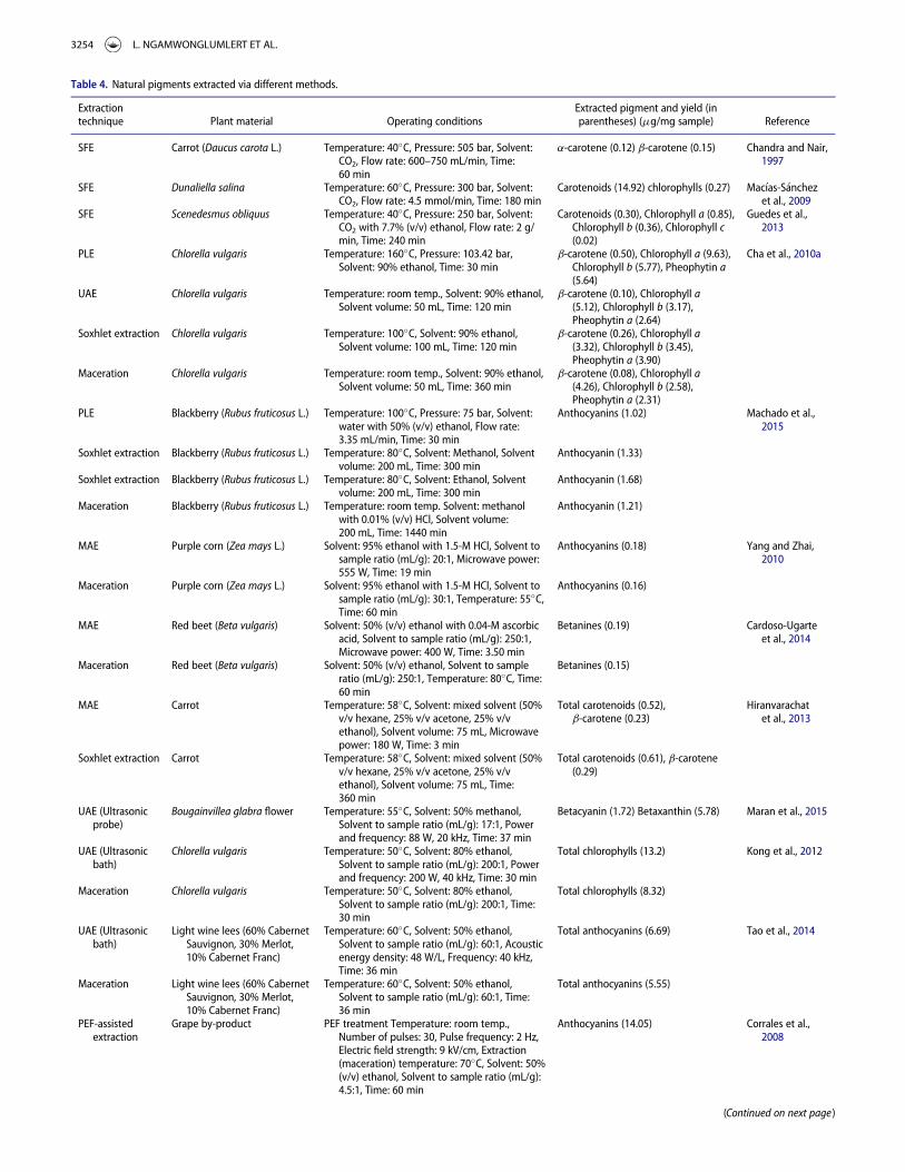
Table 4. Natural pigments extracted via different methods.
Extractiontechnique Plant material Operating conditions
Extracted pigment and yield (inparentheses) (mg/mg sample) Reference
SFE Carrot (Daucus carota L.) Temperature: 40�C, Pressure: 505 bar, Solvent:CO2, Flow rate: 600–750 mL/min, Time:60 min
a-carotene (0.12) b-carotene (0.15) Chandra and Nair,1997
SFE Dunaliella salina Temperature: 60�C, Pressure: 300 bar, Solvent:CO2, Flow rate: 4.5 mmol/min, Time: 180 min
Carotenoids (14.92) chlorophylls (0.27) Mac�ıas-S�anchezet al., 2009
SFE Scenedesmus obliquus Temperature: 40�C, Pressure: 250 bar, Solvent:CO2 with 7.7% (v/v) ethanol, Flow rate: 2 g/min, Time: 240 min
Carotenoids (0.30), Chlorophyll a (0.85),Chlorophyll b (0.36), Chlorophyll c(0.02)
Guedes et al.,2013
PLE Chlorella vulgaris Temperature: 160�C, Pressure: 103.42 bar,Solvent: 90% ethanol, Time: 30 min
b-carotene (0.50), Chlorophyll a (9.63),Chlorophyll b (5.77), Pheophytin a(5.64)
Cha et al., 2010a
UAE Chlorella vulgaris Temperature: room temp., Solvent: 90% ethanol,Solvent volume: 50 mL, Time: 120 min
b-carotene (0.10), Chlorophyll a(5.12), Chlorophyll b (3.17),Pheophytin a (2.64)
Soxhlet extraction Chlorella vulgaris Temperature: 100�C, Solvent: 90% ethanol,Solvent volume: 100 mL, Time: 120 min
b-carotene (0.26), Chlorophyll a(3.32), Chlorophyll b (3.45),Pheophytin a (3.90)
Maceration Chlorella vulgaris Temperature: room temp., Solvent: 90% ethanol,Solvent volume: 50 mL, Time: 360 min
b-carotene (0.08), Chlorophyll a(4.26), Chlorophyll b (2.58),Pheophytin a (2.31)
PLE Blackberry (Rubus fruticosus L.) Temperature: 100�C, Pressure: 75 bar, Solvent:water with 50% (v/v) ethanol, Flow rate:3.35 mL/min, Time: 30 min
Anthocyanins (1.02) Machado et al.,2015
Soxhlet extraction Blackberry (Rubus fruticosus L.) Temperature: 80�C, Solvent: Methanol, Solventvolume: 200 mL, Time: 300 min
Anthocyanin (1.33)
Soxhlet extraction Blackberry (Rubus fruticosus L.) Temperature: 80�C, Solvent: Ethanol, Solventvolume: 200 mL, Time: 300 min
Anthocyanin (1.68)
Maceration Blackberry (Rubus fruticosus L.) Temperature: room temp. Solvent: methanolwith 0.01% (v/v) HCl, Solvent volume:200 mL, Time: 1440 min
Anthocyanin (1.21)
MAE Purple corn (Zea mays L.) Solvent: 95% ethanol with 1.5-M HCl, Solvent tosample ratio (mL/g): 20:1, Microwave power:555 W, Time: 19 min
Anthocyanins (0.18) Yang and Zhai,2010
Maceration Purple corn (Zea mays L.) Solvent: 95% ethanol with 1.5-M HCl, Solvent tosample ratio (mL/g): 30:1, Temperature: 55�C,Time: 60 min
Anthocyanins (0.16)
MAE Red beet (Beta vulgaris) Solvent: 50% (v/v) ethanol with 0.04-M ascorbicacid, Solvent to sample ratio (mL/g): 250:1,Microwave power: 400 W, Time: 3.50 min
Betanines (0.19) Cardoso-Ugarteet al., 2014
Maceration Red beet (Beta vulgaris) Solvent: 50% (v/v) ethanol, Solvent to sampleratio (mL/g): 250:1, Temperature: 80�C, Time:60 min
Betanines (0.15)
MAE Carrot Temperature: 58�C, Solvent: mixed solvent (50%v/v hexane, 25% v/v acetone, 25% v/vethanol), Solvent volume: 75 mL, Microwavepower: 180 W, Time: 3 min
Total carotenoids (0.52),b-carotene (0.23)
Hiranvarachatet al., 2013
Soxhlet extraction Carrot Temperature: 58�C, Solvent: mixed solvent (50%v/v hexane, 25% v/v acetone, 25% v/vethanol), Solvent volume: 75 mL, Time:360 min
Total carotenoids (0.61), b-carotene(0.29)
UAE (Ultrasonicprobe)
Bougainvillea glabra flower Temperature: 55�C, Solvent: 50% methanol,Solvent to sample ratio (mL/g): 17:1, Powerand frequency: 88 W, 20 kHz, Time: 37 min
Betacyanin (1.72) Betaxanthin (5.78) Maran et al., 2015
UAE (Ultrasonicbath)
Chlorella vulgaris Temperature: 50�C, Solvent: 80% ethanol,Solvent to sample ratio (mL/g): 200:1, Powerand frequency: 200 W, 40 kHz, Time: 30 min
Total chlorophylls (13.2) Kong et al., 2012
Maceration Chlorella vulgaris Temperature: 50�C, Solvent: 80% ethanol,Solvent to sample ratio (mL/g): 200:1, Time:30 min
Total chlorophylls (8.32)
UAE (Ultrasonicbath)
Light wine lees (60% CabernetSauvignon, 30% Merlot,10% Cabernet Franc)
Temperature: 60�C, Solvent: 50% ethanol,Solvent to sample ratio (mL/g): 60:1, Acousticenergy density: 48 W/L, Frequency: 40 kHz,Time: 36 min
Total anthocyanins (6.69) Tao et al., 2014
Maceration Light wine lees (60% CabernetSauvignon, 30% Merlot,10% Cabernet Franc)
Temperature: 60�C, Solvent: 50% ethanol,Solvent to sample ratio (mL/g): 60:1, Time:36 min
Total anthocyanins (5.55)
PEF-assistedextraction
Grape by-product PEF treatment Temperature: room temp.,Number of pulses: 30, Pulse frequency: 2 Hz,Electric field strength: 9 kV/cm, Extraction(maceration) temperature: 70�C, Solvent: 50%(v/v) ethanol, Solvent to sample ratio (mL/g):4.5:1, Time: 60 min
Anthocyanins (14.05) Corrales et al.,2008
(Continued on next page )
3254 L. NGAMWONGLUMLERT ET AL.
subsequently leads to better release of cellular constituents, andhence enhanced extraction (Dons�ı et al., 2010; Azmir et al., 2013).
Most solvents used to extract betalains and anthocyanins arepolar solvents, possessing electrical conductivity, and can letelectricity pass thorough to sample cells. On the other hand,electric field cannot pass thorough a non-polar solvent, since itis an electrical resistance possessing low or negligible conduc-tivity (Yuhas, 1995). For this reason, PEF is more suitable forthe extraction of betalains and anthocyanins than for chloro-phylls and carotenoids.
A number of works exist on the use of PEF to extract (or toassist the extraction of) plant pigments. Pu�ertolas et al. (2013),for instance, applied PEF at 3.4 kV/cm and 105 ms (35 pulsesof 3 ms) to extract anthocyanins from purple-fleshed potato.The anthocyanins yield was noted, as expected, to be higherthan those from the non-PEF-treated sample and also from themacerated sample. pH value also exerts a significant effect onthe PEF extraction yield. L�opez et al. (2009), for example, stud-ied the effect of pH of the solvent on the PEF extraction yieldof betanines from red beetroot, and found that McIlvaine bufferat pH 3.5 led to the highest yield of betanines. This is probablybecause an acidic solvent could prevent the degradation of beta-nines during extraction (Bilyk and Howard, 1982).
Notwithstanding its potential, PEF-based extractor, espe-cially at an industrial scale, suffers from the need of a high-power supply equipment and treatment chamber (Nip, 2007),which are nowadays still rather expensive.
Enzyme-assisted extractionEnzyme-assisted extraction involves the use of enzymes toenhance the extraction of bioactive compounds, including pig-ments, from a plant material. Various enzymes, such as pecti-nase, cellulase, and hemicellulase, are used to hydrolyze plantcellulosic cell walls (Socaciu, 2008), resulting in easier release ofcellular constituents, and hence facilitated extraction. EAE is ofinterest due to its non-thermal nature as well as lower toxicityand solvent consumption (Socaciu, 2008). EAE can be dividedinto two common approaches, which are enzyme-assisted
aqueous extraction (EAAE) and enzyme-assisted cold pressing(EACP) (Latif and Anwar, 2009). In EAAE, enzymes are used tohelp destroy cell walls, and hence rupturing polysaccharide–pro-tein colloids. On the other hand, enzymes are used only to hydro-lyze cell walls in the case of EACP (Hassan and G€OkCe, 2014).
Many researchers investigated the effect of enzyme treat-ment on the extraction yields of chlorophylls, carotenoids, beta-lains, and anthocyanins. Barzana et al. (2002), for example,noted that the extraction yield of carotenoids from marigoldflower treated by enzymes (viscozyme, proteolytic, and pectino-lytic enzymes) was higher than that from the untreated sample.Kammerer et al. (2005) proposed a two-stage process for theextraction of anthocyanins from grape pomace via EAE. Theprocess involved a pre-extraction step, which was then followedby EAE. Release of phenolic acids during the pre-extractionstep nevertheless caused a decrease in pH, leading to an unfa-vorable condition for enzyme activity. For this reason, beforeEAE, the pH of the residue obtained in the pre-extraction stepwas adjusted to an optimum range for enzyme hydrolysis,which resulted in enhanced extraction.
Enzyme treatment can be also applied to assist other greenextraction methods. Lenucci et al. (2015), for instance, proposedan enzyme-aided supercritical carbon dioxide extraction, andfound that enzymatic pretreatment could help increase theextractable lycopene concentration from the matrix of tomatopuree due to the degradation of tomato cell walls by enzymes.However, the denseness of the enzyme-treated sample hinderedthe diffusion of supercritical carbon dioxide through the samplematrix. Addition of a co-matrix (hazelnut seeds) into the basematrix was therefore proposed to facilitate the penetration ofsupercritical carbon dioxide through the sample matrix, andhence increased lycopene extractability. Although EAE canimprove extraction yield and can be applied to extract heat-sensi-tive pigments, EAE requires a very long extraction time; enzymesare also normally quite expensive (Hardouin et al., 2014).
Examples of pigment extraction via different methods, andpros and cons of each extraction method are listed in Tables 4and 5, respectively.
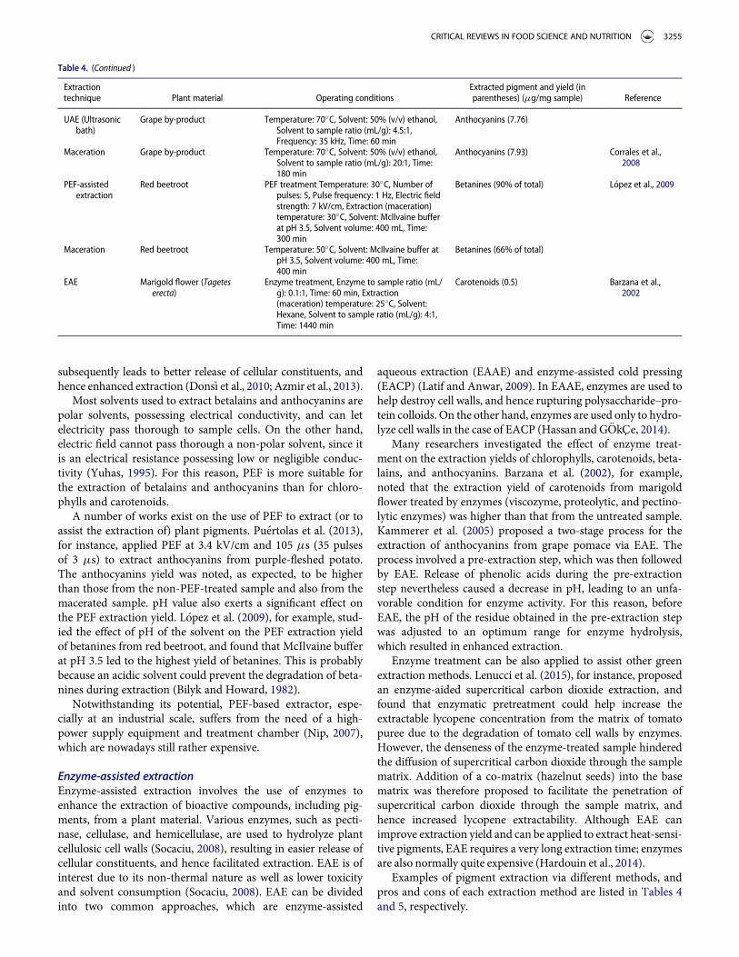
Table 4. (Continued )
Extractiontechnique Plant material Operating conditions
Extracted pigment and yield (inparentheses) (mg/mg sample) Reference
UAE (Ultrasonicbath)
Grape by-product Temperature: 70�C, Solvent: 50% (v/v) ethanol,Solvent to sample ratio (mL/g): 4.5:1,Frequency: 35 kHz, Time: 60 min
Anthocyanins (7.76)
Maceration Grape by-product Temperature: 70�C, Solvent: 50% (v/v) ethanol,Solvent to sample ratio (mL/g): 20:1, Time:180 min
Anthocyanins (7.93) Corrales et al.,2008
PEF-assistedextraction
Red beetroot PEF treatment Temperature: 30�C, Number ofpulses: 5, Pulse frequency: 1 Hz, Electric fieldstrength: 7 kV/cm, Extraction (maceration)temperature: 30�C, Solvent: McIlvaine bufferat pH 3.5, Solvent volume: 400 mL, Time:300 min
Betanines (90% of total) L�opez et al., 2009
Maceration Red beetroot Temperature: 50�C, Solvent: McIlvaine buffer atpH 3.5, Solvent volume: 400 mL, Time:400 min
Betanines (66% of total)
EAE Marigold flower (Tageteserecta)
Enzyme treatment, Enzyme to sample ratio (mL/g): 0.1:1, Time: 60 min, Extraction(maceration) temperature: 25�C, Solvent:Hexane, Solvent to sample ratio (mL/g): 4:1,Time: 1440 min
Carotenoids (0.5) Barzana et al.,2002
CRITICAL REVIEWS IN FOOD SCIENCE AND NUTRITION 3255
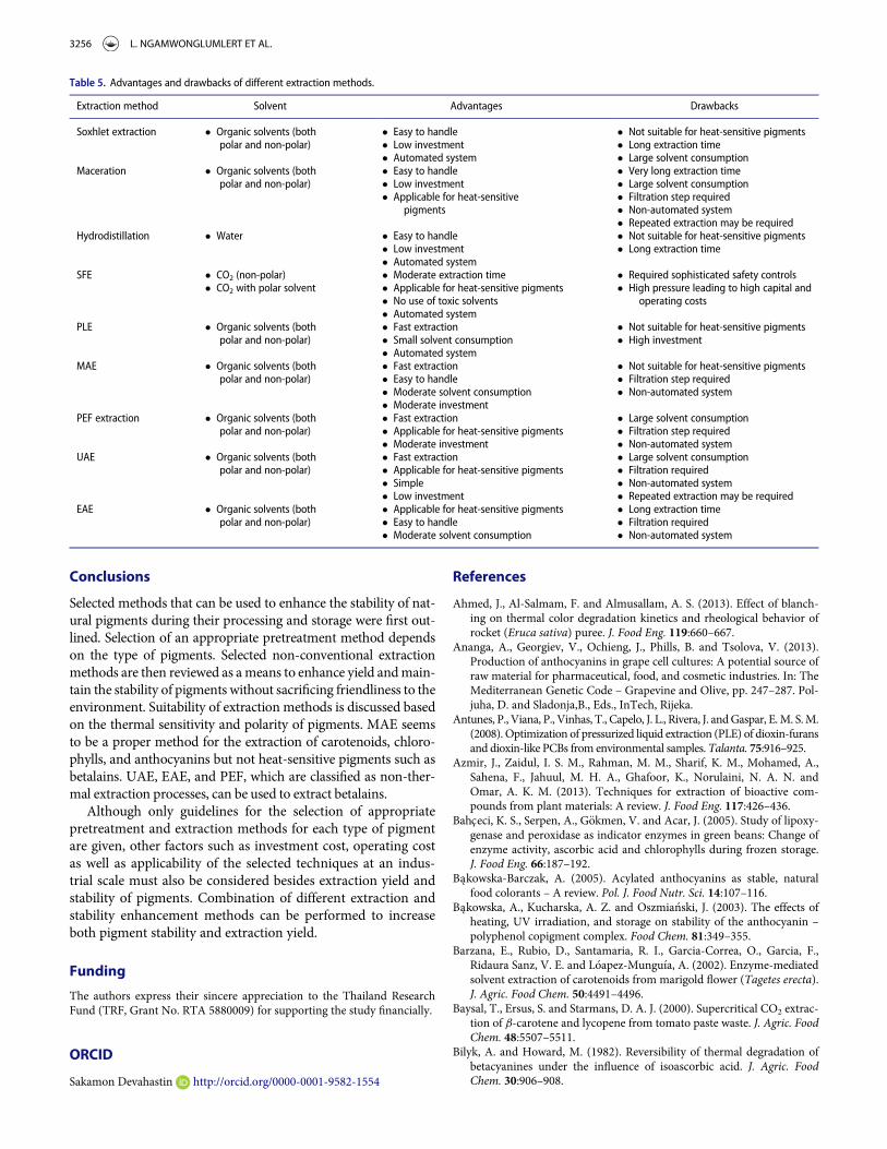
Conclusions
Selected methods that can be used to enhance the stability of nat-ural pigments during their processing and storage were first out-lined. Selection of an appropriate pretreatment method dependson the type of pigments. Selected non-conventional extractionmethods are then reviewed as ameans to enhance yield andmain-tain the stability of pigments without sacrificing friendliness to theenvironment. Suitability of extraction methods is discussed basedon the thermal sensitivity and polarity of pigments. MAE seemsto be a proper method for the extraction of carotenoids, chloro-phylls, and anthocyanins but not heat-sensitive pigments such asbetalains. UAE, EAE, and PEF, which are classified as non-ther-mal extraction processes, can be used to extract betalains.
Although only guidelines for the selection of appropriatepretreatment and extraction methods for each type of pigmentare given, other factors such as investment cost, operating costas well as applicability of the selected techniques at an indus-trial scale must also be considered besides extraction yield andstability of pigments. Combination of different extraction andstability enhancement methods can be performed to increaseboth pigment stability and extraction yield.
Funding
The authors express their sincere appreciation to the Thailand ResearchFund (TRF, Grant No. RTA 5880009) for supporting the study financially.
ORCID
Sakamon Devahastin http://orcid.org/0000-0001-9582-1554
References
Ahmed, J., Al-Salmam, F. and Almusallam, A. S. (2013). Effect of blanch-ing on thermal color degradation kinetics and rheological behavior ofrocket (Eruca sativa) puree. J. Food Eng. 119:660–667.
Ananga, A., Georgiev, V., Ochieng, J., Phills, B. and Tsolova, V. (2013).Production of anthocyanins in grape cell cultures: A potential source ofraw material for pharmaceutical, food, and cosmetic industries. In: TheMediterranean Genetic Code – Grapevine and Olive, pp. 247–287. Pol-juha, D. and Sladonja,B., Eds., InTech, Rijeka.
Antunes, P., Viana, P., Vinhas, T., Capelo, J. L., Rivera, J. andGaspar, E.M. S.M.(2008). Optimization of pressurized liquid extraction (PLE) of dioxin-furansand dioxin-like PCBs from environmental samples. Talanta. 75:916–925.
Azmir, J., Zaidul, I. S. M., Rahman, M. M., Sharif, K. M., Mohamed, A.,Sahena, F., Jahuul, M. H. A., Ghafoor, K., Norulaini, N. A. N. andOmar, A. K. M. (2013). Techniques for extraction of bioactive com-pounds from plant materials: A review. J. Food Eng. 117:426–436.
Bahceci, K. S., Serpen, A., G€okmen, V. and Acar, J. (2005). Study of lipoxy-genase and peroxidase as indicator enzymes in green beans: Change ofenzyme activity, ascorbic acid and chlorophylls during frozen storage.J. Food Eng. 66:187–192.
Bakowska-Barczak, A. (2005). Acylated anthocyanins as stable, naturalfood colorants – A review. Pol. J. Food Nutr. Sci. 14:107–116.
Bakowska, A., Kucharska, A. Z. and Oszmia�nski, J. (2003). The effects ofheating, UV irradiation, and storage on stability of the anthocyanin –polyphenol copigment complex. Food Chem. 81:349–355.
Barzana, E., Rubio, D., Santamaria, R. I., Garcia-Correa, O., Garcia, F.,Ridaura Sanz, V. E. and L�oapez-Mungu�ıa, A. (2002). Enzyme-mediatedsolvent extraction of carotenoids from marigold flower (Tagetes erecta).J. Agric. Food Chem. 50:4491–4496.
Baysal, T., Ersus, S. and Starmans, D. A. J. (2000). Supercritical CO2 extrac-tion of b-carotene and lycopene from tomato paste waste. J. Agric. FoodChem. 48:5507–5511.
Bilyk, A. and Howard, M. (1982). Reversibility of thermal degradation ofbetacyanines under the influence of isoascorbic acid. J. Agric. FoodChem. 30:906–908.
Table 5. Advantages and drawbacks of different extraction methods.
Extraction method Solvent Advantages Drawbacks
Soxhlet extraction � Organic solvents (bothpolar and non-polar)
� Easy to handle � Not suitable for heat-sensitive pigments� Low investment � Long extraction time� Automated system � Large solvent consumption
Maceration � Organic solvents (bothpolar and non-polar)
� Easy to handle � Very long extraction time� Low investment � Large solvent consumption� Applicable for heat-sensitive
pigments� Filtration step required� Non-automated system� Repeated extraction may be required
Hydrodistillation � Water � Easy to handle � Not suitable for heat-sensitive pigments� Low investment � Long extraction time� Automated system
SFE � CO2 (non-polar) � Moderate extraction time � Required sophisticated safety controls� CO2 with polar solvent � High pressure leading to high capital and
operating costs� Applicable for heat-sensitive pigments� No use of toxic solvents� Automated system
PLE � Organic solvents (bothpolar and non-polar)
� Fast extraction � Not suitable for heat-sensitive pigments� Small solvent consumption � High investment� Automated system
MAE � Organic solvents (bothpolar and non-polar)
� Fast extraction � Not suitable for heat-sensitive pigments� Easy to handle � Filtration step required� Moderate solvent consumption � Non-automated system� Moderate investment
PEF extraction � Organic solvents (bothpolar and non-polar)
� Fast extraction � Large solvent consumption� Applicable for heat-sensitive pigments � Filtration step required
� Non-automated system� Moderate investmentUAE � Organic solvents (both
polar and non-polar)� Fast extraction � Large solvent consumption� Applicable for heat-sensitive pigments � Filtration required� Simple � Non-automated system� Low investment � Repeated extraction may be required
EAE � Organic solvents (bothpolar and non-polar)
� Applicable for heat-sensitive pigments � Long extraction time� Filtration required� Easy to handle
� Moderate solvent consumption � Non-automated system
3256 L. NGAMWONGLUMLERT ET AL.
Bilyk, A., Kolodij, M. A. and Sapers, G. M. (1981). Stabilization of red beetpigments with isoascorbic acid. J. Food Sci. 46:1616–1617.
Bobbio, P. A. and Guedes, M. C. (1990). Stability of copper and magne-sium chlorophylls. Food Chem. 36:165–168.
Boon, C. S., McClements, D. J., Weiss, J. and Decker, E. A. (2010). Factorsinfluencing the chemical stability of carotenoids in foods. Crit. Rev.Food Sci. Nutr. 50:515–532.
Bordignon-Luiz, M. T., Gauche, C., Gris, E. F. and Falc~ao, L. D. (2007).Colour stability of anthocyanins from Isabel grapes (Vitis labrusca L.)in model systems. LWT Food Sci. Technol. 40:594–599.
Britton, G. (1996). Carotenoids. In: Natural Food Colorants, 2nd ed., pp.197–243. Hendry, G. A. F. and Houghton, J. D., Eds., Chapman & Hall,New York, NY.
Buchweitz, M., Brauch, J., Carle, R. and Kammerer, D. R. (2013). Applica-tion of ferric anthocyanin chelates as natural blue food colorants inpolysaccharide and gelatin based gels. Food Res. Int. 51:274–282.
Buchweitz, M., Nagel, A., Carle, R. and Kammerer, D. R. (2012). Charac-terisation of sugar beet pectin fractions providing enhanced stability ofanthocyanin-based natural blue food colourants. Food Chem.132:1971–1979.
Cardoso-Ugarte, G. A., Sosa-Morales, M. E., Ballard, T., Liceaga, A. andMart�ın-Gonz�alez, M. F. S. (2014). Microwave-assisted extraction ofbetalains from red beet (Beta vulgaris). LWT Food Sci. Technol.59:276–282.
Casta~neda-Ovando, A., Pacheco-Hern�andez, M. D. L., P�aez-Hern�andez,M. E., Rodr�ıguez, J. A. and Gal�an-Vidal, C. A. (2008). Chemical studiesof anthocyanins: A review. Food Chem. 113:859–871.
Cevallos-Casals, B. A. and Cisneros-Zevallos, L. (2004). Stability of antho-cyanin-based aqueous extracts of Andean purple corn and red-fleshedsweet potato compared to synthetic and natural colorants. Food Chem.86:69–77.
Cha, K. H., Lee, H. J., Koo, S. Y., Song, D. G., Lee, D. U. and Pan, C. H.(2010a). Optimization of pressurized liquid extraction of carotenoids andchlorophylls from Chlorella vulgaris. J. Agric. Food Chem. 58:793–797.
Cha, K. H., Kang, S. W., Kim, C. Y., Um, B. H., Na, Y. R. and Pan, C. H.(2010b). Effect of pressurized liquids on extraction of antioxidantsfrom Chlorella vulgaris. J. Agric. Food Chem. 58:4756–4761.
Chandra, A. and Nair, M. G. (1997). Supercritical fluid carbon dioxideextraction of a- and b-carotene from carrot (Daucus carota L.). Phyto-chem. Anal. 8:244–246.
Chandrasekhar, J., Sonika, G., Madhusudhan, M. C. and Raghavarao,K. S. M. S. (2015). Differential partitioning of betacyanins andbetaxanthins employing aqueous two-phase extraction. J. Food Eng.144:156–163.
Cheok, C. Y., Salman, H. A. K. and Sulaiman, R. (2014). Extraction andquantification of saponins: A review. Food Res. Int. 59:16–40.
Choi, M. H., Kim, G. H. and Lee, H. S. (2002). Effects of ascorbic acidretention on juice color and pigment stability in blood orange (Citrussinensis) juice during refrigerated storage. Food Res. Int. 35:753–759.
Corrales, M., Toepfl, S., Butz, P., Knorr, D. and Tauscher, B. (2008).Extraction of anthocyanins from grape by-products assisted by ultra-sonics, high hydrostatic pressure or pulsed electric fields: A compari-son. Innovative Food Sci. Emerging Technol. 9:85–91.
Cowan, M. M. (1999). Plant products as antimicrobial agents. Clin. Micro-biol. Rev. 12:564–582.
Dabiri, M., Salimi, S., Ghassempour, A., Rassouli, A. and Talebi, M. (2005).Optimization of microwave-assisted extraction for alizarin and pur-purin in Rubiaceae plants and its comparison with conventional extrac-tion methods. J. Sep. Sci. 28:387–396.
Dahmoune, F., Moussia, S. K., Reminia, H., Cherbalc, A. and Madania, K.(2014). Pistacia lentiscus leaves as a source of phenolic compounds:Microwave-assisted extraction optimized and compared with ultra-sound-assisted and conventional solvent extraction. Ind. Crops Prod.61:31–40.
Delgado-Vargas, F., Jim�enez, A. R. and Paredes-L�opez, O. (2000). Naturalpigments: Carotenoids, anthocyanins, and betalains – Characteristics,biosynthesis, processing, and stability. Crit. Rev. Food Sci. Nutr.40:173–289.
Delgado-Vargas, F. and Paredes-L�opez, O. (2003). Natural Colorants forFood and Nutraceutical Uses. CRC Press, Boca Raton, FL.
Dons�ı, F., Ferrari, G. and Pataro, G. (2010). Application of pulsed electricfields treatments for the enhancement of mass transfer from vegetabletissue. Food Eng. Rev. 2:109–130.
Fellows, P. (2000). Food Processing Technology – Principles and Practice,2nd ed. CRC Press, Boca Raton, FL.
Francis, F. J. (1996). Safety of food colorants. In: Natural Food Colorants,2nd ed., pp. 112–126. Hendry, G. A. F. and Houghton, J. D., Eds.,Chapman & Hall, New York, NY.
Gauche, C., Malagoli, E. D. S. and Bordignon-Luiz, M. T. (2010). Effect ofpH on the copigmentation of anthocyanins from Cabernet Sauvignongrape extracts with organic acids. Sci. Agric. 67:41–46.
Gliemmo, M. F., Latorrea, M. E., Gerschensona, L. N. and Campos, C. A.(2009). Color stability of pumpkin (Cucurbita moschata, Duchesne exPoiret) puree during storage at room temperature: Effect of pH, potas-sium sorbate, ascorbic acid and packaging material. LWT Food Sci.Technol. 42:196–201.
G�omez-Miguez, M., Gonz�alez-Manzano, S., Escribano-Bail�on, M. T.,Heredia, F. J. and Santos-Buelga, C. (2006). Influence of different phe-nolic copigments on the color of malvidin 3-glucoside. J. Agric. FoodChem. 54:5422–5429.
GonCalves, L. C. P., Genova, B. M. D., D€Orr, F. A., Pinto, E. and Bastos, E.L. (2013). Effect of dielectric microwave heating on the color and anti-radical capacity of betanine. J. Food Eng. 118:49–55.
Greibrokk, T. (1991). Fluid extraction of chlorinated compounds and otherpollutants. In: Organic Micropollutants in the Aquatic Environment,pp. 112–114. Angeletii, G. and Bjørseth, A., Eds., Springer, Brussels,Belgium.
Gris, E. F., Ferreira, E. A., Falc~ao, L. D. and Bordignon-Luiz, M. T. (2007).Caffeic acid copigmentation of anthocyanins from Cabernet Sauvignongrape extracts in model systems. Food Chem. 100:1289–1296.
Guedes, A. C., Giao, M. S., Matias, A. A., Nunes, A. V. M., Pintado, M. E.,Duarte, C. M. M. and Malcata, F. X. (2013). Supercritical fluid extrac-tion of carotenoids and chlorophylls a, b and c from a wild strain ofScenedesmus obliquus for use in food processing. J. Food Eng.116:478–482.
Hackett, M. M., Lee, J. H., Francis, D. and Schwartz, S. J. (2004). Thermalstability and isomerization of lycopene in tomato oleoresins from dif-ferent varieties. J. Food Sci. 69:C536–C541.
Han, D., Kim, S. J., Kim, S. H. and Kim, D. M. (1998). Repeated regenera-tion of degraded red beet juice pigments in the presence of antioxi-dants. J. Food Sci. 63:69–72.
Hardouin, K., Bedoux, G., Burlot, A. S., Nyvall-Coll�en, P. and Bourgoug-non, N. (2014). Enzymatic recovery of metabolites from seaweeds:Potential applications. In: Advances in Botanical Research, pp. 280–313. Jacquot, J. P., Gadal, P. and Bourgougnon, N., Eds., AcademicPress, Oxford, UK.
Harp, B. P. and Barrows, J. N. (2015). US regulation of color additives infoods. In: Colour Additives for Foods and Beverages, pp. 75–88. Scot-ter, M. J., Ed., Woodhead Publishing, Cambridge.
Hassan, E. and G€OkCe, A. (2014). Production and consumption of biopes-ticides. In: Advances in Plant Biopesticides, pp. 361–380. Singh, D.,Ed., Springer, New Delhi, India.
Heaton, J.W. andMarangoni, A. G. (1996). Chlorophyll degradation in proc-essed foods and senescent plant tissues. Trends Food Sci. Technol. 7:8–15.
Hendry, G. A. F. (1996). Natural pigments in biology. In: Natural FoodColorants, 2nd ed., pp. 1–39. Hendry, G. A. F. and Houghton, J. D.,Eds., Chapman & Hall, New York, NY.
Henry, B. S. (1996). Natural food colours. In: Natural Food Colorants, 2nded., pp. 40–79. Hendry, G. A. F. and Houghton, J. D., Eds., Chapman &Hall, New York, NY.
Herbach, K. M., Rohe, M., Stintzing, F. C. and Carle, R. (2006a). Structuraland chromatic stability of purple pitaya (Hylocereus polyrhizus [Weber]Britton & Rose) betacyanins as affected by the juice matrix and selectedadditives. Food Res. Int. 39:667–677.
Herbach, K. M., Stintzing, F. C. and Carle, R. (2006b). Betalain stabilityand degradation – Structural and chromatic aspects. J. Food Sci. 71:R41–R50.
Hiranvarachat, B., Devahastin, S. and Chiewchan, N. (2011). Effects of acidpretreatments on some physicochemical properties of carrot undergo-ing hot air drying. Food Bioprod. Process. 89:116–127.
CRITICAL REVIEWS IN FOOD SCIENCE AND NUTRITION 3257

Hiranvarachat, B., Devahastin, S., Chiewchan, N. and Raghavan, G. S. V.(2013). Structural modification by different pretreatment methods toenhance microwave-assisted extraction of b-carotene from carrots. J.Food Eng. 115:190–197.
Hiranvarachat, B., Devahastin, S. and Soponronnarit, S. (2015). Compara-tive evaluation of atmospheric and vacuum microwave-assisted extrac-tion of bioactive compounds from fresh and dried Centella asiatica L.leaves. Int. J. Food Sci. Technol. 50:750–757.
Hosikian, A., Lim, S., Halim, R. and Danquah, M. K. (2010). Chlorophyllextraction from microalgae: A review on the process engineeringaspects. Int. J. Chem. Eng. 2010:1–11.
Jackman, R. L. and Smith, J. L. (1996). Anthocyanins and betalains. In:Natural Food Colorants, 2nd ed., pp. 244–309. Hendry, G. A. F. andHoughton, J. D., Eds., Chapman & Hall, New York, NY.
Kammerer, D., Claus, A., Schieber, A. and Carle, R. (2005). A novel processfor the recovery of polyphenols from grape (Vitis vinifera L.) pomace. J.Food Sci. 70:C157–C163.
Khan, M. I. and Giridhar, P. (2014). Enhanced chemical stability, chro-matic properties and regeneration of betalains in Rivina humilis L.berry juice. LWT Food Sci. Technol. 58:649–657.
Khoo, H. E., Prasad, K. N., Kong, K. W., Jiang, Y. and Ismail, A. (2011).Carotenoids and their isomers: Color pigments in fruits and vegetables.Molecules. 16:1710–1738.
Kong, W., Liu, N., Zhang, J., Yang, Q., Hua, S., Song, H. and Xia, C. (2012).Optimization of ultrasound-assisted extraction parameters of chloro-phyll from Chlorella vulgaris residue after lipid separation usingresponse surface methodology. J. Food Sci. Technol. 51:2006–2013.
Kujala, T., Loponen, J. and Pihlaja, K. (2001). Betalains and phenolics inred beetroot (Beta vulgaris) peel extracts: Extraction and characterisa-tion. J. Biosci. 56:343–348.
Lakkakula, N., Lima, M. and Walker, T. (2004). Rice bran stabilization andrice bran oil extraction using Ohmic heating. Bioresource Technol.92:157–161.
Latif, S. and Anwar, F. (2009). Physicochemical studies of hemp (Cannabissativa) seed oil using enzyme-assisted cold-pressing. Eur. J. Lipid Sci.Technol. 111:1042–1048.
Lau, M. H., Tang, J. and Swanson, B. G. (2000). Kinetics of textural andcolor changes in green asparagus during thermal treatments. J. FoodEng. 45:231–236.
Lavelli, V., Zanoni, B. and Zaniboni, A. (2007). Effect of water activity on carot-enoid degradation in dehydrated carrots. Food Chem. 104:1705–1711.
Lenucci, M. S., Caroli, M. D., Marrese, P. P., Iurlaro, A., Rescio, L., B€Ohm,V., Dalessandro, G. and Piro, G. (2015). Enzyme-aided extraction oflycopene from high-pigment tomato cultivars by supercritical carbondioxide. Food Chem. 170:193–202.
Lianfu, Z. and Zelong, L. (2008). Optimization and comparison of ultra-sound/microwave assisted extraction (UMAE) and ultrasonic-assistedextraction (UAE) of lycopene from tomatoes. Ultrason. Sonochem.15:731–737.
L�opez, N., Pu�ertolas, E., Cond�on, S., Raso, J. and Alvarez, I. (2009).Enhancement of the extraction of betanine from red beetroot by pulsedelectric fields. J. Food Eng. 90:60–66.
Machado, A. P. D. F., Pasquel-Re�ategui, J. L., Barbero, G. F. and Mart�ınez,J. (2015). Pressurized liquid extraction of bioactive compounds fromblackberry (Rubus fruticosus L.) residues: A comparison with conven-tional methods. Food Res. Int. 77:675–683.
Machmudah, S. and Goto, M. (2013). Methods for extraction and analysisof carotenoids. In: Natural Products, pp. 3368–3407. Ramawat, K. G.and M�erillon, J. M., Eds., Springer, Heidelberg, Germany.
Mac�ıas-S�anchez, M. D., Mantell, C., Rodr�ıguez, M., Mart�ınez de la Ossa, E.,Lubi�an, L. M. and Montero, O. (2005). Supercritical fluid extraction ofcarotenoids and chlorophyll a from Nannochloropsis gaditana. J. FoodEng. 66:245–251.
Mac�ıas-S�anchez, M. D., Mantell, C., Rodr�ıguez, M.,Mart�ınez de la Ossa, E.,Lubi�an, L. M. and Montero, O. (2009). Comparison of supercriticalfluid and ultrasound-assisted extraction of carotenoids and chlorophylla from Dunaliella salina. Talanta. 77:948–952.
Maharaj, V. and Sankat, C. K. (1996). Quality changes in dehydrateddasheen leaves: Effects of blanching pre-treatments and drying condi-tions. Food Res. Int. 29:563–568.
Maran, J. P., Priyab, B. and Nivetha, C. V. (2015). Optimization of ultra-sound-assisted extraction of natural pigments from Bougainvillea gla-bra flowers. Ind. Crops Prod. 63:182–189.
Marquez, U. M. L. and Sinnecker, P. (2008). Chlorophylls in foods: Sourcesand stability. In: Food Colorants: Chemical and Functional Properties,pp. 195–212. Socaciu, C., Ed., CRC Press, Boca Raton, FL.
Marx, M., Stuparic, M., Schieber, A. and Carle, R. (2003). Effects of ther-mal processing on trans-cis-isomerization of b-carotene in carrot juicesand carotene-containing preparations. Food Chem. 83:609–617.
Masone, D. and Chanforan, C. (2015). Study on the interaction of artificialand natural food colorants with human serum albumin: A computa-tional point of view. Comput. Biol. Chem. 56:152–158.
Mazza, G. and Brouillard, R. (1990). The mechanism of co-pigmenta-tion of anthocyanins in aqueous solutions. Phytochemistry.29:1097–1102.
Mercadante, A. Z. and Bobbio, F. O. (2008). Anthocyanins in foods:Occurrence and physicochemical. In: Food Colorants: Chemical andFunctional Properties, pp. 241–274. Socaciu, C., Ed., CRC Press, BocaRaton, FL.
Mustafa, A. and Turner, C. (2011). Pressurized liquid extraction as a greenapproach in food and herbal plants extraction: A review. Anal Chim.Acta. 703:8–18.
Negi, P. S. and Roy, S. K. (2001). Effect of drying conditions on qual-ity of green leaves during long-term storage. Food Res. Int. 34:283–287.
Ngo, T. and Zhao, Y. (2005). Retaining green pigments on thermally proc-essed peels-on green pears. J. Food Sci. 70:C568–C574.
Nip, W. K. (2007). Fundamentals of food manufacturing. In: Handbook ofFood Products Manufacturing, pp. 5–44. Hui, Y. H., Chandan, R. C.,Clark, S., Cross, N., Dobbs, J., Hurst, W. J., Nollet, L. M. L., Shimoni,E., Sinha, N., Smith, E. B., Surapat, S., Titchenal, A. and Toldr�a, F.,Eds., Wiley, Hoboken, UK.
Olivera, D. F., Vi~na, A. Z., Marani, C. M., Ferreyra, R. M., Mugridge, A.,Chaves, A. R. and Mascheroni, R. H. (2008). Effect of blanching on thequality of Brussels sprouts (Brassica oleracea L. gemmifera DC) afterfrozen storage. J. Food Eng. 84:148–155.
€Otles, S. and Cagindi, €O. (2008). Carotenoids as natural colorants. In: FoodColorants: Chemical and Functional Properties, pp. 51–70. Socaciu, C.,Ed., CRC Press, Boca Raton, FL.
€Ozkan, G. and Bilek, S. E. (2015). Enzyme-assisted extraction of stabilizedchlorophyll from spinach. Food Chem. 176:152–157.
Perera, O. C. and Baldwin, E. A. (2001). Biochemistry of fruits and itsimplications on processing. In: Fruit Processing: Nutrition, Productsand Quality Management, 2nd ed., pp. 25–27. Arthey, D. and Ashurst,P. R., Eds., Aspen, Gaithersburg, Germany.
Pitt, W. G., Husseini, G. A. and Staples, B. J. (2004). Ultrasonic drug deliv-ery – A general review. Expert Opin. Drug Deliv. 1:37–56.
Pokorny, J. (2007). Antioxidants in food preservation. In: Handbook ofFood Preservation, 2nd ed., pp. 259–286. Rahman, M. S., Ed., CRCPress, Boca Raton, FL.
Pongmalai, P., Devahastin, S., Chiewchan, N. and Soponronnarit, S.(2015). Enhancement of microwave-assisted extraction of bioactivecompounds from cabbage outer leaves via the application of ultrasonicpretreatment. Sep. Purif. Technol. 144:37–45.
Provesi, J. G. and Amante, E. R. (2015). Carotenoids in pumpkin andimpact of processing treatments and storage. In: Processing and Impacton Active Components in Food, pp. 71–80. Preedy, V. R., Ed., Elsevier,San Diego, CA.
Pu�ertolas, E., Cregenz�an, O., Luengo, E., �Alvarez, I. and Raso, J. (2013).Pulsed electric field-assisted extraction of anthocyanins from purple-fleshed potato. Food Chem. 136:1330–1336.
Qian, C., Decker, E. A., Xiao, H. and McClement, D. J. (2012). Physical andchemical stability of b-carotene-enriched nanoemulsions: Influence ofpH, ionic strength, temperature, and emulsifier type. Food Chem.132:1221–1229.
Rastogi, N. K. (2011). Opportunities and challenges in application of ultra-sound in food processing. Crit. Rev. Food Sci. Nutr. 51:705–722.
Reshmi, S. K., Aravindhan, K. M. and Devi, P. S. (2012). The effect of light,temperature, pH on stability of betacyanin pigment in basella alba fruit.Asian J. Pharm. Clin. Res. 5:107–110.
3258 L. NGAMWONGLUMLERT ET AL.
Reynoso, R., Garcia, F. A., Morales, D. and de Mejia, E. G. (1997). Stabilityof betalain pigments from a cactacea fruit. J. Agric. Food Chem.45:2884–2889.
Rodr�ıguez-Saona, L. E., Giusti, M. M. and Wrolstad, R. E. (1999). Colorand pigment stability of red radish and red-fleshed potato anthocya-nins in juice model systems. J. Food Sci. 64:451–456.
Sharif, A., Saim, N., Jasmani, H. and Ahmad, W. Y. W. (2010). Effects ofsolvent and temperature on the extraction of colorant from onion(Allium cepa) skin using pressurized liquid extraction. Asian J. Appl.Sci. 3:262–268.
Silva, L. V., Nelson, D. L., Drummond, M. F. B., Dufoss�e, L. and Gl�oria, M.B. A. (2005). Comparison of hydrodistillation methods for the deodor-ization of turmeric. Food Res. Int. 38:1087–1096.
Socaciu, C. (2008). Updated technologies for extracting and formulatingfood colorants. In: Food Colorants: Chemical and Functional Proper-ties, pp. 303–322. Socaciu, C., Ed., CRC Press, Boca Raton, FL.
Stamatopoulos, K., Katsoyannos, E., Chatzilazarou, A. and Konteles, S. J.(2012). Improvement of oleuropein extractability by optimising steamblanching process as pre-treatment of olive leaf extraction via responsesurface methodology. Food Chem. 133:344–351.
Steet, J. A. and Tong, C. H. (1996). Degradation kinetics of green color andchlorophylls in peas by colorimetry and HPLC. J. Food Sci. 61:924–928.
Stintzing, F. C. and Carle, R. (2008). Betalains in food: Occurrence, stabil-ity, and post-harvest modifications. In: Food Colorants: Chemical andFunctional Properties, pp. 277–290. Socaciu, C., Ed., CRC Press, BocaRaton, FL.
Takyi, E. E. K. (2001). Bioavailability of carotenoids from vegetables versussupplements. In: Vegetables, Fruits and Herbs in Health Promotion,pp. 19–34. Watson, R. R., Ed., CRC Press, Boca Raton, FL.
Tan, J. B. L., Lim, Y. Y. and Lee, S. M. (2014). Rhoeo spathacea (Swartz)stearn leaves, a potential natural food colorant. J. Funct. Foods. 7:443–451.
Tao, Y. and Sun, D. W. (2015). Enhancement of food processes by ultra-sound: A review. Crit. Rev. Food Sci. Nutr. 55:570–594.
Tao, Y., Wu, D., Zhang, Q. A. and Sun, D. W. (2014). Ultrasound-assistedextraction of phenolics from wine lees: Modeling, optimization and sta-bility of extracts during storage. Ultrason. Sonochem. 21:706–715.
Tonucci, L. H. and von Elbe, J. H. (1992). Kinetics of the formation of zinccomplexes of chlorophyll derivatives. J. Agric. Food Chem. 40:2341–2344.
V�agi, E., Sim�andi, B., Daood, H. G., De�ak, A. and Sawinsky, J. (2002).Recovery of pigments from Origanum majorana L. by extraction withsupercritical carbon dioxide. J. Agric. Food Chem. 50:2297–2301.
Veggi, P. C., Martinez, J. and Meireles, M. A. A. (2013). Fundamentals ofmicrowave extraction. In: Microwave-Assisted Extraction for BioactiveCompounds: Theory and Practice, pp. 15–52. Chemat, F. and Cravotto,G., Eds., Springer, New York, NY.
Vi~na, S. Z., Olivera, D. F., Marani, C. M., Ferreyra, R. M., Mugridge, A.,Chaves, A. R. and Mascheroni, R. H. (2007). Quality of Brussels sprouts(Brassica oleracea L. gemmifera DC) as affected by blanching method.J. Food Eng. 80:218–225.
von Elbe, J. H. and Schwartz, S. J. (1996). Colorants. In: Food Chemistry,3rd ed., pp. 651–722. Fennema, O. R., Ed., Marcel Dekker, New York,NY.
Wang, J. X., Xiao, H. X. and Li, G. K. (2008). Study of vacuum microwave-assisted extraction of polyphenolic compounds and pigment from Chi-nese herbs. J. Chromatogr. A. 1198–1199:45–53.
Wu, X. and Prior, R. L. (2005). Identification and characterizationof anthocyanins by high-performance liquid chromatography-electro-spray ionization-tandem mass spectrometry in common foods in theUnited States: Vegetables, nuts, and grains. J. Agric. Food Chem.53:3101–3113.
Xiao, X., Song, W., Wang, J. and Li, G. (2012). Microwave-assistedextraction performed in low temperature and in vacuo for theextraction of labile compounds in food samples. Anal Chim. Acta.712:85–93.
Yang, Z. and Zhai, W. (2010). Optimization of microwave-assisted extrac-tion of anthocyanins from purple corn (Zea mays L.) cob and identifica-tion with HPLC-MS. Innovative Food Sci. Emerging Technol. 11:470–476.
Yuhas Jr., S. A. (1995). Solvents. In: Paint and Coating Testing Manual, 4thed., pp. 125–158. Koleske, J. V., Ed., ASTM, Ann Arbor, MI.
Zou, T., Wu, H., Li, H., Jia, Q. and Song, G. (2013). Comparison of micro-wave-assisted and conventional extraction of mangiferin from mango(Mangifera indica L.) leaves. J. Sep. Sci. 36:3457–3462.
CRITICAL REVIEWS IN FOOD SCIENCE AND NUTRITION 3259

![Isolation and characterization of pigment producing marine ... · commercially available colorants [6]. Currently, the whole world is looking towards the usage of traditional products](https://static.fdocuments.net/doc/165x107/5ed61715bcb22c51e2620aff/isolation-and-characterization-of-pigment-producing-marine-commercially-available.jpg)